In vivo anticancer and histopathology studies of Schiff bases on Ehrlich ascitic carcinoma cells
9
ORIGINAL ARTICLE 1st Cancer Update In vivo anticancer and histopathology studies of Schiff bases on Ehrlich ascitic carcinoma cells Dhanya Sunil a , Arun M. Isloor b, * , Prakash Shetty c , Pawan G. Nayak d , K.S.R. Pai d a Department of Chemistry, Manipal Institute of Technology, Manipal University, Manipal 576 104, India b Organic Chemistry Division, Department of Chemistry, National Institute of Technology-Karnataka, Surathkal, Mangalore 575 025, India c Department of Printing and Media Engineering, Manipal Institute of Technology, Manipal University, Manipal 576 104, India d Department of Pharmacology, Manipal College of Pharmaceutical Sciences, Manipal University, Manipal 576 104, India Received 26 October 2010; accepted 14 December 2010 Available online 21 December 2010 KEYWORDS Schiff base; EAC; Hematology; Biochemical; Histopathology Abstract Three Schiff bases in two different concentrations were evaluated for their anti-tumor activity against Ehrlich ascites carcinoma (EAC) bearing Swiss albino mice. The in vivo anti-tumor potency of Schiff bases was assessed by measuring the increase in mean survival time of the drug treated over untreated control mice and treated standard (cisplatin) mice. Their toxicity was assessed in vivo in normal, standard, and EAC-bearing mice by measuring the drug-induced changes in biochemical as well as hematological parameters. The histopathology studies to assess the toxic- ity of these compounds on vital organs also have been studied. Among the three Schiff bases stud- ied, 4-({[3-(4-fluorophenyl)-1H-pyrazol-4-yl]methylene}amino)-5-[(2-methylphenoxy)methyl]-1,2,4- triazole-3-thiol (SB-3) at an optimal dose of 100 mg/kg body weight was found to enhance the mean survival time of infected mice. Deviated hematological parameters and mean survival time in tumor * Corresponding author. Tel.: +91 824 2473206; fax: +91 824 2474033. E-mail addresses: [email protected] (D. Sunil), isloor@ yahoo.com (A.M. Isloor), [email protected] (P. Shetty), [email protected] (K.S.R. Pai). 1878-5352 ª 2011 King Saud University. Production and hosting by Elsevier B.V. All rights reserved. Peer review under responsibility of King Saud University. doi:10.1016/j.arabjc.2010.12.016 Production and hosting by Elsevier Arabian Journal of Chemistry (2013) 6, 25–33 King Saud University Arabian Journal of Chemistry www.ksu.edu.sa www.sciencedirect.com
-
Upload
independent -
Category
Documents
-
view
3 -
download
0
Transcript of In vivo anticancer and histopathology studies of Schiff bases on Ehrlich ascitic carcinoma cells

Arabian Journal of Chemistry (2013) 6, 25–33
King Saud University
Arabian Journal of Chemistry
www.ksu.edu.sawww.sciencedirect.com
ORIGINAL ARTICLE
1st Cancer Update
In vivo anticancer and histopathology studies of Schiff
bases on Ehrlich ascitic carcinoma cells
Dhanya Sunila, Arun M. Isloor
b,*, Prakash Shettyc, Pawan G. Nayak
d,
K.S.R. Paid
a Department of Chemistry, Manipal Institute of Technology, Manipal University, Manipal 576 104, Indiab Organic Chemistry Division, Department of Chemistry, National Institute of Technology-Karnataka, Surathkal,Mangalore 575 025, Indiac Department of Printing and Media Engineering, Manipal Institute of Technology, Manipal University, Manipal 576 104, Indiad Department of Pharmacology, Manipal College of Pharmaceutical Sciences, Manipal University, Manipal 576 104, India
Received 26 October 2010; accepted 14 December 2010
Available online 21 December 2010
*
24
E
ya
ks
18
El
Pe
do
KEYWORDS
Schiff base;
EAC;
Hematology;
Biochemical;
Histopathology
Corresponding author. Te
74033.
-mail addresses: dhanyad
hoo.com (A.M. Isloor), pr
[email protected] (K.S.R
78-5352 ª 2011 King Saud
sevier B.V. All rights reserve
er review under responsibilit
i:10.1016/j.arabjc.2010.12.01
Production and h
l.: +91
ss3@gma
akash.she
. Pai).
Universit
d.
y of King
6
osting by E
Abstract Three Schiff bases in two different concentrations were evaluated for their anti-tumor
activity against Ehrlich ascites carcinoma (EAC) bearing Swiss albino mice. The in vivo anti-tumor
potency of Schiff bases was assessed by measuring the increase in mean survival time of the drug
treated over untreated control mice and treated standard (cisplatin) mice. Their toxicity was
assessed in vivo in normal, standard, and EAC-bearing mice by measuring the drug-induced changes
in biochemical as well as hematological parameters. The histopathology studies to assess the toxic-
ity of these compounds on vital organs also have been studied. Among the three Schiff bases stud-
ied, 4-({[3-(4-fluorophenyl)-1H-pyrazol-4-yl]methylene}amino)-5-[(2-methylphenoxy)methyl]-1,2,4-
triazole-3-thiol (SB-3) at an optimal dose of 100 mg/kg body weight was found to enhance the mean
survival time of infected mice. Deviated hematological parameters and mean survival time in tumor
824 2473206; fax: +91 824
il.com (D. Sunil), isloor@
[email protected] (P. Shetty),
y. Production and hosting by
Saud University.
lsevier

26 D. Sunil et al.
bearing mice were found to be significantly restored towards normal after treatment with SB-3
100 mg/kg body weight of mice. The ALP and SGOT values were found to approach the normal
range. A:G ratios also did not deviate from normal on treatment with SB-3. The histopathology
studies revealed only mild hepatotoxicity and nephrotoxicity when compared to the normal and
standard. The splenic cellularity also did not show much variation from normal. SB-3 at a prime
dose of 100 mg has shown promising anticancer activity in vivo against EAC when compared to
standard drug with minimum toxic effects.
ª 2011 King Saud University. Production and hosting by Elsevier B.V. All rights reserved.
1. Introduction
Cancer or malignant neoplasm is a class of diseases in which a
group of cells display uncontrolled growth, invasion and evensometimes metastasis (De Vita et al., 2005; Thomas and Vinay,2007). It continues as a serious public health problem through-
out the world as the most feared diagnosis. It is the second lead-ing cause of human death after cardiovascular diseases indeveloping as well as in developed countries (Babasaheb et al.,2010). Currently, the treatment for cancer primarily includes
surgery and chemotherapy, but the curative effects of the exist-ing chemotherapeutic drugs are not good enough and they haveplentiful side effects. The development of more effective drugs
for treating patients with cancer has been a main attempt overthe past 50 years. In recent years, various 1,2,4-triazole deriva-tives have been found to be associated with anticancer (Al-Soud
et al., 2006; Holla et al., 2002; Ikizler et al., 2003) properties.AK-2123 (Sanazol), a nitrotriazole hypoxic cell sensitizer hassupposedly improved results in head and neck cancers, uterinecervical cancers and other solid tumors when added to radical
radiotherapy (Dobrowsky et al., 2005). Non-steroidal aroma-tase inhibitors obtained from triazole derivatives are used inthe treatment of breast cancer (Robert et al., 2004). Current lit-
erature shows 1,2-pyrazole derivatives to possess various bio-logical activities (Mathew et al., 2007; Konagai et al., 2002). Itis also observed that incorporation of aryl substituents and hal-
ogen atoms into the heterocyclic ring systems enhances the bio-logical activities considerably (Padmavathi et al., 2009). SeveralSchiff bases were reported to possess potential anticancer prop-
erties (Holla et al., 2003). In view of the wide spectrum ofmedic-inal applications, especially anticancer properties of varioustriazole derivatives and in continuation of our research on bio-logically active new molecules, we hereby report the anticancer
activity of three Schiff bases in vivo.In vitro screening in hepatic carcinoma cell lines (Hep G2)
using MTT assay, revealed the cytotoxicity of the Schiff bases
(Dhanya et al., in press). Their in vivo anti-tumor potency wasassessed in Ehrlich ascites carcinoma (EAC) cells by assessingthe increase in median survival times and body weights of drug
treated (T) over untreated control (C) and standard (S) mice.Results showed that the survival time of treated mice wasmarkedly increased by SB-3 at an optimal dose of 100 mg/kg
body weight. Its toxic effects was studied in vivo in normaland EAC-bearing mice by measuring drug-induced changesin hematological, biochemical parameters as well as organ tox-icities on day 15 following drug treatment. The hematological
and biochemical parameters were mostly within normal limit.Histology of liver and kidney revealed mild abnormality upontreatment with SB-3 (100 mg/kg body weight). Minor splenic
toxicity in treated groups SB-3 (100 mg/kg body weight) simi-lar to standard group was observed.
2. Materials and methods
All the chemicals used in the present study were from Sigma–
Aldrich, USA. Adult female swiss albino mice of 6–8 weeks oldweighing 25–30 g, inbred at Central Animal Research Facility,Manipal University, Karnataka, India were used throughout
the study. Animals were housed in polypropylene cages con-taining sterile paddy husk as bedding material under hygienicconditions with a maximum of four animals in a cage. Theywere maintained under controlled conditions (10:14 h light:-
dark), temperature (23 ± 3 �C). Animals were fed on auto-claved standard mice food pellets (Hindustan Lever) andwater ad libitum. The animal experiments were performed
according to the rules and regulations of the Institutional Ani-mal Ethics Committee (IAEC).
Three Schiff bases, 4-({[3-(4-chlorophenyl)-1H-pyrazol-
4-yl]methylene}amino)-5-[(4-methylphenoxy)methyl]-1,2,4-tri-azole-3-thiol (SB-1), 4-({[3-(4-fluorophenyl)-1H-pyrazol-4-yl]methylene}amino)-5-[(4-methylphenoxy)methyl]-1,2,4-tri-azole-3-thiol (SB-2) and 4-({[3-(4-fluorophenyl)-1H-pyrazol-
4-yl]methylene}amino)-5-[(2-methylphenoxy)methyl]-1,2,4-tri-azole-3-thiol (SB-3) were prepared by the method as describedin the literature (Kalluraya et al., 2009; Dhanya et al., in press).
Their structures were confirmed by IR, NMR and mass spec-tral studies. IR spectra of Schiff bases showed an absorptionband at 1600–1610 cm�1 characteristic of –C‚N– group in
the molecule. Also the absence of carbonyl stretching bandat around 1700 cm�1 clearly indicated amino condensationand hence the formation of Schiff bases. The 1H NMR spec-
trum of Schiff base showed a singlet at d 9.95 due to the pres-ence of proton in –N‚CH group in the molecule, confirmingthe formation of Schiff bases. Mass spectrum of the threeSchiff bases showed molecular ion peaks which were in agree-
ment with their molecular formula. The elemental analysis wasalso carried out. The purity of the compounds was checkedusing LC–MS technique. The structure of the Schiff bases
are given in Fig. 1.
2.1. In vivo anticancer studies
The drug solutions were prepared daily just prior to the injec-tion by suspending them in 0.25% CMC (carboxy methyl cel-lulose) and was administered intraperitoneally. The dose of the
standard drug, cisplatin was selected as 3.5 mg/kg mice bodyweight. This was calculated from the human dose using anappropriate conversion factor (Sathisha et al., 2008).
Ehrlich ascites carcinoma cells to induce cancer in animalmodel (mice) were obtained from Amala Cancer ResearchCenter, Amala Nagar, Kerala, India. The cells were main-
tained as ascites tumor in swiss albino mice by intraperitonealinoculation of 1 · 106 viable cells (Sathisha et al., 2008; Devi

R N
NN
O
N
N
NH
R1
SH
SB-1: R = 4-methyl benzene, R1= Cl, SB-2: R = 4-methyl benzene, R1= F
SB-3: R = 2-methyl benzene, R1= F
Figure 1 Structure of Schiff bases.
In vivo anticancer and histopathology studies of Schiff bases on Ehrlich ascitic carcinoma cells 27
et al., 1998). Acute toxicity studies were carried out as per theOECD guidelines, 2001. For determining the maximum toler-ated dose of the Schiff bases, the standard protocol was fol-lowed by administering the Schiff bases to normal swiss
albino mice at the dose range up to 2000 mg/kg body weightafter depriving them of food for 18 h. Animals were observedfor any symptoms of toxicity continuously for 4 h, then after
24 h and finally the number of survivors were noted after a per-iod of 72 h. Depending on the results obtained, the therapeuticdoses for further studies were selected (1/10th to 1/20th of the
maximum tolerated dose).For the in vivo assay, nine groups were formed. Each group
comprised of 12 female swiss albino mice. Formed groups areas follows:
� Group 1: The normal mice.� Group 2: The EAC-bearing mice (Control).
� Group 3: The EAC-bearing mice on a single dose of cis-platin (Standard).� Groups 4–9 represented the study group, treated with Schiff
bases as depicted below:– Group 4: SB-1 (50 mg/kg).– Group 5: SB-1 (100 mg/kg).
– Group 6: SB-2 (50 mg/kg).– Group 7: SB-2 (150 mg/kg).– Group 8: SB-3 (50 mg/kg).– Group 9: SB-3 (100 mg/kg).
The ascitic carcinoma bearing donor mice was taken 15 daysafter tumor transplantation. The ascitic fluid was drawn
using an 18-gauge needle into a sterile syringe. A smallamount was tested for microbial contamination. Tumor via-bility was determined by Trypan blue exclusion test and cells
were counted using haemocytometer. The ascitic fluid wassuitably diluted in normal saline to get a concentration of10 · 106 cells/mL of tumor cell suspension. From this stocksuspension, 0.25 mL (2.5 million cells/mice) was injected
intraperitoneally to normal mice in order to obtain ascitictumor (day 0). Cisplatin (single dose) was given intraperito-neally to the standard group on day 1. The three Schiff bases
in two different concentrations were administered on days 3,5, 7, 10, 12 and 14 (6 doses) intraperitoneally. The controlgroup was treated with the vehicle (0.25% CMC). On day
15, from each group six mice were sacrificed for hematolog-ical, biochemical and histopathological studies. The remain-
ing six animals from each group were monitored daily for
30 days and mortality was recorded to calculate the meansurvival time.
2.1.1. Mean survival time and increase in lifespan (% ILS)(Sathisha et al., 2008)Total number of days an animal survived from the day of tu-
mor inoculation was counted. Subsequently mean survivaltime (MST) for each group was calculated. Survival time fortreated group was compared with that of control group using
the following calculation:
Increase of life span ðILSÞ
¼ MST of treated group�MST of control group
MST of control group
� �� 100
where MST ¼P
survival time ðdaysÞ of each mice in a group
Total number of mice.
2.1.2. Body weight (Eckchardt et al., 1982)Body weights of all animals were measured on days 0, 3, 5, 7,10, 12 and 14. The percentage increase in weight was calculatedas per the following equation:
% increase in weight
¼ Animal weight on respective day�Animal weight on day 0
Animal weight on day 0
� �
� 100
2.2. In vivo toxicological studies
On day 15, blood samples from six animals in each group weretaken in microfuge tubes containing EDTA for hematologicalstudies and the serum was collected for biochemical studies.
The animals were then sacrificed by cervical dislocation,dissection was done and the liver, kidney and spleen wereremoved for histopathology studies.
2.2.1. Hematological parameters (Mukherjee et al., 2007)To study the hematological changes associated with Schiff
base administration, four parameters – (a) hemoglobin count(b) erythrocyte count, (c) leukocyte count and (d) differentialcount were determined in peripheral blood of mice. The blood
was withdrawn from retro-orbital plexus on day 15 for thestudy. The values were then compared with the normal andstandard values.
2.2.2. Biochemical parameters (Pain et al., 2003)Numerous laboratory investigations have been proposed in the
assessment of liver dysfunction. For evaluating drug inducedhepatotoxicity, liver function tests were done and valuesobtained for treated groups were compared with the normal
value. From among the host of liver function tests, the follow-ing battery of blood tests namely bilirubin, SGOT, SGPT,ALP and protein levels were carried out.
2.3. Histopathology studies (Roy et al., 1981; Hassan et al.,2009)
The animals were sacrificed by cervical dislocation. The liver,kidneys and spleen were removed and stored in neutral forma-
28 D. Sunil et al.
lin and histopathology studies were conducted. These were
processed by standard methods to prepare slides of tissuesby hematoxylin and eosin staining. The slides were viewedunder light microscope. Drug induced hepatotoxicity, nephro-toxicity and spleen toxicity studies were performed
sequentially.
2.4. Statistical analysis
The statistical analyses were performed by one-way ANOVA,followed by Tukey’s post hoc test using GraphPad Prism 5.02.
The results were expressed as the mean ± SEM. Differenceswere considered significant p< 0.05.
3. Results and discussion
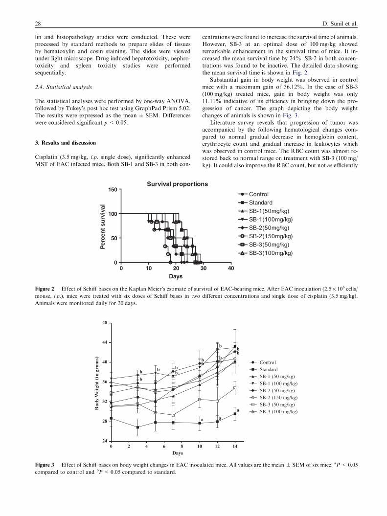
Cisplatin (3.5 mg/kg, i.p. single dose), significantly enhanced
MST of EAC infected mice. Both SB-1 and SB-3 in both con-
Survival proportio
0 10 200
50
100
150
Days
Per
cen
t su
rviv
al
Figure 2 Effect of Schiff bases on the Kaplan Meier’s estimate of sur
mouse, i.p.), mice were treated with six doses of Schiff bases in two
Animals were monitored daily for 30 days.
0 2 4 6 8 1024
28
32
36
40
44
48
bbb
b
b
Days
Bod
y W
eigh
t (i
n gr
ams)
Figure 3 Effect of Schiff bases on body weight changes in EAC inocu
compared to control and bP < 0.05 compared to standard.
centrations were found to increase the survival time of animals.
However, SB-3 at an optimal dose of 100 mg/kg showedremarkable enhancement in the survival time of mice. It in-creased the mean survival time by 24%. SB-2 in both concen-trations was found to be inactive. The detailed data showing
the mean survival time is shown in Fig. 2.Substantial gain in body weight was observed in control
mice with a maximum gain of 36.12%. In the case of SB-3
(100 mg/kg) treated mice, gain in body weight was only11.11% indicative of its efficiency in bringing down the pro-gression of cancer. The graph depicting the body weight
changes of animals is shown in Fig. 3.Literature survey reveals that progression of tumor was
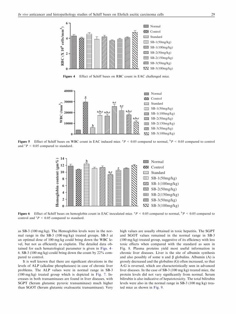
accompanied by the following hematological changes com-
pared to normal gradual decrease in hemoglobin content,erythrocyte count and gradual increase in leukocytes whichwas observed in control mice. The RBC count was almost re-stored back to normal range on treatment with SB-3 (100 mg/
kg). It could also improve the RBC count, but not as efficiently
ns
30 40
ControlStandardSB-1(50mg/kg)SB-1(100mg/kg)SB-2(50mg/kg)SB-2(150mg/kg)SB-3(50mg/kg)SB-3(100mg/kg)
vival of EAC-bearing mice. After EAC inoculation (2.5 · 106 cells/
different concentrations and single dose of cisplatin (3.5 mg/kg).
12 14
ControlStandardSB-1 (50 mg/kg)SB-1 (100 mg/kg)SB-2 (50 mg/kg)SB-2 (150 mg/kg)SB-3 (50 mg/kg)SB-3 (100 mg/kg)
a
a
a
bb
b
b
bbb
lated mice. All values are the mean ± SEM of six mice. aP < 0.05
0
2
4
6Normal
Control
Standard
SB-1(50mg/kg)
SB-1(100mg/kg)
SB-2(50mg/kg)
SB-2(150mg/kg)
SB-3(50mg/kg)
SB-3(100mg/kg)
RB
C (
X 1
06 cel
ls/m
m3 )
Figure 4 Effect of Schiff bases on RBC count in EAC challenged mice.
0
10000
20000
30000
40000Normal
Control
Standard
SB-1(50mg/kg)
SB-1(100mg/kg)
SB-2(50mg/kg)
SB-2(150mg/kg)
SB-3(50mg/kg)
SB-3(100mg/kg)
a
a,b,c
a,c
a,b,c
a,b,c
b
a,b,c
a,c
WB
C (
/mm
3 )
Figure 5 Effect of Schiff bases on WBC count in EAC induced mice. aP < 0.05 compared to normal, bP < 0.05 compared to control
and cP < 0.05 compared to standard.
0
2
4
6
8
10
12
14NormalControlStandardSB-1(50mg/kg)SB-1(100mg/kg)SB-2(50mg/kg)SB-2(150mg/kg)SB-3(50mg/kg)SB-3(100mg/kg)
a,c
b
c
Hem
oglo
bin
con
ten
t (g
m%
)
Figure 6 Effect of Schiff bases on hemoglobin count in EAC inoculated mice. aP < 0.05 compared to normal, bP < 0.05 compared to
control and cP < 0.05 compared to standard.
In vivo anticancer and histopathology studies of Schiff bases on Ehrlich ascitic carcinoma cells 29
as SB-3 (100 mg/kg). The Hemoglobin levels were in the nor-
mal range in the SB-3 (100 mg/kg) treated groups. SB-3 atan optimal dose of 100 mg/kg could bring down the WBC le-vel, but not as efficiently as cisplatin. The detailed data ob-
tained for each hematological parameter is given in Figs. 4–6. SB-3 (100 mg/kg) could bring down the count by 22% com-pared to control.
It is well known that there are significant elevations in thelevels of ALP (alkaline phosphatases) in case of chronic liverproblems. The ALP values were in normal range in SB-3
(100 mg/kg) treated group which is depicted in Fig. 7. In-creases in both transaminases are found in liver diseases, withSGPT (Serum glutamic pyruvic transaminase) much higherthan SGOT (Serum glutamic oxaloacetic transaminase). Very
high values are usually obtained in toxic hepatitis. The SGPT
and SGOT values remained in the normal range in SB-3(100 mg/kg) treated group, suggestive of its efficiency with lesstoxic effects when compared with the standard as seen in
Fig. 8. Plasma proteins yield most useful information inchronic liver diseases. Liver is the site of albumin synthesisand also possibly of some a and b globulins. Albumin (A) is
grossly decreased and the globulins (G) often increased, so thatA:G is reversed, which are characteristically seen in advancedliver diseases. In the case of SB-3 (100 mg/kg) treated mice, the
protein levels did not vary significantly from normal. Serumbilirubin is also indicative of hepatotoxicity. The total bilirubinlevels were also in the normal range in SB-3 (100 mg/kg) trea-ted mice as shown in Fig. 9.

SGOTSGPT
ALP
0
50
100
150
200
250
300
350
400
450NormalControl
CisplatinSB-1(50mg/kg)
SB-1(100mg/kg)
a
a,b
b,c
a,b
SB-3(50mg/kg)
SB-3(100mg/kg)a,b
b,c
ab,cc
a
a,b
a,b,c a,b.c
bb,c
(in
IU/L
)
Figure 7 Effect of Schiff bases on transaminases and ALP in EAC challenged mice. aP < 0.05 compared to normal and bP < 0.05
compared to control.
Total
Protei
ns
Album
in
Globulin
0
3
6
9
12
15NormalControlCisplatin
SB-1(50mg/kg)SB-1(100mg/kg)
a
bb
b
SB-3(50mg/kg)SB-3(100mg/kg)
b b
a a ab b ba a a a
b,c b
(in
g/d
l)
Figure 8 Effect of Schiff bases on protein content in EAC induced mice. aP < 0.05 compared to normal and bP < 0.05 compared to
control.
Total
Biliru
bin
Direct
Biliru
bin
Indire
ct Bili
rubin
0.0
0.2
0.4
0.6
0.8
1.0
NormalControlCisplatinSB-1(50mg/kg)
SB-1(100mg/kg)SB-3(50mg/kg)
SB-3(100mg/kg)
a
a,ba,b
b
b,cb,c
a
a,b
b,c b,c b,cb,c
a
a,bb,c
b b
b,c(in
mg/
dl)
Figure 9 Effect of Schiff bases on Bilirubin count in EAC induced mice. aP < 0.05 compared to normal and bP < 0.05 compared to
control.
30 D. Sunil et al.

Figure 10 Histopathology of liver (40·). Animals were sacrificed on day 15 and histopathology of liver was studied for signs of
hepatotoxicity. The histology of liver of standard group showed cellular infiltration, congestion and mild central vein dilatation. SB-3
(50 mg/kg) treated group showed only mild central vein dilation suggesting less hepatotoxicity compared to control and standard.
Figure 11 Histopathology of kidney (40·). Animals were sacrificed on day 15 and histopathology of kidney was studied for signs of
nephrotoxicity. The standard showed signs of nephrotoxicity due to glomerular infiltration. The SB-3 (100 mg/kg) treated group also
showed mild glomerular infiltration. There were no signs of tubular necrosis, casts and glomerular congestion, which was indicative of
only mild nephrotoxicity with SB-3 (100 mg/kg).
In vivo anticancer and histopathology studies of Schiff bases on Ehrlich ascitic carcinoma cells 31

Figure 12 Histopathology of spleen (40·). Animals were sacrificed on day 15 and tissue sections of spleen were studied for signs of spleen
toxicity. Mild loss in spleen architecture is observed in SB-3 (100 mg/kg) and cisplatin treated mice spleen.
32 D. Sunil et al.
The histopathology studies also revealed the relatively lesstoxic nature of SB-3 (100 mg/kg) as compared to control and
standard group when viewed under light microscope of magni-fication 40·. The liver is known to accumulate significantamounts of cisplatin, second to kidney. The histology studiesof liver of standard group showed cellular infiltration (inflam-
mation), congestion and mild central vein dilatation as seen inFig. 10. Whereas the SB-3 (100 mg/kg) treated group showedonly mild central vein dilation suggesting less hepatotoxicity
compared to standard group. The histopathology of the kid-ney tissues of normal, control, standard and SB-3 (100 mg/kg) treated mice are shown in Fig. 11. The control group
showed some cellular infiltration, while the standard groupshowed signs of nephrotoxicity due to cellular and glomerularinfiltration. The SB-3 (100 mg/kg) treated group also showedmild glomerular infiltration. There were no signs of tubular
necrosis, casts and glomerular congestion, which were indica-tive of only mild nephrotoxicity, with SB-3 (100 mg/kg). Thestandard group showed loss of splenic architecture and conges-
tion as depicted in Fig. 12. The treated group also showed mildloss of splenic architecture but congestion was not observedindicating lower splenic toxicity due to treatment with SB-3
(100 mg/kg).
4. Conclusions
In the light of the above observations, it can be concluded thatSB-3, at a dose of 100 mg/kg, optimally inhibits the growth of
EAC cells in vivo. This is evident from the reduced tumorweight and enhanced life span of that study group. The treat-ment with SB-3 (100 mg/kg) restored the deviated hematolog-ical and biochemical parameters to the normal range. It is an
effective antineoplastic agent with comparatively less toxic ef-fects. It is necessary that the anti-tumor activity of this Schiff
base should be carried out against other tumor cell lines whichmay bring promising results in cancer chemotherapy.
Acknowledgements
The authors are thankful to the Director, Innovation centre
for providing with necessary funds. AMI is grateful to theDepartment of Atomic Energy, Board for Research in NuclearSciences, Government of India for ‘Young Scientist’ award.
D.S. is thankful to the Director and Head-Department ofChemistry, Manipal Institute of Technology, Manipal Univer-sity, Manipal for rendering us the necessary laboratoryfacilities.
References
Al-Soud, Y.A., Al-Masoudi, I.A., Saeed, B., Beifub, U., Al-Masoudi,
N.A., 2006. Synthesis of new 1H-1,2,4-triazolylcoumarins and their
antitumor and anti-HIV activities. Chemistry of Heterocyclic
Compounds 42, 583–590.
Babasaheb, P.B., Shrikant, S.G., Ragini, G.B., Jalinder, V.T., Chan-
drahas, N.K., 2010. Synthesis and biological evaluation of simple
methoxylated chalcones as anticancer, anti-inflammatory and
antioxidant agents. Bioorganic and Medicinal Chemistry 18,
1364–1370.
Devi, P.U., Rao, B.S.S., Solomon, F.E., 1998. Effect of plumbagin on
the radiation induced cytogenetic and cell cycle changes in mouse
Ehrlich Ascitic Carcinoma in vivo. Indian Journal of Experimental
Biology 36, 891–895.

In vivo anticancer and histopathology studies of Schiff bases on Ehrlich ascitic carcinoma cells 33
De Vita Jr., V.T., Samuel, H., Steven, A.R., 2005. Cancer – Principles
and Practice of Oncology, seventh ed. Lippincott Williams &
Wilkins, New York.
Dhanya, S., Shetty, P., Satyamoorthy, K., Isloor, A.M., in press.
Synthesis, characterization and in vitro anticancer properties of
some new Schiff and Mannich bases in Hep G2 cells. Medicinal
Chemistry Research.
Dobrowsky, W., Huigol, N.G., Jayatilake, R.S., Kizilbash, N.I.,
Okkan, S., Kagiya, T.V., Tatsuzaki, H., 2005. AK-2123 (Sanazol)
as a radiation sensitizer in the treatment of stage III cancer cervix:
initial results of an IAEA multicentre randomized trial. Journal of
Cancer Research Therapy 1 (2), 75–78.
Eckchardt, A.E., Malone, B.N., Goldstein, I.J., 1982. Inhibition of
Ehrlich Ascites tumor cell growth by Griffonia Simplifolia lectin
in vivo. Cancer Research 42, 2977–2979.
Hassan, I.E., Mohamed, F.I., Shalaby, F.M., Abou-El-Magd, R.F.,
Gaur, R.L., Fernando, A.M., Raj, H.G., Ouhtit, A., 2009.
Histopathological effects of cisplatin, doxorubicin and 5-flurouracil
on the liver of male albino rats. International Journal of Biological
Sciences 5, 466–473.
Holla, B.S., Veerendra, B., Shivananda, M.K., Poojary, B., 2003.
Synthesis, characterization and anticancer activity studies on some
Mannich bases derived from 1,2,4-triazoles. European Journal of
Medicinal Chemistry 38, 759–767.
Holla, B.S., Poojary, K.N., Rao, B.S., Shivananda, M.K., 2002. New
bis-aminomercaptotriazoles and bis-triazolothiadiazoles as possible
anticancer agents. European Journal of Medicinal Chemistry 37,
511–517.
Ikizler, A.A., Uzunali, E., Demirbas, A., 2003. Synthesis of some 1,2,4-
triazole derivatives as potential antitumor agents. Indian Journal of
Pharmaceutical Sciences 62, 371–375.
Kalluraya, B., Isloor, A.M., Shetty, P., 2009. Regioselective reaction-
synthesis, characterization and pharmacological studies of some
new Mannich bases derived from 1,2,4-triazoles. European Journal
of Medicinal Chemistry 44, 3784–3787.
Konagai, A., Maebashi, K., Asaoka, T., Matsumoto, M., Ishida, K.,
2002. Strong antifungal activity of SS750, a new triazole derivative,
is based on its selective binding affinity to cytochrome P450 of
fungi. Antimicrobial Agents and Chemotherapy 46, 308–314.
Mathew, V., Keshavaya, J., Vaidya, V.P., Giles, D., 2007. Studies on
synthesis and pharmacological activities of 3,6-disubstituted-1,2,4-
triazolo[3,4-b]-1,3,4-thiadiazoles and their dihydro analogues.
European Journal of Medicinal Chemistry 42, 823–840.
Mukherjee, A., Dutta, S., Sanyal, U., 2007. Evaluation of Dimeth-
oxydop-NU as novel anti-tumor agent. Journal of Experimental
Clinical Cancer Research 26, 489–497.
Padmavathi, V., Sudhakar, R.G., Padmaja, A., Kondaiah, P., Ali-
Shazia, 2009. Synthesis, antimicrobial and cytotoxic activities of
1,3,4-oxadiazoles, 1,3,4-thiadiazoles and 1,2,4-triazoles. European
Journal of Medicinal Chemistry 44, 2106–2112.
Pain, A., Samanta, S., Dutta, S., 2003. Evaluation of Naphthalymus-
tine, a nitrogen mustard derivative of naphthalimide as a rationally
designed anticancer agent. Journal of Experimental Clinical Cancer
Research 22, 411–418.
Robert, W., Brueggemeier, John, C.H., Edgar, S.D., 2004. Aromatase
inhibitors in the treatment of breast cancer. Endocrine Review 26
(3), 331–345.
Roy, M.R., Guhathakurta, S., Roychowdhury, J., 1981. Hematolog-
ical changes in experimental tumors. Indian Journal of Medical
Research 74, 896–903.
Sathisha, M.P., Revankar, V.K., Pai, K.S.R., 2008. Synthesis, struc-
ture, electrochemistry and spectral characterization of bis-isatin
thiocarbohydrazone metal complexes and their antitumor activity
against Ehrlich ascites carcinoma in swiss albino mice. Metal based
Drugs, pp. 1–11.
Thomas, P.S., Vinay, K., 2007. Robbins Basic Pathology, eighth ed.
Saunders, Philadelphia.