Scientific Committee on Consumer Safety SCCS Opinion on triclosan Scientific Committee members
Upload
independentCategory
view
5download
0
Reproductive Toxicology 27 (2009) 177–185
Contents lists available at ScienceDirect
Reproductive Toxicology
journa l homepage: www.e lsev ier .com/ locate / reprotox
Alteration of testicular steroidogenesis and histopathology of reproductivesystem in male rats treated with triclosan
Vikas Kumara,1,2, Ajanta Chakrabortya,2, Mool Raj Kuralb, Partha Roya,∗
a Molecular Endocrinology Laboratory, Department of Biotechnology, Indian Institute of Technology Roorkee, Roorkee, Uttarakhand 247667, Indiab Institute Hospital, Indian Institute of Technology Roorkee, Roorkee, Uttarakhand 247667, India
a r t i c l e i n f o
Article history:Received 7 May 2008Received in revised form10 November 2008Accepted 3 December 2008Available online 11 December 2008
Keywords:TriclosanGonadotropinsTesticular gene expressionSteroidogenesisHistopathology
a b s t r a c t
Triclosan (TCS), a chlorophenol, is widely used as a preservative in different types of commercial prepara-tions. The reports on TCS-mediated endocrine disruption are controversial and the present study aimed toelucidate the probable mode of action of TCS as an antiandrogenic compound using a robust study design.Male albino rats, Rattus norvegicus, were treated with three doses of triclosan for a period of 60 daysfollowed by the analysis of various biochemical parameters. RT-PCR analysis demonstrated a significantdecrease in mRNA levels for testicular steroidogenic acute regulatory (StAR) protein, cytochrome P450SCC,cytochrome P450C17, 3�-hydroxysteroid dehydrogenase (3�-HSD), 17�-hydroxysteroid dehydrogenase(17�-HSD) and androgen receptor (AR) in TCS treated rats (p < 0.05). TCS also induced a perturbed trans-lation of testicular StAR, and AR proteins as shown by Western blot analysis in treated groups of rats. Areduced level of StAR was further indicated by immunohistochemistry in testicular Leydig cells. Further,there was a significant decrease (p < 0.05) in the level of serum lutenizing hormone (LH), follicle stimulat-ing hormone (FSH), cholesterol, pregnenolone, and testosterone. In vitro assays demonstrated more than30% decrease in testicular 3�-HSD and 17�-HSD enzyme activities in treated group of animals. Extensivehistopathological malformations were observed in the testis and sex accessory tissues of the treated rats.
Overall this study showed that TCS decreased the synthesis of androgens followed by reduced spermle ratituita
1
ooaa[ft[pc(gi
R
0d
production in treated mainvolving hypothalamo–p
. Introduction
Endocrine-disrupting chemicals (EDC) comprise a categoryf environmental contaminants that interferes with the functionf endocrine system [1]. An increasing body of evidence revealsn association between various environmental compounds thatct as EDC and lead to sex hormone-sensitive disease/disorders2,3]. Chemicals that mimic the structure of the natural hormonesound in the animal/human body may pose species-specific riskshat are difficult to investigate because of latent adverse effects4]. A body of literature exists for various EDC demonstratingotential estrogenic activities which have been identified and
lassified [2]. Although there are similar health concerns regardinganti)androgenic EDC that interfere with sperm production, alterenital development and contribute to neurological syndromesn males, the identification and classification of these putative∗ Corresponding author. Fax: +91 1332 273560.E-mail address: [email protected] (P. Roy).
1 Present address: Department of Biotechnology, The ICFAI University, Dehradun,ajawala Road, Dehradun 248197, Uttarakhand, India.
2 Contributed equally to this work.
890-6238/$ – see front matter © 2009 Elsevier Inc. All rights reserved.oi:10.1016/j.reprotox.2008.12.002
s which could be mediated by a decreased synthesis of LH and FSH thusry–gonadal axis.
© 2009 Elsevier Inc. All rights reserved.
health hazards have progressed comparatively slowly [3]. Recentreports of several non-steroidal compounds that have the ability toalter the androgen dependent functions are of particular concernbecause many of them are ubiquitously used in our daily life.
A number of antimicrobial agents and preservatives are com-monly used in the personal care products such as soaps, shampoos,detergents, disinfectants, cosmetics and pharmaceutical products[5–7]. The continuous use of these chemicals results in theiraccumulation at detectable concentrations within different partsof our body like blood, milk, and various organs and tissues[5,8–10]. Triclosan (TCS; 2,4,4′-trichloro-2′-hydroxydiphenyl ether;a chlorophenol) is an antimicrobial agent widely used as preserva-tive in toothpastes, soaps, shampoos, and cosmetics [11]. The chem-ical structure is shown in Fig. 1. In general, TCS has been known tobe a highly toxic chemical for aquatic flora and fauna [12] and thushas been included in the probable list of endocrine disruptors onaccount of its resemblance with known non-steroidal estrogens orits mimetic (e.g. diethylestradiol, bisphenol A). Further, TCS and its
chlorinated derivatives are readily converted into various chlori-nated dibenzo-p-dioxins by heat and ultraviolet irradiation whichmay also be harmful for biological systems [13–15]. The mode ofaction of TCS as an EDC is controversial and various studies indicateit to be of different nature, viz. estrogenic or weak androgenic or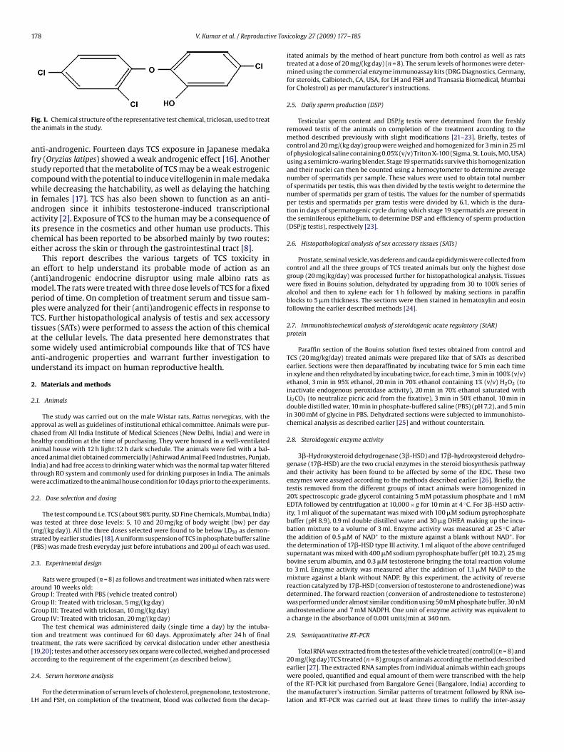
178 V. Kumar et al. / Reproductive Tox
Ft
afscwiaaice
a(mppTtasau
2
2
achaaItw
2
w(s(
2
aGGGG
tt[a
2
L
ig. 1. Chemical structure of the representative test chemical, triclosan, used to treathe animals in the study.
nti-androgenic. Fourteen days TCS exposure in Japanese medakary (Oryzias latipes) showed a weak androgenic effect [16]. Anothertudy reported that the metabolite of TCS may be a weak estrogenicompound with the potential to induce vitellogenin in male medakahile decreasing the hatchability, as well as delaying the hatching
n females [17]. TCS has also been shown to function as an anti-ndrogen since it inhibits testosterone-induced transcriptionalctivity [2]. Exposure of TCS to the human may be a consequence ofts presence in the cosmetics and other human use products. Thishemical has been reported to be absorbed mainly by two routes:ither across the skin or through the gastrointestinal tract [8].
This report describes the various targets of TCS toxicity inn effort to help understand its probable mode of action as ananti)androgenic endocrine disruptor using male albino rats as
odel. The rats were treated with three dose levels of TCS for a fixederiod of time. On completion of treatment serum and tissue sam-les were analyzed for their (anti)androgenic effects in response toCS. Further histopathological analysis of testis and sex accessoryissues (SATs) were performed to assess the action of this chemicalt the cellular levels. The data presented here demonstrates thatome widely used antimicrobial compounds like that of TCS haventi-androgenic properties and warrant further investigation tonderstand its impact on human reproductive health.
. Materials and methods
.1. Animals
The study was carried out on the male Wistar rats, Rattus norvegicus, with thepproval as well as guidelines of institutional ethical committee. Animals were pur-hased from All India Institute of Medical Sciences (New Delhi, India) and were inealthy condition at the time of purchasing. They were housed in a well-ventilatednimal house with 12 h light:12 h dark schedule. The animals were fed with a bal-nced animal diet obtained commercially (Ashirwad Animal Feed Industries, Punjab,ndia) and had free access to drinking water which was the normal tap water filteredhrough RO system and commonly used for drinking purposes in India. The animalsere acclimatized to the animal house condition for 10 days prior to the experiments.
.2. Dose selection and dosing
The test compound i.e. TCS (about 98% purity, SD Fine Chemicals, Mumbai, India)as tested at three dose levels: 5, 10 and 20 mg/kg of body weight (bw) per day
mg/(kg day)). All the three doses selected were found to be below LD50 as demon-trated by earlier studies [18]. A uniform suspension of TCS in phosphate buffer salinePBS) was made fresh everyday just before intubations and 200 �l of each was used.
.3. Experimental design
Rats were grouped (n = 8) as follows and treatment was initiated when rats wereround 10 weeks old:roup I: Treated with PBS (vehicle treated control)roup II: Treated with triclosan, 5 mg/(kg day)roup III: Treated with triclosan, 10 mg/(kg day)roup IV: Treated with triclosan, 20 mg/(kg day)
The test chemical was administered daily (single time a day) by the intuba-ion and treatment was continued for 60 days. Approximately after 24 h of finalreatment, the rats were sacrificed by cervical dislocation under ether anesthesia19,20]; testes and other accessory sex organs were collected, weighed and processed
ccording to the requirement of the experiment (as described below)..4. Serum hormone analysis
For the determination of serum levels of cholesterol, pregnenolone, testosterone,H and FSH, on completion of the treatment, blood was collected from the decap-
icology 27 (2009) 177–185
itated animals by the method of heart puncture from both control as well as ratstreated at a dose of 20 mg/(kg day) (n = 8). The serum levels of hormones were deter-mined using the commercial enzyme immunoassay kits (DRG Diagnostics, Germany,for steroids, Calbiotech, CA, USA, for LH and FSH and Transasia Biomedical, Mumbaifor Cholestrol) as per manufacturer’s instructions.
2.5. Daily sperm production (DSP)
Testicular sperm content and DSP/g testis were determined from the freshlyremoved testis of the animals on completion of the treatment according to themethod described previously with slight modifications [21–23]. Briefly, testes ofcontrol and 20 mg/(kg day) group were weighed and homogenized for 3 min in 25 mlof physiological saline containing 0.05% (v/v) Triton X-100 (Sigma, St. Louis, MO, USA)using a semimicro-waring blender. Stage 19 spermatids survive this homogenizationand their nuclei can then be counted using a hemocytometer to determine averagenumber of spermatids per sample. These values were used to obtain total numberof spermatids per testis, this was then divided by the testis weight to determine thenumber of spermatids per gram of testis. The values for the number of spermatidsper testis and spermatids per gram testis were divided by 6.1, which is the dura-tion in days of spermatogenic cycle during which stage 19 spermatids are present inthe seminiferous epithelium, to determine DSP and efficiency of sperm production(DSP/g testis), respectively [23].
2.6. Histopathological analysis of sex accessory tissues (SATs)
Prostate, seminal vesicle, vas deferens and cauda epididymis were collected fromcontrol and all the three groups of TCS treated animals but only the highest dosegroup (20 mg/kg/day) was processed further for histopathological analysis. Tissueswere fixed in Bouins solution, dehydrated by upgrading from 30 to 100% series ofalcohol and then to xylene each for 1 h followed by making sections in paraffinblocks to 5 �m thickness. The sections were then stained in hematoxylin and eosinfollowing the earlier described methods [24].
2.7. Immunohistochemical analysis of steroidogenic acute regulatory (StAR)protein
Paraffin section of the Bouins solution fixed testes obtained from control andTCS (20 mg/kg/day) treated animals were prepared like that of SATs as describedearlier. Sections were then deparaffinated by incubating twice for 5 min each timein xylene and then rehydrated by incubating twice, for each time, 3 min in 100% (v/v)ethanol, 3 min in 95% ethanol, 20 min in 70% ethanol containing 1% (v/v) H2O2 (toinactivate endogenous peroxidase activity), 20 min in 70% ethanol saturated withLi2CO3 (to neutralize picric acid from the fixative), 3 min in 50% ethanol, 10 min indouble distilled water, 10 min in phosphate-buffered saline (PBS) (pH 7.2), and 5 minin 300 mM of glycine in PBS. Dehydrated sections were subjected to immunohisto-chemical analysis as described earlier [25] and without counterstain.
2.8. Steroidogenic enzyme activity
3�-Hydroxysteroid dehydrogenase (3�-HSD) and 17�-hydroxysteroid dehydro-genase (17�-HSD) are the two crucial enzymes in the steroid biosynthesis pathwayand their activity has been found to be affected by some of the EDC. These twoenzymes were assayed according to the methods described earlier [26]. Briefly, thetestis removed from the different groups of intact animals were homogenized in20% spectroscopic grade glycerol containing 5 mM potassium phosphate and 1 mMEDTA followed by centrifugation at 10,000 × g for 10 min at 4 ◦C. For 3�-HSD activ-ity, 1 ml aliquot of the supernatant was mixed with 100 �M sodium pyrophosphatebuffer (pH 8.9), 0.9 ml double distilled water and 30 �g DHEA making up the incu-bation mixture to a volume of 3 ml. Enzyme activity was measured at 25 ◦C afterthe addition of 0.5 �M of NAD+ to the mixture against a blank without NAD+. Forthe determination of 17�-HSD type III activity, 1 ml aliquot of the above centrifugedsupernatant was mixed with 400 �M sodium pyrophosphate buffer (pH 10.2), 25 mgbovine serum albumin, and 0.3 �M testosterone bringing the total reaction volumeto 3 ml. Enzyme activity was measured after the addition of 1.1 �M NADP to themixture against a blank without NADP. By this experiment, the activity of reversereaction catalyzed by 17�-HSD (conversion of testosterone to androstenedione) wasdetermined. The forward reaction (conversion of androstenedione to testosterone)was performed under almost similar condition using 50 mM phosphate buffer, 30 nMandrostenedione and 7 mM NADPH. One unit of enzyme activity was equivalent toa change in the absorbance of 0.001 units/min at 340 nm.
2.9. Semiquantitative RT-PCR
Total RNA was extracted from the testes of the vehicle treated (control) (n = 8) and
20 mg/(kg day) TCS treated (n = 8) groups of animals according the method describedearlier [27]. The extracted RNA samples from individual animals within each groupswere pooled, quantified and equal amount of them were transcribed with the helpof the RT-PCR kit purchased from Bangalore Genei (Bangalore, India) according tothe manufacturer’s instruction. Similar patterns of treatment followed by RNA iso-lation and RT-PCR was carried out at least three times to nullify the inter-assay
V. Kumar et al. / Reproductive Toxicology 27 (2009) 177–185 179
Table 1Primers used for semiquantitative RT-PCR.
Gene Primer Sequence (5′–3′) Product size Cycle used Annel Temp Gene bank accession no.
P450SCC (F) CgCTCAgTgCTggTCAAAA 688 23 55 J 05156P450SCC (R) TCTggTAgACggCgTCgAT
P450C17 (F) GACCAAGGGAAAGGCGT 302 24 55 M 22204P450C17 (R) GCATCCACGATACCCTC
3�-HSD (F) CCgCAAgTATCATgACAgA 547 24 55 M 381783�-HSD (R) CCgCAAgTATCATgACAgA
17�-HSD (F) TTCTgCAAggCTTTACCAgg 653 26 55 AF 03515617�-HSD (R) ACAAACTCATCggCggTCTT
AR (F) TTACgAAgTgggCATgATgA 570 26 55 M 20133AR (R) ATCTTgTCCAggACTCggTg
StAR (F) TTgggCATACTCAACAACCA 389 25 58 NM 031558S
G 21 58 NM 017008G
vt6atdr(Tuifipfu
2
ecwplStp(�
2
als
3
3
cWandiTta
Table 2Effects of three different dosage level of triclosan on the body weights of the animalsbefore and after the completion of 60 days of treatment.
Groups Body weight (g ± S.E.M.)
Initial Final
Control 168 ± 6.3 187 ± 7.1Triclosan (5 mg/kg) 166 ± 7.9 185 ± 5.7
tAR (R) ATgACACCgCTTTgCTCAg
APDH (F) AgACAgCCgCATCTTCTTgT 207APDH (R) CTTgCCgTgggTAgAgTCAT
ariations. PCR was performed by denaturing at 94 ◦C for 60 s, annealing at variousemperatures (depending on primer pairs used) for 30 s and extension at 72 ◦C for0 s followed by varying number of cycles for amplification. The primer sequences,nnealing temperature and number of cycles for PCR were all designed accordinghe earlier report by Ohsako et al. [30] except for StAR, glyceraldehyde-3-phosphateehydrogenase (GAPDH) [28]. Primer sequence for StAR was adopted from earliereport [29]. Primers for GAPDH were designed with the help of Primer3 softwareSteve Rozen, Helen J. Skaletsky, 1998, Primer3) and standardized in the laboratory.he PCR products were then separated on 2% agarose gel and visualized in a gel doc-mentation system (Bio Rad, USA). The intensity of the bands on gels was converted
nto digital image with a gel analyzer and amounts of RT-PCR products were quanti-ed with Scion Images software (Scion Corporation, Fredrick, MD, USA). GAPDH PCRroducts were used as internal standards and each of the RT-PCR was carried outour times. Primer sequence, product size, annealing temperature, number of cyclessed and gene bank accession number of all primers are presented in Table 1.
.10. Western blot analysis
The testis of the control and 20 mg/(kg day) of TCS treated male rats were homog-nized in phosphate buffer saline having 20% (v/v) glycerol and 1 mM EDTA andentrifuged at 10,000 × g for 30 min at 4 ◦C. The pellet was discarded, supernatantsere quantified and an equal quantity of protein samples were analyzed by 12%olyacrylamide gel according to the method of described earlier [30]. This was fol-
owed by Western blot analysis of the protein samples. Membranes were probed withtAR and androgen receptor (AR) antibodies (dilutions 1:1000 and 1:250 respec-ively). Color development was performed in 30 ml AP-buffer (100 mM Tris/HClH 9.5, 100 mM NaCl, 5 mM MgCl2), with 200 �l NBT (50 mg/ml) and 100 �l BCIP50 mg/ml). The developed blots were subjected to densitometric analysis using the-actin as internal control.
.11. Statistical analysis
Origin 6.1 software (Origin Lab Corporation, USA) was used for statistical analysisnd data were expressed as mean ± S.E.M. For statistical analysis of data, ANOVA fol-owed by multiple two-tail comparison t-test was used and p < 0.05 was consideredignificant.
. Results
.1. Body weight and weight of testis and SATs
Treatment of rats with test samples did not induce significanthanges in the body weight at any of the test doses (Table 2).
hatever minimal increase in the body weight observed could bettributed to normal aging. Administration of TCS did not cause sig-ificant change in the weight of testis and SATs at the 5 mg/(kg day)
osage. In contrast, the higher test doses (10 and 20 mg/(kg day))nduced a significant decrease in the weight of testis and SATs.CS exposure decreased the weights of testis, epididymis, ven-ral prostate, vas deferens and seminal vesicles between 20–50%t 10 mg/(kg day) and 35–49% at 20 mg/(kg day) (Table 3) (p < 0.05).
Triclosan (10 mg/kg) 169 ± 5.1 188 ± 6.8Triclosan (20 mg/kg) 165 ± 7.2 183 ± 7.5
Each value denotes mean ± S.E.M. of eight animals.
3.2. Gene expression analysis
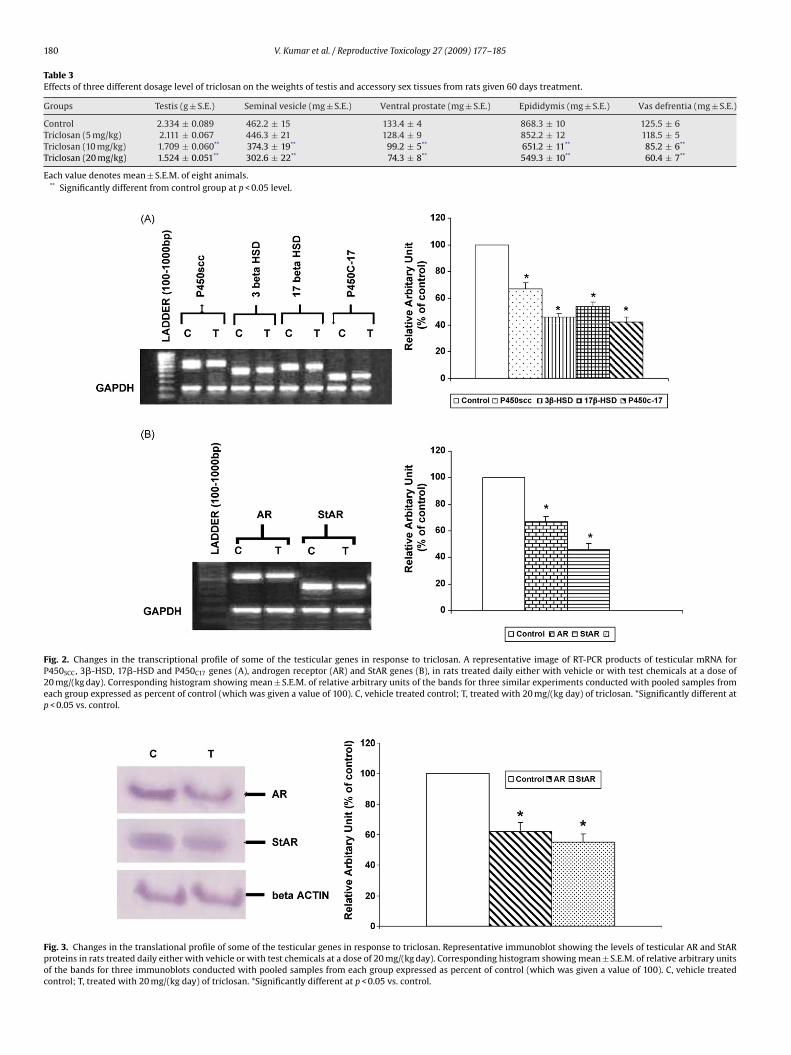
Rats treated with 20 mg/(kg day) TCS showed a statisticallysignificant down-regulation in the testicular levels of mRNA forcytochrome P450SCC (P450SCC), cytochrome P450C17 (P450C17), 3�-HSD, 17�-HSD, StAR and AR as compared to control. Level ofP450SCC, 3�-HSD, 17�-HSD and P450C17 mRNA decreased up to33, 54, 46 and 58% respectively as compared to control (p < 0.05)(Fig. 2A). In the case of AR and StAR, the expression decreased up to33 and 54% respectively as compared to control (Fig. 2B) (p < 0.05).
Western blot analysis demonstrated that the rats treated withTCS at a dose of 20 mg/(kg day) showed reduced translation of StAR(30 kDa) and AR (100 kDa) proteins as compared to control (vehi-cle treated animals) and this decrease was statistically significant(Fig. 3) (p < 0.05). The uniform band intensities of �-actin in all thewells indicated equal gel loading.
3.3. Testicular 3ˇ-HSD and 17ˇ-HSD levels in vitro
In vitro spectrophotometric enzyme assays for 3�-HSD and 17�-HSD demonstrated that the treatment of animals with test chemicalcaused a statistically significant decrease in the activity of boththe testicular steroidogenic enzymes at the two higher dose levels(10 and 20 mg/(kg day)) (p < 0.05). The decrease was not significantat a dose of 5 mg/(kg day) for both the enzymes. Doses of 10 and20 mg/(kg day) TCS decreased 3�-HSD enzyme activity up to 27 and39% respectively (Fig. 4) while that of 17�-HSD enzyme activity upto 31 and 46% respectively (Fig. 4) as compared to control (p < 0.05).
3.4. Serum hormone levels
There was a statistically significant decrease in the serum LH(38.5%), FSH (17%), cholesterol (35%), pregnenolone (31%) andtestosterone (41%) levels in male rats treated with a dose of20 mg/(kg day) as compared to control (p < 0.05) (Table 4).
180 V. Kumar et al. / Reproductive Toxicology 27 (2009) 177–185
Table 3Effects of three different dosage level of triclosan on the weights of testis and accessory sex tissues from rats given 60 days treatment.
Groups Testis (g ± S.E.) Seminal vesicle (mg ± S.E.) Ventral prostate (mg ± S.E.) Epididymis (mg ± S.E.) Vas defrentia (mg ± S.E.)
Control 2.334 ± 0.089 462.2 ± 15 133.4 ± 4 868.3 ± 10 125.5 ± 6Triclosan (5 mg/kg) 2.111 ± 0.067 446.3 ± 21 128.4 ± 9 852.2 ± 12 118.5 ± 5Triclosan (10 mg/kg) 1.709 ± 0.060** 374.3 ± 19** 99.2 ± 5** 651.2 ± 11** 85.2 ± 6**
Triclosan (20 mg/kg) 1.524 ± 0.051** 302.6 ± 22** 74.3 ± 8** 549.3 ± 10** 60.4 ± 7**
Each value denotes mean ± S.E.M. of eight animals.** Significantly different from control group at p < 0.05 level.
Fig. 2. Changes in the transcriptional profile of some of the testicular genes in response to triclosan. A representative image of RT-PCR products of testicular mRNA forP450SCC, 3�-HSD, 17�-HSD and P450C17 genes (A), androgen receptor (AR) and StAR genes (B), in rats treated daily either with vehicle or with test chemicals at a dose of20 mg/(kg day). Corresponding histogram showing mean ± S.E.M. of relative arbitrary units of the bands for three similar experiments conducted with pooled samples fromeach group expressed as percent of control (which was given a value of 100). C, vehicle treated control; T, treated with 20 mg/(kg day) of triclosan. *Significantly different atp < 0.05 vs. control.
Fig. 3. Changes in the translational profile of some of the testicular genes in response to triclosan. Representative immunoblot showing the levels of testicular AR and StARproteins in rats treated daily either with vehicle or with test chemicals at a dose of 20 mg/(kg day). Corresponding histogram showing mean ± S.E.M. of relative arbitrary unitsof the bands for three immunoblots conducted with pooled samples from each group expressed as percent of control (which was given a value of 100). C, vehicle treatedcontrol; T, treated with 20 mg/(kg day) of triclosan. *Significantly different at p < 0.05 vs. control.
V. Kumar et al. / Reproductive Toxicology 27 (2009) 177–185 181
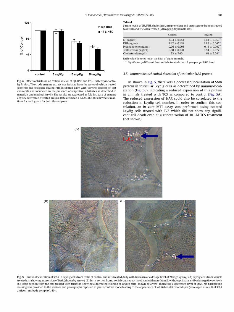
Fig. 4. Effects of triclosan on testicular level of 3�-HSD and 17�-HSD enzyme activ-ity in vitro. The crude enzyme extract was isolated from the testes of vehicle treated(control) and triclosan treated rats intubated daily with varying dosages of testchemicals and incubated in the presence of respective substrates as described inmaterials and methods (n = 8). The results are expressed as fold increase of enzymeactivity over vehicle treated groups. Data are mean ± S.E.M. of eight enzymatic reac-tions for each group for both the enzymes.
Table 4Serum levels of LH, FSH, cholesterol, pregnenolone and testosterone from untreated(control) and triclosan treated (20 mg/(kg day)) male rats.
Control Treated
LH (ng/ml) 1.04 ± 0.054 0.64 ± 0.056**
FSH (ng/ml) 8.12 ± 0.168 6.82 ± 0.045**
Pregnenolone (ng/ml) 0.26 ± 0.008 0.18 ± 0.007**
Testosterone (ng/ml) 6.60 ± 0.130 3.94 ± 0.077**
**
Fig. 5. Immunolocalization of StAR in Leydig cells from testis of control and rats treated dtreated rats showing expression of StAR (shown by arrow). (B) Testis section from a vehicle t(C) Testis section from the rats treated with triclosan showing a decreased staining of Lstaining was provided to the sections and photographs captured in phase contrast mode leantigen–antibody complex). 40×.
Cholesterol (mg/dl) 93 ± 7.00 61 ± 5.00
Each value denotes mean ± S.E.M. of eight animals.** Significantly different from vehicle treated control group at p < 0.05 level.
3.5. Immunohistochemical detection of testicular StAR protein
As shown in Fig. 5, there was a decreased localization of StARprotein in testicular Leydig cells as determined by immunolocal-ization (Fig. 5C), indicating a reduced expression of this proteinin animals treated with TCS as compared to control (Fig. 5A).The reduced expression of StAR could also be correlated to thereduction in Leydig cell number. In order to confirm this cor-
relation, an in vitro MTT assay was performed using isolatedLeydig cells treated with TCS which did not show any signifi-cant cell death even at a concentration of 10 �M TCS treatment(not shown).aily with triclosan at a dosage level of 20 mg/(kg day). (A) Leydig cells from vehiclereated rat incubated with non-fat milk without primary antibody (negative control).eydig cells (shown by arrow) indicating a decreased level of StAR. No backgroundading to the appearance of whitish violet colored spot (developed as result of StAR

182 V. Kumar et al. / Reproductive Tox
Fig. 6. Daily sperm production/g of testis weight (DSP/g) in vehicle treated (control)adDt
3
ttswAmwr
dsrteb
ar
F(
nd triclosan treated (20 mg/(kg day)) rats. Data are presented as mean ± S.E.M. ofuplicate determinations from individual testes; n = 8 for control and treated testes.SP was significantly decreased (p < 0.05) in the treated group as compared to con-
rol.
.6. Histopathology of testis and SATs
A number of histopathological malformations were observed inhe testis and SATs of 20 mg/(kg day) group as compared to con-rol which probably affected the production and maturation of theperms. This was supported by a 34% decrease in the DSP/g of testiseight (p < 0.05) in treated group as compared to control (Fig. 6).lthough the cauda epididymis (CE) from control rats showed a nor-al structure and sperm density (Fig. 7A), a reduced sperm densityas observed in the lumina of epididymal tubule from the treated
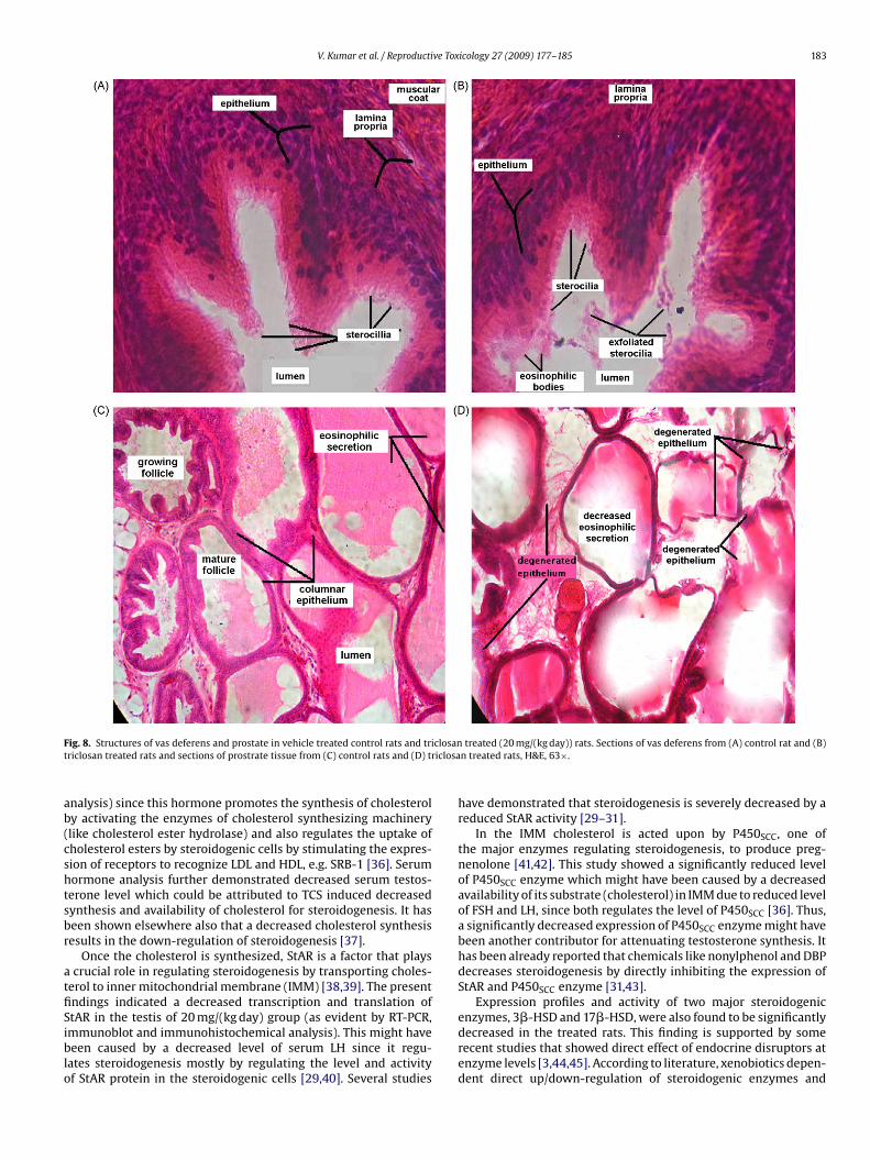
ats (Fig. 7B).A normal thickness as well as arrangement of ciliated brush bor-
er was observed in vas deference from control rats (Fig. 8A) whileeveral malformations were observed in vas deference from treatedats (Fig. 8B). Lumen of vas deference from the treated rats showedhe presence of sterocilia detached from the epithelium and pres-
nce of eosinophillic bodies (Fig. 8B). The stereocilia were found toe thin, few or absent in the epithelium of treated rats (Fig. 8B).In the case of prostate tissues, folliculli appeared to be normalnd large sized in the control rats (Fig. 8C) while in the treatedats the folliculli were found to be comparatively degenerated
ig. 7. Photomicrographs of rat cauda epididymes showing reduction in sperm mass as a rA) Vehicle treated rat and (B) triclosan treated rats, H&E, 20×.
icology 27 (2009) 177–185
and empty and follicular walls were thinner (Fig. 8D). However,surprisingly the seminal vesicles did not display any noticeablehistopathological changes in control and treated rats (data notshown).
4. Discussion
TCS is a synthetic chemical widely used as an antimicrobial agentin different commercial preparations [11]. Since TCS possess a phe-nolic moiety like many of the common EDC it could be presumedto display similar activities as demonstrated by other EDC of thesame chemical family [31,32]. The dosage for the test chemicalsused in this study were selected based on LD50 values and also someearlier reports where a similar compound, triclocarbon (TCC), wasused in rat models [33,34]. In this synthesis it should be noted thatTCS is considered as a traditional EDC whereas the group of chem-icals represented by TCC may rather act as a new-type of EDC as an“enhancer” through mechanisms that are yet to be identified. Basedon these reports, in the present study 5, 10 and 20 mg/(kg day) of TCSwere administered by oral routes. Although, 10 and 20 mg/(kg day)showed significant responses against the parameters tested here,we presented the data only for the latter in majority of the experi-ments since the response were almost similar by those two dosagelevels.
Testicular androgenesis involves several crucial steps right fromthe synthesis of cholesterol, the parent molecule for all the steroidhormones, to its transport within the steroidogenic tissues and thenits further metabolism to form steroids. Cholesterol is acquired bysteroidogenic cells either by de novo synthesis or from the high den-sity/low density lipoprotein (HDL and LDL) circulating in the blood[35]. In this study, 20 mg/kg/day group demonstrated a decreasedlevel of serum cholesterol. In addition, some of our preliminarydata also showed the reduction in the level of SRB-1 expression(data not shown). SRB-1 is a type of lipoprotein receptor present on
the surface of steroidogenic cells to recognize HDL and LDL and isresponsible for their uptake in those cells. It could be presumed thatreduction in SRB-1 level might interfere with the uptake of choles-terol by those cells. Both these events might have been achievedby a reduced LH production (as demonstrated by serum hormoneesult of 60 days consecutive triclosan treatment at a dosage level of 20 mg/(kg day).
V. Kumar et al. / Reproductive Toxicology 27 (2009) 177–185 183
F closant iclosa
ab(cshtsbr
atfiSiblo
ig. 8. Structures of vas deferens and prostate in vehicle treated control rats and tririclosan treated rats and sections of prostrate tissue from (C) control rats and (D) tr
nalysis) since this hormone promotes the synthesis of cholesteroly activating the enzymes of cholesterol synthesizing machinerylike cholesterol ester hydrolase) and also regulates the uptake ofholesterol esters by steroidogenic cells by stimulating the expres-ion of receptors to recognize LDL and HDL, e.g. SRB-1 [36]. Serumormone analysis further demonstrated decreased serum testos-erone level which could be attributed to TCS induced decreasedynthesis and availability of cholesterol for steroidogenesis. It haseen shown elsewhere also that a decreased cholesterol synthesisesults in the down-regulation of steroidogenesis [37].
Once the cholesterol is synthesized, StAR is a factor that playscrucial role in regulating steroidogenesis by transporting choles-
erol to inner mitochondrial membrane (IMM) [38,39]. The presentndings indicated a decreased transcription and translation of
tAR in the testis of 20 mg/(kg day) group (as evident by RT-PCR,mmunoblot and immunohistochemical analysis). This might haveeen caused by a decreased level of serum LH since it regu-ates steroidogenesis mostly by regulating the level and activityf StAR protein in the steroidogenic cells [29,40]. Several studies
treated (20 mg/(kg day)) rats. Sections of vas deferens from (A) control rat and (B)n treated rats, H&E, 63×.
have demonstrated that steroidogenesis is severely decreased by areduced StAR activity [29–31].
In the IMM cholesterol is acted upon by P450SCC, one ofthe major enzymes regulating steroidogenesis, to produce preg-nenolone [41,42]. This study showed a significantly reduced levelof P450SCC enzyme which might have been caused by a decreasedavailability of its substrate (cholesterol) in IMM due to reduced levelof FSH and LH, since both regulates the level of P450SCC [36]. Thus,a significantly decreased expression of P450SCC enzyme might havebeen another contributor for attenuating testosterone synthesis. Ithas been already reported that chemicals like nonylphenol and DBPdecreases steroidogenesis by directly inhibiting the expression ofStAR and P450SCC enzyme [31,43].
Expression profiles and activity of two major steroidogenic
enzymes, 3�-HSD and 17�-HSD, were also found to be significantlydecreased in the treated rats. This finding is supported by somerecent studies that showed direct effect of endocrine disruptors atenzyme levels [3,44,45]. According to literature, xenobiotics depen-dent direct up/down-regulation of steroidogenic enzymes and
1 ve Tox
siaao
aaTtst[
ontlptitTacotoatcaa
miptodfuoaeaitiioeeoettbwtd
C
[
[
84 V. Kumar et al. / Reproducti
teroidogenesis can be affected at several levels, viz. direct bind-ng of these chemicals to steroid receptors, steroidogenic enzymesnd proteins associated with steroidogenesis, e.g. StAR protein [46]nd increasing the stability of transcripts and transcriptional ratef the promoter of steroidogenic enzymes [47].
Furthermore, the expression patterns of AR, both at transcriptionnd translation level was found to be decreased in treated groupss demonstrated by RT-PCR and immnunoblot analysis respectively.his might have been achieved by a reduced level of available testos-erone although autologous regulation of AR gene in the testes istill a matter of controversy. Several reports exist on both regula-ion and non-regulation of AR mRNA expression by androgen itself48–50].
Another interesting result of this study was the developmentf histopathological abnormalities in SATs of 20 mg/(kg day) group,amely CE, ductus deference and prostate. The decreased testos-erone and AR level in treated rats (as indicated above) might haveed to the degenerative changes and atrophy in the SATs as sup-orted by their decreased weight and size (data not shown) inhis study. Results also demonstrated a decreased sperm countn the testis of treated rats as compared to control, probably dueo reduced testicular spermatogenesis induced by TCS [51,52].his could also be attributed to the reduced level of serum FSH,hormone directly involved in maintaining spermatogenesis in
onjunction with testosterone [53]. Other degenerative changesbserved in the cauda were occurrence of epithelial degenera-ion in the form of nuclear karyolysis and pyknosis. These typesf histopathological changes in cauda have been reported by otherslso [54,55]. Similar degeneration and atrophy were also found inhe other SATs like ductus deferens and prostate glands. All theseould be attributed to the decrease in the level of androgen andndrogen receptors which are known to support the functioningnd continuous persistence of these organs.
In conclusion, TCS, a commonly used chemical in various cos-etics and other applications, may act as an endocrine disruptor
n male rats and has the potential to impair the pituitary–gonadalathway at various levels. Inhibition of androgen production byhis chemical may be explained by its action at various stepsf steroidogenesis: reduction of LH and cholesterol production;epressed StAR expression, one of the crucial protein responsibleor cholesterol transport to inner mitochondrial membrane for itstilization by steroidogenic enzymes; and finally down-regulationf several key steroidogenic enzymes (P450SCC, P450C17, 3�-HSD,nd 17�-HSD). Apparently gonadotrophic hormones, steroidogenicnzymes, and StAR, key proteins involved in androgen productionnd maintenance of SATs, are all potential targets for TCS-mediatedmpairment of steroidogenesis. To the best of our knowledgehis is the first ever report on the anti-androgenic effects of TCSn male rats. However, further studies are needed to designatet as an anti-androgenic EDC since the effects are dependentn several factors like composition of chemicals, animal mod-ls, dose and time of treatment to name a few of them. Anarlier report exists in the effect of polychlorinated biphenylsn testicular steroidogenesis through oxidative stress [56]. Theffects observed here for TCS could involve similar testicular oxida-ive stress as a mechanism. All these facts reinforces the notionhat these common chemicals pose a hazard to human healthy acting through various targets in endocrine disruption andarrants further detailed investigation to pin-point the specific
arget site(s) for these chemical induced toxicity and endocrineisruption.
onflict of interest
None declared.
[
[
icology 27 (2009) 177–185
Acknowledgements
Kind help of Prof. Ilpo Huhtaniemi, Imperial College London, UKwith all steroidal test chemicals is greatly acknowledged. We wouldalso like to thank Prof. D.M. Stocco, Texas Tech University, Lubbock,TX, and Dr. R.K. Tyagi, Jawaharlal Nehru University, New Delhi, Indiafor kindly providing the StAR and androgen receptor antibodies,respectively. The study was supported by the Ministry of HumanResource and Development as fellowship to V.K., and by the Depart-ment of Biotechnology (DBT, no. BT/BCE/08/355/04), Governmentof India and Council of Scientific and Industrial Research (CSIR,no. 37(1200)/04/EMR II), Government of India as funded projectsto P.R. The authors would thus like to thank all these fundingagencies.
References
[1] Cargouet M, Perdiz D, Mouatassim A, Tamisier S, Levi Y. Assessment of rivercontamination by estrogenic compounds in Paris area (France). Sci Tot Environ2004;324:55–66.
[2] Chen J, Ahn KC, Gee NA, Gee SJ, Hammock BD, Lasley BL. Antiandrogenic prop-erties of parabens and other phenolic containing small molecules in personalcare products. Toxicol Appl Pharmacol 2007;221:278–84.
[3] Kumar V, Chakraborty A, Viswanath G, Roy P. Androgenic endocrine disruptorsin wastewater treatment plant effluents in India: their influence on repro-ductive processes and systemic toxicity in male rats. Toxicol Appl Pharmacol2007;226(1):60–3.
[4] Fenton SE. Endocrine-disrupting compounds and mammary gland devel-opment: early exposure and later life consequences. Endocrinology2006;147(6):18–24.
[5] Darbre PD. Environmental oestrogens, cosmetics and breast cancer. Res ClinEndocrinol Metab 2006;20:121–43.
[6] Cabana H, Jiwan JL, Rozenberg R, Elisashvili V, Penninckx M, Agathos SN, et al.Elimination of endocrine disrupting chemicals nonylphenol and bisphenol Aand personal care product ingredient triclosan using enzyme preparation fromthe white rot fungus Coriolopsis polyzona. Chemosphere 2007;67:770–8.
[7] Lakeram M, Lockley DJ, Sanders DJ, Pendlington R, Forbes B. Paraben trans-port and metabolism in the biomimetic artificial membrane permeabilityassay (BAMPA) and 3-day and 21-day Caco-2 cell systems. J Biomol Screen2006;12:84–91.
[8] Dayan AD. Response to: ethics, data and the pharmaceutical physician. Int J ClinPract 2006;60:1009.
[9] Heidler J, Sapkota A, Halden RU. Partitioning, persistence and accumulation indigested sludge of the topical antiseptic triclocarban during wastewater treat-ment. Environ Sci Technol 2006;40:3634–9.
[10] Nakada N, Tanishima T, Shinohara H, Kiri K, Takada H. Pharmaceutical chemicalsand endocrine disrupters in municipal wastewater in Tokyo and their removalduring activated sludge treatment. Water Res 2006;40:3297–303.
[11] Black JG, Howes DRT. Percutaneous absorption and metabolism of Irgasan DP300. Toxicology 1975;3:33–47.
12] Tatarazako N, Ishibashi H, Teshima K, Kishi K, Arizono. K Effects of triclosan onvarious aquatic organisms. Environ Sci 2004;11(2):133–40.
[13] Kanetoshi A, Ogawa H, Katsura E, Kaneshima H. Chlorination of IrgasanDP300 and formation of dioxins from its chlorinated derivatives. J Chromatogr1987;389:139–53.
[14] Kanetoshi A, Ogawa H, Katsura E, Kaneshima H, Miura T. Formation of polychlo-rinated dibenzo-p-dioxins upon combustion of commercial textile productscontaining 2,4,4-trichloro-2-hydroxydiphenyl ether (Irgasan DP300). J Chro-matogr 1998;442:289–99.
[15] Kanetoshi A, Ogawa H, Katsura E, Kaneshima H, Miura T. Formation of poly-chlorinated dibenzo-p-dioxins from 2,4,4-trichloro-2-hydroxydiphenyl ether(Irgasan DP300) and its chlorinated derivatives by exposure to sunlight. J Chro-matogr 1998 b;454:145–55.
[16] Foran CM, Bennett ER, Benson WH. Developmental evaluation of a potentialnon-steroidal estrogen: triclosan. Mar Environ Res 2000;50(1–5):153–6.
[17] Ishibashi H, Matsumura N, Hirano M, Matsuoka M, Shiratsuchi H, Ishibashi Y, etal. Effects of triclosan on the early life stages and reproduction of medaka Oryziaslatipes and induction of hepatic vitellogenin. Aquat Toxicol 2004;67:167–79.
[18] Bhargava HN, Leonard PA. Triclosan: applications and safety. Am J Infect Control1996;24:209–18.
[19] Biswas NM, Gupta RS, Chattopadhyay A, Choudhury GR, Sarkar M. Effect ofatenolol on cadmium induced testicular toxicity in male rats. Reprod Toxicol2001;15:699–704.
20] Blesson CS, Awasthi S, Kharkwal G, Daverey A, Dwivedi A. Steroids
2006;71:993–1000.21] Robb GW, Amann RP, Killian GJ. Daily sperm production and epididymal spermreserves of pubertal and adult rats. J Reprod Fertil 1978;54:103–7.
22] Cooke PS, Kirby JD, Porcelli J. Increased testis growth and sperm production inadult rats following transient neonatal goitrogen treatment: optimization of thepropylthiouracil dose and effect of methimazole. J Reprod Fertil 1993;97:493–9.

ve Tox
[
[
[
[
[
[
[
[
[
[
[
[
[
[
[
[
[
[
[
[
[
[
[
[
[
[
[
[
[
[
[
[
[
V. Kumar et al. / Reproducti
23] Fernandes GSA, Arena AC, Fernandez CDB, Mercadante A, Barbisan LF, Kemp-inas WG. Reproductive effects in male rats exposed to diuron. Reprod Toxicol2007;23:106–12.
24] Mukherjee KL. Routine biochemical tests and histological techniques. In:Mukherjee K, editor. Medical laboratory technology. New Delhi: Tata McGraw-Hill Publishing Company Limited; 2003. p. 1124–90.
25] Kroft TL, Patterson J, Won Yoon J, Doglio L, Walterhouse DO, Iannaccone PM,et al. GLI1 localization in the germinal epithelial cells alternates between cyto-plasm and nucleus: upregulation in transgenic mice blocks spermatogenesis inpachytene. Biol Reprod 2000;65:1663–71.
26] Sarkar M, Biswas NM, Ghosh D. Effect of sodium arsenite on testicular 3 and17 hydroxysteroid dehydrogenase activities in albino rats: dose and duration-dependent responses. Med Sci Res 1991;19:789–90.
27] Chomczynski P, Sacchi N. Single step method of RNA isolation by acid guani-dium thiocyanate-phenol-chloroform extraction. Anal Biochem 1987;162:156–9.
28] Ohsako S, Kubota K, Kurosawa S, Takeda K, Qing W, Ishimura R, et al. Alter-ations of gene expression in adult male rat testis and pituitary shortly aftersub acute administration of the antiandrogen flutmide. J Reprod Dev 2003;49:275–90.
29] Murugesan P, Muthusamy T, Balasubramanian K, Arunakaran J. Effects ofvitamins C and E on steroidogenic enzymes mRNA expression in polychlo-rinated biphenyl (Aroclor 1254) exposed adult rat Leydig cells. Toxicology2007;232:170–82.
30] Laemmli UK. Cleavage of structural proteins during the assembly of the headof bacteriophage T4. Nature 1970;227:680–5.
31] Kitamura S, Suzuki T, Sanoh S, Kohta R, Jinno N, Sugihara K, et al. Compara-tive study of the endocrine-disrupting activity of bisphenol A and 19 relatedcompounds. Toxicol Sci 2005;84:249–59.
32] Stocco DM. StAR protein and the regulation of steroid hormone biosynthesis.Ann Rev Physiol 2001;63:193–213.
33] Nollen GA, Dierckman TA. Reproduction and teratogenic studies of 2:1 mixtureof 3,4,4′-trichlorocarbanilide and 3-trifluoromethyl-4,4′-dichlorocarbnilide inrats and rabbits. Toxicol Appl Pharmacol 1979;51:417–25.
34] Chen J, Ahn KC, Gee NA, Ahmed MI, Duleba AJ, Zhao L, et al. Triclocarbanenhances testosterone action: a new type of endocrine disruptor? Endocrinol-ogy 2008;149(3):1173–9.
35] Cao G, Zhao L, Strangle H, Hasegawa T, Richardson JA, Parker KL, et al. Develop-mental and hormonal regulation of murine scavenger receptor, class B, type 1.Mol Endocrinol 1999;13:1460–73.
36] Fauser BCJM. Molecular biology of steroidogenesis. In: Molecular biol-ogy in the reproductive medicine. New York: Parthenon Publishing; 1999.p. 234–51.
37] Barlow NJ, Phillips SI, Wallace DC, Gaido WK, Foster PMD. Qunatitative changesin gene expression in fetal rat testis following exposure to di(n-butyl) phatha-late. Toxicol Sci 2003;73:431–51.
38] Hasegawa T, Zhao L, Caron KM, Majdic G, Suzuki T, Shizava S, et al. Develop-
mental roles of the steroidogenic acute regulatory protein (StAR) as revealedby StAR knockout mice. Mol Endocrinol 2000;14:1462–71.39] Manna PR, Roy P, Clark BJ, Stocco DM, Huhtaniemi IT. Interaction of thyroidhormone and steroidogenic acute regulatory (StAR) protein in the regulationof murine Leydig cell steroidogenesis. J Steroid Biochem Mol Biol 2001;76:167–77.
[
icology 27 (2009) 177–185 185
40] Arakane F, King SR, Du Y, Kallen CB, Walsh LP, Watari H, et al. Phosphorylationof steroidogenic acute regulatory protein (StAR) modulates its steroidogenicactivity. J Biol Chem 1997;272(51):32656–62.
41] Miller WL. Molecular biology of steroid hormone synthesis. Endocr Rev1988;9:295–318.
42] Omura T, Morohashi K. Gene regulation of steroidogenesis. J Steroid BiochemMol Biol 1995;53:19–25.
43] Arukwe A. Modulation of brain steroidogenesis by affecting transcriptionalchanges of steroidogenic acute regulatory (StAR) protein and cholesterol sidechain cleavage (P450scc) in juvenile Atlantic salmon (Salmo salar) is a novelaspect of nonylphenol toxicity. Environ Sci Technol 2005;39:9791–8.
44] Andric NL, Kostic TS, Zoric SN, Stanic BD, Andric SA, Kovacevic RZ. Effectof PCB-based transformer oil on testicular steroidogenesis and xenobiotic-metabolizing enzymes. Reprod Toxicol 2006;22:102–10.
45] Lyssimachou A, Jessen BM, Arukew A. Brain cytochrome P450 aromatase geneisoforms and activity levels in Atlantic salmon after waterborne exposure tonominal environmental concentrations of the pharmaceutical ethynylestradioland antifoulant tributyltin. Toxicol Sci 2006;91:82–92.
46] Rice S, Mason HD, Whitehead SA. Phytoestrogens and their low dose com-binations inhibit mRNA expression and activity of aromatase in humangranulosa-luteal cells. J Steroid Biochem Mol Biol 2006;101:216–25.
47] Lin TC, Chien SC, Hsu PC, Li LA. Mechanistic study of polychlorinatedbiphenyl 126-induced CYP 11B1 and CYP 11B2 up-regulation. Endocrinology2006;147:1536–44.
48] Blok LJ, Barlett JM, Bolt-De Vries J, Themmen AP, Brinkmann AO, Weinbauer GF,et al. Effect of testosterone deprivation on expression of the androgen receptorin rat prostate, epididymis and testis. Int J Androl 1992;15:182–98.
49] Prins GS, Woodham C. Autologous regulation of androgen receptor messen-ger ribonucleic acid in the separate lobes of the prostate gland. Biol Reprod1995;53:609–19.
50] Shan LX, Bardin CW, Hardy MP. Immunohistochemical analysis of androgeneffects on androgen receptor expression in developing Leydig and Sertoli cells.Endocrinology 1997;138:1259–66.
51] Ku WE, Chapin RE, Wine RN, Gladen BC. Testicular toxicity of boric acid (BA):relationship of dose to lesion development and recovery in the F334 rat. ReprodToxicol 1993;7:305–19.
52] Poon R, Rigden M, Chu I, Valli VE. Short-term oral toxicity of pentylether, 1,4-diethoxybutane, and 1,6-dimethoxyhexane in male rats. Toxicol Sci2004;77:142.
53] Plant TM, Marshall GR. The functional significance of FSH in spermato-genesis and the control of its secretion in male primates. Endocr Rev2001;22(6):764–86.
54] Chitra KC, Ramachandra KR, Mathur PP. Effect of bisphenol A and co-administration of bisphenol A and vitamin C on epididymis of adult rats: ahistological and biochemical study. Asian J Androl 2003;5:203–8.
55] Narayana K, Prashanthi N, Nayanatara A, Kumard SG, Kumare HHC, BairyKL, et al. A broad-spectrum organophosphate pesticide O,O-dimethyl O-
4-nitrophenyl phosphorothioate (methyl parathion) adversely affects thestructure and function of male accessory reproductive organs in the rat. EnvironToxicol Pharmacol 2006;2:315–24.56] Murugesan P, Balaganesh M, Balasubramanian K, Arunakaran J. Effects ofpolychlorinated biphenyl (Arochlor 1254) on steroidogenesis and antioxidantsystem in cultured adult rat Leydig cells. J Endocrinol 2007;192:325–38.
Copyright © 2022 FDOKUMEN