White spot syndrome virus infection in cultured Penaeus vannamei (Boone) in Ecuador with emphasis on...
12
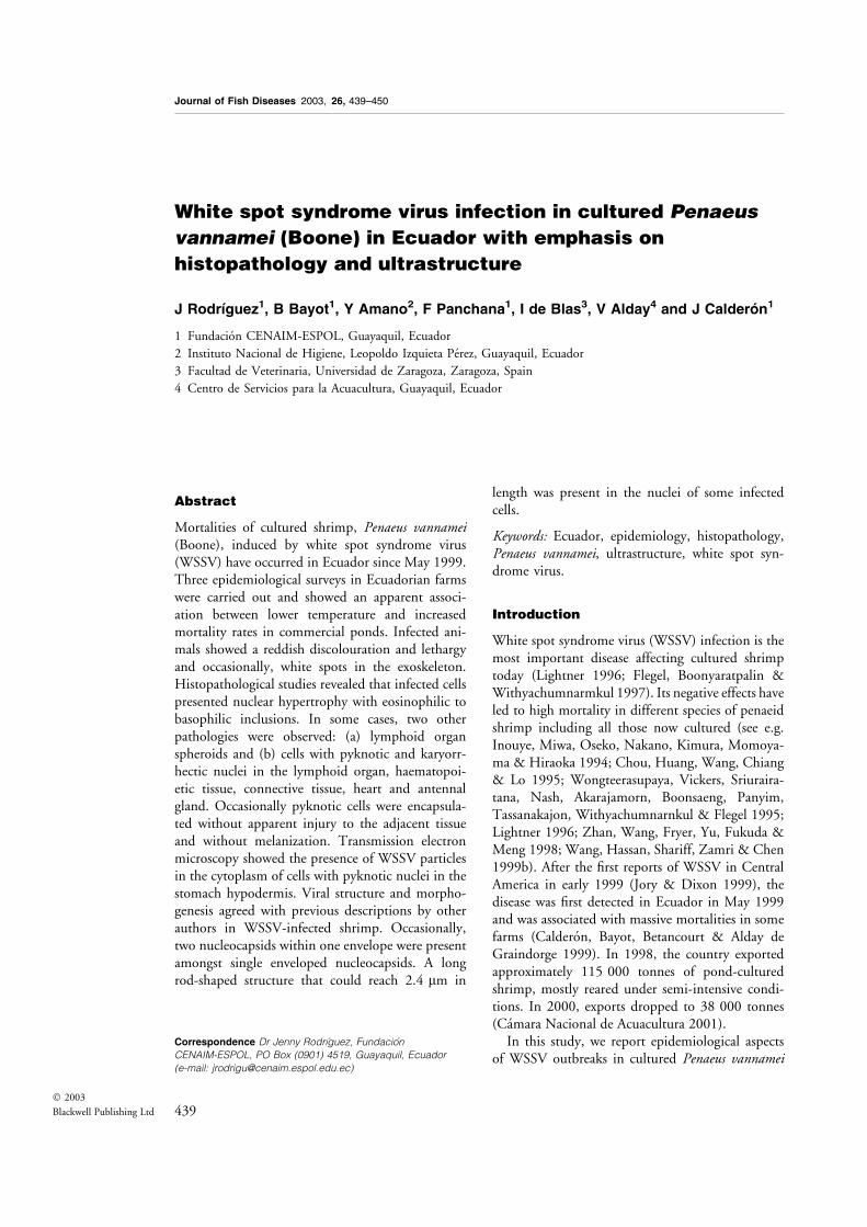
White spot syndrome virus infection in cultured Penaeus vannamei (Boone) in Ecuador with emphasis on histopathology and ultrastructure J Rodrȷguez 1 , B Bayot 1 , Y Amano 2 , F Panchana 1 , I de Blas 3 , V Alday 4 and J CalderɃn 1 1 Fundacio ´n CENAIM-ESPOL, Guayaquil, Ecuador 2 Instituto Nacional de Higiene, Leopoldo Izquieta Pe ´rez, Guayaquil, Ecuador 3 Facultad de Veterinaria, Universidad de Zaragoza, Zaragoza, Spain 4 Centro de Servicios para la Acuacultura, Guayaquil, Ecuador Abstract Mortalities of cultured shrimp, Penaeus vannamei (Boone), induced by white spot syndrome virus (WSSV) have occurred in Ecuador since May 1999. Three epidemiological surveys in Ecuadorian farms were carried out and showed an apparent associ- ation between lower temperature and increased mortality rates in commercial ponds. Infected ani- mals showed a reddish discolouration and lethargy and occasionally, white spots in the exoskeleton. Histopathological studies revealed that infected cells presented nuclear hypertrophy with eosinophilic to basophilic inclusions. In some cases, two other pathologies were observed: (a) lymphoid organ spheroids and (b) cells with pyknotic and karyorr- hectic nuclei in the lymphoid organ, haematopoi- etic tissue, connective tissue, heart and antennal gland. Occasionally pyknotic cells were encapsula- ted without apparent injury to the adjacent tissue and without melanization. Transmission electron microscopy showed the presence of WSSV particles in the cytoplasm of cells with pyknotic nuclei in the stomach hypodermis. Viral structure and morpho- genesis agreed with previous descriptions by other authors in WSSV-infected shrimp. Occasionally, two nucleocapsids within one envelope were present amongst single enveloped nucleocapsids. A long rod-shaped structure that could reach 2.4 lm in length was present in the nuclei of some infected cells. Keywords: Ecuador, epidemiology, histopathology, Penaeus vannamei, ultrastructure, white spot syn- drome virus. Introduction White spot syndrome virus (WSSV) infection is the most important disease affecting cultured shrimp today (Lightner 1996; Flegel, Boonyaratpalin & Withyachumnarmkul 1997). Its negative effects have led to high mortality in different species of penaeid shrimp including all those now cultured (see e.g. Inouye, Miwa, Oseko, Nakano, Kimura, Momoya- ma & Hiraoka 1994; Chou, Huang, Wang, Chiang & Lo 1995; Wongteerasupaya, Vickers, Sriuraira- tana, Nash, Akarajamorn, Boonsaeng, Panyim, Tassanakajon, Withyachumnarnkul & Flegel 1995; Lightner 1996; Zhan, Wang, Fryer, Yu, Fukuda & Meng 1998; Wang, Hassan, Shariff, Zamri & Chen 1999b). After the first reports of WSSV in Central America in early 1999 (Jory & Dixon 1999), the disease was first detected in Ecuador in May 1999 and was associated with massive mortalities in some farms (Caldero ´n, Bayot, Betancourt & Alday de Graindorge 1999). In 1998, the country exported approximately 115 000 tonnes of pond-cultured shrimp, mostly reared under semi-intensive condi- tions. In 2000, exports dropped to 38 000 tonnes (Ca ´mara Nacional de Acuacultura 2001). In this study, we report epidemiological aspects of WSSV outbreaks in cultured Penaeus vannamei Journal of Fish Diseases 2003, 26, 439–450 Correspondence Dr Jenny Rodrı´guez, Fundacio´n CENAIM-ESPOL, PO Box (0901) 4519, Guayaquil, Ecuador (e-mail: [email protected]) 439 Ó 2003 Blackwell Publishing Ltd
-
Upload
independent -
Category
Documents
-
view
1 -
download
0
Transcript of White spot syndrome virus infection in cultured Penaeus vannamei (Boone) in Ecuador with emphasis on...
White spot syndrome virus infection in cultured Penaeus
vannamei (Boone) in Ecuador with emphasis on
histopathology and ultrastructure
J Rodr�guez1, B Bayot1, Y Amano2, F Panchana1, I de Blas3, V Alday4 and J Calder�n1
1 Fundacion CENAIM-ESPOL, Guayaquil, Ecuador
2 Instituto Nacional de Higiene, Leopoldo Izquieta Perez, Guayaquil, Ecuador
3 Facultad de Veterinaria, Universidad de Zaragoza, Zaragoza, Spain
4 Centro de Servicios para la Acuacultura, Guayaquil, Ecuador
Abstract
Mortalities of cultured shrimp, Penaeus vannamei(Boone), induced by white spot syndrome virus(WSSV) have occurred in Ecuador since May 1999.Three epidemiological surveys in Ecuadorian farmswere carried out and showed an apparent associ-ation between lower temperature and increasedmortality rates in commercial ponds. Infected ani-mals showed a reddish discolouration and lethargyand occasionally, white spots in the exoskeleton.Histopathological studies revealed that infected cellspresented nuclear hypertrophy with eosinophilic tobasophilic inclusions. In some cases, two otherpathologies were observed: (a) lymphoid organspheroids and (b) cells with pyknotic and karyorr-hectic nuclei in the lymphoid organ, haematopoi-etic tissue, connective tissue, heart and antennalgland. Occasionally pyknotic cells were encapsula-ted without apparent injury to the adjacent tissueand without melanization. Transmission electronmicroscopy showed the presence of WSSV particlesin the cytoplasm of cells with pyknotic nuclei in thestomach hypodermis. Viral structure and morpho-genesis agreed with previous descriptions by otherauthors in WSSV-infected shrimp. Occasionally,two nucleocapsids within one envelope were presentamongst single enveloped nucleocapsids. A longrod-shaped structure that could reach 2.4 lm in
length was present in the nuclei of some infectedcells.
Keywords: Ecuador, epidemiology, histopathology,Penaeus vannamei, ultrastructure, white spot syn-drome virus.
Introduction
White spot syndrome virus (WSSV) infection is themost important disease affecting cultured shrimptoday (Lightner 1996; Flegel, Boonyaratpalin &Withyachumnarmkul 1997). Its negative effects haveled to high mortality in different species of penaeidshrimp including all those now cultured (see e.g.Inouye, Miwa, Oseko, Nakano, Kimura, Momoya-ma & Hiraoka 1994; Chou, Huang, Wang, Chiang& Lo 1995; Wongteerasupaya, Vickers, Sriuraira-tana, Nash, Akarajamorn, Boonsaeng, Panyim,Tassanakajon, Withyachumnarnkul & Flegel 1995;Lightner 1996; Zhan, Wang, Fryer, Yu, Fukuda &Meng 1998; Wang, Hassan, Shariff, Zamri & Chen1999b). After the first reports of WSSV in CentralAmerica in early 1999 (Jory & Dixon 1999), thedisease was first detected in Ecuador in May 1999and was associated with massive mortalities in somefarms (Calderon, Bayot, Betancourt & Alday deGraindorge 1999). In 1998, the country exportedapproximately 115 000 tonnes of pond-culturedshrimp, mostly reared under semi-intensive condi-tions. In 2000, exports dropped to 38 000 tonnes(Camara Nacional de Acuacultura 2001).
In this study, we report epidemiological aspectsof WSSV outbreaks in cultured Penaeus vannamei
Journal of Fish Diseases 2003, 26, 439–450
Correspondence Dr Jenny Rodrıguez, Fundacion
CENAIM-ESPOL, PO Box (0901) 4519, Guayaquil, Ecuador
(e-mail: [email protected])
439� 2003
Blackwell Publishing Ltd
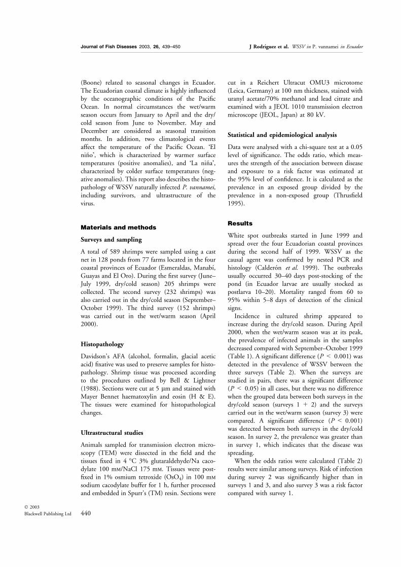
(Boone) related to seasonal changes in Ecuador.The Ecuadorian coastal climate is highly influencedby the oceanographic conditions of the PacificOcean. In normal circumstances the wet/warmseason occurs from January to April and the dry/cold season from June to November. May andDecember are considered as seasonal transitionmonths. In addition, two climatological eventsaffect the temperature of the Pacific Ocean. �Elnino�, which is characterized by warmer surfacetemperatures (positive anomalies), and �La nina�,characterized by colder surface temperatures (neg-ative anomalies). This report also describes the histo-pathology of WSSV naturally infected P. vannamei,including survivors, and ultrastructure of thevirus.
Materials and methods
Surveys and sampling
A total of 589 shrimps were sampled using a castnet in 128 ponds from 77 farms located in the fourcoastal provinces of Ecuador (Esmeraldas, Manabı,Guayas and El Oro). During the first survey (June–July 1999, dry/cold season) 205 shrimps werecollected. The second survey (232 shrimps) wasalso carried out in the dry/cold season (September–October 1999). The third survey (152 shrimps)was carried out in the wet/warm season (April2000).
Histopathology
Davidson’s AFA (alcohol, formalin, glacial aceticacid) fixative was used to preserve samples for histo-pathology. Shrimp tissue was processed accordingto the procedures outlined by Bell & Lightner(1988). Sections were cut at 5 lm and stained withMayer Bennet haematoxylin and eosin (H & E).The tissues were examined for histopathologicalchanges.
Ultrastructural studies
Animals sampled for transmission electron micro-scopy (TEM) were dissected in the field and thetissues fixed in 4 �C 3% glutaraldehyde/Na caco-dylate 100 mm/NaCl 175 mm. Tissues were post-fixed in 1% osmium tetroxide (OsO4) in 100 mm
sodium cacodylate buffer for 1 h, further processedand embedded in Spurr’s (TM) resin. Sections were
cut in a Reichert Ultracut OMU3 microtome(Leica, Germany) at 100 nm thickness, stained withuranyl acetate/70% methanol and lead citrate andexamined with a JEOL 1010 transmission electronmicroscope (JEOL, Japan) at 80 kV.
Statistical and epidemiological analysis
Data were analysed with a chi-square test at a 0.05level of significance. The odds ratio, which meas-ures the strength of the association between diseaseand exposure to a risk factor was estimated atthe 95% level of confidence. It is calculated as theprevalence in an exposed group divided by theprevalence in a non-exposed group (Thrusfield1995).
Results
White spot outbreaks started in June 1999 andspread over the four Ecuadorian coastal provincesduring the second half of 1999. WSSV as thecausal agent was confirmed by nested PCR andhistology (Calderon et al. 1999). The outbreaksusually occurred 30–40 days post-stocking of thepond (in Ecuador larvae are usually stocked aspostlarva 10–20). Mortality ranged from 60 to95% within 5–8 days of detection of the clinicalsigns.
Incidence in cultured shrimp appeared toincrease during the dry/cold season. During April2000, when the wet/warm season was at its peak,the prevalence of infected animals in the samplesdecreased compared with September–October 1999(Table 1). A significant difference (P < 0.001) wasdetected in the prevalence of WSSV between thethree surveys (Table 2). When the surveys arestudied in pairs, there was a significant difference(P < 0.05) in all cases, but there was no differencewhen the grouped data between both surveys in thedry/cold season (surveys 1 + 2) and the surveyscarried out in the wet/warm season (survey 3) werecompared. A significant difference (P < 0.001)was detected between both surveys in the dry/coldseason. In survey 2, the prevalence was greater thanin survey 1, which indicates that the disease wasspreading.
When the odds ratios were calculated (Table 2)results were similar among surveys. Risk of infectionduring survey 2 was significantly higher than insurveys 1 and 3, and also survey 3 was a risk factorcompared with survey 1.
440� 2003
Blackwell Publishing Ltd
Journal of Fish Diseases 2003, 26, 439–450 J Rodrıguez et al. WSSV in P. vannamei in Ecuador

Histopathology and ultrastructure
In ponds undergoing a disease outbreak, animalsreduced food intake or stopped feeding completely.White spots in the infected shrimp were small andmostly located on the inside surface of the cephalo-thorax. These spots were not always present, but areddish body and pinkish colouration of the haemo-lymph were common in moribund animals, whichalso presented a general weakness and lethargy.The histopathology of infected cells was charac-
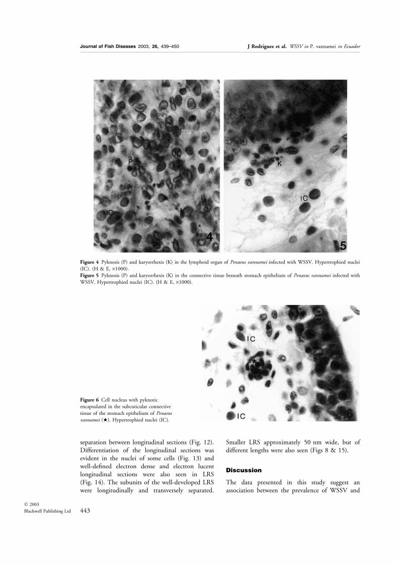
terized by nuclear hypertrophy with eosinophilic(Cowdry A inclusion) to basophilic inclusions.Several tissues were infected, mainly the cuticularepithelium, the stomach cuticular epithelium, con-nective tissue, antennal gland, gills, heart andhaematopoietic tissue (Figs 1 & 2). In the lymphoidorgan, in addition to the presence of infected cellswith inclusion bodies, the presence of spheroids[lymphoid organ spheroids (LOS)] was observed(Fig. 3). In some instances, no cells infected withWSSV were observed in the lymphoid organ andonly LOS were present in this tissue, while othertissues of the same specimen were infected. Otherpathologies affecting the animals, which had severeinfections of WSSV, were the presence of light tosevere pyknosis and karyorrhexis. The tissues affec-ted were the lymphoid organ presenting a loss of
structure (in the most severe cases) (Fig. 4), thespongy connective tissue beneath the stomachcuticular epithelium (Fig. 5), haematopoietic tissue(to much lesser degree than in the lymphoid organand without loss of organ structure), heart andantennal gland. Encapsulation of cells with pyk-notic nuclei was observed in some infected animals.The encapsulation was not accompanied by mela-nization and the adjacent tissues did not showlesions that could be attributed to inflammatoryfoci (Fig. 6). This condition was more evident inthe spongy connective tissue basal to the stomachcuticular epithelium. Animals examined that pre-sented pyknosis and karyorrhexis were alwaysseverely infected with WSSV but no signs of aninfection by Taura syndrome virus (TSV) wereobserved. TEM examination of cells of the stomachcuticular epithelium with pyknotic nuclei showedthe presence of WSSV viral particles in the cyto-plasm (Fig. 7). The pyknotic nuclei showedchromatin condensation at the nuclear membraneor a complete condensation of the nucleus. Thecytoplasm was condensed and vacuolated, while themitochondria remained relatively intact (Fig. 7).
Transmission electron microscopy of infectedlymphoid organ cells showed hypertrophied nucleiwith marginated chromatin and the presence ofrod-shaped enveloped virions. The virions werepresent in a paracrystalline arrangement or scatteredin the nuclei (Fig. 8). The virions were from 245 to308 nm in length and 98–124 nm in width. Thenucleocapsids ranged from 183 to 262 nm inlength to 60–95 nm in width (Fig. 8). The presenceof two nucleocapsids within one envelope wasoccasionally observed (Fig. 8). The virus envelopewas 8–9 nm thick and trilaminar, consisting of twoelectron opaque layers separated by an electronlucent layer.
A membrane profile consisting of a series ofvesicles joined together was present at the early stageof virion development and empty capsids insidethese vesicles had asymmetric ends (Fig. 9). Partially
Table 1 Sea surface temperatures and the prevalence of WSSV in cultured Penaeus vannamei obtained from epidemiological surveys
conducted in the four coastal provinces (Esmeraldas, Manabı, Guayas and El Oro) of Ecuador, during different seasons
Survey Date Season Temperature (�C)aTemperature
anomalies (�C)a n
WSSV
negative
WSSV
positive
%
prevalence
Survey 1 June–July 1999 Dry/cold 21.83, 20.44 )0.85,)1.14 205 185 20 9.76
Survey 2 September–October 1999 Dry/cold 19.75, 19.23 )0.89,)1.09 232 135 97 41.81
Survey 3 April 2000 Wet/warm 25.64 )0.88 152 123 29 19.07
a Sea surface temperature (SST) and anomalies (variations of seasonal averages) of SST for �El nino 1 + 2� region (oceanic region located between the
western coast of South America and 90� W and from 0� to 10� S). The values correspond to the respective monthly average for each case.
Table 2 Chi-square (v2) and odds ratio (OR) values determined
from three epidemiological surveys conducted in the four coastal
provinces (Esmeraldas, Manabı, Guayas and El Oro) of Ecuador
during different seasons
Surveys
Risk
factor
v2 OR
Value d.f. P Value
95%
Lower
limit
95%
Upper
limit
1, 2, 3 – 63.561 2 0.000 – – –
1, 2 2 57.037 1 0.000 6.646 4.063 10.870
1, 3 3 6.406 1 0.011 2.181 1.191 3.992
2, 3 2 21.524 1 0.000 3.048 1.902 4.883
1 + 2, 3 1 + 2 3.581 1 0.058 1.551 0.984 2.444
441� 2003
Blackwell Publishing Ltd
Journal of Fish Diseases 2003, 26, 439–450 J Rodrıguez et al. WSSV in P. vannamei in Ecuador

enveloped empty capsids were present singly orappeared surrounded by the same envelope (Fig. 10),but with an open end. These were 100–188 nmlong and 62–64 nm wide. Single enveloped emptycapsids presented segmentation, but this was notclearly evident in all cases as the segmentation wasonly slightly electron dense (Fig. 10). An electrondense central filament was present in some envel-oped capsids (Fig. 11), and these developing capsidswere longer and narrower than fully developedvirions.
A long rod-shaped structure (LRS) was present inthe nucleus of some infected cells alongside fullydeveloped virions (Fig. 8). The LRS reached2.4 lm in length by 50 nm in width. Along thelongitudinal axis of the whole structure an electronlucent core was present which averaged 13–17 nm.Electron dense striations were regularly present inthe LRS, however, these striations did not appear tocross the electron lucent core.
Presumptive early stage LRS were made upof numerous repeating subunits but without
Figure 1 Tissues of Penaeus vannamei infected with white spot syndrome virus (WSSV). Stomach cuticular epithelium showing
hypertrophied nucleus and viral inclusion (IC). Note an eosinophilic Cowdry Type A inclusion (CI) (H & E, ·1000).Figure 2 Tissues of Penaeus vannamei infected with white spot syndrome virus (WSSV). Hypertrophied nucleus and viral inclusion
(IC) in gill tissue (H & E, ·1000).
Figure 3 Lymphoid organ of Penaeusvannamei infected with WSSV, showing
lymphoid organ spheroids (H & E, ·1000).
442� 2003
Blackwell Publishing Ltd
Journal of Fish Diseases 2003, 26, 439–450 J Rodrıguez et al. WSSV in P. vannamei in Ecuador
separation between longitudinal sections (Fig. 12).Differentiation of the longitudinal sections wasevident in the nuclei of some cells (Fig. 13) andwell-defined electron dense and electron lucentlongitudinal sections were also seen in LRS(Fig. 14). The subunits of the well-developed LRSwere longitudinally and transversely separated.
Smaller LRS approximately 50 nm wide, but ofdifferent lengths were also seen (Figs 8 & 15).
Discussion
The data presented in this study suggest anassociation between the prevalence of WSSV and
Figure 6 Cell nucleus with pyknotic
encapsulated in the subcuticular connective
tissue of the stomach epithelium of Penaeusvannamei (w). Hypertrophied nuclei (IC).
Figure 4 Pyknosis (P) and karyorrhexis (K) in the lymphoid organ of Penaeus vannamei infected with WSSV. Hypertrophied nuclei
(IC). (H & E, ·1000).Figure 5 Pyknosis (P) and karyorrhexis (K) in the connective tissue beneath stomach epithelium of Penaeus vannamei infected with
WSSV. Hypertrophied nuclei (IC). (H & E, ·1000).
443� 2003
Blackwell Publishing Ltd
Journal of Fish Diseases 2003, 26, 439–450 J Rodrıguez et al. WSSV in P. vannamei in Ecuador

mainly dry/cold climatological conditions in thisgeographical area. The first epidemiological surveywas carried out at the beginning of the dry/coldseason in June–July 1999. During June and Julywater temperature is normally cold, but in this year
cold (negative) anomalies of )0.85 and )1.14 �Cbelow seasonal average, respectively, were detected.The second survey was during the height of the dry/cold season in September–October 1999, when thewater temperature was colder than in June–July and
Figure 7 Electron micrograph of the stomach epithelium of Penaeus vannamei infected with WSSV. Condensed nucleus (N),
mitochondrion (M), virus particles in the cytoplasm (arrows) (bar ¼ 1 lm).
444� 2003
Blackwell Publishing Ltd
Journal of Fish Diseases 2003, 26, 439–450 J Rodrıguez et al. WSSV in P. vannamei in Ecuador

negative anomalies persisted ()0.89 and )1.09 �C).Thus, at these times water temperatures were evenlower than the normal cold conditions because ofthe �La nina� event. This could be a cause of thehigher prevalence of WSSV during the secondsurvey when compared with the other surveys. Theodds ratios between survey 1 and survey 2 andbetween survey 2 and survey 3, show a higher risk ofthe disease during the second survey. In April 2000(survey 3), when the wet/warm season is normally
fully developed, the prevalence of WSSV decreasedcompared with survey 2. The water temperatureanomaly remained negative ()0.88 �C) at this timebecause of the continuing �La nina� event. The oddsratio showed that there was a higher risk of diseasedevelopment in the third survey than in the first one.The increase in WSSV prevalence between survey 1and survey 2 might be a consequence of theexpanding distribution of the virus as the epizooticspread and/or the effect of the drop in water
Figure 8 Electron micrograph of a cell nucleus of the lymphoid organ of Penaeus vannamei infected with WSSV. Virus particles in
paracrystalline arrangement (V), nucleocapsid (arrow), envelope (e), two nucleocapsids within one envelope (w) (bar ¼ 200 nm).
445� 2003
Blackwell Publishing Ltd
Journal of Fish Diseases 2003, 26, 439–450 J Rodrıguez et al. WSSV in P. vannamei in Ecuador

temperature as the cold season developed. Thiseffect would be exacerbated by the �La nina� event.The effect of temperature could explain the decreasein prevalence in survey 3 during the warm season,when temperatures were almost 6 �C higher than insurvey 2, despite the continuing �La nina� event. Thehigher prevalence and odds ratio of WSSV in survey3 compared with survey 1 might be due to thespread of the virus. Survey 1 was conducted duringthe early phase of the WSSV epizootic. The
continued occurrence of the �La nina� event loweredthe average temperatures at the time of survey 3which may have reduced the magnitude of thetemperature effect on the virus. Our results suggestthat the dry/cold season is an important risk factor inthe occurrence of WSSV in Ecuador and that therisk increases with time over this season.
Further research is needed to determine themechanisms and effects of environmental stress intriggering a disease outbreak in shrimp ponds.
Figure 9 Electron micrograph of the lymphoid organ of Penaeus vannamei infected with WSSV. Nucleus at early stage of viral
infection; empty capsids (arrow), joined vesicles (jv) (bar ¼ 200 nm).
Figure 10 Electron micrograph of the lymphoid organ of Penaeus vannamei infected with WSSV. Empty capsid with asymmetric ends
(a) (bar ¼ 200 nm).
Figure 11 Electron micrograph of the lymphoid organ of Penaeus vannamei infected with WSSV. Nucleocapsid with central filament
(arrow) (bar ¼ 200 nm).
446� 2003
Blackwell Publishing Ltd
Journal of Fish Diseases 2003, 26, 439–450 J Rodrıguez et al. WSSV in P. vannamei in Ecuador

Outbreaks have been empirically associated withenvironmental changes (Flegel et al. 1997), or watertemperature (Zhan et al. 1998; Jimenez, Barniol, deBarniol & Machuca 2000). In this study there wasan apparent association between low temperaturesand increased mortality rates in ponds, and a similarscenario has been reported elsewhere in Ecuador inP. vannamei infected by WSSV. It is interestingthat some authors have reported high survival of
P. vannamei, challenged with WSSV, under hyper-thermic conditions (Vidal, Granja & Aranguren2001; Sonnenholzner, Rodrıguez & Calderon2002).
Although the histopathology of P. vannameiexperimentally infected with WSSV has been des-cribed in detail by different researchers (Tapay, Lu,Gose, Brock & Loh1997; Lightner, Hasson, White& Redman 1998; Nunan, Poulos & Lightner
Figure 12 Electron micrograph of long rod-shaped structure (LRS) in the nucleus of WSSV-infected cell. Presumptive early stage of
LRS formation. LRS with repeating subunits without separation in long sections (arrow) (bar ¼ 200 nm).
Figure 13 Electron micrograph of long rod-shaped structure (LRS) in the nucleus of WSSV-infected cell. LRS with slight
differentiation between longitudinal sections (arrow); note virions in cross section arranged next to LRS (w) (bar ¼ 200 nm).
Figure 14 Electron micrograph of long rod-shaped structure (LRS) in the nucleus of WSSV-infected cell. LRS with three repeating
units (arrow) (bar ¼ 200 nm).
Figure 15 Electron micrograph of long rod-shaped structure (LRS) in the nucleus of WSSV-infected cell. LRS consisting of 1 subunit
(arrow) (bar ¼ 200 nm).
447� 2003
Blackwell Publishing Ltd
Journal of Fish Diseases 2003, 26, 439–450 J Rodrıguez et al. WSSV in P. vannamei in Ecuador

1998), the present study indicates that animalsundergoing natural infections under field condi-tions can present additional histological changes,such as LOS, pyknosis and karyorrhexis. LOS havebeen reported in penaeids infected by differentviruses, including WSSV, although in the latter casenot in specific pathogen-free P. vannamei experi-mentally infected with the virus (Owens, de Beer &Smith 1991; Bonami, Lightner, Redman & Poulos1992; Spann, Vickers & Lester 1995; Lightner1996; Hasson, Lightner, Mohney, Redman, Poulos& White 1999a; Vidal et al. 2001) and bacteria(Alday-Sanz, Roque & Turnbull 2002; Van deBraak, Botterblom, Taverne, Van Muiswinkel,Rombout & Van der Knaap 2002). Thus, althoughLOS appear to be strongly associated with WSSVthis cannot be unequivocally stated without addi-tional experimental evidence. It appears that thelymphoid organ and the formation of LOS areinvolved in the immune response of penaeids toinfections (Hasson, Lightner, Mohney, Redman &White 1999b; Anggraeni & Owens 2000). LOSseem to appear when the animal is able to controlthe infection or at least to react against it, as in thecase of survivors or in chronic infections. Nuclearpyknosis and karyorrhexis have been reported to beassociated with several viral infections includingyellow head virus (YHV), TSV and lymphoid organvacuolization virus (Lightner 1996). Nuclear pyk-nosis and karyorrhexis of the haematopoietic tissueand the lymphoid organ, together with loss ofstructure, have been considered pathognomiclesions of YHV infection. However, in the samplesexamined here, these lesions were associated withsevere WSSV infection. In Ecuador the presence ofYHV has not yet been determined by any techniqueemployed. Pantoja & Lightner (2003) also observedpyknosis and karyorrhexis in animals experimen-tally infected with WSSV. In addition, pyknosisand karyorrhexis in haematopoietic tissue have beendescribed in Farfantepenaeus duorarum challengedwith WSSV (Wang, White, Redman & Lightner1999a).Ultrastructural transformations (nuclear pykno-
sis, cytoplasmic vacuolization) observed in deadcells in this study resemble apoptotic changes. Inaddition, cells with pyknotic nuclei were encapsu-lated by haemocytes without melanization. Similarmodifications have been described in apoptoticevents. Apoptosis in WSSV infected shrimp hasbeen reported in Penaeus monodon (Sahtout, Hassan& Shariff 2001) and P. vannamei (Granja,
Aranguren, Vidal, Aragon & Salazar 2003). Inhigher vertebrates apoptosis is a mechanism toeliminate cells infected by virus (O’Brien 1998).The presence of virions in the cytoplasm of cellswith pyknotic nuclei (without nuclear replication)suggests a similar mechanism in shrimp.
More studies are required to establish if apoptosisis a mechanism to eliminate viral infection inshrimp, however, there is now some publishedevidence suggesting that this occurs. Hasson et al.(1999b) reported cellular changes characteristic ofapoptotic cells in the lymphoid organ of shrimpwhich have controlled TSV infection, and Wanget al. (1999a) observed pyknosis and karyorrhexis inanimals that survived WSSV induced infections. Inaddition, Granja et al. (2003) have seen a highdegree of apoptosis in shrimp surviving WSSVinfections.
Our TEM observations of viral structure andmorphogenesis agree with the descriptions of otherauthors on penaeid shrimp infected by WSSV. Thestructure we refer to as LRS has been reported inP. vannamei (Durand, Lightner, Redman& Bonami1997) and P. monodon (Wang et al. 1999b)infected with WSSV. We observed up to eightlongitudinal electron dense bands and four lighterbands in one LRS, as reported by Wang et al.(1999b). Durand et al. (1997) considered thisstructure as an assembly of nucleocapsid precursors,while Wang et al. (1999b) proposed that �the LRSmay compromise WSSV nucleosomes that areassociated with early viral replication�. In ourinvestigation the LRS was present both in the earlyand late stages of viral replication and assembly.The use of specific antibodies against parts of theWSSV virion such as the nucleocapsid proteinVP26 (van Hulten, Westenberg, Goodall & Vlak2000) and the immunogold labelling techniquecould be used to conclusively establish the nature ofthe LRS.
The occasional presence of two nucleocapsidswithin one envelope amongst singly envelopednucleocapsids as observed here has been reportedin Fenneropenaeus chinensis infected by WSSV(Zhan et al. 1998), and in the crab, Carcinusmaenas (L.), infected by a rod-shaped nuclear virus,although it is considered as a rare occurrence incrabs (Johnson 1988). This occurrence is alsouncommon in penaeids infected by WSSV, as ithas not been reported in other ultrastructuralinvestigations in P. vannamei (Durand et al. 1997;Tapay, Lu, Gose, Brock & Loh 1997; Rajendran,
448� 2003
Blackwell Publishing Ltd
Journal of Fish Diseases 2003, 26, 439–450 J Rodrıguez et al. WSSV in P. vannamei in Ecuador
Vijayan, Santiago & Krol 1999; Wang et al. 1999a)or other penaeid shrimps (see e.g. Inouye et al.1994; Takahashi, Itami, Kondo, Maeda, Fujii,Tomonaga, Supamattaya & Boonyaratpalin 1994;Wongteerasupaya et al. 1995; Wang et al.1999a,b).
Acknowledgements
The authors wish to thank the different shrimpfarmers who provided access and collaboration dur-ing sampling and fieldwork in these studies. Theauthors also wish to thank Xavier Romero for hisadvice on the pathological aspects of this paperand July Nieto for English corrections. Dr ClaudiaVenegas (Universidad de Santiago, Chile) is acknow-ledged for her comments and suggestions on theoriginal manuscript.
References
Alday-Sanz V., Roque A. & Turnbull J.F. (2002) Clearing
mechanisms of Vibrio vulnificus biote I in the black tiger
shrimp Penaeus monodon. Diseases of Aquatic Organisms 48,91–99.
Anggraeni M.S. & Owens L. (2000) The haemocytic origin of
lymphoid organ spheroid cells in the penaeid prawn Penaeusmonodon. Diseases of Aquatic Organisms 40, 85–92.
Bell T.A. & Lightner D.V. (1988) A Handbook of Normal Pen-aeid Shrimp Histology. The World Aquaculture Society, Baton
Rouge, LA.
Bonami J.R., Lightner D.V., Redman R.M. & Poulos B.T.
(1992) Partial characterization of a togavirus (LOVV)
associated with histopathological changes of the lymphoid
organ of penaeid shrimp. Diseases of Aquatic Organisms 14,145–152.
Calderon J.V., Bayot B., Betancourt I. & Alday de Graindorge
V. (1999) Monitoreo del virus de la mancha blanca en
Ecuador. El Mundo Acuicola 5, 211–214.
Camara Nacional de Acuacultura (2001) Ecuador compite logro
un acuerdo de voluntades. Acuacultura del Ecuador 45, 60–63.
Chou H.Y., Huang C.Y., Wang C.H., Chiang H.C. & Lo C.F.
(1995) Pathogenicity of a baculovirus infection causing white
spot syndrome in cultured penaeid shrimp in Taiwan. Diseasesof Aquatic Organisms 23, 165–173.
Durand S., Lightner D.V., Redman R.M. & Bonami J.R. (1997)
Ultrastructure and morphogenesis of white spot syndrome
baculovirus. Diseases of Aquatic Organisms 29, 205–211.
Flegel T.W., Boonyaratpalin S. & Withyachumnarmkul B.
(1997) Progress in research on yellow-head virus and white-
spot virus in Thailand. In: Diseases in Asian Aquaculture III(ed. by T.W. Flegel & I.H. MacRae), pp. 285–295. Asian
Fisheries Society, Fish Health Section, Manila, Philippines.
Granja C.B., Aranguren L.F., Vidal O.M., Aragon L. & Salazar
M. (2003) Does hyperthermia increase apoptosis in white spot
syndrome virus (WSSV)-infected Litopenaeus vannamei? Dis-eases of Aquatic Organisms 54, 73–78.
Hasson K.W., Lightner D.V., Mohney L.L., Redman R.M.,
Poulos B.T. & White B.M. (1999a) Taura syndrome virus
(TSV) development and the disease cycle in the Pacific white
shrimp Penaeus vannamei. Diseases of Aquatic Organisms 36,81–93.
Hasson K.W., Lightner D.V., Mohney L.L., Redman R.M. &
White B.M. (1999b) Role of lymphoid organ spheroids in
chronic Taura syndrome virus (TSV) infections in Penaeusvannamei. Diseases of Aquatic Organisms 38, 93–105.
van Hulten M.C.W., Westenberg M., Goodall S.D. & Vlak J.M.
(2000) Identification of two major virion protein genes of
white spot syndrome virus of shrimp. Virology 266, 227–236.
Inouye K., Miwa S., Oseko N., Nakano H., Kimura T.,
Momoyama K. & Hiraoka M. (1994) Mass mortalities of
cultured kuruma shrimp Penaeus japonicus in Japan in 1993:
electron microscopic evidence of the causative virus. FishPathology 29, 149–158.
Jimenez R., Barniol R., de Barniol L. & Machuca M. (2000)
Periodic occurrence of epithelial necrosis outbreaks in Penaeusvannamei in Ecuador. Diseases of Aquatic Organisms 42,91–99.
Johnson P.T. (1988) Rod-shaped nuclear viruses of crustaceans:
hemocyte-infecting species. Diseases of Aquatic Organisms 5,111–122.
Jory D.E. & Dixon H.M. (1999) Shrimp white spot virus in the
western hemisphere. Aquaculture Magazine 25, 83–91.
Lightner D.V. (1996) A Handbook of Shrimp Pathology andDiagnostic Procedures for Diseases of Cultured Penaeid Shrimp.The World Aquaculture Society, Baton Rouge, LA.
Lightner D.V., Hasson K.W., White B.L. & Redman R.M.
(1998) Experimental infection of western hemisphere penaeid
shrimp with Asian white spot syndrome virus and Asian yellow
head virus. Journal of Aquatic Animal Health 10, 271–281.
Nunan L.M., Poulos B.T. & Lightner D.V. (1998) The detec-
tion of white spot syndrome virus (WSSV) and yellow head
virus (YHV) in imported commodity shrimp. Aquaculture160, 19–30.
O’Brien V. (1998) Viruses and apoptosis. Journal of GeneralVirology 79, 1833–1845.
Owens L., de Beer S. & Smith J. (1991) Lymphoidal parvovirus-
like particles in Australian penaeid prawns. Diseases of AquaticOrganisms 11, 129–134.
Pantoja C.R. & Lightner D.V. (2003) Similarity between the
histopathology of white spot syndrome virus and yellow head
syndrome virus and its relevance to diagnosis of YHV disease
in the Americas. Aquaculture 218, 47–54.
Rajendran K.V., Vijayan K.K., Santiago T.C. & Krol R.M.
(1999) Experimental host range and histopathology of white
spot syndrome virus (WSSV) infection in shrimp, prawns,
crabs and lobsters from India. Journal of Fish Diseases 22,183–191.
Sahtout A.H., Hassan M.D. & Shariff M. (2001) DNA frag-
mentation, an indicator of apoptosis, in cultured black tiger
shrimp Penaeus monodon. Diseases of Aquatic Organisms 44,155–159.
449� 2003
Blackwell Publishing Ltd
Journal of Fish Diseases 2003, 26, 439–450 J Rodrıguez et al. WSSV in P. vannamei in Ecuador
Sonnenholzner S., Rodrıguez J. & Calderon J. (2002) Tem-
perature and WSSV: CENAIM studies promising shrimp
culture technique. Global Aquaculture Advocate 5, 55–57.
Spann K.M., Vickers J.E. & Lester R.J. (1995) Lymphoid organ
virus of Penaeus monodon from Australia. Diseases of AquaticOrganisms 23, 127–137.
Takahashi Y., Itami K., Kondo M., Maeda M., Fujii R.,
Tomonaga S., Supamattaya K. & Boonyaratpalin S. (1994)
Electron microscopic evidence of bacilliform virus infection
in kuruma shrimp (Penaeus japonicus). Fish Pathology 29,121–125.
Tapay L.M., Lu Y., Gose R.B., Brock J.A. & Loh P.C. (1997)
Infection of white spot baculovirus (WSSV) in two species of
penaeid shrimp Penaeus stylirostris (Stimpson) and P. vannamei(Boone). In: Diseases in Asian Aquaculture III (ed. by T.W.
Flegel & I.H. MacRae), pp. 297–302. Asian Fisheries Society,
Fish Health Section, Manila, Philippines.
Thrusfield M. (1995) Veterinary Epidemiology. Blackwell Science,Edinburgh.
Van de Braak C.B.T, Botterblom M.H.A., Taverne N., Van
Muiswinkel W.B., Rombout J.H.W.M. & Van der Knaap
P.W. (2002) The roles of haemocytes and the lymphoid organ
in the clearance of injected Vibrio bacteria in Penaeus vanna-mei shrimp. Fish and Shellfish Immunology 13, 293–309.
Vidal O.M., Granja C.B. & Aranguren F. (2001) A profound
effect of hyperthermia on survival of Litopenaeus vannamei
juveniles infected with white spot syndrome virus. Journal ofthe World Aquaculture Society 32, 364–372.
Wang Q., White B.L., Redman R.M. & Lightner D.V.
(1999a) Per os challenge of Litopenaeus vannamei post-larvaeand Farfantepenaeus duorarum juveniles with six geographic
isolates of white spot syndrome virus. Aquaculture 170,179–194.
Wang Y.G., Hassan M.D., Shariff M., Zamri S.M. & Chen X.
(1999b) Histopathology and cytopathology of white spot
syndrome virus (WSSV) in cultured Penaeus monodon from
peninsular Malaysia with emphasis on pathogenesis and the
mechanism of white spot formation. Diseases of AquaticOrganisms 39, 1–11.
Wongteerasupaya C., Vickers J.E., Sriurairatana S., Nash G.L.,
Akarajamorn A., Boonsaeng V., Panyim S., Tassanakajon A.,
Withyachumnarnkul B. & Flegel T.W. (1995) A non-
occluded systemic baculovirus that occurs in cells of ecto-
dermal and mesodermal origin and causes high mortality in
the black tiger prawn Penaeus monodon. Diseases of AquaticOrganisms 21, 69–77.
Zhan W.B., Wang Y.H., Fryer J.L., Yu K.K., Fukuda H. &
Meng Q.X. (1998) White spot syndrome virus infection of
cultured shrimp in China. Journal of Aquatic Animal Health10, 405–410.
Received: 18 September 2002Accepted: 1 April 2003
450� 2003
Blackwell Publishing Ltd
Journal of Fish Diseases 2003, 26, 439–450 J Rodrıguez et al. WSSV in P. vannamei in Ecuador