Histopathology Image Classification Using Bag of Features and Kernel Functions
10
Histopathology Image Classification using Bag of Features and Kernel Functions Juan C. Caicedo, Angel Cruz and Fabio A. Gonzalez Bioingenium Research Group National University of Colombia {jccaicedoru,aacruzr,fagonzalezo}@unal.edu.co http://www.bioingenium.unal.edu.co Abstract. Image representation is an important issue for medical im- age analysis, classification and retrieval. Recently, the bag of features approach has been proposed to classify natural scenes, using an analogy in which visual features are to images as words are to text documents. This process involves feature detection and description, construction of a visual vocabulary and image representation building through visual- word occurrence analysis. This paper presents an evaluation of different representations obtained from the bag of features approach to classify histopathology images. The obtained image descriptors are processed us- ing appropriate kernel functions for Support Vector Machines classifiers. This evaluation includes extensive experimentation of different strate- gies, and analyses the impact of each configuration in the classification result. 1 Introduction Medical imaging applications are challenging because they require effective and efficient content representations to manage large image collections. The first stage for medical image analysis is modeling image contents by defining an ap- propriate representation. This is a fundamental problem for all image analysis tasks such as image classification, automatic image annotation, object recogni- tion and image retrieval, which require discriminative representations according to the application domain. During the last few years, the bag of features ap- proach has attracted great attention from the computer vision community. This approach is an evolution of texton-based representations and is also influenced by the bag of words assumption in text classification. In text documents, a word dictionary is defined and all documents are processed so that the frequency of each word is quantified. This representation ignores word relationships in the document, i.e., it does not take into account the document structure. An anal- ogy is defined for images in which a feature dictionary is built to identify visual patterns in the collection. This representation has shown to be effective in dif- ferent image classification, categorization and retrieval tasks [1,2,3]. The bag of features representation is an adaptive approach to model image structure in a robust way. In contrast to image segmentation, this approach does
-
Upload
independent -
Category
Documents
-
view
7 -
download
0
Transcript of Histopathology Image Classification Using Bag of Features and Kernel Functions
Histopathology Image Classification using Bag ofFeatures and Kernel Functions
Juan C. Caicedo, Angel Cruz and Fabio A. Gonzalez
Bioingenium Research GroupNational University of Colombia
{jccaicedoru,aacruzr,fagonzalezo}@unal.edu.co
http://www.bioingenium.unal.edu.co
Abstract. Image representation is an important issue for medical im-age analysis, classification and retrieval. Recently, the bag of featuresapproach has been proposed to classify natural scenes, using an analogyin which visual features are to images as words are to text documents.This process involves feature detection and description, construction ofa visual vocabulary and image representation building through visual-word occurrence analysis. This paper presents an evaluation of differentrepresentations obtained from the bag of features approach to classifyhistopathology images. The obtained image descriptors are processed us-ing appropriate kernel functions for Support Vector Machines classifiers.This evaluation includes extensive experimentation of different strate-gies, and analyses the impact of each configuration in the classificationresult.
1 Introduction
Medical imaging applications are challenging because they require effective andefficient content representations to manage large image collections. The firststage for medical image analysis is modeling image contents by defining an ap-propriate representation. This is a fundamental problem for all image analysistasks such as image classification, automatic image annotation, object recogni-tion and image retrieval, which require discriminative representations accordingto the application domain. During the last few years, the bag of features ap-proach has attracted great attention from the computer vision community. Thisapproach is an evolution of texton-based representations and is also influencedby the bag of words assumption in text classification. In text documents, a worddictionary is defined and all documents are processed so that the frequency ofeach word is quantified. This representation ignores word relationships in thedocument, i.e., it does not take into account the document structure. An anal-ogy is defined for images in which a feature dictionary is built to identify visualpatterns in the collection. This representation has shown to be effective in dif-ferent image classification, categorization and retrieval tasks [1,2,3].
The bag of features representation is an adaptive approach to model imagestructure in a robust way. In contrast to image segmentation, this approach does
not attempt to identify complete objects inside images, which may be a hardertask than the image classification itself. Instead, the bag of features approachlooks for small characteristic image regions allowing the representation of com-plex image contents without explicitly modeling objects and their relationships,a task that is tackled in another stage of image content analysis. In addition,an important advantage of the bag of features approach is its adaptiveness tothe particular image collection to be processed. In the same way as text docu-ments, in which the appropriate word-list to be included in the dictionary maybe identified earlier in the process, the bag of features approach allows to iden-tify visual patterns that are relevant to the whole image collection. That is, thepatterns that are used in a single image representation come from the analysisof patterns in the complete collection. Other important characteristics of thisapproach are the robustness to occlusion and affine transformations as well asits computational efficiency [2].
Some of these properties are particularly useful for medical image analysisand, in fact, the bag of features representation has been successfully applied tosome problems in medical imaging [4,5]. Histopathology images have a particularstructure, with a few colors, particular edges and a wide variety of textures. Also,objects such as glands or tissues may appear anywhere in the image, in differentproportions and at different zoom levels. All those properties make the bag offeatures a potentially appropriate representation for that kind of visual contents.Up to our knowledge, the bag of features representation has not been evaluatedyet on histopathology images and that is the main goal of this paper.
This paper presents a systematic evaluation of different representations ob-tained from the bag of features approach to classify histopathology images. Thereare different possibilities to design an image descriptor using the bag of featuresframework and each one lead to different image representations that may bemore or less discriminative. In addition, the obtained image descriptors are pro-cessed using two kernel functions for Support Vector Machine classifiers. Theperformed experiments allow to analyze the impact of different strategies in thefinal classification result. The paper is organized as follows: Section 2 presentsthe previous work on histopathology image classification. Section 3 describesthe bag of features methodology and all applied techniques. Section 4 presentsexperimental results, and finally the concluding remarks are in Section 5.
2 Histopathology Image Classification
2.1 Previous work
In different application contexts, medical images have been represented using awide variety of descriptors including colors, textures, regions and transformation-based coefficients. Such descriptors are usually motivated by visual propertiesthat can be identified by domain experts in target images. For instance, Long et.al [6] developed an algorithm based on active contours to segment vertebrae inx-ray spine images, and then match nine semantic points to evaluate a particulardisease. Although the algorithm is very effective in such a task, it has two main
disadvantages: first, the computational effort required to process each image, andsecond, the method is devised to work only on that particular kind of medicalimages. Other more generic descriptors have been proposed for classification andretrieval of medical images. Guld et. al [7] proposes to down-scale medical imagesup to 32x32 pixels and use that as a feature vector for classification tasks. In[8] the use of global histogram features are used to retrieve a wide variety ofmedical images. Even though that descriptors are by nature simple and generic,they lack of direct semantics and may lead to poor results in large-scale realapplications. Hence, there is a trade-off between the semantics and the generalityof the representation.
In histopathology images that tendency can also be observed. Most of theprevious works in the context of histology, pathology and tissue image classifi-cation have approached the problem using segmentation techniques [9,10]. Theyfirst define the target object to be segmented (e.g. cells, nuclei, tissues) and thenapply a computational strategy to identify it. Global features have also been usedto retrieve and classify histology images [11,12]. Those two global approaches arein one extreme of the balance between explicit semantics and practical gener-alization or adaptation. Other kind of works have oriented the image contentanalysis by window-based features, under the observation that histology imagesare “usually composed of different kinds of texture components” [13]. In [14] thosesub-images are classified individually and then a semantic analyzer is used to de-cide the final image classification on the complete image. This approach is closeto the bag of features one since the unit of analysis is a small sub-image and alearning algorithm is applied to evaluate the categorization of small sub-images.However, the approach requires the annotation of example sub-images to trainfirst-stage classifiers, a process that is performed in an unsupervised fashionunder the bag of features framework.
2.2 Histopathology image dataset
The image dataset has been previously used in an unrelated clinical study todiagnose a special skin cancer known as basal-cell carcinoma. Basal-cell carci-noma is the most common skin disease in white populations and its incidenceis growing world wide [15]. It has different risk factors and its development ismainly due to ultraviolet radiation exposure. Pathologists confirm whether ornot this disease is present after a biopsied tissue is evaluated under microscope.The database is composed of 1,502 images annotated by experts into 18 cate-gories. Each label corresponds to a histopathology concept which may be foundin a basal-cell carcinoma image. An image may have one or several labels, thatis to say, different concepts may be recognized within the same image and theother way around.
3 The Bag of Features Framework
The bag of features framework is an adaptation of the bag of words scheme usedfor text categorization and text retrieval. The key idea is the construction of a

Fig. 1. Overview of the Bag of Features framework
codebook, that is, a visual vocabulary, in which the most representative patternsare codified as codewords or visual words. Then, the image representation isgenerated through a simple frequency analysis of each codeword inside the image.Csurka et. al [2] describe four steps to classify images using a bag of featuresrepresentation: (1) Feature detection and description, (2) Codebook generation,(3) the bag of features construction and finally (4) training of learning algorithms.Figure 1 shows an overview of those steps. The bag of features approach is aflexible and adaptable framework, since each step may be determined by differenttechniques according to the application domain needs. The following subsectionspresent the particular methods and techniques that have been evaluated in thiswork.
3.1 Feature detection and description
Feature detection is the process in which the relevant components of an imagemust be identified. Usually, the goal of feature detection is set to identify a spa-tially limited image region that is salient or prominent. Different strategies havebeen proposed by the computer vision community to detect local features, thatare motivated by different visual properties such as corners, edges or saliency.Once local features are detected, the next step is to describe or characterize thecontent of such local regions. Ideally, two local features should have the samedescriptor values if they refer to the same visual concept. That motivates theimplementation of descriptors that are invariant to affine transformations andillumination changes.
In this work two feature detection strategies with their corresponding featuredescriptor have been evaluated. The first strategy is dense random sampling. Thegoal of this strategy is to select points in the image plane randomly and then,define a block of pixels around that coordinate. The size of the block is set to 9×9

pixels, and the descriptor for these blocks is the vector with explicit pixel valuesin gray scales. This descriptor will be called raw block, but it is also known astexton or raw pixel descriptor. The advantage of this strategy is its simplicity andcomputational efficiency. In addition, a large number of blocks may be extractedfrom different image scales, and that sample is a good approximation of theprobability distribution of visual patterns in the image [16].
The second strategy is based on Scale-Invariant Feature Transform (SIFT)points [17]. This strategy uses a keypoint detector based on the identification ofinteresting points in the location-scale space. This is implemented efficiently byprocessing a series of difference-of-Gaussian images. The final stage of this algo-rithm calculates a rotation invariant descriptor using predefined orientations overa set of blocks. We use SIFT points with the most common parameter configura-tion: 8 orientations and 4× 4 blocks, resulting in a descriptor of 128 dimensions.The SIFT algorithm has demonstrated to be a robust keypoint descriptor in dif-ferent image retrieval and matching applications, since it is invariant to commonimage transformations, illumination changes and noise.
3.2 Codebook construction
The visual dictionary or codebook is built using a clustering or vector quantiza-tion algorithm. In the previous stage of the bag of features framework, a set oflocal features has been extracted. All local features, over a training image set, arebrougth together independently of the source image and are clustered to learn aset of representative visual words from the whole collection. The k-means algo-rithm is used in this work to find a set of centroids in the local features dataset.An important decision in the construction of the codebook is the selection ofits size, that is, how many codeblocks are needed to represent image contents.According to different works on natural image classification, the larger the code-book size the better [16,2]. However, Tomassi et. al [4] found that the size of thecodebook is not a significant aspect in a medical image classification task. Weevaluated different codebook sizes, to analyze the impact of this parameter in theclassification of histopathology images.
The goal of the codebook construction is to identify a set of visual patternsthat reflects the image collection contents. Li et. al [18] have illustrated a code-book for natural scene image categorization that contains several visual primi-tives, such as orientations and edges. The result is consistent with the contents ofthat image collection. In the same way, we illustrate a codebook extracted fromthe collection of histopathology images. It is composed of 150 codeblocks as isshown in Figure 2. In this case the codeblocks are also reflecting the contents ofthe histopathology image collection. Visual primitives in this case may be rep-resenting cells and nuclei of different sizes and shapes. That codebook has beengenerated using the raw-block descriptor described in the previous Subsection.

Fig. 2. A codebook with 150 codeblocks for the histopathology image collection. Code-blocks are sorted by frequency.
3.3 Image Representation
The final image representation is calculated by counting the occurrence of eachcodeblock in the set of local features of an image. This is why the frameworkreceives its name, the bag of features, since the geometric relationships of localfeatures are not taken into account. This representation is known as term fre-quencies (TF) in text applications and is also adopted in this work. Normalizedterm frequencies can also be calculated by normalizing according to the numberof features extracted from the image. This may be specially useful for the SIFTpoints strategy, in which the number of features from image to image may havea large variation. Those are the standard image representations, commonly usedfor image categorization. In addition, the inverse document frequency (IDF) hasalso been used as weighting scheme to produce a new image representation.
3.4 Kernel Functions
Classifiers used in this work are Support Vector Machines (SVM), that receivesas input a data representation impliciltly defined by a kernel function [19]. Kernelfunctions describe a similarity relationship between the objects to be classified.The image representation that we are dealing with are histograms with termfrequencies. In that sense, a natural choice of a kernel function would be a simi-larity measure between histogram structures. The histogram intersection kernelis the first kernel function evaluated in this work:
D∩(H,H ′) =M∑
m=1
min(Hm, H′m)
Where H and H ′ are the term frequency histograms of two images, calcu-lated using a codebook with M codeblocks. In addition, a Radial Basis Function

composed with the histogram intersection kernel has been also implemented andtested:
K(H,H ′) = exp(−γD∩(H,H ′))
4 Experimental Evaluation
4.1 Experimental setup
The collection has 1,502 histopathology images with examples of 18 differentconcepts. The collection is split into 2 datasets, the first one for training andvalidation, and the second one for testing. The dataset partition is done us-ing stratified sampling in order to preserve the original distribution of examplesin both datasets. This is particularly important due to the high imbalance ofclasses. In the same way, the performance measures reported in this work areprecision, recall and F-measure to evaluate the detection rate of positive exam-ples, since the class imbalance may produce trivial classifiers with high accuracythat do not recognize any positive sample. In addition, since one image can beclassified in many classes simultaneously, the classification strategy is based onbinary classifiers following the one-against-all rule. Experiments to evaluate dif-ferent configurations of the bag of features approach have been done. For eachexperiment, the regularization parameter of the SVM is controlled using 10-foldcross validation in the training dataset, to guarantee good generalization on thetest dataset. Reported results are calculated on the test dataset and averagedover all 18 classes.
4.2 Results
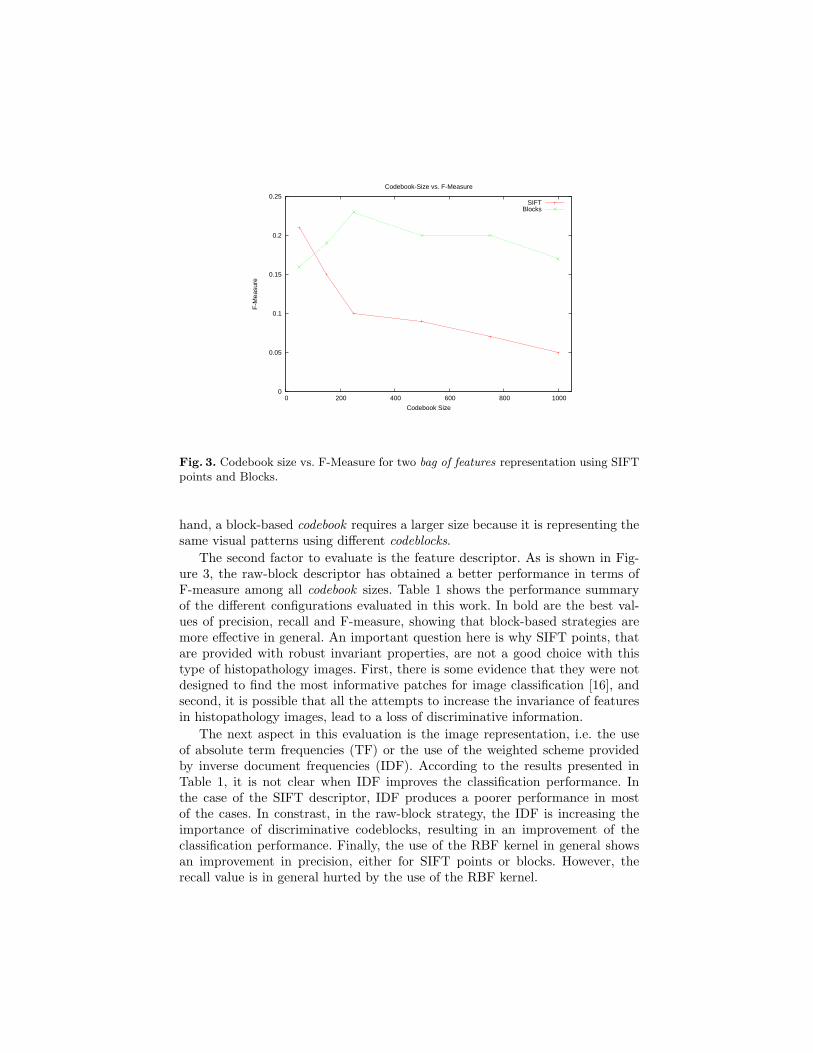
The first evaluation focuses on the codebook size. We have tested six differentcodebook sizes, starting with 50 codeblocks and following with 150, 250, 500,750 and 1000. Figure 3 shows a plot for codebook size vs. F-measure using twodifferent bag of features configurations. The first strategy, is based on SIFT pointsand the second is based on raw blocks. Perhaps surprisingly, the plot shows thatthe classification performance decreases while the codebook size increases. Thisbehaviour is explained by the intrinsic structure of histopathology images: theyare usually composed of some kinds of textures, that is, the number of distinctivepatterns in the collection is limited. This fact can also be seen in the codebookillustrated in Figure 2, which shows several repeated patterns, even with just150 codeblocks. In the limit, it is reasonable that a large codebook size does nothave any discriminative power because each different pattern in an image has itsown codeblock.
The nature of the descriptor is also a determinant factor in this behavioursince the performance of the SIFT points decreases faster than the performanceof raw blocks. This suggests that a SIFT-based codebook requires less codeblocksto express all different patterns in the image collection, which is consistent withthe rotation and scale invariance properties of that descriptor. On the other
0
0.05
0.1
0.15
0.2
0.25
0 200 400 600 800 1000
F-M
easu
re
Codebook Size
Codebook-Size vs. F-Measure
SIFTBlocks
Fig. 3. Codebook size vs. F-Measure for two bag of features representation using SIFTpoints and Blocks.
hand, a block-based codebook requires a larger size because it is representing thesame visual patterns using different codeblocks.
The second factor to evaluate is the feature descriptor. As is shown in Fig-ure 3, the raw-block descriptor has obtained a better performance in terms ofF-measure among all codebook sizes. Table 1 shows the performance summaryof the different configurations evaluated in this work. In bold are the best val-ues of precision, recall and F-measure, showing that block-based strategies aremore effective in general. An important question here is why SIFT points, thatare provided with robust invariant properties, are not a good choice with thistype of histopathology images. First, there is some evidence that they were notdesigned to find the most informative patches for image classification [16], andsecond, it is possible that all the attempts to increase the invariance of featuresin histopathology images, lead to a loss of discriminative information.
The next aspect in this evaluation is the image representation, i.e. the useof absolute term frequencies (TF) or the use of the weighted scheme providedby inverse document frequencies (IDF). According to the results presented inTable 1, it is not clear when IDF improves the classification performance. Inthe case of the SIFT descriptor, IDF produces a poorer performance in mostof the cases. In constrast, in the raw-block strategy, the IDF is increasing theimportance of discriminative codeblocks, resulting in an improvement of theclassification performance. Finally, the use of the RBF kernel in general showsan improvement in precision, either for SIFT points or blocks. However, therecall value is in general hurted by the use of the RBF kernel.

Table 1. Performance measures for the evaluated configurations of the bag of features.
Kernel function BOF SIFT Raw-Blocks
Precision Recall F-Measure Precision Recall F-Measure
Hist. Intersection TF 0.480 0.152 0.207 0.610 0.162 0.234Hist. Intersection IDF 0.473 0.128 0.189 0.634 0.152 0.231
RBF Kernel TF 0.393 0.146 0.205 0.647 0.123 0.190RBF Kernel IDF 0.506 0.136 0.165 0.673 0.155 0.237
5 Conclusions and Future Work
This paper presented an evaluation of the bag of features approach to classifyhistopathology images. This is the first systematic evaluation of this representa-tion scheme on this particular medical image domain. The developed evaluationincludes a comparative study of different methods and techniques in each stageof the bag of features approach. The main advantage of the proposed approachis its adaptiveness to the particular contents of the image collection. Previouswork in histology and pathology image analysis used global-generic features orsegmentation-based approaches that are not easily extended to other applica-tions even in the same domain.
The adaptiveness property of this framework is obtained with an automatedcodebook construction, which should have enough patterns to describe the imagecollection contents. In this domain, we found that the codebook size is very smallcompared with the codebook size required in other applications such as naturalscene classification or even in other kinds of medical images (i.e. mamographyand x-rays). The main reason of this smaller size is due to the structure ofhistopathology images which exhibit homogeneous tissues and the representativevisual patterns among the whole collection tends to be uniform.
The bag of features representation is a flexible framework that may be adaptedin different ways, either in the visual feature descriptors and the codebook con-struction. The future work includes a more extensive evaluation of codebookconstruction methods and different strategies to include color information intovisual words as well as more robust texture descriptors. This evaluation will alsoinclude a comparison against other commonly used representation schemes.
Acknowledgments
This work has been partially supported by Ministerio de Educacion Nacional deColombia grant 1101393199, according to the COLCIENCIAS call 393-2006 tosupport research projects using the RENATA network.
References
1. Bosch, A., Munoz, X., Martı, R.: Which is the best way to organize/classify imagesby content? Image and Vision Computing 25(6) (June 2007) 778–791

2. Csurka, G., Dance, C.R., Fan, L., Willamowski, J., Bray, C.: Visual categorizationwith bags of keypoints. In: Workshop on Statistical Learning in Computer Vision.(2004)
3. Sivic, J., Zisserman, A.: Video Google: a text retrieval approach to object matchingin videos. (2003) 1470–1477 vol.2
4. Tommasi, T., O.F.C.B.: CLEF2007 Image annotation task: An SVM-based cueintegration approach. In: Working Notes of the 2007 CLEF Workshop, Budapest,Hungary (2007)
5. Iakovidis, D.K., Pelekis, N., Kotsifakos, E.E., Kopanakis, I., Karanikas, H.,Theodoridis, Y.: A pattern similarity scheme for medical image retrieval. In-formation Technology in Biomedicine, IEEE Transactions on (2008)
6. Long, L.R., Antani, S.K., Thoma, G.R.: Image informatics at a national researchcenter. Computerized Medical Imaging and Graphics 29 (2005) 171–193
7. Guld, M.O., Keysers, D., Deselaers, T., Leisten, M., Schubert, H., Ney, H.,Lehmann, T.M.: Comparison of global features for categorization of medical im-ages. Medical Imaging 5371 (2004) 211–222
8. Deselaers, T., Keysers, D., Ney, H.: FIRE - Flexible Image Retrieval Engine:imageCLEF 2004 evaluation. In: CLEF 2004, LNCS 3491. (2004) 688–698
9. Datar, M., Padfield, D., Cline, H.: Color and texture based segmentation of molec-ular pathology images using hsoms. In: Biomedical Imaging: From Nano to Macro,2008. ISBI 2008. 5th IEEE International Symposium on. (2008) 292–295
10. Comaniciu, D., Meer, P., Foran, D.: Shape-based image indexing and retrievalfor diagnostic pathology. In: Pattern Recognition, 1998. Proceedings. FourteenthInternational Conference on. Volume 1. (1998) 902–904 vol.1
11. Caicedo, J.C., Gonzalez, F.A., Romero, E.: A semantic content-based retrievalmethod for histopathology images. Information Retrieval Technology LNCS 4993(2008) 51–60
12. Zheng, L., Wetzel, A.W., Gilbertson, J., Becich, M.J.: Design and analysis ofa content-based pathology image retrieval system. Information Technology inBiomedicine, IEEE Transactions on 7(4) (2003) 249–255
13. Lam, R.W.K., Ip, H.H.S., Cheung, K.K.T., Tang, L.H.Y., Hanka, R.: A multi-window approach to classify histological features. In: Pattern Recognition, 2000.Proceedings. 15th International Conference on. Volume 2. (2000) 259–262 vol.2
14. Tang, H.L., Hanka, R., Ip, H.H.S.: Histological image retrieval based on semanticcontent analysis. Information Technology in Biomedicine, IEEE Transactions on7(1) (2003) 26–36
15. Fletcher, C.D.M.: Diagnostic Histopathology of tumors. Elsevier Science (2003)16. Nowak, E., Jurie, F., Triggs, B.: Sampling strategies for bag-of-features image
classification. (2006) 490–50317. Lowe, D.G.: Distinctive image features from scale-invariant keypoints. Interna-
tional Journal of Computer Vision 60(2) (November 2004) 91–11018. Li, F.F., Perona, P.: A bayesian hierarchical model for learning natural scene cate-
gories. In: CVPR ’05: Proceedings of the 2005 IEEE Computer Society Conferenceon Computer Vision and Pattern Recognition (CVPR’05) - Volume 2, Washington,DC, USA, IEEE Computer Society (2005) 524–531
19. Shawe-Taylor, J., Cristianini, N.: Kernel Methods for Pattern Analysis. CambridgeUniversity Press (2004)