Year 1 Histopathology OSPE Objective structured practical ...
18
Year 1 Histopathology OSPE Objective structured practical examination (OSPE) SAMPLE OSPE QUESTIONS The following OSPE questions are examples. They are intended to allow candidates to become familiar with the style and content of the Year 1 OSPE. Answers, together with marks for each question are also provided below. Please remember that the marking schemes are a general guide for the assessors/markers. When marking papers, they are able to consider additional information provided by the candidates and may assess the overall standard of the responses. All questions are double-marked. SAMPLE QUESTIONS No Topic Additional materials provided at station Assessor present? 1 Gynaecological cytology • 2 photomicrographs No 2 Post-mortem pathology • 1 macroscopic photograph of brain No 3 Gastrointestinal pathology • 1 sheet microscopic photographs No 4 Breast pathology • H&E slide of an unequivocal grade 3 ductal carcinoma of the breast. • Graph of mitotic and field diameter for grading purposes. • Photographs of ER and PR immunostains • Microscope No 5 Double-headed microscope • 1 glass slide • 1 double-headed microscope Yes 6 Laboratory techniques • None No 7 Non-Gynaecological Cytology • None No
-
Upload
khangminh22 -
Category
Documents
-
view
8 -
download
0
Transcript of Year 1 Histopathology OSPE Objective structured practical ...

Year 1 Histopathology OSPE Objective structured practical examination (OSPE)
SAMPLE OSPE QUESTIONS
The following OSPE questions are examples. They are intended to allow candidates to become familiar with the style and content of the Year 1 OSPE.
Answers, together with marks for each question are also provided below. Please remember that the marking schemes are a general guide for the assessors/markers. When marking papers, they are able to consider additional information provided by the candidates and may assess the overall standard of the responses.
All questions are double-marked. SAMPLE QUESTIONS No
Topic
Additional materials provided at station
Assessor present?
1 Gynaecological cytology
• 2 photomicrographs
No
2 Post-mortem pathology
• 1 macroscopic photograph of brain
No
3 Gastrointestinal pathology
• 1 sheet microscopic photographs
No
4 Breast pathology • H&E slide of an unequivocal grade 3 ductal carcinoma of the breast. • Graph of mitotic and field diameter for grading purposes. • Photographs of ER and PR immunostains • Microscope
No
5 Double-headed microscope
• 1 glass slide • 1 double-headed microscope
Yes
6 Laboratory techniques
• None
No
7 Non-Gynaecological Cytology
• None
No
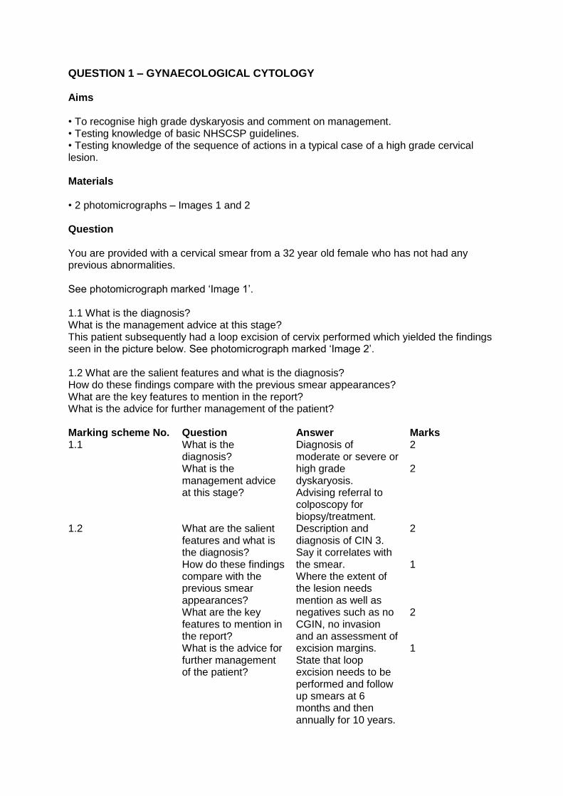
QUESTION 1 – GYNAECOLOGICAL CYTOLOGY Aims • To recognise high grade dyskaryosis and comment on management. • Testing knowledge of basic NHSCSP guidelines. • Testing knowledge of the sequence of actions in a typical case of a high grade cervical lesion. Materials • 2 photomicrographs – Images 1 and 2 Question You are provided with a cervical smear from a 32 year old female who has not had any previous abnormalities. See photomicrograph marked ‘Image 1’. 1.1 What is the diagnosis? What is the management advice at this stage? This patient subsequently had a loop excision of cervix performed which yielded the findings seen in the picture below. See photomicrograph marked ‘Image 2’. 1.2 What are the salient features and what is the diagnosis? How do these findings compare with the previous smear appearances? What are the key features to mention in the report? What is the advice for further management of the patient? Marking scheme No.
Question
Answer
Marks
1.1 What is the diagnosis? What is the management advice at this stage?
Diagnosis of moderate or severe or high grade dyskaryosis. Advising referral to colposcopy for biopsy/treatment.
2 2
1.2 What are the salient features and what is the diagnosis? How do these findings compare with the previous smear appearances? What are the key features to mention in the report? What is the advice for further management of the patient?
Description and diagnosis of CIN 3. Say it correlates with the smear. Where the extent of the lesion needs mention as well as negatives such as no CGIN, no invasion and an assessment of excision margins. State that loop excision needs to be performed and follow up smears at 6 months and then annually for 10 years.
2 1 2 1
Image 1
Image 2

QUESTION 2 – POST-MORTEM PATHOLOGY Aims • To test gross autopsy pathology observation and interpretation skills. • To test knowledge of basic neuropathology. • To test knowledge of subsidiary tests in the autopsy. • To test appreciation of wider issues in autopsy practice. Materials • 1 macroscopic photograph of brain (online on the College website). Question Case history: A 75-year-old man is found dead in bed at home. The ‘sudden death report’ supplied by the coroner’s officer is rather brief but indicates that his wife reported that he had been unwell for a couple of days and had retired to bed with a headache. The GP confirms that he was generally fit and well, having last seen a doctor six months previously for a minor complaint. You have been asked to carry out an autopsy on behalf of the Coroner. You are provided with a picture of the brain. 2.1a Describe the specimen. 2.1b Describe the abnormalities that are present. 2.2 What is your diagnosis? 2.3 What subsidiary tests would you consider at the time of the autopsy? What would
you expect them to show? 2.4 What issues may influence your use of subsidiary tests? Marking scheme No.
Question
Answer
Marks
2.1a 2.1b
Describe the specimen. Describe the abnormalities that are present.
Adequate description of the anatomy shown (inferior surface of posterior part of brain/cerebellum). Indicate the presence of opacification of the basal meninges with a cream/green exudate within the subarachnoid space.
2 2
2.2 What is your diagnosis?
Diagnosis of purulent meningitis.
1
2.3 What subsidiary tests would you consider at the time of the autopsy? What would you expect them to show?
Suggestion of histology with reasonable description of likely results. Suggestion of microbiology with reasonable description of likely results.
2 2

2.4 What issues may influence your use of subsidiary tests?
Description of coronial consent issues around retaining samples. Describe/discuss accurate microbiological diagnosis for notification of infectious disease purposes.
1

QUESTION 3 – GASTROINTESTINAL PATHOLOGY Aims • To test clinicopathological correlation and understanding of the role of biopsy in clinical investigation. • Recognition of normal histology and description of pathological changes. Materials • 1 sheet microscopic photographs (online on the College website). Question A 35-year-old diabetic woman is found to have iron deficiency anaemia. An upper GI endoscopy is arranged as part of her investigation. 3.1 Why has this investigation been selected? 3.2 Examine the two images. Both are from duodenal biopsies.
Which one is normal? Describe three abnormalities in the abnormal biopsy.
3.3 What confirmatory investigations could be conducted? 3.4 Four biopsies are recommended in order to make an accurate diagnosis of this
condition. Why is this number of biopsies recommended? Marking scheme No.
Question
Answer
Marks
3.1 Why has this investigation been selected?
Upper GI endoscopy may be indicated in iron deficiency anaemia. To confirm or to exclude coeliac disease. To identify a bleeding source.
1 1 1
3.2 Examine the two images. Both are from duodenal biopsies. Which one is normal? Describe three abnormalities in the abnormal biopsy.
Histological appearances: Biopsy A is abnormal. Villous atrophy. Increased intraepithelial lymphocytes. Crypt hyperplasia. Increased inflammatory cells in lamina propria.
1 0.5 0.5 0.5 0.5 (3 max)
3.3 What confirmatory investigations could be conducted?
Serology for antigliadin/antiendomysial antibodies. TTG. Trial of gluten exclusion and rebiopsy to show histological improvement.
1 1 1 (3 max)

3.4 Four biopsies are recommended in order to make an accurate diagnosis of this condition. Why is this number of biopsies recommended?
Changes are typically patchy.
1

QUESTION 4 – BREAST PATHOLOGY Aims • To test microscope diagnostic skills and histology interpretation. • To test approach (knowledge) to staging of breast carcinoma. • To test basic knowledge of immunostains. Materials • H&E slide of an unequivocal grade 3 ductal carcinoma of the breast. • Graph of mitotic and field diameter for grading purposes (online on the College website) • Photographs of ER and PR immunostains (online on the College website). • Microscope. Question Case history: A 33-year-old female undergoes a mastectomy for breast carcinoma. You are provided with a haematoxylin and eosin (H&E)-stained section from the tumour. 4.1 Grade this tumour according to the modified Bloom Richardson system (i.e. the
standard system in use in the UK). [High power field diameter to be indicated to candidates at start.]
4.2 Comment on the immunostaining pattern of the tumour for oestrogen receptors (ER) and progesterone receptors (PR). (A score is not required.)
4.3 Why is the ER status of this tumour important in patient management? Marking scheme No.
Question
Answer
Marks
4.1 Grade this tumour according to the modified Bloom Richardson system (i.e. the standard system in use in UK).
Grading of tumour: 1. Tubule formation 2. Nuclear pleomorphism 3. Mitotic count 4. Overall grade. Appropriate score for each component and overall grade. Components for grading given but inappropriate scores and grade. Not all component scores given. No component scores or grade given.
1 1 1 2 Up to 5 Up to 3 1 0 (5 max)
4.2 Comment on the immunostaining pattern of the tumour for oestrogen receptors (ER) and progesterone receptors (PR). (A score is not
Correctly describes the immunoprofile with comments of staining patterns or Correctly describes the immunoprofile but without comments of staining patterns or Incorrect description of the immunoprofile. and Suggests that the PR stain may not have worked and
2 1 0 1

needs repeating.
4.3 Why is the ER status of this tumour important in patient management?
Comments on use of hormone manipulation in ER positive tumours. Does not comment on use of hormone manipulation in ER positive tumours.
2 0 (2 max)


QUESTION 5 – DOUBLE-HEADED MICROSCOPE Aims • To test microscope diagnostic skills and histology interpretation. • To test basic knowledge of pathological processes. Materials • 1 glass slide. • 1 double-headed microscope. Question Case history: A 20-year-old female has mediastinal and abdominal lymphadenopathy. You are provided with a haematoxylin and eosin (H&E)-stained section of a lymph node. You have 4 minutes to examine the slide. The assessor will then ask you to point out certain features and make comments. 5.1 Questions asked by the assessor:
Identify the following normal structures of the lymph node: a) Pericapsular sinus b) Lymphoid follicle c) Paracortex d) Germinal centre. 5.2 Identify the following normal cellular constituents of a lymph node: a) Lymphocyte b) Centroblast c) Centrocyte. 5.3 Describe the pathological features in this lymph node. 5.4 What other types of granulomas do you know? 5.5 How would you proceed to investigate this case further?
No. Question Answer Marks 5.1 Questions asked by
the assessor: Identify the following normal structures of the lymph node: a) Pericapsular sinus b) Lymphoid follicle c) Paracortex d) Germinal centre
Normal structures identified correctly: 1. Pericapsular sinus 2. Lymphoid follicle 3. Paracortex 4. Germinal centre.
See marking scheme checklist below
5.2 Identify the following normal cellular constituents of a lymph node: a) Lymphocyte b) Centroblast c) Centrocyte
Normal cells identified correctly: 1. Lymphocyte 2. Centroblast 3. Centrocyte.
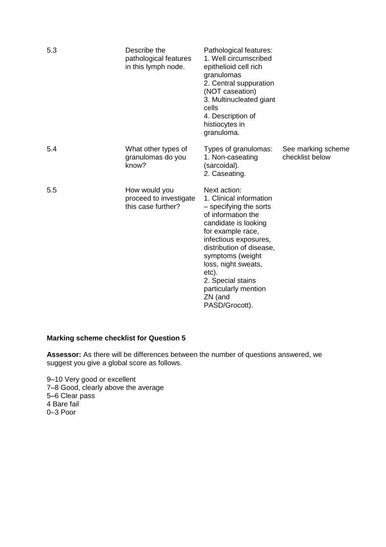
5.3 Describe the pathological features in this lymph node.
Pathological features: 1. Well circumscribed epithelioid cell rich granulomas 2. Central suppuration (NOT caseation) 3. Multinucleated giant cells 4. Description of histiocytes in granuloma.
5.4 What other types of granulomas do you know?
Types of granulomas: 1. Non-caseating (sarcoidal). 2. Caseating.
See marking scheme checklist below
5.5 How would you proceed to investigate this case further?
Next action: 1. Clinical information – specifying the sorts of information the candidate is looking for example race, infectious exposures, distribution of disease, symptoms (weight loss, night sweats, etc). 2. Special stains particularly mention ZN (and PASD/Grocott).
Marking scheme checklist for Question 5 Assessor: As there will be differences between the number of questions answered, we suggest you give a global score as follows. 9–10 Very good or excellent 7–8 Good, clearly above the average 5–6 Clear pass 4 Bare fail 0–3 Poor

QUESTION 6 – LABORATORY TECHNIQUES Aims • To assess knowledge of immunohistochemistry techniques. • To assess knowledge and attitudes relating to use of human tissues. Materials required • None. Question Your laboratory is introducing a new primary antibody for immunohistochemistry. 6.1 How might your laboratory overcome the effects of formalin fixation to allow
demonstration of the antigen? (2 marks) 6.2 How would you ensure that the results produced are sensitive and specific? (2
marks) 6.3 What causes of false positive immunostaining can you think of? (2 marks) 6.4 How should your laboratory ensure that their results are similar to those produced by
other similar laboratories? (1 mark) 6.5 You find a case which produces excellent staining and your laboratory asks you to
take some further blocks to use as controls. What issues do you consider before doing this? (3 marks)
Marking scheme No.
Question
Answer
Marks
6.1 Give two ways in which your laboratory might overcome the effects of formalin fixation to allow demonstration of this antigen and how do they work?
Discussion of principles of antigen retrieval. Should include methods of heat mediated antigen retrieval eg microwave, pressure cooker. Use of proteolytic enzymes.
Up to 2
6.2 How would you ensure that the results produced are sensitive and specific?
Discussion of assessment of internal positive controls (tissues within test section expected to stain) and external positive controls (known positive test sections) and negative control slides (eg using irrelevant primary antibody or omitting primary antibody).
Up to 2
6.3 What causes of false positive immunostaining can you think of?
Reasonable causes such as edge effects, non-specific antibody binding, cross reactivity, endogenous peroxidase, endogenous biotin.
Up to 2

6.4 How should your laboratory ensure that their results are similar to those produced by other similar laboratories?
Mention of participation in national EQA scheme.
1
6.5 You find a case which produces excellent staining and your laboratory asks you to take some further blocks to use as controls. What issues do you consider before doing this?
Discussion of legal implications of consent for storage and use of tissue for non-diagnostic purposes.
Up to 3
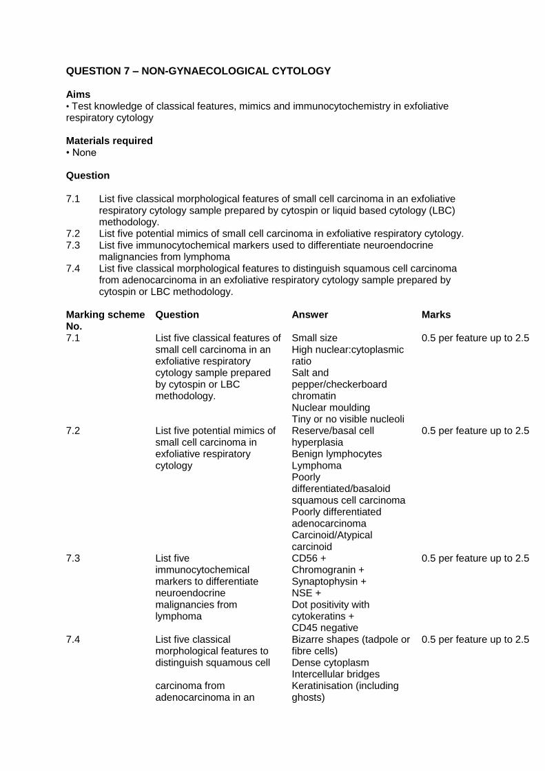
QUESTION 7 – NON-GYNAECOLOGICAL CYTOLOGY Aims • Test knowledge of classical features, mimics and immunocytochemistry in exfoliative respiratory cytology Materials required • None Question 7.1 List five classical morphological features of small cell carcinoma in an exfoliative
respiratory cytology sample prepared by cytospin or liquid based cytology (LBC) methodology.
7.2 List five potential mimics of small cell carcinoma in exfoliative respiratory cytology. 7.3 List five immunocytochemical markers used to differentiate neuroendocrine
malignancies from lymphoma 7.4 List five classical morphological features to distinguish squamous cell carcinoma
from adenocarcinoma in an exfoliative respiratory cytology sample prepared by cytospin or LBC methodology.
Marking scheme No.
Question
Answer
Marks
7.1 List five classical features of small cell carcinoma in an exfoliative respiratory cytology sample prepared by cytospin or LBC methodology.
Small size High nuclear:cytoplasmic ratio Salt and pepper/checkerboard chromatin Nuclear moulding Tiny or no visible nucleoli
0.5 per feature up to 2.5
7.2 List five potential mimics of small cell carcinoma in exfoliative respiratory cytology
Reserve/basal cell hyperplasia Benign lymphocytes Lymphoma Poorly differentiated/basaloid squamous cell carcinoma Poorly differentiated adenocarcinoma Carcinoid/Atypical carcinoid
0.5 per feature up to 2.5
7.3 List five immunocytochemical markers to differentiate neuroendocrine malignancies from lymphoma
CD56 + Chromogranin + Synaptophysin + NSE + Dot positivity with cytokeratins + CD45 negative
0.5 per feature up to 2.5
7.4 List five classical morphological features to distinguish squamous cell
Bizarre shapes (tadpole or fibre cells) Dense cytoplasm Intercellular bridges
0.5 per feature up to 2.5
carcinoma from adenocarcinoma in an
Keratinisation (including ghosts)

exfoliative respiratory cytology sample prepared by cytospin or LBC methodology.
Pearls Pyknotic/India-ink nuclei Lack of vacuolation or foamy cytoplasm Eccentric nuclei