Influence of squid extracts on the triggering of secondary vitellogenesis in Penaeus vannamei
Upload
khangminh22Category
view
1download
0
MICROSCOPICAL ANALYSIS OF THE HEPATOPANCREAS IN WHITELEG SHRIMP
(Penaeus vannamei) USING COMPUTER-ASSISTED
IMAGE ANALYSIS
Microscopical analysis of the hepatopancreas in whiteleg shrim
p (Penaeus vannamei) using com
puter-assisted image analysis Fabio Cervellione
Department of MorphologyFaculty of Veterinary Medicine
Ghent University
Academic year 2016 – 2017
Fabio Cervellione

ii
Fabio Cervellione (2017) Microscopical analysis of the hepatopancreas in whiteleg shrimp (Penaeus
vannamei) using computer-assisted image analysis
On the front cover: photo editing of a light microscopic image of the tubular strucuture of the hepatopancreas in whiteleg shrimp (Penaeus vannamei)
Printed by University Press, Zelzate, Belgium. www.universitypress.be This thesis was funded by Skretting ARC and the Research Council of Norway. The author and promoters give the permission to consult and copy parts of this work for personal use only. Every other use is subject to copyright laws. Permission to reproduce any material should be obtained from the author.

i

iii
MICROSCOPICAL ANALYSIS OF THE HEPATOPANCREAS IN
WHITELEG SHRIMP (Penaeus vannamei)
USING COMPUTER-ASSISTED IMAGE ANALYSIS
Fabio Cervellione
A dissertation submitted in partial fulfilment of the requirements for the degree of Doctor in Veterinary Sciences (PhD)
Promoters Prof. dr. Wim Van den Broeck
Dr. Charles McGurk
Department of Morphology Faculty of Veterinary Medicine
Ghent University
Academic year 2016 - 2017

iv

v
Promoters
Prof. dr. Wim Van den Broeck
Faculty of Veterinary Medicine, UGent
Dr. Charles McGurk
Skretting Aquaculture Research Centre, Norway
Members of the Examination Committee
Chair of the Examination Committee Prof. dr. Geert Janssens
Faculty of Veterinary Medicine, UGent
Prof. dr. Koen Chiers
Faculty of Veterinary Medicine, UGent
Prof. dr. Dominique Adriaens
Faculty of Science, UGent
Prof. dr. Peter Bossier
Faculty of Bioscience Engineering, UGent
Dr. Dantas Lima João
IMAQUA, Gent

vi

vii
Table of contents
List of abbreviations ........................................................................................... 1
General Introduction ........................................................................................... 3
Scientific aims ................................................................................................... 47
Chapter 1 ........................................................................................................... 49
Optimization of fixation methods for image analysis of the hepatopancreas in whiteleg shrimp, Penaeus vannamei (Boone)
Chapter 2 ........................................................................................................... 71
Use of computer-assisted image analysis for semi-quantitative histology of the hepatopancreas in whiteleg shrimp, Penaeus vannamei (Boone)
Chapter 3 ......................................................................................................... 101
Effect of starvation and refeeding on the hepatopancreas of whiteleg shrimp Penaeus vannamei (Boone) using computer-assisted image analysis
Chapter 4 ......................................................................................................... 127
Effect of starvation and refeeding on the ultrastructure of the hepatopancreas of whiteleg shrimp Penaeus vannamei (Boone) using transmission electron microscopy
Chapter 5 ......................................................................................................... 151
“Perigastric organ”: a replacement name for the “hepatopancreas” in Decapoda
General discussion ......................................................................................... 163
Summary/Samenvatting ................................................................................ 179
Acknowledgments .......................................................................................... 187
Curriculum Vitae ............................................................................................. 188
Bibliography .................................................................................................... 189

viii

1
List of abbreviations
The following list provides the full names of all abbreviations used in the test
presented in this PhD thesis in alphabetical order.
1 in = single injection inside
4 in = four injections inside
4 out = four injections outside
APP = protocol for image analysis
CAIA = computer-assited image analysis
FCA = F-cell area
FE = fed group
H&E = haematoxylin and eosin
HIA = haemocytic infiltration area
HP = hepatopancreas
IHC = immunohistochemistry
Mabs = monoclonal antibodies
MVB = multivescicular body
Ob1/Ob2 = observer 1/ observer 2
PM = peritrophic membrane
PNR = point of no return
PO = perigastric organ
REF = refed group
RER = rough endoplasmic reticulum
ROI = region of interest
SER = smooth endoplasmic reticulum
ST = starved group
TLA = total lumen area
TTA = total tissue area
VBA = vacuole B-cell area
WSH8 = monoclonal antibodies targenting haemocytes

2

3
General Introduction
1.1 Shrimp production
Modern shrimp farming began in the late 1960s, when French researchers in Tahiti
developed techniques for intensive breeding and rearing of various shrimp species
belonging to the family Penaeidae. Penaeid shrimp are decapods, possessing distinctive
morphological features including an extensive rostrum that projects as far as the eye stalk
and three pairs of clawed legs (Martin & Hose 2010). Early penaeid culture efforts in
America concentrated on indigenous species including northern white shrimp (Penaeus
setiferus) and blue shrimp (Penaeus stylirostris) in Panama, northern brown shrimp
(Penaeus aztecus) and western white shrimp (Penaeus occidentalis) in Honduras,
northern brown shrimp and northern pink shrimp (Penaeus duorarum) in southern USA,
southern white shrimp (Penaeus schmitti) and redspotted shrimp (Penaeus braziliensis) in
Brazil. In 1972, the initial work on whiteleg shrimp (Penaeus vannamei) gave much better
results than the other species and eyestalk ablation techniques (used to stimulate female
shrimp to develop mature ovaries and spawn) led to successful year-round production.
Whiteleg shrimp were introduced into Asia experimentally in 1978-79, and commercially in
1996 (FAO 2004).
Farmed crustaceans contribute to a significant proportion of the total aquaculture sector
with an annual production exceeding ten million metric tonnes yielding

General introduction
4
first sale value of 40 billion US dollars. Average annual growth in penaeid shrimp
production has been approximately 14% per annum since 1989 (Bondad-Reantaso et al.
2012). The sector is dominated by farmed tropical marine shrimp, the fastest growing
sector of global aquaculture industry (Stentiford et al. 2012). The leading producers of
farmed shrimp are primarily located in South East Asia (China, Thailand, Vietnam,
Indonesia and India) and Central and South America (Ecuador, Mexico and Brazil). Main
farmed species are whiteleg shrimp and giant tiger prawn (Penaeus monodon) (Lightner,
et al. 2012).
Shrimp remain the most valuable farmed species in the seafood industry, with the highest
trade value: the international market is concentrated in the USA, EU and Japan, with the
major exporting countries consisting of Thailand, China and Vietnam. Trends in cultured
shrimp production of whiteleg shrimp are summarized in Figure 1 (FAO 2017).
Fig. 1 Global aquaculture production of whiteleg shrimp (Penaeus vannamei) from 1950 to 2014 (FAO 2017).

General Introduction
5
1.2 Whiteleg Shrimp (Penaeus vannamei)
1.2.1 Taxonomy
The taxonomic position of whiteleg shrimp is outlined below (ITIS 2017).
Kingdom: Animalia
Subkingdom: Bilateria
Infrakingdom: Ecdysozoa
Phylum: Arthropoda
Subphylum: Cruscacea
Class: Malacostraca
Subclass: Eumalacostraca
Superorder: Eucarida
Order: Decapoda
Suborder: Dendrobrachiata
Superfamily: Penaeoidea
Family: Penaeidae
Genus: Penaeus
Species: Penaeus vannamei, Boone 1931
1.2.2 Biology
Whiteleg shrimp live in tropical marine habitats (depth 0 to 72 m), native to the Eastern
Pacific coast from Mexico to South America, where water temperature is normally over
20°C all year-round. Maximum total body length is 230 mm and maximum carapace
length is 90 mm (Holthuis 1980). Adults live and spawn in the open ocean, while post
larvae migrate inshore to spend their sub-adult stages in coastal estuaries. Males become
mature from 20 g and females from 28 g onwards at the age of six–seven months.
Females weighing 30–45 g will spawn approximately 100.000–250.000 eggs (Ø 0.22
mm). Hatching occurs about 16 hours after spawning and fertilization. The first stage

General introduction
6
larvae (nauplii) do not feed and live on their yolk reserves. They swim intermittently and
are attracted by light (positively phototactic). The next larval stages (protozoea, mysis)
remain planktonic and eat phytoplankton and zooplankton, carried towards the shore by
tidal currents. Five days after moulting into post larvae, shrimp move inshore and start
feeding on benthic detritus (FAO 2017). Farming of whiteleg shrimp offers some
advantages over giant tiger prawn: the ability to close the life cycle and produce brood
stock (allowing for domestication and genetic selection), rapid growth rate, tolerance to
high stocking density and to low salinities and temperatures, and high larval survival
during rearing (FAO 2017).

General Introduction
7
1.2.3 Morphology
Siebold (1848) was the first author to describe the general morphology of the Crustacea.
Figure 2 and Figure 3 show the original illustrations of the internal morphology of
European crayfish (Astacus fluviatilis) published in 1880 by Huxley.
Fig. 2 Historical illustration of the internal morphology of the Decapoda
(Huxley 1880).

General introduction
8
Fig. 3 Historical illustration of the digestive tract of the Decapoda (Huxley 1880).

General Introduction
9
1.2.3.1 External and internal morphology
The shrimp body is laterally compressed and divided into two regions: the cephalothorax
and the abdomen. The anterior cephalothorax comprises the fusion of five head segments
and eight thoracic segments. The abdomen is composed of six distinct segments. The
external surface is covered by the exoskeleton (Martin & Hose 2010). External and
internal morphology of the Decapoda are illustrated in Figure 4 and Figure 5.
Fig. 4 External morphology of penaeid shrimp. Adapted from FAO (2017).

General introduction
10
Fig. 5 Internal morphology of the Decapoda. AG = antennal gland; BR = brain; CO = cardiac ostium; CS = cardiac stomach; DA = dorsal artery; GG = ganglion; GI = gills; HP = hepatopancreas; HT = heart; M = midgut; MM = muscles; MO = mouth; NC = neural cord; PS = pyloric stomach; SP = sperm duct; TT = testicle; VA = ventral artery. Adapted from SlidePlayer (2017).

General Introduction
11
1.3 Digestive tract of the Decapoda
The digestive tract can be divided in three parts: foregut (mouth, oesophagus and
stomach), midgut (including hepatopancreas and midgut caeca) and hindgut. It extends
the length of the body from the anterior mouth to the posterior anus (Figure 6) (Gimenez
2013). The anatomy and cellular composition of the digestive tract of decapods is
considerably different from vertebrates. Differences primarily include the gastric mill, a
sophisticated filter apparatus in the stomach, and the tubules of the hepatopancreas (HP).
Additional differences are the lack of strongly acid pH and pepsin in the stomach. The
lumen of the digestive tract must be considered as being located outside the animal itself.
The internal wall is impregnated, in its fore and hind parts (foregut and hindgut), with
complexes of chitin and proteins forming a cuticle (comparable to the exoskeleton) which
are removed with the exoskeleton at each moult. Tegumental glands are located along
the walls of the oesophagus and the junction between midgut and hindgut. They secrete
mucus that lubricates the wall and facilitates the transition of food and faeces
(Felgenhauer 1992; McGaw & Curtis 2013).
Fig. 6 Schematic representation of the digestive tract of the Decapoda. F = foregut; H = hindgut; HP = hepatopancreas; M = midgut. Adapted from Dall (1965).

General introduction
12
1.3.1 Foregut
The foregut is composed of the mouth, oesophagus and stomach. The oesophagus is
usually short, straight, positioned vertically, and lined by a thick cuticle (Ceccaldi 1989).
The stomach is a dual-chambered, chitinous sac, divided into a larger anterior (or cardiac)
chamber and a narrower posterior (or pyloric) chamber. The anterior chamber is a
spacious sac facilitating mastication and sorting ingested food. It bears a longitudinal row
of small ridges which lead to the lateral teeth and the larger dorsal median tooth. The
cardio-pyloric valve separates the anterior chamber from the posterior chamber. The
posterior chamber has a dorsal region which allows ingested material to pass directly into
the midgut and a ventral region called the filter-press, which permits only the finest
particles to enter the HP. Both chambers are composed of a varying number of chitinous
plates/ossicles that differ in size and morphology (Felgenhauer 1992).
1.3.2 Midgut
The midgut is composed of the intestine and many evaginations, namely the HP, anterior
and posterior midgut caeca (McLaughlin 1983). It starts in the cephalothorax at the
junction with the pyloric sac of the stomach and ends in a coiled tube, the posterior midgut
caecum, at the anterior half of the sixth abdominal segment (Ceccaldi 1989; McGaw &
Curtis 2013). The midgut is the only region of the digestive tract which is not lined by
cuticle and is in contact with the external environment. The midgut is lined by a columnar
epithelium sitting on a basement membrane, usually exhibiting a prominent brush border.
The functions of the midgut are not entirely clear, and include osmoregulation, limited
nutrient absorption, and the production of the peritrophic membrane (PM). The PM is a

General Introduction
13
non-cellular membrane lining the midgut, which serves as a barrier between the contents
of the gut lumen and the midgut epithelium, protecting the midgut from abrasive food
particles and pathogens. The PM is composed of chitin fibrils associated to proteoglycans
and assists digestion process by compartmentalization of the gut and immobilization of
the digestive enzymes. It is an important component of the intestinal immune system,
inactivates ingested toxin, and prevents oxidation (Wang et al. 2012). The midgut caeca
are two blind ending extensions of the midgut, and histologically differ from the intestine in
lacking longitudinal muscular fibres and in possessing considerably infolded walls. They
play a role in ion and water regulation (Barker & Gibson 1977; Ceccaldi 1989).

General introduction
14
1.3.3 Hepatopancreas
The HP is a large bi-lobed organ composed of many blindly ending tubules, which wrap
over the dorsal and lateral sides of the posterior part of the stomach and the anterior part
of the midgut (Franceschini-Vicentini et al. 2009; Martin & Hose 2010). It represents 2-6%
of the total body weight and reaches its greatest degree of complexity in the Decapoda
(Gibson & Barker 1979). Its colour (brown, red, green, yellow, blue) mainly depends on
the stored reserves (carotene, zeaxanthine, astaxanthine, and cantaxantine) (Gibson &
Barker 1979; Ceccaldi 1989). It has two separate lobes, enclosed together in a connective
tissue capsule and separated by a thin septum. Each lobe is composed of two or three
lobules and it is connected to the intestine through the corresponding principal duct
(Esteve & Herrera 2000; Vasagam et al. 2007). Principal ducts divide in secondary ducts
which branch in the HP tubules. Around each duct, there are fine circular and longitudinal
muscular fibres, which allow for peristaltic and longitudinal contractions, facilitating the
movement of liquids through the organ. The HP is well supplied with haemolymph: the
hepatic arteries pass into the HP lobes where they subdivide among ducts and tubules
(Herreid & Full 1988; Ceccaldi 1989). Transverse sections of the tubules through the
medial region of the HP exhibit a normal star-shape lumen (Esteve & Herrera 2000;
Vasagam et al. 2007).
Each hepatopancreatic tubule can be subdivided into a distal, medial and proximal
zone relative to the distance from the midgut (Figure 7) (Felgenhauer 1992; Franceschini-
Vicentini et al. 2009). Each tubule is made up of four basic cell types: E-, F-, R- and B-
cells (Figure 8) (Nakamura 1987; Franceschini-Vicentini et al. 2009). Figure 9 shows the
four cell types comprising the HP tubule observed under light microscope.

General Introduction
15
Fig. 7 Schematic representation of the hepatopancreas tubule structure in the Decapoda. = B-cell; DZ = distal zone; E = E-cell; F = F-cell; L = lumen; MZ = medial zone; PZ = proximal zone; R = R-cell; SD = secondary duct. Adapted from Van Weel (1955).
Fig. 8 Schematic representation of a cross section of the medial part hepatopancreatic tubule in the Decapoda. B = B-cell; F = F-cell; L = lumen; R = R-cell. Adapted from Johnson (1995).

General introduction
16
Fig. 9 Light microscopic structure of the hepatopancreas (a, b, c) and midgut (d) in whiteleg shrimp (Penaeus vannamei). Five µm paraffin sections. H&E. (a) B-cells, showing a single large vacuole and peripheral nucleus (white arrow); (b) F-cell with basophilic cytoplasm and R-cell with multiple lipid droplets (black arrows); (c) E-cells at the distal end of the tubule, in mitotic division (black/white arrow); (d) midgut, showing the epithelium and peritrophic membrane. B = B-cell; E = E-cell; F = F-cell; ME = midgut epithelium; PM = peritrophic membrane; R = R-cell. Scale bar = (a,b,c) 20 µm, (c) = 50 µm (Skretting ARC).
The E-cells, or embryonic cells, are small cuboidal cells found at the distal blind
ends of the tubules and give rise to the other three cell types. E-cells are characterized by
a large nucleus with prominent nucleolus and usually lack a brush border. The
ultrastructure of the medial zone of the HP tubule is summarized in Figure 10. F-cells, or
fibrillar cells, are cylindrical/prismatic cells located at the medial and proximal zones of the
tubules among R-cells and B-cells. They have a basally located nucleus and a developed

General Introduction
17
rough endoplasmic reticulum (RER), giving them a fibrillar appearance, typically exhibiting
a strong basophilia in H&E staining due to a high RNA content. They present small
vesicles throughout the cytoplasm and have a prominent brush border (Barker & Gibson
1977) (Felgenhauer 1992). This cell type has a high rate of protein synthesis and is the
only site of digestive enzyme synthesis (Franceschini-Vicentini et al. 2009). B-cells, or
blister cells, are large, primary secretory cells, more frequent at the proximal zone of the
tubules. They are defined by the presence of a single enormous vesicle surrounded by a
dense cytoplasm filled with RER (Barker & Gibson 1977; Felgenhauer 1992; Cuartas et
al. 2002). They are responsible for intracellular digestion, concentrating the absorbed
materials in the large vacuole and secreting the vacuolar content at the end of the
digestive process, and it is believed that the digestion-derived nutrients are released for
reabsorption by R-cells (Franceschini-Vicentini et al. 2009). The R-cells are the most
numerous cell type, located at medial and proximal zones. These tall, columnar cells are
characterized by a prominent brush border and a large number of irregularly-shaped
storage vesicles (primarily lipids) in their cytoplasm (Franceschini-Vicentini et al. 2009).
These cells function in food absorption and storage of nutrients and minerals
(Felgenhauer 1992).

General introduction
18
Fig. 10 Schematic representation of the ultrastructure of the medial zone of the hepatopancreas tubule in the Decapoda. B = B-cell; bb = brush boarder; F = F-cell; g = Golgi complex; h = haemocyte; L = lumen; ld = lipid droplet; m = mitochondria; n= nucleus; R = R-cell; rer = rough endoplasmic reticulum; vc = vacuole. Adapted from Dall & Moriarty (1983).

General Introduction
19
1.3.4 Hindgut
The hindgut is of ectodermal origin and consequently lined with chitin (Barker & Gibson
1977). It is located in the posterior half of the sixth abdominal segment and includes the
rectum and anus (Ceccaldi 1989). It is characterized by the presence of cuticular scales
or groups of spines, posteriorly directed facilitating the movement of the faecal mass
towards the anus (Felgenhauer 1992).
1.4 Digestive physiology
The foregut, midgut and hindgut are specialized structures serving specific functions. The
foregut is responsible for mechanical and extracellular enzymatic digestion (enzymes
released by the HP into the anterior chamber), midgut regulates movements of digesta
into the HP where intracellular digestion takes place, and peristaltic movements of the
hindgut expel the PM containing faeces (Figure 11).
The mouth opens into a short oesophagus, lined with tegumental glands, secreting mucus
that lubricates the food. The pyloric region filters out particulate matter, so that only liquid
and particles less than 100 nm in diameter enter the HP (Martin & Hose 2010; McGaw &
Curtis 2013). The primary role of the HP is the synthesis and secretion of digestive
enzymes, final digestion of ingested food and uptake of nutrients (Laohabanjong et
al.2009). Enzymes from the HP are released into the anterior (cardiac) chamber and flow
ventrally into the filter-press where setae filter particles. The excluded particles are
passed posteriorly into the midgut, while the smallest particles and fluid enter the HP
tubules for absorption (McGaw & Curtis 2013).

General introduction
20
The microvilli in the midgut epithelia are involved with absorption of nutrients in larval
shrimp, but minimal absorption occurs in adults. They synthesise the PM, a chitinous
wrapper that encapsulates ingested materials as soon as they enter this region of the gut
and persist around the faecal pellet (Martin & Hose 2010). Anterior and posterior midgut
caeca play also a role in ion and water regulation, and maintain pH balance. The hindgut
is involved in active ion uptake and transport. Tegumental glands along its length secrete
mucus to lubricate the walls (McGaw & Curtis 2013).

General Introduction
21
Fig. 11 Schematic diagram of digestive processes in the Decapoda. Organs (in black) and their main functions (in red between brackets). Organs: A = anus; AMC = anterior midgut caecum; BC = B-cells; CS = cardiac stomach; EC= E-cells; FC = F-cells; HP = hepatopancreas; M = midgut; MO = mouth; PMC = posterior midgut caecum; PS = pyloric stomach; RC = R-cells; TG = tegumental glands. Functions: ED = extracellular digestion; EP = enzyme production; F = filtration; FC = faecal compaction; ID = intracellular digestion; IWA = ion and water uptake; MS = mucus secretion; NA = nutrient absorption; PPM = production of peritrophic membrane; SC = staminal cells. Adapted from McGaw & Curtis (2013).

General introduction
22
1.5 Feeding and moulting
Crustaceans alternate episodes of feeding and fasting during development, which occurs
through moulting and results in growing by sequential steps. Increase in body size at each
ecdysis is non-linear; this is a hormonally controlled process which might last days or
weeks, is continuous and accompanied by morphological, physiological, and behavioral
alterations occurring almost daily (Dall et al. 1990). Moulting is a natural continuous
process used by crustaceans to shed their exoskeleton and grow. The moult cycle is
divided into four major stages: pre-moult (D), moult (E), post-moult (A and B) and inter-
moult (C). In stage A, the exoskeleton is soft and the shrimp is incapable of taking food.
Feeding begins in stage B, when the exoskeleton is sufficient rigid to handle food. During
stage C, shrimp feeds actively and exoskeleton becomes very rigid. Feeding declines
during stage D, just before ecdysis (stage E), where the old cuticle is shed and the
organism is unable to feed (Sánchez-Paz et al. 2007).
1.6 Immunology
Crustaceans lack true adaptive immunity and solely depend on innate immunity (cellular
and humoral).
1.6.1 Cellular innate immunity
In crustaceans, the circulating haemocytes are crucial in protecting the animal against
invading microorganisms by participating in recognition, phagocytosis, melanisation and
cytotoxicity. Three morphologically different classes of haemocytes have been described:
hyaline, semigranular and granular (Cerenius et al. 2010; Dantas Lima 2013; Musthaq &
Kwang 2014).

General Introduction
23
Pathogen recognition stimulates the degranulation of granular and semigranular cells. The
release of the prophenoloxidase system and other plasma factors from the stored
granules promotes phagocytosis by hyaline cells or encapsulation by semigranular cells
(Johansson et al. 2000).
Phagocytosis is the major cellular defence mechanism towards infection in crustaceans. If
the number of the foreign entities invading the organism is too high or the sizes too large
to be eliminated by phagocytosis, crustaceans apply nodulation (bacteria and fungus) or
encapsulation (parasite). The process is characterized by trapping and isolating the
foreign entities by attachment of concentric layers of adherent haemocytes around them.
It follows the killing of the invaders by asphyxiation and action of toxic compounds and
enzymatic cascades (Dantas Lima 2013).
1.6.2 Humoral innate immunity
Humoral defences of invertebrates are a multiplicity of serum or plasma factors which act
against micro-organisms, foreign cells or abiotic materials. These factors include
agglutinins, lytic agents, bactericidal peptides, cytokines, micro-biocidal pigments,
antiviral, cytotoxic, clotting, and prophenoloxidase factors (Smith & Chisholm 1992).
1.6.3 Mucosal immunity
In vertebrates the mucosal immunity is composed of mechanical and chemical barriers
preventing the entry of foreign bodies or potential pathogens. The PM constitutes a clear
example of non-vertebrate barrier epithelia. The inflammatory process involves changes
in vascular tissue and the rapid recruitment and activation of immune cells to damaged or

General introduction
24
infected tissue. The main mechanisms of mucosal immunity in the gut of crustaceans are:
receptors of pathogen recognition, as PI-PET (thioesther containing protein), and
antimicrobial peptides (Garcia-Garcia et al. 2013).
1.6.4 Innate immunity with specificity
There is increasing evidence that shrimp and other arthropods exhibit immune specificity
and immune memory. Invertebrate host, after its first exposure to a pathogen, aquires
memory that gives the host an enhanced immunity when the same pathogen is
encountered again. The invertebrate immune response is now called “innate immunity
with specificity” or “immune priming”, and its mechanisms are still unclear. While
vertebrate antibodies have no invertebrate homolog, the Down syndrome cell adhesion
molecule (Dscam) can function as a pathogen-specific recognizing molecule in athropods
(Ng et al. 2014).

General Introduction
25
1.7 Health issues related to shrimp farming
The aquaculture of penaeid shrimp has grown from its experimental beginnings roughly
three decades ago into a major industry. Almost from the start, diseases were recognized
as a biological threat to the shrimp industry, causing serious economic losses. The
diseases of cultured penaeid shrimp include syndromes with infectious (viral, bacterial,
fungal, parasitic) and non-infectious aetiologies (environmental stressors, nutritional
imbalances, toxicants and genetic factors) (Lightner & Redman 1998).
According to a survey conducted by the Global Aquaculture Alliance, approximately 60%
of the disease losses in shrimp aquaculture could be attributed to viral diseases and
approximately 20% to bacterial diseases. The remaining 20% of losses can be attributed
to a variety of other pathogens (parasites and fungi) and to abiotic or unknown causes
(Flegel 2012).
Very recently shrimp farming has been affected by a new syndrome, targeting the
HP, called acute hepatopancreatic necrosis disease (AHPND). AHPND was first reported
in 2009 (officialy in 2010) in China and initially named early mortality syndrome (EMS).
The disease has rapidly spread to Vietnam, Malaysia, Thailand, Mexico and the
Philippines (Leaño 2016). This disease specified with mass mortality (up to100%) 20-30
days after stocking. The most important clinical symptoms consist of lethargy, low speed
growth rate, spiral swimming, as well as empty gut (Lightner et al. 2013). Affected shrimp
consistently show an abnormal HP, shrunken and discolored. The key diagnostic feature
for diagnosis is the sloughing off of the HP cells observed in histological sections (Leaño
2016). The disease is caused by a highly virulent strain of Vibrio parahaemolyticus,

General introduction
26
carrying plasmids containing the genes that code for the toxins pirA and pirB (Tran et al.
2013).
1.8 Pathophysiology of the hepatopancreas
1.8.1 Structural changes observed with light microscopy
The HP is routinely examined by pathologists and monitored for health (Johnson 1995;
Laohabanjong et al. 2009; Calvo et al. 2011). Morphological changes in the HP have
been described in response to physiological demands (Adiyodi & Adiyodi 1972),
imbalanced diets (Gimenez et al. 2004; Vasagam et al. 2007; Laohabanjong et al. 2009),
food deprivation (Vogt et al. 1985; Nakamura 1987; Ong & Johnston 2006; Leser et al.
2008; Berillis et al. 2013; Sacristán et al. 2016), environmental stressors (Diaz et al. 2010;
Kuhn et al. 2010), and diseases (Frelier et al. 1992; Lightner & Redman 1994; Del Rio-
Rodriguez et al. 2006; Joshi et al. 2014).
Haemocyte infiltration in the intertubular spaces is the response of the immune
system to HP tissue damage (traumatic or due to infection), toxicity and environmental
stress (Johnson 1980; Chiodi Boudet et al. 2015; Longo & Diaz 2015). Main
morphological changes reported in the HP literature are: atrophy of the tubular epithelium,
reduction of the epithelium thickness, desquamation of the epithelium, enlargement of the
tubular lumen, reduction of the number of R-, F-, and B-cells, reduction of lipid droplets,
damage and infolding of the basal lamina, enlargement of B-cell vacuole, nodule
formation, loss of the normal star-like shape of the lumen.

General Introduction
27
1.8.2 Ultrastructural changes observed with electron microscopy
A few studies have described the ultrastructural changes occurring in the HP of decapods
and, to our best knowledge, none of them in whiteleg shrimp during food deprivation.
Three types of cell death have been described in literature so far: necrosis, apoptosis and
autophagy (Sonakowska et al. 2016).
Ultrastructural changes in the HP have been described in response to imbalanced
diets (Gopinath et al. 2012), food deprivation (Storch & Anger 1983; Elendt & Storch 1990;
Storch et al. 1984; Tam & Avenant-Oldewage 2009; Yoshida et al. 2009; Sonakowska et
al. 2016), environmental stressors (Diaz et al. 2010), toxic compounds (Znidarsic, Strus &
Drobne 2003; Abdelmeguid, Awad, Ibrahim & Yousef 2009) and diseases (Frelier et al.
1992).
The main ultrastructural changes described in the HP are: depletion of lipid droplets
within R-cells, swelling of organelles (mitochondria, RER, Golgi complex), rupture of the
brush boarder and cell membranes, infolding and thickening of the basal lamina, nuclear
fragmentation and vacuolation, chromatin condensation, autophagy, haemocytic
infiltration in the intertubular spaces, and tubular necrosis.
1.9 Histological analysis
1.9.1 Fixatives and fixation methods
Histology has proved to be an essential discipline in the medical and veterinary
professions since cellular pathology was initially described by Virchow in 1858
(Mawdesley-Thomas 1972). The foundation of all good histological preparations is
adequate and complete sample fixation. Faults in fixation cannot be remedied at any later

General introduction
28
stage, and the finished section can only be as good as its primary fixation. The object of
fixation is to preserve the tissues as much as possible by preventing autolysis and
putrefaction. The most important reaction for maintaining morphology is the stabilization of
proteins. Fixatives have the property of forming cross-links between proteins, thereby
forming a gel, ideally keeping everything in their in vivo relations to each other. In
conventional histological techniques, lipids are largely removed during preparation of
tissues: if they are to be visualized, then frozen sections should be used (Bancroft &
Marylin 2002). Fixation can be performed using physical (heat, freezing) and chemical
methods (liquid fixatives). Liquid fixatives affect the tissues both physically and
chemically. The principal changes produced are swelling or shrinkage, and most of the
fixatives are formulated to balance these two effects. Dehydration and paraffin embedding
produce some further shrinkage and hardening (Kiernan 2008). Solid specimens are fixed
by immersion in at least ten times their own volumes of the appropriate solution or by
perfusion of the fixative through the vascular system, or by injection of the fixative in the
target tissue (Kiernan 2008).
1.9.2 Fixatives in crustaceans
The HP undergoes rapid autolysis immediately after death. Delays of even a few seconds
in fixative penetration into this organ can result in the whole specimen being useless for
diagnosis. Thus, specimens must be immersed or injected with fixative while still alive. In
tropical areas, it is best to use cold fixative to help arrest autolysis and secondary
microbial proliferation. Larvae and early post larvae should be immersed directly in a
minimum of ten volumes of fixative to one volume of shrimp tissue. This 10:1 ratio is
critical for effective preservation (Bondad-Reantaso et al. 2001). Larger shrimp should be

General Introduction
29
injected before being immersed in fixative (Bell & Lightner 1988). The chitinous
exoskeleton of juvenile and adult crustaceans does not allow for adequate fixative
penetration by simple immersion: following removal from the water, specimens should be
fixed immediately by injecting the fixative into vital areas (Bell & Lightner 1988).
Various fixatives have been used for preservation of crustaceans: Helly´s, Bouin´s,
Dietrich´s, and Davidson´s fixatives, and 4% fomaldehyde (Nakamura 1987; Bell &
Lightner 1988; Sindermann & Lightner 1988; Esteve & Herrera 2000; Lewbart 2006; Ong
& Johnston 2006; Alexandre et al. 2014).
Helly´s fixative (25 g potassium dichromate, 10 g sodium sulphate, 50 g mercuric chloride,
1000 mL distilled water; before use, add 40% formaldehyde) is considered excellent for
bone marrow and intercalated discs of cardiac muscle. Helly´s is used mainly to fix
cytoplasmic elements such as mitochondria and granlues. The non-coagulant
componenets (formaldehyde, dichromate at neutral pH) offset the coagulant action of the
mercuric chloride so that the organelles are not destroyed by coarse coagulation of the
cytoplasm. It produces mercury pigments which should be removed from sections before
staining (Kiernan 2008; Leica Biosystem 2017). Bouin´s fixative (750 mL picric acid
saturated aqueous solution 2.1%, 250 mL formaldehyde, 50 mL acetic acid glacial) gives
good results with trichrome staining and is recommended for gastro-intestinal biopsies
and endocrine gland tissue. Bouin´s fixative preserves morphological features, especially
of nuclei and connective tissue. The fixative reacts with bacis proteins and forms
crystalline picrates with amino acids (Hopwood 2002). Physical distortion of tissues is
minimal and paraffin sections are easy to cut. Because of its acidic nature it will slowly
remove small calcium and iron deposits (Kiernan 2008; Leica Biosystem 2017).

General introduction
30
4% formaldehyde is the most widely used fixative for routine histology. Many epitopes
require antigen retrival for successful immunohistochemistry following its use (Leica
Biosystems 2017). Davidson´s fixative (30 mL 95% ethanol, 20 mL 37% formaldehyde, 10
mL glacial acetic acid, and 30 mL distilled water) is the most common fixative used in
crustaceans (Bell & Lightner 1988; Lightner et al. 1993; Cuartas et al. 2002; Gimenez et
al. 2004; Hasson et al. 2009; Diaz et al. 2010; Kuhn et al. 2010; Andrade et al. 2008 Joshi
et al. 2014). The swelling caused by acetic acid is present to counter the shrinkage
caused by ethanol. Most lipids are extracted using this fixative (Kiernan 2008; Leica
Biosystem 2017). Carnoy´s fixative (60 mL absolute ethanol, 30 mL chloroform, and 10
mL glacial acetic acid) is reccomended for the preservation of nucleic acids and
macromolecular carbohydrates. The fixative penetrates rapidly coagulating protein and
nucleic acids. It extracts lipids but maintains many carbohydrate components (Kiernan
2008). Zinc salt fixation (0.05 g calcium acetate, 0.5 g zinc acetate, 0.5 g zinc chloride, 1.2
g 0.1 M tris buffer, bidistilled water to 100 mL) has been reported to work excellently for
the preservation of fixation-sensitive antigens in immunohistochemistry (Rieger et al.
2013).
1.9.3 Fixation protocols for crustaceans
Bell and Lightner´s (1988) and the CEFAS (European Union reference laboratory for
crustacean diseases) protocols are the fixation protocols most commonly used for shrimp
histology (CEFAS 2013).

General Introduction
31
1.9.3.1 Bell´s and Lightner´s protocol
Bell and Lightner´s protocol (1988) is considered by many researchers to be the reference
protocol for shrimp histology. The protocol is here briefly described.
For post larvae (>20mm in length) use a fine needle to make a small shallow incision at
the cuticular junction between the cephalothorax and first abdominal segment to allow the
fixative to penetrate quickly. For larger post larvae, juveniles and adults, place the shrimp
briefly in ice water to sedate them. Immediately after sedation, inject Davidson´s fixative
(approximately 10% of the shrimp´s body weight) at the following sites: the HP, region
anterior to the HP, anterior and posterior abdominal regions. The HP should receive a
larger proportion of the injected fixative than the abdominal region. In larger shrimp it is
better to inject the HP at several points. All signs of life should cease and the colour
should change at the injection sites. Immediately following injection slit the cuticle with
dissecting scissors along the side of the body from the sixth abdominal segment to the
cuticle overlying the “head region” (cephalothorax). From there, angle the cut forward and
upward until it reaches the base of the rostrum. Avoid cutting too deeply into the
underlying tissue. Shrimp over 12 g should be transversely dissected, at least once,
posterior of the abdomen/cephalothorax junction or again mid-abdominally. The tissues
should then be immersed in a 10:1 volume ratio of fixative to tissue, at room temperature.
The fixative can be changed after 24-72 hours to 70% ethanol, for long term storage.
1.9.3.2 European Union reference laboratory for crustacean diseases protocol
CEFAS is the reference European laboratory for crustacean diseases. The protocol is
here briefly described (CEFAS 2013).

General introduction
32
Ensure that the area around the cephalothorax is injected several times with small
amounts of Davidson’s fixative, to ensure good fixation of the HP. Place in Davidson’s
fixative and allow fixation for at least 24 hours after the last shrimp was placed in the
fixative before sectioning samples. Using razor blade, carefully remove antennae and
pereiopods, leaving the base of appendages attached to enable any protozoan infections
to be identified in section. Remove eyes, rostrum and mandible, tail and cut shrimp along
the mid-sagittal line. Place half of the cephalothorax into the cassette. Remove pleopods
from tail and section transversally first and third abdominal segments. Longitudinally cut
sixth segment in half as shown above to include midgut posterior caecum and hindgut in
the block. Place one half of the sixth segment (longitudinally sectioned) and the first and
third segment (transversally sectioned) into the cassette. Take the second half of the
cephalothorax and section across the middle line to include the lymphoid organ in the
block. Place this second half into cassette. Remove the carapace from the remaining
cephalothorax and section gills, place segment in the cassette.
1.9.4 Stainings
In many biological fields, tissue samples are treated with dyes that have selective affinities
for different biological substances (Macenko et al. 2009). Stainings are chosen for their
efficacy in differentiating normal elements in tissues, their ability to demonstrate abnormal
conditions and to ease the detection of microorganisms and viral inclusions (Johnson
1980). Affinity describes the tendency of a stain to transfer from solution onto a section.
The affinity´s magnitude is affected by every factor aiding or hindering this process. Main
factors are: stain-tissue (coulombic attractions, Van der Waal´s forcesm hydrogen and

General Introduction
33
covalent bonding, solvent-solvent (hydrophobic bonding), stain-solvent, and stain-stain
interactions (Horobin 2002).
1.9.4.1 Stainings in crustacean
In crustaceans, the main stainings reported in literature to study the digestive tract are:
Mayer´s hematoxylin-eosin, sudan B for lipids, bromophenol blue for proteins, von Kossa
for calcium detection, PAS/alcian blue for detection of neutral and acid polysaccharides,
phloxine-eosin, and Massons trichromic technique (Barker & Gibson 1977; Bell & Lightner
1988; Frelier et al. 1993; Lightner & Redman 1994; Karunasagar et al. 1997; Ong &
Johnston 2006; Vasagam et al. 2007; Bortolini & Alvarez 2008; Li et al. 2008; Ding et al.
2013; Alexandre et al. 2014; Nunes et al. 2014).
1.10 Image analysis
Histological analysis is the standard method for pathology investigations and disease
diagnosis (Silva et al. 2015). In current practice, histological analysis is based on
qualitative features interpreted by pathologists (Miedema et al. 2012). Pathologists’
quantification is in general time-consuming, poorly objective with significant discrepancies
in scoring results reported between pathologists (He et al. 2012; Higgins 2015). This has
motivated the development of computer-assisted image analysis (CAIA) methods for
producing unbiased, reproducible, and reliable data (Nativ et al. 2014; Silva et al. 2015).
The goal of CAIA in histopathology is to increase the amount and quality of data derived
from a specimen (Fuchs & Buhmann 2011; He et al. 2012; Miedema et al. 2012; Nativ et
al. 2014). CAIA assists but not replaces experienced pathologists as additional

General introduction
34
morphological features need to be considered for clinical appraisal (He et al. 2012;
Miedema et al. 2012; Nativ et al. 2014). The basic principle of automated image analysis
for histology is the use of a series of mathematical algorithms to process images, enabling
the segmentation of picture elements into regions of interest based on their colour, texture
and/or context (Webster & Dunstan 2014). CAIA is currently used by Skretting
Aquaculture Research Centre for investigating different organs (gills, gut and skin) in
some fish species.
1.10.1 Image analysis in crustaceans
A limited amount of research has been done on the use of CAIA in crustacean and no
studies have been conducted in whiteleg shrimp (Odendaal & Reinecke 2007; Berillis et
al. 2013). Berillis et al. (2013) described an image-processing method for determining the
nutritional condition in the HP of lobsters. However, the method proposed was not
completely automated.

General Introduction
35
1.11 Terminology for the HP
The use of name “hepatopancreas” still remains controversial in crustacean and authors
continue to refer to it using various names, often interchangeably (Cornelius 1985;
Warburg 2012). Authors have been referring to the “hepatopancreas” of crustaceans as:
“midgut gland”, “gastric gland”, “digestive gland”, “hepatic gland”, “pancreatic gland”,
“racemose gland”, “pancreas”, “liver”, “liver diverticulum”, “midgut diverticulum”, “branched
midgut caeca”, “branched diverticula”, “hepatopancreatic caeca”, “intestinal caecum”,
“hepatic caecum”, “lateral caecum”, “pyloric caecum”, “midgut organ”, “digestive organ”,
“digestive diverticula”, “gland diverticulum”, and “caeca anteriores” (Van Weel 1974;
Gibson & Barker 1979; McLaughlin 1980; Bliss 1983; McLaughlin 1983; Cornelius 1985;
Herreid & Full 1988). The most accepted name is “hepatopancreas”. In vertebrates, this
name is used in some fish species when the exocrine pancreas forms islets of tissue
dispersed in the liver. This exocrine pancreatic tissue is formed around the portal vein,
and it penetrates deeply into the liver parenchyma during ontogenesis, separated from the
rest of the liver by a thin layer of connective tissue (Seyrafi et al. 2013). The use of the
name “hepatopancreas” for the putative analogue invertebrate structure has resulted in
some confusion (Dall & Moriartry 1983; Cornelius 1985). The name “hepatopancreas” is
not appropriate, because this organ differs from the vertebrate liver and pancreas (Vonk
1960; Warburg 2012; Rőszer 2014).

General introduction
36
1.12 Conclusions
The fast growth in shrimp farming in the last decades was followed by the increase of
mortality outbreaks. Adverse environmental conditions and disease outbreaks force
farmers to starve shrimp for several days. The HP is the main organ of the gastro-
intestinal tract of decapods, and it is frequently checked by pathologists for diseases and
for health monitoring. Pathologist evaluation is subjective and time consuming. CAIA has
the potential to support the pathologist in the slide interpretation. However, CAIA requires
proper fixation and staining uniformity of the sections before scanning and image
processing. Bell and Lightner´s (1988) and the CEFAS protocols are the fixation protocols
most commonly used for shrimp histology (CEFAS 2013). However, the use of these
protocols do not allow to process sections for CAIA due to the incomplete fixation of the
HP. Moreover, these protocols do not specify the volume of the fixative that has to be
injected and the number of injection within the HP.
Many stainings have been used in shrimp and crustaceans in the past. However, staining
protocols are not optimized for CAIA, which requires consistent staining uniformity of the
sections.

General Introduction
37
References
Abdelmeguid, N.E., Awad, H.E., Ibrahim, A.H. & Yousef, N.A. (2009) Ultrastructural changes in hepatopancreas of Palaemon serratus, following treatment with petroleum carcinogenic compounds. Pakistan Journal of Nutrition, 8: 770-781. Adiyodi, R.G. & Adiyodi, K.G (1972) Hepatopancreas of Paratelphusa hydrodromous (Herbst): histopathology and the pattern of proteins in relation to reproduction and molt. The Biological Bulletin, 142 (3), 359-369. Alexandre, D., Ozorio, R.A., Derner, R.B., Fracalossi, D.M., Oliveira, G.B., Samuels, R.I., Terra, W.R. & Silva, C.P. (2014) Spatial distribution of digestive proteinases in the midgut of the Pacific white shrimp (Litopenaeus vannamei) indicates the existence of endo-ectoperitrophic circulation in Crustacea. Comparative Biochemistry and Physiology B-Biochemistry & Molecular Biology, 172, 90-95. Andrade, T.P.D., Redman, R.M. & Lightner, D.V. (2008) Evaluation of the preservation of shrimp samples with Davidson's AFA fixative for infectious myonecrosis virus (IMNV) in situ hybridization. Aquaculture, 278, 179-183. Bancroft, J.D. & Marylin, G. (2002) Theory and practice of histological techniques, Churchill Livingstone, Edinburgh, Scotland, UK. Barker, P.L. & Gibson, R. (1977) Observation on feeding mechanism, structure of gut, and digestive physiology of european lobster Homarus gammarus (L) (Decapoda-Nephropidae). Journal of Experimental Marine Biology and Ecology, 26, 297-324. Bell, T.A. & Lightner, D.V. (1988) A handbook of normal penaeid shrimp histology, The World Aquaculture Society, Baton Rouge. Berillis, P., Simon, C., Mente, E., Sofos, F. & Karapanagiotidis, I.T. (2013) A novel image processing method to determine the nutritional condition of lobsters. Micron, 45, 140-144. Bliss, D.E. (1983) General preface. In: The Biology of Crustacea: systematics, the fossil record and biogeography, Vol. 1, (ed. D.E. Bliss), pp. xiii–xiv. Academic Press, New York.
Bondad-Reantaso, M.G., Mcgladdery, S.E., East, I. & Subasinghe, R.P. (2001) Asia Diagnostic Guide to Aquatic Animal Diseases. In: FAO Fisheries Technical Paper No. 402, Supplement 2, pp. 154-191, Rome.
Bondad-Reantaso, M.G., Subasinghe, R.P., Josupeit, H., Cai, J.N. & Zhou, X.W. (2012) The role of crustacean fisheries and aquaculture in global food security: Past, present and future. Journal of Invertebrate Pathology, 110, 158-165.

General introduction
38
Bortolini, J.L. & Alvarez, F. (2008) Hepatopancreas alteration of the blue crab Callinectes sapidus by the rhizocephalan barnacle Loxothylacus texanus. Journal of Invertebrate Pathology, 103, 144-144. Calvo, N.S., Stumpf, L., Pietrokovsky, S. & Greco, L.S.L. (2011) Early and late effects of feed restriction on survival, growth and hepatopancreas structure in juveniles of the red claw crayfish Cherax quadricarinatus. Aquaculture, 319, 355-362. Ceccaldi, H.J. (1989) Anatomy and physiology of digestive tract of crustacean decapods reared in aquaculture. In: Advances in Tropical Aquaculture, pp. 243-259, Tahiti, French Polynesia.
CEFAS (2013) Method for testing crustaceans for White Spot Disease, Yellowhead Disease and Taura Syndrome by histological analysis. Weymouth, U.K.
Cerenius, L., Jiravanichpaisal, P., Liu, H. & Söderhäll, I. (2010) Crustacean immunity. In: Invertebrate Immunity (ed. by K. Söderhäll), pp. 239-259. Spring Science, New York. Chiodi Boudet, L.N., Polizzi, P., Romero, M.B., Robles, A., Marcovecchio, J.E. & Gerpe, M.S. (2015) Histopathological and biochemical evidence of hepatopancreatic toxicity caused by cadmium in white shrimp, Palaemonetes argentinus. Ecotoxicology and Environmental Safety, 113, 231-240. Cornelius, C.E. (1985) Hepatic ontogenesis. Hepatology, 5, 1213-1221. Cuartas, E.I., Diaz, A.C. & Petriella, A.M. (2002) Morphological and histological study of the hepatopancreas of shrimp Pleoticus muelleri (Bate) (Crustacea, Penaeoidea). Revista de Investigacion y Desarrollo Pesquero, 15, 5-13. Dall, W. (1965) Studies on the physiology of a shrimp, Metapenaeus sp. (Crustacea Decapoda: Penaeidae), Carbohydrate metabolism. Australian Journal of Marine & Freshwater Research, 16:163–80. Dall, W. & Moriarty, D.J.W. (1983) Functional aspects of nutrition and digestion. In: The Biology of Crustacea: internal anatomy and physiological regulation, Vol. 5 (ed. D.E. Bliss), pp. 215–261. Academic Press, New York. Dantas Lima, J. (2013) Development of Techniques to Culture Shrimp Haemocytes and Purify White Spot Syndrome Virus (WSSV) in Order to Study WSSV-Haemocyte Interactions. In: Department of Virology, Parasitology and Immunology, p. 173. Ghent University.
Del Rio-Rodriguez, R.E., Soto-Rodriguez, S., Lara-Flores, M., Cu-Escamilla, A.D. & Gomez-Solano, M.I. (2006) A necrotizing hepatopancreatitis (NHP) outbreak in a shrimp farm in Campeche, Mexico: A first case report. Aquaculture, 255, 606-609.

General Introduction
39
Diaz, A.C., Sousa, L.G. & Petriella, A.M. (2010) Functional cytology of the hepatopancreas of Palaemonetes argentinus (Crustacea, Decapoda, Caridea) under osmotic stress. Brazilian Archives of Biology and Technology, 53, 599-608. Ding, Z.F., Yao, W., Du, J., Ren, Q., Li, W.J., Wu, T., Xiu, Y.J., Meng, Q.G., Gu, W., Xue, H., Tang, J.Q. & Wang, W. (2013) Histopathological characterization and in situ hybridization of a novel spiroplasma pathogen in the freshwater crayfish Procambarus clarkii. Aquaculture, 380, 106-113. Elendt, B.-P. & Storch, V. (1990) Starvation-Induced Alterations of the Ultrastructure of the Midgut of Daphnia Magna Straus, 1820 (Cladocera). Journal of Crustacean Biology, 10, 79-86. Esteve, M. & Herrera, F.C. (2000) Hepatopancreatic alterations in Litopenaeus vannamei (Boone, 1939) (Crustacea-Decapoda-Penaeidae) experimentally infected with a Vibrio alginolyticus strain. Journal of Invertebrate Pathology, 76, 1-5. FAO (2004) Introductions and movements of Penaeus vannamei and Penaeus stylirostris in Asia and the Pacific. RAP Pubblication 2004/10, Bankok. FAO (2017) Cultured Aquatic Species Information Programme. Penaeus vannamei. Cultured Aquatic Species Information Programme. In: FAO Fisheries and Aquaculture Department [online],http://www.fao.org/fishery/species/3404/en. Felgenhauer, B.E. (1992) Internal Anatomy of the Decapods: An Overview. In: Microscopic Anatomy of Invertebrates, pp. 45-75. Wiley-Liss, New York. Flegel, T.W. (2012) Historic emergence, impact and current status of shrimp pathogens in Asia. Journal of Invertebrate Pathology, 110, 166-173. Franceschini-Vicentini, I.B., Ribeiro, K., Papa, L.P., Marques, J., Vicentini, C.A. & Valenti, P. (2009) Histoarchitectural features of the hepatopancreas of the amazon river prawn Macrobrachium amazonicum. International Journal of Morphology, 27, 121-128. Frelier, P.F., Loy, J.K. & Kruppenbach, B. (1993) Trasmission of necrotizing hepatopancreatitis in Penaeus vannamei. Journal of Invertebrate Pathology, 61, 44-48. Frelier, P.F., Sis, R.F., Bell, T.A. & Lewis, D.H. (1992) Microscopic and ultrastructural studies of necrotizing hepatopancreatitis in pacific white shrimp (Penaeus vannamei) cultured in Texas. Veterinary Pathology, 29, 269-277. Fuchs, T.J. & Buhmann, J.M. (2011) Computational pathology: challenges and promises for tissue analysis. Computerized Medical Imaging and Graphics, 35, 515-530. Gibson, R. & Barker, P.L. (1979) The decapod hepatopancreas. Oceanography and Marine Biology an Annual Review, 17, 285-346.

General introduction
40
Gimenez, A.V.F. (2013) Digestive physiology of three species of decapod crustaceans of Argentina. Journal of Shellfish Research, 32, 767-777. Gimenez, A.V.F., Fenucci, J.L. & Petriella, A.M. (2004) The effect of vitamin E on growth, survival and hepatopancreas structure of the Argentine red shrimp Pleoticus muelleri Bate (Crustacea, Penaeidea). Aquaculture Research, 35, 1172-1178. Gopinath, R., Paul Raj, R., George, K.C. & Sanil, N.K. (2012) Ultrastructural changes in the hepatopancreas of Penaeus monodon Fabricius 1798 given aflatoxin B1 diets. Aquaculture Research, 43, 32-43. Garcia-Garcia, E., Galindo-Villegas, J. & Mulero, V. (2013) Mucosal immunity in the gut: The non-vertebrate perspective. Developmental and Comparative Immunology, 40, 278-288. Hasson, K.W., Wyld, E.M., Fan, Y.P., Lingsweiller, S.W., Weaver, S.J., Cheng, J.L. & Varner, P.W. (2009) Streptococcosis in farmed Litopenaeus vannamei: a new emerging bacterial disease of penaeid shrimp. Diseases of Aquatic Organisms, 86, 93-106. He, L., Long, L.R., Antani, S. & Thoma, G.R. (2012) Histology image analysis for carcinoma detection and grading. Computer Methods and Programs in Biomedicine, 107, 538-556. Herreid, C.F., & Full, R.J. (1988) Energetic and locomotion. In: Biology of the land crabs (eds. W.W Burggren & B.R. Mcmahon), pp. 341–345. Cambridge University Press, Cambridge, UK. Higgins, C. (2015) Applications and challenges of digital pathology and whole slide imaging. Biotech Histochem, 90, 341-347. Holthuis, L.B. (1980) FAO specie catalogue, volume 1 – Shrimps and prawns of the world. FAO Fisheries Synopsis n. 125. Rome. Huxley, T.H. (1880). The crayfish. Appleton, New York. ITIS (2017) Litopenaeus vannamei (Boone, 1931) Taxonomic Serial number 551682. Integrated Taxonomic Information System (ITIS) [online], http://www.itis.gov. Johansson, M.W., Keyser, P., Sritunyalucksana, K. & Soderhall, K. (2000) Crustacean haemocytes and haematopoiesis. Aquaculture, 191, 45-52. Johnson, P.T. (1980) Histology of the blue crab Callinectes sapidus. A model for the Decapoda, Praeger Publishers, New York. Johnson, S.K. (1995) Handbook of Shrimp Diseases, Texas A&M University, Texas.

General Introduction
41
Joshi, J., Srisala, J., Truong, V.H., Chen, I.T., Nuangsaeng, B., Suthienkul, O., Lo, C.F., Flegel, T.W., Sritunyalucksana, K. & Thitamadee, S. (2014) Variation in Vibrio parahaemolyticus isolates from a single Thai shrimp farm experiencing an outbreak of acute hepatopancreatic necrosis disease (AHPND). Aquaculture, 428, 297-302. Karunasagar, I., Otta, S.K. & Karunasagar, I. (1997) Histopathological and bacteriological study of white spot syndrome of Penaeus monodon along the west coast of India. Aquaculture, 153, 9-13. Kiernan, J.A. (2008) Histological and histochemical methods - Theory and practice. Cold Spring Harbor Laboratory Press, New York. Kuhn, D.D., Smith, S.A., Boardman, G.D., Angier, M.W., Marsh, L. & Flick, G.J. (2010) Chronic toxicity of nitrate to Pacific white shrimp, Litopenaeus vannamei: Impacts on survival, growth, antennae length, and pathology. Aquaculture, 309, 109-114. Laohabanjong, R., Tantikitti, C., Benjakul, S., Supamattaya, K. & Boonyaratpalin, M. (2009) Lipid oxidation in fish meal stored under different conditions on growth, feed efficiency and hepatopancreatic cells of black tiger shrimp (Penaeus monodon). Aquaculture, 286, 283-289. Leaño, E. M. (2016). Regional response on AHPND and other emerging shrimp diseases in the Asia-Pacific. Proceedings of the ASEAN Regional Technical Consultation on EMS/AHPND and Other Transboundary Diseases for Improved Aquatic Animal Health in Southeast Asia: pp. 24-32. Makati City, Philippines. Leica Biosytem (2017) Fixation and fixatives – popular fixative solutions. www.leicabiosystem.com. Leser, V., Drobne, D., Vilhar, B., Kladnik, A., Znidarsic, N. & Strus, J. (2008) Epithelial thickness and lipid droplets in the hepatopancreas of Porcellio scaber (Crustacea: Isopoda) in different physiological conditions. Zoology, 111, 419-432. Lewbart, G.A. (2006) Invertebrate medicine. Blackwell Publishing, Oxford, UK. Li, E.C., Chen, L.Q., Zeng, C., Yu, N., Xiong, Z.Q., Chen, X.F. & Qin, J.G. (2008) Comparison of digestive and antioxidant enzymes activities, haemolymph oxyhemocyanin contents and hepatopancreas histology of white shrimp, Litopenaeus vannamei, at various salinities. Aquaculture, 274, 80-86. Lightner, D.V., Redman, R.M., Moore, D.W. & Park, M.A. (1993) Development and application of a simple and rapid diagnostic method to studies on hepatopancreatic parvovirus in Penaeid shrimp. Aquaculture, 116, 15-23.

General introduction
42
Lightner, D.V. & Redman, R.M. (1994) An epizootic of necrotizing hepatopancreatitis in cultured penaeid shrimp (Crustacea: decapoda) in northwestern Peru. Aquaculture, 122, 9-18. Lightner, D.V. & Redman, R.M. (1998) Shrimp diseases and current diagnostic methods. Aquaculture, 164, 201-220. Lightner, D.V. (2012) Global transboundry disease politics: The OIE perspective. Journal of Invertebrate Pathology, 110, 184-187. Lightner, D.V., Redman, R.M., Pantoja, C.R., Tang, K.F.J., Noble, B.L., Schofield, P., Mohney, L.I., Nunan, L.M. & Navarro, S.A. (2012) Historic emergence, impact and current status of shrimp pathogens in the Americas. Journal of Invertebrate Pathology, 110, 174-183. Lightner, D.V., Redman, R.M., Pantoja, C.R., Noble, B.L., Nunan, L.M.L& oc Tran (2013). Documentation of anEmerging Disease (Early Mortality Syndrome) in SE Asia & Mexico. OIE Reference Laboratory forShrimp Diseases, Department of Veterinary Science & Microbiology, School of Animal and ComparativeBiomedical Sciences. Longo, M.V. & Diaz, A.O. (2015) Histological and Histochemical Study of the Hepatopancreas of Two Estuarine Crab Species, Cyrtograpsus angulatus and Neohelice granulata (Grapsoidea, Varunidae): Influence of Environmental Salinity. Zoological Science, 32, 163-170. Macenko, M., Niethammer, M., Marron, J.S., Borland, D., Woosley, J.T., Guan, X.J., Schmitt, C., Thomas, N.E. & Ieee (2009) A method for normalizing histology slides for quantitative analysis, Ieee, New York. Martin, G.G. & Hose, H. (2010) Functional anatomy of Penaeid shrimp. In: The Shrimp Book (V. Alday-Sanz, ed), Notthingham University Press, UK. Mawdesley-Thomas, L.E. (1972) Diseases of fish. In. Academic Pres Inc., New York. McGaw, I.J. & Curtis, D.L. (2013) A review of gastric processing in decapod crustaceans. Journal of Comparative Physiology B-Biochemical Systemic and Environmental Physiology, 183, 443-465. McLaughlin, P.A. (1980). Comparative morphology of Recent Crustacea. W.H Freeman, San Francisco. McLaughlin, P.A. (1983) Internal anatomy. In: The biology of Crustacea: internal anatomy and physiological regulation, Vol. 5 (ed. D.E. Bliss), pp. 1–41. Academic Press, New York.

General Introduction
43
Miedema, J., Marron, J.S., Niethammer, M., Borland, D., Woosley, J., Coposky, J., Wei, S., Reisner, H. & Thomas, N.E. (2012) Image and statistical analysis of melanocytic histology. Histopathology, 61, 436-444. Musthaq, S.K.S. & Kwang, J. (2014) Evolution of specific immunity in shrimp - A vaccination perspective against white spot syndrome virus. Developmental and Comparative Immunology, 46, 279-290. Nativ, N.I., Chen, A.I., Yarmush, G., Henry, S.D., Lefkowitch, J.H., Klein, K.M., Maguire, T.J., Schloss, R., Guarrera, J.V., Berthiaume, F. & Yarmush, M.L. (2014) Automated image analysis method for detecting and quantifying macrovesicular steatosis in hematoxylin and eosin-stained histology images of human livers. Liver Transplant, 20, 228-236. Nakamura, K. (1987) Classification of diverticular cells of the midgut gland in the prawn Penaeus japonicus. Memoirs of Faculty of Fisheries Kagoshima University, 36, 207-214. Nejedli, S. & Tlak Gajger, I. (2013) Hepatopancreas in some sea fish from different species and the structure of the liver in teleost fish, common pandora, Pagellus erythinus (Linnaeus, 1758) and whiting, Merlangius merlangus euxinus (Nordmann, 1840). Veterinarski Archiv, 83, 441-452. Ng, T.H., Chiang, Y-A., Yeh, Y-C. & Wang, H-C. (2014) Review of Dscam-mediated immunity in shrimp and other arthropods. Developmental and Comparative Immunology, 46: 129-138. Nunes, E.T., Braga, A.A. & Camargo-Mathias, M.I. (2014) Histochemical study of the hepatopancreas in adult females of the pink-shrimp Farfantepenaeus brasiliensis Latreille, 1817. Acta Histochemica, 116, 243-251. Odendaal, J.P. & Reinecke, A.J. (2007) Quantitative assessment of effects of zinc on the histological structure of the hepatopancreas of terrestrial isopods. Archives of Environmental Contamination and Toxicology, 53, 359-364. Ong, B.L. & Johnston, D. (2006) Influence of feeding on hepatopancreas structure and digestive enzyme activities in Penaeus monodon. Journal of Shellfish Research, 25, 113-121. Peeler, E.J. (2012) Costs and benefits of freedom from shrimp diseases in the European Union. Journal of Invertebrate Pathology, 110, 188-195. Rieger, J.; Twardziok, S.; Huenigen, H., Hirschberg, RM. & Plendi, J. (2013) Porcine intestinal mast cells. Evaluation of different fixatives for histochemical staining techniques considering tissue shrinkage. European Journal of Histochemistry, 57 (3): 133-142.

General introduction
44
Rőszer, T. (2014) The invertebrate midintestinal gland (“hepatopancreas”) is an evolutionary forerunner in the integration of immunity and metabolism. Cell and Tissue Research, 358, 685-695.. Sacristán, H.J., Ansaldo, M., Franco-Tadic, L.M., Gimenez, A.V.F. & Greco, L.S.L. (2016) Long-Term starvation and posterior feeding effects on biochemical and physiological responses of midgut gland of Cherax quadricarinatus Juveniles (Parastacidae). PloS one, 11, e0150854. Sánchez-Paz, A., García-Carreño, F., Hernández-López, J., Muhlia-Almazán, A. & Yepiz-Plascencia, G. (2007) Effect of short-term starvation on hepatopancreas and plasma energy reserves of the Pacific white shrimp (Litopenaeus vannamei). Journal of Experimental Marine Biology and Ecology, 340, 184-193. Seyrafi, R., Najafi, G., Rahmati-Holasoo, H., Hajimohammadi, B. & Shamsadin, A.S. (2009) Histological Study of Hepatopancreas in Iridescent Shark Catfish (Pangasius hypophthalmus). Journal of Animal and Veterinary Advances, 8, 1305-1307. Siebold, C.TH. (1848) Lehrbuch der vergleichenden Anatomie der Wirbellosen Thiere, Veir, Berlin. Silva, P.F., McGurk, C., Thompson, K.D., Jayasuriya, N.S. & Bron, J.E. (2015) Development of a quantitative semi-automated system for intestinal morphology assessment in Atlantic salmon, using image analysis. Aquaculture, 442, 100-111. Sindermann, C.J. & Lightner, D.V. (1988) Disease diagnosis and control in North American marine aquaculture. Developments in Aquaculture and Fisheries Science, 17, 1-431. SlidePlayer (2017). Artropodi Crostacei. Canale alimentare negli insetti: intestino completo e differenziato [online] http://slideplayer.it/slide/5544006/ Smith, V.J. & Chisholm, J.R.S. (1992) Non-cellular immunity in crustaceans. Fish & Shellfish Immunology, 2, 1-31.
Sonakowska, L., Wlodarczyk, A., Wilczek, G., Wilczek, P., Student, S. & Rost-Roszkowska, M.M. (2016) Cell Death in the Epithelia of the Intestine and Hepatopancreas in Neocaridina heteropoda (Crustacea, Malacostraca). PloS one, 11, e0147582.
Stentiford, G.D., Neil, D.M., Peeler, E.J., Shields, J.D., Small, H.J., Flegel, T.W., Vlak, J.M., Jones, B., Morado, F., Moss, S., Lotz, J., Bartholomay, L., Behringer, D.C., Hauton, C. & Lightner, D.V. (2012) Disease will limit future food supply from the global crustacean fishery and aquaculture sectors. Journal of Invertebrate Pathology, 110, 141-157.

General Introduction
45
Storch, V. & Anger, K. (1983) Influence of starvation and feeding on the hepatopancreas of larval Hyas araneus (Decapoda, Majidae). Helgoländer Meeresuntersuchungen, 36, 67-75. Storch, V., Juario, J.V. & Pascual, F.P. (1984) Early effects of nutritional stress on the liver of milkfish, Chanos chanos (Forsskal), and on the hepatopancreas of the tiger prawn, Penaeus monodon (Fabricius). Aquaculture, 36, 229-236. Tam, Q. & Avenant-Oldewage, A. (2009) The effect of starvation on the ultrastructure of the digestive cells of Dolops ranarum (Stuhlmann, 1891) (Crustacea: Branchiura). Arthropod Structure & Development, 38, 391-399. Tran, L., L. Nunan, R.M. Redman, L.L.Mohney, C.R. Pantoja, K. Fitzsimmons & Lightner D.V. (2013) Determination of the infectious nature of the agent of acute hepatopancreatic necrosis syndrome affecting penaeid shrimps. Disease of Aquatic Organisms,105:45-55. Van Weel, P. (1955). Processes of Secretion, Restitution, and Resorption in Gland of Mid-Gut (Glandula Media Intestini) of Atya spinipes Newport (Decapoda-Brachyura). Physiological Zoology, 28(1), 40-54. Van Weel, P.B. (1974) Hepatopancreas? Comparative Biochemistry and Physiology Part A: Physiology, 47, 1-9. Vasagam, K.P.K., Balasubramanian, T. & Venkatesan, R. (2007) Apparent digestibility of differently processed grain legumes, cow pea and mung bean in black tiger shrimp, Penaeus monodon Fabricius and associated histological anomalies in hepatopancreas and midgut. Animal Feed Science and Technology, 132, 250-266. Vonk, H.J. (1960) Digestion and metabolism. In: The physiology of the Crustacea, Vol. 1 (T.H. Waterman, ed.), pp. 291–311. Academic Press, New York. Vogt, G., Storch, V., Quinitio, E.T. & Pascual, F.P. (1985) Midgut gland as monitor organ for the nutritional value of diets in Penaeus monodon (Decapoda). Aquaculture, 48, 1-12.
Wang, L.Y., Li, F.H., Wang, B. & Xiang, J.H. (2012) Structure and partial protein profiles of the peritrophic membrane (PM) from the gut of the shrimp Litopenaeus vannamei. Fish & Shellfish Immunology, 33, 1285-1291. Warburg, M.R. (2012) Reviewing the structure and function of the scorpion's hepatopancreas. Arthropods, 1, 79-93. Webster, J.D. & Dunstan, R.W. (2014) Whole-slide imaging and automated image analysis: considerations and opportunities in the practice of pathology. Veterinary Pathology, 51, 211-223.

General introduction
46
Yoshida, T., Kawagushi, S., Meyer, B., Virtue, P., Penschow, J. & Nash, G. (2009) Structural changes in the digestive glands of larval Antarctic krill (Euphausia superba) during starvation. Polar Biology, 32, 503-507.
Znidarsic, N., Strus, J. & Drobne, D. (2003) Ultrastructural alterations of the hepatopancreas in Porcellio scaber under stress. Environmental Toxicolology Pharmacology, 13, 161-174.

47
Scientific aims
The hepatopancreas (HP) is a key organ of the gastro-intestinal tract in crustaceans,
serving five functions: the absorption of nutrients, secretion of digestive enzymes, storage
of metabolic reserves, breakdown of toxic substances, and elimination of waste products.
It is routinely checked by pathologists being vulnerable to diseases, environmental and
dietary changes.
The general aim of this thesis was to study if it was possible to monitor health in whiteleg
shrimp based on histological analysis of the morphology of the HP.
The particular aims of this thesis were:
(1) To optimize sampling and fixation methods for paraffin and frozen sections of the HP
that can be used for computer-assisted image analysis (CAIA).
(2) To investigate which morphological parameters can be used to monitor the health
status of whiteleg shrimp using CAIA.
(3) To develop a standardized starvation trial to validate the developed CAIA methodology
and to describe the ultrastructural changes using transmission electron microscopy.
(4) To propose a new terminology for the HP, based on the differences between
vertebrates and invertebrates (Decapoda).

48

49
Chapter 1
Optimization of fixation methods for image analysis of the
hepatopancreas in whiteleg shrimp, Penaeus vannamei (Boone)
F Cervellione1, C McGurk1, P Silva1, MAG Owen1, W Van den Broeck2
1Skretting Aquaculture Research Centre, Stavanger, Norway.
2Department of Morphology, University of Ghent, Ghent, Belgium.
Journal of Fish Disease 2016, DOI:10.1111/jfd.12531

Chapter 1: Optimization of fixation methods
50
Abstract
Pathology in Penaeid shrimps relies on histology, which is subjective, time consuming and
difficult to grade in a reproducible manner. Automated image analysis is faster, objective
and suitable for routine screening, however it requires standardized protocols. The first
critical step is proper fixation of the target tissue. Bell and Lightner´s (1988) fixation
protocol, widely used for routine histology of paraffin sections, is not optimized for image
analysis and no protocol for frozen sections is described in the available literature.
Therefore the aim of this study was to optimize fixation of the hepatopancreas from
whiteleg shrimp (Penaeus vannamei) for both paraffin and frozen sections using a semi-
quantitative scoring system. For paraffin sections four injection volumes and three
injection methods were compared, for frozen sections four freezing methods and four
fixation methods. For paraffin sections optimal fixation was achieved by increasing three
fold the fixative volume recommended by Bell and Lightner, from 10% to 30% of the
shrimp body weight, combined with single injection into the hepatopancreas. Optimal
fixation for frozen sections was achieved by freezing the cephalothorax with liquid
nitrogen, followed by fixation of the section with 60% isopropanol. These optimized
methods enable the future use of image analysis and improve classical histology.
Keywords: histology; hepatopancreas; fixation methods; Penaeus vannamei; whiteleg
shrimp; penaeid.

Chapter 1: Optimization of fixation methods
51
Introduction
Pathologists studying penaeid shrimp rely mainly on well-established diagnostic methods,
such as macroscopic examination, classical microbiology and microscopic analysis,
including routine histology and histochemistry (Bell & Lightner 1988; Lightner, Hasson et
al. 1996). Routine histological assessments are core to shrimp pathology diagnostic
laboratories (Lightner & Redman 1998).
Histology is defined as the study of the microscopic anatomy of cells and tissues of
organisms, and is performed by examining a thin section of tissue under microscope.
Classic diagnostic methods have limitations, due to the methods being subjective. The
analysis of tissue sections depends on the experience and expertise of histopathologists,
and it is laborious and difficult to grade in a reproducible manner. It is known that there
are substantial intra- and inter-observation variations between experts (Asaoka et al.
2016). Computer assisted image analysis refers to the field of using computer algorithms
to extract quantitative information from digital images of sections (Kårsnäs 2014). Image
analysis can be faster than traditional methods, generating objective numerical data and
can be suitable for routine screening of high number of samples, both for diagnostic and
research applications (Gurcan et al. 2009). However, image analysis requires
standardization of histology procedures for preparation of the sample: the first critical step
is to achieve proper fixation of the target tissue (He et al. 2012). The basis of high quality
histological preparation is complete and adequate fixation. The purpose of fixation is to
preserve tissues by preventing autolysis and putrefaction: inadequate fixation cannot be
remedied at any later stage, and the finished section can only be as good as its primary
fixation (Bucke 1972). In crustaceans, the hepatopancreas (HP) is affected by multiple

Chapter 1: Optimization of fixation methods
52
diseases and is routinely examined by pathologists (Johnson 1995). The HP is also
assessed as a general indicator of health, being the site of digestion, nutrient absorption,
detoxification, reserve storage, synthesis and secretion of digestive enzymes
(Laohabanjong et al. 2009; Calvo et al. 2011). However, the HP undergoes rapid autolysis
immediately after death, and delays of even a few seconds in fixative penetration into this
organ can result in the whole specimen being unsuitable for diagnosis (Bondad-Reantaso
et al. 2012). Inadequate or improper fixation can often lead to misinterpretation of the
sectioned material (Bell & Lightner 1988). Therefore the aim of the present study was to
semi-quantitatively optimize the fixation of the HP in whiteleg shrimp (Penaeus
vannamei), so that it can be used later on for histological computer assisted image
analysis.

Chapter 1: Optimization of fixation methods
53
Materials and methods
Shrimp and rearing conditions
Experiments were conducted in collaboration with IMAQUA, located at the Faculty of
Veterinary Medicine (Ghent, Belgium). Specific pathogen-free whiteleg shrimp were
imported from Shrimp Improvement Systems (Florida, USA). Shrimp were housed in 50 L
glass tanks, each supplied with constant aeration and independent biological/mechanical
filters. Water temperature was kept at 27 ± 1 °C, with a pH of 7.8 ─ 8.1 and salinity of 20 ±
1 g L-1. The biological filters and regular water exchanges kept the total ammonium
concentrations below 0.5 mg L-1 and the total nitrites below 0.15 mg L-1. The room was
illuminated 12 h d-1 by a dimmed luminescent light-tube. Shrimp were fed a commercial
diet for 30 d, at 5% of the body weight (bw), 4 times per day. Thereafter, shrimp of 2 ± 1
g, within the C-intermoult stage (Corteel et al. 2009) were collected and transported to the
Department of Morphology (Ghent University, Belgium) in aerated buckets. Shrimp were
then euthanized as described by Lightner (2015) for paraffin sections and euthanized in
cold ice for frozen sections.
Paraffin sections
In a previous experiment (data not shown), through comparison of different fixatives
(Davidson´s, Bouin´s, formaldehyde, Carnoy and Zinc salt based fixatives) it was
ascertained that Davidson’s fixative resulted in the best fixation quality.

Chapter 1: Optimization of fixation methods
54
Shrimp were individually weighed, fixed with Davidson´s solution (30 mL 95% ethanol, 20
mL 37% formaldehyde, 10 mL glacial acetic acid, and 30 mL distilled water), and further
processed according to the protocol of Bell and Lightner (1988). Four distinct volumes of
Davidson´s fixative were compared, using a 1 mL syringe with a 26 G needle: 10%, 20%,
30%, and 40% of the bw were injected (15 shrimp per volume). The speed of injection
was approximately 0.02 mL s-1. Seventy-five percent of the fixative was allocated to the
cephalothorax (from which 50% into or around the HP and the other 50% into the region
anterior to the HP) and 25% to the abdomen (from which 50% into the posterior
abdominal region and 50% into the anterior abdominal region). For each volume, three
different injection methods into or around the HP were tested (five shrimp per method):
single injection into the HP (1 in), four injections into the HP (4 in), and four injections
around the HP (4 out). After injection and lateral slitting of the cuticle from the sixth
abdominal segment to the base of the rostrum, shrimp were immersed in Davidson´s
fixative (ratio of fixative to tissue volume of 10:1) for 48 h at 22 °C. The shrimp were then
bisected transversally at the junction of the cephalothorax and abdomen, the
cephalothorax bisected longitudinally on the mid-line, and both parts placed in tissue
processing cassettes. Tissues were dehydrated in a graded series of alcohol baths (70%,
80%, 95%, and 100%) and cleared in xylene, using an automated tissue processor
(Microm STP420D, ThermoScientific™). Then, the tissues were embedded in paraffin
(melting point 54 – 56 °C, Paraclean Klinipath BV, VWR International®) using manual
embedding station (Microm EC350, ThermoScientific™). Based on a previous experiment
(data not shown) comparing different thicknesses of the section, eight µm thick
longitudinal sections were cut with an automated microtome (Microm HM 360,

Chapter 1: Optimization of fixation methods
55
ThermoScientific™), placed on APES (3-Aminopropyltriethoxysilane) coated glass slides,
dried for 1 h at 56 °C and kept at 37 °C overnight. Sections were then stained with
Mayer´s haematoxylin and eosin combination (Wilson & Gamble 2002) through an
automated slide-stainer (Gemini AS, ThermoScientific™) and mounted in non-aqueous
medium (DPX, Sigma-Aldrich®).
Frozen sections
Shrimp were bisected transversally at the junction of the cephalothorax and the abdomen,
and immediately anterior to the HP. Four freezing methods were compared (five animals
per method): (i) immersion and storing in pre-cooled (-20 °C) isopentane (2-
Methylbutane); (ii) injection and storing in pre-cooled isopentane (single injection laterally
into the HP); (iii) immersion for 20 s in liquid nitrogen followed by immersion and storing in
pre-cooled isopentane; (iv) immersion for 20 s in liquid nitrogen and storing in dry air.
After freezing, all samples were kept at -80 °C. Samples were embedded in water-soluble
media (Tissue-Tek® OCT, VWR International®) and, based on a previous experiment
(data not shown) comparing different thicknesses of the section (5-8-10-12-14 and 16
µm), 16 µm thick longitudinal sections cut, by use of an automated cryostat (Microm
HM505E, ThermoScientific™) at -25 °C and placed on glass slides (HistoBond®+,
Marienfeld). Sections were fixed for 10 s in 70% alcohol at 22 °C, stained with Mayer´s
haematoxylin and eosin combination (Wilson & Gamble 2002) using an automated slide-
stainer (Gemini AS, ThermoScientific™) and mounted in aqueous media (Aquatex®,
VWR International®). In addition, aiming to optimize the lipid staining for sections frozen
according to method (iv), four different fixation methods were employed: (a) 10 s in 60%
isopropanol at 22 °C; (b) 10 s in 70% alcohol at 22 °C; (c) air drying for 30 min at 45 °C;

Chapter 1: Optimization of fixation methods
56
(d) air drying for 10 min at 22 °C. Thereafter, sections were stained with Lillie and
Ashburn´s isopropanol Oil Red O staining (Culling 1974) using the same automated
stainer and mounted in aqueous media (Aquatex®, VWR International®).
Microscopic visualization
All sections were label-coded, blindly observed and randomized to avoid any bias
potentially caused by personal preferences. One experienced examiner evaluated every
slide (each five times) over a period of five consecutive days, under light microscope
(BX53, Olympus©). A semi-quantitative scoring system was developed to evaluate the
fixation of the HP. Criteria are summarized in Tables 1 and 2, and Figures 1 and 2.
Statistical analysis
Slide scores were treated as non-parametric data. One-way ANOVA (Kruskal-Wallis) was
applied for testing equality of score medians among treatments (Gibson-Corley et al.
2013). Dunn´s multiple comparisons test with median ranks (p = 0.05) was used to
compare injection methods (Säfhlom et al. 2016).

Chapter 1: Optimization of fixation methods
57
Table 1. Semi-quantitative scoring system for light microscopic evaluation of the hepatopancreas of whiteleg shrimp (Penaeus vannamei) in paraffin sections. H&E = haematoxylin and eosin; na = not applicable.
SEMI-QUANTITATIVE SCORING SYSTEM OF PARAFFIN SECTIONS
CRITERIA SCORE Description 0 1 2 3 4 5
Adhesion (H&E)
Adhesion of the tissue to the slide
Extremely poor <5%
Poor
5%<x<25%
Scarce
25%<x<50%
Moderate
50%<x<75%
Good
75%<x<95%
Excellent
100% Morphology and Architecture (H&E)
Tubules: normal morphology and attachment to the basal membrane
Extremely poor <5%
Poor
5%<x<25%
Scarce
25%<x<50%
Moderate
50%<x<75%
Good
75%<x<95%
Excellent
100%
Tissue Integrity (H&E)
Tissue damage due to the needle; artifacts
Severe Moderate Light Absent na na
Table 2. Semi-quantitative scoring system for light microscopic evaluation of the hepatopancreas of whiteleg shrimp (Penaeus vannamei) in frozen sections. H&E = haematoxylin and eosin.
SEMI-QUANTITATIVE SCORING SYSTEM OF FROZEN SECTIONS
CRITERIA SCORE
Description 0 1 2 3 4 5 General Morphology (H&E)
Appearance and morphology of tubules
Extremely poor <5%
Poor
5%<x<25%
Scarce
25%<x<50%
Moderate
50%<x<75%
Good
75%<x<95%
Excellent
100%
Lipid droplets (Oil Red O)
Morphology and location of lipid droplets
Not present
Widespread in the whole
section
Not uniform in size and
location
Not uniform and present in the lumen
Uniform in size and
present in the lumen
Uniform in size and inside
tubules

Chapter 1: Optimization of fixation methods
58
Figure 1. Criteria for light microscopic evaluation of paraffin sections of the hepatopancreas in whiteleg shrimp (Penaeus vannamei). Adhesion (a - f), morphology and architecture (g, g1 and g2), and tissue integrity (h - k); ad = tissue adhesion; HP = hepatopancreas; lu = tubular lumen; mm = muscles; nad = tissue adhesion not present; tb = hepatopancreatic tubule; white arrows = detachment of tubule from the basal membrane; black arrows = tissue damage. H&E. 8 µm section. Scale bar a – f = 2500 µm; scale bar g1 – g2 = 200µm; scale bar g – k = 700µm. Adhesion: a = score 0 (extremely poor); b = score 1 (poor); c = score 2 (scarce); d = score 3 (moderate); e = score 4 (good); f = score 5 (excellent). Morphology and architecture: g = score 2 (scarce); g1 = magnification of g, showing detachment of the tubules from the basal membrane; g2 = magnification of g, showing the complete attachment of the tubules to the basal membrane. Tissue integrity: h = score 0 (severe tissue damage); i = score 1 (moderate tissue damage); j = score 2 (light tissue damage); k= score 3 (no tissue damage).

Chapter 1: Optimization of fixation methods
59
Figure 2. Criteria for light microscopic evaluation of frozen sections of the hepatopancreas in whiteleg shrimp (Penaeus vannamei). General morphology (a-f), location and morphology of lipid droplets (g-l). lu = tubular lumen; tb = hepatopancreatic tubule; black arrows = lipid droplets in red. H&E (a-f); Oil Red O (g-l). 16 µm section. Scale bar a – l =200 µm. General morphology: a = score 0 (extremely poor); b = score 1 (poor); c = score 2 (scarce); d = score 3 (moderate); e = score 4 (good); f = score 5 (excellent). Location and morphology of lipid droplets: g = score 0 (not present); h = score 1 (widespread in the whole section); i = score 2 (not uniform in size and location); j = score 3 (not uniform in size and present in the lumen); k = score 4 (uniform in size and present in the lumen); l = score 5 (uniform in size and inside tubules).

Chapter 1: Optimization of fixation methods
60
Results
Paraffin sections
The effect of volume and injection method on adhesion to the glass slide is shown in
Figure 3. Adhesion improved as the injection volumes increased from 10% to 30%, while
it slightly decreased when 40% was injected. This tendency was seen with every injection
method. Furthermore, 1 in always provided slightly better scores than 4 in, and it was
significantly higher than 4 out. The effect of volume and injection method on the
morphology and tissue architecture is shown in Figure 4. Method 1 in was always
significantly better than method 4 out. Method 1 in provided slightly better scores than 4 in
when 10% and 30% were injected, slightly worse than 4 in with 20% and similar to 4 in
with 40%. Slide score for 4 in slightly increased form 10% to 30% while it decreased when
40% was injected. Slide score for 4 out was not highly influenced by the volume injected.
Morphology and tissue architecture had highest scores when 30% was injected using
methods 1 in and 4 in. Finally, Figure 5 shows the effect of volume and injection methods
on tissue integrity. When 10% was injected no differences in slide score was observed,
while for 20%, 30% and 40% 4 in was generally worse than 4 in and 4 out. Method 1 in
was better than 4 in when 30% and 40% were injected.

Chapter 1: Optimization of fixation methods
61
Frozen sections
The effect of freezing methods on general morphology is shown in Figure 6. Immersion in
isopentane (i) and injection with isopentane (ii) gave a slide score significantly lower than
did the freezing in liquid nitrogen (iii and iv). Furthermore, the use of liquid nitrogen
followed by storing in dry air (iv) gave the highest slide scores. The effect of the fixation
methods on the morphology and location of the lipid droplets is shown in Figure 7. Lipid
droplets displayed the characteristic morphology and appropriate location into the
hepatopancreatic tubules when frozen slides were fixed with 60% isopropanol. Slide
scores worsened progressively when frozen slides where fixed with 70% alcohol, with
heat and air-dried. Fixation with 60% isopropanol was significantly higher than the heating
and air drying methods
Figure 3. Effect of volume and injection methods on adhesion of hepatopancreas (HP) in paraffin sections from whiteleg shrimp (Penaeus vannamei). Single injection into the HP (1 in); four injections into the HP (4 in); four injections around the HP (4 out). 1 in and 4 out are significantly different (* = p<0.05). Error bars indicate the standard deviation of the median. Sample size for each bar is n=5.
Slide score (Adhesion)
10%
20%
30%
40%

Chapter 1: Optimization of fixation methods
62
Figure 4. Effect of volume and injection method on morphology and architecture of hepatopancreas (HP) in paraffin sections from whiteleg shrimp (Penaeus vannamei). Single injection into the HP (1 in); four injections into the HP (4 in); four injections around the HP (4 out). 1 in and 4 out are significantly different (* = p<0.05). Error bars indicate the standard deviation of the median. Sample size for each bar is n=5.
Figure 5. Effect of volume and injection method on tissue integrity of hepatopancreas (HP) in paraffin sections from whiteleg shrimp (Penaeus vannamei). Single injection into the HP (1 in); four injections into the HP (4 in); four injections around the HP (4 out). No statistically significant differences were observed. Error bars indicate the standard deviation of the median. Sample size for each bar is n=5.
Fixative volume
10%
20%
30%
40%
0
1
2
3
4
51 in
4 in
4 out
* * * *
Slide score (Integrity)
10%
20%
30%
40%

Chapter 1: Optimization of fixation methods
63
Figure 6. Effect of freezing method on general morphology of hepatopancreas (HP) in frozen sections from whiteleg shrimp (Penaeus vannamei). Freezing method: (i) immersion and storing in pre-cooled (-20 °C) isopentane; (ii) injection and storing in pre-cooled isopentane (single injection laterally into the HP); (iii) immersion for 20 s in liquid nitrogen followed by immersion and storing in pre-cooled isopentane; (iv) immersion for 20 s in liquid nitrogen and storing in dry air. i and ii are significantly lower than iv (* = p<0.05). Error bars indicate the standard deviation of the median. Sample size for each bar is n=5.
i ii iii iv

Chapter 1: Optimization of fixation methods
64
Figure 7. Effect of fixation method on morphology and location of lipid droplets of hepatopancreas in frozen sections from whiteleg shrimp (Penaeus vannamei). Tissue samples were immersed in liquid nitrogen for 20 s and stored in dry air. Fixation method: (a) 10 seconds in 60% isopropanol at 22 °C; (b) 10 s in 70% alcohol at 22 °C; (c) air drying for 30 minutes at 45 °C; (d) a drying for 10 min at 22 °C. a is significantly higher than c and d (* = p<0.05). Error bars indicate the standard deviation of the median. Sample size for each bar is n=5.
Discussion
Computer-assisted image analysis requires effective consistent fixation of the sections
(He et al. 2012). Established histological protocols for the HP of crustaceans are not
satisfactory for this purpose. Therefore, the aim of the present study was to optimize
fixation for paraffin sections and for frozen sections. The chitinous exoskeleton of
crustaceans does not allow for adequate fixative penetration by simple immersion:
Fixation method
a b c d
0
1
2
3
4
5
*

Chapter 1: Optimization of fixation methods
65
following removal from the water, specimens should be fixed immediately for paraffin
sections by injecting the fixative into vital areas (Bell & Lightner 1988).
In the present study, a semi-quantitative scoring system evaluated fixation in paraffin
sections assessing three parameters: adhesion of the section to the glass slide, general
structural morphology and architecture of the tubules (attachment of tubules to the basal
membrane), and tissue integrity of the HP (damage due to the needle and fixative
penetration, and artifacts). Bell and Lightner´s protocol (1988) is considered by many
shrimp health practitioners to be the reference work for shrimp histology: it recommends
injecting fixative at 5 - 10% volume relative to the bw of the shrimp, with a single injection
into the HP. Different methods have been implemented and suggested as standard
operating procedures for shrimp histology diagnostic laboratories, however all are
adapted from Bell and Lightner´s protocol. The Centre for Environment, Fisheries and
Aquaculture Science (CEFAS) in the United Kingdom suggests injecting the
cephalothorax several times, with small amounts of fixative to ensure good fixation of the
HP region (CEFAS 2014). The Food and Agriculture Organization of the United Nations
(FAO) recommends injecting the HP at several points (Bondad-Reantaso et al. 2001). The
World Organization for Animal Health (OIE) recommends injecting the HP at two or more
sites (Lightner 2015). In all these manuals the number of injections into the HP is not
specified.
The present study showed that 1 in fixed the HP better than 4 in and 4 out. We suggest
following Bell and Lightner´s protocol but increasing the volume of the fixative being
injected. Although 30% might appear to be a large relative volume, it allows complete
fixation of the HP without compromising morphology. The protocol described in the

Chapter 1: Optimization of fixation methods
66
present study can be used not only in the research environment but also in a farm site,
where animals can be easily fixed and then sent to diagnostic laboratories to be
processed.
Paraffin wax continues to be the most popular embedding medium for histology
(Anderson & Bancroft 2002). However, alcohol soluble tissue lipids are extracted during
the dehydration process. Some alternatives were proposed to overcome lipid extraction,
including plastic embedding (Van Goor et al. 1986) and fixation of lipids in formalin fixed
samples (Tracy & Walia 2002). Both methodologies were tested in a previous experiment
(data not shown) but were found to be time consuming and without generating successful
results. Cryosectioning is the standard method for lipid preservation in histology
(Hopwood 2002). This is a simple and rapid procedure but adversely affects
morphological features, because crystal formation disrupts the cells, and freezing rate is
critical to control ice crystal formation and thus cell disruption (Frederick & Busing 1981).
This limitation of the method dissuades many researchers from using it in their studies (Xu
& Xu 2001).
For the first time, in the present study a standardized protocol for cryopreservation and
cryosectioning of the HP was described in crustaceans. A semi-quantitative scoring
system evaluated fixation in frozen sections assessing two parameters: general
morphology of the HP and tubules (freezing method), and morphology and location of the
lipid droplets in the HP (fixation method). Immediate immersion in liquid nitrogen was
demonstrated to be the best freezing method. Slow freezing of tissues produces artifacts,
due to aggregation of water molecules into the ice crystals, which is significantly reduced
with immediate immersion in liquid nitrogen (Steu et al. 2008). Based on a previous
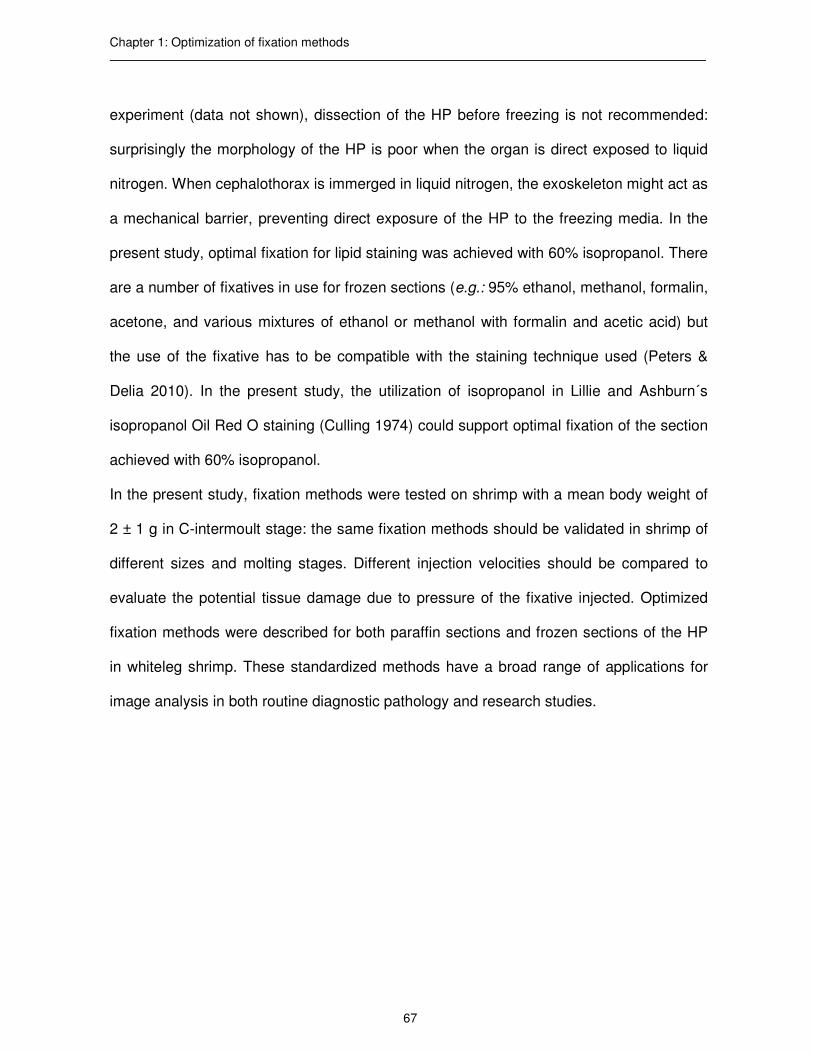
Chapter 1: Optimization of fixation methods
67
experiment (data not shown), dissection of the HP before freezing is not recommended:
surprisingly the morphology of the HP is poor when the organ is direct exposed to liquid
nitrogen. When cephalothorax is immerged in liquid nitrogen, the exoskeleton might act as
a mechanical barrier, preventing direct exposure of the HP to the freezing media. In the
present study, optimal fixation for lipid staining was achieved with 60% isopropanol. There
are a number of fixatives in use for frozen sections (e.g.: 95% ethanol, methanol, formalin,
acetone, and various mixtures of ethanol or methanol with formalin and acetic acid) but
the use of the fixative has to be compatible with the staining technique used (Peters &
Delia 2010). In the present study, the utilization of isopropanol in Lillie and Ashburn´s
isopropanol Oil Red O staining (Culling 1974) could support optimal fixation of the section
achieved with 60% isopropanol.
In the present study, fixation methods were tested on shrimp with a mean body weight of
2 ± 1 g in C-intermoult stage: the same fixation methods should be validated in shrimp of
different sizes and molting stages. Different injection velocities should be compared to
evaluate the potential tissue damage due to pressure of the fixative injected. Optimized
fixation methods were described for both paraffin sections and frozen sections of the HP
in whiteleg shrimp. These standardized methods have a broad range of applications for
image analysis in both routine diagnostic pathology and research studies.

Chapter 1: Optimization of fixation methods
68
References
Anderson G. & Bancroft J. (2002) Tissue processing and microtomy. In: Theory and practice of histological techniques (ed. by J. D. Bancroft & G. M. Gamble), pp. 85-99. Elsevier Health Publications, Edinburgh.
Asaoka Y., Togashi Y., Mutsuga M., Imura N. & Miyoshi T. & Miyamoto Y. (2016) Histological image analysis of chemical-induced hepatocellular hypertrophy in mice. Experimental and Toxicologic Pathology 68, 233-239. Bell T.A. & Lightner D.V. (1988) A handbook of normal penaeid shrimp histology, pp. 2-6. The World Aquaculture Society, Baton Rouge. Bondad-Reantaso M.G., McGladdery S.E., East I. & Subasinghe R.P. (2001) Asia Diagnostic Guide to Aquatic Animal Diseases. In: FAO Fisheries Technical Paper No. 402, Supplement 2, pp. 154-191. FAO and NACA, Rome.
Bondad-Reantaso M.G., Subasinghe R.P., Josupeit H., Cai J. & Zhou X. (2012) The role of crustacean fisheries and aquaculture in global food security: Past, present and future. Journal of Invertebrate Pathology 110, 158-165. Bucke D. (1972) Some histological techniques applicable to fish tissues. In: Diseases of Fish (ed. by L.E. Mawdesley-Thomas), pp. 153-189. Symposia of The Zoological Society of London, number 30. Academic Press, New York. Calvo N.S., Stumpf L., Pietrokovsky S. & López Greco L.S. (2011) Early and late effects of feed restriction on survival, growth and hepatopancreas structure in juveniles of the red claw crayfish Cherax quadricarinatus. Aquaculture 319, 355-362. CEFAS (2014) Sampling of crustaceans for infectious diseases. In: Standard Operating Procedure SOP 2014, Revision number 1, pp. 10. Center for Environment Fisheries and Aquaculture Science, Weymouth. Corteel M., Dantas-Lima J.J., Wille M., Alday-Sanz V., Pensaert M.B., Sorgeloos P. & Nauwynck H.J. (2009) Molt stage and cuticle damage influence white spot syndrome virus immersion infection in penaeid shrimp. Veterinary Microbiology 137, 209-216. Culling C.F.A. (1974) Identification of Lipids. In: Handbook of Histopathological and Histochemical Techniques, pp. 360-361. Butterworths & Co. Ltd, London. Frederick P.M. & Busing W.M. (1981) Ice crystal damage in frozen thin sections: freezing effects and their restoration. Journal of Microscopy 121 (2), 191-199. Gibson-Corley K.N., Olivier A.K. & Meyerholz D.K. (2013) Principles for Valid Histopathologic Scoring in Research. Veterinary Pathology 50, 1007-1015.

Chapter 1: Optimization of fixation methods
69
Gurcan M.N., Boucheron L., Can A., Madabhushi A., Rajpoot N. & Yener B. (2009) Histopathological image analysis: a review. IEEE reviews in biomedical engineering 2, 147-171. He L., Long L.R., Antani S. & Thoma G.R. (2012) Histology image analysis for carcinoma detection and grading. Computer Methods and Programs in Biomedicine 107, 538-556. Hopwood D. (2002) Fixation and fixatives. In: Theory and practice of histological techniques (ed. by J. D. Bancroft & GM. Gamble), pp. 67-68. Elsevier Health Publications, Edinburgh.
Johnson S.K. (1995) Handbook of Shrimp Diseases, pp. 10. Texas A&M University, Texas. Kårsnäs A. (2014) Image analysis methods and tools for digital histopathology applications relevant to breast cancer diagnosis. Digital Comprehensive Summaries of Uppsala Dissertations from the Faculty of Science and Technology 1128, pp. 129. Uppsala. Laohabanjong R., Tantikitti C., Benjakul S., Supamattaya K. & Boonyaratpalin M. (2009) Lipid oxidation in fish meal stored under different conditions on growth, feed efficiency and hepatopancreatic cells of black tiger shrimp (Penaeus monodon). Aquaculture 286, 283-289. Lightner D.V., Hasson K.W., White B.L. & Redman R.M. (1996) Chronic toxicity and histopathological studies with Benlate®, a commercial grade of benomyl, in Penaeus vannamei (Crustacea: Decapoda). Aquatic Toxicology 34, 2: 105-118. Lightner D.V. & Redman R.M. (1998) Shrimp diseases and current diagnostic methods. Aquaculture 164, 201-220. Lightner D.V. (2015) Diseases of Crustaceans. In: Manual of Diagnostic Tests for Aquatic Animals (ed. by OIE), Section 2.2.0, pp. 1-10. World Organization of Animal Health, Paris. Peters, S.R. & Delia C. S. (2010) Fixation, staining and coverslipping of frozen section slides. In: A Practical Guide to Frozen Section Technique (ed. by S. R. Peter), pp. 117-129. Springer, New York. Säfholm M., Jansson E., Fick J. & Berg C. (2016) Molecular and histological endpoints for developmental reproductive toxicity in Xenopus tropicalis: Levonorgestrel perturbs anti-Müllerian hormone and progesterone receptor expression. Comparative Biochemistry and Physiology, Part C 181-182, 9-18. Steu S., Baucamp M., Von Dach G., Bawohl M., Dettwiler S., Storz M., Moch H. & Schraml P. (2008) A procedure for tissue freezing and processing applicable to both intra-

Chapter 1: Optimization of fixation methods
70
operative frozen section diagnosis and tissue banking in surgical pathology. Virchows Archiv 452, 305-312. Tracy R.E. & Walia P. (2002) A method to fix lipids for staining fat embolism in paraffin sections. Histopathology 41, 75-79. Van Goor H., Gerrits P.O. & Grond J. (1986) The application of lipid-soluble stains in plastic-embedded sections. Histochemistry 85, 251 253. Wilson I. & Gamble M. (2002) The Hematoxylins and Eosin. In: Theory and practice of histological techniques (ed. by J. D. Bancroft & GM. Gamble), pp. 125-138. Elsevier Health Publications, Edinburgh.
Xu X. & Xu P.X. (2001) A modified cryosection method for mouse testis tissue. Tissue and Cell 33, 208-210.

71
Chapter 2
Use of computer assisted image analysis for semi-quantitative histology of the
hepatopancreas in whiteleg shrimp, Penaeus vannamei (Boone)
Fabio Cervellione1, Charles McGurk1, Tommy Berger Eriksen1, Wim Van den Broeck2
1Skretting Aquaculture Research Centre, Stavanger, Norway
2Department of Morphology, Ghent University, Ghent, Belgium
Journal of Fish Disease 2016, DOI:10.1111/jfd.12599

Chapter 2: Development of computer-assisted image analysis
72
Abstract
Despite the increasing use of novel molecular techniques in pathology; histology remains
the standard method for monitoring tissue alterations and for assessing pathology.
Histopathological evaluation is generally laborious and subjective with risk of
discrepancies in semi-quantitative scoring between pathologists. In contrast, computer
assisted image analysis (CAIA) is potentially faster, more objective and thus suitable for
routine screening. Limited research has been done on CAIA in crustacean histopathology
and the methods described were not fully automated. Therefore, the aim of the present
study was to develop CAIA in whiteleg shrimp (Penaeus vannamei) for the study of the
hepatopancreas. Paraffin sections were immunohistochemically stained with monoclonal
antibodies WSH8 against haemocytes and counterstained with Mayer´s haematoxylin for
detection of haemocytes and B-cell vacuoles, and modified Toluidine Blue protocol was
used for detection of F-cells; frozen sections were stained with Oil Red O for detection of
lipid droplets within R-cells. Visiopharm® software was used to develop and validated
protocols for the quantification of morphological parameters (areas of haemocyte
infiltration, F-cells, B-cell vacuoles, lipid droplets and their ratios to total tissue area and
total lumen area). These protocols enable the future use of CAIA for determining the
degree of severity of morphological changes reflecting a pathological or nutritional
alteration of the organ.
Key words: computer assisted image analysis, semi-quantitative histology, hepatopancreas, whiteleg shrimp, Penaeus vannamei.

Chapter 2: Development of computer-assisted image analysis
73
Introduction
In crustaceans, the hepatopancreas (HP) is a large bilobed organ, located in the
cephalothorax, composed of many blindly ending tubules, which wrap over the dorsal
and lateral sides of the posterior part of the stomach and the anterior part of the midgut
trunk (Franceschini-Vicentini et al. 2009; Alday-Sanz 2010). The primary role of the HP is
the synthesis and secretion of digestive enzymes, final digestion, uptake of nutrients and
detoxification (Laohabanjong et al. 2009). In decapods, two separate lobes are enclosed
together in a connective tissue capsule and are separated by a thin septum. The lobes
are connected separately to the intestine through the corresponding principal duct
(Esteve & Herrera 2000; Vasagam et al. 2007). Hepatopancreatic tubules diverge
dendritically and each tubule can be subdivided into distal, medial and proximal zones
relative to the distance from the midgut (Nakamura 1987; Felgenhauer 1992;
Franceschini-Vicentini et al. 2009). From the distal to the proximal zone of the tubules the
epithelium becomes smaller and the lumen larger (Cuartas et al. 2002). Tubules are lined
up by an epithelium which, apart from the distal closed end, is only a single layer thick
(Gibson & Barker 1979). The simple columnar epithelium is made up of four basic cell
types: E-, F-, R- and B-cells (Nakamura 1987; Franceschini-Vicentini et al. 2009). Normal
structure of the HP is shown in Figure 1.

Chapter 2: Development of computer-assisted image analysis
74
Figure 1. Normal structure of the medial zone of the hepatopancreas tubule. Cross section. lu = lumen. Adapted from Johnson (1995).
The E-cells, or embryonic cells, are small cuboidal cells entirely confined to the distal
blind ends of the tubules and give rise to the other three cell types of the organ. F-cells,
or fibrillar cells, are cylindrical/prismatic cells located at the medial and proximal zones of
the tubules among R- and B-cells, and are characterized by a strong basophilia due to a
well-developed rough endoplasmatic reticulum, giving them a fibrillar appearance. This
cell type has a high rate of protein synthesis and synthetizes enzyme for extracellular
digestion (Barker & Gibson 1977; Felgenhauer 1992; Sousa et al. 2005; Franceschini-
Vicentini et al. 2009). B-cells, or blister cells, are large primary secretory cells more
frequent at the proximal zone of the tubules and are defined by the presence of a single
large membrane-bound vesicle surrounded by a dense cytoplasm. These cells produce
digestive enzymes in the HP and are responsible for intracellular digestion, concentrating

Chapter 2: Development of computer-assisted image analysis
75
the absorbed materials in the large vacuole and secreting the vacuolar content into the
tubular lumen at the end of the digestive process for reabsorption by R-cells (Barker &
Gibson 1977; Gibson & Barker 1979; Felgenhauer 1992; Franceschini-Vicentini et al.
2009). The R-cells, or reabsorption cells, are the most numerous cell types in the HP.
These tall, columnar cylindrical cells are located, as F-cells, at medial and proximal
zones and characterized by a large numbers of irregularly-shaped storage vesicles
(primarily lipids) in their cytoplasm. These cells function in food absorption, storing lipid
droplets, glycogen and mineral deposits (Felgenhauer 1992; Sousa et al. 2005;
Franceschini-Vicentini et al. 2009).
Although new molecular methods in pathology are developing fast, histology remains the
standard method for pathology investigations and disease diagnosis (Silva et al. 2015). In
current practice, histological analysis is based most commonly on qualitative features as
interpreted by pathologists (Miedema et al. 2012). Pathologists’ quantification is in
general time-consuming, poorly objective with significant discrepancies in scoring results
reported between pathologists (He et al. 2012; Higgins 2015). This has motivated the
development of computer-assisted image analysis (CAIA) methods for producing
unbiased, reproducible, and reliable data (Nativ et al. 2014; Silva et al. 2015). This
method could assist but not replace experienced pathologists as additional morphological
features need to be considered for clinical appraisal (He et al. 2012; Miedema et al.
2012; Nativ et al. 2014). This methodology requires optimization of histological
procedures for preparation of samples. In a previous study, our research group proposed
a standardized protocol for paraffin and frozen sections of the HP in whiteleg shrimp
(Penaeus vannamei) for image analysis (Cervellione et al. 2016). A limited amount of

Chapter 2: Development of computer-assisted image analysis
76
research has been done on the application of CAIA in crustacean histopathology and to
our best knowledge, no studies have been conducted in whiteleg shrimp (Odendaal &
Reinecke 2003; Odendaal & Reinecke 2007; Berillis et al. 2013). Therefore, the aim of
the present study was to develop CAIA in whiteleg shrimp for the study of the HP
structure and future determination of the nutritional and pathological status of this organ.
Materials and Methods
Shrimp and rearing conditions
Experiments were conducted in collaboration with IMAQUA, located at the Faculty of
Veterinary Medicine (Ghent, Belgium). Specific pathogen-free whiteleg shrimp were
imported from Shrimp Improvement Systems (Florida, USA). Shrimp were housed in 50 L
glass tanks, each supplied with constant aeration and independent biological/mechanical
filters. Water temperature was kept at 27 ± 1 °C, with a pH of 7.8 ─ 8.1 and salinity of 20
± 1 g L-1. The biological filters and regular water exchanges kept the total ammonium
concentrations below 0.5 mg L-1 and the total nitrites below 0.15 mg L-1. The room was
illuminated 12 h d-1 by a dimmed luminescent light-tube. Shrimp were fed a commercial
diet for 30 d, at 5% of the body weight (bw), four times per day. Thereafter, shrimp of 2 ±
1 g, within the C-intermoult stage (Corteel et al. 2009) were collected and transported to
the Department of Morphology (Ghent University, Belgium) in aerated buckets. Shrimp
were then euthanized as described by Lightner (2015) for paraffin sections and
euthanized in cold ice for frozen sections.

Chapter 2: Development of computer-assisted image analysis
77
Preparation of paraffin and frozen sections
Ten shrimp were individually weighed, fixed with Davidson´s solution (30 mL 95%
ethanol, 20 mL 37% formaldehyde, 10 mL glacial acetic acid, and 30 mL distilled water),
as described by Cervellione et al. (2016). After injection and lateral slitting of the cuticle
from the sixth abdominal segment to the base of the rostrum, shrimp were immersed in
Davidson´s fixative (ratio of fixative to tissue volume of 10:1) for 48 h at 22 °C. The
shrimp were then bisected transversally at the junction of the cephalothorax and
abdomen, the cephalothorax bisected longitudinally on the mid-line, and both parts
placed in tissue processing cassettes. Tissues were dehydrated in a graded series of
alcohol baths (70%, 80%, 95%, and 100%) and cleared in xylene, using an automated
tissue processor (Microm STP420D, ThermoScientific™). Then, the tissues were
embedded in paraffin (melting point 54 – 56 °C, Paraclean Klinipath BV, VWR
International®) using manual embedding station (Microm EC350, ThermoScientific™).
After further optimization of section thickness, five µm thick longitudinal sections were
cut with an automated microtome (Microm HM 360, ThermoScientific™), placed on APES
(3-Aminopropyltriethoxysilane) coated glass slides, dried for 1 h at 56 °C and kept at 37
°C overnight.
For frozen sections, ten shrimp were processed as described by Cervellione et al (2016).
Shrimp were bisected transversally at the junction of the cephalothorax and the
abdomen, and immediately anterior to the HP. Cephalothorax was frozen by immersion
for 20 s in liquid nitrogen and storing in dry air. After freezing, all samples were kept at -
80 °C. Samples were embedded in water-soluble media (Tissue-Tek® OCT, VWR
International®) and 16 µm thick longitudinal sections cut, by use of an automated

Chapter 2: Development of computer-assisted image analysis
78
cryostat (Microm HM505E, ThermoScientific™) at -25 °C and placed on glass slides
(HistoBond®+, Marienfeld). Then frozen sections were fixed for 10 s in 60% isopropanol
at 22 °C.
Selection of morphometric parameters for CAIA
In crustaceans, the HP is routinely examined by pathologists and assessed as a general
indicator of health (Johnson 1995; Laohabanjong et al. 2009; Calvo et al. 2011).
Morphological alterations of the HP, such as reduction of the tubular area and
enlargment of the lumen area, variation in the number of B-cells, F-cells and R-cells,
infiltration of haemocytes in the intertubular spaces, have been described by different
authors in case of pathogens, food deprivation, changes in salinity and food ingredients
(Nakamura 1987; Sánchez-Paz et al. 2007; Tam & Avendant-Oldewage 2009; Diaz et al.
2010; Masson et al. 2012; Sacristán et al. 2016). Haemocyte infiltration has been
reported as initial response of the HP to tissue damage (traumatic or due to infection),
toxicity and environmental stress (Johnson 1980; McGallery 2011; Chiodi Boudet et al.
2015; Longo & Diaz 2015). F-cells have a high rate of protein synthesis, producing
enzymes for extracellular digestion (Barker & Gibson 1977). The quantification of F-cells
(enzymatic activity of the HP) could help to understand how the HP copes with different
diets, feeding regimes and starvation periods. Morphometric parameters were selected
for the development of protocols (APPs) and quantification of the main morphological
changes occurring in the HP (Table 1). Total Tissue Area (TTA) represented the total
area covered by tubular epithelium, composed by B-, F- and R-cells, and Total Lumen
Area (TLA) represented the area covered by tubular lumen. The ratio TLA:TTA indicated
the variation of tubular lumen. Haemocyte Infiltration Area (HIA) indicated the area of

Chapter 2: Development of computer-assisted image analysis
79
haemocytes infiltrating the intertubular spaces of the HP, and ratio HIA:TTA indicated the
response of the HP to noxa. Vacuoles B-cells Area (VBA) represented the area of the
tubule composed by vacuole of the B-cells, and ratio VBA:TTA measured the intracellular
digestion taking place in the HP. F-Cell Area (FCA) represented the area of the tubule
composed by F-cells, and ratio FCA:TTA indicated the enzymatic activity of the HP.
Finally, Lipid Droplet Area (LDA) indicated the area of the tubule composed by lipid
droplets within R-cells, and ratio LDA:TTA measured the lipid reserves in the HP. All
ratios were indicated as % of TTA.

Chapter 2: Development of computer-assisted image analysis
80
Table 1. Selection of morphometric parameters and their ratios used for the development of computer assisted image analysis of the hepatopancreas in whiteleg shrimp (Penaeus vannamei).
Morphometric parameter Description
Total Tissue Area (TTA)
Total area covered by tubular epithelium, composed by B-
cells, F-cells and R-cells
Total Lumen Area (TLA)
Total area covered by lumen
TLA:TTA
Ratio of Total Lumen Area to Total Tissue Area, as measure of
the variation of the tubular lumen, expressed as %
Haemocyte Infiltration Area (HIA)
Area of the haemocytes infiltrating the intertubular spaces of
the HP
HIA:TTA
Ratio of Haemocyte Infiltration Area to Total Tissue A, as
measure of the HP response to noxa, expressed as %
Vacuoles of B-cells Area (VBA)
Area of the tubule composed by vacuoles of B-cells
VBA:TTA
Ratio of Vacuoles of B-cells Area to Total Tissue Area, as
measure of intra-cellular digestion of the HP, expressed as %
F-Cells Area (FCA)
Area of the tubule composed by F-cells
FCA:TTA
Ratio of F-Cells Area to Total Tissue Area, as measure of the
enzymatic activity of the HP, expressed as %
Lipid Droplets Area (LDA)
Area of the tubule composed by lipid droplets within R-cells
LDA:TTA
Ratio of Lipid Droplets Area to Total Tissue Area, as measure
of the lipid reserves in the HP, expressed as %
Note: HP = hepatopancreas.

Chapter 2: Development of computer-assisted image analysis
81
Stainings
Stainings used for the development of APPs for CAIA are reported in Table 2. Detection
of haemocytes in the intertubular spaces was based on immunohistochemistry (IHC).
WSH8 monoclonal antibodies (Mabs) were produced by the Wageningen Institute of
Animal Science (Wageningen University, The Netherlands) immunizing mice with
membrane lysates of tiger shrimp (Penaeus monodon, Fabricus 1798) haemocytes (Van
de Braak et al. 2000). A previous experiment (data not shown) proved the specificity of
WSH8 for whiteleg shrimp haemocytes. After deparaffination in xylene and rehydration in
ethanol series, paraffin sections (one section per shrimp) were subjected to heat-induced
epitope retrieval using citrate solution in microwave for 150 s at 750 W followed by 10
min at 160 W and stored for 30 min at 4 °C. Then sections were incubated for 5 min at 22
°C with Dako REAL™ Peroxidase-Blocking Solution followed by incubation with 30%
rabbit serum blocking solution for 30 min at 22 °C. After incubation of the slides for 1h at
22 °C with the Mabs dilution (100x), peroxidase-conjugated reaction was carried out
using Dako REAL™ EnVision™ Labelled Polymer HRP anti-mouse for 30 min at 22 °C,
followed by DAB chromogen solution for 5 min at 22 °C. Sections were then
counterstained with Mayer´s haematoxylin for 5 min and mounted in non-aqueous
medium (DPX, Sigma-Aldrich®). Mayer´s haematoxylin was used on the same section
for counterstaining IHC and identification of B-cell vacuoles. Detection of F-cells was
based on modified Toluidine Blue protocol (Gurr 1962). Paraffin sections (one section per
shrimp) were stained for 3 min in Toluidine Blue followed by 1 min in ThermoScientific™
Carifier 1 through an automated slide-stainer (Gemini AS, ThermoScientific™) and
mounted in non-aqueous medium (DPX, Sigma-Aldrich®). Detection of lipid droplets

Chapter 2: Development of computer-assisted image analysis
82
within the R-cells was based on Oil Red O staining. Therefore, frozen sections (one
section per shrimp) were stained with Lillie and Ashburn´s isopropanol Oil Red O staining
(ORO) (Culling 1974) using an automated slide-stainer (Gemini AS, ThermoScientific™)
and mounted in aqueous media (Aquatex®, VWR International®). All sections were label-
coded and one experienced examiner observed slides under light microscope to evaluate
the suitability of the sections for image analysis (BX53, Olympus©).
Table 2. List of stainings used for the development of computer assisted image analysis of the hepatopancreas in whiteleg shrimp (Penaeus vannamei).
Staining APP
IHC with Mabs WSH8 counterstained with Mayer´s
haematoxylin
HIVB (Haemocyte Infiltration and Vacuole of
B-cells)
modified Toluidine Blue protocol F (F-cells)
Oil Red O LD (Lipid Droplets within R-cells)
Note: Each APP was developed analyzing region of interests (ROI) of 13372 µm2 for HIVB and F, and 1650 µm2 for LD. APP = protocol for computer assisted image analysis; IHC = immunohistochemistry; Mabs = monoclonal antibodies.

Chapter 2: Development of computer-assisted image analysis
83
Digital image acquisition
Slides were scanned using automated slide-scanner (Leica SCN400, Leica
Microsystems©, Germany) producing digitized images (scn file format) in 20x
magnification.
Image processing
The software Visiopharm (version VIS 6.0.0.1765 with Author™ module, Visiopharm®,
Denmark) was used for image analysis. Author™ module allows developing customized
APPs. For each section a region of interest (ROI; 13372 µm2 for APP HIVB and F and
1650 µm2 for APP F) was selected for image processing. An example of a representative
ROI is shown in Figure 2. ROI was selected avoiding the boarders of the HP (where
distal ends of the tubules are present) and selecting the medial part of the cross section
of the tubule.
Figure 2. Region of interest (ROI)
selected as representative area a
cross section of the hepatopancreas
(HP) of whiteleg shrimp (Penaeus
vannamei) for the development of
computer assisted image analysis
protocol (APP).

Chapter 2: Development of computer-assisted image analysis
84
The workflow for each APP was divided in four phases: 1) pre-processing, 2) image
classification, 3) post-processing, and 4) calculations.
1) Pre-processing
The purpose of pre-processing is to enhance phenomena that can improve the
subsequent image segmentation and/or to remove phenomena that will disturb the
subsequent image segmentation. A combination of two color bands was used for each
APP. Main filters used were Mean and Median (filter size 5 x 5).
2) Classification
During classification the image is segmented into multiple segments (sets of pixels)
sharing common features in the selected color bands. APPs were developed using
Linear Bayesian classification method and trained on representative sets of images.
3) Post-processing
In post-processing the image segmentation can be further refined based on
morphological and contextual information. Main steps used were Change, Erode, Dilate,
Open, Close, Fill holes and Separate objects.
4) Calculations
After post-processing, quantitative data of the morphological parameters were generated
by the software.

Chapter 2: Development of computer-assisted image analysis
85
APPs validation
Quantitative data generated using the proposed APPs (automated method) were
compared to data generated with a semi-automated method. For semi-automated
method, two trained observers (Ob1 and Ob2) used the same software (Visiopharm®,
Denmark) to extract the same morphological parameters manually (without the
developed APPs). Each operator manually drew labels of the different tissue types
(tubule, lumen, F-cells, vacuoles of B-cells and lipid droplets within R-cells, and
haemocytes) within the selected ROI. Then, calculations and ratios were automatically
extracted by the software. For each APP, operators evaluated ten sections (one ROI per
section) to measure time efficiency, accuracy, and reproducibility (inter- and intra-
observer variability) (Silva et al. 2015). For efficiency, total computational time for data
generation with automated and semi-automated methods were compared. To show the
robustness of the proposed APPs, inter- and intra-observer variability between
automated and semi-automated methods were compared. For intra-observer variability,
operators evaluated the same sections in three consecutive day. For intra-observer
variability, data generated on the same sections by the two observers were compared.
Statistical analysis
Data were tested and treated as parametric data. For accuracy and reproducibility,
Pearson's correlation coefficient r (95% confidence interval) was used to measure the
strength of association between automated and semi-automated methods and intra- and
inter-observer variability (Galarraga et al. 2012; Mukaka 2012; Shi et al. 2012).

Chapter 2: Development of computer-assisted image analysis
86
Results
In the present study, three APPs (HIVB, LD, and F) were developed to identify
haemocytes and B-cell vacuoles, lipid droplets within R-cells, and F-cells. Morphological
parameters assessed by each APP are reported in Table 3.
Table 3. Protocols (APPs) and morphological parameters for the development of computer assisted image analysis of the hepatopancreas in whiteleg shrimp (Penaeus vannamei).
APP Morphological parameters assessed
HIVB (Haemocyte Infiltration and Vacuoles of B-
cells)
HIA, VBA, TLA, and their ratios to TTA
F (F-cells) FCA, TLA, and their ratios to TTA
LD (Lipid Droplets within R-cells) LDA, TLA, , and their ratios to TTA
Note: FCA = F-cell area; LDA = total lipid area; HIA = haemocyte infiltration area; TLA = total lumen area; TTA = total tissue area; VBA = vacuole of B-cell area.
Figure 3 shows main steps for the development of HIVB. Haemocytes were stained with
IHC using WHS8 and no background staining was observed (a). In the post-processing
phase (d) 17 steps allowed identifying lumen (grey), tissue area (pink), B-cells vacuoles
(light blue) and haemocytes (dark blue). Development of LD is shown in Figure 4. In the
post-processing phase (d) 26 steps allowed identifying lumen (white), tissue area (blue),
and lipid droplets (red). The workflow for F is reported in Figure 5. In the post-processing
phase (d) 30 steps allowed identifying F-cells (dark blue), lumen (pink) and tissue area
(light blue).

Chapter 2: Development of computer-assisted image analysis
87
Figure 3. Development of computer assisted image analysis protocol (APP) for the detection of haemocytes and B-cell vacuoles in the hepatopancreas (HP) of whiteleg shrimp (Penaeus vannamei). Paraffin section (5 µm) of the HP stained with immunohistochemistry using monoclonal antibodies WSH8 counterstained with Mayer´s haematoxylin (a). APP workflow (b,c,d): pre-processing (b), classification (c), post-processing (d). In d: total tissue area (pink), lumen area (grey), B-cell vacuoles (light blue), haemocytes (blue). lu = lumen; tb = hepatopancreatic tubule; vB= B-cell vacuole; black arrows = haemocytes. Scale bar = 100 µm.

Chapter 2: Development of computer-assisted image analysis
88
Figure 4. Development of computer assisted image analysis protocol (APP) for the detection of lipid droplets within R-cells in the hepatopancreas (HP) of whiteleg shrimp (Penaeus vannamei). Frozen section (16 µm) of the HP stained with Oil Red O (a). APP workflow (b,c,d): pre-processing (b), classification (c), post-processing (d). In d: total tissue area (blue), lumen area (white), lipid droplets (red). lu = lumen; tb = hepatopancreatic tubule. Scale bar = 150 µm.

Chapter 2: Development of computer-assisted image analysis
89
Figure 5 Development of computer assisted image analysis protocol (APP) for the detection of F-cells in the hepatopancreas (HP) of whiteleg shrimp (Penaeus vannamei). Paraffin section (5 µm) of the HP stained with modified Toluidine blue protocol (a). APP workflow (b,c,d): pre-processing (b), classification (c), post-processing (d). In d: total tissue area (light blue), lumen area (pink), F-cells (blue). lu = lumen; tb = hepatopancreatic tubule; arrows = F-cells. Scale bar = 85 µm.

Chapter 2: Development of computer-assisted image analysis
90
APPs validation and statistical analysis
Transparency slider in the label drawing dialog was used to examine the results during
each phase of image processing. Data for time efficiency are summarized in Table 4.
Automated method was about 200 to 700 times faster than semi-automated method. In
particular, APPS generated results in less than 1 s. Ob2 resulted to be always faster than
Ob1. Accuracy is reported in Table 5. Pearson‘correlation coefficient r (95% confidence
interval) ranged from 0.91 to 0.99, depending on the morphological parameter assessed.
Finally, inter and intra observer variability is reported in Table 6. Pearson‘correlation
coefficient r for intra-observer variability ranged from 0.98 to 0.99, while automated
method gave always 1. Coefficient for inter-observer variability was 0.96 for the semi-
automated method and 1 for the automated method.
Table 4. Comparison of time efficiency between automated and semi-automated methods for computer assisted image analysis of the hepatopancreas in whiteleg shrimp (Penaeus vannamei).
Morphological parameters assessed Total average processing time
HIVB (HIA, VBA, TTA, TLA)
Automated
Semi-automated
0´0.5´´± 00´´ (always)
5´53´´± 43´´(Ob1) 5´06´´± 53´´ (Ob2)
F (FCA, TTA, TLA)
Automated
Semi-automated
0´0.5´´± 00´´ (always)
2´05´´± 22´´ (Ob1) 1´53´´± 35´´ (Ob2)
LD (LDA, TTA, TLA)
Automated
Semi-automated
0´0.5´´± 00´´ (always)
3´12´´± 47´´(Ob1) 2´42´´± 29´´ (Ob2)
Note: for each protocol (HIVB, F, and LD), ten histological sections (one ROI per section) were analyzed by two operators (Ob1, Ob2). FCA = F-cell area; LDA = total lipid area; HIA = haemocyte infiltration area; ROI = region of interest; TTA = total tissue area; TLA = total lumen area; VBA = vacuole of B-cell area.

Chapter 2: Development of computer-assisted image analysis
91
Table 5. Comparison of accuracy between automated and semi-automated methods for computer assisted image analysis of the hepatopancreas in whiteleg shrimp (Penaeus vannamei).
Morphological parameter evaluated Pearson´s coefficient (r)
Protocol HIVB
HIA
VBA
TTA
TLA
0.92
0.91
0.95
0.95
Protocol F
FCA
TTA
TLA
0.95
0.94
0.95
Protocol LD
LDA
TTA
TLA
0.93
0.91
0.99
Note: for each protocol (HIVB, F, and LD), ten histological sections (one ROI per section) were analyzed by two operators (Ob1, Ob2). Pearson´correlation coefficient (r) is calculated with 95% confidence interval. FCA = F-cell area; LDA = total lipid area; HIA = haemocyte infiltration area; ROI = region of interest; TTA = total tissue area; TLA = total lumen area; VBA = vacuole of B-cell area.
Table 6. Reproducibility of automated and semi-automated methods for computer assisted image analysis of the hepatopancreas in whiteleg shrimp (Penaeus vannamei).
Method Intra-observer variability Inter observer variability
Automated
Semi-automated
r = 1 (always)
r = 0.99 (Ob1) r = 0.98 (Ob2)
r = 1 (always)
r = 0.96
Note: ten histological sections (one ROI per section) were analyzed by two operators (Ob1, Ob2). For inter-observer variability, sections were analyzed three times in three consecutive days. Pearson´correlation coefficient (r) is calculated with 95% confidence interval. ROI = region of interest.

Chapter 2: Development of computer-assisted image analysis
92
Discussion
The goal of CAIA in histopathology is to increase the amount and quality of data derived
from a specimen, providing quantitative measurements to assist the pathologist (Fuchs &
Buhmann 2011; He et al. 2012; Miedema et al. 2012; Nativ et al. 2014). The basic
principle of automated image analysis for histology is the use of a series of mathematical
algorithms to process images, enabling the segmentation of picture elements into regions
of interest based on their color, texture, and/or context (Webster & Dunstan 2014).
Although many image analysis software systems utilize similar image segmentation and
image-processing concepts to their algorithms, a key differentiating factor of commercial
software packages is the amount of supervision required, or allowed, by the user. It is
possible to classify software in two categories: unsupervised and supervised.
Unsupervised packages do not need extensive training of operators and rely on
algorithms built into the software. Supervised packages, such as Visiopharm®, instead
require upfront training in image analysis, but allow users to program specific algorithms
and to develop unique analyses (Webster & Dunstan 2014). Berillis et al. (2013) have
described an image processing method for the HP to determine the nutritional condition
in lobsters: however the method was not fully automatized and required manual marking
of tubules by drawing the contours. Moreover, the algorithm measured R-cells total area
without quantifying lipid droplets. The present study describes a fully automated CAIA
method for the study of the HP in whiteleg shrimp. Three APPs were developed and
validated for detection and quantification of morphological parameters.
Morphometric parameters, such as HIA and ratio HIA:TTA, could be used in future
studies for monitoring the health condition of the HP and improve the understanding of

Chapter 2: Development of computer-assisted image analysis
93
the pathophysiological mechanisms of this organ. In the future, morphological
parameters as FCA, VCA, LDA, TLA and their ratios to the TTA could support the study
of the HP physiology comparing the effect of different diets and feeding regimes. The
TISSUEalign™ module in Visiopharm® software allows automated high precision
alignment of an unlimited amount of serial sections and it could be used to extract
morphologic parameters from serial sections stained with different dyes. For automated
analysis systems, each source of variability presents a significant obstacle (McCann et
al. 2015). There are three main sources of variability in a histology-based diagnosis:
biological, inter- and intra-observer, and technical variability. Fixation, staining and
scanning are sources of technical variability (Kothari et al. 2013; McCann et al. 2015).
For clinical application of CAIA, it is essential to minimize these sources of variability and
validate APPs on multiple datasets (Kothari et al. 2013). In the present study, shrimp of
the same intermoult stage were fixed with a standardized method developed for image
analysis (Cervellione et al. 2016). Automated stainer and automated scanner were used
to minimize technical variability. The APPs validation was done by analyzing data
generated by two observers on ten sections (one ROI per section) using automated and
semi-automated methods. Three parameters were assessed: efficiency, accuracy and
reproducibility. The automated method was about 200 to 700 faster than the semi-
automated method. Efficiency in time allows the proposed method to analyze big
datasets in a short time.
The accuracy of image analysis methods greatly affects the robustness of downstream
analysis (Kothari et al. 2013). Accuracy values for the proposed methods ranged from
0.91 to 0.99 and are comparable with those reported in literature (Sieren 2010;

Chapter 2: Development of computer-assisted image analysis
94
Galarraga et al. 2012; Pajor et al. 2012; Shi et al. 2012; Wang et al. 2016). Correlation
value for intra- and inter-observer variability in the automated method was always higher
than in the semi-automated method. The proposed method is fully automated and
reduces the intra- and inter-observer variability to zero, increasing workflow efficiency
and reproducibility (Bueno et al. 2016). The exact parameters and assumptions can be
used across every sample with no-inter-observer bias, which can significantly impact the
reproducibility of data analysis (Inman et al. 2005; Diller & Kellar 2015). In the present
study, CAIA was developed using shrimp with a mean body weight of 2 ± 1 g in C-
intermoult stage: the same methodology should be validated in shrimp of different sizes
and moulting stages. Development of immunohistochemistry methods and test of other
stainings for targeting specific structures in the HP should also be investigated.
Development of immunohistochemistry, in situ hybridization methods and test of other
stainings for targeting specific structures and pathogens should be also investigated.
To our best knowledge, the present study describes for the first time the use of CAIA in
whiteleg shrimp. CAIA allows detection of initial histopathological changes useful for early
diagnosis of diseases: for example, infiltration of haemocytes associated with lack of
differentiation in the tubule epithelial cells have been described in the early stages of
acute hepatopancreatic necrosis disease (AHPND). CAIA could also be used for
detecting emerging diseases, as hepatopancreatic microsporidiodis caused by
Enterocytozoon hepatopenaei (EHP), in combination with in situ hybridization method
(Thitamadee et al. 2016). Furthermore, CAIA could be helpful in identifying
histopathological changes of the HP in case of starvation, as reduction of the epithelial
thickness, loss of the typical star-shaped lumen, reduction of B-cells and lipid droplets

Chapter 2: Development of computer-assisted image analysis
95
within R-cells, observed during disease outbreaks and adverse environmental conditions
(Vogt et al. 1985; Nakamura 1987; Lavilla-Pitogo et al. 1998; Ong & Johnston 2006;
Leser et al. 2008; Calvo et al. 2011; Sacristán et al. 2016). In the future, the application
of CAIA on routine health screening of shrimp farms might support the shrimp industry
anticipating diagnosis, facilitating therapeutical and management solutions.
References Alday-Sanz, V. (2010) The shrimp book, Nottingham University Press, Loughborough. Arevalo, J., Cruz-Roa, A. & Gonzàles, F.A. (2014) Histopathology image representation for automatic analysis: a state-of-the-art review. Revista Facultad de Medicina, 22 (2), 79-91. Barker, P.L. & Gibson, R. (1977) Observation on feeding mechanism, structure of gut, and digestive physiology of european lobster Homarus gammarus (L) (Decapoda-Nephropidae). Journal of Experimental Marine Biology and Ecology, 26, 297-324. Berillis, P., Simon, C., Mente, E., Sofos, F. & Karapanagiotidis, I.T. (2013) A novel image processing method to determine the nutritional condition of lobsters. Micron, 45, 140-144. Bueno, G., Fernandez-Carrobles, M.M., Deniz, O. & Garcia-Rojo, M. 2016. New Trends of Emerging Technologies in Digital Pathology. Pathobiology, 83, 61-69. Calvo, N.S., Stumpf, L., Pietrokovsky, S. & Greco, L.S.L. 2011. Early and late effects of feed restriction on survival, growth and hepatopancreas structure in juveniles of the red claw crayfish Cherax quadricarinatus. Aquaculture, 319, 355-362. Cervellione, F., McGurk, C., Silva, P., Owen, M.A.G. & Van den Broeck, W. 2016. Optimization of fixation methods for image analysis of the hepatopancreas in whiteleg shrimp, Penaeus vannamei (Boone). Journal of Fish Diseases, doi 10.1111/jfd.12531. Chiodi Boudet, L.N., Polizzi, P., Romero, M.B., Robles, A., Marcovecchio, J.E. & Gerpe, M.S. (2015) Histopathological and biochemical evidence of hepatopancreatic toxicity caused by cadmium in white shrimp, Palaemonetes argentinus. Ecotoxicology and Environmental Safety, 113, 231-240. Corteel M., Dantas-Lima J.J., Wille M., Alday-Sanz V., Pensaert M.B., Sorgeloos P. & Nauwynck H.J. (2009) Molt stage and cuticle damage influence white spot syndrome virus immersion infection in penaeid shrimp. Veterinary Microbiology 137, 209-216.

Chapter 2: Development of computer-assisted image analysis
96
Cuartas, E.I., Diaz, A.C. & Petriella, A.M. (2002) Morphological and histological study of the hepatopancreas of shrimp Pleoticus muelleri (Bate) (Crustacea, Penaeoidea). Revista de Investigacion y Desarrollo Pesquero, 15, 5-13. Culling, C.F.A. (eds.) (1974) Identification of lipids. In Handbook of Histopathological and Histochemical Techniques, pp. 360–361. Butterworths & Co. Ltd, London. Diaz, A.C., Sousa, L.G. & Petriella A.M. 2010. Functional Cytology of the Hepatopancreas of Palaemonetes argentinus (Crustacea, Decapoda, Caridea) Under Osmotic Stress. Brazilian Archives of Biology and Technology, 53, 599-608. Diller, R.B. & Kellar, R.S. 2015. Validating whole slide digital morphometric analysis as a microscopy tool. Microscopy and Microanalysis, 21, 249-255. Esteve, M. & Herrera, F.C. (2000) Hepatopancreatic alterations in Litopenaeus vannamei (Boone, 1939) (Crustacea : Decapoda : Penaeidae) experimentally infected with a Vibrio alginolyticus strain. Journal of Invertebrate Pathology, 76, 1-5. Felgenhauer, B.E. (1992) Internal Anatomy of the Decapods: An Overview. In: Microscopic Anatomy of Invertebrates, pp. 45-75. Wiley-Liss, New York. Franceschini-Vicentini, I.B., Ribeiro, K., Papa, L.P., Marques, J., Vicentini, C.A. & Valenti, P. (2009) Histoarchitectural Features of the Hepatopancreas of the Amazon River Prawn Macrobrachium amazonicum. International Journal of Morphology, 27, 121-128. Fuchs, T.J. & Buhmann, J.M. (2011) Computational pathology: challenges and promises for tissue analysis. Computerized Medical Imaging and Graphics, 35, 515-530. Galarraga, M., Campión, J., Munõz-Barrutia, A., Boqué, N., Moreno, H., Martínez, J.A., Milagro, F. & Ortiz-De-Solórzano, C. 2012. Adiposoft: automated software for the analysis of white adipose tissue cellularity in histological sections. Journal of Lipid Research, 53, 2791-2796. Gibson, R. & Barker, P.L. (1979) The decapod hepatopancreas. Oceanography and Marine Biology an Annual Review, 17, 285-346. Gurr, E. (eds.) (1962) Stainings Animal Tissues, Practical and Theoretical, pp. 442. Leonard Hills Books Ltd., London. He, L., Long, L.R., Antani, S. & Thoma, G.R. (2012) Histology image analysis for carcinoma detection and grading. Computer Methods and Programs in Biomedicine, 107, 538-556. Higgins, C. 2015. Applications and challenges of digital pathology and whole slide imaging. Biotechnic & Histochemistry, 90, 341-347.

Chapter 2: Development of computer-assisted image analysis
97
Inman, C.F., Rees, L.E., Barker, E., Haverson, K., Stokes, C.R. & Bailey, M. 2005. Validation of computer-assisted, pixel-based analysis of multiple-colour immunofluorescence histology. Journal of Immunological Methods, 302, 156-167. Johnson, P.T. (1980) Histology of the blue crab Callinectes sapidus. A model for the Decapoda, Praeger Publishers, New York. Johnson, S.K. (1995) Handbook of Shrimp Diseases, Texas A&M University, Texas. Kothari, S., Phan, J.H., Stokes, T.H. & Wang, M.D. (2013) Pathology imaging informatics for quantitative analysis of whole-slide images. Journal of the American Medical Informatics Association, 20, 1099-1108. Laohabanjong, R., Tantikitti, C., Benjakul, S., Supamattaya, K. & Boonyaratpalin, M. (2009) Lipid oxidation in fish meal stored under different conditions on growth, feed efficiency and hepatopancreatic cells of black tiger shrimp (Penaeus monodon). Aquaculture, 286, 283-289. Lavilla-Pitogo, C.R., Leaño, E.M. & Paner, M.G. (1998) Mortalities of pond-cultured juvenile shrimp, Penaeus monodon, associated with dominance of luminescent vibrios in the rearing environment. Aquaculture, 164, 337-349. Leser, V., Drobne, D., Vilhar, B., Kladnik, A., Znidarsic, N. & Strus, J. (2008) Epithelial thickness and lipid droplets in the hepatopancreas of Porcellio scaber (Crustacea: Isopoda) in different physiological conditions. Zoology, 111, 419-432. Lightner D.V. (2015) Diseases of Crustaceans. In: Manual of Diagnostic Tests for Aquatic Animals (ed. by OIE), Section 2.2.0, pp. 1-10. World Organization of Animal Health, Paris. Longo, M.V. & Diaz, A.O. (2015) Histological and Histochemical Study of the Hepatopancreas of Two Estuarine Crab Species, Cyrtograpsus angulatus and Neohelice granulata (Grapsoidea, Varunidae): Influence of Environmental Salinity. Zoological Science, 32, 163-170. Masson, I., Diaz, A. C. & Petriella, A. M. 2012. Effect of salinity changes on the midgut gland of Artemesia longinaris (Decapoda, Penaeidae). Latin American Journal of Aquatic Research, 40, 358-366. Mccann, M.T., Ozolek, J.A., Castro, C.A., Parvin, B. & Kovacevic, J. (2015) Automated histology analysis opportunities for signal processing. Ieee Signal Processing Magazine, 32, 78-87. McGallery, S.E. (2011) Shellfish diseases (viral, baterial and fungal). In: Fish Diseases and Disorders, volume 3, (ed. by P.T.K. Woo & D.W. Bruno, 2nd edition), pp.809-810. CAB International, Wallingford.

Chapter 2: Development of computer-assisted image analysis
98
Miedema, J., Marron, J.S., Niethammer, M., Borland, D., Woosley, J., Coposky, J., Wei, S., Reisner, H. & Thomas, N.E. (2012) Image and statistical analysis of melanocytic histology. Histopathology, 61, 436-444. Mukaka, M.M. 2012. A guide to appropriate use of Correlation coefficient in medical research. Malawi Medical Journal : The Journal of Medical Association of Malawi, 24, 69-71. Nakamura, K. (1987) Classification of diverticular cells of the midgut gland in the prawn Penaeus japonicus. Memoirs of Faculty of Fisheries Kagoshima University, 36, 207-214. Nativ, N.I., Chen, A.I., Yarmush, G., Henry, S.D., Lefkowitch, J.H., Klein, K.M., Maguire, T.J., Schloss, R., Guarrera, J.V., Berthiaume, F. & Yarmush, M.L. (2014) Automated image analysis method for detecting and quantifying macrovesicular steatosis in hematoxylin and eosin-stained histology images of human livers. Liver Transplantation, 20, 228-236. Odendaal, J.P. & Reinecke, A.J. (2003) Quantifying histopathological alterations in the hepatopancreas of the woodlouse Porcellio laevis (Isopoda) as a biomarker of cadmium exposure. Ecotoxicology and Environmental Safety, 56, 319-325. Odendaal, J.P. & Reinecke, A.J. (2007) Quantitative assessment of effects of zinc on the histological structure of the hepatopancreas of terrestrial isopods. Archives of Environmental Contamination and Toxicology, 53, 359-364. Ong, B.L. & Johnston, D. (2006) Influence of feeding on hepatopancreas structure and digestive enzyme activities in Penaeus monodon. Journal of Shellfish Research, 25, 113-121. Pajor, G., Alpar, D., Kajtar, B., Melegh, B., Somogyi, L., Kneif, M., Bollmann, D., Pajor, L. & Sule, N. 2012. Automated signal pattern evaluation of a bladder cancer specific multiprobe-fish assay applying a user-trainable workstation. Microscopy Research and Technique, 75, 814-820. Sacristán, H.J., Ansaldo, M., Franco-Tadic, L.M., Gimenez, A.V.F. & Greco, L.S.L. 2016. Long-term starvation and posterior feeding effects on biochemical and physiological responses of midgut gland of Cherax quadricarinatus juveniles (Parastacidae). PloS one, 11, e0150854. Sánchez-Paz, A., García-Carreño, F., Hernández-López, J., Muhlia-Almázan, A. & Yepiz-Plascencia, G. 2007. Effect of short-term starvation on hepatopancreas and plasma energy reserves of the Pacific white shrimp (Litopenaeus vannamei). Journal of Experimental Marine Biology and Ecology, 340, 184-193.

Chapter 2: Development of computer-assisted image analysis
99
Shi, L., Liu, S., Wang, D., Wong, H.-L., Hunag, W.-H., Wang, Y.-X. J., Griffith, J. F., Leung, P.-C. & Ahuja, A. T. 2012. Computerized quantification of bone tissue and marrow in stained microscopic images. Cytometry Part A, 81A, 916-921. Sieren, J. C., Weydert, J., Bell, A., De Young, B., Smith, A. R., Thiesse, J., Namati, E. & Mclennan, G. 2010. An Automated Segmentation Approach for Highlighting the Histological Complexity of Human Lung Cancer. Annals of Biomedical Engineering, 38, 3581-3591. Silva, P.F., McGurk, C., Thompson, K.D., Jayasuriya, N.S. & Bron, J.E. (2015a) Development of a quantitative semi-automated system for intestinal morphology assessment in Atlantic salmon, using image analysis. Aquaculture, 442, 100-111. Sousa, L.G., Cuartas, E.I. & Petriella, A.M. (2005) Fine structural analysis of the epithelial cells in the hepatopancreas of Palaemonetes argentinus (Crustacea, Decapoda, Caridea) in intermoult. Biocell, 29, 25-31. Tam, Q. & Avenant-Oldewage, A. 2009. The effect of starvation on the ultrastructure of the digestive cells of Dolops ranarum (Stuhlmann, 1891) (Crustacea: Branchiura). Arthropod Structure & Development, 38, 391-399. Thitamadee, S., Prachumwat, A., Srisala, J., Jaroenlak, P., Salachan, P.V., Sritunyalucksana, K., Flegel, T.W. & Itsathitphaisarn, O. (2016) Review of current disease threats for cultivated penaeid shrimp in Asia. Aquaculture, 452, 69-87.
Van De Braak, C.B.T., Taverne, N., Botterblom, M.H.A., Van Der Knaap, W.P.W. & Rombout, J. (2000) Characterisation of different morphological features of black tiger shrimp (Penaeus monodon) haemocytes using monoclonal antibodies. Fish & Shellfish Immunology, 10, 515-530.
Vasagam, K.P.K., Balasubramanian, T. & Venkatesan, R. (2007) Apparent digestibility of differently processed grain legumes, cow pea and mung bean in black tiger shrimp, Penaeus monodon Fabricius and associated histological anomalies in hepatopancreas and midgut. Animal Feed Science and Technology, 132, 250-266.
Vogt, G., Storch, V., Quinitio, E.T. & Pascual, F.P. (1985) Midgut gland as monitor organ for the nutritional value of diets in Penaeus monodon (Decapoda). Aquaculture, 48, 1-12.
Wang, P., Hu, X., Li, Y., Liu, Q. & Zhu, X. 2016. Automatic cell nuclei segmentation and classification of breast cancer histopathology images. Signal Processing, 122, 1-13. Webster, J.D. & Dunstan, R.W. 2014. Whole-slide imaging and automated image analysis: considerations and opportunities in the practice of pathology. Veterinary Pathology, 51, 211-223.

Chapter 2: Development of computer-assisted image analysis
100

101
Chapter 3
Effect of starvation and refeeding on the hepatopancreas in
whiteleg shrimp, Penaeus vannamei (Boone) using computer assisted image
analysis
Fabio Cervellione1, Charles McGurk1, Tommy Berger Eriksen1, Wim Van den Broeck2
1Skretting Aquaculture Research Centre, Stavanger, Norway
2Department of Morphology, Ghent University, Ghent, Belgium
Journal of Fish Disease 2017, DOI:10.1111/jfd.12639

Chapter 3: Effect of starvation and refeeding on light microscopic level
102
Abstract
Under normal farming conditions, shrimp can experience starvation periods attributable to
disease outbreaks or adverse environmental conditions. Starvation leads to significant
morphological changes in the hepatopancreas (HP), being the main organ for absorption
and storage of nutrients. In literature, limited research has described the effect on the HP
of periods of starvation followed by refeeding, and none in whiteleg shrimp (Penaeus
vannamei) using computer-assisted image analysis (CAIA). The present study describes
the effect of starvation and starvation followed by refeeding on the HP of whiteleg shrimp
using CAIA. Visiopharm® software was used to quantify the following morphological
parameters, measured as ratio to the total tissue area (TTA): total lumen area (TLA:TTA),
haemocytic infiltration area in the intertubular spaces (HIA:TTA), B-cell vacuole area
(VBA:TTA), lipid droplet area within R-cells (LDA:TTA), and F-cell area (FCA:TTA).
Significant changes were measured for HIA:TTA and LDA:TTA during starvation (increase
of HIA:TTA associated with decreased of LDA:TTA) and starvation followed by refeeding
(decrease of HIA:TTA associated with increase of LDA:TTA). In the future, HIA:TTA and
LDA:TTA have the potential to be used in a pre-emptive manner to monitor the health of
the HP, facilitate early diagnosis of diseases and study the pathophysiology of the organ.
Key words: hepatopancreas, starvation, computer-assisted image analysis, whiteleg shrimp, Penaeus vannamei.

Chapter 3: Effect of starvation and refeeding on light microscopic level
103
Introduction
Over the last 30 years the production of crustaceans, notably penaeid shrimp, has
increased in aquaculture, with an average annual growth of approximately 14% per year
since 1989 (Peeler 2012). This rapid growth largely reflects the dramatic increase in the
production of the whiteleg shrimp (Penaeus vannamei). Due to this rapid intensification of
shrimp production, farms have become increasingly affected by pathogenic diseases and
stressful environmental challenges (Bondad-Reantaso et al. 2012). Farmed shrimp can
experience starvation periods during disease outbreaks, adverse environmental
conditions, and molting (Lavilla-Pitogo et al.1998; Sacristán et al. 2016). The
hepatopancreas (HP), being the principal organ for storage of nutrients (mainly lipids)
which can be mobilized during non-feeding periods, plays a fundamental role during
starvation (Sánchez-Paz et al. 2007; Sacristán et al. 2016). The HP is a large, bilobed
organ that occupies most of the cephalotorax. It flanks the stomach and the anterior
midgut, and each lobe is composed of many blind ending tubules (Gibson & Barker 1979).
Each hepatopancreatic tubule is composed of four cell types: E-, F-, R- and B-cells
(Nakamura 1987; Franceschini-Vicentini et al. 2009). The E-cells are undifferentiated cells
located at the blind end of the tubules and develop into R-, F-, and B-cells (Brunet,
Arnaud & Mazza 1994). The R-cells are the most abundant cell type in the HP and the
primary site of uptake and storage of nutrients, resembling the absorptive cells of the
vertebrate intestine (Barker and Gibson 1977; Ong & Johnston 2006). F-cells are
responsible for the release of digestive enzymes for extracellular digestion, while B-cells
are responsible for the intracellular digestion (Brunet et al. 1994). Starvation and
refeeding affect the HP morphology (Sacristán et al. 2016). The main morphological

Chapter 3: Effect of starvation and refeeding on light microscopic level
104
changes described by pathologists are disorganization of the tubular structure and
reduction of the epithelial thickness, loss of the typical star-shaped lumen, reduction of the
number of R-, F- and B-cells, reduction of lipid droplets within R-cells and hypertrophy of
B-cells (Vogt et al. 1985; Nakamura 1987; Ong & Johnston 2006; Leser et al. 2008;
Berillis, Simon, Mente, Sofos & Karapanagiotidis 2013; Sacristán et al. 2016). However,
pathologists’ quantification is generally time-consuming, mainly subjective and open to
operator variations (He et al. 2012; Higgins 2015). This has motivated the development of
computer assisted image analysis (CAIA) methods for producing unbiased, reproducible,
and reliable data (Nativ et al. 2014). This methodology requires optimization of
histological procedures for preparation of samples. In a previous study, our research
group proposed standardized protocols for preparation of paraffin and frozen sections of
the HP in whiteleg shrimp and developed three protocols for quantification of
morphological parameters of the HP using CAIA (Cervellione et al. 2016a and b). To our
best knowledge, CAIA has not been used in starvation and refeeding studies in whiteleg
shrimp. Therefore the aim of the present study was to describe the effect of starvation on
the HP morphology in whiteleg shrimp and whether or not these morphological changes
are reversible over time, in controlled experimental conditions. A detailed description of
the morphological changes was done using the previously developed CAIA method.

Chapter 3: Effect of starvation and refeeding on light microscopic level
105
Materials and Methods
Shrimp and rearing conditions
Experiments were conducted in collaboration with IMAQUA, located at the Faculty of
Veterinary Medicine (Ghent, Belgium). Specific pathogen-free whiteleg shrimp were
imported from Shrimp Improvement Systems (Florida, USA). One hundred and twenty
shrimp were housed individually in 50 L glass tanks divided by plastic separators in five
compartments (one shrimp per compartment, five shrimp per tank, 8 tanks per treatment,
and total of 24 tanks), each supplied with constant aeration and independent
biological/mechanical filters. Water temperature was kept at 27 ± 1 °C, with a pH of 7.8 ─
8.1 and salinity of 20 ± 1 g L-1. The biological filters and regular water exchanges kept the
total ammonium concentrations below 0.5 mg L-1 and the total nitrites below 0.15 mg L-1.
The room was illuminated 12 h d-1 by a dimmed luminescent light-tube. Shrimp were
acclimatized for 14 d and fed a commercial diet at 5% of the body weight, distributed in 4
times/d.
Experimental design
Experimental design is summarized in Table 1. Shrimp of 2 ± 1 g were divided into three
groups (40 shrimp each), receiving three treatments (one treatment per group) for 15 days:
FE (fed), ST (starved), and REF (starved for 5 d and then re-fed for 10 d). Shrimp were
sampled at day 0, 5, 10, and 15 (six shrimp per treatment and sampling point; three shrimp
for paraffin sections, three shrimp for frozen sections) and selected for the C-intermolt
stage (Corteel et al. 2009) before being transported to the Department of Morphology
(Ghent University, Belgium) in aerated buckets. Shrimp were then euthanized as

Chapter 3: Effect of starvation and refeeding on light microscopic level
106
described by Lightner (2015) for paraffin sections and euthanized in cold ice for frozen
sections.
Table 1. Experimental design to study the effect of starvation and refeeding on the hepatopancreas in whiteleg shrimp (Penaeus vannamei) using computer assisted image analysis. Group Treatment
FE Fed for 15 days
ST Starved for 15 days
REF Starved for 5 days and then refed for 10 d
Note: four sampling points (day 0, 5, 10, 15) and six shrimp per sampling point/group. Feeding regime was 5% of the body weight, distributed in 4 times/d.
Preparation of paraffin and frozen sections Shrimp were processed for paraffin and frozen sections as described by Cervellione et al.
(2016a). For paraffin sections, shrimp were individually weighed and fixed with
Davidson´s solution (30 mL 95% ethanol, 20 mL 37% formaldehyde, 10 mL glacial acetic
acid, and 30 mL distilled water). After injection and lateral slitting of the cuticle from the
sixth abdominal segment to the base of the rostrum, shrimp were immersed in Davidson´s
fixative (ratio of fixative to tissue volume of 10:1) for 48 h at 22 °C. The shrimp were then
bisected transversally at the junction of the cephalothorax and abdomen, the
cephalothorax bisected longitudinally on the mid-line, and both parts placed in tissue
processing cassettes. Tissues were dehydrated in a graded series of alcohol baths (70%,

Chapter 3: Effect of starvation and refeeding on light microscopic level
107
80%, 95%, and 100%) and cleared in xylene, using an automated tissue processor
(Microm STP120D, ThermoScientific™). Then, the tissues were embedded in paraffin
(melting point 54 – 56 °C, Paraclean Klinipath BV, VWR International®) using manual
embedding station (Microm EC350, ThermoScientific™). Five µm thick longitudinal
sections were cut with an automated microtome (Microm HM 360, ThermoScientific™),
placed on APES (3-Aminopropyltriethoxysilane) coated glass slides, dried for 1 h at 56 °C
and kept at 37 °C overnight. For frozen sections, shrimp were bisected transversally at
the junction of the cephalothorax and the abdomen, and immediately anterior to the HP.
Cephalothorax was frozen by immersion for 20 s in liquid nitrogen and storing in dry air.
After freezing, all samples were kept at -80 °C. Samples were embedded in water-soluble
media (Tissue-Tek® OCT, VWR International®) and 16 µm thick longitudinal sections cut,
by use of an automated cryostat (Microm HM505E, ThermoScientific™) at -25 °C and
placed on glass slides (HistoBond®+, Marienfeld). Then frozen sections were fixed for 10
s in 60% isopropanol at 22 °C. For each group and each sampling point, three stainings
were used: immuno-histochemistry, modified toluidine blue staining (both on paraffin
sections), and Oil red O staining (on frozen sections) (Table 2). Detection of haemocytes
in the intertubular spaces was based on immunohistochemistry (IHC). WSH8 monoclonal
antibodies (Mabs) were produced by the Wageningen Institute of Animal Science
(Wageningen University, The Netherlands) using membrane lysates of tiger shrimp
(Penaeus monodon, Fabricus 1798) haemocytes as immunogen (Van de Braak et al.
2000). Sections (one section per shrimp) were processed as described by Cervellione et
al. (2016a). After deparaffination in xylene and rehydration in ethanol series, paraffin
sections were subjected to heat-induced epitope retrieval using citrate solution in

Chapter 3: Effect of starvation and refeeding on light microscopic level
108
microwave for 150 s at 750 W followed by 10 min at 160 W and stored for 30 min at 4 °C.
Then sections were incubated for 5 min at 22 °C with Dako REAL™ Peroxidase-Blocking
Solution followed by incubation with 30% rabbit serum blocking solution for 30 min at 22
°C. After incubation of the slides for 1h at 22 °C with the Mabs dilution (100x), peroxidase-
conjugated reaction was carried out using Dako REAL™ EnVision™ Labelled Polymer
HRP anti-mouse for 30 min at 22 °C, followed by DAB chromogen solution for 5 min at 22
°C. Sections were then counterstained with Mayer´s haematoxylin for 5 min and mounted
in non-aqueous medium (DPX, Sigma-Aldrich®). Mayer´s haematoxylin was used on the
same section for counterstaining IHC and identification of B-cell vacuoles.
Table 2. Stainings used for computer assisted image analysis of the hepatopancreas in whiteleg shrimp (Penaeus vannamei).
Staining APP
IHC with Mabs WSH8 counterstained
with Mayer´s haematoxylin
HIVB (Haemocyte Infiltration and
Vacuole of B-cells)
modified Toluidine Blue protocol F (F-cells)
Oil Red O LD (Lipid Droplets within R-cells)
Note: For each shrimp, four regions of interest (ROIs, 198611 µm2 each) of the hepatopancreas were analyzed. APP = protocol for computer assisted image analysis; IHC = immunohistochemistry; Mabs = monoclonal antibodies.
For the identification of F-cells, paraffin sections (one section per shrimp) were stained for
3 min in Toluidine Blue followed by 1 min in ThermoScientific™ Carifier 1 through an
automated slide-stainer (Gemini AS, ThermoScientific™) and mounted in non-aqueous
medium (DPX, Sigma-Aldrich®). Detection of lipid droplets within the R-cells was based
on Oil Red O staining. Therefore, frozen sections (one section per shrimp) were stained
with Lillie and Ashburn´s isopropanol Oil Red O staining (ORO) (Culling 1974) using an

Chapter 3: Effect of starvation and refeeding on light microscopic level
109
automated slide-stainer (Gemini AS, ThermoScientific™) and mounted in aqueous media
(Aquatex®, VWR International®). All sections were label-coded and one experienced
examiner observed slides under light microscope to evaluate the suitability of the sections
for image analysis (BX53, Olympus©).
Digital image acquisition
Slides were scanned using an automated slide-scanner (Leica SCN400, Leica
Microsystems©, Germany) producing digitized images (scn file format) in 20x
magnification.
Image processing
The software Visiopharm (version VIS 6.5.0.2303 with Author™ module, Visiopharm®,
Denmark) was used for image analysis. In each hepatopancreas, four representative
regions of interest (ROIs; 198611 µm2 each) were randomly selected for image
processing. An example of a representative ROI is shown in Figure 2. ROI was selected
avoiding the boarders of the HP (where distal ends of the tubules are present) and
selecting the medial part of the cross section of the tubule.

Chapter 3: Effect of starvation and refeeding on light microscopic level
110
Figure 2. Region of interest (ROI)
selected as representative area a
cross section of the hepatopancreas
(HP) of whiteleg shrimp (Penaeus
vannamei) for the development of
computer assisted image analysis
protocol (APP).
Image analysis workflow and protocols (APPs) were used as described by Cervellione et
al. (2016b). Three APPs (HIVB, LD, and F) were used to quantify total lumen area (TLA),
haemocytic infiltration (HIA), B-cell vacuoles (VBA), lipid droplets within R-cells (LDA),
and F-cells (FCA), and their ratios to total tissue area (TTA). Before data extraction, one
experienced observer assessed the result of the post-processing using the transparency
tool bar and applied manual corrections when needed. Description of morphometric
parameters is summarized in Table 3.
Statistical analysis
Data extracted were tested and treated as parametric data and analyzed using GraphPad
Prism® version 6 (GraphPad Software Inc., USA). One-way ANOVA was applied for
testing equality of means among treatments. Dunnett´s multi comparison test with mean
ranks (p = 0.05) was used to compare sampling points within the treatment.

Chapter 3: Effect of starvation and refeeding on light microscopic level
111
Table 3. Morphometric parameters used for measuring the effect of starvation and refeeding on the hepatopancreas of whiteleg shrimp (Penaeus vannamei) using computer assisted image analysis.
Morphometric parameter Description
Total Tissue Area
(TTA)
Total area covered by tubular epithelium, composed by B-
cells, F-cells and R-cells
Total Lumen Area (TLA) Total area covered by lumen
TLA:TTA
Ratio of Total Lumen Area to Total Tissue Area, as measure
of the variation of the tubular lumen, expressed as %
Haemocyte Infiltration Area (HIA) Area of the haemocytes infiltrating the intertubular spaces of
the HP
Morphometric parameter Description
HIA:TTA Ratio of Haemocyte Infiltration Area to Total Tissue A, as
measure of the HP response to noxa, expressed as %
Vacuoles of B-cells Area (VBA) Area of the tubule composed by vacuoles of B-cells
VBA:TTA Ratio of Vacuoles of B-cells Area to Total Tissue Area, as
measure of intra-cellular digestion of the HP, expressed as %
F-Cells Area (FCA) Area of the tubule composed by F-cells
FCA:TTA Ratio of F-Cells Area to Total Tissue Area, as measure of the
enzymatic activity of the HP, expressed as %
Lipid Droplets Area (LDA) Area of the tubule composed by lipid droplets within R-cells
LDA:TTA Ratio of Lipid Droplets Area to Total Tissue Area, as
measure of the lipid reserves in the HP, expressed as %
Note: HP = hepatopancreas. For each shrimp, four regions of interest (ROIs, 198611 µm2 each) were analyzed.

Chapter 3: Effect of starvation and refeeding on light microscopic level
112
Results
Morphometric parameters extracted were TLA:TTA (Fig. 2), HIA:TTA (Fig. 3), LDA:TTA
(Fig. 4), VBA:TTA(Fig. 5) and FCA:TTA (Fig. 6).
For the fed group, variation of the TLA:TTA was not statistically significant. However, a
slight increase was observed over time (from 33% to 45%). HIA:TTA and LDA:TTA did not
vary significantly over time (HIA:TTA avg 0.1%; LDA:TTA avg 42.9%). Variation of the
VBA:TTA and FCA:TTA was not statistically significant. However, for VBA:TTA a slight
increase followed by decrease was observed (from 11.2% to 19.9% and down to 7.8%),
while FCA:TTA slightly increased over time (from 9.7% to 14.1%).
For the starved group, variation of the TLA:TTA was not statistically significant. However, a
slight decrease was observed over time (from 46% to 36%). HIA:TTA and LDA:TTA varied
significantly during starvation, with an increase of HIA:TTA (from 0.1% to 1.4%), and a
decrease of LDA:TTA (from 42.8% to 0%). VBA:TTA and FCA:TTA did not vary
significantly over time. However, both parameters slightly decreased over time (VBA:TTA
from 13.3% to 6.1%; FCA:TTA from 11.3% to 5.5%).
For the refeed group, variation of the TLA:TTA was not statistically significant. However, a
slight increase was observed over time (from 44% to 61%). HIA:TTA and LDA:TTA varied
significantly during refeeding, with a decrease of HIA:TTA (from 0.58% to 0.3%) and an
increase of LDA:TTA (from 0% to 35.8%). VBA:TTA slightly decreased (from 14.2% to
9.6%) but non significantly. FCA:TTA decreased significantly after 5 days of refeeding
(from 11.3% to 3.1%).

Chapter 3: Effect of starvation and refeeding on light microscopic level
113
The application of CAIA on light microscopic images of the HP is shown in Fig. 7
(haemocytic infiltration) and Fig. 8 (lipid droplets). After 15 d of starvation it was possible to
observe an increase in the number of haemocytes infiltrating the intertubular spaces
associated with the complete depletion of lipid droplets within R-cells. Refeeding
contributed instead to a reduction of the haemocytic infiltration associated with an increase
of lipid droplets.
No mortality was observed during the experiment. In the starved group, a progressive
reduction of the muscular texture was observed overtime during manipulation.

Chapter 3: Effect of starvation and refeeding on light microscopic level
114
Fig 2. Effect of starvation and refeeding on the lumen area of the tubules of the hepatopancreas in whiteleg shrimp (Penaeus vannamei). Note: TLA:TTA = ratio of Total Lumen Area to Total tissue Area (as %). FE = fed; ST = starved; REF = refed; R = start of refeeding after 5 days of starvation. Error bars indicate the standard deviation of the mean. Sample size for each point is n=5.

Chapter 3: Effect of starvation and refeeding on light microscopic level
115
Fig 3. Effect of starvation and refeeding on the haemocyctic infiltration in the intertubular spaces of the hepatopancreas in whiteleg shrimp (Penaeus vannamei). Note: HIA:TTA = ratio of Haemocytic Infiltration Area to Total Tissue Area (as %). FE = fed; ST = starved; REF = refed; R = start of refeeding after 5 days of starvation. One-way ANOVA with 95% confidence interval and multicomparison (* = p<0.05). Error bars indicate the standard deviation of the mean. Sample size for each point is n=5.

Chapter 3: Effect of starvation and refeeding on light microscopic level
116
Fig 4. Effect of starvation and refeeding on the lipid droplets of the hepatopancreas in whiteleg shrimp (Penaeus vannamei). Note: LDA:TTA = ratio of Lipid Droplet Area to Total Tissue Area (as %). FE = fed; ST = starved; REF = refed; R = start of refeeding after 5 days of starvation. One-way ANOVA with 95% confidence interval and multicomparison (* = p<0.05). Error bars indicate the standard deviation of the mean. Sample size for each point is n=5.

Chapter 3: Effect of starvation and refeeding on light microscopic level
117
Fig 5. Effect of starvation and refeeding on B-cell vacuoles of the hepatopancreas in whiteleg shrimp (Penaeus vannamei). Note: VBA:TTA = ratio of Vacuole of B-cells Area to Total tissue Area (as %). FE = fed; ST = starved; REF = refed; R = start of refeeding after 5 days of starvation. Error bars indicate the standard deviation of the mean. Sample size for each point is n=5.

Chapter 3: Effect of starvation and refeeding on light microscopic level
118
Fig 6. Effect of starvation and refeeding on F-cells of the hepatopancreas in whiteleg shrimp (Penaeus vannamei). Note: LDA:FCA = ratio of F-Cell Area to Total tissue Area (as %). Group FE = fed group; Group ST = starved; Group REF = refed; R = start of refeeding after 5 days of starvation. One-way ANOVA with 95% confidence interval and multicomparison (* = p<0.05). Error bars indicate the standard deviation of the mean. Sample size for each point is n=5.

Chapter 3: Effect of starvation and refeeding on light microscopic level
119
Fig 7. Effect of feeding, starvation and refeeding on the haemocytic infiltration in the intertubular spaces of the hepatopancreas in whiteleg shrimp (Penaeus vannamei) using computer assisted image analysis. FE, fed for15 days; ST, starved for15 days; REF, starved for 5 days and refed for 10 days. Total Tissue Area (pink), Total Lumen Area (grey), B-cell Vacuoles Area (light blue), Haemocytic Infiltration Area (blue). Scale bar = 150 µm.

Chapter 3: Effect of starvation and refeeding on light microscopic level
120
Fig 8. Effect of feeding, starvation and refeeding on the lipid droplets of the hepatopancreas in whiteleg shrimp (Penaeus vannamei) using computer assisted image analysis. FE, fed for15 days; ST, starved for15 days; REF, starved for 5 days and refed for 10 days. Total tissue area (blue), Total Lumen Area (white), Lipid Droplets Area (red). Scale bar = 150 µm.

Chapter 3: Effect of starvation and refeeding on light microscopic level
121
Discussion
Moulting and development stage are influenced by starvation. Five days of starvation
corresponds to the approximate time that juvenile shrimp starve during the moulting cycle:
shrimp feed actively during C-intermoult stage and stop eating during stage E (ecdysis)
and stage A-B (post-moult) (Nakamura 1987; Sanchez-Paz et al. 2006). During starvation,
lipid droplets stored within R-cells are used as initial energy source, while for longer
periods protein is used as main energy source (Nakamura 1987). In the present study,
although not statistically significant TLA:TTA slightly increase during feeding and
refeeding while slightly decreased during starvation. A systematic interpretable change
was not observed by our research group: future studies should repeat the same
experimental design to better understand which effect starvation and refeeding has on the
lumen area of the tubules. Nakamura (1987) described with subjective appraisal an
increase of the lumen area in adults of kuruma shrimp (Penaeus japonicus) after 30 d of
starvation. Future studies should verify if longer starvation periods can affect TLA and
TTA in whiteleg shrimp.
In the decapod HP, haemocytic infiltration has been reported as initial response of the
immune system to tissue damage (traumatic or due to infection), starvation, toxicity and
environmental stress (Johnson 1980; McGallery 2011; Chiodi Boudet et al. 2015; Longo &
Diaz 2015). Tissue damage stimulates the degranulation of granular and semigranular
haemocytes. The release of the prophenoloxidase system and other plasma factors from
the stored granules promotes phagocytosis by hyaline cells or encapsulation by
semigranular cells (Johansson et al. 2000).

Chapter 3: Effect of starvation and refeeding on light microscopic level
122
In the present study, starvation induced the activation of the inflammatory pathway,
stimulating the migration of haemocytes to the intertubular spaces of the HP. Refeeding
had a progressive reduction of the haemocytic infiltration leading to the partial recovery of
the organ within 10 d. HIA:TTA has the potential to be used in the future in a very early
state to monitor the health of the HP, facilitate early diagnosis of diseases and study the
pathophysiology of the organ. Future studies should also investigate if the HP has the
potential to recover completely after more prolonged refeeding periods and if recovery is
possible after more prolonged starvation periods.
VBA:TTA and FCA:TTA did not vary significantly during the starvation and refeeding
periods, with the exception of FCA:TTA in the Refed group. From previous studies, it is
not clear what the impact of starvation on the B- and F-cells is (Ong & Johnston 2006;
Calvo et al. 2011; Berillis et al. 2013; Sacristán et al. 2016). In the present study, our
research group did not see a systematic interpretable change for VBA:TTA and FCA:TTA.
It is important to notice that B- and F-cell distribution is not uniform in the tubule. B-cells
are mainly located in proximal zone of the tubule, while F-cells are located in the proximal
and medial zones of the tubule (Sousa et al. 2015). Consequently, the variability of the
plane of section could have partially influenced the results. Furthermore, longer period of
starvation could be necessary to modify the B- and F-cell population in whiteleg shrimp,
but this should be linked to cell turnover as well. However, information about cell turnover
of B- and F-cells is scarce in the available literature (Vogt 2008; Gherardi et al. 2010).
Physiological cell turnover plays a crucial role in maintaining normal tissue function and
architecture and it can be up- or down-regulated by external factors (Medh & Thompson

Chapter 3: Effect of starvation and refeeding on light microscopic level
123
2000). Future studies should investigate if starvation and refeeding can modulate the
physiological cell turnover of the different cell type composing the HP.
The importance of the HP for lipid storage was first described in 1893 and depletion of
lipid droplets during starvation and their recovery during refeeding has been observed
using subjective appraisal (Cuénot 1893; Barker & Gibson 1977; Vogt et al. 1985;
Sacristán et al. 2016). In the present study, data show that juvenile shrimp deplete lipid
droplets within 5 d of starvation at 27 ± 1 °C and salinity of 20 ± 1 g L-1. LDA:TTA is a very
clear indicator of starvation: CAIA method has already been validated and has the
potential to be utilized as measure of starvation and to understand the dynamics of
starvation. Future studies are required to quantify the LDA depletion profile over time at
different temperatures, salinities, shrimp size and molting stage. After the complete
depletion of lipid droplets, shrimp start to utilize proteins as energy source (Nakamura
1987). In the present study, “soft” muscular texture was observed in the Starved group
during manipulation. This softness increased progressively over time and it could be
attributed to the utilization of the muscular protein as energy source.
To our best knowledge, the present study describes for the first time the effect of
starvation and refeeding on the HP in whiteleg shrimp using CAIA. For the first time, F-
cells, B-cells, lipid droplets and haemocytic infiltration are calculated objectively as % of
the TTA. CAIA has the potential to be use in the future to evaluate the effect of diets and
feeding regimes on the HP, and to study the HP affected by diseases, facilitating the
anticipation of diagnosis.

Chapter 3: Effect of starvation and refeeding on light microscopic level
124
References
Barker, P.L. & Gibson, R. (1977) Observation on feeding mechanism, structure of gut, and digestive physiology of european lobster Homarus gammarus (L) (Decapoda-Nephropidae). Journal of Experimental Marine Biology and Ecology, 26, 297-324. Berillis, P., Simon, C., Mente, E., Sofos, F. & Karapanagiotidis, I.T. (2013) A novel image processing method to determine the nutritional condition of lobsters. Micron, 45, 140-144.
Bondad-Reantaso, M.G., Subasinghe, R.P., Josupeit, H., Cai, J.N. & Zhou, X.W. (2012) The role of crustacean fisheries and aquaculture in global food security: Past, present and future. Journal of Invertebrate Pathology, 110, 158-165. Brunet, M., Arnaud, J. & Mazza, J. (1994) Gut structure and digestive cellular processes in marine crustacea. Oceanography and Marine Biology: An Annual Review, 32, 335-367. Calvo, N.S., Stumpf, L., Pietrokovsky, S. & Greco, L.S.L. (2011) Early and late effects of feed restriction on survival, growth and hepatopancreas structure in juveniles of the red claw crayfish Cherax quadricarinatus. Aquaculture, 319, 355-362. Cervellione, F., McGurk, C., Berger Eriksen, T. & Van den Broeck, W. (2016b) Use of computer-assisted image analysis for semi-quantitative histology of the hepatopancreas in whiteleg shrimp, Penaeus vannamei (Boone). Journal of Fish Diseases, doi 10.1111/jfd.12599. Cervellione, F., McGurk, C., Silva, P., Owen, M.A.G. & Van den Broeck, W. (2016a) Optimization of fixation methods for image analysis of the hepatopancreas in whiteleg shrimp, Penaeus vannamei (Boone). Journal of Fish Diseases, doi 10.1111/jfd.12531. Chiodi Boudet, L.N., Polizzi, P., Romero, M.B., Robles, A., Marcovecchio, J.E. & Gerpe, M.S. (2015) Histopathological and biochemical evidence of hepatopancreatic toxicity caused by cadmium in white shrimp, Palaemonetes argentinus. Ecotoxicology and Environmental Safety, 113, 231-240. Corteel M., Dantas-Lima J.J., Wille M., Alday-Sanz V., Pensaert M.B., Sorgeloos P. & Nauwynck H.J. (2009) Molt stage and cuticle damage influence white spot syndrome virus immersion infection in penaeid shrimp. Veterinary Microbiology 137, 209-216. Cuénot, L. (1893) Éstudes physiologiques sur les crustacés décapodes. Archives de Biologie, 13, 245-303.
Culling C.F.A. (1974) Identification of Lipids. In: Handbook of Histopathological and Histochemical Techniques, pp. 360-361. Butterworths & Co. Ltd, London. Franceschini-Vicentini, I.B., Ribeiro, K., Papa, L.P., Marques, J., Vicentini, C.A. & Valenti, P. (2009) Histoarchitectural Features of the Hepatopancreas of the Amazon River Prawn Macrobrachium amazonicum. International Journal of Morphology, 27, 121-128.

Chapter 3: Effect of starvation and refeeding on light microscopic level
125
Gherardi, F., Souty-Grosset, C., Vogt, G., Diéguez-Uribeondo, J. & Crandall, K.A. (2010). Infraorder Astacidea Latreille, 1802 P.P.: the freshwater crayfish. In: The Crustacea, volume 9A (67) (ed. by F.R. Schram & J.C. con Vaupel Klein), pp. 269-423. Brill, Boston. Gibson, R. & Barker, P.L. (1979) The decapod hepatopancreas. Oceanography and Marine Biology an Annual Review, 17, 285-346. He, L., Long, L.R., Antani, S. & Thoma, G.R. (2012) Histology image analysis for carcinoma detection and grading. Computer Methods and Programs in Biomedicine, 107, 538-556. Higgins, C. 2015. Applications and challenges of digital pathology and whole slide imaging. Biotechnic & Histochemistry, 90, 341-347. Johnson, P.T. (1980) Histology of the blue crab Callinectes sapidus. A model for the Decapoda, Praeger Publishers, New York. Lavilla-Pitogo, C.R., Leaño, E.M. & Paner, M.G. (1998) Mortalities of pond-cultured juvenile shrimp, Penaeus monodon, associated with dominance of luminescent vibrios in the rearing environment. Aquaculture, 164, 337-349. Leser, V., Drobne, D., Vilhar, B., Kladnik, A., Znidarsic, N. & Strus, J. (2008) Epithelial thickness and lipid droplets in the hepatopancreas of Porcellio scaber (Crustacea: Isopoda) in different physiological conditions. Zoology, 111, 419-432. Lightner D.V. (2015) Diseases of Crustaceans. In: Manual of Diagnostic Tests for Aquatic Animals (ed. by OIE), Section 2.2.0, pp. 1-10. World Organization of Animal Health, Paris. Longo, M.V. & Diaz, A.O. (2015) Histological and Histochemical Study of the Hepatopancreas of Two Estuarine Crab Species, Cyrtograpsus angulatus and Neohelice granulata (Grapsoidea, Varunidae): Influence of Environmental Salinity. Zoological Science, 32, 163-170. Medh, R.D. & Thompson, E.B. (2000) Hormonal regulation of physiological cell turnover and apoptosis. Cell and Tissue Research, 301, 101-124.
Nakamura, K. (1987) Classification of diverticular cells of the midgut gland in the prawn Penaeus japonicus. Memoirs of Faculty of Fisheries Kagoshima University, 36, 207-214. Nativ, N.I., Chen, A.I., Yarmush, G., Henry, S.D., Lefkowitch, J.H., Klein, K.M., Maguire, T.J., Schloss, R., Guarrera, J.V., Berthiaume, F. & Yarmush, M.L. (2014) Automated image analysis method for detecting and quantifying macrovesicular steatosis in hematoxylin and eosin-stained histology images of human livers. Liver Transplantation, 20, 228-236.

Chapter 3: Effect of starvation and refeeding on light microscopic level
126
Ong, B.L. & Johnston, D. (2006) Influence of feeding on hepatopancreas structure and digestive enzyme activities in Penaeus monodon. Journal of Shellfish Research, 25, 113-121. Peeler, E.J. (2012) Costs and benefits of freedom from shrimp diseases in the European Union. Journal of Invertebrate Pathology, 110, 188-195. Sacristán, H.J., Ansaldo, M., Franco-Tadic, L.M., Gimenez, A.V.F. & Greco, L.S.L. (2016) Long-Term Starvation and Posterior Feeding Effects on Biochemical and Physiological Responses of Midgut Gland of Cherax quadricarinatus Juveniles (Parastacidae). PloS one, 11, e0150854. Sanchez-Paz, A., Garcia-Carreño, F., Muhlia-Almazan, A., Peregrino-Uriarte, A.B., Hernandez-Lopez, J. & Yepiz-Plascencia, G. (2006) Usage of energy reserves in crustaceans during starvation: Status and future directions. Insect Biochemistry and Molecular Biology, 36, 241-249. Sánchez-Paz, A., García-Carreño, F., Hernández-López, J., Muhlia-Almazán, A. & Yepiz-Plascencia, G. (2007) Effect of short-term starvation on hepatopancreas and plasma energy reserves of the Pacific white shrimp (Litopenaeus vannamei). Journal of Experimental Marine Biology and Ecology, 340, 184-193. Sousa, L.G., Cuartas, E.I. & Petriella, A.M. (2005) Fine structural analysis of the epithelial cells in the hepatopancreas of Palaemonetes argentinus (Crustacea, Decapoda, Caridea) in intermoult. Biocell, 29, 25-31.
Van De Braak, C.B.T., Taverne, N., Botterblom, M.H.A., Van Der Knaap, W.P.W. & Rombout, J. (2000) Characterisation of different morphological features of black tiger shrimp (Penaeus monodon) haemocytes using monoclonal antibodies. Fish & Shellfish Immunology, 10, 515-530.
Vogt, G., Storch, V., Quinitio, E.T. & Pascual, F.P. (1985) Midgut gland as monitor organ for the nutritional value of diets in Penaeus monodon (Decapoda). Aquaculture, 48, 1-12.
Vogt, G. (2008) How to minimize formation and growth of tumors: potential benefits of decapod crustaceans for cancer research. International Journal of Cancer, 123, 2727-2734.

127
Chapter 4
Effect of starvation and refeeding computer on the ultrastructure of the
hepatopancreas in whiteleg shrimp, Penaeus vannamei (Boone) using transmission
electron microscopy
Fabio Cervellione1, Charles McGurk1, Wim Van den Broeck2
1Skretting Aquaculture Research Centre, Stavanger, Norway
2Department of Morphology, Ghent University, Ghent, Belgium
Submitted to Journal of Crustacean Biology

Chapter 4: Effect of starvation and refeeding on electron microscopic level
128
Abstract
Shrimp can experience starvation periods attributable to disease outbreaks or adverse
environmental conditions in modern farming. The hepatopancreas (HP), being the
principal organ for storage of nutrients which can be mobilized during non-feeding
periods, plays a fundamental role during starvation. Limited research has described the
effect of periods of starvation followed by refeeding on the ultrastructure of the HP, and
none of them in whiteleg shrimp (Penaeus vannamei). Therefore, the present study
describes the ultrastructural changes in the HP of whiteleg shrimp occurring during
starvation and refeeding. Starvation induced a progressive increase of cellular immunity
(haemocytic infiltration, both hyaline and granular/semigranular haemocytes), signs of
potential necrosis (swelling of organelles and cytolysis), and autophagy (formation of
phagophores, autophagosomes, multivesicular and residual bodies). The complete
depletion of lipid droplets and a few signs of apoptosis (chromatin condensation and
nuclear fragmentation) were also observed during starvation. Refeeding did not result in a
complete recovery the HP. The present findings indicate the capacity of the HP to recover
when refed for 10 days after 5 days of starvation. Longer starvation periods severely
affect the HP, causing potential economic losses to the famers.
Keywords: hepatopancreas, whiteleg shrimp, starvation, ultrastructure.

Chapter 4: Effect of starvation and refeeding on electron microscopic level
129
Introduction
In crustaceans, the primary role of the HP is the synthesis and secretion of digestive
enzymes, digestion and uptake of nutrients, and detoxification (Laohabanjong et al.
2009). The HP is composed of many blindly ending tubules, and each tubule can be
subdivided into a distal, medial and proximal zone relative to the distance from the midgut
(Nakamura 1987; Felgenhauer 1992; Franceschini-Vicentini et al. 2009). Since the
classification of Hirsch and Jacobs (1928), it is possible to distinguish four basic cell types
composing the tubular epithelium: E-, F-, R- and B-cells (Nakamura 1987; Gibson &
Barker 1979; Franceschini-Vicentini et al. 2009). E-cells, or embryonic cells, are located at
the blind end of the tubule. E-cells differentiate and give origin to the other cell type of the
tubule, namely F-, R-, and B-cells. E-cell cytoplasm has a nucleus located centrally or
basally, many free ribosomes, and mitochondria concentrated mainly in the apical part.
The rough endoplasmic reticulum (RER) and Golgi complex are poorly developed,
whereas the smooth endoplasmic reticulum (SER) is well-developed. F-cells, or fibrillar
cells, are responsible for enzyme production and are located at the medial and proximal
zones of the tubule. Their cytoplasm contains abundant both free and membrane-bound
ribosomes (long and narrow shape), scattered mitochondria, dilated Golgi cisternae, and
a well-developed SER. The RER surrounds the nucleus and occupies most of the
cytoplasm, giving it a fibrillar appearance (Stanier, Woodhouse & Griffin 1968; Muskó
1988; Sousa et al. 2005). R-cells, or reabsorptive cells, are the most abundant cell type in
the tubule. These cells are responsible for absorption and storage of nutrients. Their
cytoplasm has a well-developed RER, a Golgi complex with dilated cisternae,
mitochondria and lipid droplets. Apically, R-cells resemble vertebrate intestinal epithelial

Chapter 4: Effect of starvation and refeeding on electron microscopic level
130
cells with a well-developed brush boarder (Muskó 1988; Hemambika & Paulraj 1996). B-
cells, or blister cells, are more frequent at the proximal zone of the tubule at different
stages of their life cycle. These cells are responsible for intracellular digestion. Their
cytoplasm is characterized by a large supranuclear vacuole originating by coalescence of
small vacuoles (Sousa et al. 2005). They contain many free ribosomes, a Golgi complex
with flattened elongated cisternae and a poorly developed RER (Muskó 1988).
In multicellular organisms, the normal life span of a cell may vary between a few days and
several years, depending on the cell type (Medh & Thomson 2000). Cell death can occur
as a natural process or can be activated by cellular stress or inflammation, as toxic
metals, pathogens, and starvation. Morphological and ultrastructural features of cell death
allow to characterize and quantify early and late cytopathological changes occurring in
cells undergoing degeneration (Tinari et al. 2008). In crustaceans, three types of
morphological features indicating cell death have been described in literature so far:
necrosis, apoptosis, and autophagy (Sonakowska et al. 2016). Necrosis is a type of cell
death that is not programmed but rather accidental. Cellular swelling, breakdown of cell
organelles and denaturation of cytoplasmic proteins are characteristic of necrosis. In
contrast to apoptosis, necrosis is characterized by an inflammation of surrounding tissues
due to the release of intracellular enzymes contained in lysosomes after cell membrane
rupture. Apoptosis is a regulated, energy-dependent and programmed form of cell death
that occurs in normal as well as in pathologically altered tissues (Ziegler & Groscurth
2004; Tinari et al. 2008). Chromatin condensation and nuclear fragmentation, membrane
preservation, blebbing of the plasma membrane and formation of apoptotic bodies are
observed during apoptosis (Van Cruchten & Van Den Broeck 2002). Apoptosis occurs

Chapter 4: Effect of starvation and refeeding on electron microscopic level
131
without associated inflammation due to the containment of the cellular constituents by an
intact membrane and the subsequent engulfment of apoptotic bodies. Finally, autophagy
is a catabolic, tight regulated process participating in organelle turnover, playing an
essential role in the maintenance of cellular homeostasis and allowing cells to survive
periods of starvation (Ziegler & Groscurth 2004; Tinari et al. 2008; Larsoon & Masucci
2016). Phagophores, autophagosomes, multivescicular bodies (MVBs), autolysosomes
and residual bodies are characteristic of autophagy (Tsujimoto & Shimizu 2005; Tinari et
al. 2008). If not well-controlled and regulated, autophagy can cause cell death. Farmed
shrimp can experience starvation periods during disease outbreaks, adverse
environmental conditions, and molting (Lavilla-Pitogo, Leaño & Paner 1998; Sacristán et
al. 2016). The HP, being the principal organ for storage of nutrients which can be
mobilized during non-feeding periods, plays a fundamental role during starvation
(Sánchez-Paz et al. 2007; Sacristán et al. 2016). Few authors have described the effect
of starvation on the ultrastructure of the HP in crustaceans and, to our best knowledge,
none of them in whiteleg shrimp (Penaeus vannamei) (Storch & Anger 1983; Elendt &
Storch 1990; Storch, Juario & Pascual 1984; Tam & Avenant-Oldewage 2009; Yoshida et
al.2009; Sonakowska et al. 2016). Our research group recently published a study on the
effect of starvation and refeeding on light microscopic level using computer-assisted
image analysis (Cervellione et al. 2017a). Significant changes were measured for
haemocytic infiltration in the intertubular spaces and content of lipid droplets within R-cells
during starvation (increase of haemocytic infiltration associated with decreased of lipid
droplets) and starvation followed by refeeding (decrease of haemocytic infiltration
associated with increase of lipid droplets). No interpretable changes were observed for B-

Chapter 4: Effect of starvation and refeeding on electron microscopic level
132
and F-cells. Therefore, the aim of the present study was to describe the effect of
starvation and refeeding on the ultrastructure of the HP in whiteleg shrimp on electron
microscopic level.
Materials and Methods
Shrimp and rearing conditions
Experiments were conducted in collaboration with IMAQUA, located at the Faculty of
Veterinary Medicine (Ghent, Belgium). Specific pathogen-free whiteleg shrimp were
imported from Shrimp Improvement Systems (Florida, USA). Sixty shrimp were housed
individually in 50 L glass tanks divided by plastic separators in five compartments (one
shrimp per compartment, five shrimp per tank), each supplied with constant aeration and
independent biological/mechanical filters. Water temperature was kept at 27 ± 1 °C, with a
pH of 7.8 ─ 8.1 and salinity of 20 ± 1 g L-1. The biological filters and regular water
exchanges kept the total ammonium concentrations below 0.5 mg L-1 and the total nitrites
below 0.15 mg L-1. The room was illuminated 12 h d-1 by a dimmed luminescent light-tube.
Shrimp were acclimatized for 14 days (d) and fed a commercial diet at 5% of the body
weight, distributed 4 times/d.
Experimental design
Experimental design is summarized in Table 1. Shrimp of 2 ± 1 g were divided into three
groups (20 shrimp each), receiving three treatments (one treatment per group) for 15 d:
fed, starved, and refed (starved for 5 d and then refed for 10 d). Shrimp were sampled at
d 0, 5, 10, and 15 (three shrimp per treatment and sampling point) and selected for the C-

Chapter 4: Effect of starvation and refeeding on electron microscopic level
133
intermoult stage (Corteel et al. 2009) before being transported to the Department of
Morphology (Ghent University, Belgium) in aerated buckets and euthanized in cold ice.
Table 1. Experimental design to study the effect of starvation and refeeding on the ultrastructure of the hepatopancreas (perigastric organ) in whiteleg shrimp (Penaeus vannamei) using transmission electron microscopy.
Group Treatment
FE Fed for 15 days
ST Starved for 15 days
REF Starved for 5 days and then refed for 10 d
Note: four sampling points (day 0, 5, 10, 15) and six shrimp per sampling point/group. Feeding regime was 5% of the body weight, distributed in 4 times/d.
Preparation of ultra-thin sections
After dissecting the HP, samples of four mm3 were cut and fixed with 2%
paraformaldehyde and 2.5% glutaraldehyde in 0.1 M cacodylate buffer (pH 7.2 at 4 °C).
After six hours, samples were further cut in blocks of one mm3 and put in the same but
fresh fixative overnight. Samples were then washed in 0.1 M cacodylate buffer at 22 °C
(four times, 15 min each), postfixed in 1% osmium tetroxide for 90 min and washed in
distilled water (three times, 15 min each). Samples were then dehydrated in a graded
series of alcohols (10%, 30%, 50%, 70%, 94%, absolute alcohol with CuSO4, 10 min
each) and acetone (four times, 10 min each) using an automated tissue processor (EM
TP, Leica©). Samples were embedded in epoxy resin using epoxy embedding kit (EMbed
812 Resin Kit, EMS©) and left at 60 °C for 72 hours. One µm semi-thin sections were cut
with an automated ultramicrotome (EM UC6, Leica©), stained with toluidine blue, and
observed under light microscope (BX53, Olympus©). Then ninety nm ultra-thin sections

Chapter 4: Effect of starvation and refeeding on electron microscopic level
134
(two sections per shrimp) were cut with diamond knife (DiAtome 45°, EMS©) using the
same ultramicrotome and placed on coated copper grids (Formvar, EMS©). Sections
were then stained with 1% uranylacetate for 25 min followed by lead citrate for 10 min,
and examined with transmission electron microscope (JEM-1400Plus, JEOL USA Inc.).
Selection of morphometric parameters for electron microscopy
Ultrastructural changes of the HP, such as depletion of lipid droplets, swelling of the
mitochondria and reduction in the number of cristae, infolding and increased thickness of
the basal lamina, fragmentation and dilation of the cisternae of RER, nuclear
fragmentation and chromatin condensation, have been described by different authors in
case of starvation and refeeding in crustacean (Storch & Anger 1983; Papathanassiou &
King 1984; Storch et al. 1984; Yoshida et al. 2009; Tam & Avenant-Oldewage 2009;
Sonakowska et al. 2016). In the present study, morphological features as potential sings
of cell death (necrosis, apoptosis and autophagy) and activation of the cellular immunity
(haemocytic infiltration) were used to describe the effect of starvation and refeeding on
the HP in whiteleg shrimp (Table 2). For necrosis, morphological features considered
were swelling and breakdown of cell organelles (mitochondria, RER and Golgi complex),
and partial or complete cytolysis (observed as electron-lucent area) that can be caused by
denaturation of cytoplasmic proteins. For apoptosis, morphological features considered
were chromatin condensation and nuclear fragmentation, membrane preservation,
blebbing of the plasma membrane and formation of apoptotic bodies. For autophagy,
morphological features considered were the presence of phagophores, autophagosomes,
multivescicular bodies, autolysosomes and residual bodies. Finally, haemocytic infiltration
in the intertubular spaces was considered as initial response of cellular immunity, as

Chapter 4: Effect of starvation and refeeding on electron microscopic level
135
described in literature in case of tissue damage, starvation, toxicity and environmental
stress (Johnson 1980; Chiodi Boudet et al. 2015; Longo & Diaz 2015). Three
morphologically different classes of haemocytes have been described: hyaline,
semigranular and granular. Pathogen recognition stimulates the degranulation of granular
and semigranular cells. The release of the prophenoloxidase system and other plasma
factors from the storage granules promotes phagocytosis by hyaline cells or
encapsulation by semigranular cells (Cerenius et al. 2010; Dantas Lima 2013; Musthaq &
Kwang 2014).
Table 2. Morphological features selected to study the effect of starvation and refeeding on the ultrastructure of hepatopancreas (perigastric organ) in whiteleg shrimp (Penaeus vannamei) using transmission electron microscopy.
Morphological features Cellular process
potentially linked to
swelling and breakdown of cell organelles
(mitochondria, RER and Golgi complex),
cytolysis
Necrosis
Chromatin condensation and nuclear
fragmentation, membrane preservation,
blebbing of the plasma membrane and
formation of apoptotic bodies
Apoptosis
Presence of phagophores, autophagosomes,
multivescicular bodies, autolysosomes and
residual bodies
Autophagy
Infiltration of haemocytes in the intertubular
spaces
Cellular immunity

Chapter 4: Effect of starvation and refeeding on electron microscopic level
136
Results
The present study was performed on 36 shrimp (three groups, four time points, three
shrimp per group, and two sections per shrimp). Ultrastructural changes were observed
mainly in R- and F-cells during starvation and starvation followed by refeeding. No
changes were observed in B-cells. In the fed group, no signs of cell death (necrosis,
apoptosis, and autophagy) were observed (Figure 1). Brush boarder was well-developed
and uniform (about 1000nm in height), basal lamina was intact and not infolded.
Mitochondria showed a normal shape, RER was well developed both in R- and F-cells.
Lipid droplets were abundantly dispersed within R-cells and a few haemocytes were
present in the intertubular spaces (Figure 1). In the starved group, morphological features
indicating potential necrosis and autophagy (Figure 2 for necrosis, and Figure 3 for
autophagy) and haemocytic infiltration (Figure 4) were observed during food deprivation,
increasingly overtime. Haemocytes (granular/semigranluar and hyaline) infiltrating the
intertubular spaces and swelling of mitochondria increased progressively during 15 d of
starvation. Haemocytes adhering to the tubular epithelium showed an electron-dense
area with microfilament structure in the region proximal to the basal lamina. After five days
of starvation, complete depletion of lipid droplets, mild infolding of the basal lamina,
swelling of mitochondria and increase of haemocytic infiltration were observed. No signs
of autophagy or dilatation of RER and Golgi complex cisternae were shown within the
tubular epithelium. RER developed a concentric shape occasionally in R-cells. After ten
days of starvation, the brush boarder lost integrity and uniformity, appearing damaged
and reduced in height. Basal lamina had multiple infoldings and swollen mitochondria
showed a few altered and disrupted cristae. Furthermore, more frequent signs of

Chapter 4: Effect of starvation and refeeding on electron microscopic level
137
autophagy (phagophores, autophagosomes, and MVBs) were present within the
cytoplasm. Only few nuclei showed signs of chromatin condensation and nuclear
fragmentation. Haemocytic infiltration increased considerably. After 15 days of starvation,
ultrastructural changes increased compared to previous sampling point. Diffused
cytolysis, swelling of mitochondria, dilation of the cisternae of SER, RER and Golgi
complex, haemocytic infiltration and signs of autophagy were observed. Basal lamina was
considerably infolded but not increased in thickness. In the refed group, signs of partial
restitution of the HP were observed during refeeding. After five days of refeeding, a
reduction of mitochondria swelling was observed. Authophagosomes and MVBs,
dilatation of the cisternae of RER and Golgi complex, and infolding of the basal lamina
were still present. After ten days of refeeding, brush boarder appeared well-developed
and uniform (same length of the fed group, approximately 1µm), and basal lamina did not
show sign of infolding. No signs of necrosis or apoptosis were observed. Lipid droplets
within R-cells increased and haemocytic infiltration decreased over the refeeding period.

Chapter 4: Effect of starvation and refeeding on electron microscopic level
138
Fig 1. Normal ultrastructure of the hepatopancreas (perigastric organ) in whiteleg shrimp (Penaeus vannamei) at electron microscopic level. (a) well-developed brush boarder and mitochondria; (b) B-, F- and R-cells; (c) F-cells, well-developed rough endoplasmic reticulum; d) B-cell, white arrows show endocytosis of extracellular particles; (e) R-cell, lipid droplets; f) R-cell showing a well-developed rough endoplasmic reticulum and normal size mitochondria, black arrow indicates the intertubular space. bb = brush boarder; bl = basal lamina; hae = haemocyte; is = intertubular space; lid = lipid droplet; m = mitochondria; n = nucleus; rer = rough endoplasmic reticulum; tl = tubular lumen; B = B-cell; F = F-cell; R = R-cell. Scale bar = (a, d, e) 1 µm, (b) 5 µm, (c) 2 µm, (f) 500 nm.

Chapter 4: Effect of starvation and refeeding on electron microscopic level
139
Fig 2. Effect of starvation on the ultrastructure of the hepatopancreas (perigastric organ) in whiteleg shrimp (Penaeus vannamei) at electron microscopic level showing potential signs of necrosis. (a) F-cell, disruption of the brush boarder, swelling of the Golgi cisternae; (b) R-cell, swelling of the mitochondria associated with few altered and disrupted cristae; (c) infolding of the basal lamina (black arrows); (d) R-cell, dilatation of the cisternae of the smooth endoplasmic reticulum. bb = brush boarder; bl = basal lamina; go = Golgi complex; is = intertubular space; ph = phagophore; sm = swollen mitochondria; ser = smooth endoplasmic reticulum; F = F-cell; R = R-cell. Scale bar = (a, d) 1µm, (b) 200nm, (c) 5 µm.

Chapter 4: Effect of starvation and refeeding on electron microscopic level
140
Fig 3. Effect of starvation on the ultrastructure of the hepatopancreas (perigastric organ) in whiteleg shrimp (Penaeus vannamei) at electron microscopic level showing signs of autophagy. (a) Mutivesicular bodies present in the cell cytoplasm, containing degraded organelles. (b) residual body; (c) white arrows indicate lysosomes; (e) black arrows indicate the formation of the autophagosome. mvb = multivesicular body; n = nucleus; sm = swollen mitochondria; ph = phagophore; rs = residual body. Scale bar = (a, b, c, d, f) 500 nm, (e) 200 nm.

Chapter 4: Effect of starvation and refeeding on electron microscopic level
141
Fig 4. Effect of starvation on the ultrastructure of the hepatopancreas (perigastric organ) in whiteleg shrimp (Penaeus vannamei) at electron microscopic level showing haemocytic infiltration in the intertubular spaces. (a) high number of haemocytes (white arrow) infiltrating the intertubular spaces, infolding of the basal membrane of the tubular epithelium (black arrow) (b) granular/semigranular and hyaline haemocytes (arrow indicates the infolding of the basal lamina); (c) haemocyte adhering to the tubular epithelium showing an electron-dense area (white/black arrow) in the region proximal to the basal lamina (white and black arrow); (d) high magnification of (c) showing the microfilament structure of the electron-dense area (white/black arrow); of the haemocyte. bl = basal lamina; ep= tubular epithelium; gh = granular/semigranular haemocyte; hae = haemocyte; hh = hyaline haemocyte; is = intertubular space. Scale bar = (a) 10 1µm, (b) 2 µm, (c) 500 nm, (d) 250 nm.

Chapter 4: Effect of starvation and refeeding on electron microscopic level
142
Fig 5. Effect of starvation followed by refeeding on the ultrastructure of the hepatopancreas (perigastric organ) in whiteleg shrimp (Penaeus vannamei) at electron microscopic level. (a) R-cell, lipid droplets; (b) integrity and uniformity of the brush boarder; (c) dilation of the cisternae of the rough endoplasmic reticulum; (d) hyaline haemocyte infiltrating the intertubular space. bb = brush boarder; bl = basal lamina; ld = lipid droplets; hh = hyaline haemocyte; is = intertubular space; n = nucleus; sm = swollen mitochondria; n = nucleus; rer = rough endoplasmic reticulum; R = R-cell. Scale bar = (a) 500nm, (b, d) 2 µm, (c) 1µm.

Chapter 4: Effect of starvation and refeeding on electron microscopic level
143
Discussion
During starvation, ultrastructural changes can be observed in the digestive system of
crustaceans, particularly in the HP (Storch & Anger 1983; Papathanassiou & King 1984;
Storch et al. 1984; Yoshida et al. 2009; Tam & Avenant-Oldewage 2009; Sonakowska et
al. 2016) The capacity of crustaceans to survive transitory periods of starvation and to
subsequently recover depends on physiological and behavioral marks, especially on their
capacity of storing and mobilizing energy in the HP (Calvo et al. 2012). In the present
study, morphological features as potential signs of cell death (necrosis, apoptosis and
autophagy) and cellular immunity (haemocytic infiltration) were observed during starvation
and refeeding in the HP of whiteleg shrimp. Starvation induced a progressive increase of
cellular immunity (haemocytic infiltration, both hyaline and granular/semigranular
haemocytes), determing potential signs of necrosis (swelling of organelles and cytolysis),
and autophagy (formation of phagophores, autophagosomes, MVBs and residual bodies).
Lipid droplets were completely depleted after five days of starvation. The depletion of lipid
droplets and increase in haemocytic infiltration observed during food deprivation followed
by the partial restitution of lipid droplets and reduction of the haemocytic infiltration during
refeeding are supported by our previous light microscopic study describing the effect of
starvation and refeeding using computer-assisted image analysis (Cervellione et al.
2017a).
Haemocytes adhering to the tubular epithelium showed an electron-dense area,
composed by microfilaments, in the region proximal to the basal lamina. These
microfilaments might be formed by the assembling of actin subunits for the formation of
pseudopodia. In leukocytes, the formation of pseudopodia is described as a cellular

Chapter 4: Effect of starvation and refeeding on electron microscopic level
144
response to chemotactic factors for promoting migration, adhesion and phagocytosis
(Moazzam et al. 1997). Further ultrastructural studies are requested to better understand
this process in decapods. Disruption of the brush boarder was only observed after ten
days of starvation indicating that five days of starvation do not compromise the absorptive
capacity of the epithelial cells. Regarding apoptosis, no blebbing and formation of
apoptotic bodies were observed. Infolding of the basal lamina can be caused by the
shrinkage of the cells during necrosis, however during necrosis swelling of the cell is
expected rather than shrinkage. Future studies should what causes the infolding of the
basal lamina. Nuclear fragmentation and chromatin segmentation were rarely observed,
suggesting that apoptosis plays a minor role during short term starvation. Future studies
should investigate the role of apoptosis when shrimp are starved for longer periods. Signs
of necrosis were frequently observed during starvation and refeeding. Swelling of
mitochondria (observed after five days of starvation) and alteration and disorganization of
the mitochondrial cristae (observed after ten days of starvation) were observed during
starvation and refeeding. Mitochondria are the sites of respiration, and generate chemical
energy in the form of adenosine triphosphate (ATP) by metabolizing sugars, fats, and
other chemical fuels with the assistance of molecular oxygen. Damage of mitochondria
can lead to ATP depletion, and furthermore ion deregulation and altered mitochondrial
permeability contribute to both apoptotic and necrotic death (Nieminen 2003). Refeeding
for ten days did not result in a complete recovery of the cell organelles, as described in
other crustaceans and in teleost and mammalian hepatocytes (Pokrovskii et al. 1976;
Storch & Anger 1983). Future studies should evaluate if complete recovery is possible
after more prolonged refeeding periods. As reported by Elendt & Storch (1990), when

Chapter 4: Effect of starvation and refeeding on electron microscopic level
145
shrimp are fixed right after molting, the uptake of water leads to a short change of the
intracellular osmotic value that can result in swollen mitochondria even in well-fed shrimp.
Our research group strongly recommends to sample shrimp in C-intermolt stage for both
light and electron microscopic studies to minimize this event.
The term autophagy (from the Greek autos for “self”, and phago for “digestion”) was first
coined by Christian de Duve in 1963, and was largely based on the observed degradation
of mitochondria and other intra-cellular structures within lysosomes of rat liver perfused
with the pancreatic hormone glucagone (Glick et al. 2010). Since then, many researchers
have been studying the machinery of autophagy, culminating with the 2016 Nobel Prize
awarded to Dr. Ohsumi for his discoveries of mechanisms for autophagy. Autophagy is a
complex process involving degradation of cellular components to maintain organelles and
protein homeostasis in response to stress-induced damage as well as starvation. It
operates engulfing non-functional cellular components into double-membrane vesicles
and delivering them to the lysosome for degradation. By this mechanism, autophagy
mediates the digestion and recycling of non-essential parts of the cell for macromolecular
synthesis and energy production when external nutrient supply is limited. Deficiency in the
clearing and restoring function of autophagy leads to cell death (Suwansa-Ard et al. 2016;
Antonioli et al. 2017). A few studies have shown the presence of autophagy within cells of
the midgut in crustaceans by using electron microscopy (Suwansa et al. 2016). In the
present study, autophagy was one of the most frequent phatophysiological process
observed during starvation and refeeding. The presence of autophagosomes,
multivesicular bodies (MVBs), and autolysosomes increased over time during starvation,
reflects the organism switching over to a forced endogenous alimentation. MVBs are

Chapter 4: Effect of starvation and refeeding on electron microscopic level
146
specialized subsets of endosomes that contain membrane-bound intraluminal vesicles,
formed by budding into the lumen of the MVBs. Their content can be degraded, via fusion
with lysosomes, or released in the extracellular space, via fusion with the plasma
membrane. In the present study, electron microscopy was conducted on cross sections of
the HP and did not investigate the effect of starvation and refeeding on embryonic E-cells,
located at the blind end of the tubule. Future studies should evaluate if food deprivation
and refeeding have an impact on the ultrastructure of E-cells.
Depending on the species, the reestablishment of the ultrastructure of R-cell is not
possible after a prolonged period of food deprivation. After this period, known as point of
no return (PNR), animals are unable to recover from the effect of prolonged starvation
(Storch & Anger 1983). Future studies should also investigate if the HP has the potential
to recover completely after more prolonged refeeding periods and if subsequent recovery
is possible (identification of PNR).
To our best knowledge, the present study describes for the first time the ultrastructural
changes occurring in the HP of whiteleg shrimp during starvation and refeeding.
Autophagy, followed by necrosis and a few signs of apoptosis, were the most frequent cell
death mechanisms observed. The present findings should contribute in the future to better
understand the impact of starvation on the HP and shrimp health and describe the
different cell death types occurring in crustaceans during disease outbreaks and adverse
environmental conditions.

Chapter 4: Effect of starvation and refeeding on electron microscopic level
147
References Antonioli, M., Di Rienzo, M., Piacentini, M. & Fimia, G.M. (2017) Emerging Mechanisms in Initiating and Terminating Autophagy. Trends Biochem Sci, 42, 28-41. Calvo, N.S., Tropea, C., Anger, K.A. & Greco, L.L. (2012) Starvation resistance in juvenile freshwater crayfish. Aquatic Biology, 16, 287-297. Cerenius, L., Jiravanichpaisal, P., Liu, H. & Söderhäll, I. (2010) Crustacean immunity. In: Invertebrate Immunity (ed. by K. Söderhäll), pp. 239-259. Spring Science, New York. Cervellione, F., McGurk, C., Berger Eriksen, T. & Van den Broeck, W. (2017a) Effect of starvation and refeeding on the hepatopancreas in whiteleg shrimp, Penaeus vannamei (Boone). Journal of Fish Diseases, doi 10.1111/jfd.12639. Chiodi Boudet, L.N., Polizzi, P., Romero, M.B., Robles, A., Marcovecchio, J.E. & Gerpe, M.S. (2015) Histopathological and biochemical evidence of hepatopancreatic toxicity caused by cadmium in white shrimp, Palaemonetes argentinus. Ecotoxicology and Environmental Safety, 113, 231-240. Corteel M., Dantas-Lima J.J., Wille M., Alday-Sanz V., Pensaert M.B., Sorgeloos P. & Nauwynck H.J. (2009) Molt stage and cuticle damage influence white spot syndrome virus immersion infection in penaeid shrimp. Veterinary Microbiology 137, 209-216. Dantas Lima, J. (2013). Development of Techniques to Culture Shrimp Haemocytes and Purify White Spot Syndrome Virus (WSSV) in Order to Study WSSV-Haemocyte Interactions. PhD thesis, Ghent University. Elendt, B.-P. & Storch, V. (1990) Starvation-Induced Alterations of the Ultrastructure of the Midgut of Daphnia Magna Straus, 1820 (Cladocera). Journal of Crustacean Biology, 10, 79-86. Felgenhauer, B.E. (1992) Internal Anatomy of the Decapods: An Overview. In: Microscopic Anatomy of Invertebrates, pp. 45-75. Wiley-Liss. Franceschini-Vicentini, I.B., Ribeiro, K., Papa, L.P., Marques, J., Vicentini, C.A. & Valenti, P. (2009) Histoarchitectural Features of the Hepatopancreas of the Amazon River Prawn Macrobrachium amazonicum. International Journal of Morphology, 27, 121-128. Gibson, R. & Barker, P.L. (1979) The decapod hepatopancreas. Oceanography and Marine Biology an Annual Review, 17, 285-346. Glick, D., Barth, S. & Macleod, K.F. (2010) Autophagy: cellular and molecular mechanisms. Journal of Pathology, 221, 3-12.

Chapter 4: Effect of starvation and refeeding on electron microscopic level
148
Hemambika, M. & Paulraj, R. (1996) An Ultrastructural Study of the Hepatopancreas of Indian White Prawn Penaeus indicus H. Milne Edwards. In: The Fourth Indian Fisheries Forum Proceedings, pp. 291-294. Hirsch, G.C. & Jacobs, W. (1928) Der Arbeitrhythmus der Mitteldarmdruse con Astacus leptodactylus. Zeitschrift für vergleichende Physiologie, 8, 102-144. Johnson, P.T. (1980) Histology of the blue crab Callinectes sapidus. A model for the Decapoda, Praeger Publishers, New York. Laohabanjong, R., Tantikitti, C., Benjakul, S., Supamattaya, K. & Boonyaratpalin, M. (2009) Lipid oxidation in fish meal stored under different conditions on growth, feed efficiency and hepatopancreatic cells of black tiger shrimp (Penaeus monodon). Aquaculture, 286, 283-289. Larsson, N.G. & Masucci, M.G. (2016) Scientific background, Discoveries of Mechanisms for autophagy. Nobelförsanlingen, the Nobel Assembly at Karolinska Institutet, 1-7. Karolinska Institute, Solna, Sweden. Lavilla-Pitogo, C.R., Leaño, E.M. & Paner, M.G. (1998) Mortalities of pond-cultured juvenile shrimp, Penaeus monodon, associated with dominance of luminescent vibrios in the rearing environment. Aquaculture, 164, 337-349. Lightner D.V. (2015) Diseases of Crustaceans. In: Manual of Diagnostic Tests for Aquatic Animals (ed. by OIE), Section 2.2.0, pp. 1-10. World Organization of Animal Health, Paris. Longo, M.V. & Diaz, A.O. (2015) Histological and Histochemical Study of the Hepatopancreas of Two Estuarine Crab Species, Cyrtograpsus angulatus and Neohelice granulata (Grapsoidea, Varunidae): Influence of Environmental Salinity. Zoological Science, 32, 163-170. Medh, R.D. & Thompson, E.B. (2000) Hormonal regulation of physiological cell turnover and apoptosis. Cell and Tissue Research, 301, 101-124. Moazzam, F., Delano, F.A., Zweifach, B.W., Schmid, S., Xf & Nbein, G.W. (1997) The Leukocyte Response to Fluid Stress. Proceedings of the National Academy of Sciences of the United States of America, 94, 5338-5343. Musthaq, S.K.S. & Kwang, J. (2014) Evolution of specific immunity in shrimp - A vaccination perspective against white spot syndrome virus. Developmental and Comparative Immunology, 46, 279-290. Muskó, I.B. (1988) Ultrastructure of the Midgut Gland of Gammarus roeselii Gervais (Amphipoda, Gammaridea). Crustaceana, 54, 207-217.

Chapter 4: Effect of starvation and refeeding on electron microscopic level
149
Nakamura, K. (1987) Classification of diverticular cells of the midgut gland in the prawn Penaeus japonicus. Memoirs of Faculty of Fisheries Kagoshima University, 36, 207-214. Nieminen, A.-L. (2003) Apoptosis and necrosis in health and disease: Role of mitochondria. In: International Review of Cytology, pp. 29-55. Academic Press. Papathanassiou, E. & King, P.E. (1984) Effects of starvation on the fine structure of the hepatopancreas in the common prawn Palaemon serratus (Pennant). Comparative Biochemistry and Physiology a-Molecular & Integrative Physiology, 77, 243–249. Pokrovskii, A.A., Tashev, T.A., Krystev, L.P., Tutel'ian, V.A. & Kravchenko, L.V. (1976) Ultrastructural changes in the subcellular membranes of hepatocytes during the early periods of starvation. Voprosy Pitaniia, 26-31. Sacristán, H.J., Ansaldo, M., Franco-Tadic, L.M., Gimenez, A.V.F. & Greco, L.S.L. (2016) Long-Term Starvation and Posterior Feeding Effects on Biochemical and Physiological Responses of Midgut Gland of Cherax quadricarinatus Juveniles (Parastacidae). PloS one, 11, e0150854. Sánchez-Paz, A., García-Carreño, F., Hernández-López, J., Muhlia-Almazán, A. & Yepiz-Plascencia, G. (2007) Effect of short-term starvation on hepatopancreas and plasma energy reserves of the Pacific white shrimp (Litopenaeus vannamei). Journal of Experimental Marine Biology and Ecology, 340, 184-193. Sonakowska, L., Wlodarczyk, A., Wilczek, G., Wilczek, P., Student, S. & Rost-Roszkowska, M.M. (2016) Cell Death in the Epithelia of the Intestine and Hepatopancreas in Neocaridina heteropoda (Crustacea, Malacostraca). PloS one, 11, e0147582. Sousa, L.G., Cuartas, E.I. & Petriella, A.M. (2005) Fine structural analysis of the epithelial cells in the hepatopancreas of Palaemonetes argentinus (Crustacea, Decapoda, Caridea) in intermoult. Biocell, 29, 25-31. Stanier, J.E., Woodhouse, M.A. & Griffin, R.L. (1968) The Fine Structure of the Hepatopancreas of Carcinus maenas (L.) (Decapoda Brachyura). Crustaceana, 14, 56-66. Storch, V. & Anger, K. (1983) Influence of starvation and feeding on the hepatopancreas of larval Hyas araneus (Decapoda, Majidae). Helgoländer Meeresuntersuchungen, 36, 67-75. Storch, V., Juario, J.V. & Pascual, F.P. (1984) Early effects of nutritional stress on the liver of milkfish, Chanos chanos (Forsskal), and on the hepatopancreas of the tiger prawn, Penaeus monodon (Fabricius). Aquaculture, 36, 229-236. Suwansa-Ard, S., Kankuan, W., Thongbuakaew, T., Saetan, J., Kornthong, N., Kruangkum, T., Khornchatri, K., Cummins, S.F., Isidoro, C. & Sobhon, P. (2016)
Chapter 4: Effect of starvation and refeeding on electron microscopic level
150
Transcriptomic analysis of the autophagy machinery in crustaceans. BMC Genomics, 17, 587.
Tam, Q. & Avenant-Oldewage, A. (2009) The effect of starvation on the ultrastructure of the digestive cells of Dolops ranarum (Stuhlmann, 1891) (Crustacea: Branchiura). Arthropod Structure & Development, 38, 391-399.
Tinari, A., Giammarioli, A.M., Manganelli, V., Ciarlo, L. & Malorni, W. (2008) Analyzing morphological and ultrastructural features in cell death. Methods in Enzymology, 442, 1-26. Tsujimoto, Y. & Shimizu, S. (2005) Another way to die: autophagic programmed cell death. Cell Death Differentation, 12 Suppl 2, 1528-1534. Van Cruchten, S. & Van Den Broeck, W. (2002) Morphological and biochemical aspects of apoptosis, oncosis and necrosis. Anatomie Histologie Embryologie, 31, 214-223.
Yoshida, T., Kawagushi, S., Meyer, B., Virtue, P., Penschow, J. & Nash, G. (2009) Structural changes in the digestive glands of larval Antarctic krill (Euphausia superba) during starvation. Polar Biology, 32, 503-507.
Ziegler, U. & Groscurth, P. (2004) Morphological features of cell death. News Physiol Sci, 19, 124-128.

151
Chapter 5
“Perigastric organ”: a replacement name for the “hepatopancreas”
in Decapoda
Fabio Cervellione1, Charles McGurk1 and Wim Van den Broeck2
1Skretting Aquaculture Research Centre, Stavanger, Norway
2Department of Morphology, Ghent University, Ghent, Belgium
Journal of Crustacean Biology 2017, DOI: 10.1093/jcbiol/rux020

Chapter 5: “Perigastric organ” as a replacement for “hepatopancreas”
152
Abstract
Since its first description in 1848, many names have been given to the key organ of the
gastro-intestinal tract of Decapoda, which have caused much confusion. Even the most
commonly accepted name, “hepatopancreas”, is inappropriate because this organ
inherently differs from the liver and pancreas of vertebrates. We briefly discuss the
embryology, gross anatomy, histology, and physiology of the “hepatopancreas”. The
names “perigastric organ” and “perigaster” (from the Greek peri- for “around” and
“enclosing”, and gaster for “stomach”) are proposed as a replacement of
“hepatopancreas”. This new name considers the topography of the organ without referring
to its anatomy and physiology.
Key Words: perigaster, terminology.

Chapter 5: “Perigastric organ” as a replacement for “hepatopancreas”
153
Authors have been referring to the “hepatopancreas” of crustaceans using different
names: “midgut gland”, “gastric gland”, “digestive gland”, “hepatic gland”, “pancreatic
gland”, “racemose gland”, “pancreas”, “liver”, “liver diverticulum”, “midgut diverticulum”,
“branched midgut ceca”, “branched diverticula”, “hepatopancreatic caeca”, “intestinal
caecum”, “hepatic caecum”, “lateral caecum”, “pyloric caecum”, “midgut organ”, “digestive
organ”, “digestive diverticula”, “gland diverticulum”, and “caeca anteriores” (Van Weel
1974; Gibson & Barker 1979; McLaughlin 1980; Bliss 1983; McLaughlin 1983; Cornelius
1985; Herreid & Full 1988). Scientists continue to use these various names, often
interchangeably (Cornelius 1985), even if it adds to the confusion. The greatest degree of
complexity of this organ among crustaceans is seen in Decapoda, and it is in this group
that research began, attracting the interest of scientists for over 150 years (Gibson &
Barker 1979). As a result, most of the terminology appearing in the literature has been
coined by decapod researchers, and secondarily applied to non-decapod crustaceans.
Siebold (1848, 1874) first named the organ die leber (the liver) and described it in
the Order Siphonostoma of Decapoda. Felix Hoppe-Seyler demonstrated in 1877 that the
organ was involved in food digestion (see Gibson & Barker 1979). Huxley (1880) retained
the name liver, linking the secretory functions of the organ with digestion in the European
crayfish (Astacus fluviatilis Linnaeus, 1758).
Guieyesse (1906) named the organ the organ entérique and indicated it served five
functions: the absorption of nutrients, secretion of digestive enzymes, storage of metabolic
reserves, breakdown of toxic substances, and elimination of waste products (Gibson &
Barker 1979; Sánchez-Paz et al. 2007). The most accepted name since 1960 has been
the “hepatopancreas.” Van Weel (1974) and Phillips et al. (1977) argued that this organ

Chapter 5: “Perigastric organ” as a replacement for “hepatopancreas”
154
did not function as the homologous liver or pancreas in mammalians and, therefore,
“hepatopancreas” was both inappropriate and inaccurate when applied to any of the
evaginations of the crustacean midgut. Consequently, they suggested the use of “midgut
gland” (Van Weel 1974). Gibson and Barker (1979) nevertheless concluded that
“hepatopancreas” was an appropriate name. McLaughlin (1980) described the organ in
Decapoda using “digestive gland,” “hepatic cecum/ceca,” and “hepatopancreas.” Bliss
(1983) and McLaughlin (1983) recommended to refer the ventral, glandular, digestive
diverticula of all crustaceans as “midgut glands.” Dall & Moriarty (1983) argued for the use
of “digestive gland,” based primarily on functionality, supporting the inappropriateness of
the name “hepatopancreas.” Davie et al. (2015) more recently referred to the terminology
of Dall & Moriarty (1983).
The use of “hepatopancreas” still remains controversial (Warburg 2012).
“Hepatopancreas” is used in some fish species when the exocrine pancreas forms islets of
tissue dispersed in the liver. This exocrine pancreatic tissue is formed around the portal
vein, and it penetrates deeply into the liver parenchyma during ontogenesis, separated
from the rest of the liver by a thin layer of connective tissue (Seyrafi et al. 2009; Nejedli &
Gajger 2013). The use of the same name for the putative analog invertebrate structure has
resulted in some confusion (Dall & Moriartry 1983; Cornelius 1985). The name
“hepatopancreas” is not appropriate, because this organ differs from the vertebrate liver
and pancreas (Table 1) (Vonk 1960; Warburg 2012; Rőszer 2014). It thus seems important
to review these names and propose the most appropriate alternative.

Chapter 5: “Perigastric organ” as a replacement for “hepatopancreas”
155
Table 1. Schematic comparison of the main characteristics of the “hepatopancreas” in vertebrates and invertebrates (Decapoda).
Vertebrates Invertebrates (Decapoda)
Embryological origin foregut endoderm midgut endoderm
Tissue type
Exocrine pancreas yes no
Endocrine pancreas yes no
Hepatocytes yes no
Haemopoietic tissue yes no
Functional units
Acinar and lobular structure yes no
Tubular structure no yes
Physiology
Production of bile salts and bile pigments yes no
Production of serum and blood-clotting protein yes no
Ability to regulate sugar levels in the “blood” yes no
Absorption of nutrients no yes
Storage of nutrients and minerals yes yes
Detoxification yes yes
The digestive tract of decapods is divided into three regions: fore-, mid-, and
hindgut (McLaughlin 1980; Dall & Moriarty 1983; Ceccaldi 1989; Ceccaldi 1997; Davie et
al. 2015). The midgut starts in the cephalothorax at its junction with the pyloric sac of the
stomach and ends in a coiled tube, the posterior midgut caecum, at the anterior half of the
sixth abdominal segment (Ceccaldi 1989; McGaw & Curtis 2013). The midgut is composed
of the intestine and many evaginations, namely the “hepatopancreas” and the anterior and
posterior midgut caeca (McLaughlin 1983). The intestine is lined by a simple columnar
epithelium surrounded by circular and longitudinal muscular fibres. It has a limited
absorptive function and mainly produces a mucous substance which forms the peritrophic

Chapter 5: “Perigastric organ” as a replacement for “hepatopancreas”
156
membrane that coats the non-digested products from the stomach, aiding the movement
of food towards the hindgut (McLaughlin 1983; Ceccaldi 1989; Davie et al. 2015).
The midgut caeca are two blind ending extensions of the midgut, and histologically
differ from the intestine in lacking longitudinal muscular fibres and in possessing
considerably infolded walls. They play a role in ion and water regulation (Barker & Gibson
1977; Ceccaldi 1989). The “hepatopancreas” forms a large, compact, paired mass that
occupies most of the cephalothorax, flanking the stomach and anterior midgut. It reaches
its greatest degree of complexity in Decapoda. Each half of the organ is composed of two
or three lobes and the entire structure is surrounded by a capsule of connective tissue
(Gibson & Barker 1979; Davie et al. 2015). Each lobe is connected to the intestine through
the corresponding principal duct (Esteve & Herrera 2000; Vasagam et al. 2007). Fine
circular and longitudinal muscular fibres are found around each duct. The fibres allow for
peristaltic and longitudinal contractions, facilitating the movement of liquids through the
organ. The hepatopancreas is well supplied with hemolymph as the hepatic arteries pass
into the lobes of the hepatopancreas, where they subdivide among ducts and tubules
(Herreid & Full 1988; Ceccaldi 1989).
Each lobe is composed by many blind ending tubules, lined by a simple columnar
epithelium made up of four basic cell types (E-, F-, R- and B-cells) (Nakamura 1987;
Franceschini-Vicentini et al. 2009; Martin & Hose 2010; Davie et al. 2015). The E-cells, or
embryonic cells, are small cuboidal cells located at the blind end of the tubule and give rise
to the three other cell types. F-cells, or fibrillar cells, are cylindrical/prismatic cells located
among R- and B-cells, and synthetize enzyme for extracellular digestion (Barker & Gibson
1977; Felgenhauer 1992; Sousa et al. 2005; Franceschini-Vicentini et al. 2009). B-cells, or

Chapter 5: “Perigastric organ” as a replacement for “hepatopancreas”
157
blister cells, are large primary secretory cells producing digestive enzymes in the
hepatopancreas and are responsible for intracellular digestion, concentrating the absorbed
materials in the large vacuole and secreting the vacuolar content into the tubular lumen at
the end of the digestive process for reabsorption by R-cells (Barker & Gibson 1977;
Gibson & Barker 1979; Felgenhauer 1992; Franceschini-Vicentini et al. 2009). The R-cells,
or reabsorption cells, are the most numerous cell types in the hepatopancreas. These tall
and cylindricalcolumnar cells are characterized by a large number of irregularly-shaped
storage vesicles (primarily lipids) in their cytoplasm. These cells function in food
absorption, storage of lipid droplets, glycogen and mineral deposits and resemble the
absorptive cells of the vertebrate intestine (Herreid & Full 1988; Felgenhauer, 1992; Sousa
et al. 2005; Franceschini-Vicentini et al. 2009; Davie et al. 2015).
The embryological origin of the digestive system is different in vertebrates and
invertebrates. In vertebrates, the foregut, midgut, and hindgut derive from the endoderm,
and liver and pancreas arise from the ventral foregut endoderm (Zorn 2008). In
crustaceans, the foregut and hindgut derive from the ectoderm, whereas the midgut is
from the endoderm and the “hepatopancreas” arises from the anterior midgut endoderm
(Felgenhauer 1992). The histological structure of the hepatopancreas is completely
different from that of a vertebrate liver and pancreas. Moreover, most of the functions
present in vertebrates are not described in Decapoda, such as secretion of bile salts, bile
pigments and endocrine substances, liver-derived serum proteins or blood-clotting
components, and ability to regulate the “blood” sugar level (Cornelius 1985). Furthermore,
there are some processes occurring in decapods that are not found in liver and pancreas,

Chapter 5: “Perigastric organ” as a replacement for “hepatopancreas”
158
such as the reabsorption of extracellular digested food (Van Weel 1974). It is evident that
the name “hepatopancreas” is an inappropriate one when used for decapods.
Is the “hepatopancreas” an organ or a gland? A gland is an organ derived from
epithelial tissue, specialized to secrete or excrete materials not related to their ordinary
metabolic needs: glands do not absorb or store nutrients. The name “gland” is too
restrictive because it considers only the secretory function of the hepatopancreas,
excluding its absorption and storage functions. The name “organ,” a collection of tissues
joined into a structural unit to serve common function, is more appropriate, being less
specific than gland. The names “digestive organ” and “midgut organ” are also
physiologically incorrect. “Digestive organ” is too broad and only descriptive, and it can
refer to any of the organs of the digestive system. Moreover, this name does not describe
the nutrient storage function of the organ. The name “midgut organ” is also inappropriate
because the midgut is an organ itself and it can also refer to the midgut caeca.
The physiological role of the “hepatopancreas” as a nutrient absorptive organ has
been suggested for at least 75 years and this organ in fact functions as a true intestine
(Burgents et al. 2004). As a result, Van Weel (1974) coined the name
“intestinohepatopancreas” for “hepatopancreas.” The hepatopancreas is clearly a complex
organ that incorporates many functions. These functions cannot be described using a
single name, nor combining names of organs present in vertebrates with similar (but not
identical) functions, such as liver, pancreas, and intestine. The hepatopancreas of
vertebrates can be considered as an amalgamation of two organs (liver and pancreas)
having different structures and functions. The hepatopancreas of decapods is a single
organ with uniform structure serving different functions.

Chapter 5: “Perigastric organ” as a replacement for “hepatopancreas”
159
Therefore, in replacement of “hepatopancreas,” we suggest to name the structure
after its precise location. We propose to call it the “perigastric organ” (PO), from perigaster
(from the Greek peri-, for “around” or “enclosing,” and gaster for “stomach”). This organ
occupies most of the cephalothorax, and surrounds and flanks the posterior part of the
stomach. The name defines its location without reference to its physiological functions.
The PO is a complex three-dimensional structure that consists of epithelial elements
arranged in repetitive microscopic functional units. The functional unit of the PO is the
tubule, lined by a simple columnar epithelium made up of four basic cell types (E-, F-, R-
and B-cells). The PO serves five functions: the production and secretion of enzymes,
intracellular digestion, absorption of nutrients, storage of metabolic reserves and minerals,
and breakdown of toxic substances.
Other vertebrate organs have been named based on their position, such as
“hypothalamus” (from the Greek ipo- for “under” and thalamus), “parathyroid” (from the
Greek para- for “near” and thyroid), and suprarenal gland (from the Latin supra for “above”
and renes for “kidney”).
There is great heterogeneity in the structure and function of accessory digestive
organs in Mollusca and Crustacea. These glands perform analogous functions and differ in
location, structure, and complexity (Cornelius, 1985). Future studies should review if the
terminology in use in these taxa is appropriate.
When an improper name is frequently used, it becomes generally accepted and as
such it appears in publications, especially textbooks. It is then difficult to have it replaced
by more accurate ones (Van Weel, 1974). The name “hepatopancreas” is widely used, but
this organ differs fundamentally from those of the liver, pancreas, and hepatopancreas

Chapter 5: “Perigastric organ” as a replacement for “hepatopancreas”
160
described in vertebrates. The proposed name “perigastric organ” and its equivalent
“perigaster” would aid in standardizing the terminology of this key organ, minimizing
confusion and misunderstandment between scientists.
References Barker, P.L. & Gibson, R. (1977) Observation on feeding mechanism, structure of gut, and digestive physiology of european lobster Homarus gammarus (L) (Decapoda-Nephropidae). Journal of Experimental Marine Biology and Ecology, 26: 297–324. Bliss, D.E. (1983) General preface. In: The Biology of Crustacea: systematics, the fossil record and biogeography, Vol. 1, (ed. D.E. Bliss), pp. xiii–xiv. Academic Press, New York. Burgents, J.E., Burnett, K.G. & Burnett, L.E. (2004) Disease resistance of Pacific white shrimp, Litopenaeus vannamei, following the dietary administration of a yeast culture food supplement. Aquaculture, 231: 1–8. Ceccaldi, H.J. (1989) Anatomy and physiology of digestive tract of crustacean decapods reared in aquaculture. Advances in tropical aquaculture, Tahiti, Feb. 20 - March 4, 1989, AQUACOP, IFREMER. Actes de Colloque, Vol. 9, pp. 243–259. Ceccaldi, H.J. (1997) Anatomy and physiology of the digestive gland. In: Crustacean nutrition (eds. L.R. D'Abramo, D.E. Conklin & D.M. Akiyama), pp. 261–291. Advances in World Aquaculture, Vol. 6. World Aquaculture Society, Baton Rouge, LA. Cornelius, C.E. (1985) Hepatic ontogenesis. Hepatology, 5: 1213–1221. Dall, W. & Moriarty, D.J.W. (1983) Functional aspects of nutrition and digestion. In: The Biology of Crustacea: internal anatomy and physiological regulation, Vol. 5 (ed. D.E. Bliss), pp. 215–261. Academic Press, New York. Davie, P.J.F., Guinot, D. & Ng, P.K.L. (2015) Anatomy and Functional Morphology of Brachyura. In: Treatise on Zoology - Anatomy, Taxonomy, Biology. The Crustacea (eds. P. Castro, P.J.F. Davie, D. Guinot, F. R. Schram & J. C. von Vaupel Klein), 9C-I, pp. 11–163. Brill, Boston. Esteve, M. & Herrera, F.C. (2000) Hepatopancreatic alterations in Litopenaeus vannamei (Boone, 1939) (Crustacea: Decapoda: Penaeidae) experimentally infected with a Vibrio alginolyticus strain. Journal of Invertebrate Pathology, 76: 1–5. Felgenhauer, B.E. (1992). Internal anatomy of the decapods: an overview. In: Microscopic anatomy of invertebrates (eds. F.W. Harrison & A.G. Humes), 10: pp. 45–75. Wiley-Liss, New York.

Chapter 5: “Perigastric organ” as a replacement for “hepatopancreas”
161
Franceschini-Vicentini, I.B., Ribeiro, K., Papa, L.P., Marques, J., Vicentini, C.A. & Valenti, P. (2009) Histoarchitectural features of the hepatopancreas of the Amazon river prawn Macrobrachium amazonicum. International Journal of Morphology, 27: 121–128. Gibson, R. & Barker, P.L. (1979) The decapod hepatopancreas. Oceanography and Marine Biology: an Annual Review, 17: 285–346. Guieyesse, A. (1906) Étude du système digestif de la langouste. Bulletin de la Société Philomatique, série 9, 6: 117–128. Herreid, C.F., & Full, R.J. (1988) Energetic and locomotion. In: Biology of the land crabs (eds. W.W Burggren & B.R. Mcmahon), pp. 341–345. Cambridge University Press, Cambridge, UK. Hoppe-Seyler, F. (1877) Ueber Unterschiede in chemischen Bau und Verdauung hdherer und niederer Tiere. Pflügers Archiv – European Journal of Physiology, 14: 395–400. Huxley, T.H. (1880) The crayfish. Appleton, New York. Martin, G.G & Hose, J.E. (2010) Functional anatomy of penaeid shrimp. In: The shrimp book (ed. V. Alday-Sanz), pp. 47–72. Nottingham University Press, Loughborough, UK. McGaw, I.J. & Curtis, D.L. (2013) A review of gastric processing in decapod crustaceans. Journal of Comparative Physiology B, 183: 443–465. McLaughlin, P.A. (1980) Comparative morphology of Recent Crustacea. W.H Freeman, San Francisco. McLaughlin, P.A. (1983) Internal anatomy. In: The biology of Crustacea: internal anatomy and physiological regulation, Vol. 5 (ed. D.E. Bliss), pp. 1–41. Academic Press, New York. Nakamura, K. (1987) Classification of diverticular cells of the midgut gland in the prawn Penaeus japonicus. Memoirs of Faculty of Fisheries Kagoshima University, 36: 207–214. Nejedli, S. & Gajger, I.T. (2013) Hepatopancreas in some sea fish from different species and the structure of the liver in teleost fish, common pandora, Pagellus erythinus (Linnaeus, 1758) and whiting, Merlangius merlangus euxinus (Nordmann, 1840). Veterinarski Archiv, 83: 441–452. Phillips, J.W., McKinney, R.J.W., Hird, F.J.R. & Macmillan, D.L. (1977) Lactic acid formation in crustaceans and the liver function of the midgut gland questioned. Comparative Biochemistry and Physiology B, 56: 427–433.

Chapter 5: “Perigastric organ” as a replacement for “hepatopancreas”
162
Rőszer, T. (2014) The invertebrate midintestinal gland (“hepatopancreas”) is an evolutionary forerunner in the integration of immunity and metabolism. Cell and Tissue Research, 358: 685–695. Sánchez-Paz, A., García-Carreño, F., Hernández-López, J., Muhlia-Almazán, A. & Yepiz-Plascencia, G. (2007) Effect of short-term starvation on hepatopancreas and plasma energy reserves of the Pacific white shrimp (Litopenaeus vannamei). Journal of Experimental Marine Biology and Ecology, 340: 184–193. Seyrafi, R., Najafi, G., Rahmati-Holasoo, H., Hajimohammadi, B. & Shamsadin, A.S. (2009) Histological study of hepatopancreas in iridescent shark catfish (Pangasius hypophthalmus). Journal of Animal and Veterinary Advances, 8: 1305–1307. Siebold, C.TH. (1848) Lehrbuch der vergleichenden Anatomie der Wirbellosen Thiere. Veir, Berlin. Siebold, C.TH. (1874) Anatomy of the Invertebrata. James Campbell, Boston. Sousa, L.G., Cuartas, E.I. & Petriella, A.M. (2005) Fine structural analysis of the epithelial cells in the hepatopancreas of Palaemonetes argentinus (Crustacea, Decapoda, Caridea) in intermoult. Biocell, 29: 25–31. Van Weel, P.B. (1974) Hepatopancreas? Comparative Biochemistry and Physiology A, 47: 1–9. Vasagam, K.P.K., Balasubramanian, T. & Venkatesan, R. (2007) Apparent digestibility of differently processed grain legumes, cow pea and mung bean in black tiger shrimp, Penaeus monodon Fabricius and associated histological anomalies in hepatopancreas and midgut. Animal Feed Science and Technology, 132: 250–266. Vonk, H.J. (1960) Digestion and metabolism. In: The physiology of the Crustacea, Vol. 1 (ed. T.H. Waterman), pp. 291–311. Academic Press, New York. Warburg, M.R. (2012) Reviewing the structure and function of the scorpion's hepatopancreas. Arthropods, 1: 79–93. Zorn, A.M. (2008) Liver development. In: Stembook (ed. L. Girard), pp. 1–26. The Stem Cell Research Community, StemB.

163
General discussion
The final goal of the present PhD was to develop a semi-quantitative histological
method to monitor health in whiteleg shrimp using computer-assisted image analysis
(CAIA) on microscopical sections. To monitor health, we focused on the hepatopancreas
(HP), being the principal organ of the digestive tract and vulnerable to pathophysiological
changes. The HP is site of synthesis and secretion of digestive enzymes, digestion and
nutrients absorption, reserve storage, and detoxification (Laohabanjong et al. 2011).
Optimization of fixation methods for paraffin sections
The purpose of fixation is to preserve cells and tissue components in a “life-like
state” by preventing autolysis and putrefaction. Inadequate fixation cannot be remedied at
any later stage, and the finished section can only be as good as its primary fixation
(Hopwood 2002). No matter how much care is subsequently taken in tissue processing,
sectioning and staining, the morphological and histochemical information obtainable from
the specimen will be compromised if fixation is not adequate. During fixation and the steps
that follow, there are substantial changes to the composition and appearance of cell and
tissue components. It is important to realize that fixatives produce a number of changes to
the tissues, including shrinkage, swelling and hardening of various components. However,
standardization of fixation and tissue processing can lead to consistent chemical and
physical characteristics in tissue sections which allow patterns to be observed,
morphological and chemical changes to be noted and comparisons to be made (Leica
Biosystem 2017). Fixation of tissues can be achieved by chemical or physical means.

General discussion
164
Physical methods include heating, micro-waving and cryo-preservation. Chemical fixation
is achieved instead by immersion, injection or perfusion of the specimen in a chemical
fixative solution. Chemical fixative solutions can contain a single fixative agent (dissolved
in a solvent such as water, alcohol, or buffer solution) or several different fixing agents in
combination (Kiernan 2008). Different fixing agents are used to compensate negative
effects of one solution by the addition of another. In Davidson´s fixative, for example
swelling caused by acetic acid is present to counter the shrinkage caused by ethanol.
As inadequate or improper fixation makes histological sections unsuitable to be
further processed for CAIA, the initial objective of this PhD was to develop a standardized
operating protocol for sample collection and fixation. The rapid autolysis of the HP occurs
immediately after death (Bell & Lightner 1988). Autolysis results in tissue digestion by
intracellular enzymes released by rupture of organelle membranes, and bacterial
decomposition or putrefaction brought by microorganisms which may already present in
the specimen. Loss and diffusion of soluble substances must be avoided as far as possible
by precipitation or coagulation of these components or by cross-linking them to other
insoluble structural components. Through comparison of different fixatives (Davidson,
Bouin, formaldehyde, Carnoy and Zinc salt based fixatives) and techniques (immersion
and injection), it was ascertained that Davidson’s fixative injected directly into the organ
resulted in the best fixation quality. In our experience, for juvenile and adult shrimp
immersion in the fixative was shown to be insufficient. The HP lobes are surrounded by a
capsule of connective tissue that slows down the penetration rate of the fixative within the
organ. During injection, artefacts are created at each injection point, modifying the normal
morphology and architecture of the organ. Therefore, multiple injections into the HP are

General discussion
165
not recommended. We suggest one single injection lateral to the HP in the right or left lobe
using a small needle (26/28 Gauge). However, even when fixative is injected once, a
certain degree of artefacts cannot be completely avoided. The increase of the volume
injected from the previously recommended 10% (Bell and Lightner´s protocol) to our
recommendation of 30% of the body weight improved the fixation of the organ. Ten
percent is not sufficient to preserve the morphology and tissue architecture of the entire
organ: the HP is often fixed only in the outermost layers resulting in the detachment of the
tubules from the basal membranes in the innermost layers and consequently the alteration
of the normal tubular architecture, causing the specimen to be unsuitable for CAIA. Shrimp
should be fixed immediately after sampling in both laboratory and field conditions. It is
crucial to fix the shrimp while still alive, immediately after sedation in cold ice. Delays of
few seconds in the injection can determine improper fixation. If ice is not available on a
farming site, we recommend refrigerating the fixative, to minimize the time occurring
between injection and dead of the animal and to slow down the enzymatic activity of the
organ. The use of this protocol for paraffin sections can be used in both laboratory and
field conditions, and facilitates the preparation of consistently samples for sectioning,
staining and subsequent automated analysis.
Optimization of fixation methods for frozen sections
Chemical fixation and paraffin wax continue to be the most popular procedure for
histological analysis. However, alcohol-soluble tissue lipids are extracted during the
dehydration process, and if lipids have to be demonstrated paraffin sections are not
adequate (Bancroft & Marylin 2002). In the past, some alternatives were proposed to

General discussion
166
overcome lipid extraction, including plastic embedding (Van Goor et al. 1986) and fixation
of lipids in formaldehyde-fixed samples (Tracy & Walia 2002). We tested both
methodologies (data not shown) but we found them to be time-consuming and not
successful. However, this alternative approach could be worthy of further investigation and
future studies should evaluate more thoroughly the potential of plastic embedding for the
preserving lipids in the HP. In general, plastic embedding preserves morphology better
than frozen sections. Cryosectioning is the standard method for lipid preservation in
histology (Hopwood 2002). We focused then on the optimization of frozen sections. We
optimized Oil Red O staining for identifying neutral lipids and fatty acids. This is a simple
staining that can be used for both fresh smears and frozen sections. Cryo-sectioning
adversely affects morphology of the sections, because crystal formation disrupts the cells,
and freezing rate is critical to control ice crystal formation and thus cell disruption. The
methodology proposed for frozen sections requires snap freezing in liquid nitrogen
followed by storing samples in cold ice. Because of these substances, this methodology
can only be implemented in specialist laboratories with little feasibility of application on
farming sites. To overcome these limitations, in a recent experiment (data not shown) we
test Oil Red O staining on fresh smears of the HP for preservation of lipid droplets (Figure
1).

General discussion
167
Fig.1 Oil Red O staining on fresh smears of the hepatopancreas of whiteleg shrimp (Penaeus vannamei).
This methodology is fast (requires about 20 minutes), straightforward and can be adopted
in field conditions. Fresh smears are fixed with heat, stained with Oil Red O and
counterstained with haematoxylin and eosin. In the future, protocols for fresh smears
should be further optimized and validated on both well-fed and starved shrimp. The
comparison between fresh smears and CAIA on frozen sections could permit the
development of scoring tables containing images that can be used in the field to evaluate
the lipid droplet content in the HP. Because of the linear relationship between nutritional
status and lipid droplet content, these tables could allow farmers to asses if shrimp are
well-fed and adapt consequently the feeding regime.
Optimization of staining protocols for image analysis
CAIA refers to the field of using computer algorithms to extract quantitative information
from digital images of sections (Kårsnäs 2014). During image classification/segmentation,
the image is portioned into multiple segments, each of which corresponds to a group of

General discussion
168
pixels that share common visual characteristics (colour, intensity, etc). Maximization of
staining uniformity and minimization of staining variability are then fundamental for
accuracy and reproducibility of image processing protocols (Kothari et al. 2013; McCann et
al. 2015). Manual staining cannot guarantee staining uniformity and automated stainers
are crucial for staining sections. However, staining intensity can also vary from batch to
batch when an automated stainer is used, greatly affecting the whole image analysis
process. Classical automated stainers contain different stations (boxes filled with reagents)
arranged in a circle or a line. Batches of slides (usually around 20 per cycle) are stained by
consecutive programmed movement of holders. If reagents within boxes are not
substituted after each batch, staining uniformity is not guaranteed. Even the use of the
same consumables provided by different suppliers can result in different staining intensitis.
Recently, automated stainers, such as the Ventana BenchMark (Ventana Medical
Systems, Roche Ltd) adopted a different system, where reagents are dropped directly on
the single section, similar to automatic stainers for IHC. These newer machines are
recommended for maximal staining uniformity (He et al. 2012; Miedema et al. 2012; Nativ
et al. 2014).
We recommend storing some control paraffin blocks to check the impact of different
consumables on the developed image analysis protocols. The comparison between the
quantitative data generated with different consumables allows validating or adapting the
existing protocols or the need to develop new ones.
Ideally for image analysis of the HP, stainings should be selective for each of the cell types
comprising the tubular epithelium (F-, B-, E- and R-cells).

General discussion
169
Development of immunohistochemistry (IHC), in situ hybridization methods and testing of
other stainings for targeting specific structures and pathogens should be investigated for
future development of image analysis of the HP.
Role of computer-assisted image analysis (CAIA)
In crustaceans, routine histological assessments are core to diagnostic laboratories
(Lightner & Redman 1998). However, classic diagnostic methods have some limitations in
both human and veterinary medicine. The analysis of tissue sections depends on the
experience and expertise of histopathologists, it is time consuming and subjective. To
overcome these limitations, CAIA has been developing fast in the last years in human
medicine. CAIA does not replace the pathologist, but increases the amount and quality of
data derived from a histological specimen, providing quantitative measurements to assist
the pathologist in the slide interpretation (Fuchs & Buhmann 2011; He et al. 2012;
Miedema et al. 2012; Nativ et al. 2014). CAIA is fast, objective and is suitable for high
throughput data. Visiopharm® software is currently mainly used for cancer diagnostic in
human medicine and specific protocols (called APPs) are already available in the market
for the detection of specific cell structures/markers. In the Skretting Aquaculture Research
Centre, CAIA has been used in the past seven years for semi-quantitative histology on
skin, gills and intestine of different farmed fish species for morphological studies and
pathogen detection.
Visiopharm® offers APPs “ready to use” for specific staining and IHC. However, the
methodology is quite expensive, both for the software and the hardware. It also demands
intensive training of the operator for familiarizing with the software and time for the
development of customized image analysis protocols. It is not recommended for every

General discussion
170
histology laboratories but only for reference centres, where operators can use it on a daily
basis. Moreover, APPs have to be validated carefully before implementation to avoid
generation of false data. Image segmentation/classification is the most crucial step during
the image analysis workflow and mistakes during this step cannot be easily corrected
during post-processing.
Selection of morphological parameters for computer assisted image analysis (CAIA)
Three image analysis protocols were developed and validated for the quantification of
morphological parameters of the HP. Ratio of Haemocyte Infiltration Area to Total Tissue
Area (HIA:TTA, or inflammatory index) and ratio of Lipid Droplet Area to Total Tissue Area
(LDA:TTA, or nutritional index) have the potential to be used in the future respectively for
detecting early inflammatory stages and evaluate the nutritional status of shrimp.
Ratio of Vacuoles B-cells Area and F-Cell Area to Total Tissue Area (VBA:TTA and
FCA:TTA) have the potential to be used for monitoring the intracellular digestion and
enzyme production in the HP. However, because of some difficulties in the development of
protocols for detecting these structures (VBA measures only the vacuole area and
Toluidine Blue for FCA was not counterstained), we recommend in the future to develop
specific IHC stainings for both B- and F-cells. IHC could be developed by laser capture
dissection or centrifugal elutriation of the different cell types and production of monoclonal
antibodies targeting enzymes present in the cells (Vogt et al. 1989; Emmert-Buck et al.
1996; Espina et al. 2007)
For all the morphological parameters, ratios were indicated as % of TTA. Total Tissue
Area (TTA) has some limitations, representing not only the total area covered by tubular

General discussion
171
epithelium (B-, F- and R-cells), but also including the intertubular space. The intertubular
space is the space where haemolymph circulates within the HP. It is in fact extremely
difficult to train the software to recognize automatically the connective tissue with
haematoxylin and eosin staining. Masson Trichrome staining could help in the
identification of connective tissue, but it would require the development of a separate
image analysis protocol. It is difficult for the software to distinguish the intertubular space
from the lumen when the latter is reduced. It is possible for the operator to correct the
post-processing manually but we recommend keeping the protocols as much automated
as possible to minimise operator bias. We preferred to optimise the protocol HIVB for both
detection of haemocytes using IHC and haematoxylin and eosin for vacuoles of B-cell.
Total Lumen Area (TLA) represented the area covered by tubular lumen.
All these morphological parameters can only be interpreted within properly controlled trials.
There are no standard reference values that have been established for any of these
indices yet. The ratio TLA:TTA indicated the variation of tubular lumen, tubular epithelium
thickness and intertubular space. Atrophy of the tubular epithelium determines a reduction
of TTA and subsequently increase of TLA, determining an increase of TLA:TTA.
Hypertrophy of the tubular epithelium determines an increase of TTA and subsequently
decrease of TLA, determining a reduction of TLA:TTA. At the same time, during
inflammation an increase of the intertubular space due to the migration of haemocytes and
production of connective tissue can increase TTA value even if the tubular epithelium is
reduced in thickness. These potential cases are speculative and future studies should
verify if the presented scenarios are correct or not.

General discussion
172
To overcome the limitation of the morphological parameter TTA, we recommend in the
future calculating TTA by subtracting the lumen area to the tubular epithelium (Figure 2).
The tubular epithelium could be identified developing IHC targeting the basal lamina of the
tubule. By doing this the intertubular space will not be counted as part of TTA.
Fig.2 Schematic drawing for the potential development of image analysis protocols for the calculation of the total tissue area (TTA) of the hepatopancreas of whiteleg shrimp (Penaeus vannamei) by immunohistochemistry targeting the basal lamina of the tubule.
Effect of starvation and refeeding on the HP morphology using computer assisted image
analysis on light microscopy
Shrimp experience starvation periods during moulting, disease outbreaks and adverse
environmental conditions (Lavilla-Pitogo et al. 1998; Sacristán et al. 2016). In the present
PhD, CAIA was used to study the effect of starvation and starvation followed by refeeding
on the HP morphology and evaluate whether or not these morphological changes were

General discussion
173
reversible over time. Shrimp can partially recover when starved for five days and
subsequently refed for ten days.
Longer periods of refeeding might be necessary for a complete restitution of the lipid
droplets within R-cells and a reduction of the haemocytic infiltration in the intertubular
spaces of the HP. Data extracted using CAIA on light microscopicy indicated that F-cells
and B-cells did not react to the different feeding regimes. F-cells and B-cells are
responsible respectively for enzyme production and intracellular digestion. However, on
electron microscopic level, R- and F-cells reacted to the different feeding regimes, while B-
cells were not apparently affected. We elaborated three hypothesis to interpret the lack of
variation of VBA and FBA during starvation. One, starvation could not have an influence
on F- and B-cells. Two, the decrease in number of cells is compensated by the increase in
area due to the swelling of the cell caused by reversible tissue damage. Three, the
decrease in the area is compensated by the increase of cell number. Further research is
needed to calculate the mitotic index of B and F-cells and to develop image analysis
protocols cable to quantify the number of cells. For F-cells, the combination of nuclear
staining and toluidine blue could allow to quantify the number of cells by counting the
nuclei surrounded by FCA. For B-cells, VBA does not quantify the area of the cell but the
vacuole area. The same approach then is not feasible and the nucleus is not centrally
located in the B-cell. Development of IHC for B-cell should allow in the future to quantify
the total area and number of B-cells. Moreover, CAIA should also investigate if starvation
and refeeding affect the E-cell population, by developing specific IHC and image analysis
protocols for the embryonic cells.

General discussion
174
Effect of starvation and refeeding on the HP morphology using electron microscopy
Transmission electron microscopy was used to study the ultrastructural changes in the HP
caused by starvation and refeeding and correlate them to the light microscopic findings.
Ultrastructural changes were observed in R- and F-cells, while B-cells were not apparently
affected by food deprivation. Signs indicating potential autophagy and necrosis were the
main ultrastructural changes observed during starvation and refeeding, while apoptosis
was rarely observed.
Electron microscopic findings supported the light microscopic results, indicating that
starvation is mainly characterized by lipid depletion, haemocytic infiltration and
degenerative processes typical of cell death. Electron microscopy could be also used in
the future to measure the area of the different cell types of the tubule to better understand
the influence of starvation and refeeding on the cell population.
Transmission electron microscopy has the potential to be used in the future to further
clarify the degenerative cell processes occurring during disease outbreaks or more
prolonged starvation periods.
Terminology for the hepatopancreas
At present, the HP in crustaceans has been named using different terms, creating
confusions between researchers. The HP is not a liver, pancreas or a combination of both
organs. It is an organ, with a peculiar structure that is not comparable with any organ in
vertebrates. We reviewed the terms adopted in the past considering the gross anatomy,
histology and physiology of the organ and concluded that the term “hepatopancreas” was
not appropriate when applied to this organ in the Decapoda. We decided then to propose

General discussion
175
the new name of perigaster (or perigastic organ) considering primarily the topography of
the organ, following the convention of certain other organs in human and veterinary
anatomy. Future studies should evaluate if the appropriateness of the name
“hepatopancreas” in the other crustacean groups.
Conclusions and future perspectives
The optimization of fixation stainings methods permitted the development of CAIA on light
microscopic sections for monitoring morphological parameters of the HP in whiteleg
shrimp. CAIA showed great potential for its efficiency, accuracy and reproducibility.
However, more research has to be conducted for improving the image analysis protocols
for overcoming the limitations of the current developed method. The main finding of this
PhD can be considered as a starting base to implement image analysis in the shrimp
histology labs for diagnosis purposes and to start collecting reference values of the
developed morphological parameters to be used for screening health in crustaceans. The
same principles applied for the HP can be used in the future for implementing CAIA in
different organs in various crustacean species.

General discussion
176
Based on the findings described on this PhD, future research shoud consider in the future:
� to use of laser capture microdissection or centrifugal elutriation for isolating different
cell types of the HP for the development of antibodies allowing to stain selectively
F-, B, R, and E-cells in immohistochemistry;
� to develop “ready to use” image analysis protocols for immunohistochemistry and
identification of new morphological parameters in the HP and gastro-intestinal tract
to monitor health;
� to collect reference values of the developed morphological parameters to be used
for screening health in crustaceans;
� to study the effect of long term starvation on the HP with CAIA and electron
microscopy (identify point of no return);
� to apply quantitative measurement for cell death with electron microscopy.

General discussion
177
References
Bell T.A. & Lightner D.V. (1988) A handbook of normal penaeid shrimp histology, pp. 2-6. The World Aquaculture Society, Baton Rouge. Emmert-Buck, M.R., Bonner, R.F., Smith, P.D., Chuaqui, R.F., Zhuang, Z., Goldstein, S.R., Weiss, R.A. & Liotta, L.A. (1996) Laser capture microdissection. Science, 274, 998-1001. Espina, V., Heiby, M., Pierobon, M. & Liotta, L.A. (2007) Laser capture microdissection technology. Expert Review of Molecular Diagnostics, 7, 647-657. Fuchs, T.J. & Buhmann, J.M. (2011) Computational pathology: challenges and promises for tissue analysis. Computerized Medical Imaging and Graphics, 35, 515-530. He, L., Long, L.R., Antani, S. & Thoma, G.R. (2012) Histology image analysis for carcinoma detection and grading. Computer Methods and Programs in Biomedicine, 107, 538-556. Hopwood D. (2002) Fixation and fixatives. In: Theory and practice of histological techniques (ed. by J. D. Bancroft & GM. Gamble), pp. 67-68. Elsevier Health Publications, Edinburgh. Kårsnäs A. (2014) Image analysis methods and tools for digital histopathology applications relevant to breast cancer diagnosis. Digital Comprehensive Summaries of Uppsala Dissertations from the Faculty of Science and Technology 1128, pp. 129. Uppsala. Kothari, S., Phan, J.H., Stokes, T.H. & Wang, M.D. (2013) Pathology imaging informatics for quantitative analysis of whole-slide images. Journal of the American Medical Informatics Association, 20, 1099-1108. Lavilla-Pitogo, C.R., Leaño, E.M. & Paner, M.G. (1998) Mortalities of pond-cultured juvenile shrimp, Penaeus monodon, associated with dominance of luminescent vibrios in the rearing environment. Aquaculture, 164, 337-349. Mccann, M.T., Ozolek, J.A., Castro, C.A., Parvin, B. & Kovacevic, J. (2015) Automated Histology Analysis Opportunities for signal processing. Ieee Signal Processing Magazine, 32, 78-87. Miedema, J., Marron, J.S., Niethammer, M., Borland, D., Woosley, J., Coposky, J., Wei, S., Reisner, H. & Thomas, N.E. (2012) Image and statistical analysis of melanocytic histology. Histopathology, 61, 436-444. Nativ, N.I., Chen, A.I., Yarmush, G., Henry, S.D., Lefkowitch, J.H., Klein, K.M., Maguire, T.J., Schloss, R., Guarrera, J.V., Berthiaume, F. & Yarmush, M.L. (2014) Automated

General discussion
178
image analysis method for detecting and quantifying macrovesicular steatosis in hematoxylin and eosin-stained histology images of human livers. Liver Transplantation, 20, 228-236. Sacristán, H.J., Ansaldo, M., Franco-Tadic, L.M., Gimenez, A.V.F. & Greco, L.S.L. (2016) Long-Term Starvation and Posterior Feeding Effects on Biochemical and Physiological Responses of Midgut Gland of Cherax quadricarinatus Juveniles (Parastacidae). PloS one, 11, e0150854. Tracy R.E. & Walia P. (2002) A method to fix lipids for staining fat embolism in paraffin sections. Histopathology 41, 75-79. Van Goor H., Gerrits P.O. & Grond J. (1986) The application of lipid-soluble stains in plastic-embedded sections. Histochemistry 85, 251 253. Vogt, G., Stocker, W., Storch, V. & Zwilling, R. (1989) Biosynthesis of Astacus protease, a digestive enzyme for crayfish. Histochemistry, 91, 373-381.

179
Summary - Samenvatting
Summary
The aquaculture of penaeid shrimp has grown from its experimental beginnings roughly
three decades ago into a major industry. Almost from the start, diseases and adverse
environmental conditions were recognized as threats to the shrimp industry, causing
serious economic losses. Clinical examination of penaeid shrimp largely relies upon
histological analysis, which is subjective and time consuming. Computer-assisted image
analysis (CAIA) is instead faster, objective and suitable for routine screening; however it
requires standardized protocols.
In the General introduction, we described the anatomy, histology and physiology of the
gastro-intestinal tract of whiteleg shrimp (Penaeus vannamei), focusing mainly on the
hepatopancreas (HP), being responsible for digestion, absorption and storage of nutrients
in decapods. The first critical step was to ensure appropriate fixation of the target tissue.
Bell and Lightner´s (1988) fixation protocol (injection of Davidson´s fixative), widely used
for routine histology of paraffin sections, had not been optimized for image analysis and
no protocol for frozen sections was described in literature. Therefore the aim of Chapter 1
was to optimize the fixation of the HP from whiteleg shrimp for both paraffin sections (for
morphology) and frozen sections (for lipid detection) using a semi-quantitative scoring
system. For paraffin sections, four injection volumes and three injection methods, and for
frozen sections four freezing methods and four fixation methods were compared. For
paraffin sections optimal fixation was achieved by increasing three fold the fixative volume

Summary
180
recommended by Bell and Lightner, from 10% to 30% of the shrimp body weight,
combined with single injection into the HP. Optimal fixation for frozen sections was
achieved by freezing the cephalothorax with liquid nitrogen, followed by fixation of the
section with 60% isopropanol. The aim of Chapter 2 was then to develop CAIA in whiteleg
shrimp for the study of HP. Paraffin sections were immunohistochemically stained with
monoclonal antibodies WSH8 against haemocytes and counterstained with Mayer´s
haematoxylin for detection of haemocytes and B-cell vacuoles, and modified Toluidine
Blue protocol was used for detection of F-cells; frozen sections were stained with Oil Red
O for detection of lipid droplets within R-cells. Visiopharm® software was used to develop
and validate protocols for the quantification of morphological parameters (areas of
haemocyte infiltration, F-cells, B-cell vacuoles, lipid droplets and their ratios to total tissue
area and total lumen area). These protocols were then used for determination of the
nutritional condition of the HP. Under normal farming conditions, shrimp can experience
starvation periods attributable to disease outbreaks or adverse environmental conditions.
Starvation leads to significant morphological changes in the HP. In literature, limited
research has described the effect on the HP of periods of starvation followed by
refeeding, and none in whiteleg shrimp using CAIA. Chapter 3 described the effect of
starvation and starvation followed by refeeding on the HP of whiteleg shrimp using CAIA.
Visiopharm® software was used to quantify the following morphological parameters,
measured as ratio to the total tissue area (TTA): total lumen area (TLA:TTA), haemocytic
infiltration area in the intertubular spaces (HIA:TTA), B-cell vacuole area (VBA:TTA), lipid
droplet area within R-cells (LDA:TTA), and F-cell area (FCA:TTA). Significant changes
were measured for HIA:TTA and LDA:TTA during starvation (increase of HIA:TTA

Summary
181
associated with decreased of LDA:TTA) and starvation followed by refeeding (decrease of
HIA:TTA associated with increase of LDA:TTA). Chapter 4 described the ultrastructural
changes in the HP of whiteleg shrimp occurring during starvation and refeeding using
transition electron microscopy. Starvation induced a progressive increase of cellular
immunity (haemocytic infiltration, both hyaline and granular/semigranular haemocytes),
potential indicators of necrosis (swelling of organelles and cytolysis), and autophagy
(formation of phagophores, autophagosomes, multivesicular and residual bodies). The
complete depletion of lipid droplets and a few signs of apoptosis (chromatin condensation
and nuclear fragmentation) were observed during starvation. Refeeding did not result in a
complete recovery of the HP. The present findings should contribute in the future to better
understand the impact of starvation on the HP and shrimp health, and describe the
different cell death types occurring in crustaceans during disease outbreaks and adverse
environmental conditions.
Since its first description in 1848, many names have been given to the key organ of the
gastro-intestinal tract of Decapoda, which have caused much confusion. Even the most
commonly accepted name, “hepatopancreas,” is inappropriate because this organ
inherently differs from the liver and pancreas of vertebrates. In Chapter 5 we briefly
discussed the embryology, gross anatomy, histology, and physiology of the
“hepatopancreas.” The names “perigastric organ” and “perigaster” (from the Greek peri-
for “around” and “enclosing,” and gaster for “stomach”) were proposed as a replacement
of “hepatopancreas.”
In the General discussion, the main findings of each chapter were critically discussed and
conclusions were drawn. The optimization of fixation methods and staining and the

Summary
182
development of CAIA for the HP represented a solid background for future use of CAIA in
crustaceans. In the future, the technical knowledge generated in this thesis could be used
in a pre-emptive manner to monitor the health of the HP, facilitate early diagnosis of
diseases and study the pathophysiology of the organ

Samenvatting
183
Samenvatting
De aquacultuur van Penaeidae-garnalen is de laatste drie decennia enorm in omvang
toegenomen. Maar reeds vanaf het prille begin had de sector te kampen met ongunstige
omstandigheden en ziekte uitbraken, met serieuze economische verliezen tot gevolg. Het
klinisch onderzoek van deze garnalen is voor een groot stuk gebaseerd op histologisch
onderzoek, wat toch tijdrovend en enigszins subjectief is. Computergestuurde
beeldanalyse (computer-assisted image analysis, CAIA) daarentegen is sneller, objectief,
en geschikt voor een eerste routine screening. Een belangrijke voorwaarde echter is dat
alle stappen in dit proces volgens een gestandaardiseerde methode dienen te verlopen.
In de Algemene Inleiding wordt de anatomie, histologie en fysiologie van het gastro-
intestinaal kanaal van de Witpootgarnaal Penaeus vannamei beschreven, waarbij
voornamelijk de hepatopancreas (HP), in Decapoda verantwoordelijk voor de vertering,
absorptie en opslag van nutriënten, besproken wordt. Een eerste belangrijke stap is een
efficiënte fixatie van dit orgaan. Het fixatieprotocol van Bell en Lightner (1988) dat op dit
ogenblik het meest gebruikt wordt voor het vervaardigen van paraffinecoupes, is echter
niet geoptimaliseerd voor het gebruik van computergestuurde beeldanalyse van deze
weefselcoupes. Bovendien is een gestandaardiseerd protocol voor het maken van
vriescoupes niet voorhanden.
Daarom werd in Hoofdstuk 1 de procedure voor het fixeren van de hepatopancreas voor
het maken van zowel paraffinecoupes als vriescoupes die beiden gebruikt kunnen worden
voor computergestuurde beeldanalyse, geoptimaliseerd. Voor het vervaardigen van
paraffinecoupes werden drie injectiemethoden en vier injectievolumes getest en
vergeleken; voor het maken van vriescoupes werden vier invriesmethoden en vier

Samenvatting
184
fixatieven gebruikt. In het geval van de paraffinecoupes werd de beste fixatie bekomen
door driemaal zoveel fixatief als beschreven door Bell en Lightner (namelijk 30% in plaats
van 10% van het lichaamsgewicht) te injecteren als één dosis in de hepatopancreas. De
optimale invriesmethode voor het maken van vriescoupes bestond erin de cephalothorax
als dusdanig onder te dompelen in vloeibare stikstof en nadien de vriescoupes te fixeren
in 60% isopropanol. In Hoofdstuk 2 wordt vervolgens de ontwikkeling van de protocols
voor de computergestuurde beeldanalyse van de histologische coupes van de
hepatopancreas beschreven. Hiervoor werden paraffinecoupes immunohistochemisch
gekleurd, gebruik makend van de antistoffen WSH8, voor het aankleuren van de
haemocyten, en tegengekleurd met Mayer’s haematoxyline voor de detectie van de
vacuolen in de B-cellen. Een gemodificeerde toluidineblauw kleuring werd toegepast op
paraffinecoupes om de F-cellen aan te tonen. Op de vriescoupes werd een Oil Red O
kleuring toegepast om de vetvacuolen, aanwezig in de R-cellen te kleuren. De software
Visiopharm werd gebruikt om de volgende morfologische parameters automatisch te
kwantificeren, uitgedrukt in oppervlakte-eenheid: totale lumen (total lumen area, TLA),
haemocyten-infiltratie in de intertubulaire ruimte (haemocytic infiltration area, HIA), B-cel
vacuolen (vacuoles B-cell area, VBA), vetvacuolen in de R-cellen (lipid droplet area,
LDA), en de F-cellen (F-cell area, FCA). Al deze parameters werden vervolgens
uitgedrukt als een verhouding tot het totale weefsel oppervlak (total tissue area, TTA).
Deze beeldanalyseprotocols werden vervolgens gebruikt om de morfologie van de
hepatopancreas bij bepaalde nutritionele condities te evalueren. In
aquacultuuromstandigheden worden garnalen regelmatig geconfronteerd met periodes
van uithongering. Deze uithongering kan tot significante morfologische veranderingen

Samenvatting
185
leiden ter hoogte van de hepatopancreas. In Hoofdstuk 3 wordt dan ook het effect van
gecontroleerde uithongering en uithongering gevolgd door voederen, op de
hepatopancreas van de Witpootgarnaal beschreven. Significante verschillen werden
opgemerkt voor de HIA:TTA en LDA:TTA verhoudingen tijdens het uithongeren (namelijk
een toename van de HIA:TTA verhouding na verloop van tijd, geassocieerd met een
afname van de LDA:TTA verhouding), en tijdens het uithongeren gevolgd door voederen
(namelijk een afname van de HIA:TTA verhouding en een toename van de LDA:TTA
verhouding). In Hoofdstuk 4 worden de ultrastructurele veranderingen ter hoogte van de
hepatopancreas van de Witpootgarnaal beschreven, zowel tijdens uithongeren als
uithongeren gevolgd door voederen. Uithongering induceert een progressieve toename
van de cellulaire immuniteit (infiltratie van zowel hyaliene als granulaire en semigranulaire
haemocyten), necrose (zwelling van verschillende organellen, cytolyse), en autofagie
(vorming van fagoforen, autofagosomen, multivesiculaire lichaampjes, en residuele
lichaampjes). Bovendien verdwenen de vetvacuolen volledig tijdens uithongeren en
werden enkele tekenen van apoptose waargenomen (chromatine condensatie, nucleaire
fragmentatie). Opnieuw voederen resulteerde niet in een volledig herstel van de
hepatopancreas.
Sinds de eerste beschrijving in 1848 werden verschillende namen gegeven aan het
belangrijke orgaan in het gastro-intestinaal kanaal van Decapoda, wat vaak leidde tot
verwarring. Zelfs de meest gebruikte benaming “hepatopancreas” is eigenlijk niet
geschikt, aangezien dit orgaan duidelijk verschilt van de lever en pancreas van
vertebraten. In Hoofdstuk 5 wordt kort de embryologie, anatomie, histologie en fysiologie
van de “hepatopancreas” aangehaald. De namen “perigastrisch orgaan” en “perigaster”

Samenvatting
186
(gebaseerd op het Grieks peri- wat “rondom” en “omheen” betekent, en gaster wat “maag”
betekent) werden voorgesteld als nieuwe benaming voor de hepatopancreas.
In de Algemene Discussie tenslotte worden de belangrijkste bevindingen van elk
hoofdstuk kritisch bediscussieerd en worden enkele conclusies getrokken. Het
optimaliseren van de fixatiemethodes en kleurprotocols enerzijds, en het ontwikkelen van
enkele automatische computergestuurde beeldanalyseprocessen anderzijds, vormt een
soliede basis voor het verder gebruik van deze beeldanalyse. In de nabije toekomst kan
deze knowhow gebruikt worden om op een preventieve manier de gezondheidsstatus van
garnalen te monitoren, en vroegtijdig ongunstige leefomstandigheden en zelfs ziekte
uitbraken te detecteren.

187
Aknowledgements
I would like to thank Skretting ARC and the Research Council of Norway for sponsoring
this research project. In particular, dr. Charles McGurk, dr. Alex Obach and all Skretting
colleagues who supported me during these three years.
I would like to thank prof. Wim Van den Broeck for his support, availability and valuable,
constructive help throughout these years. Many thanks to Lobke De Bels, Jurgen De
Craene, Liesbeth Couck and and Bart De Pauw for their excellent technical assistance.
I would like to thank prof. Geert Wiegertjes from Wageningen University and dr. Karin van
Den Braak for their collaboration and for providing monoclonal antibodies.
Many thanks to my family and close friends for their support and love.

188
Curriculum vitae
Fabio Cervellione was born on August 4th, 1980.
He took his degree in Veterinary Medicine at the University of Milan in 2004 with a thesis
entitled “Experimental infection with Flavobacterium psychrophilum in rainbow trout
(Oncorhynchus mykiss) by intraperitoneal and bath challenges”. Then he moved to
Scotland, where he took the title of MSc in Aquatic Veterinary Studies at the Univesity of
Stirling in 2005, with a thesis entitled “Detection of aquatic mycobacteria using single and
duplex PCR”. From 2005 to 2014, he worked for Skretting Italy (fish and shrimp feed
company) as veterinarian- technical and sales advisor for fresh water species. In same the
period, from 2008 to 2010, he attended a three-year post-graduate course for veterinarians
at the Univesity of Milan, getting the title of Specialist in food inspection, safety, breeding
and disease of aquatic animals and their products, with a thesis entitled “Red Mark
syndrome in rainbow trout (Oncorhynchus mykiss) farmed in North Italy:
anatomopathology, histology and aetiology”. In 2014 he moved to Norway and joined
Skretting Aquaculture Reasearch Center as jounior researcher for the Health Department,
as PhD candidate at Ghent University.
Presenter at six international conferences, co-author of seven peer-reviewed papers, two
conference proceedings and four posters.

189
Bibliography
Publications: Manfrin, A., Tonon, P., Cervellione, F., Qualtieri, K., Selli, L. & Bovo G. (2005) Experimental infection with Flavobacterium psychrophilum in rainbow trout (Oncorhynchus mykiss) by intraperitoneal and bath challenges. Ittiopatologia, 2: 171-182.
Pourahmad, F., Cervellione, F. , Thompson, KD, Taggart, JB, Adams, A. & Richards, RH (2008) Mycobacterium stomatepiae sp. nov., a slowly growing, non-chromogenic species isolated from fish. International Journal of Systemic and Evolutionary Microbiology, 58 (12): 2821-7.
Salogni, C., Cervellione, F., Guarnera, S., Mioso, PM, Zanoni, M., Giovannini, S. & Alborali, GL (2010) Gram positive cocci infections in Acipenser baeri reared in the Po Valley. Ittiopatologia, 7: 25-32.
Cervellione, F., McGurk, C., Silva, P., Owen, MAG & Van den Broeck, W. (2016) Optimization of fixation methods for image analysis of the hepatopancreas in whiteleg shrimp, Penaeus vannamei (Boone). Journal of Fish Diseases, doi:10.1111/jfd.12531.
Cervellione, F., McGurk, C., Berger Eriksen, T. & Van den Broeck, W. (2016) Use of computer-assisted image analysis for semi-quantitative histology of the hepatopancreas in whiteleg shrimp Penaeus vannamei (Boone). Journal of Fish Diseases, doi:10.1111/jfd.12599.
Cervellione, F., McGurk, C. & Van den Broeck, W. (2017) Effect of starvation and refeeding on the hepatopancreas of whiteleg shrimp Penaeus vannamei (Boone) using computer-assisted image analysis. Journal of Fish Diseases, doi:10.1111/jfd.12599.
Cervellione, F., McGurk, C. & Van den Broeck, W. (2017) “Perigastric organ”: a replacement name for the “hepatopancreas” of Decapoda. Journal of Crustacean Biology, doi: 10.1093/jcbiol/rux020.

190
Oral presentations: Cervellione, F., Rogato, F., Sarti, M. & Luzzana U. (2013) Protec: a nutritional approach for controlling nodavirosis in sea bass (Dicentrarchus labrax). XIX National Conference S.I.P.I., 35, Siracusa, Italy. Cervellione, F., Rogato, F., Sarti, M., Luzzana, U. & Zarza Araque, C. (2011) Preliminary investigations on usage of rapid test for stress assessment in field. XVII National Conference S.I.P.I., 36, Ostuni, Italy. Sarti, M., Cervellione, F., Giusto, C., Manzano, M., Pecchini, A. & Luzzana U. (2008) Gut microflora modulation as a control strategy for rainbow trout gastro enteric syndrome (RTGE). XV National Conference S.I.P.I., 23, Erice, Italy. Cervellione, F., McGurk, C., Berger Eriksen, T. & Van den Broeck, W. (2016) Use of image analysis on paraffin and frozen sections of the hepatopancreas in whiteleg shrimp Penaeus vannamei. The Crustacean Society Mid-year meeting, 31, Singapore. Cervellione, F., McGurk, C., Berger Eriksen, T. & Van den Broeck, W. (2016) Effect of starvation on the hepatopancreas of whiteleg shrimp Penaeus vannamei using computer-assisted image analysis on paraffin and frozen sections. 3rd International Conference on Aquaculture & Fisheries, 62, London. Cervellione, F., McGurk, C., Berger Eriksen, T. & Van den Broeck, W. (2017) Development of computer-assisted image analysis for semi-quantitative histology of the hepatopancreas for monitoring health in whiteleg shrimp (Penaeus vannamei). 18th EAFP International Conference on Diseases of Fish and Shellfish, Belfast. Sept 2017. Conference Proceedings: Salogni, C., Cervellione, F., Alborali, GL, Gibelli, L. & Gelmetti, D. (2013) Atypical outbreak of Rainbow Trout Gastro Enteric Syndrome (RTGE). XIX National Conference S.I.P.I., 39, Siracusa, Italy. Salogni, C., Cervellione, F., Zanoni, M., Gelmetti, D. & Alborali GL (2008) Description of an outbreak of Mycobacterium marinum in rainbow trout (Oncorhynchus mykiss). XV National Conference S.I.P.I., 29, Erice, Italy.

191
Posters: Cervellione, F., Salogni, C., Mioso, PM, Alborali, GL & Gelmetti, D. (2011) Red Mark Syndrome in rainbow trout (Oncorhynchus mykiss) farmed in North Italy: anatomopathology, histology and aetiology. XVII National Conference S.I.P.I., 46, Ostuni Italy. Cervellione, F., McGurk, C., Silva, P., & Van den Broeck, W. (2015) Comparison of different fixatives and fixation methods for light micrsocopic examination of the gastro-intestinal tract of Pacific white shrimp (Penaeus vannamei). 17th EAFP International Conference on Diseases of Fish and Shellfish, 352, Gran Canaria. Silva, P., Berger Eriksen, T., Owen, MAG, Lightner, DV, Noble, BL, Cervellione, F. & McGurk, C. (2015) Development of a quantitative semi-automated system, for hepatopancreas histology assessment in penaeid shrimp, using image analysis. 17th EAFP International Conference on Diseases of Fish and Shellfish, 386, Gran Canaria. Cervellione, F., McGurk, C., Berger Eriksen, T. & Van den Broeck, W. (2017) Development Effect of starvation and refeeding on the hepatopancreas morphology in whiteleg shrimp (Penaeus vannamei) using light and electron microscopy. 18th EAFP International Conference on Diseases of Fish and Shellfish, Belfast. Sept 2017. Lectures: Strawberry disease and Red Mark Syndrome (2011). Faculty of Veterinary Medicine, School of Food inspection, safety, breeding and disease of aquatic animals and their products. University of Padova, Italy.
Copyright © 2022 FDOKUMEN