Abundance and diversity of zooplankton in semi-intensive shrimp (Penaeus monodon) farm
Upload
khangminh22Category
view
1download
0
Vol. 21: 53-59,1995 DISEASES OF AQUATIC ORGANISMS
Dis. aquat. Org. Published February 2
I
Taura syndrome in Penaeus vannamei (Crustacea: Decapoda): gross signs, histopathology
and ultrastructure
D. V. Lightner, R. M. Redman, K. W. Hasson, C. R. Pantoja
Department of Veterinary Science, University of Arizona, Tucson, Arizona 85721, USA
ABSTRACT: Taura syndrome (TS) is an economically important disease of Penaeus vannamei (Crustacea- Decapoda) that was first recognized in commercial penaeid shrimp farms located near the mouth of the Taura River in the Gulf of Guayaquil, Ecuador, in June 1992. The syndrome is now known from shrimp farms throughout the Gulf of Guayaquil, as well as from single or multiple farm sites in Peru, Colomb~a, Honduras, and Oahu, Hawaii, USA. Both toxic and infectious et~ologies for TS have been proposed, but TS appears to have a viral etiology due to a previously unrecognized agent now called Taura syndrome virus or TSV The disease has peracute and recovery [or chron~c) phases, which are grossly distinguishable. Peracute episodes of TS are the most common manifestation of TS and occur in juvenile shrimp (of 0.1 to 5.0 g ) within 14 to 40 d of stocking into grow-out ponds or tanks. Gross signs displayed by moribund shrimp with peracute TS include expansion of the red chromato- phores giving the affected shrimp a pale reddish coloration and making the tail fan and pleopods dis- tinctly red. Peracutely affected anlmals usually die d u r ~ n g the process of molting. Those with peracute or acute TS that survive molting either recover or are chronically affected by TS and typically display multiple melamzed cuticular lesions suggestive of 'shell disease' Shrimp acutely affected with TS d ~ s - play a distinctive hstopathology that consists of multifocal areas of necrosis of the cuticular epithelium and subcutis (of the general cuticle, gills, appendages, foregut and hindgut), which are characterized by the presence of several to extremely numerous, variably sized eosinophilic to basophilic cytoplasmic inclusion bodies that give TS lesions a characteristic 'peppered' or 'buckshot' appearance, which is considered to be pathognomonic for TS. Transmission electron microscopy of affected cells shows these inclusion b o d ~ e s to be composed of an amorphous, granular, electron-dense matrix in which are often embedded numerous needle-like crystals of presumed calc~um phosphate. The purpose of the present paper is to provide a definition of TS (based primarily on gross signs and histopathology supported by electron microscopy) as a basis for future studies on the dlsease
KEY WORDS: Penaeid shrimp . Penaeus . Crustacea . Taura syndrome . Diagnosis . Virus . TSV
INTRODUCTION
Taura syndrome (TS) is the name given to an econom- ically important disease of cultured and wild penaeid shrimp. TS was first recognized in farms near the mouth of the Taura River, Ecuador, in June 1992 (Jimenez 1992, Rosenberry 1993, 1994a, b, Lightner et al. 1994, Wigglesworth 1994), but it has since been observed in wild and cultured penaeid shrimp in much of the Gulf of Guayaquil in Ecuador. Episodes of TS were reported by local shrimp farmers to often follow periods of heavy rain, and by late 1993, TS had spread to most shrimp
growing areas in the Guayas River delta (Lightner et al. 1994, Wigglesworth 1994). The TS epizootic in shrimp farms bordering the Gulf of Guayaquil has accounted for a significant reduction in Ecuador's annual shrimp production. This is illustrated by the decline from a peak production of approximately 100 million t in 1991 to 70 ndlion tin 1993. The loss of 30 million t translates to a 30 % reduction in shrimp production and, with current shrimp prices, to more than $120 million in annual losses (Rosenberry 1993, 1994a, b).
Following its recognition in Ecuador, cases of TS have been observed in other shrimp growing areas of
O Inter-Research 1995
Dis. aquat. Org. 21: 53-59, 1995
the region. Although generally much less severe than the TS epizootic in Ecuador, TS has been observed in sporadic episodes in cultured Peneaus vannamei near Tumbes, Peru (June-July 1993); Tumaco, Colombia (January-February 1994; Lightner & Redman 1994, Lightner et al. 1994, Wigglesworth 1994); Coletuca, Honduras (February-? 1994); and Oahu, Hawaii, USA (May-September 1994; Brock et al. in press). Recent studies carried out independently in Hawaii and in Arizona, USA, indicate that TS has a viral etiology (Brock et al. in press). Until more is known about the virus, it has been named TSV for Taura syndrome virus (Brock et al. in press).
The purpose of the present paper is to provide a definition of Taura syndrome in Penaeus vannamei (based pnmarily on gross signs and histopathology supported by electron microscopy) as a basis for future studies on the disease.
MATERIALS AND METHODS
Juvenile Penaeus vannamei displaying gross signs of peracute and recovery (or chronic) phase of TS were selected from samples taken from affected farm ponds in Ecuador, Peru, and Colombia by use of cast nets, trap nets, or seining of affected ponds. Using David- son's AFA fixative, selected shrimp were preserved for light microscopy by one of the authors or by shrimp farm personnel. All histological materials were pre- pared using standard histological procedures for shrimp as described in Bell & Lightner (1988). Shrimp selected for electron microscopic study were preserved in 6 % phosphate buffered glutaraldehyde and pro- cessed for examination as was previously described (Bonami et al. 1992).
RESULTS
Gross signs of TS
TS typically occurs in nursery phase Penaeus van- namei within 14 to 40 d of stocking as postlarvae. Hence, shrimp with TS are typically small juveniles of 0.1 g to less than 5 g. Larger shrimp may also be affected, but apparently much less often. Moribund shrimp display expansion of red chromatophores in the appendages, especially of the uropods, telson, and pleopods, but also those of the general body surface. Because these gross signs are typical of small juvenile P vannamei with TS, many farmers in the Guayas area have referred to TS as 'red tail disease'. In such shrimp, close inspection of the cu.ticular epithelium in thin appendages (such as the edges of the uropods or
pleopods) reveals signs of focal epithelia1 necrosis (Fig. 1). Shrimp showing these gross signs of peracute TS are typically in the late D stages of the molt cycle and, hence, they possess a soft cuticle and have an empty gut. Shrimp with severe peracute TS typically die during the molting process. The higher frequency of molting in juveniles of less than 5 g compared to larger juveniles suggests that molting is important in the pathogenesis of TS.
Fair to moderate numbers of shrimp in affected ponds show multifocal, melanized cuticular lesions of the bacterial shell disease type (Fig. 2) (Rosen 1970, Cipriani et al. 1980, Sindermann 1990, Brock & Lightner 1990, Lightner 1993, Brock 1993). These shrimp may or may not have soft cuticles and red- chromatophore expansion. Such shrimp may be behaving and feeding normally. These shrimp are survivors of an acute episode of TS, that (after surviv- ing a TS episode and successfully molting) display resolving, melanized TS cuticular lesions (Fig. 2). These shrimp are then assumed to be in the second phase of the disease and to be displaying signs of recovery or a chronic form of TS.
Histopathology of TS
In H&E preparations, TS lesions appear as multifocal areas of necrosis in the cuticular epithelium of the general body surface, all appendages, gills, hindgut, esophagus and stomach (Figs. 3 to 7). Rarely, the antenna1 gland tubule epithelium is affected. Fre- quently, the subcuticular connective tissue and adja- cent striated muscle fibers basal to cuticular epithelia1 cells are also involved (Fig. 4; see also Fig. 12). Promi- nent in the multifocal cuticular lesions are conspicuous and often extremely abundant generally spherical, cytoplasmic inclusion bodies (of 1 to 20 pm diameter) that range in staining from eosinophilic, to grayish, to intensely basophilic. Some of the cell nuclei in affected tissues are pyknotic or karyorrhectic. However, the lack of a Feulgen reaction indicates that most of the inclusion bodies present in TS lesions do not contain significant amounts of DNA and, therefore, they can- not be pyknotic or fragmented nuclei. When abundant in focal lesions, these cytoplasmic inclusions give the lesion a 'peppered' or 'buckshot' appearance (Figs. 4 to 7 ) , which is considered to be pathognomonic for TS. The absence of hemocytic infiltration or other signs of a significant host inflammatory response dis- tinguishes the peracute phase of TS from the recovery or chronic phase of the disease.
In recovery or chronic phase TS specimens, cuticular lesions resemble those of classic bacterial shell disease (Rosen 1970, Cipriani et al. 1980, Brock & Lightner

Figs. 1 to 7. Penaeus vannamei. Fig. 1. Tail fan of a pond-reared juvenile l? vannamei from Ecuador peracutely affected by Taura syndrome (TS). Multifocal necrotic foci in the cuticular epithelium are apparent (arrows). F i g . A juvenile P vannamei affected with the recovery or chronic form of TS. Multifocal resolving cuticular lesions are shown as melanized foci. Fig. A histological section through the base of a mastigobranchial process in the gllls of a juvenile P vannamei with peracute T S . Prominent areas of necrosis in the cuticular epithelium, which secretes the cuticle (C), are apparent (arrows) adjacent to normal appearing epithelial cells (NI. Scale bar = 25 pm. A higher magnification of one of the lesions shown in Flg. 3. Necrotic cuticular epithelial and subcuticular connective tissue cells wlth pyknotic (P) nuclei and very numerous, variably staining, cytoplasmic inclusions are apparent which give the lesion a 'peppered' or 'buckshot' appearance. The peracute nature of the lesion is sug- gested by the absence of hemocytes in the lesion or in the more basal muscle tissue. Scale bar = 10 pm- Figs. 5 to 7. Similar focal peracute leslons (from a juvenile P vannamei), which are pathognomonic for TS, in the cuticular epithelium and subcutis of the stomach (Fig. 5), of the carapace (Fig. 6), and the gills (Fig. 7). Nuclear pyknosis (P) and an abundance of variably staining generally spherical cytoplasmic inclusions and/or intercellular bodies (arrows) are distinguishing characteristics of the lesions.
Scale bars = 10 pm (Figs. 5 & 7) and 5 pm (Fig. 6)
Dis. aquat. Org. 21: 53-59, 1995
1990), and such lesions may show erosion of the cuti- cle, surface colonization and invasion of the affected cuticle by presumed Vibrio spp. A marked hemocytic infiltrate, which may or may not be melanized, is often present basal to such melanized lesions of the cuticle.
Ultrastructure of TS
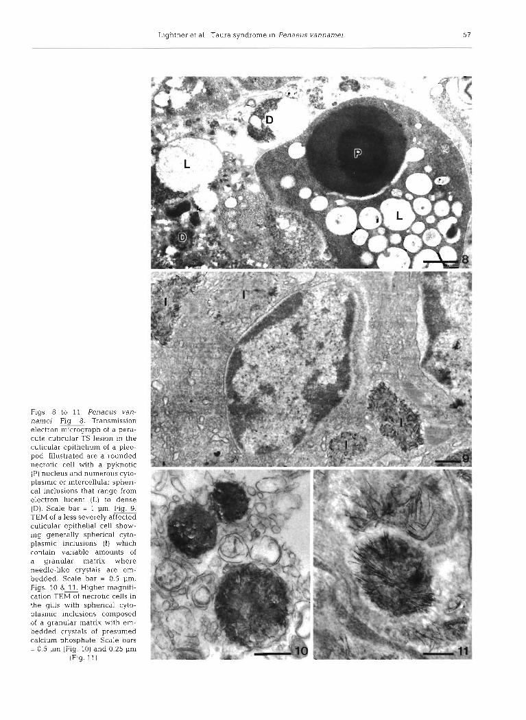
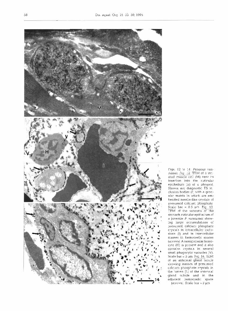
Transmission electron microscopy of focal lesions in peracute phase TS confirmed many of the characteris- tics of the lesions as discerned by histopathology. The spherical intracytoplasmic inclusions were found to be abundant in affected tissues examined (gills and cutic- ular epithelium, subcutis and muscle in appendages) and to have contents that ranged from sparse and elec- tron lucent to electron dense (Figs. 8 to 13). Some were found to contain a few to very abundant needle-like crystals (Fig. 9 to 14). In some specimens exam.ined, masses of these crystals were present in hemocoel spaces in the gills, subcutis, and in association with adjacent muscle fibers (Figs. 12 & 13). In the antennal gland of 1 specimen examined by TEM, morphologi- cally similar crystals were observed in the hemocoel basal to the antennal gland epithelium and in the antennal gland lumen (Fig. 14). Because the morpho- logy of these needle-like crystals is identical to that reported for calcium phosphate in certain metabolic disorders of vertebrate animals (Cheville 1983), they are presumed to be calcium phosphate. Of 9 shrimp presenting gross signs of peracute TS and examined by TEM, all displayed presumed calcium phosphate crystals associated with TS lesions.
DISCUSSION
Until recently, TS was considered to be an idiopathic disease whose distribution was determined as a func- tion of a toxicant or infectious agent common to all the locations where the disease had been recognized. An alternative explanation for TS suggested that it may have been an emerging toxic or infectious disease syn- drome present in many shrimp culture regions that went unrecognized until it was first discovered in Ecuador. Coincidental with the discovery of TS in the Taura region of Ecuador in 1992 was the initiation of aerial spraying of agricultural fungicides in the Taura River drainage basin, which were applied in an effort to control Sigtoka Negra or Black Leaf Wilt of bananas. Most commonly applied were BenlateTM (benomyl, DuPont) and the ergosterol (a sterol) synthesis inhibitors ~ i l t ~ (propiconazole, Ciba-Geigy) and CalixinTM (tridemorph, BASF) (Lightner et al. 1994, Rosenberry 1994a, b, Wigglesworth 1994). Because
crustacean molting hormone, or ecdysone, is also a sterol (Grieneisen 1994) that is structurally very similar to ergosterol, there were compelling reasons to suspect that these fungicides were involved in the etiology of TS. However, despite the numerous studies on a pre- sumed toxicological etiology that have been run by the research components of the affected farms, univer- sity groups or the pesticide companies, a relationship of these fungicides to the etiology of TS has not been confirmed experimentally (Lightner et al. 1994, Rosenberry 1994a, b, Wigglesworth 1994, Brock et al. in press).
Histopathology and TEM studies carried out on juve- nile Penaeus vannamei suggest an etiology in which calcium metabolism (calcification and de-calcification of the cuticle during the molting process) is disrupted resulting in accumulations of calcium phosphate crys- tals in affected tissues and organs. This finding might be considered to support an hypothesis of a toxic etiology for TS. However, recent studies carried out in Hawaii and in Arizona have implicated a previously unrecognized, small (-30 nm), icosahedral, cyto- plasmic virus (called TS virus or TSV) as the causative agent of the disease (Brock et al. in press). In these studies, TS was induced in healthy, juvenile P van- namei by exposure to the virus via injection of cell-free homogenates prepared from the carcasses of P van- namei (from Ecuador and Hawaii) with the disease or by feeding directly the same carcasses. Identical results were obtained with TS-positive shrimp from both Hawaii and Ecuador (Brock et al. in press).
The histological lesions presented by juvenile Penaeus vannamei with peracute or acute TS are unique and pathognomonic for the disease. While other infectious and noninfectious diseases of penaeid shrimp affect the same target tissues or display a histopathology somewhat similar to TS, none are suffi- ciently similar to TS to be confused. The diseases of penaeid shrimp warrant discussion and comparison to TS. Like TS, infectious hypodermal and hematopoietic necrosis virus (IHHNV) (Lightner 1993) has a viral etiology and has among its target tissues the cuticular epithelium and antennal gland where it causes a dif- fuse to multifocal necrosis. However, IHHNV can be readily distinguished from TS by the presence of char- acteristic intranuclear inclusion bodies in affected cells. Further distinction between TSV infection and IHHNV can be accomplished by the use of a gene probe to IHHNV (Man et al. 1993) in an in situ hybridization assay with histological materials, which is available commercially (Shrimp probeTM, DiagXotics, Wilton, CT, USA).
Yellowhead disease is another virus disease of penaeid shrimp in which the cuticular epithelium is among the target tissues (Chantanachookin et al.
Lightner et a1 Taura syndrome in Penaeus vannamei 57
Figs. 8 to 11. Penaeus van- narnei. Fig. 8. Transmission electron micrograph of a pera- cute cuticular TS lesion in the cuticular epithelium of a pleo- pod. Illustrated are a rounded necrotic cell with a pyknotic . . (P) nucleus and numerous cyto- plasmic or intercellular spheri- cal inclusions that range from electron lucent (L) to dense (D). Scale bar = l pm. Fig. 9. TEM of a less severely affected cuticular epithelia1 cell show- ing generally spherical cyto- plasmic inclusions (I) whch contain variable amounts of a granular matrix where needle-like crystals are em- bedded. Scale bar = 0.5 pm. Figs. 10 & 11. Higher magnifi- cation TEM of necrotic cells in the gills with spherical cyto- plasmic inclusions composed of a granular matrix with em- bedded crystals of presumed calcium phosphate Scale bars = 0.5 pm (Fig. 10) and 0.25 pm
(Fig. 11)
5 8 Dis. aquat. Org. 21. 53-59, 1995
Figs. 12 to 14. Penaeus van- name]. Fig. 12 . TEM of a stri- ated muscle cell (M) near its insertion into the cuticular epithelium (E) of a pleopod. Shown are diagnostic TS in- clusion bodies ( I ) with a gran- ular matrix in which are em- bedded needle-like crystals of presumed calcium phosphate. Scale bar = 0.5 pm. Fig. 13. TEM of the subcutis of the stomach cuticular epithelium of a juvenile F? vannarnei show- ing large accumulations of presumed calcium phosphate crystals in intracellular inclu- sions (I) and in intercellular masses in hemocoelic spaces (arrows). A semigranular hemo- cyte (H) is present and it also contains crystals in several small phagocytlc vacuoles (V). Scale bar = 3 pm. Fig.* TEM of an antennal gland tubule showlng masses of presumed calc~um phosphate crystals in the lumen ( L ] of the antennal gland tubule and in the adjacent hemocoelic space
[arrows). Scale bar = 3 pm
Lightner et al.. Taura syndrome in Penaeus uannaniel 5 9
1993). Like TS, spherical cytoplasmic inclusion bodies are an important diagnostic feature of infection by yellowhead virus (YHV). However, TSV-caused lesions contain far more spherical cytoplasmic inclu- sions (giving them their characteristic 'peppered' or 'buckshot' appearance) than do those due to YHV. Fur- thermore, the principal target organ for YHV seems to be the lymphoid organ where severe necrosis is often apparent, whereas in Penaeus vannamei with TS no discernable pathology is apparent in this organ.
Finally, various forms of shell disease may be con- fused with TS, especially when shrimp in the recovery or chronic phases of the disease are present in affected populations. Bacterial shell disease (Cipriani et al. 1980, Lightner 1993) is indistinguishable from lesions present in shrimp that are in the recovery or chronic phases of TS. In this situation, histopathological studies of shrimp in the peracute or acute phases of TS are required to provide the correct diagnosis.
Acknowledgements. Grant support for this work has been from the U.S.D.A. Cooperative State Research Service, Marine Shrimp Farming Consortium grant number 88-38808- 3320; from technical assistance contracts from the following Ecuadorian, Peruvian, and Colombian shrimp culture compa- nies: El Rosario, Acuespecies. Cosemar, and Dibsa; from the Asociacion Langostinera Peruana de Tumbes, Peru; and from Asociacion Nacional de Acuicultores de Colombia.
LITERATURE CITED
Bell TA, Lightner DV (1988) A handbook of normal penae~d shrimp histology. World Aquaculture Society, Baton Rouge. LA, p 2-6
Bonami JR. Lightner DV, Redman RM. Poulos BT (1992) Par- tial characterization of a togavirus (LOVV) associated with histopathological changes of the lymphoid organ of penaeid shrimps. Dis aquat Org 14:145-152
Brock JA (1993) Current diagnostic methods for agents and diseases of farmed marine shrimp In: Fulks W, Main K (eds) Diseases of cultured penae~d shrimp in Asia and the United States. Oceanic Institute, Honolulu, HI, p 210-231
Brock JA. Gose R, Lightner DV, Hasson KW (in press) An overview on Taura syndrome, an important disease of farmed P vannamei. Proceedings of the special session 'Swimming Through Troubled Water' J World Aquacult soc
Brock JA, Lightner DV (1990) Diseases of crustacea. In: l n n e
Responsible Subject Editor: J. E. Stewart, Dartmouth, Nova Scotia, Canada
0 (ed) Diseases of marine animals, Vol 111 Biologische Anstalt Helgoland, Hamburg, p 245-349
Chantanachookin C, B0onya1-atpalin S, Kasornchandra J , Direkbusarakom S, Ekpanithanpong U, Supamataya K , Srlurairatana S, Flegel TW (1993) Histology and ultra- structure reveal a new granulosis-like virus in Penae~ls monodon affected by yellow-head disease. Dis aquat Org 17:145-157
Cheville NF (1983) Cell pathology. lowa State Unlv Press, Ames, p 158-160
Cipriani GR, Wheeler RS, Sizemore RK (1980) Characteriza- tion of brown spot disease of Gulf Coast Shrlmp. J Inver- tebr Path01 36:255-263
Grieneisen ML (1994) Recent advances in our knowledge of ecdysteroid biosynthesis in insects and crustaceans. Insect Biochem. Molec Biol 241115-132
Jimenez R (1992) Sindrome de Taura (resumen). Acuacull ~ ~ r a del Ecuador. In: Jimenez R (ed) Revista especlallzada dc la Camara Nacional de Acuacultura, Guayaquil 1.1-16
Lightner DV (1993) Diseases of cultured penaeld shrimp. In. McVey J . (ed) CRC handbook of mariculture, 2nd edn, Vol 1, Crustacean aquaculture. CRC Press, Boca Raton, p 393-486
Lightner DV, Jones LS, Ware GW (1994) Proceedings of the Taura Syndrome Workshop: executive summary, submit- ted reports, and transcribed notes. Jan 21-22, 1994, Univ Arizona, Tucson
Lightner DV, Redman RM (1994) Histopathology and ultra- structural studies of Taura syndrome, a putdtive toxicity syndrome of penaeid shrimp. In: Book of abstracts, World Aquaculture '94, January 14-18, New Orleans, LA. World Aquaculture Society, Louisiana State Univ, Baton Rouge, p 227
Mari J , Bonarni J-R, Lightner DV (1993) Partial cloning of the genome of infectious hypodermal and haematopoietic necrosls virus, an unusual parvovirus pathogenic for penaeid shrimps; diagnosis of the disease using a specific probe. J gen Virol ?4:2637-2643
Rosen B (1970) Shell disease of aquatic crustaceans. In: Snieszko SF (ed) A Symposium on Diseases of Fishes and Shellfishes. Publ. No. 5. American Fisheries Society. Mlashington. DC, p 409-415
Rosenberry B (ed) (1993) Taura syndrome hits farms in Ecuador - again. Shrimp News lnt 18(3):6
Rosenberry B (ed) (1994a) Update on Taura syndrome in Ecuador. Shrimp News Int 19(3)-2-4
Rosenberry B (ed) (1994b) Taura syndrome ravages Ecuador Shrimp News Int 19(5):10-12
Sindermann CJ (1990) Principal diseases of marine fish and shellfish. Vol 11, Diseases of marine shellfish. Academic Press, San Diego, p 48-53
Wigglesworth J (1994) 'Taura syndrome' hits Ecuador farms. Fish Farmer 17(3):30-31
Manuscript first received: July 28. 1994 Revised version accepted: September 30, 1994
Copyright © 2022 FDOKUMEN