Relations between cochlear histopathology and hearing loss in experimental cochlear implantation
9
Research paper Relations between cochlear histopathology and hearing loss in experimental cochlear implantation S.J. O’Leary * , P. Monksfield 1 , G. Kel, T. Connolly 2 , M.A. Souter 3 , A. Chang, P. Marovic 4 , J.S. O’Leary, R. Richardson 5 , H. Eastwood Department of Otolaryngology, University of Melbourne, 2nd Floor, Peter Howson Wing, Royal Victorian Eye and Ear Hospital, 32 Gisborne St, East Melbourne, Victoria 3002, Australia article info Article history: Received 23 October 2012 Received in revised form 17 January 2013 Accepted 18 January 2013 Available online 5 February 2013 abstract This study reviews the cochlear histology from four hearing preservation cochlear implantation exper- iments conducted on 73 guinea pigs from our institution, and relates histopathological findings to re- sidual hearing. All guinea pigs had normal hearing prior to surgery and underwent cochlear implantation via a cochleostomy with a silastic-platinum dummy electrode. Pure tone auditory brainstem response (ABR) thresholds from 2 to 32 kHz were recorded prior to surgery, and at one and four weeks post- operatively. The cochleae were then fixed in paraformaldehyde, decalcified, paraffin embedded, and mid- modiolar sections were prepared. The treatment groups were as follows: 1) Systemic dexamethasone, 0.2 mg/kg administered 1 h before implantation, 2) Local dexamethasone, 2% applied topically to the round window for 30 min prior to cochlear implantation, 3) Local n-acetyl cysteine, 200 mg applied topically to the round window for 30 min prior to implantation, 4) inoculation to keyhole-limpet he- mocyanin (KLH) prior to implantation, and 5) untreated controls. There was a significant correlation between the extent of the tissue reaction in the cochlea and the presence of foreign body giant cells (FBGCs), new bone formation and injury to the osseous spiral lamina (OSL). The extent of the tissue response, as a percentage of the area of the scala tympani, limited the best hearing that was observed four weeks after cochlear implantation. Poorer hearing at four weeks correlated with a more extensive tissue response, lower outer hair cell (OHC) counts and OSL injury in the basal turn. Progressive hearing loss was also correlated with the extent of tissue response. Hearing at 2 kHz, which corresponds to the region of the second cochlear turn, did not correspond with loco-regional inner hair cell (IHC), OHC or SGC counts. We conclude that cochlear injury is associated with poorer hearing early after implantation. The tissue response is related to indices of cochlear inflammation and injury. An extensive tissue response limits hearing at four weeks, and correlates with progressive hearing loss. These latter effects may be due to inflammation, but would also be consistent with interference of cochlear mechanics. Crown Copyright Ó 2013 Published by Elsevier B.V. All rights reserved. 1. Introduction Delayed hearing loss following cochlear implantation remains a problem for hearing preservation surgery. Although hearing may be preserved immediately after surgery in a significant proportion of patients undergoing implantation, it has been reported that approximately a third of recipients lose their residual hearing slowly over the next few months (Barbara et al., 2003; Gstoettner Abbreviations: ABR, auditory brainstem response; CI, cochlear implant; FBGC, foreign body giant cell; IHC, inner hair cell; KLH, keyhole-limpet hemocyanin; NAC, n-acetyl cysteine; OHC, outer hair cell; OSL, osseous spiral lamina; SGC, spiral ganglion cell; ST, scala tympani. * Corresponding author. Tel.: þ61 3 9929 8366; fax: þ61 3 9663 1958. E-mail addresses: [email protected] (S.J. O’Leary), monksfi[email protected] (P. Monksfield), [email protected] (G. Kel), [email protected] (T. Connolly), [email protected] (M.A. Souter), [email protected] (A. Chang), [email protected] (P. Marovic), [email protected] (J.S. O’Leary), rricharson@ bionicsinstitute.org (R. Richardson), [email protected] (H. Eastwood). 1 Present address: Queen Elizabeth Medical Centre, Birmingham, West Midlands B15 2TH, United Kingdom. 2 Permanent address: Geelong Hospital, 72-322 Ryrie Street, Geelong, Victoria, Australia. 3 Permanent address: Christchurch Hospital, Riccarton Avenue, Private Bag 4710, Christchurch, NZ. 4 Present address: Southern Health, Monash Medical Centre, Victoria, Australia. 5 Permanent address: Bionics Institute, 384-388 Albert St, East Melbourne, Victoria, Australia. Contents lists available at SciVerse ScienceDirect Hearing Research journal homepage: www.elsevier.com/locate/heares 0378-5955/$ e see front matter Crown Copyright Ó 2013 Published by Elsevier B.V. All rights reserved. http://dx.doi.org/10.1016/j.heares.2013.01.012 Hearing Research 298 (2013) 27e35
-
Upload
bionicsinstitute -
Category
Documents
-
view
0 -
download
0
Transcript of Relations between cochlear histopathology and hearing loss in experimental cochlear implantation

at SciVerse ScienceDirect
Hearing Research 298 (2013) 27e35
Contents lists available
Hearing Research
journal homepage: www.elsevier .com/locate/heares
Research paper
Relations between cochlear histopathology and hearing loss in experimentalcochlear implantation
S.J. O’Leary*, P. Monksfield 1, G. Kel, T. Connolly 2, M.A. Souter 3, A. Chang, P. Marovic 4, J.S. O’Leary,R. Richardson 5, H. EastwoodDepartment of Otolaryngology, University of Melbourne, 2nd Floor, Peter HowsonWing, Royal Victorian Eye and Ear Hospital, 32 Gisborne St, East Melbourne, Victoria 3002, Australia
a r t i c l e i n f o
Article history:Received 23 October 2012Received in revised form17 January 2013Accepted 18 January 2013Available online 5 February 2013
Abbreviations: ABR, auditory brainstem response; Ccysteine; OHC, outer hair cell; OSL, osseous spiral lam* Corresponding author. Tel.: þ61 3 9929 8366; fax
E-mail addresses: [email protected] (S.J. [email protected] (M.A. Souter), [email protected] (R. Richardson), haydente@unime
1 Present address: Queen Elizabeth Medical Centre2 Permanent address: Geelong Hospital, 72-322 Ry3 Permanent address: Christchurch Hospital, Riccar4 Present address: Southern Health, Monash Medic5 Permanent address: Bionics Institute, 384-388 Al
0378-5955/$ e see front matter Crown Copyright � 2http://dx.doi.org/10.1016/j.heares.2013.01.012
a b s t r a c t
This study reviews the cochlear histology from four hearing preservation cochlear implantation exper-iments conducted on 73 guinea pigs from our institution, and relates histopathological findings to re-sidual hearing. All guinea pigs had normal hearing prior to surgery and underwent cochlear implantationvia a cochleostomy with a silastic-platinum dummy electrode. Pure tone auditory brainstem response(ABR) thresholds from 2 to 32 kHz were recorded prior to surgery, and at one and four weeks post-operatively. The cochleae were then fixed in paraformaldehyde, decalcified, paraffin embedded, and mid-modiolar sections were prepared. The treatment groups were as follows: 1) Systemic dexamethasone,0.2 mg/kg administered 1 h before implantation, 2) Local dexamethasone, 2% applied topically to theround window for 30 min prior to cochlear implantation, 3) Local n-acetyl cysteine, 200 mg appliedtopically to the round window for 30 min prior to implantation, 4) inoculation to keyhole-limpet he-mocyanin (KLH) prior to implantation, and 5) untreated controls. There was a significant correlationbetween the extent of the tissue reaction in the cochlea and the presence of foreign body giant cells(FBGCs), new bone formation and injury to the osseous spiral lamina (OSL). The extent of the tissueresponse, as a percentage of the area of the scala tympani, limited the best hearing that was observedfour weeks after cochlear implantation. Poorer hearing at four weeks correlated with a more extensivetissue response, lower outer hair cell (OHC) counts and OSL injury in the basal turn. Progressive hearingloss was also correlated with the extent of tissue response. Hearing at 2 kHz, which corresponds to theregion of the second cochlear turn, did not correspond with loco-regional inner hair cell (IHC), OHC orSGC counts. We conclude that cochlear injury is associated with poorer hearing early after implantation.The tissue response is related to indices of cochlear inflammation and injury. An extensive tissueresponse limits hearing at four weeks, and correlates with progressive hearing loss. These latter effectsmay be due to inflammation, but would also be consistent with interference of cochlear mechanics.
Crown Copyright � 2013 Published by Elsevier B.V. All rights reserved.
1. Introduction
Delayed hearing loss following cochlear implantation remainsa problem for hearing preservation surgery. Although hearing may
I, cochlear implant; FBGC, foreignina; SGC, spiral ganglion cell; ST,: þ61 3 9663 1958.’Leary), [email protected] (P.o.com (A. Chang), [email protected] (H. Eastwood)., Birmingham, West Midlands B15rie Street, Geelong, Victoria, Austraton Avenue, Private Bag 4710, Chrial Centre, Victoria, Australia.bert St, East Melbourne, Victoria, A
013 Published by Elsevier B.V. All
be preserved immediately after surgery in a significant proportionof patients undergoing implantation, it has been reported thatapproximately a third of recipients lose their residual hearingslowly over the next few months (Barbara et al., 2003; Gstoettner
body giant cell; IHC, inner hair cell; KLH, keyhole-limpet hemocyanin; NAC, n-acetylscala tympani.
Monksfield), [email protected] (G. Kel), [email protected] (T. Connolly),gmail.com (P. Marovic), [email protected] (J.S. O’Leary), rricharson@
2TH, United Kingdom.lia.stchurch, NZ.
ustralia.
rights reserved.

Table 1Summary of treatments applied to the animals included in this analysis. All animalsunderwent cochlear application. Some were pre-treated with either topical appli-cation of drug to the cochlea, via its application to the round window on a carbox-ymethylcellulose-hyaluronic acid polymer. Others were treated systemically, viaintravenous injection. “KLH” animals were inoculated to induce an immuneresponse against keyhole limpert hemocyanin.
Study Treatment(all received implant)
Numbers Descriptor
1. Topicaldexamethasone(James et al., 2008)
Local application to roundwindow 2% dexamethasone
14 2% dex
2. Topical NAC(Eastwoodet al., 2010)
Local application to roundwindow 200 mg n-acetylcysteine
14 NAC
3. Systemicdexamethasone(Connollyet al., 2010)
Intravenous administration0.2 mg/kg dexamethasone
6 Low dose
4. KLH/Topical Local application to round 10 KLH steroid
S.J. O’Leary et al. / Hearing Research 298 (2013) 27e3528
et al., 2006; Woodson et al., 2010). The reasons for this are not wellunderstood, but it has been proposed that the tissue response toimplantation may be implicated. Studies of human temporal boneshave demonstrated that the electrode is surrounded by a fibroussheath (Nadol et al., 2001). It has been proposed that this tissueresponse6 may adversely affect residual hearing in several ways.The fibrous tissue could dampen cochlear mechanics if it were toinvolve the basilar membrane at the site of implantation in thebasal turn, where it has been predicted that high frequency hearingloss will result (Choi and Oghalai, 2005; Kiefer et al., 2006).Whereas fibrosis occupying a significant proportion of the scalatympani (ST) is predicted to cause a low frequency hearing loss,according to the modelling of Choi and Oghalai (2005). Alter-natively, it has been proposed that biological activity within thetissue response may be toxic to the organ of Corti (Eshraghi et al.,2005). The data presented here provide some insights into howresidual hearingmay change over time after cochlear implantation.
Of the small numbers of human temporal bones available, mostinvolve patients that have had little, if any, residual hearing andhave been implanted years prior to their death (Fayad et al., 2009;Nadol and Eddington, 2006; Nadol et al., 2001). Therefore it isunlikely that this material will provide insights into postoperativedelayed hearing impairment in the foreseeable future. In contrast,experimental animals have a histopathological response to animplant which resembles that seen in humans, with fibrosis, newbone growth (osteoneogenesis) and the presence of foreign bodygiant cells (FBGCs) being prominent features (James et al., 2008;Xu et al., 1997). Therefore an experimental approach is more likelyto provide insights into the aetiology of delayed hearing loss andits relationship to the intracochlear tissue response fromimplantation.
In our laboratories, we now have a sizeable cohort of guineapigs that have undergone hearing preservation cochlear im-plantation, where the nature and extent of the tissue responsepresent within the basal turn of the cochlea has been assessed.These animals received a cochlear implant (CI) into the basal turnwith or without a short application of a hearing preservationdrug, such as dexamethasone or n-acetyl cysteine. A robustanalysis of cochlear histopathology and its impact upon hearingloss has been precluded due to marked variability in the extent ofthe tissue response in separate experiments. Now, sufficientnumbers of animals have been studied across a range of experi-ments to minimise the influence of inter-animal variability, so ananalysis has been conducted incorporating subjects across studygroups.
Here we correlate the histopathological appearance of the co-chlea, with changes in auditory brainstem response (ABR)thresholds over time in an attempt to determine whether thecochlear histopathology has influenced these hearing outcomes. Inall of these experiments there was an initial elevation in ABRthreshold, which was still apparent one week after surgery. Overthe next three weeks, ABR thresholds either partially recovered,remained unchanged or worsened slightly. These divergent hear-ing outcomes were correlated with the histopathological analysesperformed on cochlear tissue collected four weeks after implan-tation. In addition, we examined whether there was a treatmenteffect upon the tissue response from the drugs applied to theseanimals.
6 tissue response e this is the term used to describe the tissue response tocochlear implantation. It is thought to arise from both the response to surgery andalso the response to the presence of the implant (i.e. the foreign body reaction). Thetissue response was quantified as the percentage of the area of scala tympani (in thelower basal turn) that it occupied.
2. Methods
2.1. Animals
All procedures in this study were approved by the Animal EthicsCommittee of the Royal Victorian Eye and Ear Hospital (Projects 05/113A, 06/132A, 07/140A and 07/146A). The guinea pigs were bred inthe Biological Resource Centre at the Royal Victorian Eye and EarHospital and weighed �300 g. The anaesthesia used for all ABRtesting was 60 mg/kg ketamine and 4 mg/kg xylazine, intra-muscularly. For surgical procedures, after induction with ketamineand xylazine, gaseous anaesthesia was titrated against respirationwith 0.5e1% isoflurane delivered in oxygen set at a rate of 500 ml/min.
2.2. Experimental groups
The experimental groups have been described in detail previ-ously (Table 1) (Connolly et al., 2010; Eastwood et al., 2010; Jameset al., 2008; Maini et al., 2009; Souter et al., 2012). In the topicaldexamethasone study (James et al., 2008), a carbox-ylmethylcellulose hyaluronic acid polymeric pledget (Seprapack�,Genzyme Corporation) was loaded with 5 ml of either 2% dex-amethasone phosphate or saline and placed on the round windowmembrane for 30minprior to cochlear implantation. In the n-acetylcysteine (NAC) study (Eastwood et al., 2010) Seprapak pledgetswereloaded with 5 ml of either 40 mg/ml n-acteyl cysteine or saline andagain applied to the round window membrane for 30 min prior tocochlear implantation. In the systemic dexamethasone study(Connolly et al., 2010), a low dose of dexamethasone (0.2 mg/kg) orsaline was injected into the internal jugular vein 60 min prior tocochlear implantation. The experimental groups described abovewere typified by there having been hearing protection observed at32 kHz only. In the keyhole limpet hemocyanin (KLH) study (Souteret al., 2012), animals were injected subcutaneously with KLH ata dose of 1 mg a week prior to implantation. KLH-primed animalswere then randomised to receive a 5 ml volume of either 20% dex-amethasone or normal saline delivered to the round window ona Seprapak pledget for 30 min prior to cochlear implantation. The
dexamethasone(Souter et al., 2012)
window 20% dexamethasoneKLH primed
5. KLH control(Souter et al., 2012)
Local application to roundwindow normal salineKLH primed
10 KLH control
6. Controls fromnon-KLH studies
19 Control
Total 73

Fig. 1. The scala tympani (ST) was divided into quadrants (1, 2, 3, and 4) as shown inorder to qualitatively describe the electrode position and tissue response. The scalavestibule (SV), scala media (SM), basilar membrane (BM) and Rosenthal’s canal (RC) aremarked. The tissue response was found in quadrants 2 and 4 (Q2,4) in 20 animals,Q1,2,4 in 12 animals, Q1,2,3,4 in 11, Q1 in 4, Q2 in 3, Q1,2 in 3, Q4 in 1, Q1,3,4 in 1 and notissue response was evident in 10. The tissue response was found in SV in 3, and bothSV and ST in 5.
S.J. O’Leary et al. / Hearing Research 298 (2013) 27e35 29
animals were given a second dose of KLH aweek after implantation.Four weeks after implantation, all animals in these studies under-went a final ABR measurement and were then euthanised with anoverdose of pentobarbitone (1 g/kg) prior to transcardiac perfusionwith a phosphate-buffered normal saline solution.
2.3. Auditory Brainstem evoked potential recordings
ABR recordings were completed in the ear to be implantedimmediately prior to surgery, then at one and four weeks aftersurgery (Chang et al., 2009; James et al., 2008). Acoustic stimuliwere delivered via a loudspeaker (Richard Allen DT 20, UK) placed0.1 m from the test ear. The stimuli were computer generated tonepips of 5 ms durationwith 1 ms rise/fall times, presented at 2, 8, 16,24 and 32 kHz. The contralateral ear was masked using an earmould compound (Otoform K2, DLT, West Yorkshire).
Subcutaneous needle electrodes were placed upon the skullvertex, dorsal neck and midline torso of the guinea pig to recordABRs. Responses were amplified by 100,000� (DAM-5A, W-P In-struments Inc, New Haven, Conn., USA) and band-pass filtered(Krohn-Hite 3750, Avon, Mass., USA) between 150 Hz and 3 kHz(6 dB/octave). The signal was fed to a 16-bit analogue-to-digitalconverter (Tucker Davis Technologies, USA) and sampled at20 kHz for 10 ms after stimulus onset then averaged over 250stimulus repetitions. Stimulus intensity was decreased by 5 dBdecrements to sub-threshold levels. The waveforms were analysedusing a custom program (software written by Dr James Fallon andadapted by Prof Stephen O’Leary) in Igor Pro 5.02 (WavemetricsInc.) with the researcher blinded to the treatment group. Thresholdwas defined as the lowest stimulus intensity to evoke a wave IIIresponse greater than 0.4 mV.
2.4. Cochlear electrode and implantation
The CI electrode array has been described previously (Changet al., 2009; James et al., 2008). Its total length was 15 mm, con-sisting of 3 platinum rings placed 0.75 mm apart along a silasticcarrier (MDX4-4210 DowCorning Products, USA). Platinumwires of25 mm diameter were welded to each electrode and embeddedwithin the silastic. The platinum rings served as a depth gaugewithinsertion to the third ring indicative of 2.25 mm insertion. Themaximum diameter was 0.45 mm, narrowing to 0.41 mm towardsthe tip of the electrode. Sterility was achieved with ultrasoniccleaning and ethylene oxide sterilisation. The electrode wasimplanted under general anaesthesia. Aftermaking a post-auricularincision, the bulla was exposed and opened with a 3 mm cuttingburr. Under magnification, a 0.7 mm cutting burr was used to placea cochleostomy into the basal turn of the cochlea. The silastic-platinum dummy electrode was passed gently into the cochlea us-ing soft surgical principles to the level of the third platinum ring(2.25 mm) and left in situ. A fascial graft was used to seal the coch-leostomy around the implant. The extracochlear portion was trim-med to fit within the bulla.
2.5. Histological methods
After transcardiac perfusion with phosphate-buffered normalsaline, cochleae from all animals were removed and placed in 10%formalin before decalcification in 4% (w/v) ethylenediaminetetra-acetic acid (SigmaeAldrich, USA) for one week. Cochleae weretrimmed, the electrode was removed, and the specimen was ori-ented using agar prior to paraffin embedding and sectioning. Whenthe histologist judged (by visual inspection) that the mid-modiolarregion had been reached, three sequential mid-modiolar sections of5 mm thickness were mounted, cover-slipped, stained with
haematoxylin and eosin and examined by light microscopy (CarlZeiss, Göttingen, Germany).
The position of the tissue response in the lower basal turn of thecochlea was analysed. The ST was divided into four quadrants (seeFig. 1) for the purpose of recording the location(s) of the tissueresponse and the electrode. The upper inner quadrant (Q1) wasproximate to the spiral ganglion and osseous spiral lamina (OSL), theupper outer quadrant (Q2) was in the vicinity of the lateral osseousspiral lamina, basilar membrane and the adjacent lateral wall.Quadrant 3 (Q3) was medio-inferior and quadrant 4 (Q4), latero-inferior. The electrode left a circular lumen in the tissue responsethat indicated its position prior to electrode removal. Therefore, anelectrode position could only be estimated when a tissue responsewas present. Specific features examined on all sections included thenature of the tissue response, and whether this was constituted ofloose areolar fibrosis, dense fibrosis and/or osteoneogenesis. Thepercentage of STarea occupied by the tissue responsewas reported.The presence or absence of fracture of the OSL was also noted. Theorgan of Corti was assessed bycounting inner hair cells (IHCs, 0 or 1)outer hair cells (OHCs, 0e3) and the presence of the tunnel of Cortiacross the three mid-modiolar sections. The IHC and tunnel weredeemed to be present if recognised on one of the three sectionsexamined. OHC counts were averaged across the three sections. Theextent of the osteoneogenesis was assessed subjectively, and whenpresent was classed as 0e absent, 1eminimal, 2e occupying up toonequarter of the tissue response, or 3eoccupying greater than onequarter of the tissue response. Spiral ganglion cell (SGC) densitieswere calculated by dividing the number of Type I SGCs exhibiting anidentifiable nucleus by the area of Rosenthal’s canal.
2.6. Methods of data synthesis and statistical analysis
In all experiments, timelines for histological analysis and func-tional testing of the auditory system were standardised. ABRthreshold shifts (between preoperative and postoperative levels)

S.J. O’Leary et al. / Hearing Research 298 (2013) 27e3530
were acquired one and four weeks after cochlear implantation.Histological analyses were performed at the four-week post-operative time.
Bivariate analyses (Pearson’s correlation) were performed toexplore relationships between histological features and/or the ABRthreshold shifts. This was followed, where appropriate by multipleregression, to determine whether factors had either collective orunique effects. The treatment groups were ranked according to thevalue of the threshold shift averaged across frequency (leading toan order from lower to higher of 2% local (KLH), control (KLH), 2%local, NAC, systemic low dose steroid and control) in preparation formultiple regression analysis. A repeated measures analysis of var-iance was employed to evaluate whether factors such as cochlearturn or treatment group accounted for the variance in ABRthresholds across time or frequency.
Data was collated in Microsoft Office Excel 2010 and statisticswere performed in IBM SPSS version 20.
2.7. Rational for analysis
The primary goal of this study was to investigate whether therewere correlations between the histological features within thecochlea after implantation and hearing threshold changes. In orderto perform these correlations, the position of histological responseneeded to be related to a frequency specific place map. The lowerthree half turns (lower basal, upper basal and lower second turn) ofthe cochleae were assessed in mid-modiolar sections (cutting fromthe round window). Based on the (Greenwood, 1990) frequencyplace map, the 32 and 24 kHz frequency responses would haveoriginated from the lower basal, the 16 and 8 kHz responses fromthe upper basal and the 2 kHz response from the lower second turn.When performing multiple regression, threshold shifts at 32 kHzwere used to represent the lower basal turn, 8 kHz to represent theupper basal turn and 2 kHz to represent the lower second turn.Given that the electrode was inserted approximately 2.25 mm intothe scala, its tip reached to the cochlear region that gives rise tohearing in the 16e24 kHz range. Therefore, thresholds elevated at 2and 8 kHz are in a region apical to the tip of the electrode.
Fig. 2. Summary of the histopathological findings and auditory brainstem thresholdshifts from the lower basal to the lower second cochlear turns. The lower panelpresents threshold shifts from the lower basal turn (from left to right, 32, 24 and16 kHz), the upper basal turn (8 kHz) and the lower second turn (2 kHz). Thethresholds at one and four weeks after surgery are presented. The inner hair cell (IHC),outer hair cell (OHC) and spiral ganglion cell (SGC) counts from each cochlear half-turnare also presented. The presence of the Tunnel of Corti was recorded as either present(1) or absent (0), and the values were averaged to generate the values for the Figure.Bars represent the standard error of the mean.
3. Results
3.1. Hearing and quantitative analyses of neurosensory elements
At one and four weeks after implantation ABR thresholds wereon average 20e30 dB higher for frequencies in the range between 8and 32 kHz; the threshold shift was greatest at 8 kHz, and was<10 dB at 2 kHz. This can be seen in Fig. 2, which presents the ABRthreshold shifts one and four weeks after implantation, with theplace-matched histological observations of the organ of Corti (IHC,OHC, presence of the tunnel of Corti) and SGC counts. In the presentstudy, hearing loss that increased in magnitude by 10 dB or moreover the four weeks since surgery was seen in 19% of all animals.The IHC, OHC and SGC counts were poorer in the lower basal turnthan in either the upper basal or the lower second turns.
Multiple regression was performed to determine factors thatmay have contributed to the variation in ABR thresholds. For thisanalysis, IHC, OHC and SGN counts were pairedwith place-matchedABR thresholds for each of the lower three half turns of the cochleaand entered as dependent variables. Cochlear half turn and treat-ment were also entered as dependent variables. The multipleregression was weakly predictive of the week four threshold shifts(r ¼ 0.401, r2 ¼ 0.163, p < 0.001), but only the cochlear (half) turnwas found to be a significant factor (beta ¼ �0.337, p < 0.001). Thisindicated that ABR thresholds differed across cochlear place, but
that neither treatment group, nor place-matched neural and sen-sory counts explained the variation in ABR thresholds.
3.2. Position and pattern of the tissue response
The cochlear electrode occupied approximately 20% of the areaof the ST, and was in most cases surrounded by tissue. The location

S.J. O’Leary et al. / Hearing Research 298 (2013) 27e35 31
of the tissue response was determined relative to quadrants of theST as represented in Fig. 1. The tissue response was most oftenlocated laterally, in the vicinity of Q2, or between this quadrant andQ1 (Q1,2) or Q4 (Q2,4). The tissue surrounded a centrally locatedelectrode (Q1,2,3,4) in 11 of the 73 cochleae. Medially placed tissue(Q1 or Q1,3,4) was seen in 4 cochleae only. A tissue response wasfound within the scala vestibuli in 8 animals. A comprehensivelisting of tissue locations is found in the legend of Fig. 1.
The tissue reaction four weeks after implantation was typifiedby a loose areolar matrix of fibrocytes, which extended from theOSL, the lateral wall, or both, to engulf the electrode (Fig. 3). Thistissue typically arose from the cochlear wall nearest to the elec-trode, and was broadly based along the scalar margin. When theelectrode was more centrally located, loose areolar tissue was seento extend to both the OSL and the lateral wall, oftenwith sparing ofthe region immediately inferior to the basilarmembrane. Therewasfrequently a dense layer of fibrosis, often involving FBGCs, imme-diately surrounding the electrode. This tissue arose from either thecochlear wall adjacent to the electrode or de novo when the elec-trode was more centrally placed within the ST. Osteoneogenesiswas sometimes present within the tissue response, and varied inextent from being minimal to widespread.
3.3. Relations between histopathological features present withinthe lower basal turn
The electrode was present in the lower basal turn of the cochlea,and it was here that the majority of the cochlear pathology wasfound. It was revealed that the percentage of the SToccupied by thetissue response correlated with the presence of FBGCs (Fig. 4a),
Fig. 3. Examples of cochlear histopathology. Bar: 200 mm. (a) Loose areolar tissue extends f(black arrow head). The outer hair cells are present. (b) There is a dense fibrotic reaction sheads). Significant new bone growth extends from the osseous spiral lamina (black arrow heof the region below the basilar membrane (BM). (c) Extensive loose areolar tissue extendingpreservation of the tunnel of Corti.
osteoneogenesis (Fig. 4b), OSL fracture and poorer OHC counts(Table 2). On multiple regression, collectively these factors werepredictors of the extent of the tissue response (r¼ 0.504, r2¼ 0.254,p ¼ 0.003), but only the presence of FBGCs was demonstrated tohave a unique effect (beta ¼ 0.263, p ¼ 0.045).
OHC survival in the lower basal turn could be excellent, even inthe presence of a tissue response within the ST. In fact, a fullcomplement of OHC was present in 53% of cochleae analysed.However, OHC counts were negatively correlated with the extent oftissue response, the extent of new bone growth and the presence ofFBGCs (Table 2), and while on multiple regression the collectiveeffect of these factors (on OHC count) was not significant(p ¼ 0.071) the unique effect of osteoneogenesis was (p ¼ 0.026).
3.4. Effect of treatment group upon the tissue response
Amultivariate ANOVAwas performed to determinewhether thetreatment group had an effect on the variability of the tissue-related factors. Treatment group was found not to have an effectupon either the extent of the tissue response (p ¼ 0.310), thepresence of foreign body giant cells (p ¼ 0.071) or OSL fracture(p¼ 0.288). Similarly, there was not a significant effect of treatmentgroup upon SGC counts in either the lower basal (p ¼ 0.547), upperbasal (p ¼ 0.115), lower second (p ¼ 0.072) or upper second(p ¼ 0.461) turns. However, treatment group did have a significanteffect upon OHC counts in the lower basal turn (p < 0.001), theupper basal (p¼ 0.028) turn and the upper middle (p¼ 0.045) turn.Post hoc testing revealed that this effect could be accounted for byNAC-treated cochleae having significantly lower OHC counts thanother treatment groups in the basal turn.
rom the osseous spiral lamina (OSL) and the lateral wall, with some new bone growthurrounding the electrode, associated with some foreign body giant cells (white arrowad). There is loose areolar fibrosis extending from the OSL and lateral wall, with sparingfrom the scalar walls. Note the presence of a full complement of outer hair cells, with
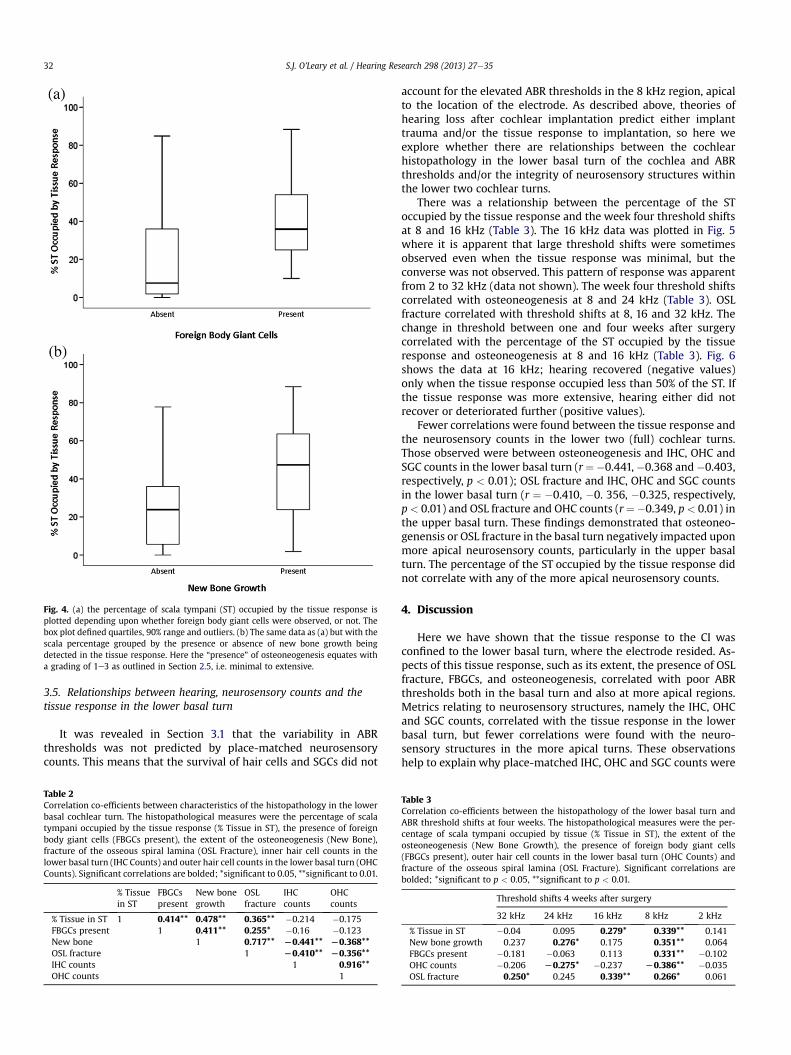
Fig. 4. (a) the percentage of scala tympani (ST) occupied by the tissue response isplotted depending upon whether foreign body giant cells were observed, or not. Thebox plot defined quartiles, 90% range and outliers. (b) The same data as (a) but with thescala percentage grouped by the presence or absence of new bone growth beingdetected in the tissue response. Here the “presence” of osteoneogenesis equates witha grading of 1e3 as outlined in Section 2.5, i.e. minimal to extensive.
S.J. O’Leary et al. / Hearing Research 298 (2013) 27e3532
3.5. Relationships between hearing, neurosensory counts and thetissue response in the lower basal turn
It was revealed in Section 3.1 that the variability in ABRthresholds was not predicted by place-matched neurosensorycounts. This means that the survival of hair cells and SGCs did not
Table 2Correlation co-efficients between characteristics of the histopathology in the lowerbasal cochlear turn. The histopathological measures were the percentage of scalatympani occupied by the tissue response (% Tissue in ST), the presence of foreignbody giant cells (FBGCs present), the extent of the osteoneogenesis (New Bone),fracture of the osseous spiral lamina (OSL Fracture), inner hair cell counts in thelower basal turn (IHC Counts) and outer hair cell counts in the lower basal turn (OHCCounts). Significant correlations are bolded; *significant to 0.05, **significant to 0.01.
% Tissuein ST
FBGCspresent
New bonegrowth
OSLfracture
IHCcounts
OHCcounts
% Tissue in ST 1 0.414** 0.478** 0.365** �0.214 �0.175FBGCs present 1 0.411** 0.255* �0.16 �0.123New bone 1 0.717** L0.441** L0.368**OSL fracture 1 L0.410** L0.356**IHC counts 1 0.916**OHC counts 1
account for the elevated ABR thresholds in the 8 kHz region, apicalto the location of the electrode. As described above, theories ofhearing loss after cochlear implantation predict either implanttrauma and/or the tissue response to implantation, so here weexplore whether there are relationships between the cochlearhistopathology in the lower basal turn of the cochlea and ABRthresholds and/or the integrity of neurosensory structures withinthe lower two cochlear turns.
There was a relationship between the percentage of the SToccupied by the tissue response and the week four threshold shiftsat 8 and 16 kHz (Table 3). The 16 kHz data was plotted in Fig. 5where it is apparent that large threshold shifts were sometimesobserved even when the tissue response was minimal, but theconverse was not observed. This pattern of response was apparentfrom 2 to 32 kHz (data not shown). The week four threshold shiftscorrelated with osteoneogenesis at 8 and 24 kHz (Table 3). OSLfracture correlated with threshold shifts at 8, 16 and 32 kHz. Thechange in threshold between one and four weeks after surgerycorrelated with the percentage of the ST occupied by the tissueresponse and osteoneogenesis at 8 and 16 kHz (Table 3). Fig. 6shows the data at 16 kHz; hearing recovered (negative values)only when the tissue response occupied less than 50% of the ST. Ifthe tissue response was more extensive, hearing either did notrecover or deteriorated further (positive values).
Fewer correlations were found between the tissue response andthe neurosensory counts in the lower two (full) cochlear turns.Those observed were between osteoneogenesis and IHC, OHC andSGC counts in the lower basal turn (r¼�0.441,�0.368 and�0.403,respectively, p < 0.01); OSL fracture and IHC, OHC and SGC countsin the lower basal turn (r ¼ �0.410, �0. 356, �0.325, respectively,p< 0.01) and OSL fracture and OHC counts (r¼�0.349, p< 0.01) inthe upper basal turn. These findings demonstrated that osteoneo-genensis or OSL fracture in the basal turn negatively impacted uponmore apical neurosensory counts, particularly in the upper basalturn. The percentage of the ST occupied by the tissue response didnot correlate with any of the more apical neurosensory counts.
4. Discussion
Here we have shown that the tissue response to the CI wasconfined to the lower basal turn, where the electrode resided. As-pects of this tissue response, such as its extent, the presence of OSLfracture, FBGCs, and osteoneogenesis, correlated with poor ABRthresholds both in the basal turn and also at more apical regions.Metrics relating to neurosensory structures, namely the IHC, OHCand SGC counts, correlated with the tissue response in the lowerbasal turn, but fewer correlations were found with the neuro-sensory structures in the more apical turns. These observationshelp to explain why place-matched IHC, OHC and SGC counts were
Table 3Correlation co-efficients between the histopathology of the lower basal turn andABR threshold shifts at four weeks. The histopathological measures were the per-centage of scala tympani occupied by tissue (% Tissue in ST), the extent of theosteoneogenesis (New Bone Growth), the presence of foreign body giant cells(FBGCs present), outer hair cell counts in the lower basal turn (OHC Counts) andfracture of the osseous spiral lamina (OSL Fracture). Significant correlations arebolded; *significant to p < 0.05, **significant to p < 0.01.
Threshold shifts 4 weeks after surgery
32 kHz 24 kHz 16 kHz 8 kHz 2 kHz
% Tissue in ST �0.04 0.095 0.279* 0.339** 0.141New bone growth 0.237 0.276* 0.175 0.351** 0.064FBGCs present �0.181 �0.063 0.113 0.331** �0.102OHC counts �0.206 L0.275* �0.237 L0.386** �0.035OSL fracture 0.250* 0.245 0.339** 0.266* 0.061

Fig. 5. The percentage of scala tympani (ST) occupied by the tissue response is plottedagainst the auditory brainstem response threshold shift (dB) at 16 kHz four weeks afterimplantation.
S.J. O’Leary et al. / Hearing Research 298 (2013) 27e35 33
not significant covariates for ABR threshold shifts on repeatedmeasures ANOVA; the organ of Corti was substantially preservedon light microscopic examination in the upper basal turn andabove, while ABR thresholds were significantly elevated.
The treatment groups did not have a main effect on either theABR thresholds or the tissue response, presumably because thestudies selected for this analysis involved treatments leading toa minimal effect on hearing, and when present was confined to32 kHz. We attribute the lack of a treatment effect in the localsteroid studies to the relatively short, 30 min period of drugapplication to the round window (Chang et al., 2009). We havesubsequently shown that application periods of 60e120 min arerequired to cause effective hearing protection (Chang et al.,
Fig. 6. The auditory brainstem response (ABR) threshold shift (dB) at 16 kHz fourweeks after implantation is plotted against the change in ABR thresholds betweenweeks one and four after implantation (dB) e positive values indicate deterioration inthresholds.
2009). The low dose systemic steroid group included in this anal-ysis was not effective in preserving hearing. Effective hearing pro-tection required a higher dose of systemic steroid. The pooling ofcontrol animals across studies may have masked some of thetreatment effects seen at 32 kHz noted in previous studies.
An important observation of this study is that four weeks aftersurgery lower ABR threshold shifts were not observed when therewas extensive fibrosis. This is apparent from Fig. 5 where all thedata points where in the lower right triangle of the graph, dem-onstrating that significant hearing loss can be associated withminimal fibrosis, but not the converse. Furthermore, there was analmost (log-)linear relationship between the best hearing thresh-olds at any level of fibrosis, abovew40% fibrosis. The latter suggeststhat fibrosis and hearing may be related, either directly or indi-rectly. One possibility is that fibrosis reflects the extent of intra-cochlear inflammation in the postoperative period that in itselfcauses greater hearing loss (an inflammatory theory) (Eshraghiet al., 2005). Another possibility is that fibrosis reduces hearingthresholds by a direct effect upon cochlear mechanics, as suggestedby Choi (a mechanical theory) (Choi and Oghalai, 2005). Thesepotential mechanisms and their relativemerits are nowconsidered.
In the experimental setting, the cochlear inflammation pro-voked by CI surgery has been shown to cause hearing loss bypromoting oxidative stress and apoptosis within hair cells (Dinhet al., 2011; Eshraghi et al., 2006; Haake et al., 2009). This occursnot only at the site of injury but also at remote cochlear locations,which provides a potential explanation for the low frequencyhearing loss observed following cochlear implantation. It is plau-sible that cochlear inflammation may also determine the extent ofthe tissue response by influencing cellular recruitment into thecochlea after injury; greater inflammation will attract larger num-bers of immune competent cells into the cochlea (Tornabene et al.,2006), including the macrophages and fibroblasts required for thegeneration of the tissue response. Therefore, it may be proposedthat the severity of the inflammatory response determines both themagnitude of the hearing loss and also the extent of the tissueresponse, both being different manifestations of the same under-lying mechanism. Several observations can be made that may beconsistent with this interpretation. First, consider the associationbetween the extent of the tissue response and the presence ofFBGCs (Fig. 4a). FBGCs arise when macrophages are unable tophagocytose foreign material, as is the case following cochlearimplantation (Anderson et al., 2008). The inflammatory theorypredicts that the severity of the inflammatory response willdetermine the numbers of macrophages recruited into the cochleaand thus the chance that these will coalesce into FBGCs, and this isconsistent with our observation that a greater percentage of ST wasoccupied by fibrosis when FBGCs were seen. The presence of FBGCswithin the tissue response did not affect the ABR thresholds sig-nificantly although there was a trend in that direction, suggestingthat these may not be related directly, but more likely indirectlythrough inflammation. Interestingly, a reanalysis of the data pre-sented by Xu and Shepherd (Xu et al., 1997) when cochlear elec-trodes were implanted into the hearing cat supports thisassociation. In that study, ABR thresholds were recorded inresponse to an acoustic click and these were poorer when FBGCswere seen in the tissue response (KruskaleWallis, p < 0.01).
In the present study, a relationship between fibrosis andinflammation is supported by the significant negative correlationbetween OHC counts and fibrosis. A more extensive tissue responsealso correlated with more extensive new bone growth, and bothwere correlated with OSL fracture. It is tempting to suggest in thepresent study that when OSL fracture occurred this led to a moreextensive inflammatory response provoking both an extensivetissue response and also OHC loss. However, in the present study
S.J. O’Leary et al. / Hearing Research 298 (2013) 27e3534
we cannot exclude the possibility that OSL fracturemay lead to OHCloss by other mechanisms, such as vascular compromise throughdisruption of the spiral cochlear vein.
While the ABR responses appear to be consistent with the ideathat inflammatory changes initiated at the site of electrode inser-tion extended to more apical cochlear regions, the paucity of cor-responding changes in the histopathology in the upper basal andsecond turns might suggest a more complex interpretation of thesedata. There was no evidence of fibrosis in the cochlear half turnsapical to the electrode, and IHC and OHC counts were close tonormal. Therefore, the elevation in ABR thresholds observed at8 kHz, that arose from the upper basal turn, must have arisen froma structural change in the hair cells, disruption of the afferentsynapse or a change in cochlear mechanics.
Models of passive cochlear mechanics (Choi and Oghalai, 2005;Kiefer et al., 2006) have demonstrated that if fibrosis were toincrease damping of the cochlear partition by even amodest degreeof 5e6 times then there would be a substantial reduction in basilarmembrane velocity in the region involved, resulting in an elevationin high frequency hearing thresholds for CI recipients. There isclinical evidence to support this in patients, since pure tonethresholds may improve at frequencies just apical to the position ofthe cochlear electrode (Kiefer et al., 2006). Alternatively, if thetissue response surrounding the electrode is more extensive andincreases the magnitude of damping within the ST by 2e3 orders ofmagnitude, then there is predicted to be an elevation in lowerfrequency hearing (Choi and Oghalai, 2005). Fig. 5 revealed thatabove w40% fibrosis within ST, there was an almost (log-)linearrelationship between the extent of tissue response and the besthearing observed across all animals and frequencies. This is inkeeping with Choi’s modelling both qualitatively and quantita-tively, and consistent with an interpretation that the fibrosis causessignificant damping. The correlation between fibrosis and the re-covery of ABR thresholds over weeks one to four demonstrates thatgood recovery of hearing is possible only when there is a lessextensive tissue response; when there is greater tissue response,hearing recovers only a little and sometimes worsens over time.This observation could potentially be explained by the effects ofprogressive cochlear damping as the tissue response matured.
It is known that electrode trajectory and electrode design in-fluence direct trauma to the basilar membrane or OSL (Briggs et al.,2001). The present study demonstrates how detrimental OSLfracture is in affecting hearing loss and leading to an extensivetissue response. These results strongly support the proposition thatOSL injury must be avoided when attempting hearing preservationsurgery. Hearing loss was clearly more likely with perimodiolarelectrode placement, and leads the authors to suggest that withcurrently available electrodes that lateral placement may be pref-erable for hearing preservation.
In the present study, hearing loss that increased in magnitudeby 10 dB ormore over 4weeks was seen in 19% of all animals. This isa similar percentage to that seen with delayed hearing loss in theclinical situation, where the incidence ranges from 20 to 30% be-tween studies. While it must be remembered that delayed hearingloss in humans can arise months after implantation, our observa-tion that progressive hearing loss in this animal model was asso-ciated with a more extensive tissue response may be of interestwhen attempting to understand the clinical situation.
5. Conclusions
Improving the preservation of residual hearing after CI surgery,and in particular preventing the delayed hearing loss seen in over25% of patients, requires a more thorough understanding of therelationship between the cochlear response to implantation and
hearing. Such an understanding will facilitate the rational devel-opment of hearing protection therapies, and help to further refineelectrode design specifications. Here we present evidence that thebest possible postoperative hearing is limited by the extent of thetissue response. This is consistent with the mechanical theory ofhearing impairment and suggests that reducing the extent of thetissue response is a worthy strategy for future research aimed atprotecting hearing after implantation. Alternatively, the data pre-sented is also consistent with an interpretation that the tissueresponse is related to inflammation and cochlear injury, as sug-gested by its relationship with the presence of FBGCs, osteoneo-genesis, OHC loss and OSL fracture. Whether one or bothmechanisms contribute to hearing loss will require further exper-imentation. However, from the available evidence it would beprudent to recommend that soft-surgery and inflammationreduction strategies should be considered for minimising theextent of the tissue response. Progressive hearing loss also corre-lates with the extent of the tissue response, so reducing the tissueresponse may be an effective strategy for preventing delayedhearing loss in patients after implantation.
Acknowledgements
Maria Clarke and Prudence Nielsen for preparing the histologymaterial. Helen Feng for making the cochlear electrodes. A/Prof.John Slavin from St. Vincents Health Melbourne for histopatho-logical assessment of the cochleae. Amy Hampson for proofing andpreparing of the final figures. The National Health and MedicalResearch Council of Australia, the Garnett Passe and Rodney Wil-liams Memorial Foundation, the John Mitchell Crouch Fellowshipawarded to SOL from the Royal Australasian College of Surgeonsand the Royal Victorian Eye and Ear Hospital for funding.
References
Anderson, J.M., Rodriguez, A., Chang, D.T., 2008. Foreign body reaction to bio-materials. Semin. Immunol. 20 (2), 86e100.
Barbara, M., Mattioni, A., Monini, S., Chiappini, I., Ronchetti, F., Ballantyne, D., et al.,2003. Delayed loss of residual hearing in Clarion cochlear implant users.J. Laryngol. Otol. 117 (11), 850e853.
Briggs, R.J., Tykocinski, M., Saunders, E., Hellier, W., Dahm, M., Pyman, B., et al., 2001.Surgical implications of perimodiolar cochlear implant electrode design:avoiding intracochlear damage and scala vestibuli insertion. Cochlear Implant.Int. 2 (2), 135e149.
Chang, A., Eastwood, H., Sly, D., James, D., Richardson, R., O’Leary, S., 2009. Factorsinfluencing the efficacy of round window dexamethasone protection of residualhearing post-cochlear implant surgery. Hear. Res. 255 (1e2), 67e72.
Choi, C.H., Oghalai, J.S., 2005. Predicting the effect of post-implant cochlear fibrosison residual hearing. Hear. Res. 205 (1e2), 193e200.
Connolly, T.M., Eastwood, H., Kel, G., Lisnichuk, H., Richardson, R., O’Leary, S., 2010.Pre-operative intravenous dexamethasone prevents auditory threshold shift ina guinea pig model of cochlear implantation. Audiol. Neurootol. 16 (3), 137e144.
Dinh, C.T., Bas, E., Chan, S.S., Dinh, J.N., Vu, L., Van De Water, T.R., 2011. Dex-amethasone treatment of tumor necrosis factor-alpha challenged organ of Cortiexplants activates nuclear factor kappa B signaling that induces changes in geneexpression that favor hair cell survival. Neuroscience 188, 157e167.
Eastwood, H., Pinder, D., James, D., Chang, A., Galloway, S., Richardson, R., et al.,2010. Permanent and transient effects of locally delivered n-acetyl cysteine ina guinea pig model of cochlear implantation. Hear. Res. 259 (1e2), 24e30.
Eshraghi, A.A., He, J., Mou, C.H., Polak, M., Zine, A., Bonny, C., et al., 2006. D-JNKI-1treatment prevents the progression of hearing loss in a model of cochlearimplantation trauma. Otol Neurotol. 27 (4), 504e511.
Eshraghi, A.A., Polak, M., He, J., Telischi, F.F., Balkany, T.J., Van De Water, T.R., 2005.Pattern of hearing loss in a rat model of cochlear implantation trauma. OtolNeurotol. 26 (3), 442e447.
Fayad, J.N., Makarem, A.O., Linthicum Jr., F.H., 2009. Histopathologic assessment offibrosis and new bone formation in implanted human temporal bones using 3Dreconstruction. Otolaryngol. Head Neck Surg. 141 (2), 247e252.
Greenwood, D.D., 1990. A cochlear frequency-position function for several species e29 years later. J. Acoust. Soc. Am. 87 (6), 2592e2605.
Gstoettner, W.K., Helbig, S., Maier, N., Kiefer, J., Radeloff, A., Adunka, O.F., 2006.Ipsilateral electric acoustic stimulation of the auditory system: results of long-term hearing preservation. Audiol. Neurootol. 11 (suppl. 1), 49e56.
S.J. O’Leary et al. / Hearing Research 298 (2013) 27e35 35
Haake, S.M., Dinh, C.T., Chen, S., Eshraghi, A.A., Van De Water, T.R., 2009. Dex-amethasone protects auditory hair cells against TNFalpha-initiated apoptosisvia activation of PI3K/Akt and NFkappaB signaling. Hear. Res. 255 (1e2), 22e32.
James, D.P., Eastwood, H., Richardson, R.T., O’Leary, S.J., 2008. Effects of roundwindow dexamethasone on residual hearing in a Guinea pig model of cochlearimplantation. Audiol. Neurootol. 13 (2), 86e96.
Kiefer, J., Bohnke, F., Adunka, O., Arnold, W., 2006. Representation of acoustic signalsin the human cochlea in presence of a cochlear implant electrode. Hear. Res.221 (1e2), 36e43.
Maini, S., Lisnichuk, H., Eastwood, H., Pinder, D., James, D., Richardson, R.T., et al.,2009. Targeted therapy of the inner ear. Audiol. Neurootol. 14 (6), 402e410.
Nadol Jr., J.B., Eddington, D.K., 2006. Histopathology of the inner ear relevant tocochlear implantation. Adv. Otorhinolaryngol. 64, 31e49.
Nadol Jr., J.B., Shiao, J.Y., Burgess, B.J., Ketten, D.R., Eddington, D.K., Gantz, B.J., et al.,2001. Histopathology of cochlear implants in humans. Ann. Otol. Rhinol Lar-yngol. 110 (9), 883e891.
Souter, M., Eastwood, H., Marovic, P., Kel, G., Wongprasartsuk, S., Ryan, A.F., et al.,2012. Systemic immunity influences hearing preservation in cochlear implan-tation. Otology Neurotol. 4 (12)
Tornabene, S.V., Sato, K., Pham, L., Billings, P., Keithley, E.M., 2006. Immune cellrecruitment following acoustic trauma. Hear. Res. 222 (1e2), 115e124.
Woodson, E.A., Reiss, L.A., Turner, C.W., Gfeller, K., Gantz, B.J., 2010. The hybridcochlear implant: a review. Adv. Otorhinolaryngol. 67, 125e134.
Xu, J., Shepherd, R.K., Millard, R.E., Clark, G.M., 1997. Chronic electrical stimulationof the auditory nerve at high stimulus rates: a physiological and histopatho-logical study. Hear. Res. 105 (1e2), 1e29.