Nochmals die NEUROI. Versuch einer ethnolinguistischen Namensdeutung
Upload
independentCategory
view
6download
0
310 Oper Orthop Traumatol 2007 · No. 3 © Urban & Vogel
Operative Orthopädie und Traumatologie
Minimalinvasiver, direkter vorderer Zugang zur Implantation einer HüfttotalendoprotheseTotal Hip Arthroplasty by a Minimally Invasive, Direct Anterior ApproachKazuhiro Oinuma1, Christoph Eingartner2, Yasufumi Saito1, Hideaki Shiratsuchi1
ZusammenfassungOperationsziel
Implantation einer Hüfttotalendoprothese über einen di-rekten vorderen Zugang zum Hüftgelenk mit minimaler Traumatisierung des umgebenden Gewebes.
IndikationenKonventionelle Totalendoprothetik des Hüftgelenks unge-achtet des Alters oder der Knochenqualität.
KontraindikationenDestruktion des proximalen Femurs bei Tumor oder Fraktur.Ausgeprägte Dysplasie oder Hüftluxation.Übergewicht (Body-Mass-Index [BMI] > 30 kg/m2) ist be-sonders während der Lernkurve als relative Kontraindika-tion zu werten.
OperationstechnikRückenlage des Patienten mit der Möglichkeit der Hy-perextension des Hüftgelenks in der Tischmitte, um die Darstellung des Femurs zu erleichtern.Vorderer Hautschnitt von 6–9 cm Länge, beginnend etwa 2 cm lateral und 5 cm distal der Spina iliaca anterior.Inzision der Faszie, stumpfes Präparieren des intermusku-lären Raums zwischen dem Musculus tensor fasciae latae und dem Musculus sartorius. Ausschneiden der vorderen Kapselanteile.Osteotomie des Femurhalses, Entfernung des Kopfes. Frä-sen des Azetabulums und Implantation der Pfanne. Dar-stellung des Femurs durch Hyperextension, Adduktion und Außenrotation des Beins, Inzision der hinteren Kapsel zur leichteren Vorverlagerung des Femurs. Vorbereiten des Implantatlagers und Implantation des Schafts.
Ergebnisse116 Hüften bei 111 Patienten wurden zwischen August 2004 und Dezember 2005 mit der vorgestellten Technik operiert. 17 Patienten wurden aufgrund von Frakturen oder ausgeprägter Dysplasie (Crowe 3 und 4) ausge-schlossen. Das Durchschnittsalter der Patienten betrug 62,5 Jahre (46–84 Jahre), der durchschnittliche BMI lag bei 23,1 kg/m2 (18,1–37,7 kg/m2).
AbstractObjective
Implantation of a total hip replacement device through a direct anterior approach to the hip joint with minimal trauma to adjacent tissue.
IndicationsAll conventional total hip replacements, irrespective of age and bone quality.
ContraindicationsDestruction of the proximal femur (tumor, fracture).Severe dysplasia and hip dislocation.Morbid obesity (body mass index [BMI] > 30 kg/m2) can be a relative contraindication during the learning curve.
Surgical TechniqueSupine position of the patient on the operating table with the possibility of hyperextension in the mid-table in order to facilitate femoral exposure.Anterior incision, 6–9 cm long, starting approximately 2 cm lateral and 5 cm distal of the anterior iliac spine.Incision of the fascia, blunt preparation in the intermuscu-lar space between tensor fasciae latae muscle and sartori-us muscle. Excision of the anterior parts of the capsule.Osteotomy of the femoral neck, removal of the head. Ream-ing of the acetabulum and implantation of the acetabular component. Exposure of the femur by hyperextension, ad-duction and external rotation of the leg, incision of the pos-terior capsule for easy anteriorization of the femur. Ream-ing and implantation of the femoral component.
Results116 consecutive hips in 111 patients were operated on be-tween August 2004 and December 2005. 17 patients were
Oper Orthop Traumatol 2007;19:310–26
DOI 10.1007/s00064-007-1209-3
1 Funabashi Orthopaedic Hospital, Hazama Funabashi-shi Chiba, Japan,
2 Caritas-Krankenhaus, Bad Mergentheim.

Oinuma K, et al. Direkter vorderer Zugang für die Totalendoprothetik der Hüfte
311Oper Orthop Traumatol 2007 · No. 3 © Urban & Vogel
VorbemerkungenMinimalinvasive Methoden zur Hüftprothetik erfreu-en sich zunehmender Popularität, wobei verschiedene Zugänge in der Literatur erwähnt werden. Diese um-fassen minimalinvasive laterale und hintere Zugänge sowie verschiedene Methoden mit zwei Zugängen [2, 3, 6, 10, 17].
Trotz der wachsenden Verbreitung minimalinva-siver Zugänge gibt es keine Definition des Begriffs „minimalinvasiv“. Dennoch wird allgemein akzeptiert, dass die Länge des Hautschnitts weit weniger wichtig ist als die Traumatisierung von Muskelgewebe, die Be-einträchtigung der Weichteildurchblutung und die In-nervation sowie die Erhaltung von Knochenmasse. Andererseits wird die Beliebtheit der minimalinva-siven orthopädischen Chirurgie vom Patientenwunsch gesteuert, und die einzige vom Patienten unmittelbar messbare Größe ist die Länge des Hautschnitts.
Bis jetzt gibt es keine Vergleichsstudien, die bewei-sen würden, dass ein minimalinvasives Vorgehen bei Hüftendoprothesen zu einer schnelleren Rehabilita-tion und weniger Komplikationen führt als konventio-nelle Techniken. Einige Studien scheinen jedoch zu-mindest bessere Kurzzeitverläufe zu dokumentieren [8, 14, 18].
Einige neue minimalinvasive Verfahren haben eine lange Lernkurve, speziell wenn seltene Zugänge wie Methoden mit zwei Hautschnitten verwendet werden [1, 4].
Introductory RemarksMinimally invasive procedures for total hip arthro-plasty are being promoted increasingly and different approaches have been suggested in the literature like minimally invasive lateral approaches, modified poste-rior approaches, and some two-incision techniques [2, 3, 6, 10, 17].
Despite the fact that minimally invasive procedures are getting increasingly popular, a definition of “mini-mally invasive” has not yet been established, but it seems to be accepted that the length of the skin inci-sion is far less important than disruption of muscles, impairment of soft-tissue vascularization and innerva-tion, and preservation of bone. On the other hand, the popularity of minimally invasive orthopedic surgery is also market-driven, and the length of skin incision is the only parameter that can be measured and com-pared directly by the patients themselves.
To date, no comparative study could demonstrate statistically that minimally invasive procedures for to-tal hip replacement lead to a faster rehabilitation pro-cess and less complications, but there is a trend toward better short-term results in some studies [8, 14, 18].
Some new minimally invasive procedures require an extended learning curve, especially when using a very uncommon approach like the double-incision techniques [1, 4].
The direct anterior approach is basically a modified Smith-Petersen approach to the hip joint. Independent
Die Implantation der Totalendoprothese der Hüfte wurde bei allen Patienten sicher bewerkstelligt. Wesentliche in-traoperative Komplikationen mit Verfahrenswechsel ka-men nicht vor. Die durchschnittliche Operationszeit be-trug 79 min (45–150 min). Die Operationsdauer verkürzte sich im Lauf der Studie. Die durchschnittliche präoperative Bewertung nach der Skala der Japanischen Orthopä-dischen Vereinigung (JOA) lag bei 47,2 Punkten (18–63 Punkte) und verbesserte sich 3 Monate nach der Opera-tion auf durchschnittlich 92,3 Punkte (67–100 Punkte; p < 0,001). Bei der letzten Nachuntersuchung nach durch-schnittlich 17 Monaten (9–26 Monate) betrug die Punkt-zahl 94,2 (72–100).
SchlüsselwörterTotale Hüftendoprothese · Minimalinvasiv · Direkter vorderer Zugang
excluded due to fracture or severe dysplasia (Crowe 3 and 4). Mean age was 62.5 years (range, 46–84 years), mean BMI amounted to 23.1 kg/m2 (range, 18.1–37.7 kg/m2).The implantation of a total hip replacement device could be accomplished safely in all patients. No severe intraop-erative complication requiring a change of the planned procedure or any additional surgical measures was noted. Mean surgical time was 79 min (45–150 min). The opera-tive time was decreasing gradually during the study period.The mean preoperative Japanese Orthopaedic Association (JOA) score of 47.2 points (range, 18–63 points) improved to 92.3 points (range, 67–100 points) at 3 months post-operatively (p < 0.001) and 94.2 (range, 72–100 months) at the latest follow-up at an average of 17 months (range, 9–26 months).
Key Words Total hip replacement · Minimally invasive · Direct anterior approach

Oinuma K, et al. Direct Anterior Approach for Total Hip Arthroplasty
312 Oper Orthop Traumatol 2007 · No. 3 © Urban & Vogel
Der direkte vordere Zugang ist im Grunde ein mo-difizierter Smith-Petersen-Zugang zum Hüftgelenk. Unabhängig von der Länge des Hautschnitts wird kei-ne Muskeldurchtrennung zur Darstellung des Hüftge-lenks benötigt. Dennoch hat der vordere Zugang zur Hüfttotalprothetik bisher nur wenig Verwendung in der Orthopädie gefunden. Die erste Hüftprothese wurde bereits 1950 von Robert Judet über diesen Zu-gang implantiert [9]. Ursprünglich war der Hautschnitt etwas länger, und der Musculus tensor fasciae latae wurde teilweise vom Beckenkamm abgelöst. Dieser Zugang wurde später in einen vorderen Zugang über eine Miniinzision verfeinert. Eine erfolgreiche An-wendung dieser Technik ist in einer langen, durchge-henden Serie gezeigt worden [16]. Der Eingriff wird durch die Benutzung eines Judet-Tischs erleichtert, der es erlaubt, den Zug am betroffenen Bein und des-sen Position zu regulieren.
Wir benutzen den direkten vorderen Zugang für die Totalendoprothetik der Hüfte auf einem normalen Operationstisch. Andere Autoren verwenden einen ähnlichen Zugang [11]. In einer Serie wurden häufig Verletzungen des Nervus cutaneus femoris lateralis publiziert. Die Autoren zogen daraus den Schluss, dass diese Technik modifiziert werden müsse, bevor sie zum Routinegebrauch tauglich sei [18]. Rachbauer [15] operierte 100 konsekutive Patienten und zeigte die Sicherheit dieser Technik im Hinblick auf Implan-tatposition und Gewebeschutz; Nervenschädigungen beobachtete er nicht. Einige Autoren belegten, dass navigierte Verfahren die Sicherheit der Implantatpo-sitionierung besonders bei minimalinvasiven Verfah-ren mit verminderter Darstellung und Orientierungs-möglichkeit verbessern können [13].
Vorteile• Kürzester und direkter Weg zum Hüftgelenk.• Schonung der Muskulatur unabhängig von der Län-
ge des Hautschnitts, insbesondere keine Verletzung der Hüftabduktoren.
of the length of skin incision, no muscular dissection has to be done for exposure of the hip joint. However, the anterior approach for total hip arthroplasty has been little used in the orthopedic world. The first hip arthroplasty performed through this approach was by Robert Judet in 1950 [9]. The original incision was slightly longer and the tensor fasciae latae muscle was partly detached from the iliac crest. Later, this ap-proach has been developed into a mini-incision ante-rior approach. Successful application of this approach has been reported from a large continuous series [16]. The surgical procedure has been facilitated by use of the Judet table which has been developed to apply ten-sion and to manipulate the position of the operated leg.
We have used the direct anterior approach for total hip arthroplasty on a standard surgical table. Other au-thors have been using a similar approach as well [11]. In one published series there was a high rate of injuries to the lateral femoral cutaneous nerve and the authors conclude that the technique has to be modified and is not yet suitable for routine use [18]. Rachbauer [15] concluded from his series of 100 consecutive cases that the technique was safe with respect to implant posi-tioning and tissue preservation, no nerve palsies could be observed. Some authors point out that navigation might increase the safety of implant positioning espe-cially in minimally invasive approaches with limited exposure and orientation [13].
Advantages• Shortest and direct way to the hip joint.• Muscle-sparing approach independent of the
length of skin incision, no injury to hip abductor muscles.
Operationsprinzip und -zielVerringerung des Gewebetraumas durch Implanta-tion azetabulärer und femoraler Komponenten der Hüftendoprothese über einen minimalinvasiven vor-deren Zugang. Verkürzung der postoperativen Reha-bilitation.
Surgical Principles and ObjectiveImplantation of acetabular and femoral compo-nents for total hip replacement through a minimally invasive anterior approach to reduce trauma to adja-cent tissues. Shortening of the duration of postoper-ative rehabilitation.

Oinuma K, et al. Direkter vorderer Zugang für die Totalendoprothetik der Hüfte
313Oper Orthop Traumatol 2007 · No. 3 © Urban & Vogel
• Zugang durch Region mit weniger subkutanem Fett selbst bei adipösen Patienten.
• Einfache Positionierung des Patienten auf dem Operationstisch ohne spezielle Lagerungshilfen oder Traktion. Ein Extensionstisch, wie er von an-deren Autoren verwendet wurde, ist nicht notwen-dig. Der Tisch sollte jedoch die Möglichkeit der Hy-perextension zur femoralen Darstellung bieten.
• Leichte Orientierung, speziell für Chirurgen, die mit dem lateralen Zugang in Rückenlage vertraut sind.
• Einfache Benutzung des intraoperativen Bildwand-lers.
• Gute Sicht auf das Azetabulum trotz des kleinen Hautschnitts.
• Keine Gefährdung des Nervus ischiadicus oder Nervus femoralis.
Nachteile• Mögliche Schädigung des Nervus cutaneus femoris
lateralis.• Schwierigere Präparation des Femurs, besonders
bei unzureichender Mobilisation.
Indikationen• Alle Indikationen einer normalen Hüfttotalendo-
prothetik.
Kontraindikationen• Knochendefekte des proximalen Femurs bei Frak-
tur, Knochentumoren oder Revisionseingriffen.• Ausgeprägte Dysplasie und Hüftluxationen mit In-
dikation zu femoralen Verkürzungs- oder Korrek-turosteotomien.
• Relative Kontraindikation: Fettsucht (Body-Mass-Index [BMI] > 30 kg/m2), besonders während der Lernkurve. Mit etwas Erfahrung hat der direkte vordere Zugang vermutlich eher Vorteile, da das subkutane Fett in der Leistenregion dünner als an der Außen- oder Hinterseite des Hüftgelenks ist.
Patientenaufklärung• Allgemeine chirurgische Risiken.• Möglichkeit der Verletzung des Nervus cutaneus
femoris lateralis.• Keine weiteren spezifischen Risiken durch den di-
rekten vorderen Zugang.• Typische Risiken bei Implantation einer Hüfttotal-
endoprothese.
• Access through an area of less subcutaneous fat even in obese patients.
• Easy positioning of the patient on the operating ta-ble without the need for special traction or position-ing tools. A fracture table as it has been used by other authors is not necessary, but the table has to provide the possibility of hyperextension for the femoral preparation.
• Easy orientation, especially for surgeons familiar with the lateral approach in supine position.
• Easy application of intraoperative fluoroscopic con-trol.
• Good visualization of the acetabulum even with a mini-incision.
• No danger to sciatic or femoral nerve.
Disadvantages• Possible damage to the lateral femoral cutaneous
nerve.• Difficult preparation of the femur, especially if mo-
bilized insufficiently.
Indications• All indications for regular total hip replacement.
Contraindications• Defects of the proximal femur (fracture, bone tu-
mor, bone loss in revision cases).• Severe dysplasia and hip dislocation which requires
femoral shortening osteotomy or other corrective femoral osteotomies.
• Relative contraindication: morbid obesity (body mass index [BMI] > 30 kg/m2), especially during the learning curve. After some experience, however, the direct anterior approach may have special ad-vantages in obese patients, as the subcutaneous fat tends to be less thick in the groin than on the lateral or posterior side of the hip joint.
Patient Information• No specific additional risk is imposed on the patient
due to the direct anterior approach besides the possi-bility of injury to the lateral femoral cutaneous nerve.
• The usual information regarding total hip arthro-plasty must be given to the patient.
Preoperative Work Up• Recording of range of motion.• Radiographs of the pelvis and hip in two planes.• Preoperative planning using standard radiographs
and templates.

Oinuma K, et al. Direct Anterior Approach for Total Hip Arthroplasty
314 Oper Orthop Traumatol 2007 · No. 3 © Urban & Vogel
Operationsvorbereitungen• Feststellung des präoperativen Bewegungsum-
fangs.• Röntgenbilder des Becken und der Hüfte in zwei
Ebenen.• Präoperative Planung mit Standardröntgenaufnah-
men und Schablonen.• Bewertung nach der Skala der Japanischen Ortho-
pädischen Vereinigung (JOA), welche Schmerzen (40 Punkte), Gangbild (20 Punkte), Aktivitäten des täglichen Lebens (20 Punke) und Bewegungsum-fang (20 Punkte) berücksichtigt.
• Präoperative Antibiotikaprophylaxe (Einmalgabe).• Routinelabor.
Instrumentarium und Implantate• Der direkte vordere Zugang ist nicht auf die Ver-
wendung eines spezifischen Implantats beschränkt. Er eignet sich auch für gerade Schäfte und alle Pfan-nen, sowohl zementierte als auch unzementierte.
• Um die Prothesenimplantation über den minimal-invasiven Zugang zu erleichtern, können einige spe-zifische Instrumente empfohlen werden.
• Ein Satz Hohmann-Retraktoren (verschiedene Hersteller, z.B. Aesculap, 78532 Tuttlingen) wird zur Darstellung des Azetabulums und des Femoral-kanals empfohlen (Abbildung 1):
– drei um 60° gebogene schmale Hohmann-Retrak-toren mit scharfer Spitze zur Freilegung des Fe-murhalses und des Azetabulums (A, B und C),
– ein um 90° gebogener schmaler Hohmann-Re-traktor mit scharfer Spitze zur Darstellung des Fe-murhalses und des Azetabulums (D),
– ein Hebel mit zwei stumpfen Spitzen und 30° Bie-gung zur Darstellung des Azetabulums (E),
– drei Hebel mit Doppelspitze mit jeweils 30°, 45° und 60° Biegung (F).
• We determine the Japanese Orthopaedic Associa-tion (JOA) Score which consists of pain (40 points), gait (20 points), activities of daily living (20 points), and range of motion (20 points).
• Prevention of infection with one intravenous appli-cation of antibiotics preoperatively.
• Routine laboratory work up.
Surgical Instruments and Implants• Total hip arthroplasty using the direct anterior ap-
proach is not limited to a specific implant system. It can be well used for straight stems and for any cup, both uncemented and cemented.
• In order to facilitate the minimally invasive ap-proach, some specific instruments can be recom-mended in addition to the set of instruments for im-plantation of the total hip arthroplasty device.
• A set of Hohmann retractors (several manufactur-ers, e.g., Aesculap, 78532 Tuttlingen, Germany) is recommended in order to facilitate the exposure of the acetabulum and the femoral canal (Figure 1):
– three slim Hohmann retractors with a sharp tip for exposure of the femoral neck and the acetabulum, bent 60° (A, B, and C),
– a slim Hohmann retractor with a sharp tip for ex-posure of the femoral neck and the acetabulum, bent 90° (D),
– a double-pronged lever with a blunt tip for expo-sure of the acetabulum, bent 30° (E),
– three double-pronged levers, bent 30°, 45°, and 60° (F).
• A small oscillating saw with a narrow blade can be recommended in order to keep proper visualization of the operation site during femoral osteotomy.
• A curved shaft for the reamer and the cup insertion instrument makes it easier to get along with the ac-etabular preparation, but straight instruments can be used as well.
Abbildung 1Ein Satz Hohmann-Hebel wird zur Darstellung des Azetabu-lums und des Femoralkanals empfohlen.
Figure 1A set of Hohmann retractors is recommended in order to facilitate the exposure of the acetabulum and the femoral canal.
Oinuma K, et al. Direkter vorderer Zugang für die Totalendoprothetik der Hüfte
315Oper Orthop Traumatol 2007 · No. 3 © Urban & Vogel
• Eine kleine oszillierende Säge mit schmalem Blatt sollte benutzt werden, um während der Femurosteo-tomie eine ausreichende Sicht im Operationsfeld zu gewährleisten.
• Ein gebogener Schaft für die Fräse und das Pfan-neneinbringgerät erleichtert die azetabulären Ope-rationsschritte, gerade Instrumente können jedoch ebenfalls benutzt werden.
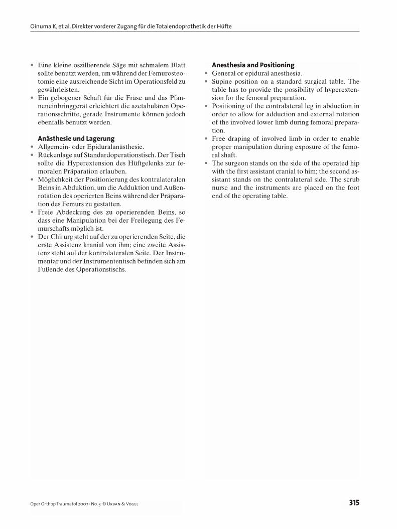
Anästhesie und Lagerung• Allgemein- oder Epiduralanästhesie.• Rückenlage auf Standardoperationstisch. Der Tisch
sollte die Hyperextension des Hüftgelenks zur fe-moralen Präparation erlauben.
• Möglichkeit der Positionierung des kontralateralen Beins in Abduktion, um die Adduktion und Außen-rotation des operierten Beins während der Präpara-tion des Femurs zu gestatten.
• Freie Abdeckung des zu operierenden Beins, so dass eine Manipulation bei der Freilegung des Fe-murschafts möglich ist.
• Der Chirurg steht auf der zu operierenden Seite, die erste Assistenz kranial von ihm; eine zweite Assis-tenz steht auf der kontralateralen Seite. Der Instru-mentar und der Instrumententisch befinden sich am Fußende des Operationstischs.
Anesthesia and Positioning• General or epidural anesthesia.• Supine position on a standard surgical table. The
table has to provide the possibility of hyperexten-sion for the femoral preparation.
• Positioning of the contralateral leg in abduction in order to allow for adduction and external rotation of the involved lower limb during femoral prepara-tion.
• Free draping of involved limb in order to enable proper manipulation during exposure of the femo-ral shaft.
• The surgeon stands on the side of the operated hip with the first assistant cranial to him; the second as-sistant stands on the contralateral side. The scrub nurse and the instruments are placed on the foot end of the operating table.

Oinuma K, et al. Direct Anterior Approach for Total Hip Arthroplasty
316 Oper Orthop Traumatol 2007 · No. 3 © Urban & Vogel
Spina iliaca ant. sup.
Inzision
Incision
N. cutaneus fem. lat.
M. sartorius
M. tensor fasciae latae
Caput fibulae
Abbildung 2Der Hautschnitt verläuft parallel zu einer imaginären Linie zwischen dem vorderen Beckenkamm (B) und dem Fibulakopf (A). Die Inzision sollte 2 cm lateral dieser Linie verlaufen und beginnt in der inguinalen Leistenbeuge. Es ist wichtig, mit dem Hautschnitt nicht zu weit nach medial zu gelangen, um den Nervus cutaneus femoris lateralis zu schonen. Die Länge der Inzision beträgt 6–9 cm, abhängig von der Größe des Pa-tienten und der Dicke des subkutanen Gewebes. Der Haut-schnitt sollte nicht zu kurz gewählt werden, um eine Deh-nung der Haut und damit verbundene Wundheilungs-probleme zu vermeiden. Theoretisch hängt die minimale Länge des Schnitts von der Größe des Pfannenimplantats ab.
Figure 2The incision is parallel to an imaginary line connecting the anterior superior iliac crest (B) to the fibular head (A). The in-cision is 2 cm lateral to this line and starts at the inguinal groin. It is important to do the incision not too medial to avoid injury to the lateral femoral cutaneous nerve. The length of the incision is usually 6–9 cm, depending on the pa-tient’s size and the thickness of the subcutaneous tissue. It is wise to make the length of the skin incision not too short in order to avoid stretching of the skin and concomitant wound healing problems. Theoretically, the minimal length of the skin incision is dependent on the size of the acetabular cup.
Operationstechnik
Abbildungen 2 bis 12
Surgical Technique
Figures 2 to 12
M. tensor fasciae latae
M. sartorius
N. cutaneus fem. lat.Intermuskuläres
Intervall
Intermuscular space
Abbildung 3Bei der Durchtrennung des subkutanen Fettgewebes sollte auf Schonung der Äste des Nervus cutaneus femoris lateralis geachtet werden. Da diese Äste oft nicht zu sehen sind, sollte eine direkte laterale Präparation benutzt werden. Das subku-tane Fett ist hier normalerweise auch bei adipösen Patienten relativ dünn. Die Faszie des Musculus tensor fasciae latae wird etwa 1 cm lateral des intermuskulären Raums durch-trennt, wiederum zur Schonung des Nervs. Stumpfes Vorprä-parieren in die Muskellücke zwischen Musculus tensor fas-ciae latae und Musculus sartorius, am besten digital, um Nervenverletzungen zu vermeiden. Die vordere Hüftgelenk-kapsel kann dann leicht getastet werden.
Figure 3During dissection of the subcutaneous fat tissues care has to be taken not to injure the branches of the lateral femoral cu-taneous nerve. As the branches cannot be identified visually, straight lateral preparation ensures avoidance of the nerve. The subcutaneous fat is normally quite thin even in obese pa-tients. The fascia of the tensor fasciae latae muscle is incised 1 cm lateral from the intermuscular space, again to protect the branches of the lateral femoral cutaneous nerve. Blunt developing of the intermuscular space between the tensor fasciae latae and the sartorius. This can be done best by fin-ger preparation in order to avoid injury to the lateral femoral cutaneous nerve. The anterior hip capsule can now easily be palpated.

Oinuma K, et al. Direkter vorderer Zugang für die Totalendoprothetik der Hüfte
317Oper Orthop Traumatol 2007 · No. 3 © Urban & Vogel
Spina iliaca ant. sup.
A./ V. circumflexa ant.
M. sartorius
M. tensor fasciae latae
Spina iliaca ant. sup.
Lig. inguinale M. sartorius
M. tensor fasciae latae+ Fascia lata
M. iliopsoas
A./V. femoralisN. femoralis
M. vastus intermed.
M. rectus fem.A
D
Abbildung 4Der erste gebogene Hohmann-Retraktor (A) wird lateral-kranial am Schenkelhals eingesetzt, außer-halb der Hüftgelenkkapsel. Ein zweiter Retraktor (B) wird um das Tuberculum innominatum des Trochan-ter major eingebracht, um den Musculus tensor fa-sciae latae aus dem Operationsfeld zu halten. Der zweite Assistent hält mit einem breiten Retraktor die Faszie und den Musculus sartorius nach medial. Die laterale Arteria und Vena circumflexa können jetzt an ihrem Übergang zur medialen Gelenkkapsel identifi-ziert und unterbunden werden.
Figure 4A first curved Hohmann retractor (A) is placed to the superior and lateral aspect of femoral neck, outside of the hip joint capsule. A second retractor (B) is placed around the innominate tubercle of the great-er trochanter; thus, the tensor fasciae latae is retract-ed out of the surgical field. The second assistant re-tracts the fascia and the sartorius muscle to the me-dial side with a broad retractor. Now, the lateral circumflex vessels can be identified crossing the an-terior aspect of the joint capsule and either be cau-terized or ligated.
Abbildung 5Die Aponeurose des Musculus rectus femoris wird entlang ihrer lateralen Begrenzung inzidiert, bis der Kopf des Muskels erreicht ist. Der Ansatz des Muscu-lus rectus am vorderen unteren Beckenkamm bleibt dabei intakt. Der Musculus rectus femoris wird dann nach medial retrahiert. Ein dritter Retraktor (C) wird außerhalb der Kapsel um die Medialseite des Schen-kelhalses eingebracht. Ein vierter Retraktor (D) wird an der vorderen Kante des Azetabulums platziert. Dabei sollte vorsichtig agiert werden, um den Nervus femoralis nicht mit der Spitze des Retraktors zu ver-letzen.
Figure 5The aponeurosis of the rectus femoris is incised along the lateral border of the muscle until the re-flected head of the muscle is reached; the insertion of the rectus on the anterior inferior iliac spine re-mains intact. The rectus femoris muscle is retracted medially and a third retractor (C) is placed outside the capsule and around the medial aspect of the femoral neck. A forth retractor (D) is placed on the anterior rim of the acetabulum. Caution should be taken not to injure the femoral nerve with the tip of this retractor.

Oinuma K, et al. Direct Anterior Approach for Total Hip Arthroplasty
318 Oper Orthop Traumatol 2007 · No. 3 © Urban & Vogel
Retraktor noch außerhalb der Kapsel
Retractor outside capsule
Retractor insidecapsule
Retraktorinnerhalb der Kapsel
T-förmige Kapselinzision
T-shaped incision
Vordere Kapsel
D
Anterior jointcapsule
Abbildung 6Nun wird die Vorderseite des Schenkelhalses dargestellt. Die Kapsel wird T-förmig inzidiert, ihre zwei lappenförmigen An-teile werden zu beiden Seiten aufgeklappt und an der Azeta-bulumkante abgesetzt. Schenkelhals und Femurkopf können jetzt durch Bewegung des Beins klar identifiziert werden. Der Schenkelhals kann mit einem Cobb-Raspatorium zirkumfe-rentiell präpariert werden. Die Retraktoren A und C werden entfernt und intrakapsulär erneut eingesetzt. Damit wird der gesamte Umfang des Schenkelhalses der Osteotomie zu-gänglich.
Figure 6Now, the anterior aspect of the femoral neck is exposed. An inverse T-shaped incision is being done and both flaps of ei-ther side of the capsule are opened up like a door and excised well onto the rim of the acetabulum in one or more steps. Now, the femoral head and neck can be clearly identified by moving the leg. The femoral neck should now be dissected circumferentially by the use of a Cobb elevator. The retractors A and C are removed and repositioned inside the capsule, thus exposing circumferentially the complete femoral neck for osteotomy.

Oinuma K, et al. Direkter vorderer Zugang für die Totalendoprothetik der Hüfte
319Oper Orthop Traumatol 2007 · No. 3 © Urban & Vogel
Schablone
Template
Collum femoris
Oszillierende Säge
Oscillating saw
M. vastus intermed.
Trochanter minor
Verbindungzwischen Collum femorisund Trochanter major
Connection between femoral neck and greater trochanter
Trochanter major
Abbildung 7Der Übergang des Schenkelhalses in den Trochanter major kann leicht palpiert und als Orientierungsmarke für die Schenkelhalsosteotomie benutzt werden. Eine Schablone er-leichtert die Bestimmung des korrekten Osteotomiewinkels, der für einen Bicontact-Schaft etwa 55° betragen sollte.Um den Femurkopf leicht entfernen zu können, sollte die an-teromediale Kapsel komplett reseziert werden. Osteophyten im Bereich des Femurkopfes werden mit einem Meißel ent-fernt. Eine doppelte Osteotomie mit einem medialen und la-teralen Schnitt an der Stelle der geplanten Osteotomie er-zeugt eine Schenkelhalsscheibe von etwa 1 cm Dicke. Diese Knochenscheibe sollte vor der Luxation des Femurkopfes ent-fernt werden. Dadurch kann der Kopf leichter herausgezogen werden, was sonst aus geometrischen Gründen Schwierig-keiten bereiten kann. Der Femurkopf wird mit einem Korken-zieher oder V-förmigen Meißel entfernt.Das korrekte Niveau der Femurkopfosteotomie kann mit einem Taster bestätigt werden. Falls der Schnitt im Vergleich zur präoperativen Planung zu weit proximal liegt, sollte nachreseziert werden.
Figure 7The junction between femoral neck and the greater trochan-ter can be used as a landmark for femoral neck osteotomy and palpated easily. A template facilitates the angulation of the osteotomy (which should be around 55° for the Bicontact stem).To remove the femoral head easily, the anteromedial capsule is completely resected. Any osteophyte on the femoral head (capital drop) is resected with a chisel. A double osteotomy with a medial cut and a lateral cut at the site of the planned osteotomy produces a slice of the femoral neck, approximate-ly 1 cm thick. This “disk” of the femoral neck has to be re-moved prior to extraction of the head. Thus, the removal of the head is eased, which otherwise can be difficult due to geometric reasons. The femoral head is removed with a cork screw extractor or a V-shaped chisel.The correct level of femoral neck osteotomy at the basis of the femoral neck can be confirmed by a caliper. If the level of the actual cut is found to be higher than planned preopera-tively, an additional resection on the neck must be done to meet the planning requirements.
Oinuma K, et al. Direct Anterior Approach for Total Hip Arthroplasty
320 Oper Orthop Traumatol 2007 · No. 3 © Urban & Vogel
Capsula articularis
Labrum acetabuli
Fossa acetabuli
E
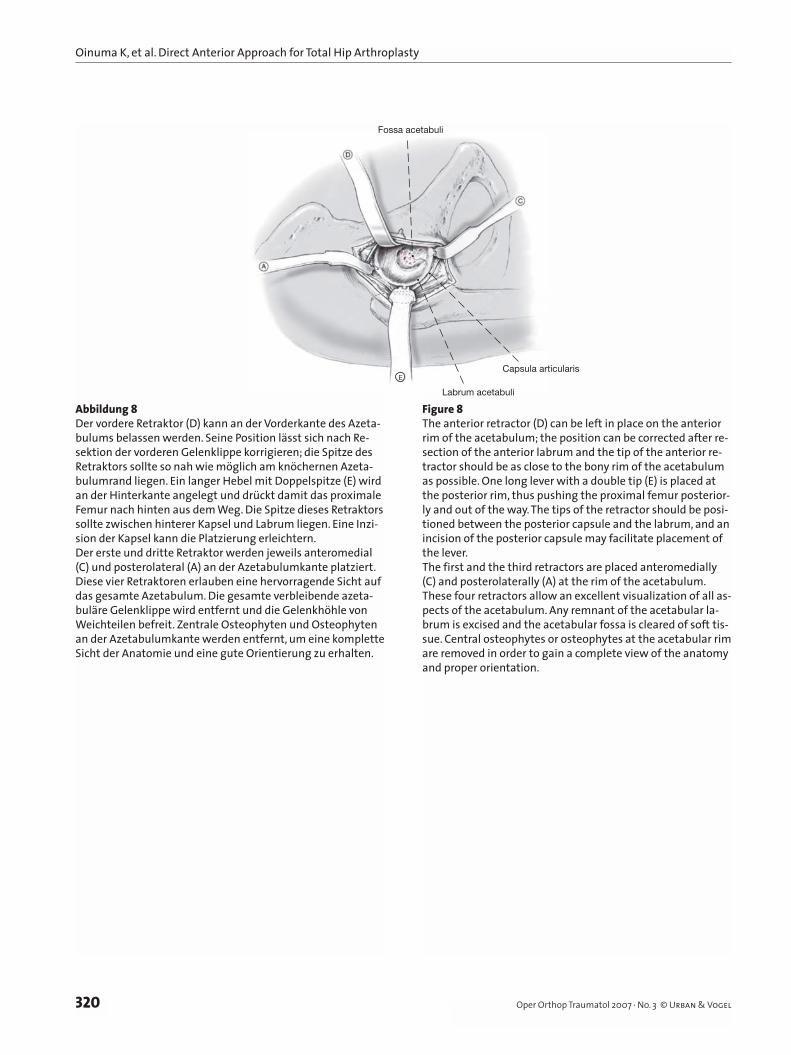
Abbildung 8Der vordere Retraktor (D) kann an der Vorderkante des Azeta-bulums belassen werden. Seine Position lässt sich nach Re-sektion der vorderen Gelenklippe korrigieren; die Spitze des Retraktors sollte so nah wie möglich am knöchernen Azeta-bulumrand liegen. Ein langer Hebel mit Doppelspitze (E) wird an der Hinterkante angelegt und drückt damit das proximale Femur nach hinten aus dem Weg. Die Spitze dieses Retraktors sollte zwischen hinterer Kapsel und Labrum liegen. Eine Inzi-sion der Kapsel kann die Platzierung erleichtern.Der erste und dritte Retraktor werden jeweils anteromedial (C) und posterolateral (A) an der Azetabulumkante platziert. Diese vier Retraktoren erlauben eine hervorragende Sicht auf das gesamte Azetabulum. Die gesamte verbleibende azeta-buläre Gelenklippe wird entfernt und die Gelenkhöhle von Weichteilen befreit. Zentrale Osteophyten und Osteophyten an der Azetabulumkante werden entfernt, um eine komplette Sicht der Anatomie und eine gute Orientierung zu erhalten.
Figure 8The anterior retractor (D) can be left in place on the anterior rim of the acetabulum; the position can be corrected after re-section of the anterior labrum and the tip of the anterior re-tractor should be as close to the bony rim of the acetabulum as possible. One long lever with a double tip (E) is placed at the posterior rim, thus pushing the proximal femur posterior-ly and out of the way. The tips of the retractor should be posi-tioned between the posterior capsule and the labrum, and an incision of the posterior capsule may facilitate placement of the lever.The first and the third retractors are placed anteromedially (C) and posterolaterally (A) at the rim of the acetabulum. These four retractors allow an excellent visualization of all as-pects of the acetabulum. Any remnant of the acetabular la-brum is excised and the acetabular fossa is cleared of soft tis-sue. Central osteophytes or osteophytes at the acetabular rim are removed in order to gain a complete view of the anatomy and proper orientation.
Oinuma K, et al. Direkter vorderer Zugang für die Totalendoprothetik der Hüfte
321Oper Orthop Traumatol 2007 · No. 3 © Urban & Vogel
Fräse
Reamer
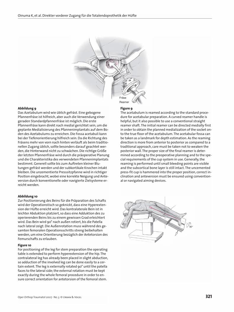
Abbildung 9Das Azetabulum wird wie üblich gefräst. Eine gebogene Pfannenfräse ist hilfreich, aber auch die Verwendung einer geraden Standardpfannenfräse ist möglich. Die erste Pfannenfräse kann direkt nach medial gerichtet sein, um die geplante Medialisierung des Pfannenimplantats auf dem Bo-den des Azetabulums zu erreichen. Die Fossa acetabuli kann bei der Tiefenorientierung hilfreich sein. Da die Richtung des Fräsens mehr von vorn nach hinten verläuft als beim traditio-nellen Zugang üblich, sollte besonders darauf geachtet wer-den, die Hinterwand nicht zu schwächen. Die richtige Größe der letzten Pfannenfräse wird durch die präoperative Planung und die Charakteristika des verwendeten Pfannenimplantats bestimmt. Generell sollte bis zum Auftreten kleiner Blu-tungen gefräst werden und der subkortikale Knochen intakt bleiben. Die unzementierte Presssitzpfanne wird in richtiger Position eingebracht, wobei eine korrekte Neigung und Ante-version durch konventionelle oder navigierte Zielsysteme er-reicht werden.
Figure 9The acetabulum is reamed according to the standard proce-dure for acetabular preparation. A curved reamer handle is helpful, but it also possible to use a conventional straight reamer shaft. The initial reamer can be directed medially first in order to obtain the planned medialization of the socket on-to the true floor of the acetabulum. The acetabular fossa can be taken as a landmark for depth estimation. As the reaming direction is more from anterior to posterior as compared to a traditional approach, care must be taken not to weaken the posterior wall. The proper size of the final reamer is deter-mined according to the preoperative planning and to the spe-cial requirements of the cup system in use. Generally, the reaming is performed until small bleeding points are visible and the subcortical bone layer is still intact. The uncemented press-fit cup is hammered into the proper position, correct in-clination and anteversion must be ensured using convention-al or navigated aiming devices.
M. piriformis
Abbildung 10Zur Positionierung des Beins für die Präparation des Schafts wird der Operationstisch so geknickt, dass eine Hyperexten-sion der Hüfte erreicht wird. Das kontralaterale Bein ist in leichter Abduktion platziert, so dass eine Adduktion des zu operierenden Beins bis zu einem gewissen Grad erleichtert wird. Das Bein wird 90° nach außen rotiert, bis die Patella nach lateral zeigt. Die Außenrotation muss während des ge-samten femoralen Operationsschritts streng beibehalten werden, um eine Orientierung bezüglich der Antetorsion des Femurschafts zu erlauben.
Figure 10For positioning of the leg for stem preparation the operating table is extended to perform hyperextension of the hip. The contralateral leg has already been placed in slight abduction, so adduction of the involved leg can be done easily to a cer-tain extent. The leg is externally rotated 90° until the patella faces to the lateral side; the external rotation must be kept exactly during the whole femoral procedure in order to en-sure correct orientation for antetorsion of the femoral stem.
Oinuma K, et al. Direct Anterior Approach for Total Hip Arthroplasty
322 Oper Orthop Traumatol 2007 · No. 3 © Urban & Vogel
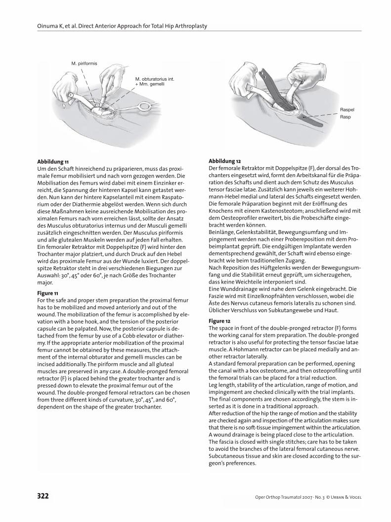
Abbildung 11Um den Schaft hinreichend zu präparieren, muss das proxi-male Femur mobilisiert und nach vorn gezogen werden. Die Mobilisation des Femurs wird dabei mit einem Einzinker er-reicht, die Spannung der hinteren Kapsel kann getastet wer-den. Nun kann der hintere Kapselanteil mit einem Raspato-rium oder der Diathermie abgelöst werden. Wenn sich durch diese Maßnahmen keine ausreichende Mobilisation des pro-ximalen Femurs nach vorn erreichen lässt, sollte der Ansatz des Musculus obturatorius internus und der Musculi gemelli zusätzlich eingeschnitten werden. Der Musculus piriformis und alle glutealen Muskeln werden auf jeden Fall erhalten. Ein femoraler Retraktor mit Doppelspitze (F) wird hinter den Trochanter major platziert, und durch Druck auf den Hebel wird das proximale Femur aus der Wunde luxiert. Der doppel-spitze Retraktor steht in drei verschiedenen Biegungen zur Auswahl: 30°, 45° oder 60°, je nach Größe des Trochanter major.
Figure 11For the safe and proper stem preparation the proximal femur has to be mobilized and moved anteriorly and out of the wound. The mobilization of the femur is accomplished by ele-vation with a bone hook, and the tension of the posterior capsule can be palpated. Now, the posterior capsule is de-tached from the femur by use of a Cobb elevator or diather-my. If the appropriate anterior mobilization of the proximal femur cannot be obtained by these measures, the attach-ment of the internal obturator and gemelli muscles can be incised additionally. The piriform muscle and all gluteal muscles are preserved in any case. A double-pronged femoral retractor (F) is placed behind the greater trochanter and is pressed down to elevate the proximal femur out of the wound. The double-pronged femoral retractors can be chosen from three different kinds of curvature, 30°, 45°, and 60°, dependent on the shape of the greater trochanter.
M. piriformis
M. obturatorius int.+ Mm. gemelli
F
Abbildung 12Der femorale Retraktor mit Doppelspitze (F), der dorsal des Tro-chanters eingesetzt wird, formt den Arbeitskanal für die Präpa-ration des Schafts und dient auch dem Schutz des Musculus tensor fasciae latae. Zusätzlich kann jeweils ein weiterer Hoh-mann-Hebel medial und lateral des Schafts eingesetzt werden.Die femorale Präparation beginnt mit der Eröffnung des Knochens mit einem Kastenosteotom; anschließend wird mit dem Oesteoprofiler erweitert, bis die Probeschäfte einge-bracht werden können.Beinlänge, Gelenkstabilität, Bewegungsumfang und Im-pingement werden nach einer Probereposition mit dem Pro-beimplantat geprüft. Die endgültigen Implantate werden dementsprechend gewählt, der Schaft wird ebenso einge-bracht wie beim traditionellen Zugang.Nach Reposition des Hüftgelenks werden der Bewegungsum-fang und die Stabilität erneut geprüft, um sicherzugehen, dass keine Weichteile interponiert sind.Eine Wunddrainage wird nahe dem Gelenk eingebracht. Die Faszie wird mit Einzelknopfnähten verschlossen, wobei die Äste des Nervus cutaneus femoris lateralis zu schonen sind. Üblicher Verschluss von Subkutangewebe und Haut.
Figure 12The space in front of the double-pronged retractor (F) forms the working canal for stem preparation. The double-pronged retractor is also useful for protecting the tensor fasciae latae muscle. A Hohmann retractor can be placed medially and an-other retractor laterally.A standard femoral preparation can be performed, opening the canal with a box osteotome, and then osteoprofiling until the femoral trials can be placed for a trial reduction.Leg length, stability of the articulation, range of motion, and impingement are checked clinically with the trial implants. The final components are chosen accordingly, the stem is in-serted as it is done in a traditional approach.After reduction of the hip the range of motion and the stability are checked again and inspection of the articulation makes sure that there is no soft-tissue impingement within the articulation.A wound drainage is being placed close to the articulation. The fascia is closed with single stitches; care has to be taken to avoid the branches of the lateral femoral cutaneous nerve. Subcutaneous tissue and skin are closed according to the sur-geon’s preferences.
Raspel
Rasp

Oinuma K, et al. Direkter vorderer Zugang für die Totalendoprothetik der Hüfte
323Oper Orthop Traumatol 2007 · No. 3 © Urban & Vogel
Postoperative Behandlung• Die postoperative Mobilisation hängt vom Nachbe-
handlungsprotokoll des spezifischen Implantatsys-tems ab. Bei unseren Patienten wurde nach Implan-tation des nichtzementierten Bicontact-Systems so-fortige Vollbelastung erlaubt.
• Am 1. postoperativen Tag wird dem Patienten das vollbelastende Aufstehen mit einem Rollator ge-stattet. Am 2. Tag wird die Drainage entfernt, und der Patient wird ermutigt, mit einer Unterarmstütze zu gehen und Treppen zu steigen. Am 10. postope-rativen Tag werden die Nähte entfernt.
• Eine Prophylaxe heterotoper Ossifikationen kann mit nichtsteroidalen Antiphlogistika analog zum Protokoll nach Hüfttotalendoprothese mit konven-tionellem Zugang durchgeführt werden.
Fehler, Gefahren, Komplikationen• Sensible Störungen im Bereich des Nervus cutaneus
femoris lateralis sind meist vorübergehend und ver-schwinden spontan. Um eine Läsion dieses Nervs zu vermeiden, sollten der Hautschnitt und die Durch-trennung der Faszie lateral des intermuskulären Raums erfolgen und anschließend der intermusku-läre Raum zwischen Musculus tensor fasciae latae und Musculus sartorius unter der Faszie präpariert werden.
• Hämatome vorn und lateral am Oberschenkel sind meist diffus und bilden sich ohne Revision zurück. Bei Wundheilungsstörungen postoperatives Wund-management, wie auch bei konventionellen Zugän-gen üblich.
• Intraoperative Fehlstellung der Prothese: Positions-änderung der Prothese unter dem Bildwandler. Es wird eine routinemäßige intraoperative Röntgen-kontrolle empfohlen.
• Ungenügende Übersicht zur Implantation der Pro-these und zur Kontrolle von intraoperativen Kom-plikationen oder Verletzungen neurovaskulärer Strukturen: Der Zugang kann jederzeit nach Smith-Petersen sowohl nach proximal als auch nach distal erweitert werden.
• Postoperative Infektion: Revision, gründliches Débridement sowie Drainage des Operationsge-biets entsprechend dem Standardvorgehen bei in-fizierter Hüftendoprothese. Die Prothese kann nötigenfalls über den gleichen Zugang entfernt werden. Bei Bedarf lässt sich der Zugang vergrö-ßern.
• Postoperative Prothesenluxation: Reposition und konservative Weiterbehandlung, vorausgesetzt, die
Postoperative Management• Postoperative mobilization depends on the postop-
erative protocol for the specific endoprosthetic sys-tem. In our patients after uncemented hip replace-ment with the Bicontact system full weight bearing is allowed immediately postoperatively.
• On the 1st day after surgery, the patient is allowed out of bed, full weight bearing with a walker. On the 2nd day, the wound drainage is removed and the pa-tient is encouraged to walk with a T-cane and go up and down the stairs. On the 10th day, the stitches can be removed.
• Prophylaxis for heterotopic ossification can be done using nonsteroidal anti-inflammatory drugs accord-ing to the protocol after total hip arthroplasty with a conventional approach.
Errors, Hazards, Complications• Sensory disturbances in field of the lateral femoral
cutaneous nerve are transient and will disappear spontaneously. In order to avoid a lesion to this nerve, it is recommended to incise the skin and split the fascia lateral from the intermuscular space. Then develop the interval between tensor fasciae latae muscle and sartorius muscle under the fascia.
• Hematomas at anterior and lateral aspect of the thigh are diffuse and regress spontaneously. Wound heal-ing problems can be handled according to the proto-col in conventional approaches.
• Intraoperative malalignment of the prosthesis: changing of the prosthesis to the correct position under fluoroscopic control. A routine intraopera-tive X-ray control is recommended.
• No sufficient exposure for implantation of any pros-thetic device or for control of any intraoperative complication like bleeding or lesion of neurovascu-lar structures: the approach can easily be enlarged according to the Smith-Petersen approach, both to the proximal and distal direction.
• Postoperative infection: revision, meticulous de-bridement and drainage of the surgical site accord-ing to standard protocols in infected total hip re-placement. Removal of the prosthesis can be ac-complished by the same approach, if necessary. The approach can easily be enlarged for revision, de-bridement or infection control, if required.
ResultsBetween August 2004 and December 2005, 116 pri-mary total hip arthroplasties were performed in 111 patients without a history of previous surgery to the

Oinuma K, et al. Direct Anterior Approach for Total Hip Arthroplasty
324 Oper Orthop Traumatol 2007 · No. 3 © Urban & Vogel
Implantatposition ist korrekt. Ansonsten Revision zur Korrektur der Ausrichtung der Implantate.
ErgebnisseZwischen August 2004 und Dezember 2005 wurden 116 Totalendoprothesen bei 111 Patienten implantiert. Die betroffenen Hüftgelenke waren nicht voroperiert. Alle Operationen wurden von zwei der Autoren durchgeführt (K.O. und H.S.). Patienten mit Femur-fraktur (sechs Hüften) und ausgeprägter Hüftdyspla-sie (fünf Hüften) wurden ausgeschlossen. Der Grad der Dysplasie wurde dabei nach der Crowe-Klassifika-tion bestimmt [5]. Ausgeschlossen wurden Arthrosen vom Crowe-Typ 3 und 4, die wegen der schwierigen anatomischen Verhältnisse eine modifizierte Technik und ein anderes Rehabilitationsprotokoll erfordern. Eine Implantation der Totalendoprothese über den direkten vorderen Zugang wäre hier prinzipiell mög-lich.
Um die Rekonvaleszenz und die Sicherheit eines beschleunigten Rehabilitationsprotokolls zu beurtei-len, wurden auch Patienten mit Gehbehinderung am kontralateralen Bein (sechs Hüften) ausgeschlossen. Die Ergebnisse umfassen also 99 Hüften von 95 Pa-tienten (82 Frauen und 13 Männer). Nachuntersu-chungen fanden mindestens 3 Monate postopera -tiv statt. Das Durchschnittsalter lag bei 62,5 Jahren (46–84 Jahre). Die Durchschnittsgröße der Patienten betrug 154 cm (134–177 cm), das Durchschnittsgewicht 55,8 kg (39–84 kg). Der durchschnittliche BMI lag bei 23,1 kg/m2 (18,1–37,7 kg/m2). Die präoperative Dia-gnose lautete bei 92 Hüften primäre Koxarthrose, bei fünf Hüften avaskuläre Hüftkopfnekrose und bei zwei Hüften rasch fortschreitende destruierende Osteo-arthritis. Bei den Patienten mit Arthrose waren 87 Hüften (94,6%) sekundäre Folge einer angeborenen Dysplasie (Crowe 1: 66 Hüften, Crowe 2: 21 Hüften). Nur fünf Hüften (5,4%) wurden aufgrund einer pri-märer Osteoarthritis nach der Definition von Naka-mura et al. [12] operiert.
Das femorale Bicontact-Implantatsystem wurde bei 83 Hüften und das femorale CentPilar-System bei 16 Hüften verwendet. Als Pfannenimplantat diente 83-mal das Bicontact-System und 16-mal das Secure-fit-AD-System. Alle Implantate wurden unzementiert eingesetzt.
Die durchschnittliche Operationszeit betrug 79 min (45–150 min). Die Operationsdauer verkürzte sich im Lauf der Studie (Abbildung 13). Der durchschnittliche intraoperative Blutverlust betrug 393 ml (73–1 053 ml). Kein Patient erhielt eine Bluttransfusion. Durch-
affected hip. All implantations were performed by two of the authors (K.O. and H.S.).We excluded patients with fracture of the femur (six hips), or severe devel-opmental dysplasia (five hips). The severity of dyspla-sia was graded according to the Crowe classification [5]. We excluded Crowe type 3 or 4 osteoarthritis which requires a modified procedure to cope with the diffi-cult anatomic situation and a different rehabilitation protocol, although it was possible to perform total hip arthroplasty with direct anterior approach.
In order to assess the recovery rate and safety of a rapid rehabilitation protocol after surgery, we also ex-cluded patients with walking disability of the unin-volved lower limb (six hips). Thus, the results include 99 hips in 95 patients (82 women and 13 men). They were followed for at least 3 months postoperatively. The mean age was 62.5 years (range, 46–84 years). The mean height was 154 cm (range, 134–177 cm), and the mean weight 55.8 kg (range, 39–84 kg). The mean BMI amounted to 23.1 kg/m2 (range, 18.1–37.7 kg/m2). The preoperative diagnosis was osteoarthritis in 92 hips, avascular necrosis of the femoral head in five hips, and rapid destructive osteoarthritis in two hips. In patients with osteoarthritis, 87 hips (94.6%) were secondary to developmental dysplasia (Crowe 1: 66 hips, Crowe 2: 21 hips). Only five hips (5.4%) were primary osteoar-thritis which was defined by Nakamura et al. [12].
The Bicontact femoral system was used in 83 hips, and the CentPilar femoral system in 16 hips. The Bi-contact plasma cup system was implanted in 83 hips, and the securefit AD shell system in 16 hips. All im-plantations were done without the use of cement.
Mean surgical time was 79 min (range, 45–150 min). The operative time was decreasing gradually (Figure 13). Mean operative blood loss amounted to 393 ml (range, 73–1,053 ml). We never gave a donor blood transfusion. Patients were able to walk over 50 m with a T-cane at an average of 5.3 days (2–30 days) after surgery.
We never observed a serious complication requir-ing surgical intervention. Complications included one traumatic dislocation, one transient femoral nerve pal-sy, one heterotopic ossification, and one asymptomatic stem subsidence (4 mm) in the early postoperative pe-riod. The dislocation was treated with closed reduction and no use of a hip brace. No lesions of lateral femoral cutaneous nerve were palsy was noted.
Radiographic evaluation was based on the routine series of postoperative radiographs. The anteroposte-rior radiographs were obtained and the cup inclination and anteversion were calculated according to the

Oinuma K, et al. Direkter vorderer Zugang für die Totalendoprothetik der Hüfte
325Oper Orthop Traumatol 2007 · No. 3 © Urban & Vogel
schnittlich 5,3 Tage (2–30 Tage) postoperativ konnten die Patienten mit einer Unterarmstütze eine 50 m lan-ge Gehstrecke bewältigen.
Ernsthafte Komplikationen mit der Notwendigkeit einer chirurgischen Intervention traten nicht auf. Die Komplikationen umfassten eine traumatische Luxa-tion, eine vorübergehende Lähmung des Nervus femoralis, eine heterotope Ossifikation sowie ein asymptomatisches Nachsinken des Implantatschafts (4 mm) in der frühen postoperativen Periode. Die Lu-xation wurde mit geschlossener Reposition und ohne Hüftschiene behandelt. Läsionen des Nervus cutane-ous femoris lateralis wurden nicht festgestellt.
Radiologisch evaluierten wir die Patienten mit Hilfe der postoperativen Röntgenaufnahmen. Die anteroposterioren Röntgenbilder dienten der Beur-teilung der Pfannenneigung und Anteversion, die nach der Methode nach Dorr & Wan [7] berechnet wurden. Die radiographische Analyse zeigte eine durchschnittliche Pfannenneigung von 44,0° ± 7,6° und eine durchschnittliche Anteversion von 16,8° ± 4,6°. Die frontale Ausrichtung des femoralen Implan-tats wich bei 96 Hüften weniger als 3° von der Neu-tralposition ab. Varisierungen von mehr als 3° wur-den bei drei Hüften festgestellt. Einer dieser varisch eingesetzten Schäfte sank in der frühen postopera-tiven Periode um 4 mm nach, war dabei aber asymp-tomatisch. Alle fehlstehenden Schaftkomponenten waren CentPilar-Schäfte. Eine Varusfehlstellung dieses Schafts wurde bei verschiedenen Zugängen gefunden und scheint ein spezifisches Problem des sehr kurzen Schafts zu sein.
Der durchschnittliche präoperative JOA-Score von 47,2 Punkten (18–63 Punkte) verbesserte sich 3 Mo-nate postoperativ auf durchschnittlich 92,3 Punkte (67–100 Punkte; p < 0,001) und bei der letzten Nachun-tersuchung nach durchschnittlich 17 Monaten (9–26 Monate) auf 94,2 Punkte (72–100 Punkte).
method of Dorr & Wan [7]. The radiographic analysis showed a mean cup inclination of 44.0° ± 7.6° and a mean anteversion angle of 16.8° ± 4.6°. The femoral component’s coronal alignment was within 3° of neu-tral position in 96 hips. Varus alignment of more than 3° was found in three hips. One of those varus-aligned stems subsided 4 mm in the early postoperative period, but was asymptomatic. All malaligned femoral com-ponents were CentPilar stems; varus malalignment of this specific stem has been reported with different ap-proaches and seems to be a specific problem of the very short femoral stem.
The mean preoperative JOA Score of 47.2 points (range, 18–63 points) improved to 92.3 points (range, 67–100 points) at 3 months postoperatively (p < 0.001) and 94.2 (range, 72–100 points) at the latest follow-up at an average of 17 months (range, 9–26 months).
In conclusion, the minimally invasive direct anterior approach has turned out to be safe and reliable and can be recommended for routine use in total hip re-placement surgery. This approach does not only per-mit a short skin incision, but also avoids detachment or incision of muscles. After a short learning curve an av-erage surgical time of < 60 min can be achieved. As far as the rehabilitation process is concerned, we do have the impression of a shorter and faster return to a nor-mal activity level, but valid data are still limited for this and other minimally invasive total hip replacement procedures.
Op
erat
ions
zeit
Sur
gica
l tim
e (m
in)
140
120
100
80
60
40
20
00 50 100
Fallzahl
Case number
Abbildung 13Die Operationszeit der ersten 99 Hüfttotalendoprothesen spiegelt die Lernkurve wider. Inzwischen kann bei normaler Anatomie die Totalendoprothese in < 60 min implantiert werden.
Figure 13The operating time of the first 99 total hip replacements re-flects the learning curve. Now, total hip replacement can be done in < 1 h in patients with normal anatomy.

Oinuma K, et al. Direct Anterior Approach for Total Hip Arthroplasty
326 Oper Orthop Traumatol 2007 · No. 3 © Urban & Vogel
Zusammenfassend erwies sich der minimalinvasive, direkte vordere Zugang als sicher und zuverlässig und kann deshalb empfohlen werden. Dieser Zugang er-laubt nicht nur einen kurzen Hautschnitt, sondern ver-meidet auch das Ablösen oder Durchtrennen von Muskulatur. Nach einer kurzen Lernkurve kann die Operationszeit auf < 60 min gesenkt werden. Bezüg-lich der Rehabilitation haben wir zwar der Eindruck einer schnelleren Wiederherstellung, doch sind die da-zu verfügbaren Daten für diese und andere minimal-invasive Methoden zur Hüfttotalendoprothetik noch begrenzt.
Literatur – References1. Archibeck MJ, White RE Jr. Learning curve for the two-incision total hip
replacement. Clin Orthop 2004;429:232–8.2. Berger RA. Total hip arthroplasty using the minimally invasive two-in-
cision approach. Clin Orthop 2003;417:232–41.3. Berger RA. Mini-incision total hip replacement using an anterolateral ap-
proach: technique and results. Orthop Clin North Am 2004;35:143–51.4. Berger RA, Jacobs JJ, Meneghini RM, et al. Rapid rehabilitation and re-
covery with minimally invasive total hip arthroplasty. Clin Orthop 2004;429:239–47.
5. Crowe JF, Mani VJ, Ranawat CS. Total hip replacement in congenital dis-location and dysplasia of the hip. J Bone Joint Surg Am 1979;61:15–23.
6. DiGioia AM III, Plakseychuk AY, Levison TJ, et al. Mini-incision technique for total hip arthroplasty with navigation. J Arthroplasty 2003;18:123–8.
7. Dorr LD, Wan Z . Causes of and treatment protocol for instability of total hip replacement. Clin Orthop 1998;355:144–51.
8. Jerosch J, Theising C, Fadel ME. Antero-lateral minimal invasive (ALMI) approach for total hip arthroplasty: technique and early results. Arch Orthop Trauma Surg 2006;126:164–73.
9. Judet J, Judet R. The use of an artificial femoral head for arthroplasty of the hip joint. J Bone Joint Surg Br 1950;32:166–73.
10. Kennon RE, Keggi JM, Wetmore RS, et al. Total hip arthroplasty through a minimally invasive anterior surgical approach. J Bone Joint Surg Am 2003;85:Suppl 4:39–48.
11. Krismer M, Nogler M, Rachbauer F. Direct, anterior, single incision ap-proach. In: Hozak WJ, Krismer M, Nogler M, et al., eds. Minimal invasive joint arthroplasty. Heidelberg–New York–Tokyo: Springer, 2004:4–10.
12. Nakamura S, Ninomiya S, Nakamura T. Primary osteoarthritis of the hip joint in Japan. Clin Orthop 1989;241:190–6.
13. Nogler M. Navigated minimal invasive total hip arthroplasty. Surg Technol Int 2004;12:259–62.
14. Ogonda L, Wilson R, Archbold P, et al. A minimal-incision technique in total hip arthroplasty does not improve early postoperative out-comes. A prospective, randomized, controlled trial. J Bone Joint Surg Am 2005;87:701–10.
15. Rachbauer F. Minimally invasive total hip arthroplasty via direct ante-rior approach. Orthopäde 2005;34:1103–8, 1110.
16. Siguier T, Siguier M, Brumpt B. Mini-incision anterior approach does not increase dislocation rate: a study of 1037 total hip replacements. Clin Orthop 2004;426:164–73.
17. Wenz JF, Gurkan I, Jibodh SR. Mini-incision total hip arthroplasty: a comparative assessment of perioperative outcomes. Orthopedics 2002;25:1031–43.
18. Wohlrab D, Hagel A, Hein W. Advantages of minimal invasive total hip replacement in the early phase of rehabilitation. Z Orthop Ihre Grenzgeb 2004;142:685–90.
Korrespondenzanschrift – Address for CorrespondenceProf. Dr. Christoph EingartnerCaritas-KrankenhausUhlandstraße 7D-97980 Bad MergentheimTelefon (+49/7931) 58-3001, Fax -3090E-Mail: [email protected]
Copyright © 2022 FDOKUMEN