Spoken Language Development in Children Following Cochlear Implantation
Upload
khangminh22Category
view
4download
0
Neuron
Article
Notch Inhibition Induces Cochlear Hair CellRegeneration and Recovery of Hearingafter Acoustic TraumaKunio Mizutari,1,2,5 Masato Fujioka,1,2,3,5 Makoto Hosoya,3 Naomi Bramhall,1,2,4 Hirotaka James Okano,3
Hideyuki Okano,3 and Albert S.B. Edge1,2,4,*1Department of Otology and Laryngology, Harvard Medical School, Boston, MA 02115, USA2Tillotson Unit, Eaton-Peabody Laboratory, Massachusetts Eye and Ear Infirmary, Boston, MA 02114, USA3Department of Physiology, Keio University School of Medicine, Tokyo 160-8582, Japan4Program in Speech and Hearing Bioscience and Technology, Division of Health Science and Technology, Harvard & MIT, Cambridge,
MA 02139, USA5These authors contributed equally to this work*Correspondence: [email protected]
http://dx.doi.org/10.1016/j.neuron.2012.10.032
SUMMARY
Hearing loss due to damage to auditory hair cells isnormally irreversible because mammalian hair cellsdo not regenerate. Here, we show that new hair cellscan be induced and can cause partial recovery ofhearing in ears damaged by noise trauma, whenNotch signaling is inhibited by a g-secretase inhibitorselected for potency in stimulating hair cell differen-tiation from inner ear stem cells in vitro. Hair cellgeneration resulted from an increase in the level ofbHLH transcription factor Atoh1 in response to inhi-bition of Notch signaling. In vivo prospective labelingof Sox2-expressing cells with a Cre-lox systemunambiguously demonstrated that hair cell genera-tion resulted from transdifferentiation of supportingcells. Manipulating cell fate of cochlear sensory cellsin vivo by pharmacological inhibition of Notch sig-naling is thus a potential therapeutic approach tothe treatment of deafness.
INTRODUCTION
The cochlear sensory epithelium contains hair cells adapted for
the detection of sound, which is transduced by stereocilia at their
apical surfaces (Hudspeth, 2008; Nayak et al., 2007). Hair cells
produced during development are postmitotic and are not
replaced after loss (Chen and Segil, 1999; Edge and Chen,
2008; Kelley, 2006; Sage et al., 2005) or as part of normal cell
turnover in mammals (Corwin and Cotanche, 1988; Fritzsch
et al., 2006; Ryals and Rubel, 1988). As a result, deafness due
to hair cell loss is irreversible. Hair cell development includes
a complex series of fate decisions, in which prosensory epithelial
cells acquire different fates, either hair cell or supporting cell,
through a process of lateral inhibition that is mediated by Notch
signaling (Adam et al., 1998; Daudet and Lewis, 2005; Kelley,
2006). Supporting cells are prevented from differentiating into
58 Neuron 77, 58–69, January 9, 2013 ª2013 Elsevier Inc.
hair cells by active Notch signaling stimulated by ligands on
adjacent hair cells.
Here, we manipulate Notch signaling to generate new hair
cells in a deafened animal. Recent insights at the cellular and
molecular level have motivated the effort to assess efficacy
in vivo. One question has been whether there were cells in the
damaged cochlea that signaled through the Notch pathway
and could serve as precursors for hair cells. Notch signaling
has been difficult to detect in the adult cochlea (Batts et al.,
2009; Doetzlhofer et al., 2009; Hartman et al., 2009), but some
have shown upregulation after damage. In addition, inner ear
stem cells isolated postnatally acted as precursors to hair cells
when treated with a g-secretase inhibitor (Jeon et al., 2011).
Mechanistic work on the role of Notch revealed a requirement
for Atoh1 for the efficacy of the g-secretase inhibitor, as prevent-
ing Atoh1 expression at the time of inhibitor treatment blocked
the differentiation to hair cells (Jeon et al., 2011). This was
consistent with the result that Atoh1 overexpression with viruses
or plasmids in an immature or adult ototoxic drug-injured
cochlea (Gubbels et al., 2008; Izumikawa et al., 2005; Zheng
and Gao, 2000) resulted in generation of new hair cells in the
organ of Corti.
We approached the problem by identifying a potent g-secre-
tase inhibitor in an assay with inner ear stem cells and assessing
its efficacy first in organ of Corti explants after damage of hair
cells and then in a mouse model of deafness. We used a lineage
tag to determine the source of the new hair cells. We show that
indeed new hair cells were formed after treatment with the inhib-
itor, that they arose by transdifferentiation of supporting cells,
and that the new hair cells contributed to a partial reversal of
hearing loss in mice.
RESULTS
Screening for g-Secretase Inhibitors that Induce HairCell Differentiation from Inner Ear Stem CellsLigand-triggered g-secretase activity catalyzes proteolytic
release of Notch intracellular domain and thereby mediates the
first step of Notch signal transduction. We previously showed
Neuron
Regeneration of Hair Cells
that g-secretase inhibitors promoted hair cell differentiation from
inner ear stem cells by an effect on Notch (Jeon et al., 2011). To
find the most potent inhibitor, we tested several known drugs,
DAPT, L-685458, MDL28170, and LY411575, for their effect on
hair cell differentiation from utricular spheres derived from
neonatal Atoh1-nGFP reporter mice (Lumpkin et al., 2003).
LY411575 had the highest potency (Figure 1A) among the
four g-secretase inhibitors. To confirm the effect of LY411575
on cochlear cells, we used spheres derived from organ of
Corti. Upon treatment with LY411575, the numbers of myosin
VIIa-positive cells (myosin VIIa is a specific marker for hair
cells) increased 1.5- to 2.5-fold above control (Figure 1B). These
cells were also positive for calretinin, another marker for hair
cells, and their hair bundles were positive for espin (data not
shown).
LY411575 Increased Hair Cell Number in Organof Corti ExplantsWe further characterized the effect of LY411575 on neonatal
organ of Corti explants. The addition of LY411575 increased
the number of myosin VIIa-positive cells in the outer hair cell
region (Figure 1C) by 30 cells/100 mm compared to the control
(Figure 1D, p < 0.05). The additional hair cells showed hair bundle
structures. These results indicated that the g-secretase inhibitor,
which was chosen by screening using inner ear stem cells, effec-
tively induced extra hair cell differentiation in the neonatal organ
of Corti.
We next used organ of Corti explants from Pou4f3-Cre; Mos-
iCsp3 double-transgenic mice to test whether hair cells could be
induced after damage (Figure 2A). This Mos-iCsp3 mouse has
a Cre-lox cassette that produces a drug-regulated dimerizable
caspase-3 (Fujioka et al., 2011) in hair cells, because Pou4f3,
which is expressed transiently in the developing inner ear, is
limited to hair cells (Sage et al., 2006). Thus, after treatment
with a drug that dimerizes caspase-3, the dimer leads to hair
cell death. Mos-iCsp3 cochleae showed loss of outer hair cells
(Figure 2B versus Figure 2C, control). LY411575 treatment of
Mos-iCsp3 organ of Corti increased the number of myosin
VIIa-positive (hair) cells in the outer hair cell region (Figure 2D;
p < 0.05) and was accompanied by a decrease in the number
of Sox2-positive (supporting) cells in the midapex and midbase
of the cochlea (Figure 2D; p < 0.05). There were no significant
differences in the number of inner hair cells in any group. The
correlation between the increase in outer hair cells and the
decrease in supporting cells after LY411575 treatment sug-
gested that supporting cells transdifferentiated into hair cells
when Notch signaling was prevented.
Systemic LY411575 Administration IncreasedHair Cell Number and Promoted Hearing Recoveryin a Noise-Damaged CochleaTo assess whether hair cell differentiation could be induced in a
mature ear, we first exposed mice to an acoustic injury (Wang
et al., 2002), producing widespread outer hair cell death and
permanent hearing loss with preservation of supporting cells
(see Figure S1 available online). Oral LY411575 at 50 mg/kg
body weight for 5 days decreased the noise-induced threshold
shift at 4, 8, and 16 kHz (Figure S2A). Outer hair cell numbers
were increased and the new hair cells had stereociliary bundles
and appeared to be innervated (Figure S2B). The treated mice
suffered significant side effects (Figure S2B). A lower dose
(10 mg/kg body weight) had no therapeutic benefit.
Local LY411575 Administration Promoted HearingRecovery by Supporting Cell Transdifferentiationinto Hair Cells after Noise-Induced Hearing Lossin the Mature CochleaDue to the dose-limiting toxicity after systemic administration of
the drug, we tested direct delivery to the inner ear via the round
window membrane, a permeable cellular barrier between the
middle and inner ear (Goycoolea and Lundman, 1997; Salt and
Plontke, 2009). We first assessed the time course of Hes5 and
Atoh1 mRNA expression levels in the deafened mature cochlea
in the presence and absence of LY411575 using quantitative
RT-PCR. Hes5 is a direct downstream target of Notch signaling
that represses Atoh1 (Zine et al., 2001). LY411575 was adminis-
tered via the round window niche 1 day after noise exposure.
After the noise exposure, Hes5 mRNA expression increased by
2.15 ± 0.26 compared to its prenoise level and its level gradually
decreased to reach the prenoise level 3 days after noise expo-
sure (Figure 3A). This induction was completely blocked in the
LY411575-treated group at 1 day (significant difference from
the control cochlea, p < 0.01). Three days after LY411575
treatment, the Hes5 expression level remained unchanged
from the control cochlea. In contrast to Hes5, Atoh1 expression
remained stable after noise exposure (Figure 3B). Its expression
was significantly increased 1 day after LY411575 treatment to
2.283 above the level postnoise exposure and remained
elevated 3 days after treatment (p < 0.05), before returning to
the prenoise level after 7 days. These results showed that
a Notch signal could be activated by intense noise trauma, and
reduction of Hes5 in the young adult mouse cochlea by local
g-secretase inhibitor treatment led to sustained upregulation of
Atoh1.
We used in vivo lineage tracing to test whether transdifferen-
tiation could account for new hair cells. We used a Cre-reporter
strain to perform lineage tracing of Sox2-positive cells since
Sox2 is expressed in supporting cells. In Sox2-CreER; mT/mG
mice, cells expressing Sox2 at the time of tamoxifen administra-
tion become positive for green fluorescent protein (GFP) and
retain expression even if they lose Sox2 expression (Figure S3).
We exposed reporter mice to noise 1 week after tamoxifen
treatment and administered LY411575 to the left ear and carrier
to the right ear 1 day after noise exposure. One month after
LY411575 treatment, numerous myosin VIIa-positive cells in
the deafened cochlea also expressedGFP, demonstrating trans-
differentiation from Sox2-positive cells. We observed green hair
bundles in the myosin VIIa/GFP double-labeled cells (Figures 4A
and 4B), and some of the bundles appeared in a V-shaped
arrangement like the original hair cells (Figures 4C and 4C0).Furthermore, the GFP-labeled cells showed positive staining
for prestin (Figure 4F), the motor protein of outer hair cells (Dallos
et al., 2006), and were negative for VGLUT3, a marker of inner
hair cells (Seal et al., 2008), as well as CtBP2 (Figure 4G),
a synaptic ribbon marker that should be expressed if the
new hair cells were active inner hair cells (Khimich et al., 2005;
Neuron 77, 58–69, January 9, 2013 ª2013 Elsevier Inc. 59
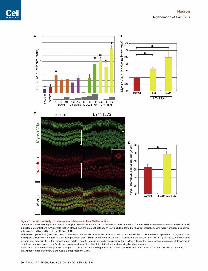
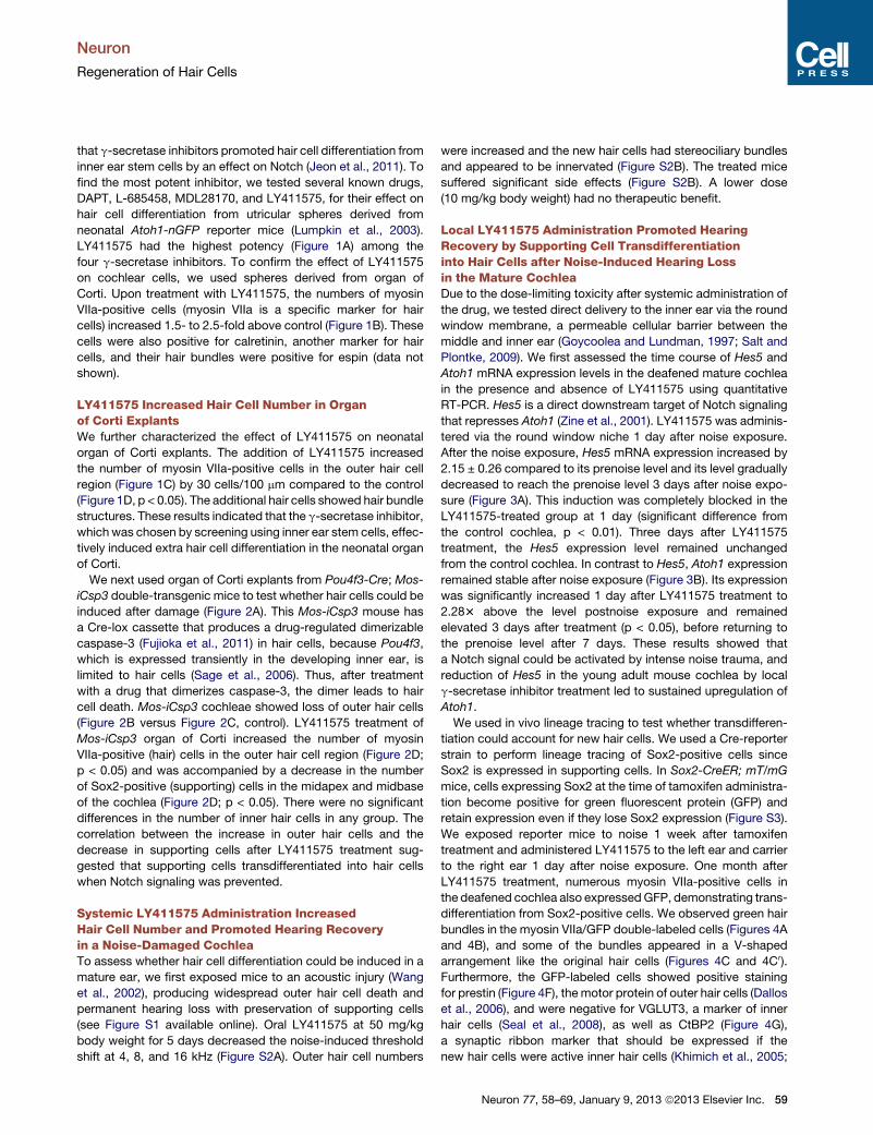
Figure 1. In Vitro Activity of g-Secretase Inhibitors in Hair Cell Induction
(A) Relative ratio of nGFP-positive cells to DAPI-positive cells after treatment of inner ear spheres made from Atoh1-nGFPmice with g-secretase inhibitors at the
indicated concentrations (mM) reveals that LY411575 had the greatest potency of four inhibitors tested for hair cell induction. Data were normalized to control
values obtained by addition of DMSO. *p < 0.01.
(B) Ratio of myosin VIIa- (labels hair cells) to Hoechst-positive cells induced by LY411575 was calculated relative to DMSO-treated spheres from organ of Corti.
(C) Explant cultures of the organ of Corti from postnatal day 1 (P1) mice cultured for 72 hr in the presence of DMSO or LY411575 (1 mM) had ectopic hair cells
(myosin VIIa; green) in the outer hair cell region (white bracket). Ectopic hair cells were positive for phalloidin (labels the hair bundle and cuticular plate; shown in
red). Inset is a high-power view (scale bar represents 2 mm) of a phalloidin-stained hair cell showing bundle structure.
(D) An increase in myosin VIIa-positive cells per 100 mm of the cultured organ of Corti explants from P1 mice was found 72 hr after LY411575 treatment.
In all graphs, error bars show SEM. Scale bar represents 50 mm.
Neuron
Regeneration of Hair Cells
60 Neuron 77, 58–69, January 9, 2013 ª2013 Elsevier Inc.
Neuron
Regeneration of Hair Cells
Liberman et al., 2011). This analysis of markers together with
their location and V-shaped bundles identified them as outer
hair cells. The double-labeled cells spanned the epithelium
frombasilarmembrane to the endolymphatic surface (Figure 4D),
which is never seen in the normal ear but has been reported
when supporting cells are transfected with Atoh1 (Izumikawa
et al., 2005). The nucleus of these cells was at the base of the
cell (Figure 4D0). Double-labeled cells were found in the upper
turns of the cochlea, with the highest numbers in the midapex
(Figure 4E; n = 5). In control ears, no double-labeled cells
were observed in any cochlear region (Figure S3). This result
indicated that blocking Notch with LY411575 promoted sup-
porting cell transdifferentiation into hair cells from the apical to
midapical turn in the mature cochlea after noise-induced hair
cell loss.
At 3 months, the number of outer hair cells was increased
throughout the middle of the cochlea (8–16 kHz) in LY411575-
treated ears, compared to the carrier-treated contralateral ear
(Figures 5A and 5B; p < 0.05). The number of supporting
cells in the outer hair cell region was decreased significantly in
the same cochleae at the 8 and 11.3 kHz areas compared
to the carrier-treated ear (Figures 5A and 5B; p < 0.05), similar
to the explant cultures (see Figure 2D). The outer hair cells
were completely absent with and without LY411575 treatment
in the most basal regions (above 22 kHz), and there were no
significant changes in the numbers of inner hair cells in the
treated group (data not shown). The differences in outer hair
cell number between LY411575- and carrier-treated ears are
larger than the corresponding differences in the number of sup-
porting cells. Furthermore, the differences in outer hair cell
number showed a similar trend, in regard to cochlear location,
as the myosin VIIa-positive cells from the Sox2 lineage observed
in Sox2-CreER; mT/mG mice (Figure 4E).
We recorded the auditory brainstem response (ABR) in
LY411575 and carrier-treated, control ears to determine the
effect of hair cell replacement on the thresholds for a response.
Threshold changes were not seen after injection of carrier alone
(Figure S4). ABR thresholds 1 day after noise exposure were >80
dB sound pressure level (SPL) at all frequencies (Figures 6A and
6B). Postexposure recovery in control ears (Figure 6A) was
minimal under these conditions, as expected (Wang et al.,
2002). Threshold recoveries after LY411575 treatment were
significantly greater than control at 8, 11.33, and 16 kHz (Fig-
ure 6D), and wave I amplitudes were increased at the same
frequencies (Figure 6E). No threshold recoveries were observed
in either ear at frequencies above 22.65 kHz by ABR and no
recoveries above the noise floor of the distortion product otoa-
coustic emissions (DPOAE) could be seen (Figure S5). The
differences in ABR threshold recovery showed a similar depen-
dence on cochlear location (significant changes at 8, 11.3, and
16 kHz) as outer hair cell number (see Figure 5).
DISCUSSION
We have demonstrated significant regeneration of hair cells in
a mammal by treatment of a damaged cochlea with a g-secre-
tase inhibitor. In vivo treatment with the inhibitor resulted in
partial recovery after noise-induced hearing loss.
The generation of physiologically active hair cells in an
adult has been a sought-after but elusive goal. Transfection of
bHLH transcription factor Atoh1, which drives hair cell differenti-
ation during development, is one approach that increases hair
cell number in embryonic or newborn tissue, but cells that
were competent to become hair cells in the embryo lost their
responsiveness as the animal matured (Doetzlhofer et al.,
2009; Gubbels et al., 2008; White et al., 2006). Delivery of
Atoh1 in an adenovirus to the damaged, adult cochlea (Izumi-
kawa et al., 2005) showed some hair cell differentiation, but
the number of new hair cells was not clear and new hair
cells could not be traced from their precursors, making it difficult
to distinguish between ‘‘new’’ hair cells and hair cells that had
recovered from trauma due to a toxin or noise damage. Stimula-
tion of cell division by silencing cell cycle inhibitors has been
suggested as an alternative route to hair cell regeneration
(Sage et al., 2005), but hair cells, due to their highly differentiated
state, tend to activate suicide programs after they divide and
proliferation can cause deafness (Chen and Segil, 1999; Lowen-
heim et al., 1999; Mantela et al., 2005). Regeneration of hair
cells is made difficult by the cellular organization of the
cochlea: minute changes in the interactions between cells of
the epithelium are a cause of deafness (Cohen-Salmon et al.,
2002). Tight junctions are required for maintaining the ionic
milieu of endolymph that bathes the surface of hair cells, and
the flexibility and spacing of outer hair cells has an impact on
the function of the cochlear amplifier, which is achieved by
outer hair cell contraction, and together with sound detection
by the transduction apparatus of inner hair cells, accounts for
the sensitivity and broad dynamic range of mammalian hearing
(Elgoyhen and Franchini, 2011; Hudspeth, 2008; Richardson
et al., 2011).
We had recently shown that inhibition of Notch increased hair
cell differentiation from stem cells and that the mechanism was
dependent on Atoh1, since silencing the transcription factor in
the g-secretase inhibitor-treated stem cells prevented the induc-
tion of hair cell fate (Jeon et al., 2011). We used inner ear stem
cells to select a potent g-secretase inhibitor. We targeted the
Notch pathway, which would only be effective on cells that
were actively signaling throughNotch. Although increasedNotch
signaling in the adult after damage had been suggested by some
(Batts et al., 2009), the loss of an effect of g-secretase inhibitors
on hair cell number in the early postnatal period (Doetzlhofer
et al., 2009) and data suggesting that Notch signaling was
extinguished after birth (Hartman et al., 2009) both suggested
that g-secretase inhibitors would have no effect on hair cell
number in the adult mammalian cochlea.
However, we have shown that inhibition of Notch after noise
damage leads to transdifferentiation of supporting cells into
hair cells. The basal location of the nucleus in the new hair cells
was consistent with the derivation from supporting cells, which
are normally located in a plane below that of the hair cells. Sup-
porting cell transdifferentiation was induced by Atoh1, which
may be acting in a similar capacity to transcription factors,
some of which are related to Atoh1, that allow cellular reprog-
ramming and transdifferentiation to neurons (Caiazzo et al.,
2011; Vierbuchen et al., 2010). The supporting cells express
stem cell markers such as Sox2, Musashi1, and GLAST (Kaneko
Neuron 77, 58–69, January 9, 2013 ª2013 Elsevier Inc. 61
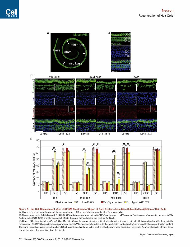
Figure 2. Hair Cell Replacement after LY411575 Treatment of Organ of Corti Explants from Mice Subjected to Ablation of Hair Cells
(A) Hair cells can be seen throughout the neonatal organ of Corti in a whole mount labeled for myosin VIIa.
(B) Three rows of outer (white bracket; OHC1–OHC3) and one row of inner hair cells (IHCs) can be seen in a P3 organ of Corti explant after staining for myosin VIIa.
Deiters’ cells (DC1–DC3) and Hensen cells (HCs) in the outer hair cell region are positive for Sox2.
(C) Organ of Corti explants from Pou4f3-Cre; Mos-iCsp3 double-transgenic mice subjected to dimerizer-induced hair cell ablation and cultured for 3 days in the
presence of LY411575 had an increased number of myosin VIIa-positive cells in the outer hair cell region (white bracket) compared to the carrier-treated explant.
The same region had a decreased number of Sox2-positive cells relative to the control. A high-power view (scale bar represents 2 mm) of phalloidin-stained tissue
shows the hair cell stereociliary bundles (inset).
(legend continued on next page)
Neuron
Regeneration of Hair Cells
62 Neuron 77, 58–69, January 9, 2013 ª2013 Elsevier Inc.
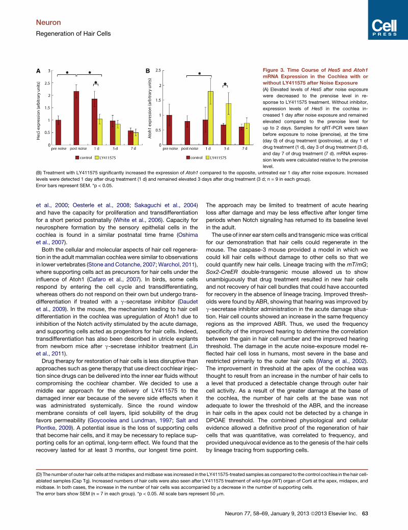
Figure 3. Time Course of Hes5 and Atoh1
mRNA Expression in the Cochlea with or
without LY411575 after Noise Exposure
(A) Elevated levels of Hes5 after noise exposure
were decreased to the prenoise level in re-
sponse to LY411575 treatment. Without inhibitor,
expression levels of Hes5 in the cochlea in-
creased 1 day after noise exposure and remained
elevated compared to the prenoise level for
up to 2 days. Samples for qRT-PCR were taken
before exposure to noise (prenoise), at the time
(day 0) of drug treatment (postnoise), at day 1 of
drug treatment (1 d), day 3 of drug treatment (3 d),
and day 7 of drug treatment (7 d). mRNA expres-
sion levels were calculated relative to the prenoise
level.
(B) Treatment with LY411575 significantly increased the expression of Atoh1 compared to the opposite, untreated ear 1 day after noise exposure. Increased
levels were detected 1 day after drug treatment (1 d) and remained elevated 3 days after drug treatment (3 d; n = 9 in each group).
Error bars represent SEM. *p < 0.05.
Neuron
Regeneration of Hair Cells
et al., 2000; Oesterle et al., 2008; Sakaguchi et al., 2004)
and have the capacity for proliferation and transdifferentiation
for a short period postnatally (White et al., 2006). Capacity for
neurosphere formation by the sensory epithelial cells in the
cochlea is found in a similar postnatal time frame (Oshima
et al., 2007).
Both the cellular and molecular aspects of hair cell regenera-
tion in the adult mammalian cochlea were similar to observations
in lower vertebrates (Stone and Cotanche, 2007; Warchol, 2011),
where supporting cells act as precursors for hair cells under the
influence of Atoh1 (Cafaro et al., 2007). In birds, some cells
respond by entering the cell cycle and transdifferentiating,
whereas others do not respond on their own but undergo trans-
differentiation if treated with a g-secretase inhibitor (Daudet
et al., 2009). In the mouse, the mechanism leading to hair cell
differentiation in the cochlea was upregulation of Atoh1 due to
inhibition of the Notch activity stimulated by the acute damage,
and supporting cells acted as progenitors for hair cells. Indeed,
transdifferentiation has also been described in utricle explants
from newborn mice after g-secretase inhibitor treatment (Lin
et al., 2011).
Drug therapy for restoration of hair cells is less disruptive than
approaches such as gene therapy that use direct cochlear injec-
tion since drugs can be delivered into the inner ear fluids without
compromising the cochlear chamber. We decided to use a
middle ear approach for the delivery of LY411575 to the
damaged inner ear because of the severe side effects when it
was administrated systemically. Since the round window
membrane consists of cell layers, lipid solubility of the drug
favors permeability (Goycoolea and Lundman, 1997; Salt and
Plontke, 2009). A potential issue is the loss of supporting cells
that become hair cells, and it may be necessary to replace sup-
porting cells for an optimal, long-term effect. We found that the
recovery lasted for at least 3 months, our longest time point.
(D) The number of outer hair cells at themidapex andmidbasewas increased in the
ablated samples (Csp Tg). Increased numbers of hair cells were also seen after L
midbase. In both cases, the increase in the number of hair cells was accompani
The error bars show SEM (n = 7 in each group). *p < 0.05. All scale bars represe
The approach may be limited to treatment of acute hearing
loss after damage and may be less effective after longer time
periods when Notch signaling has returned to its baseline level
in the adult.
The use of inner ear stem cells and transgenicmicewas critical
for our demonstration that hair cells could regenerate in the
mouse. The caspase-3 mouse provided a model in which we
could kill hair cells without damage to other cells so that we
could quantify new hair cells. Lineage tracing with the mT/mG;
Sox2-CreER double-transgenic mouse allowed us to show
unambiguously that drug treatment resulted in new hair cells
and not recovery of hair cell bundles that could have accounted
for recovery in the absence of lineage tracing. Improved thresh-
olds were found by ABR, showing that hearing was improved by
g-secretase inhibitor administration in the acute damage situa-
tion. Hair cell counts showed an increase in the same frequency
regions as the improved ABR. Thus, we used the frequency
specificity of the improved hearing to determine the correlation
between the gain in hair cell number and the improved hearing
threshold. The damage in the acute noise-exposure model re-
flected hair cell loss in humans, most severe in the base and
restricted primarily to the outer hair cells (Wang et al., 2002).
The improvement in threshold at the apex of the cochlea was
thought to result from an increase in the number of hair cells to
a level that produced a detectable change through outer hair
cell activity. As a result of the greater damage at the base of
the cochlea, the number of hair cells at the base was not
adequate to lower the threshold of the ABR, and the increase
in hair cells in the apex could not be detected by a change in
DPOAE threshold. The combined physiological and cellular
evidence allowed a definitive proof of the regeneration of hair
cells that was quantitative, was correlated to frequency, and
provided unequivocal evidence as to the genesis of the hair cells
by lineage tracing from supporting cells.
LY411575-treated samples as compared to the control cochlea in the hair cell-
Y411575 treatment of wild-type (WT) organ of Corti at the apex, midapex, and
ed by a decrease in the number of supporting cells.
nt 50 mm.
Neuron 77, 58–69, January 9, 2013 ª2013 Elsevier Inc. 63
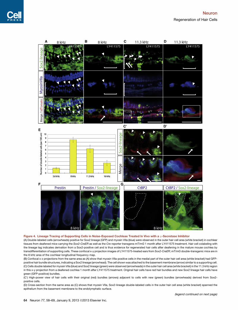
Figure 4. Lineage Tracing of Supporting Cells in Noise-Exposed Cochleae Treated In Vivo with a g-Secretase Inhibitor
(A) Double-labeled cells (arrowheads) positive for Sox2 lineage (GFP) and myosin VIIa (blue) were observed in the outer hair cell area (white bracket) in cochlear
tissues from deafened mice carrying the Sox2-CreER as well as the Cre reporter transgenemT/mG 1 month after LY411575 treatment. Hair cell colabeling with
the lineage tag indicates derivation from a Sox2-positive cell and is thus evidence for regenerated hair cells after deafening in the mature mouse cochlea by
transdifferentiation of supporting cells. These confocal x-y projection images of LY411575-treated ears from Sox2-CreER;mT/mG double-transgenic mice are in
the 8 kHz area of the cochlear longitudinal frequency map.
(B) Confocal x-z projections from the same area as (A) show that myosin VIIa-positive cells in the medial part of the outer hair cell area (white bracket) had GFP-
positive hair bundle structures, indicating aSox2 lineage (arrowhead). The cell shownwas attached to the basementmembrane (arrow) similar to a supporting cell.
(C) Cells double labeled for myosin VIIa (blue) and Sox2 lineage (green) were observed (arrowheads) in the outer hair cell area (white bracket) in the 11.3 kHz region
in this x-y projection from a deafened cochlea 1 month after LY411575 treatment. Original hair cells have red hair bundles and new Sox2 lineage hair cells have
green (GFP-positive) bundles.
(C0) High-power view of hair cells with their original (red) bundles (arrows) adjacent to cells with new (green) bundles (arrowheads) derived from Sox2-
positive cells.
(D) Cross-section from the same area as (C) shows that myosin VIIa, Sox2-lineage double-labeled cells in the outer hair cell area (white bracket) spanned the
epithelium from the basement membrane to the endolymphatic surface.
(legend continued on next page)
Neuron
Regeneration of Hair Cells
64 Neuron 77, 58–69, January 9, 2013 ª2013 Elsevier Inc.
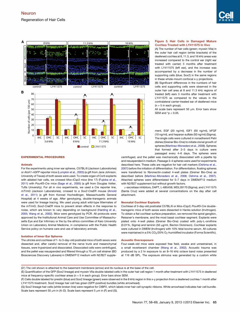
Figure 5. Hair Cells in Damaged Mature
Cochlea Treated with LY411575 In Vivo
(A) The number of hair cells (green; myosin VIIa) in
the outer hair cell region (white brackets) of the
deafened cochlea at 8, 11.3, and 16 kHz areas was
increased compared to the control ear (right ear
treated with carrier) 3 months after treatment
with LY411575 (left ear), and the increase was
accompanied by a decrease in the number of
supporting cells (blue; Sox2) in the same regions
in these whole-mount confocal x-y projections.
(B) Significant differences in the numbers of hair
cells and supporting cells were observed in the
outer hair cell area at 8 and 11.3 kHz regions of
treated (left) ears 3 months after treatment with
LY411575 as compared to the values in the
contralateral carrier-treated ear of deafened mice
(n = 5 in each group).
All scale bars represent 50 mm. Error bars show
SEM and *p < 0.05.
Neuron
Regeneration of Hair Cells
EXPERIMENTAL PROCEDURES
Animals
For the experiments using inner ear spheres, C57BL/6 (Jackson Laboratories)
or Atoh1-nGFP reporter mice (Lumpkin et al., 2003) (a gift from Jane Johnson,
University of Texas) of both sexes were used. To create organ of Corti explants
with ablated hair cells, we crossed Mos-iCsp3 mice (line 17) (Fujioka et al.,
2011) with Pou4f3-Cre mice (Sage et al., 2005) (a gift from Douglas Vetter,
Tufts University). For all in vivo experiments, we used a Cre reporter line,
mT/mG (Jackson Laboratories), crossed to a Sox2-CreER mouse (Arnold
et al., 2011) (a gift from Konrad Hochedlinger, Massachusetts General
Hospital) at 4 weeks of age. After genotyping, double-transgenic animals
were used for lineage tracing. We used young adult wild-type littermates of
the mT/mG; Sox2-CreER mice to prevent strain effects in the response to
noise, which are known to vary depending on background (Harding et al.,
2005; Wang et al., 2002). Mice were genotyped by PCR. All protocols were
approved by the Institutional Animal Care and Use Committee of Massachu-
setts Eye and Ear Infirmary or the by the ethics committee of Keio University
Union on Laboratory Animal Medicine, in compliance with the Public Health
Service policy on humane care and use of laboratory animals.
Isolation of Inner Ear Spheres
The utricles and cochleae of 1- to 3-day-old postnatal mice of both sexes were
dissected and, after careful removal of the nerve trunk and mesenchymal
tissues, were trypsinized and dissociated. Dissociated cells were centrifuged,
and the pellet was resuspended and filtered through a 70 mm cell strainer (BD
Biosciences Discovery Labware) in DMEM/F12 medium with N2/B27 supple-
(D0) The cell shown is attached to the basement membrane (arrow) and its nucleus is at the base of the cell.
(E) Quantification of the GFP (Sox2 lineage) and myosin VIIa double-labeled cells in the outer hair cell region 1
mice at frequency-specific cochlear areas (n = 5 in each group). Error bars show SEM.
(F) Cells double labeled for prestin (blue) and Sox2 lineage (green) were observed in the 8 kHz region in this x-
LY411575 treatment. Sox2 lineage hair cell has green (GFP-positive) bundles (white arrowhead).
(G) Sox2 lineage hair cells (white broken line) were negative for CtBP2, which labels inner hair cell synaptic ri
Scale bars represent 50 mm in (A–D). Scale bars represent 5 mm in (F) and (G).
Neuron 77, 58–
ment, EGF (20 ng/ml), IGF1 (50 ng/ml), bFGF
(10 ng/ml), and heparan sulfate (50 ng/ml) (Sigma).
The single cells were cultured in nonadherent Petri
dishes (Greiner Bio-One) to initiate clonal growth of
spheres (Martinez-Monedero et al., 2008). Spheres
that formed after 2–3 days in culture were
passaged every 4–6 days. The spheres were
centrifuged, and the pellet was mechanically dissociated with a pipette tip
and resuspended inmedium. Passage 3–4 spheres were used for experiments
described here. These cells are negative for hair cell markers (Oshima et al.,
2007) before the initiation of differentiation. For differentiation, floating spheres
were transferred to fibronectin-coated 4-well plates (Greiner Bio-One) as
described before (Martinez-Monedero et al., 2008; Oshima et al., 2007).
Attached spheres were differentiated for 5–7 days in DMEM/F12 medium
with N2/B27 supplement but without growth factors.
g-secretase inhibitors, DAPT, L-685458, MDL28170 (Sigma), and LY411575
(Santa Cruz) were added at several concentrations on the day after cell
attachment.
Neonatal Cochlear Explants
Cochleae of 3-day-old postnatal C57BL/6 or Mos-iCsp3; Pou4f3-Cre double-
transgenic mice of both sexes were dissected in Hanks solution (Invitrogen).
To obtain a flat cochlear surface preparation, we removed the spiral ganglion,
Reissner’s membrane, and the most basal cochlear segment. Explants were
plated onto 4-well plates (Greiner Bio-One) coated with poly-L-ornithine
(0.01%, Sigma) and laminin (50 mg/ml, Becton Dickinson). Cochlear explants
were cultured in DMEM (Invitrogen) with 10% fetal bovine serum. All cultures
weremaintained in a 5%CO2/20%O2-humidified incubator (Forma Scientific).
Acoustic Overexposure
Four-week-old mice were exposed free field, awake and unrestrained, in
a small reverberant chamber (Wang et al., 2002). Acoustic trauma was
produced by a 2 hr exposure to an 8–16 kHz octave band noise presented
at 116 dB SPL. The exposure stimulus was generated by a custom white
month after treatment with LY411575 in deafened
y projection from a deafened cochlea 1 month after
bbons. White arrowhead indicates hair cell bundle.
69, January 9, 2013 ª2013 Elsevier Inc. 65
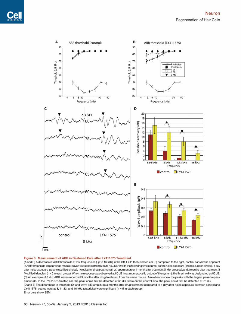
Figure 6. Measurement of ABR in Deafened Ears after LY411575 Treatment
(A and B) A decrease in ABR thresholds at low frequencies (up to 16 kHz) in the left, LY411575-treated ear (B) compared to the right, control ear (A) was apparent
inABR thresholds in recordingsmadeat seven frequencies from5.66 to45.25kHzwith the following timecourse:before noise exposure (prenoise, opencircles), 1 day
afternoiseexposure (postnoise: filledcircles), 1weekafterdrug treatment (1W,opensquares), 1monthafter treatment (1Mo,crosses), and3monthsafter treatment (3
Mo, filled triangles) (n = 5 in eachgroup).When no responsewas observedat 80dB (maximumacoustic output of the system), the thresholdwas designatedas 85dB.
(C) An example of 8 kHz ABR waves recorded 3 months after drug treatment from the same mouse. Arrowheads show the peaks with the largest peak-to-peak
amplitude. In the LY411575-treated ear, the peak could first be detected at 65 dB, while on the control side, the peak could first be detected at 75 dB.
(D and E) The differences in threshold (D) and wave I (E) amplitude 3 months after drug treatment compared to 1 day after noise exposure between control and
LY411575-treated ears at 8, 11.33, and 16 kHz (asterisks) were significant (n = 5 in each group).
Error bars show SEM.
Neuron
Regeneration of Hair Cells
66 Neuron 77, 58–69, January 9, 2013 ª2013 Elsevier Inc.
Neuron
Regeneration of Hair Cells
noise source, filtered (Brickwall Filter with a 60 dB/octave slope), amplified
(Crown power amplifier), and delivered (JBL compression driver) through an
exponential horn fitted securely to a hole in the top of a reverberant box. Sound
exposure levels were measured at four positions within each cage using a 0.25
inch Bruel and Kjær condenser microphone: sound pressure was found to vary
by <0.5 dB across these measurement positions.
Systemic or Round Window Administration of LY411575
Four-week-old mice weighing 12–16 g were used. Before surgery, the animals
were anesthetized with ketamine (20 mg/kg, intraperitoneally [i.p.]) and xyla-
zine (100 mg/kg, i.p.), and an incision was made posterior to the pinna near
the external meatus after local administration of lidocaine (1%). The otic bulla
was opened to approach the round window niche. The end of a piece of PE 10
tubing (Becton Dickinson) was drawn to a fine tip in a flame and gently inserted
into the round window niche. LY411575 was dissolved in DMSO and diluted
10-fold in polyethylene glycol 400 (Sigma) to a final concentration of 4 mM.
This solution (total volume 1 ml) was injected into the round window niche of
the left ear. Polyethylene glycol 400with 10%DMSOwas injected into the right
ear as a control. The solution was administered for 2 min. This approach is
widely used clinically and has the advantage of sparing the inner ear but still
taking advantage of the local route provided by the round window membrane
for delivery of drug into the inner ear (Mikulec et al., 2008). Gelatin was placed
on the niche to maintain the solution, and the wound was closed.
For the systemic administration, LY411575 (50 mg/kg) dissolved in
0.5% (w/v) methylcellulose (WAKO) was injected orally once daily for 5 con-
secutive days. Hearing was measured by ABR at 1 day before, 2 days,
1 week, 2 weeks, and 1, 2, and 3 months after noise exposure.
qRT-PCR
The organs of Corti were dissected in HBSS (Invitrogen) and stored in RNAlater
(Ambion) at �80�C until further use. Total RNA was extracted using the
RNeasy Mini Kit (QIAGEN) according to the manufacturer’s instructions. For
reverse transcription, SuperScript II (Invitrogen) was usedwith random hexam-
ers. The reverse transcription conditions were 25�C for 10 min followed by
37�C for 60 min. The reaction was terminated at 95�C for 5 min. cDNAs
were mixed with Taqman Gene Expression Mastermix (Applied Biosystems)
andHes5, Atoh1, or 18S primers (Applied Biosystems) according to the manu-
facturer’s instructions. Samples were analyzed in 96wells in triplicate by qPCR
(Applied Biosystems 7900HT), and PCR thermal cycling conditions were as
follows: initial denaturation at 95�C for 2 min, denaturation at 95�C for 15 s,
and annealing and extension at 60�C for 1 min for 45 cycles. Conditions
were kept constant for each primer. Each PCR reaction was carried out in trip-
licate. Relative gene expression was analyzed by using the DDCT method.
Gene expression was calculated relative to 18S RNA, and the amount of
cDNA applied was adjusted so that the Ct value for 18S RNA was between
8 and 11.
Immunohistochemistry
For spheres, cells were fixed for 10 min with 4% paraformaldehyde in PBS.
Immunostaining was initiated by blocking for 1 hr with 0.1% Triton X-100 in
PBS supplemented with 1% BSA and 5% goat serum (PBT1). Fixed and per-
meabilized cells were incubated overnight in PBT1 with polyclonal antibody to
myosin VIIa (Proteus Biosciences). Samples were washed three times for
20min with PBS. Primary antibodies were detected with secondary antibodies
conjugated with Alexa 488 (Molecular Probes), with secondary antibody alone
used as a negative control. The samples were counterstained with DAPI
(Vector Laboratories) or Hoechst 33258 (Invitrogen) for 10 min and viewed
by epifluorescence microscopy (Axioskop 2 Mot Axiocam, Zeiss).
For explants, the organs of Corti were fixed for 15 min with 4% paraformal-
dehyde in PBS. Immunostaining was initiated by blocking the tissues for 1 hr
with 0.1% Triton X-100 in PBS supplemented with 5% donkey serum
(PBT1). Fixed and permeabilized pieces were incubated overnight in PBT1
with antibodies to myosin VIIa (Proteus Biosciences), Sox2 (Santa Cruz),
GFP (Invitrogen), prestin (Santa Cruz), neurofilament H (Chemicon), and
CtBP2 (BD Biosciences). Samples were washed three times for 20 min with
PBS. Primary antibodies were detected with secondary antibodies conjugated
with Alexa 488 and 647 (Molecular Probes). The samples were stained with
rhodamine phalloidin (Invitrogen) for 15 min and viewed by confocal fluores-
cence microscopy (TCS SP5, Leica).
For collection of the mature cochlea, deeply anesthetized mice were
transcardially perfused with 0.01 M phosphate buffer (pH 7.4) containing
8.6% sucrose, followed by fixative consisting of freshly depolymerized 4%
paraformaldehyde in 0.1 M phosphate buffer (pH 7.4). After decapitation,
the temporal bones were removed and immediately placed in the same fixa-
tive at 4�C. Small openings were made at the round window, oval window, and
apex of the cochlea. After immersion in the fixative overnight at 4�C, temporal
bones were decalcified in 0.1 M EDTA (pH 7.4) containing 5% sucrose with
stirring at 4�C for 2 days. After decalcification, the cochlea was microdis-
sected into four pieces for whole-mount preparation. Immunostaining was
initiated by blocking the tissues for 1 hr with 0.1% Triton X-100 in PBS supple-
mented with 5% donkey serum (PBT1). Fixed and permeabilized pieces were
incubated overnight in PBT1 with antibodies to myosin VIIa (Proteus Biosci-
ences), Sox2 (Santa Cruz), and GFP (Invitrogen). Samples were washed three
times for 20 min with PBS. Primary antibodies were detected with secondary
antibodies conjugated with Alexa 488, 568, and 647 (Molecular Probes) and
viewed by confocal fluorescence microscopy (TCS SP5, Leica). Cochlear
lengths were obtained for each case, and a cochlear frequency map
computed to precisely localize inner hair cells from the 5.6, 8.0, 11.3, 16.0,
22.6, 32, and 45.2 kHz regions. For cross-sectioning, fixed temporal bones
were sunk in 30% sucrose in PBS at 4�C, incubated in OCT at room temper-
ature for 1 hr, and frozen in liquid nitrogen. The staining protocol was the same
as described above except for counterstaining with DAPI (Vector Laborato-
ries). Specimens were viewed by epifluorescence microscopy (Axioskop 2
Mot Axiocam, Zeiss).
Cell Counts
Cell counting for spheres was performed with MetaMorph software. The cell
number was determined from DAPI- or Hoechst-positive nuclei. Repeat cell
counting gave a test variation of <1%. For explants, inner hair cells, outer
hair cells, and supporting cells in the outer hair cell region were counted on
cochlear whole mounts. Hair cells were identified with myosin VIIa antibodies
or endogenousGFP inAtoh1-nGFPmice. High-power images of the full-length
cochlea or cochlear explant cultures were assembled and analyzed in Photo-
Shop CS4 (Adobe). ImageJ software (NIH) was used to measure the total
length of cochlear whole mounts and the length of individual counted
segments. The total number of inner hair cells, outer hair cells, and supporting
cells in the outer hair cell region was counted in each of four cochlear
segments of 1,200–1,400 mm (apical, midapical, midbasal, and basal). Density
(cells per 100 mm)was then calculated for each segment. For mature cochleae,
high-power images of frequency-specific regions (5.6, 8.0, 11.3, and 16.0 kHz)
according to the computed frequencymapwere assembled and analyzed. The
number of inner hair cells, outer hair cells, and supporting cells in the outer hair
cell region in 100 mm was counted in each of the four frequency-specific
regions of the cochlea. The number of Sox2 lineage-positive cells identified
by GFP was counted by the same method.
ABR Measurements
Auditory brain stem responses (Kujawa and Liberman, 1997; Maison et al.,
2003) were measured in each animal at seven log-spaced frequencies (half-
octave steps from 5.6 to 45.2 kHz) before and 1 day after noise exposure,
and 1 week, 1 month, and 3 months after surgery. Mice were anesthetized
with ketamine (100mg/kg, i.p.) and xylazine (20mg/kg, i.p.). Needle electrodes
were inserted at vertex and pinna, with a ground near the tail. ABRs were
evoked with 5 ms tone pips (0.5 ms rise-fall with a cos2 onset envelope deliv-
ered at 35/s). The response was amplified, filtered, and averaged in a Lab-
VIEW-driven data acquisition system. Sound level was raised in 5 dB steps
from R10 dB below threshold to <80 dB SPL. At each sound level, 1,024
responses were averaged (with stimulus polarity alternated), using an ‘‘artifact
reject,’’ whereby response waveforms were discarded when peak-to-peak
response amplitude exceeded 15 mV. On visual inspection of stacked wave-
forms, ‘‘ABR threshold’’ was defined as the lowest SPL level at which any
wave could be detected, usually corresponding to the level step just below
that at which the peak-to-peak response amplitude rose significantly above
the noise floor (approximately 0.25 mV). When no response was observed at
Neuron 77, 58–69, January 9, 2013 ª2013 Elsevier Inc. 67
Neuron
Regeneration of Hair Cells
the highest sound level available, the threshold was designated as being 5 dB
greater than that level so that statistical tests could be done. For amplitude
versus level functions, the wave I peak was identified by visual inspection at
each sound level and the peak-to-peak amplitude was computed.
Quantification and Statistical Analysis
The two-tailed Mann-Whitney U test was used to compare differences among
treatment groups. Changes before and after treatment of the same animal
were analyzed by two-tailed Wilcoxon t test. Repeated-measures ANOVA
was used to compare time-dependent differences among groups. Data are
presented in the text and in figures as mean ± SEM. p values less than 0.05
were considered significant.
Genotyping Primers
We used the following genotyping primers: LacZ F: 50-ttcactggccgtcgttttacaacgtcgtga-30 and LacZ R: 50-atgtgagcgagtaacaacccgtcggattct-30 for theMos-iCsp3 mice; Cre F: 50-tgggcggcatggtgcaagtt-30 and Cre R: 50-cggtgctaaccagcgttttc-30 for the Pou4f3Cre and Sox2CreER mice; and oIMR7318
wild-type F: 50-ctctgctgcctcctggcttct-30, oIMR7319 wild-type R: 50-cgaggcggatcacaagcaata-30, and oIMR7320 mutant R: 50-tcaatgggcgggggtcgtt-30 forthe mT/mG mice.
SUPPLEMENTAL INFORMATION
Supplemental Information includes five figures and Supplemental Experi-
mental Procedures and can be found with this article online at http://dx.doi.
org/10.1016/j.neuron.2012.10.032.
ACKNOWLEDGMENTS
We thank M.C. Liberman for critical comments on the manuscript. We thank
Yukiko Watada and Yumi Matsuzaki for assistance. This work was supported
by grants RO1 DC007174, R21 DC010440, and P30 DC05209 from the
National Institute on Deafness and other Communicative Disorders (NIDCD);
by the Tillotson Corporation, the Shulsky Foundation, and Robert Boucai; by
the Mochida Memorial Foundation for Medical and Pharmaceutical Research;
and by grants for International Activities in Life Sciences and Medicine, Keio
University Medical Science Fund.
Accepted: October 31, 2012
Published: January 9, 2013
REFERENCES
Adam, J., Myat, A., Le Roux, I., Eddison, M., Henrique, D., Ish-Horowicz, D.,
and Lewis, J. (1998). Cell fate choices and the expression of Notch, Delta
and Serrate homologues in the chick inner ear: parallels with Drosophila
sense-organ development. Development 125, 4645–4654.
Arnold, K., Sarkar, A., Yram, M.A., Polo, J.M., Bronson, R., Sengupta, S.,
Seandel, M., Geijsen, N., and Hochedlinger, K. (2011). Sox2(+) adult stem
and progenitor cells are important for tissue regeneration and survival of
mice. Cell Stem Cell 9, 317–329.
Batts, S.A., Shoemaker, C.R., and Raphael, Y. (2009). Notch signaling and Hes
labeling in the normal and drug-damaged organ of Corti. Hear. Res. 249,
15–22.
Cafaro, J., Lee, G.S., and Stone, J.S. (2007). Atoh1 expression defines
activated progenitors and differentiating hair cells during avian hair cell regen-
eration. Dev. Dyn. 236, 156–170.
Caiazzo, M., Dell’Anno, M.T., Dvoretskova, E., Lazarevic, D., Taverna, S., Leo,
D., Sotnikova, T.D., Menegon, A., Roncaglia, P., Colciago, G., et al. (2011).
Direct generation of functional dopaminergic neurons from mouse and human
fibroblasts. Nature 476, 224–227.
Chen, P., and Segil, N. (1999). p27(Kip1) links cell proliferation to morphogen-
esis in the developing organ of Corti. Development 126, 1581–1590.
68 Neuron 77, 58–69, January 9, 2013 ª2013 Elsevier Inc.
Cohen-Salmon, M., Ott, T., Michel, V., Hardelin, J.P., Perfettini, I., Eybalin, M.,
Wu, T., Marcus, D.C., Wangemann, P., Willecke, K., and Petit, C. (2002).
Targeted ablation of connexin26 in the inner ear epithelial gap junction network
causes hearing impairment and cell death. Curr. Biol. 12, 1106–1111.
Corwin, J.T., and Cotanche, D.A. (1988). Regeneration of sensory hair cells
after acoustic trauma. Science 240, 1772–1774.
Dallos, P., Zheng, J., and Cheatham, M.A. (2006). Prestin and the cochlear
amplifier. J. Physiol. 576, 37–42.
Daudet, N., and Lewis, J. (2005). Two contrasting roles for Notch activity in
chick inner ear development: specification of prosensory patches and lateral
inhibition of hair-cell differentiation. Development 132, 541–551.
Daudet, N., Gibson, R., Shang, J., Bernard, A., Lewis, J., and Stone, J. (2009).
Notch regulation of progenitor cell behavior in quiescent and regenerating
auditory epithelium of mature birds. Dev. Biol. 326, 86–100.
Doetzlhofer, A., Basch, M.L., Ohyama, T., Gessler, M., Groves, A.K., and Segil,
N. (2009). Hey2 regulation by FGF provides a Notch-independent mechanism
for maintaining pillar cell fate in the organ of Corti. Dev. Cell 16, 58–69.
Edge, A.S., and Chen, Z.Y. (2008). Hair cell regeneration. Curr. Opin.
Neurobiol. 18, 377–382.
Elgoyhen, A.B., and Franchini, L.F. (2011). Prestin and the cholinergic receptor
of hair cells: positively-selected proteins in mammals. Hear. Res. 273,
100–108.
Fritzsch, B., Beisel, K.W., and Hansen, L.A. (2006). The molecular basis of
neurosensory cell formation in ear development: a blueprint for hair cell and
sensory neuron regeneration? Bioessays 28, 1181–1193.
Fujioka, M., Tokano, H., Fujioka, K.S., Okano, H., and Edge, A.S. (2011).
Generating mouse models of degenerative diseases using Cre/lox-mediated
in vivo mosaic cell ablation. J. Clin. Invest. 121, 2462–2469.
Goycoolea, M.V., and Lundman, L. (1997). Round window membrane.
Structure function and permeability: a review. Microsc. Res. Tech. 36,
201–211.
Gubbels, S.P., Woessner, D.W., Mitchell, J.C., Ricci, A.J., and Brigande, J.V.
(2008). Functional auditory hair cells produced in the mammalian cochlea by in
utero gene transfer. Nature 455, 537–541.
Harding, G.W., Bohne, B.A., and Vos, J.D. (2005). The effect of an age-related
hearing loss gene (Ahl) on noise-induced hearing loss and cochlear damage
from low-frequency noise. Hear. Res. 204, 90–100.
Hartman, B.H., Basak, O., Nelson, B.R., Taylor, V., Bermingham-McDonogh,
O., and Reh, T.A. (2009). Hes5 expression in the postnatal and adult mouse
inner ear and the drug-damaged cochlea. J. Assoc. Res. Otolaryngol. 10,
321–340.
Hudspeth, A.J. (2008). Making an effort to listen: mechanical amplification in
the ear. Neuron 59, 530–545.
Izumikawa, M., Minoda, R., Kawamoto, K., Abrashkin, K.A., Swiderski, D.L.,
Dolan, D.F., Brough, D.E., and Raphael, Y. (2005). Auditory hair cell replace-
ment and hearing improvement by Atoh1 gene therapy in deaf mammals.
Nat. Med. 11, 271–276.
Jeon, S.J., Fujioka, M., Kim, S.C., and Edge, A.S.B. (2011). Notch signaling
alters sensory or neuronal cell fate specification of inner ear stem cells.
J. Neurosci. 31, 8351–8358.
Kaneko, Y., Sakakibara, S., Imai, T., Suzuki, A., Nakamura, Y., Sawamoto, K.,
Ogawa, Y., Toyama, Y., Miyata, T., and Okano, H. (2000). Musashi1: an evolu-
tionally conserved marker for CNS progenitor cells including neural stem cells.
Dev. Neurosci. 22, 139–153.
Kelley, M.W. (2006). Regulation of cell fate in the sensory epithelia of the inner
ear. Nat. Rev. Neurosci. 7, 837–849.
Khimich, D., Nouvian, R., Pujol, R., Tom Dieck, S., Egner, A., Gundelfinger,
E.D., andMoser, T. (2005). Hair cell synaptic ribbons are essential for synchro-
nous auditory signalling. Nature 434, 889–894.
Kujawa, S.G., and Liberman, M.C. (1997). Conditioning-related protection
from acoustic injury: effects of chronic deefferentation and sham surgery.
J. Neurophysiol. 78, 3095–3106.
Neuron
Regeneration of Hair Cells
Liberman, L.D., Wang, H., and Liberman, M.C. (2011). Opposing gradients of
ribbon size and AMPA receptor expression underlie sensitivity differences
among cochlear-nerve/hair-cell synapses. J. Neurosci. 31, 801–808.
Lin, V., Golub, J.S., Nguyen, T.B., Hume, C.R., Oesterle, E.C., and Stone, J.S.
(2011). Inhibition of Notch activity promotes nonmitotic regeneration of hair
cells in the adult mouse utricles. J. Neurosci. 31, 15329–15339.
Lowenheim, H., Furness, D.N., Kil, J., Zinn, C., Gultig, K., Fero, M.L., Frost, D.,
Gummer, A.W., Roberts, J.M., Rubel, E.W., et al. (1999). Gene disruption of
p27(Kip1) allows cell proliferation in the postnatal and adult organ of corti.
Proc. Natl. Acad. Sci. USA 96, 4084–4088.
Lumpkin, E.A., Collisson, T., Parab, P., Omer-Abdalla, A., Haeberle, H., Chen,
P., Doetzlhofer, A., White, P., Groves, A., Segil, N., and Johnson, J.E. (2003).
Math1-driven GFP expression in the developing nervous system of transgenic
mice. Gene Expr. Patterns 3, 389–395.
Maison, S.F., Emeson, R.B., Adams, J.C., Luebke, A.E., and Liberman, M.C.
(2003). Loss of alpha CGRP reduces sound-evoked activity in the cochlear
nerve. J. Neurophysiol. 90, 2941–2949.
Mantela, J., Jiang, Z., Ylikoski, J., Fritzsch, B., Zacksenhaus, E., and Pirvola, U.
(2005). The retinoblastoma gene pathway regulates the postmitotic state of
hair cells of the mouse inner ear. Development 132, 2377–2388.
Martinez-Monedero, R., Yi, E., Oshima, K., Glowatzki, E., and Edge, A.S.
(2008). Differentiation of inner ear stem cells to functional sensory neurons.
Dev. Neurobiol. 68, 669–684.
Mikulec, A.A., Hartsock, J.J., and Salt, A.N. (2008). Permeability of the round
window membrane is influenced by the composition of applied drug solutions
and by common surgical procedures. Otol. Neurotol. 29, 1020–1026.
Nayak, G.D., Ratnayaka, H.S., Goodyear, R.J., and Richardson, G.P. (2007).
Development of the hair bundle and mechanotransduction. Int. J. Dev. Biol.
51, 597–608.
Oesterle, E.C., Campbell, S., Taylor, R.R., Forge, A., and Hume, C.R. (2008).
Sox2 and JAGGED1 expression in normal and drug-damaged adult mouse
inner ear. J. Assoc. Res. Otolaryngol. 9, 65–89.
Oshima, K., Grimm, C.M., Corrales, C.E., Senn, P., Martinez Monedero, R.,
Geleoc, G.S., Edge, A., Holt, J.R., and Heller, S. (2007). Differential distribution
of stem cells in the auditory and vestibular organs of the inner ear. J. Assoc.
Res. Otolaryngol. 8, 18–31.
Richardson, G.P., de Monvel, J.B., and Petit, C. (2011). How the genetics of
deafness illuminates auditory physiology. Annu. Rev. Physiol. 73, 311–334.
Ryals, B.M., and Rubel, E.W. (1988). Hair cell regeneration after acoustic
trauma in adult Coturnix quail. Science 240, 1774–1776.
Sage, C., Huang, M., Karimi, K., Gutierrez, G., Vollrath, M.A., Zhang, D.S.,
Garcıa-Anoveros, J., Hinds, P.W., Corwin, J.T., Corey, D.P., and Chen, Z.Y.
(2005). Proliferation of functional hair cells in vivo in the absence of the retino-
blastoma protein. Science 307, 1114–1118.
Sage, C., Huang, M., Vollrath, M.A., Brown, M.C., Hinds, P.W., Corey, D.P.,
Vetter, D.E., and Chen, Z.Y. (2006). Essential role of retinoblastoma protein
in mammalian hair cell development and hearing. Proc. Natl. Acad. Sci. USA
103, 7345–7350.
Sakaguchi, H., Yaoi, T., Suzuki, T., Okano, H., Hisa, Y., and Fushiki, S. (2004).
Spatiotemporal patterns of Musashi1 expression during inner ear develop-
ment. Neuroreport 15, 997–1001.
Salt, A.N., and Plontke, S.K. (2009). Principles of local drug delivery to the inner
ear. Audiol. Neurootol. 14, 350–360.
Seal, R.P., Akil, O., Yi, E., Weber, C.M., Grant, L., Yoo, J., Clause, A., Kandler,
K., Noebels, J.L., Glowatzki, E., et al. (2008). Sensorineural deafness and
seizures in mice lacking vesicular glutamate transporter 3. Neuron 57,
263–275.
Stone, J.S., and Cotanche, D.A. (2007). Hair cell regeneration in the avian
auditory epithelium. Int. J. Dev. Biol. 51, 633–647.
Vierbuchen, T., Ostermeier, A., Pang, Z.P., Kokubu, Y., Sudhof, T.C., and
Wernig, M. (2010). Direct conversion of fibroblasts to functional neurons by
defined factors. Nature 463, 1035–1041.
Wang, Y., Hirose, K., and Liberman, M.C. (2002). Dynamics of noise-induced
cellular injury and repair in the mouse cochlea. J. Assoc. Res. Otolaryngol. 3,
248–268.
Warchol, M.E. (2011). Sensory regeneration in the vertebrate inner ear:
Differences at the levels of cells and species. Hear. Res. 273, 72–79.
White, P.M., Doetzlhofer, A., Lee, Y.S., Groves, A.K., and Segil, N. (2006).
Mammalian cochlear supporting cells can divide and trans-differentiate into
hair cells. Nature 441, 984–987.
Zheng, J.L., and Gao, W.Q. (2000). Overexpression of Math1 induces robust
production of extra hair cells in postnatal rat inner ears. Nat. Neurosci. 3,
580–586.
Zine, A., Aubert, A., Qiu, J., Therianos, S., Guillemot, F., Kageyama, R., and de
Ribaupierre, F. (2001). Hes1 and Hes5 activities are required for the normal
development of the hair cells in the mammalian inner ear. J. Neurosci. 21,
4712–4720.
Neuron 77, 58–69, January 9, 2013 ª2013 Elsevier Inc. 69
Copyright © 2022 FDOKUMEN