
1-(3-Pyridyl)-1-Butanone (NNK) arising from tobacco smoke in ...

Mol. Cryst. Liq. Cryst., Vol. 552: pp. 194–208, 2012Copyright © Taylor & Francis Group, LLCISSN: 1542-1406 print/1563-5287 onlineDOI: 10.1080/15421406.2011.591702
Synthesis, Crystal Structure, and SpectroscopicStudies of N-(4-Bromobenzylidene)-N′-(2-Pyridyl)
Hydrazine Schiff Base Molecule
TUNCAY TUNC,1,∗ HABIBE TEZCAN,2 ERTAN SAHIN,3
AND NEFISE DILEK4
1Department of Science Education, Faculty of Education, Aksaray University,Aksaray, Turkey2Department of Chemistry Education, Faculty of Education, Gazi University,Ankara, Turkey3Department of Chemistry, Faculty of Science and Arts, Ataturk University,Erzurum, Turkey4Department of Physics, Faculty of Science and Arts, Aksaray University,Aksaray, Turkey
A new Schiff base complex N-(4-bromobenzylidene)-N′-(2-pyridyl) hydrazine,C12H10N3Br, has been synthesized and characterized by elemental analyses, mass, 1HNMR, ultraviolet–visible (UV–VIS), and IR spectroscopy and single-crystal X-ray de-termination. The p-Br-benzene and pyridine rings are almost planar and the dihedralangle between the planes is 11.1(3)◦. The crystal structure is stabilized by intermolec-ular N H . . . N Py hydrogen bonding interaction. X-ray diffraction analyses showthat N-(4-bromobenzylidene)-N′-(2-pyridyl) hydrazine Schiff base molecule crystallizesin the monoclinic system, P21/c space group, a = 5.611(5) Å, b = 19.566(5) Å, c =10.715(5) Å, β = 98.766(5)◦, and V = 1162.60(12) Å3.
Keywords Condensation reaction; crystal structure; hydrazone; Schiff base; X-raydiffraction
Introduction
Schiff base complexes are often used as ligands in coordination chemistry related to catal-ysis and enzymatic reactions. Schiff base ligands and their metal complexes have beenextensively studied over the past few decades. In particular, hydrazones are useful for thesynthesis of formazans. Formazans form tetrazolium salt when they are oxidized [1]. Tetra-zolium salts are reduced back to formazans by the enzymes in the cell and stain the tissue.The tetrazolium-formazan system is classified as a marker of vitality [2]. Hydrazones arealso useful for the synthesis of metal complexes as they easily form stable complexeswith most transition metal ions [3,4]. Hydrazones and their metal complexes have gaineda special attraction for widespread application in technology and analytical chemistry.
∗Address correspondence to Tuncay Tunc, Department of Science Education, Faculty of Edu-cation, Aksaray University, 68100 Aksaray, Turkey. Tel: +90.3822802228; Fax: +90.382 2801180.E-mail: [email protected]
194
Dow
nloa
ded
by [
Tun
cay
Tun
ç] a
t 09:
51 2
7 D
ecem
ber
2011
A 10

X-Ray Analysis and Spectroscopic Studies of (C12H10N3Br) 195
Hydrazones are formed when hydrazines condense with aldehydes and ketones by thecondensation of aldehydes (or substituted aldehydes) with phenylhydrazine (or substitutedphenylhydrazine) and typically are crystalline compounds with sharp melting points [5,6].These compounds can therefore be used to identify the aldehydes and ketones from whichthe hydrazones have been formed. Hydrazide and hydrazones’ ligands, owing to theirstructural flexibility, are quite diverse in their chelating ability and can act as a neutral ormononegative ligand and as a bidentate or tridentate unit, though they have the potential toact as bridging tetradentate ligands [7]. Aroylhydrazone complexes of transition metal ionsare known to provide useful models for elucidation of the mechanism of enzyme inhibitionby hydrazine derivatives [8]. Hydrazone Schiff bases and their metal complexes exhibit awide variety of biological activities such as antimycobacterial, anticancer, antiviral, andantifungal agents [9–12]. Moreover, they have been the subjects of extensive investiga-tion due to their versatile chelating behavior, for which they are widely used in analyticalchemistry as a selective metal extracting agent, as well as in spectroscopic determinationof certain transition metals [13].
In our previous studies, the synthesis and characterization of aroylhydrazonecompounds were achieved [14,15]. In the present study, the compound N-(4-bromobenzylidene)-N′-(2-pyridyl)hydrazine (BrPyH) Schiff base had been synthesizedand its structure was determined by the use of elemental analysis, IR spectroscopy, andsingle-crystal X-ray diffraction techniques and characterized with spectroscopic techniques.
The goal of this study is to develop the spectral and crystal structure determination ofthe new Schiff base for the using fields, which are medical, analytical, drug applications,the dye industry, and new organic synthesis. It is also hoped that this compound will bemore suitable for using fields than the known other substituted Schiff bases.
Experimental Details
General Procedures and Materials
2-hydrazinopyridine and 4-bromobenzaldehyde were purchased from Aldrich and usedwithout further purification. Organic solvents and all the other chemical substances usedwere purchased from Carlo Erba. The IR spectra were recorded on a Thermo Nicolet 6700ATR spectrophotometer by using KBr disks in the range 4000–400 cm−1. The electronicspectra in the range of 200–900 nm were obtained on a Shimadzu UV-1240 spectropho-tometer. Elemental analyses for C, H, and N were performed using a LECO CHNS 932elemental analyzer. 1H-NMR spectra were performed on a Bruker AVANCE DPX-300 MHzand mass spectra were recorded using an Agilent 1100 MSD mass spectrometer.
Synthesis of BrPyH
The title compound, BrPyH, was prepared as shown in Scheme 1. A solution of 4-bromobenzaldehyde (1) (4.623 g, 0.025 mol) in hot methanol (20 ml) was gradually added
Scheme 1. Synthetic pathway for the synthesis of BrPyH Schiff base.
Dow
nloa
ded
by [
Tun
cay
Tun
ç] a
t 09:
51 2
7 D
ecem
ber
2011

196 T. Tunc et al.
to a solution of 2-hydrazinopyridine (2) (2.72 g, 0.025 mol) in hot ethanol–dioxane mixture(100:20 ml, 5% HCl) with constant stirring. Since condensation reaction is carried out inacidic conditions, synthesis was made at about pH 4–5 [5]. The procedure was completedin approximately 30 min. The resulting pale yellow Schiff base (3) was left on the benchfor 4 days and was then filtered. The pale yellow precipitate formed was dissolved in hotmethanol under the reflux for 3 hr and was kept in a cupboard for 2 days for recrystal-lization and was then filtered [5,6]. Also, unsubstituted N-benzlidene-N′-phenyl hydrazine(BPH) was synthesized in the same way for clarification of substituent effects of BrPyH.Experimental data of Schiff base are given in Table 1.
X-Ray Diffraction Study
The crystal and instrumental parameters used in the unit cell determination and data col-lection are summarized in Table 2. Diffraction measurements were performed at roomtemperature on a four-circle Rigaku R-AXIS RAPID-S diffractometer [equipped witha two-dimensional (2D) area image plate (IP) detector]. The graphite-monochromatizedMoKα radiation (λ = 0.71073 Å) and oscillation scans technique with �ω = 5◦ for oneimage were used for data collection. The lattice parameters were determined by the least-squares methods on the basis of all reflections with F2 > 2σ (F2). Integration of theintensities, correction for Lorentz and polarization effects, and cell refinement were per-formed using CrystalClear (Rigaku/MSC, Inc., The Woodlands, TX) software [16]. Thestructures were solved by direct methods using SHELXS-97 [17] and refined by a fullmatrix least-squares procedure using the program SHELXL-97 [18]. All nonhydrogenatoms were refined using anisotropic displacement parameters and hydrogen atoms wereincluded in their idealized positions and refined isotropically, except for H7 and H2N. AnORTEP (Oak Ridge Thermal-Ellipsoid Plot Program) [19] drawing of the molecule with40% probability displacement thermal ellipsoids and atom-labeling scheme is shown inFig. 1. Except for H7 and H2N, the H atoms were positioned geometrically, with C H =0.93 Å, and constrained to ride on their parent atoms, with Uiso(H) = 1.2 Ueq(C).
Figure 1. ORTEP drawing of the title compound, represented with displacement ellipsoids, is drawnat 50% probability level and showing the labeling scheme.
Dow
nloa
ded
by [
Tun
cay
Tun
ç] a
t 09:
51 2
7 D
ecem
ber
2011

Tabl
e1.
Exp
erim
enta
ldat
aof
the
BrP
yH
Ele
men
tala
naly
sis
Mol
ecul
arw
eigh
t%
C%
N%
HM
olec
ular
calc
ulat
edY
ield
MP
Abb
revi
atio
nfo
rmul
a(f
ound
)C
olor
(%)
(◦ C)
Cal
cula
ted
Foun
dC
alcu
late
dFo
und
Cal
cula
ted
Foun
d
BPH
C13
H12
N2
196.
25(1
97.0
2)Pa
leY
ello
w84
154–
155
79.5
6379
.502
14.2
7414
.302
6.16
35.
696
BrP
yHC
12H
10N
3B
r27
6.14
(279
.01)
Ora
nge
75.4
189–
190
52.1
9552
.132
15.2
1715
.173
3.65
03.
617
197
Dow
nloa
ded
by [
Tun
cay
Tun
ç] a
t 09:
51 2
7 D
ecem
ber
2011

198 T. Tunc et al.
Table 2. Crystal data and structural refinement details for the complex
Chemical formula C12H10Br N3
Formula weight 276.14Crystal system MonoclinicSpace group P21/cZ 4Crystal color Pale yellowa, b, c 5.611(5), 19.566(5), 10.715(5) Åβ 98.766(5)◦
V 1162.60(12) Å3
Dx 1.58 g cm−3
Radiation, λ MoKα, 0.71073 ŵ 3.510 mm−1
T 293(2) KTmin, Tmax 0.496, 0.501Scanning mode ω/2θ
Scan range −6 ≤ h ≤ 8, −27 ≤ k ≤ 27, −15 ≤ l ≤ 15Crystal size 0.20 × 0.20 × 0.20 mmθmin, θmax 2.19◦, 30.56◦
Number of measured/independentreflections, Rint
31,939/3539, 0.1466
Number of reflections with I > 2σ (I) 2774Number of refined parameters 151S 1.422Final R indices [I > 2σ (I)] 0.1131wR (I) 0.1447�ρmax, �ρmin 0.562, −0.315 e Å−3
Results and Discussion
Spectral Properties
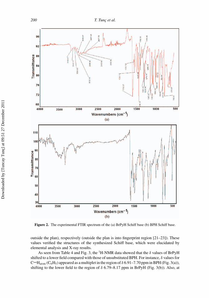
The selected IR spectral data of Schiff base are given in Table 3 and the spectrum of BrPyHis shown in Fig. 2(a). Pyridine and phenyl rings with Br substituent are attached to BrPyHSchiff base. Owing to this structure, the IR spectrum of Schiff base BrPyH is differentaccording to BPH (Fig. 2(b)). In the IR bands of BrPyH, N H and aromatic C H bandswere observed at 3189–2850 cm−1 weak and broad bands. Stretching N H band exhibitsa great broadening and low frequency shift when it forms strong intermolecular bonds andoverlapped with aromatic C H bands [20,21]. Therefore, in this study, the multiplet bandsat 3189–2850 cm−1 could be N H stretching mode forming hydrogen bond, and aromaticC H groups overlapped with the N H mode. The H of N H forms strong intermolecularhydrogen bonds with unpaired electrons of N pyridine of other molecules. These valuesconfirmed X-ray results (Fig. 3). Furthermore, strong sharp bands were observed in the IRspectrum of Schiff base for C C at 1436 cm−1 and at 1592 cm−1 for C N. The band inthe IR spectrum at approximately 811 cm−1 can be assigned to the C Br stretching mode[21,22]. In the IR spectra, three intensity bands appear at 1539, 1087–1060, and 765 cm−1,which are assigned to vibration frequency v (Py ring), v (Py ring bending), and v (Py ring
Dow
nloa
ded
by [
Tun
cay
Tun
ç] a
t 09:
51 2
7 D
ecem
ber
2011

Tabl
e3.
Sele
cted
IRsp
ectr
alda
taof
the
hydr
azon
es(K
Br,
vcm
−1)
CN
NC
PyA
r.Py
skel
eton
Abb
revi
atio
nN
HA
rH
CH
N(r
ing)
CC
NN
bend
ing
CB
r(fi
nger
prin
t)
BPH
3300
3500
–300
016
00–
1500
1250
––
930–
500
BrP
yH31
8931
43–2
850
1592
1539
1436
1130
1087
–106
090
4–67
890
4–67
8
199
Dow
nloa
ded
by [
Tun
cay
Tun
ç] a
t 09:
51 2
7 D
ecem
ber
2011
200 T. Tunc et al.
Figure 2. The experimental FTIR spectrum of the (a) BrPyH Schiff base (b) BPH Schiff base.
outside the plan), respectively (outside the plan is into fingerprint region [21–23]). Thesevalues verified the structures of the synthesized Schiff base, which were elucidated byelemental analysis and X-ray results.
As seen from Table 4 and Fig. 3, the 1H-NMR data showed that the δ values of BrPyHshifted to a lower field compared with those of unsubstituted BPH. For instance, δ values forC Harom (C6H5) appeared as a multiplet in the region of δ 6.91–7.70 ppm in BPH (Fig. 3(a)),shifting to the lower field to the region of δ 6.79–8.17 ppm in BrPyH (Fig. 3(b)). Also, at
Dow
nloa
ded
by [
Tun
cay
Tun
ç] a
t 09:
51 2
7 D
ecem
ber
2011

X-Ray Analysis and Spectroscopic Studies of (C12H10N3Br) 201
Figure 3. 1H-NMR spectrum of (a) BPH Schiff base (b) BrPyH Schiff base.
δ 7.62 ppm, the observed N H signal shifted to δ 8.17 ppm and at δ 7.13 ppm, theobserved HC N proton signal was observed at δ 8.15 ppm on the spectrum of BrPyH.These results are justifiable because there are electron-withdrawing groups such as p-Br(Hammett substituent coefficients for p-Br, σ p = 0.26) and pyridine in the structure of
Table 4. 1H-NMR data of the hydrazones (in CDCl3)
Abbreviation Harom, Hpyridine δ (ppm) N H δ (ppm) HC N δ (ppm)
BPH 7.70–6.91 7.62 7.13BrPyH 8.17–6.79 8.17 8.15
Dow
nloa
ded
by [
Tun
cay
Tun
ç] a
t 09:
51 2
7 D
ecem
ber
2011
202 T. Tunc et al.
BrPyH. Also, the downfield shift of the N H signal indicates intermolecular hydrogenbonding. Hydrogen bonding decreases electron density around the proton of amine groupand thus moves the proton absorption to a lower field. On the other hand, the assignmentof the Z-configuration to the HC N hydrazone double bond was performed consideringliterature data [6]. In fact, the chemical shift due to the CH hydrogen atom in theZ-isomer is usually upfield with respect to the E-isomer, appearing in substituted pyridinehydrazone derivatives at the range 7.45–8.30 ppm in the 1H NMR spectrum [6].
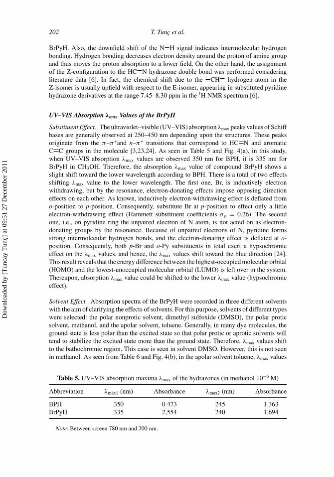
UV–VIS Absorption λmax Values of the BrPyH
Substituent Effect. The ultraviolet–visible (UV–VIS) absorption λmax peaks values of Schiffbases are generally observed at 250–450 nm depending upon the structures. These peaksoriginate from the π–π∗and n–π∗ transitions that correspond to HC N and aromaticC C groups in the molecule [3,23,24]. As seen in Table 5 and Fig. 4(a), in this study,when UV–VIS absorption λmax values are observed 350 nm for BPH, it is 335 nm forBrPyH in CH3OH. Therefore, the absorption λmax value of compound BrPyH shows aslight shift toward the lower wavelength according to BPH. There is a total of two effectsshifting λmax value to the lower wavelength. The first one, Br, is inductively electronwithdrawing, but by the resonance, electron-donating effects impose opposing directioneffects on each other. As known, inductively electron-withdrawing effect is deflated fromo-position to p-position. Consequently, substitute Br at p-position to effect only a littleelectron-withdrawing effect (Hammett substituent coefficients σ p = 0.26). The secondone, i.e., on pyridine ring the unpaired electron of N atom, is not acted on as electron-donating groups by the resonance. Because of unpaired electrons of N, pyridine formsstrong intermolecular hydrogen bonds, and the electron-donating effect is deflated at o-position. Consequently, both p-Br and o-Py substituents in total exert a hypsochromiceffect on the λmax values, and hence, the λmax values shift toward the blue direction [24].This result reveals that the energy difference between the highest-occupied molecular orbital(HOMO) and the lowest-unoccupied molecular orbital (LUMO) is left over in the system.Thereupon, absorption λmax value could be shifted to the lower λmax value (hypsochromiceffect).
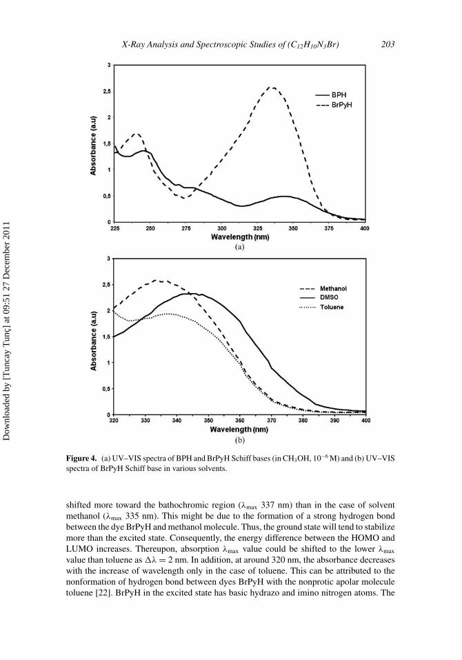
Solvent Effect. Absorption spectra of the BrPyH were recorded in three different solventswith the aim of clarifying the effects of solvents. For this purpose, solvents of different typeswere selected: the polar nonprotic solvent, dimethyl sulfoxide (DMSO), the polar proticsolvent, methanol, and the apolar solvent, toluene. Generally, in many dye molecules, theground state is less polar than the excited state so that polar protic or aprotic solvents willtend to stabilize the excited state more than the ground state. Therefore, λmax values shiftto the bathochromic region. This case is seen in solvent DMSO. However, this is not seenin methanol. As seen from Table 6 and Fig. 4(b), in the apolar solvent toluene, λmax values
Table 5. UV–VIS absorption maxima λmax of the hydrazones (in methanol 10−6 M)
Abbreviation λmax1 (nm) Absorbance λmax2 (nm) Absorbance
BPH 350 0.473 245 1.363BrPyH 335 2,554 240 1,694
Note: Between screen 780 nm and 200 nm.
Dow
nloa
ded
by [
Tun
cay
Tun
ç] a
t 09:
51 2
7 D
ecem
ber
2011
X-Ray Analysis and Spectroscopic Studies of (C12H10N3Br) 203
Figure 4. (a) UV–VIS spectra of BPH and BrPyH Schiff bases (in CH3OH, 10−6 M) and (b) UV–VISspectra of BrPyH Schiff base in various solvents.
shifted more toward the bathochromic region (λmax 337 nm) than in the case of solventmethanol (λmax 335 nm). This might be due to the formation of a strong hydrogen bondbetween the dye BrPyH and methanol molecule. Thus, the ground state will tend to stabilizemore than the excited state. Consequently, the energy difference between the HOMO andLUMO increases. Thereupon, absorption λmax value could be shifted to the lower λmax
value than toluene as �λ = 2 nm. In addition, at around 320 nm, the absorbance decreaseswith the increase of wavelength only in the case of toluene. This can be attributed to thenonformation of hydrogen bond between dyes BrPyH with the nonprotic apolar moleculetoluene [22]. BrPyH in the excited state has basic hydrazo and imino nitrogen atoms. The
Dow
nloa
ded
by [
Tun
cay
Tun
ç] a
t 09:
51 2
7 D
ecem
ber
2011

204 T. Tunc et al.
Table 6. UV–VIS absorption maxima λmax of the BrPyH in various solvents
Toluene λmax (nm) Methanol λmax (nm) DMSO λmax (nm)Abbreviation absorbance absorbance absorbance
BrPyH 337 (1.937) 335 (2.554) 346 (1.788)
basic property of hydrazo and imino nitrogen is increased by successive introduction ofelectron-donating groups by resonance and weak electron-withdrawing groups, such asBr, at the p-position of the benzene ring. Furthermore, Br stabilizes the excited state bydonating nonbonding electrons [25]. Consequently, as seen from Table 6 and Fig. 4(b), themore the solvent polarity increases, the more shifting happens to the absorption λmax valueto the bathochromic region/area.
Description of the Crystal Structure. The structure of BrPyH crystallizes into a monocliniclattice with space group P21/c. An ORTEP view of the asymmetric unit is shown in Fig. 5and selected bond distances and angles are presented in Table 7. The asymmetric unitcontains one Schiff base molecule. In the molecule, the benzene and pyridine rings and
Figure 5. View of the crystal packing of the title compound, along the b-axis. Hydrogen bonds areindicated by dashed lines.
Dow
nloa
ded
by [
Tun
cay
Tun
ç] a
t 09:
51 2
7 D
ecem
ber
2011

X-Ray Analysis and Spectroscopic Studies of (C12H10N3Br) 205
Table 7. Selected bond distances (Å), angles (◦), and torsion angles(◦)
Bond distance (Å)Br C1 1.894(5)N2 N1 1.393(5)N2 C8 1.382(6)N1 C7 1.272(6)N3 C8 1.333(6)N3 C12 1.356(7)
Bond angles (◦)N1 C7 C4 121.9(5)C2 C1 Br 120.3(4)N1 N2 N8 118.6(4)C7 N1 N2 116.9(4)
Torsion angles (◦)C3 C4 C7 N1 11.0(8)N1 N2 C8 C9 −4.5(7)
the hydrazone bridge are practically coplanar. Molecules of the title compound adopt anE-configuration about the azomethine C N double bond, with an N2-N1-C7-C4 torsionangle of 178.5(4)◦. For the molecule of BrPyH (Fig. 5), the benzene and pyridine ringsintersect at an 11.1(3)◦. The N2 C8 and N1 N2 bond distances are between 1.382(6) Åand 1.393(5) Å, respectively. The N1 C7 double bond is 1.272(6) Å.
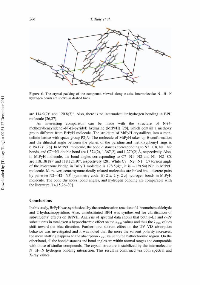
As shown in Fig. 5, each enantiomer forms head-to-tail molecular pairs. An inter-molecular hydrogen bond was observed between the H atom attached to the N atom (N2)of the hydrogen bridge and the N atom (N3) of the pyridine rings of the adjacent molecule[N2 H2···N3]. Intermolecular hydrogen bond distances and bond angles are listed in Table8. Also, π–π interactions can be seen from the packing diagram (Fig. 6). Intermolecularπ–π and hydrogen bonding interactions link the molecules and these interactions play animportant role in the stabilization of the crystal structure.
Similarly, the structure of BPH crystallizes into a monoclinic lattice with space groupP21/c [26,27]. The molecule of BPH takes up E-configuration and the angle between thetwo benzene rings is 8.1(1)◦ [26]. In BPH molecule, the bond distances correspondingto N2 C8 and N1 N2 bonds and C7 N1 double bond are 1.435(10), 1.330(8), and1.312(9) Å, respectively [26]. While the C7 N1 N2 and N1 N2 C8 bond angles inBrPyH molecule are 116.9(4)◦ and 118.6(4)◦, bond angles corresponding to them in BPH
Table 8. The hydrogen bond distances (Å) and angles (◦)
D H . . . A d(D H) (Å) d(H ··A) (Å) d(D ··A) (Å) D H ··A (◦)
N2 H2N ··N3i 1.05 2.02 3.066(2) 172
Note: Symmetry code: (i) = –x, –y, –z.
Dow
nloa
ded
by [
Tun
cay
Tun
ç] a
t 09:
51 2
7 D
ecem
ber
2011
206 T. Tunc et al.
Figure 6. The crystal packing of the compound viewed along a-axis. Intermolecular N—H···Nhydrogen bonds are shown as dashed lines.
are 114.9(7)◦ and 120.8(7)◦. Also, there is no intermolecular hydrogen bonding in BPHmolecule [26,27].
An interesting comparison can be made with the structure of N-(4-methoxybenzylidene)-N′-(2-pyridyl) hydrazine (MtPyH) [28], which contain a methoxygroup different from BrPyH molecule. The structure of MtPyH crystallizes into a mon-oclinic lattice with space group P21/c. The molecule of MtPyH takes up E-conformationand the dihedral angle between the planes of the pyridine and methoxyphenyl rings is6.19(12)◦ [28]. In MtPyH molecule, the bond distances corresponding to N2 C8, N1 N2bonds, and C7 N1 double bond are 1.374(2), 1.367(2), and 1.270(2) Å, respectively. Also,in MtPyH molecule, the bond angles corresponding to C7 N1 N2 and N1 N2 C8are 118.18(18)◦ and 118.12(19)◦, respectively [28]. While C8 N2 N1 C7 torsion angleof the hydrazone bridge in BrPyH molecule is 178.5(4)◦, it is −179.54(19)◦ in MtPyHmolecule. Moreover, centrosymmetrically related molecules are linked into discrete pairsby pairwise N2 H2···N3i [symmetry code: (i) 2-x, 2-y, 2-z] hydrogen bonds in MtPyHmolecule. The bond distances, bond angles, and hydrogen bonding are comparable withthe literature [14,15,26–30].
Conclusions
In this study, BrPyH was synthesized by the condensation reaction of 4-bromobenzaldehydeand 2-hydrazinopyridine. Also, unsubstituted BPH was synthesized for clarification ofsubstituents’ effects on BrPyH. Analysis of spectral data shows that both p-Br and o-Pysubstituents in total exert a hypsochromic effect on the λmax values and thus the λmax valuesshift toward the blue direction. Furthermore, solvent effect on the UV–VIS absorptionbehavior was investigated and it was noted that the more the solvent polarity increases,the more shifting happens to the absorption λmax value to the bathochromic region. On theother hand, all the bond distances and bond angles are within normal ranges and comparablewith those of similar compounds. The crystal structure is stabilized by the intermolecularN H···N hydrogen bonding interaction. This result is confirmed via both spectral andX-ray values.
Dow
nloa
ded
by [
Tun
cay
Tun
ç] a
t 09:
51 2
7 D
ecem
ber
2011

X-Ray Analysis and Spectroscopic Studies of (C12H10N3Br) 207
Supplementary Data
Crystallographic data for the structural analysis have been deposited with the CambridgeCrystallographic Data Centre as supplementary publication no. CCDC 798670. Copiesof this data can be obtained free of charge via www.ccdc.cam.ac.uk/data request/cif, byemailing data [email protected], or by contacting the Cambridge CrystallographicData Centre, 12 Union Road, Cambridge CB2 1EZ, UK; Fax: +44 1223 336033.
References
[1] Schiele, V. C. (1964). Ber., 30, 308.[2] Mattson, A. M., Jensen, C. O., and Dutcher, R. A. (1947). Science, 5, 294.[3] Karabocek, N., Kucukdumlu, A., Ekmekcıoglu, P., and Karabocek, S. (2009). J. Macromol. Sci.
A, 46(10), 1007.[4] Despaigne, A. A. R., Da Silva, J. G., Do Carmo, A. C. M., Piro, O. E., Castellano, E. E., and
Beraldo, H. (2009). J. Mol. Struc., 920, 97.[5] McMurry, J. E. (2004). Organic Chemistry, Brooks/Cole: Pacific Grove, CA.[6] Todeschini, A. R., de Miranda, A. L. P., da Silva, K. C. M., Parrini, S. C., and Barreiro, E.
(1998). J. Eur. J. Med. Chem., 33, 189.[7] Shit, S., Chakraborty, J., Samanta, B., Slawin, A. M. Z., Gramlich, V., and Mitra, S. (2009).
Struct. Chem., 20(4), 633.[8] Sridhar, R. and Perumal, P. T. (2003). Synth. Commun., 33(9), 1483.[9] Onnis, V., Cocco, M. T., Fadda, R., and Congiu, C. (2009). Bioorgan. Med. Chem., 17(17),
6158.[10] Bottari, B., Maccari, R., Monforte, F., Ottana, R., Vigorita, M. G., Bruno, G., Nicolo, F.,
Rotondob, A., and Rotondob, E. (2001). Bioorgan. Med. Chem., 9(8), 2203.[11] Filipovic, N., Borrmann, H., Todorovic, T., Borna, M., Spasojevic, V., Sladic, D., Novakovic,
I., and Andjelkovic, K. (2009). Inorg. Chim. Acta, 362(6), 1996.[12] Arguelles, M. C. R., Cao, R., Garcıa-Deibe, A. M., Pelizzi, C., Matalobos, J. S., and Zani, F.
(2009). Polyhedron, 28(11), 2187.[13] Barros-Garcia, F. J., Bernalte-Garcia, A., Luna-Giles, F., Maldonado-Rogado, M. A., and
Vinuelas-Zahinos, E. (2005). Polyhedron, 24(10), 1125.[14] Tezcan, H., Tunc, T., Sahin, E., and Yagbasan, R. (2004). Anal. Sci., 20, x137.[15] Tunc, T., Tezcan, H., Sarı, M., Buyukgungor, O., and Yagbasan, R. (2003). Acta Cryst., C59,
o528.[16] Rigaku Americas and Rigaku. (2005). Crystal-Clear Program for Integration of the Intensities,
Correction for Lorentz, and Polarization Effects, and Cell Refinement, Rigaku Americas: TheWoodlands, TX; Rigaku Corporation: Tokyo.
[17] Sheldrick, G. M. (1990). Acta Cryst., A46, 467.[18] Sheldrick, G. M. (1997). SHELXL-97, University of Gottingen: Germany.[19] Farrugia, L. J. (1997). J. Appl. Cryst., 30, 565.[20] Gilli, P., Bertolasi, V., Pretto, L., and Gilli, G. (2006). J. Mol. Struc., 790, 40.[21] Bellamy, L. J. (1962). The Infrared Spectra of Complex Molecules, Methuen: London.[22] Williams, D. H., and Fleming, I. (1966). Spectroscopic Methods in Organic Chemistry, McGraw-
Hill: London.[23] Ababei, L. V., Kriza, A., Musuc, A. M., Andronescu, C., and Rogozea, E. A. (2010). J. Therm.
Anal. Calorim., 101(3), 987.[24] Tezcan, H. and Uzluk, E. (2007). Dyes Pigm., 75(3), 633.[25] Ding, W. F. and Jiang, X. (1998). J. Phys. Org. Chem., 11, 809.[26] Vickery, B. B. and Willey, G. R. (1985). Acta Cryst., C41, 1072.[27] Gunes, B., Ozbey, S., and Tezcan, H. (2003). Anal. Sci., 19, 1091.[28] Tunc, T., Sarı, M., Yagbasan, R., Tezcan, H., and Sahin, E. (2003). Acta Cryst., C59, o192.
Dow
nloa
ded
by [
Tun
cay
Tun
ç] a
t 09:
51 2
7 D
ecem
ber
2011

208 T. Tunc et al.
[29] Shanmuga, S., Raj, S., Fun, H. K., Lu, Z. L., Xiao, W., Gong, X. Y., and Kang, B. Z. (1999).Acta Cryst., C55, 942.
[30] Shanmuga, S., Raj, S., Fun, H. K., Lu, Z. L., Xiao, W., Gong, X. Y., and Gen, C. M. (2000).Acta Cryst., C56, 1013.
Dow
nloa
ded
by [
Tun
cay
Tun
ç] a
t 09:
51 2
7 D
ecem
ber
2011
Copyright © 2022 FDOKUMEN


![N -[4-( N -Cyclohexylsulfamoyl)phenyl]acetamide](https://static.fdokumen.com/doc/165x107/632f4f4de68feab59a0210b7/n-4-n-cyclohexylsulfamoylphenylacetamide.jpg)
