Photoaffinity Labeling of High Affinity Nicotinic Acid Adenine Dinucleotide Phosphate...
11
Photoaffinity Labeling of High Affinity Nicotinic Acid Adenine Dinucleotide Phosphate (NAADP)-Binding Proteins in Sea Urchin Egg * □ S Received for publication, October 10, 2011, and in revised form, November 16, 2011 Published, JBC Papers in Press, November 23, 2011, DOI 10.1074/jbc.M111.306563 Timothy F. Walseth ‡1 , Yaping Lin-Moshier ‡2 , Pooja Jain § , Margarida Ruas ¶ , John Parrington ¶ , Antony Galione ¶ , Jonathan S. Marchant ‡ , and James T. Slama § From the ‡ Department of Pharmacology, University of Minnesota Medical School, Minneapolis, Minnesota 55455, the § Department of Medicinal and Biological Chemistry, University of Toledo, Toledo, Ohio 43614, and the ¶ Department of Pharmacology, University of Oxford, Mansfield Road, Oxford OX1 3QT, United Kingdom Background: Nicotinic acid adenine dinucleotide phosphate (NAADP) regulates calcium release from internal acidic stores via two-pore channels (TPCs). Results: A novel photosensitive probe (5-azido-NAADP) identified high affinity NAADP binding sites that interact with, but are distinct from, TPCs. Conclusion: High affinity NAADP-binding proteins complex with TPCs. Significance: This work provides new mechanistic insights into how NAADP regulates calcium release via TPCs. Nicotinic acid adenine dinucleotide phosphate (NAADP) is a messenger that regulates calcium release from intracellular acidic stores. Recent studies have identified two-pore chan- nels (TPCs) as endolysosomal channels that are regulated by NAADP; however, the nature of the NAADP receptor binding site is unknown. To further study NAADP binding sites, we have synthesized and characterized [ 32 P-5-azido]nicotinic acid adenine dinucleotide phosphate ([ 32 P-5N 3 ]NAADP) as a photoaffinity probe. Photolysis of sea urchin egg homoge- nates preincubated with [ 32 P-5N 3 ]NAADP resulted in spe- cific labeling of 45-, 40-, and 30-kDa proteins, which was pre- vented by inclusion of nanomolar concentrations of unlabeled NAADP or 5N 3 -NAADP, but not by micromolar concentrations of structurally related nucleotides such as NAD, nicotinic acid adenine dinucleotide, nicotinamide mononucleotide, nicotinic acid, or nicotinamide. [ 32 P- 5N 3 ]NAADP binding was saturable and displayed high affin- ity (K d 10 nM) in both binding and photolabeling experi- ments. [ 32 P-5N 3 ]NAADP photolabeling was irreversible in a high K buffer, a hallmark feature of NAADP binding in the egg system. The proteins photolabeled by [ 32 P-5N 3 ]NAADP have molecular masses smaller than the sea urchin TPCs, and antibodies to TPCs do not detect any immunoreactivity that comigrates with either the 45-kDa or the 40-kDa photola- beled proteins. Interestingly, antibodies to TPC1 and TPC3 were able to immunoprecipitate a small fraction of the 45- and 40-kDa photolabeled proteins, suggesting that these pro- teins associate with TPCs. These data suggest that high affin- ity NAADP binding sites are distinct from TPCs. NAADP 3 is a naturally occurring nucleotide that has been shown to be involved in the release of intracellular calcium from acidic intracellular stores in a wide variety of cell types (1, 2). Mounting evidence suggests that NAADP may function as a trigger to initiate a calcium signal that is then amplified by other calcium release mechanisms (3). However, a fundamental ques- tion that remains unanswered is the identity of the NAADP receptor. Recently, the two-pore channel (TPC) family of endolysosomal proteins was shown to be regulated by NAADP (4 – 6). Multiple approaches, including molecular manipulation of TPC levels by overexpression (4 –7) or knockdown (4, 5), as well as electrophysiological analyses (8 –11), all support the role of TPCs as NAADP-sensitive Ca 2 channels. However, whether TPCs directly interact with NAADP remains an important unresolved issue. Notably, overexpression of mam- malian TPC2 modestly increased [ 32 P]NAADP binding activ- ity, but the increase in binding was much lower than the increase in TPC2 mRNA levels (3-fold versus 250-fold) (4). Although [ 32 P]NAADP binding activity was also found in immunoprecipitation studies using antibodies to sea urchin TPCs (12), very little is known regarding the interaction between NAADP and TPCs. Photoaffinity labeling is a powerful technique for identifying and characterizing receptors and binding sites for appropri- ately labeled ligands (13, 14). In this study, we have synthesized and characterized [ 32 P-5N 3 ]NAADP as a photoaffinity probe for NAADP binding sites. Previous studies with 5N 3 -NAADP indicated that this NAADP analog was able to release calcium and complete with NAADP for high affinity binding sites (15). * This work was supported, in whole or in part, by National Institutes of Health Grant GM088790 (to J. S. M.). This work was also supported by University of Minnesota Academic Health Center Seed Grant 2101.95 (to T. F. W.) and a Wellcome Trust Programme Grant (to A. G. and J. P.). This article was selected as a Paper of the Week. Author’s Choice—Final version full access. □ S This article contains supplemental Figs. 1S and 2S. 1 To whom correspondence should be addressed. Tel.: 612-625-2627; Fax: 612-625-8408; E-mail: [email protected]. 2 Supported by a doctoral dissertation fellowship from the University of Min- nesota Graduate School. 3 The abbreviations used are: NAADP, nicotinic acid adenine dinucleotide phosphate; 5N 3 -NAADP, 5-azido-NAADP; NAAD, nicotinic acid adenine dinucleotide; TPC, two-pore channels. THE JOURNAL OF BIOLOGICAL CHEMISTRY VOL. 287, NO. 4, pp. 2308 –2315, January 20, 2012 Author’s Choice © 2012 by The American Society for Biochemistry and Molecular Biology, Inc. Published in the U.S.A. 2308 JOURNAL OF BIOLOGICAL CHEMISTRY VOLUME 287 • NUMBER 4 • JANUARY 20, 2012 by guest on March 17, 2016 http://www.jbc.org/ Downloaded from by guest on March 17, 2016 http://www.jbc.org/ Downloaded from by guest on March 17, 2016 http://www.jbc.org/ Downloaded from by guest on March 17, 2016 http://www.jbc.org/ Downloaded from
Transcript of Photoaffinity Labeling of High Affinity Nicotinic Acid Adenine Dinucleotide Phosphate...

Photoaffinity Labeling of High Affinity Nicotinic Acid AdenineDinucleotide Phosphate (NAADP)-Binding Proteins in SeaUrchin Egg*□S �
Received for publication, October 10, 2011, and in revised form, November 16, 2011 Published, JBC Papers in Press, November 23, 2011, DOI 10.1074/jbc.M111.306563
Timothy F. Walseth‡1, Yaping Lin-Moshier‡2, Pooja Jain§, Margarida Ruas¶, John Parrington¶, Antony Galione¶,Jonathan S. Marchant‡, and James T. Slama§
From the ‡Department of Pharmacology, University of Minnesota Medical School, Minneapolis, Minnesota 55455, the§Department of Medicinal and Biological Chemistry, University of Toledo, Toledo, Ohio 43614, and the ¶Department ofPharmacology, University of Oxford, Mansfield Road, Oxford OX1 3QT, United Kingdom
Background:Nicotinic acid adenine dinucleotide phosphate (NAADP) regulates calcium release from internal acidic storesvia two-pore channels (TPCs).Results:Anovel photosensitive probe (5-azido-NAADP) identified high affinityNAADPbinding sites that interact with, but aredistinct from, TPCs.Conclusion: High affinity NAADP-binding proteins complex with TPCs.Significance: This work provides new mechanistic insights into how NAADP regulates calcium release via TPCs.
Nicotinic acid adenine dinucleotide phosphate (NAADP) isa messenger that regulates calcium release from intracellularacidic stores. Recent studies have identified two-pore chan-nels (TPCs) as endolysosomal channels that are regulated byNAADP; however, the nature of the NAADP receptor bindingsite is unknown. To further study NAADP binding sites, wehave synthesized and characterized [32P-5-azido]nicotinicacid adenine dinucleotide phosphate ([32P-5N3]NAADP) as aphotoaffinity probe. Photolysis of sea urchin egg homoge-nates preincubated with [32P-5N3]NAADP resulted in spe-cific labeling of 45-, 40-, and 30-kDa proteins, which was pre-vented by inclusion of nanomolar concentrations ofunlabeled NAADP or 5N3-NAADP, but not by micromolarconcentrations of structurally related nucleotides such asNAD, nicotinic acid adenine dinucleotide, nicotinamidemononucleotide, nicotinic acid, or nicotinamide. [32P-5N3]NAADP binding was saturable and displayed high affin-ity (Kd �10 nM) in both binding and photolabeling experi-ments. [32P-5N3]NAADP photolabeling was irreversible in ahigh K� buffer, a hallmark feature of NAADP binding in theegg system. The proteins photolabeled by [32P-5N3]NAADPhave molecular masses smaller than the sea urchin TPCs, andantibodies to TPCs do not detect any immunoreactivity thatcomigrates with either the 45-kDa or the 40-kDa photola-beled proteins. Interestingly, antibodies to TPC1 and TPC3were able to immunoprecipitate a small fraction of the 45-and 40-kDa photolabeled proteins, suggesting that these pro-
teins associate with TPCs. These data suggest that high affin-ity NAADP binding sites are distinct from TPCs.
NAADP3 is a naturally occurring nucleotide that has beenshown to be involved in the release of intracellular calciumfrom acidic intracellular stores in a wide variety of cell types (1,2). Mounting evidence suggests that NAADPmay function as atrigger to initiate a calcium signal that is then amplified by othercalcium releasemechanisms (3).However, a fundamental ques-tion that remains unanswered is the identity of the NAADPreceptor. Recently, the two-pore channel (TPC) family ofendolysosomal proteins was shown to be regulated by NAADP(4–6).Multiple approaches, includingmolecularmanipulationof TPC levels by overexpression (4–7) or knockdown (4, 5), aswell as electrophysiological analyses (8–11), all support the roleof TPCs as NAADP-sensitive Ca2� channels. However,whether TPCs directly interact with NAADP remains animportant unresolved issue. Notably, overexpression of mam-malian TPC2 modestly increased [32P]NAADP binding activ-ity, but the increase in binding was much lower than theincrease in TPC2 mRNA levels (3-fold versus �250-fold) (4).Although [32P]NAADP binding activity was also found inimmunoprecipitation studies using antibodies to sea urchinTPCs (12), very little is known regarding the interactionbetween NAADP and TPCs.Photoaffinity labeling is a powerful technique for identifying
and characterizing receptors and binding sites for appropri-ately labeled ligands (13, 14). In this study, we have synthesizedand characterized [32P-5N3]NAADP as a photoaffinity probefor NAADP binding sites. Previous studies with 5N3-NAADPindicated that this NAADP analog was able to release calciumand complete with NAADP for high affinity binding sites (15).
* This work was supported, in whole or in part, by National Institutes of HealthGrant GM088790 (to J. S. M.). This work was also supported by University ofMinnesota Academic Health Center Seed Grant 2101.95 (to T. F. W.) and aWellcome Trust Programme Grant (to A. G. and J. P.).
� This article was selected as a Paper of the Week.Author’s Choice—Final version full access.
□S This article contains supplemental Figs. 1S and 2S.1 To whom correspondence should be addressed. Tel.: 612-625-2627; Fax:
612-625-8408; E-mail: [email protected] Supported by a doctoral dissertation fellowship from the University of Min-
nesota Graduate School.
3 The abbreviations used are: NAADP, nicotinic acid adenine dinucleotidephosphate; 5N3-NAADP, 5-azido-NAADP; NAAD, nicotinic acid adeninedinucleotide; TPC, two-pore channels.
THE JOURNAL OF BIOLOGICAL CHEMISTRY VOL. 287, NO. 4, pp. 2308 –2315, January 20, 2012Author’s Choice © 2012 by The American Society for Biochemistry and Molecular Biology, Inc. Published in the U.S.A.
2308 JOURNAL OF BIOLOGICAL CHEMISTRY VOLUME 287 • NUMBER 4 • JANUARY 20, 2012
by guest on March 17, 2016
http://ww
w.jbc.org/
Dow
nloaded from
by guest on March 17, 2016
http://ww
w.jbc.org/
Dow
nloaded from
by guest on March 17, 2016
http://ww
w.jbc.org/
Dow
nloaded from
by guest on March 17, 2016
http://ww
w.jbc.org/
Dow
nloaded from

The sea urchin egg system was chosen for this study becausehigh affinity binding in this system has been well characterizedand is considered the “gold standard” for studying NAADPfunction. [32P-5N3]NAADP photolabeling was found to specif-ically label 45-, 40-, and 30-kDa proteins in Strongylocentrotuspurpuratus homogenates. The properties of the photolabeledproteins matched NAADP binding in terms of selectivity andirreversibility in high potassium. Antibodies to TPCs did notrecognize the 45- and 40-kDa proteins. Interestingly, the 45-and 40-kDa photolabeled proteins were found in immunopre-cipitates using TPC1 and TPC3 antibodies. Overall, the datasuggest that the [32P-5N3]NAADP photoaffinity probe detectshigh affinity NAADP binding sites and that these proteins aredistinct from, but interact with, TPCs. The [32P-5N3]NAADPphotoprobe should prove to be a valuable tool in the identifica-tion of NAADP-binding proteins and elucidation of the mech-anism by which NAADP regulates mobilization of intracellularcalcium.
EXPERIMENTAL PROCEDURES
Materials—HEPES, CHAPS, potassium gluconate, N-meth-ylglucamine, NAD, NAAD, NADP, nicotinamide mononucle-otide, nicotinic acid, nicotinamide, ATP, and DTT were fromSigma. Laemmli sample buffer, 12.5% Criterion acrylamidegels, and AGMP-1 resin were obtained from Bio-Rad. Simply-Blue SafeStain was obtained from Invitrogen. Recombinanthuman NAD kinase was purchased from Alexis Biochemicals.Complete protease inhibitor tablets were from Roche AppliedScience. ECL reagents were obtained from GE Healthcare.[32P]NAD (800 Ci/mmol) was from PerkinElmer Life Sciences.NAADP, 5N3-NAADP, and 5N3-nicotinic acid were preparedas described previously (15).Sea Urchin Egg Homogenates—S. purpuratus homogenates
(25%) were prepared as described previously (16) and stored as0.5-ml aliquots at�80 °C. The homogenateswere prepared andstored in a buffer containing 250 mM potassium gluconate, 250mMN-methylglucamine, 20mMHepes, 1mMMgCl2, 2 units/mlcreatine kinase, 4 mM creatine phosphate, and 0.5 mMATP, pH7.2.Synthesis of [32P-5N3]NAADP—A two-step procedure simi-
lar to that used previously for the generation of [32P]NAADP(17) was employed to prepare [32P-5N3]NAADP. In the firststep, [32P]NAD is converted to [32P]NADP using human NADkinase. The resulting [32P]NADP is converted to [32P-5N3]NAADP through a base-exchange reaction catalyzed byAplysia ADP-ribosyl cyclase (18). [32P]NAD (1 mCi) wasdiluted to 500 �l with 20mMHepes, pH 7.3, 5 mMMgCl2, 2 mM
ATP, 1 mM DTT, 1 unit/ml creatine kinase, and 2 mM creatinephosphate. The reaction was started by adding 2 �g of humanNAD kinase and incubated for 16 h at room temperature. The[32P]NADP (80–90%yield) generatedwas purified byAGMP-1chromatography. The reaction was diluted to 1 ml with waterand injected onto a 0.5 � 5-cm column of AG MP-1. Elutionwas done with a trifluoroacetic acid gradient from 0 to 150 mM
over 30 min at 1 ml/min. Eluted radioactivity was monitoredwith a Beckman 171M radiochemical detector. [32P]NADPeluted between 15 and 17 min. The AGMP-1 chromatographysystem has been previously described (18). The [32P]NADPwas
converted to [32P-5N3]NAADP as follows. Sodium acetate, pH6.0 (175 �l of 500 mM), and 5N3-nicotinic acid (200 �l of 500mM in dimethyl sulfoxide) were added to 3.5 ml of purified[32P]NADP. The 5N3-nicotinic acid precipitates under theseconditions. The pH of the sample was raised by titration with 2M Tris base until the precipitate dissolved (�75 �l). Sodiumacetate, pH 3.7 (300�l of 500mM)was added to readjust the pHof the sample as close to pH 4.0 as possible. The base-exchangereaction was started by adding 4 �l of 100 �g/ml AplysiaADP-ribosyl cyclase and incubated at room temperature for 60–120min. The reaction was neutralized by adding 80 �l of 2 M Trisbase and was purified by AG MP-1 chromatography asdescribed above (1.1 ml of reaction/run). The [32P-5N3]-NAADP eluted at 22 min and was stored unneutralized at�20 °C in 0.5-ml aliquots. It was assumed that the specificactivity of the [32P-5N3]NAADP was the same as the commer-cial [32P]NAD used as the starting material.Photoaffinity Labeling of Egg Homogenates with [32P-5N3]-
NAADP—S. purpuratus egg homogenates were diluted 10-foldwith a buffer containing 250 mM potassium gluconate, 250 mM
N-methylglucamine, 1 mMMgCl2, 20 mMHepes, pH 7.2 (KGlubuffer). The standard photolabeling protocol was as follows.The diluted homogenates (90 �l, 120 �g of protein) were incu-bated in a total volume of 100 �l with 2–8 nM [32P-5N3]NAADP for 90 min at 4 °C. Unlabeled competitors weremade up to the final concentrations indicated in the figure leg-ends. UV photolysis was accomplished by placing the sampleson a sheet of Saran Wrap stretched over a block of ice andirradiated with UV light (1015 quanta/s) for 2 min in a Rayonetphotochemical reactor (Southern New England UltravioletCo.). After photolysis, the samples were recovered and placedin microcentrifuge tubes containing 1 ml of 20 mM Hepes, pH7.3. The samples were centrifuged at 21,000� g for 10min, andthe supernatant was discarded. The membrane pellets wereresuspended in 50 �l of 1� Laemmli SDS-sample buffer andsubjected to SDS-polyacrylamide gel electrophoresis using12.5% Criterion acrylamide gels. The gels were stained withSimplyBlue SafeStain, destained, and air-dried between twosheets of cellophane. Analysis of photolabeling was accom-plished by exposing the dried gels to MP storage phosphorscreens (Packard Instruments) for 24–120 h. The phosphorscreens were developed using a Cyclone storage phosphor sys-tem (Packard Instruments). Quantification of the phosphorimages was performed with the OptiQuant software (Ver-sion3.0, Packard Instruments). Because the egg homogenateswere prepared and stored in the presence of 0.5 mM ATP, thestandard photolabeling conditions contain 50 �M ATP.HPLC Purification of NADP—The NADP used in competi-
tion experiments for these studies was purified immediatelyprior to use because NAADP was originally discovered as acontaminant in commercial NADP (19, 20). The HPLC systemconsisted of Beckman 125 pumps and a 166 UV detector con-trolled by the Nouveau Gold software and a MonoQ HR 5/5column. The columnwas preconditioned with 10mMK2HPO4,pH 8.0, at a flow rate of 0.5ml/min. NADP (50�l of 90mM) wasinjected, and elution was controlled through a linear gradient(2–25 min) starting with 10 mM K2HPO4, pH 8.0, and endingwith 50 mM K2HPO4, 200 mM NaCl, pH 8.0.
Photoaffinity Labeling of NAADP-binding Proteins
JANUARY 20, 2012 • VOLUME 287 • NUMBER 4 JOURNAL OF BIOLOGICAL CHEMISTRY 2309
by guest on March 17, 2016
http://ww
w.jbc.org/
Dow
nloaded from

TPC Immunoprecipitation and Immunoblot Procedures—The antibodies to S. purpuratus TPCs and their coupling toprotein A agarose beads have been previously described (12).For TPC immunoprecipitation studies, egg homogenate wasphotolabeled with [32P-5N3]NAADP under standard condi-tions (see above). Twelve samples (120 �g/sample) were com-bined and processed. The resulting photolabeled pellet wasreconstituted in 400 �l of KGlu buffer � 1% CHAPS � Com-plete protease inhibitors and allowed to incubate on ice for1.5 h. TheCHAPS solubilized supernatantwas obtained by cen-trifugation at 21,000 � g for 15 min. Agarose beads (100 �l)coupled to antibodies were prewashed two times with 1 ml ofKGlu buffer �1% CHAPS before use. Immunoprecipitationwas initiated by incubating 90 �l of photolabeled CHAPS solu-bilized sample with 50 �l of prewashed agarose beads coupledto control antisera or antibodies to sea urchin TPC1, TPC2, orTPC3. The samples were incubated at 4 °C for 16 h on a rotator.The samples were centrifuged at 5000� g for 5min andwashedfour times with 1ml of KGlu buffer� 1%CHAPS. The sampleswere vigorously vortexed at each wash step, and the beads wereisolated by centrifuging at 5000 � g for 5 min, except for thefinal wash in which the samples were centrifuged at 10,000 � gfor 10 min. After the final wash, the agarose beads were sus-pended in 50�l of 1�Laemmli sample buffer and incubated for2 h at room temperature with occasional mixing. The beadswere then centrifuged at 21,000 � g for 10 min, and the super-natants were subjected to electrophoresis on 12.5% Criteriongels as described above.To control for any changes in protein migration caused by
UV or photocross-linking, samples for immunoblot analyseswere photolabeledwith 1�M5N3-NAADP, processed as above,run on 12.5% Criterion gels, and transferred to PVDF mem-brane for immunoblotting. A sample photolyzed with [32P-5N3]NAADPwas included so that the positions of the photola-beled proteins could be compared with any immunoreactivitydetected. The PVDFmembranewas cut and subjected toWest-ern analyses using antibodies to sea urchin TPC1, TPC2, andTPC3. Detection was by chemiluminescence using ECLreagents.[32P-5N3]NAADP and [32P]NAADP Binding Studies—The
binding studies using [32P-5N3]NAADP or [32P]NAADP wereconducted as described previously (15).Data Analyses—GraphPad Prism (GraphPad Software) was
used for curve fitting and statistical analyses. Error bars shownin Figs. 3–5 represent mean � S.E.
RESULTS AND DISCUSSION
Synthesis and Characterization of Novel NAADP-based Pho-toaffinity Probe—Astudy examining the structure-activity rela-tionship of NAADP analogs substituted at the 4- or 5-positionof nicotinic acid (15) revealed that 5N3-NAADPwas an agonistof calcium release from sea urchin homogenates (EC50 of 1.7�M) that also competed with [32P]NAADP with high potency(IC50 � 18 nM) in a competition binding assay. Such propertiessuggested that 5N3-NAADP could prove useful as a photoaffin-ity probe for labeling NAADP-binding proteins. Therefore, weprepared high specific activity [32P-5N3]NAADP via a base-exchange reaction by incubating high specific activity
[32P]NADP with 5-azido-nicotinic acid and AplysiaADP-ribo-syl cyclase (18) to yield [32P-5N3]NAADP (structure shown inFig. 1).The ability of the [32P-5N3]NAADP probe to detect high
affinity NAADP-binding proteins was assessed in Fig. 2. S. pur-puratus egg homogenates were incubated for 90min with [32P-5N3]NAADP in the absence or presence of 1 �M unlabeledNAADP, and then the effect of UV exposure was examined. Noproteins were labeled if the sample was not subjected to UVlight to activate the probe (Fig. 2, lane 1). However, severalproteins were labeled (45, 40, and 30 kDa) when the sample wasexposed to UV (Fig. 2, lane 2). The 45- and 40-kDa proteinslabeled to approximately the same extent, with the 30-kDa pro-tein accounting for 10% of the total specific labeling. Inclusionof unlabeled 1 �M NAADP in the incubation prevented thelabeling of the 45-, 40-, and 30-kDa proteins (Fig. 2, lane 3).These data confirm that the [32P-5N3]NAADP probe is UV-sensitive and that an excess of unlabeled NAADP protects thelabeling of the proteins. Furthermore, the time courses for [32P-5N3]NAADP photolabeling of the 45-, 40-, and 30-kDa pro-teins and [32P-5N3]NAADP binding were similar, and bothmaximized between 60 and 100 min (supplemental Fig. 1S).
FIGURE 1. Structure of 5-azido-NAADP. The * denotes the phosphoruslabeled with 32P.
FIGURE 2. [32P-5N3]NAADP photoaffinity labeling is UV-dependent andblocked by unlabeled NAADP. S. purpuratus egg homogenates were incu-bated with [32P-5N3]NAADP for 90 min in the absence (lanes 1 and 2) or pres-ence (lane 3) of 1 �M NAADP. Lane 1 represents a control that was not sub-jected to UV light. Lanes 2 and 3 were subjected to UV light. Image is aphosphor image of the resulting gel (see “Experimental Procedures” forstandard conditions).
Photoaffinity Labeling of NAADP-binding Proteins
2310 JOURNAL OF BIOLOGICAL CHEMISTRY VOLUME 287 • NUMBER 4 • JANUARY 20, 2012
by guest on March 17, 2016
http://ww
w.jbc.org/
Dow
nloaded from

This pattern of [32P-5N3]NAADP photolabeling was consistentbetween 32 different S. purpuratus egg homogenate prepara-tions, and the extent of [32P]NAADP binding correlated withthe intensity of photolabeling (supplemental Fig. 2S). Prepara-tions with low NAADP binding activity did not photolabelefficiently.Specificity of [32P-5N3]NAADP Photolabeling—How specific
was the photoaffinity labeling of the 45-, 40-, and 30-kDa pro-teins by [32P-5N3]NAADP? Inclusion of unlabeled NAADP(Fig. 3A) or 5N3-NAADP (Fig. 3B) reduced the labeling of the45-, 40-, and 30-kDa proteins in a concentration-dependent
manner. Half-maximal reduction of the photolabeling of thesebands occurred at �0.7 nM NAADP and 4 nM 5N3-NAADP(Fig. 3C). The competition kinetics were similar for all threelabeled proteins. Structurally related nucleotides such as NAD,NAAD, nicotinamide mononucleotide, nicotinic acid, and nic-otinamide did not affect the ability of [32P-5N3]NAADP to pho-tolabel the 45-, 40-, and 30-kDa proteins even at concentrationsas high as 10 �M (Fig. 3, A and D). NADP did reduce the pho-tolabeling of these bands by about 30% at 10 �M (Fig. 3, B andD), although the NADP was purified by HPLC immediatelyprior to the experiment to remove contaminatingNAADP. The
FIGURE 3. Specificity of [32P-5N3]NAADP photolabeling in sea urchin egg homogenates. Photolabeling was performed in the absence or presence of theindicated concentrations of various competitors. A, phosphor image of a gel examining the effect of NAADP, NAD, NAAD, nicotinamide mononucleotide(NMN), nicotinic acid (NA), and nicotinamide (nico). The concentrations of NAADP range from 0.1 to 10,000 nM in 10-fold increments. B, phosphor image of a gelexamining the effect of 5N3-NAADP and NADP. The concentration of unlabeled 5N3-NAADP ranges from 1 to 10,000 nM in 10-fold increments. C, competitionof [32P-5N3]NAADP photolabeling (45-, 40-, and 30-kDa bands combined) by unlabeled NAADP and 5N3-NAADP. Data represent the mean � S.E. (n � 4) ofdensitometric analyses of experiments similar to those shown in panels A and B. D, the effect of various nucleotides on [32P-5N3]NAADP photolabeling (45-, 40-,and 30-kDa bands). Data represent the mean � S.E. (n � 4) of densitometric analyses of experiments similar to those shown in panels A and B. cADPR, cyclicADP-ribose.
Photoaffinity Labeling of NAADP-binding Proteins
JANUARY 20, 2012 • VOLUME 287 • NUMBER 4 JOURNAL OF BIOLOGICAL CHEMISTRY 2311
by guest on March 17, 2016
http://ww
w.jbc.org/
Dow
nloaded from

specificity of [32P-5N3]NAADPphotolabeling shown in Fig. 3 issimilar to the specificity of [32P]NAADP binding previouslyobserved in this system (17, 21) and consistent with photolabel-ing data observed in another sea urchin species (22).Fig. 4 shows the dependence of [32P-5N3]NAADP photola-
beling and binding on the concentration of [32P-5N3]NAADP.Fig. 4A shows that the intensity of the labeling of the 45-, 40-,and 30-kDaproteins and others,most notably proteins between100 and 250 kDa, increased as the concentration of labeled pho-toprobe is increased. Only the 45-, 40-, and 30-kDa proteinswere labeled specifically as inclusion of unlabeled 1�MNAADPprevented the labeling of these proteins but not the others. Fig.4, B and C, compare saturation kinetics of [32P-5N3]NAADPphotolabeling with [32P-5N3]NAADP binding kinetics deter-mined by a conventional filtration binding assay. Nonspecificbinding was estimated in both experiments by determining thephotolabeling or binding in the presence of 1 �M NAADP ateach concentration of [32P-5N3]NAADP. Specific photolabel-ingwas determined by densitometry analyses of the data shownin Fig. 4A (specific photolabeling is the difference between thelabeling in the absence and presence of 1 �M NAADP). Bothphotolabeling and binding appear to saturate as the concentra-tion of [32P-5N3]NAADP approaches 50 nM. The Kd values cal-culated fromnon-linear regression analysis of the specific label-ing and binding data curves in Fig. 4,B andC, are comparable (8and 11 nM for photolabeling and binding, respectively). Thedata in Fig. 4 demonstrate that the [32P-5N3]NAADPprobe candetect high affinity NAADP binding sites by both photocross-linking and conventional binding assays.Perhaps the most novel feature of the NAADP system in sea
urchin eggs, but not mammalian systems, is a self-inactivation
process by which the NAADP-induced calcium release can betotally inactivated by very low, subthreshold concentrations ofNAADP (17, 23). The self-inactivation process is time- andconcentration-dependent. Full inactivation is observed with aslow as 1 nM NAADP in 10 min (the threshold NAADP concen-tration for calcium release is about 10 nM) (17, 23). This self-inactivation process occurs at the receptor level, as we haveshown that preincubation of microsomes with subthresholdconcentrations of [32P]NAADP (1–2 nM) prevents higher con-centrations of unlabeled NAADP from competing with thelabeled NAADP in a time-dependent manner (17). It appearsthat binding of subthreshold concentrations of NAADPinduces a change in the receptor that results in the inability ofthe bound NAADP to exchange with free NAADP (17). Theself-inactivation has also been shown to occur in intact eggs bymicroinjecting either NAADP (23) or caged NAADP (17). Theapparent irreversibility of binding has been shown to be a con-sequence of the high K� concentrations present in the prepa-ration (17, 21, 24). This feature does not occur in the presenceof Na� ions or at lower concentrations of K� (24). We assessedwhether a similar phenomenon occurs with [32P-5N3]NAADPphotoaffinity labeling. S. purpuratus egg homogenate was pre-incubated with 1 nM [32P-5N3]NAADP in either a high (250mM) or a lowK� (25mM) buffer, and unlabeled 500 nMNAADPwas added at 0, 2, 5, 15 or 30min.Ninetyminutes after initiatingthe reaction with label, the samples were photoaffinity-labeledby exposing to UV light. Fig. 5A shows that unlabeled NAADPis unable to completely protect photolabeling if added atvarious times after starting the preincubation with [32P-5N3]NAADP when conducted in the presence of 250 mM K�.Complete protection of photolabeling only occurs if the unla-
FIGURE 4. [32P-5N3]NAADP photolabeling and binding display saturable kinetics. A, photolabeling of sea urchin egg homogenate performed at increasingconcentrations of [32P-5N3]NAADP in the absence (�) or presence of (�) 1 �M NAADP. A phosphor image of acrylamide gel is shown. B, saturation kinetics of[32P-5N3]NAADP photolabeling. Total labeling (closed circles), nonspecific labeling (in the presence of 1 �M NAADP, closed squares), and specific labeling (opencircles) were determined from densitometric analyses of phosphor images of experiments similar to the one shown in panel A (n � 3). DLU, digital light units.C, saturation kinetics of [32P-5N3]NAADP binding. The conditions for binding were the same as used for photoaffinity labeling. Total binding (closed circles),nonspecific binding (in the presence of 1 �M NAADP, closed squares), and specific binding (open circles) were determined by a conventional filtration bindingassay.
Photoaffinity Labeling of NAADP-binding Proteins
2312 JOURNAL OF BIOLOGICAL CHEMISTRY VOLUME 287 • NUMBER 4 • JANUARY 20, 2012
by guest on March 17, 2016
http://ww
w.jbc.org/
Dow
nloaded from

beledNAADP and [32P-5N3]NAADP are added together (addi-tion at 0 min). However, if the unlabeled NAADP is added 30min after the label,�60%of control photolabeling remains (Fig.5, A and C). The magnitude of photolabeling protection isdependent on the time of preincubationwith the label, with lessprotection the longer the [32P-5N3]NAADP was allowed toincubate with homogenate before the addition of the unlabeledNAADP. Fig. 5B demonstrates that adding unlabeled NAADPat various times after starting the preincubation with [32P-5N3]NAADP does protect the photolabeling when conductedin the presence of a low K� (25 mM) buffer, suggesting that theunlabeled NAADP is able to compete with the [32P-5N3]NAADP under these conditions. Fig. 5C provides a quan-titative summary of these experiments. These data indicate thatthe proteins detected by [32P-5N3]NAADP photoaffinity label-ing in the sea urchin egg system possess the property of appar-ent irreversibility in high K� similar to that previouslydescribed for the high affinity NAADP binding (17, 21, 24).TPCs have recently been shown to be activated by NAADP
(4–6) (see Refs. 25–30 for recent reviews). Notably, the seaurchin TPCs have monomeric sizes ranging from 75 to 94 kDa,which is larger than the proteins detected by the[32P-5N3]NAADP photoprobe. To examine the relationshipbetween TPCs and the proteins specifically photolabeled by
[32P-5N3]NAADP, we compared [32P-5N3]NAADP photola-beling with TPC immunoreactivity. Fig. 6 (lanes 2–4) showsimmunoblots of S. purpuratus egg extracts probed with affini-ty-purified antibodies to sea urchin TPC1, TPC2, and TPC3.The egg homogenates subjected to immunoblotting were pho-toaffinity-labeled with 1 �M unlabeled 5N3-NAADP to controlfor any changes in proteinmigration on the gel caused by cross-linking or UV exposure. The same egg extract photoaffinity-labeled with [32P-5N3]NAADP under standard conditions wasrun on the same gel for comparison (Fig. 6, lane 1). The immu-noblot was overexposed on purpose to expose any immunore-activity in the molecular mass region (30–45 kDa) where spe-cific photolabeling occurs. As shown in Fig. 6, the antibody toTPC1 detects a major band at �75 kDa, the antibody againstTPC2 detects heavy immunoreactivity in the �250-kDa region(likely representing TPC2 dimers or multimers (31), and theantibody against TPC3detected several proteinswith themajorimmunoreactivity at �75 and 100 kDa. In each case, the pho-tolabeled proteins migrate at a much smaller size than the cor-responding TPC isoform, and the predominant 45- and 40-kDaphotolabeled proteins (implicated by immunoprecipitation, seebelow) do not align with TPC immunoreactivity. These dataargue against the [32P-5N3]NAADP photolabeled proteinsbeing TPCs or TPC degradation products.Ruas et al. (12), employing an immunoprecipitation strategy,
recently demonstrated that antibodies against sea urchin TPCswere able to immunoprecipitate NAADP binding activity. Inthese experiments, [32P]NAADP binding was found in anti-TPC1, anti-TPC2 (although at low levels), and anti-TPC3immunoprecipitates (12). The NAADP binding found in theimmunoprecipitates displayed the hallmark features of the highaffinity NAADP receptor in terms of affinity, specificity, andirreversibility in high K� buffer (12), similar to the findingsreported here (Figs. 3–5) for [32P-5N3]NAADP photoaffinitylabeling. We conducted similar immunoprecipitation exper-
FIGURE 5. [32P-5N3]NAADP photolabeling displays apparent irreversibil-ity in high K�. A and B, S. purpuratus egg homogenates were incubated with1 nM [32P-5N3]NAADP for 90 min in either high K� (250 mM, A) or low K� (25mM, B). Unlabeled 500 nM NAADP was added to the incubation at 0, 2, 5, 15,and 30 min. All samples were photolabeled after 90 min in the presence of[32P-5N3]NAADP. Control samples were incubated with [32P-5N3]NAADP only.Phosphor images of the acrylamide gels are shown. C, densitometric analysesof experiments similar to those shown in panels A and B (n � 4).
FIGURE 6. TPC immunoblot analyses of egg homogenates. S. purpuratusegg homogenates photolabeled in the presence of 1 �M unlabeled 5N3-NAADP were subjected to immunoblotting as described under “ExperimentalProcedures” using affinity-purified antibodies against TPC1 (lane 2), TPC2(lane 3), and TPC3 (lane 4). Lane 1 is a phosphor image of the same egg homo-genate photolabeled with [32P-5N3]NAADP under standard conditions.
Photoaffinity Labeling of NAADP-binding Proteins
JANUARY 20, 2012 • VOLUME 287 • NUMBER 4 JOURNAL OF BIOLOGICAL CHEMISTRY 2313
by guest on March 17, 2016
http://ww
w.jbc.org/
Dow
nloaded from

iments using affinity-purified antibodies to sea urchin eggTPCs that were coupled to protein A-agarose beads. Seaurchin egg homogenates were photoaffinity-labeled with[32P-5N3]NAADP, and the membranes were solubilized withCHAPS after the removal of free label. The CHAPS-solubilizedphotoaffinity-labeled preparation (Fig. 7, Input lane) was thensubjected to the immunoprecipitation protocol using agarosebeads coupled to control antibody and antibodies to each of thethree sea urchin TPCs (see “Experimental Procedures”). Fig. 7shows the result of a typical experiment. Both anti-TPC1 andanti-TPC3 beads, but not control or anti-TPC2 beads (lanes 1and 3), were able to immunoprecipitate 45- and 40-kDa photo-labeled proteins. Approximately 5% of the total photolabeledmaterial binds to the anti-TPC1 and anti-TPC3 beads. Theinput for this experiment is shown in Fig. 7, far right lane. Itappears that the TPC1 and TPC3 immunoprecipitates containmore of the 45-kDa protein band than the 40-kDa protein. No30-kDa protein was detected in these immunoprecipitates.Overall, the ability of anti-TPC1 and anti-TPC3 beads to pulldown a portion of the specifically photolabeled proteins indi-cates that these proteins likely form a complex with TPCs. Ruaset al. (12) postulated a similar conjecture using an immunopre-cipitation strategy. Althoughnot described in their study, about3% of total binding activity of the input extract was immuno-precipitated by anti-TPC1 and anti-TPC3 beads,4 similar to theamount of photolabel immunoprecipitated in our experiments.The TPC immunoprecipitation data strongly indicate that highaffinityNAADP-binding proteins (whether assessed by bindingor photoaffinity labeling) form complexes with TPCs.The suggestion that high affinity NAADP-binding proteins
are distinct but associate with TPCs is not inconsistent with
previous observations. When mammalian TPC2 was overex-pressed, a 3-fold increase inNAADPbinding occurred despite alarge (�250-fold) increase in TPC2 mRNA (4). This smallincrease in binding could be a compensatory change due to alarge change in an associated protein. Furthermore, the immu-noprecipitation studies reported here and those of Ruas et al.(12) are more consistent with NAADP-binding proteins form-ing a complex with TPCs than with TPCs directly bindingNAADP based on the amount of photolabeled protein orNAADP binding present in the immunocomplexes.In the companion study, Lin-Moshier et al. (22) employed a
similar strategy to investigate NAADP-binding proteins inmammalian systems and show similar findings. [32P-5N3]NAADP specifically photolabeled proteins in multiplemammalian systems that were smaller than the TPCs. Overex-pression or ablation of mammalian TPCs did not change thephotoaffinity labeling pattern, nor was label associated withendogenous or exogenously expressed TPCs (22). It is alsonotable that the apparent size of the two high affinity mamma-lian proteins (22/23 kDa) was lower than the candidate impli-cated in the sea urchin species (45/40 kDa, 41 kDa in Ref. 22).This observation is not surprising given the different character-istics of NAADP-evoked Ca2� release in urchin and mamma-lian systems, and identification of these different candidateswill be required to establish their interrelationship.In conclusion, we have synthesized and characterized [32P-
5N3]NAADP as a photoaffinity probe forNAADP-binding pro-teins. This reagent will be a useful probe in the identification ofNAADP-binding proteins, especially when combined withmass spectrometry (14). Using sea urchin egg homogenates,[32P-5N3]NAADP was found to photolabel several proteinsthat possessed properties similar to the high affinity NAADPbinding previously studied in this system (17, 21). The photo-labeled proteins were smaller than TPCs, but immunoprecipi-tation studies suggested that the photolabeled proteins associ-ated with sea urchin TPC1 and TPC3. These data suggest that alarger NAADP-sensitive complex containing the TPC channelis responsible for mediating NAADP-evoked calcium release.
REFERENCES1. Galione, A., Morgan, A. J., Arredouani, A., Davis, L. C., Rietdorf, K., Ruas,
M., and Parrington, J. (2010) NAADP as an intracellular messenger regu-lating lysosomal calcium-release channels. Biochem. Soc. Trans 38,1424–1431
2. Lee, H. C. (2005) Nicotinic acid adenine dinucleotide phosphate(NAADP)-mediated calcium signaling. J. Biol. Chem. 280, 33693–33696
3. Guse, A. H., and Lee, H. C. (2008) NAADP: a universal Ca2� trigger. Sci.Signal. 1, re10
4. Calcraft, P. J., Ruas, M., Pan, Z., Cheng, X., Arredouani, A., Hao, X., Tang,J., Rietdorf, K., Teboul, L., Chuang, K. T., Lin, P., Xiao, R., Wang, C., Zhu,Y., Lin, Y.,Wyatt, C.N., Parrington, J.,Ma, J., Evans, A.M.,Galione,A., andZhu, M. X. (2009) NAADP mobilizes calcium from acidic organellesthrough two-pore channels. Nature 459, 596–600
5. Brailoiu, E., Churamani, D., Cai, X., Schrlau,M.G., Brailoiu, G. C., Gao, X.,Hooper, R., Boulware, M. J., Dun, N. J., Marchant, J. S., and Patel, S. (2009)Essential requirement for two-pore channel 1 in NAADP-mediated cal-cium signaling. J. Cell Biol. 186, 201–209
6. Zong, X., Schieder, M., Cuny, H., Fenske, S., Gruner, C., Rotzer, K., Gries-beck, O., Harz, H., Biel, M., and Wahl-Schott, C. (2009) The two-porechannel TPCN2 mediates NAADP-dependent Ca2� release from lyso-somal stores. Pflugers Arch 458, 891–8994 M. Ruas and A. Galione, personal communication.
FIGURE 7. Antibodies to sea urchin TPC1and TPC3 specifically immuno-precipitate proteins photoaffinity-labeled with [32P-5N3]NAADP. Thefirst four lanes (left to right) are immunoprecipitated fractions from experi-ments using control antibody-, anti-TPC1-, anti-TPC2-, and anti-TPC3-cou-pled agarose beads, respectively. The far right lane (Input lane) is the inputextract used for the immunoprecipitation experiments and representsCHAPS solubilized egg extract photolabeled in the absence 5 �M NAADP. Thelane labeled Input � 1�M NAADP represents CHAPS-solubilized egg extractphotolabeled in the absence of 5 �M NAADP. A phosphor image of the gel isshown.
Photoaffinity Labeling of NAADP-binding Proteins
2314 JOURNAL OF BIOLOGICAL CHEMISTRY VOLUME 287 • NUMBER 4 • JANUARY 20, 2012
by guest on March 17, 2016
http://ww
w.jbc.org/
Dow
nloaded from

7. Brailoiu, E., Hooper, R., Cai, X., Brailoiu, G. C., Keebler, M. V., Dun, N. J.,Marchant, J. S., and Patel, S. (2010) An ancestral deuterostome family oftwo-pore channels mediates nicotinic acid adenine dinucleotide phos-phate-dependent calcium release from acidic organelles. J. Biol. Chem.285, 2897–2901
8. Brailoiu, E., Rahman, T., Churamani, D., Prole, D. L., Brailoiu, G. C.,Hooper, R., Taylor, C.W., and Patel, S. (2010)AnNAADP-gated two-porechannel targeted to the plasmamembrane uncouples triggering from am-plifying Ca2� signals. J. Biol. Chem. 285, 38511–38516
9. Pitt, S. J., Funnell, T. M., Sitsapesan,M., Venturi, E., Rietdorf, K., Ruas, M.,Ganesan, A., Gosain, R., Churchill, G. C., Zhu, M. X., Parrington, J.,Galione, A., and Sitsapesan, R. (2010) TPC2 is a novel NAADP-sensitiveCa2� release channel, operating as a dual sensor of luminal pH and Ca2�.J. Biol. Chem. 285, 35039–35046
10. Schieder,M., Rotzer, K., Bruggemann, A., Biel,M., andWahl-Schott, C. A.(2010) Characterization of two-pore channel 2 (TPCN2)-mediated Ca2�
currents in isolated lysosomes. J. Biol. Chem. 285, 21219–2122211. Yamaguchi, S., Jha, A., Li, Q., Soyombo, A. A., Dickinson, G. D., Chura-
mani, D., Brailoiu, E., Patel, S., and Muallem, S. (2011) Transient receptorpotential mucolipin 1 (TRPML1) and two-pore channels are functionallyindependent organellar ion channels. J. Biol. Chem. 286, 22934–22942
12. Ruas, M., Rietdorf, K., Arredouani, A., Davis, L. C., Lloyd-Evans, E., Koe-gel, H., Funnell, T.M.,Morgan, A. J.,Ward, J. A.,Watanabe, K., Cheng, X.,Churchill, G. C., Zhu,M. X., Platt, F. M.,Wessel, G.M., Parrington, J., andGalione, A. (2010) Purified TPC isoforms form NAADP receptors withdistinct roles for Ca2� signaling and endolysosomal trafficking.Curr. Biol.20, 703–709
13. Dorman, G., and Prestwich, G. D. (2000) Using photolabile ligands in drugdiscovery and development. Trends Biotechnol. 18, 64–77
14. Robinette, D., Neamati, N., Tomer, K. B., and Borchers, C. H. (2006) Pho-toaffinity labeling combinedwithmass spectrometric approaches as a toolfor structural proteomics. Expert Rev. Proteomics 3, 399–408
15. Jain, P., Slama, J. T., Perez-Haddock, L. A., and Walseth, T. F. (2010)Nicotinic acid adenine dinucleotide phosphate analogues containing sub-stituted nicotinic acid: effect of modification on Ca2� release. J. Med.Chem. 53, 7599–7612
16. Lee, H. C., Aarhus, R., Gee, K. R., and Kestner, T. (1997) Caged nicotinicacid adenine dinucleotide phosphate. Synthesis and use. J. Biol. Chem.272, 4172–4178
17. Aarhus, R., Dickey, D.M., Graeff, R.M., Gee, K. R.,Walseth, T. F., and Lee,H. C. (1996) Activation and inactivation of Ca2� release by NAADP�.J. Biol. Chem. 271, 8513–8516
18. Aarhus, R., Graeff, R. M., Dickey, D. M., Walseth, T. F., and Lee, H. C.(1995) ADP-ribosyl cyclase and CD38 catalyze the synthesis of a calcium-
mobilizing metabolite from NADP. J. Biol. Chem. 270, 30327–3033319. Clapper, D. L., Walseth, T. F., Dargie, P. J., and Lee, H. C. (1987) Pyridine
nucleotide metabolites stimulate calcium release from sea urchin egg mi-crosomes desensitized to inositol trisphosphate. J. Biol. Chem. 262,9561–9568
20. Lee, H. C., and Aarhus, R. (1995) A derivative of NADPmobilizes calciumstores insensitive to inositol trisphosphate and cyclic ADP-ribose. J. Biol.Chem. 270, 2152–2157
21. Billington, R. A., and Genazzani, A. A. (2000) Characterization ofNAADP� binding in sea urchin eggs. Biochem. Biophys. Res. Commun.276, 112–116
22. Lin-Moshier, Y., Walseth, T. F., Churamani, D., Slama, J. T., Hooper, R.,Brailoiu, G. C., Patel, S., andMarchant, J. S. (2012) Photoaffinity labeling ofnicotinic acid adenine dinucleotide phosphate (NAADP) targets in mam-malian cells. J. Biol. Chem. 278, 2296–2307
23. Genazzani, A. A., Empson, R. M., and Galione, A. (1996) Unique inactiva-tion properties of NAADP-sensitive Ca2� release. J. Biol. Chem. 271,11599–11602
24. Dickinson, G. D., and Patel, S. (2003) Modulation of NAADP (nicotinicacid-adenine dinucleotide phosphate) receptors by K� ions: evidence formultiple NAADP receptor conformations. Biochem. J. 375, 805–812
25. Zhu, M. X., Ma, J., Parrington, J., Galione, A., and Evans, A. M. (2010)TPCs: endolysosomal channels for Ca2� mobilization from acidic organ-elles triggered by NAADP. FEBS Lett. 584, 1966–1974
26. Zhu, M. X., Ma, J., Parrington, J., Calcraft, P. J., Galione, A., and Evans,A.M. (2010) Calcium signaling via two-pore channels: local or global, thatis the question. Am. J. Physiol. Cell Physiol. 298, C430–C441
27. Zhu, M. X., Evans, A. M., Ma, J., Parrington, J., and Galione, A. (2010)Two-pore channels for integrative Ca signaling. Commun. Integr Biol. 3,12–17
28. Patel, S., Marchant, J. S., and Brailoiu, E. (2010) Two-pore channels: reg-ulation byNAADPand customized roles in triggering calcium signals.CellCalcium 47, 480–490
29. Evans, A. M. (2010) The role of intracellular ion channels in regulatingcytoplasmic calcium in pulmonary arterial smooth muscle: which storeand where? Adv. Exp. Med. Biol. 661, 57–76
30. Galione, A., Evans, A. M., Ma, J., Parrington, J., Arredouani, A., Cheng, X.,and Zhu, M. X. (2009) The acid test: the discovery of two-pore channels(TPCs) as NAADP-gated endolysosomal Ca2� release channels. PflugersArch 458, 869–876
31. Rietdorf, K., Funnell, T. M., Ruas, M., Heinemann, J., Parrington, J., andGalione, A. (2011) Two-pore channels form homo- and heterodimers.J. Biol. Chem. 286, 37058–37062
Photoaffinity Labeling of NAADP-binding Proteins
JANUARY 20, 2012 • VOLUME 287 • NUMBER 4 JOURNAL OF BIOLOGICAL CHEMISTRY 2315
by guest on March 17, 2016
http://ww
w.jbc.org/
Dow
nloaded from
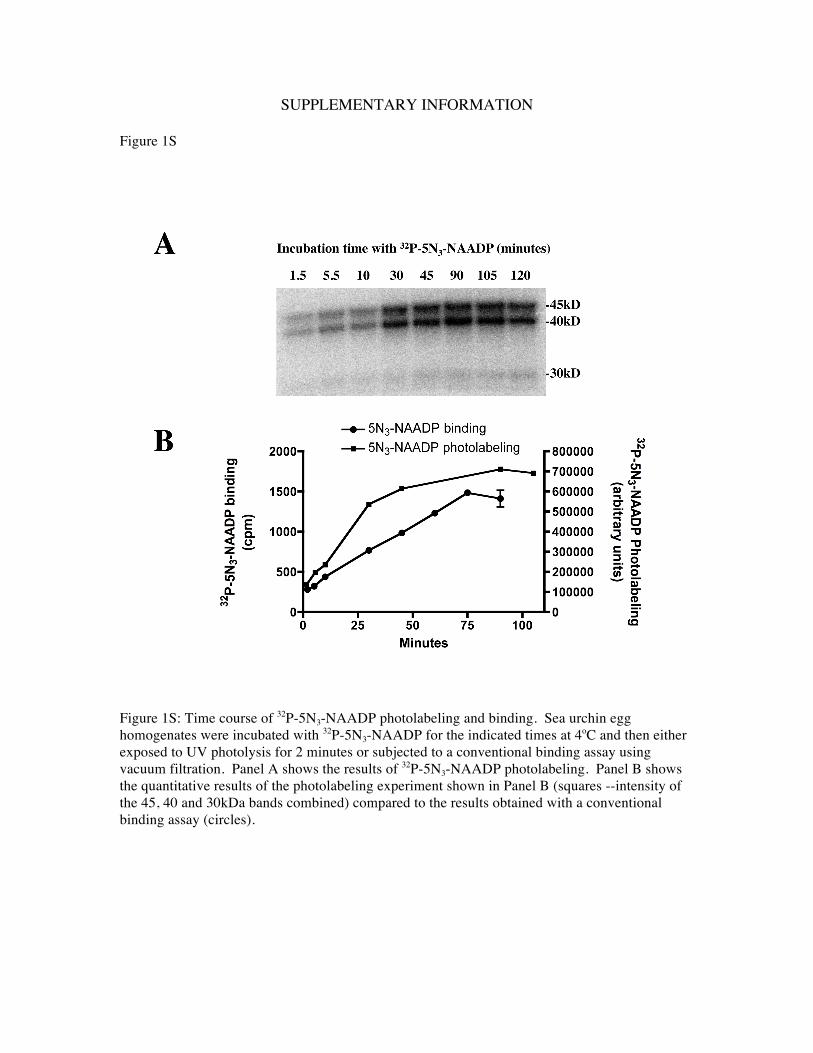
SUPPLEMENTARY INFORMATION
Figure 1S
Figure 1S: Time course of 32P-5N3-NAADP photolabeling and binding. Sea urchin egg homogenates were incubated with 32P-5N3-NAADP for the indicated times at 4oC and then either exposed to UV photolysis for 2 minutes or subjected to a conventional binding assay using vacuum filtration. Panel A shows the results of 32P-5N3-NAADP photolabeling. Panel B shows the quantitative results of the photolabeling experiment shown in Panel B (squares --intensity of the 45, 40 and 30kDa bands combined) compared to the results obtained with a conventional binding assay (circles).

Figure 2S
Figure 2S. 32P-5N3-NAADP photolabeling correlates with 32P-NAADP binding. Panel A shows 32P-5N3-NAADP photolabeling of twelve distinct S. purpuratus egg homogenate preparations. Panel B shows the comparison of the densitometric analyses of the photolabeling shown in Panel A (white bars) 45, 40 and 30kDa bands combined) with 32P-NAADP binding in these preparations (black bars). The data for photolabeling and binding is normalized to the results of homogenate #1, which was used for most of the studies described here. Note that photolabeling and binding correlate. Homogenates that have low 32P-NAADP binding activity do not photo label efficiently (i.e. homogenates #4 and 5). This correlation between binding and photolabeling applies to the 32 preparations we have examined. This correlation also extends to the ability of NAADP to induce calcium release.

Antony Galione, Jonathan S. Marchant and James T. SlamaTimothy F. Walseth, Yaping Lin-Moshier, Pooja Jain, Margarida Ruas, John Parrington,
Phosphate (NAADP)-Binding Proteins in Sea Urchin EggPhotoaffinity Labeling of High Affinity Nicotinic Acid Adenine Dinucleotide
doi: 10.1074/jbc.M111.306563 originally published online November 23, 20112012, 287:2308-2315.J. Biol. Chem.
10.1074/jbc.M111.306563Access the most updated version of this article at doi:
Alerts:
When a correction for this article is posted•
When this article is cited•
to choose from all of JBC's e-mail alertsClick here
Supplemental material:
http://www.jbc.org/content/suppl/2011/11/23/M111.306563.DC1.html
http://www.jbc.org/content/suppl/2012/01/19/M111.306563.DCAuthor_profile.htmlRead an Author Profile for this article at
http://www.jbc.org/content/287/4/2308.full.html#ref-list-1
This article cites 31 references, 17 of which can be accessed free at
by guest on March 17, 2016
http://ww
w.jbc.org/
Dow
nloaded from