Immune Response Regulation by Leishmania Secreted and Nonsecreted Antigens
Upload
independentCategory
view
0download
0
Photoaffinity Antigens for Human �� T Cells1
Ghanashyam Sarikonda,*† Hong Wang,* Kia-Joo Puan,2* Xiao-hui Liu,3‡ Hoi K. Lee,*Yongcheng Song,§ Mark D. Distefano,¶ Eric Oldfield,§ Glenn D. Prestwich,‡
and Craig T. Morita4*†
V�2V�2 T cells comprise the major subset of peripheral blood �� T cells in humans and expand during infections by recognizingsmall nonpeptide prenyl pyrophosphates. These molecules include (E)-4-hydroxy-3-methyl-but-2-enyl-pyrophosphate (HMBPP),a microbial isoprenoid intermediate, and isopentenyl pyrophosphate, an endogenous isoprenoid intermediate. Recognition of thesenonpeptide Ags is mediated by the V�2V�2 T cell Ag receptor. Several findings suggest that prenyl pyrophosphates are presentedby an Ag-presenting molecule: contact between T cells and APC is required, the Ags do not bind the V�2V�2 TCR directly, andAg recognition is abrogated by TCR mutations in CDRs distant from the putative Ag recognition site. Identification of the putativeAg-presenting molecule, however, has been hindered by the inability to achieve stable association of nonpeptide prenyl pyro-phosphate Ags with the presenting molecule. In this study, we show that photoaffinity analogues of HMBPP, meta/para-benzo-phenone-(methylene)-prenyl pyrophosphates (m/p-BZ-(C)-C5-OPP), can crosslink to the surface of tumor cell lines and be pre-sented as Ags to �� T cells. Mutant tumor cell lines lacking MHC class I, MHC class II, �2-microglobulin, and CD1, as well astumor cell lines from a variety of tissues and individuals, will all crosslink to and present m-BZ-C5-OPP. Finally, pulsing ofBZ-(C)-C5-OPP is inhibited by isopentenyl pyrophosphate and an inactive analog, suggesting that they bind to the same molecule.Taken together, these results suggest that nonpeptide Ags are presented by a novel-Ag-presenting molecule that is widely dis-tributed and nonpolymorphic, but not classical MHC class I, MHC class II, or CD1. The Journal of Immunology, 2008, 181:7738–7750.
T he �� T cell subset, which expresses T cell Ag receptors(TCR) using � and � rearranging genes (1), has functionalroles in immunity distinct from the �� T cell subset (2).
In humans, the majority of circulating �� T cells express V�2V�2(also termed V�9V�2) TCRs. V�2V�2 T cells recognize non-peptide prenyl pyrophosphate intermediates in isoprenoid bio-synthesis such as (E)-4-hydroxy-3-methyl-but-2-enyl pyrophos-phate (HMBPP)5 (3, 4) and isopentenyl pyrophosphate (IPP) (5).
V�2V�2 T cells can expand during infections to very high num-bers, accounting for half of the circulating T cells in some patients(reviewed in Ref. 2). HMBPP is the most potent Ag described (3)and is produced in the methyl-erythritol phosphate pathway forisoprenoid synthesis used by many eubacteria, some protozoa, andplant chloroplasts. By recognizing HMBPP produced by manypathogenic bacteria (such as those that cause tuberculosis and gas-troenteritis) as well as apicomplexan parasites (such as those thatcause malaria and toxoplasmosis), V�2V�2 T cells likely play im-portant roles in human immunity to both bacteria and parasites (2).
V�2V�2 T cells also kill many types of tumor cells in vitro,including malignant B cells, melanomas, prostate carcinomas, re-nal cell carcinoma, epithelial carcinomas, and others (6–10). Thisappears due to both TCR-mediated and NK receptor-mediated tu-mor cell recognition (11–13). Zoledronate and other bisphospho-nates greatly enhance tumor recognition by inhibiting the intracel-lular farnesyl pyrophosphate synthase enzyme, resulting inincreases in endogenous IPP (H. Wang and C. T. Morita, unpub-lished observations and Refs. 14–16). Importantly, treatment ofpatients with B cell malignancies (17) and metastatic prostate car-cinomas (18) with a bisphosphonate and IL-2 to activate and main-tain V�2V�2 T cells led to partial remissions and stable disease inseveral individuals. Given their broad tumor reactivity, immuno-therapy with V�2V�2 T cells appears to have promise for thetreatment of a variety of cancers.
Despite the importance of V�2V�2 T cells in human immunityto pathogens and their potential for tumor immunotherapy, little isknown about the molecular mechanisms for the presentation ofprenyl pyrophosphate Ags to these T cells. Although gene transfer
*Department of Internal Medicine, Division of Rheumatology and †InterdisciplinaryGraduate Program in Immunology, University of Iowa College of Medicine, IowaCity, IA 52242; ‡Department of Medicinal Chemistry, University of Utah School ofPharmacy, Salt Lake City, UT 84112; §Department of Chemistry and Center forBiophysics and Computational Biology, Urbana, IL 61801; and ¶Department ofChemistry, University of Minnesota, Minneapolis, MN 55455
Received for publication March 11, 2008. Accepted for publication September30, 2008.
The costs of publication of this article were defrayed in part by the payment of pagecharges. This article must therefore be hereby marked advertisement in accordancewith 18 U.S.C. Section 1734 solely to indicate this fact.1 This work was supported by grants from the National Institutes of Health, NationalInstitute of Arthritis and Musculoskeletal and Skin Disease (AR45504), the NationalInstitute of Allergy and Infectious Diseases (Midwest Regional Center of Excellencefor Biodefense and Emerging Infectious Diseases Research, AI057160), and the Na-tional Cancer Institute (CA113874) to C.T.M., the National Institute of NeurologicalDisorders and Stroke (NS29632) to G.D.P., and the National Institutes of GeneralMedical Sciences (GM073216 and GM58442) to E.O. and M.D., respectively.2 Current address: Division of Cellular and Molecular Research, Humphrey Oei In-stitute of Cancer Research, National Cancer Centre, 11 Hospital Drive Singapore169610, Singapore.3 Current address: Supergen, Inc., 2401 South Foothill Drive, Salt Lake City, UT84109.4 Address correspondence and reprint requests to Dr. Craig T. Morita, Department ofInternal Medicine, Division of Rheumatology and the Interdisciplinary Graduate Pro-gram in Immunology, University of Iowa Carver College of Medicine, EMRB 400F,Iowa City, IA 52242. E-mail address: [email protected] Abbreviations used in this paper: HMBPP, (E)-4-hydroxy-3-methyl-but-2-enyl py-rophosphate; �2M, �2-microglobulin; BZ, benzophenone; BrHPCP, bromohydrin py-rophosphonate; BrHPP, bromohydrin pyrophosphate; DATFP, diazo-3,3,3-trifluoro-
propionyloxy; EPP, ethyl pyrophosphate; FPP, farnesyl pyrophosphate; FPPS, FPPsynthase; GPP, geranyl pyrophosphate; IPP, isopentenyl pyrophosphate; m, meta;OPP, pyrophosphate; p, para.
Copyright © 2008 by The American Association of Immunologists, Inc. 0022-1767/08/$2.00
The Journal of Immunology
www.jimmunol.org

studies show that the V�2V�2 TCR mediates Ag recognition byV�2V�2 T cells (11), there is no evidence for direct binding ofprenyl pyrophosphates to the V�2V�2 TCR. Attempts to soak pre-nyl pyrophosphates into crystals of the V�2V�2 TCR (19) or todemonstrate prenyl pyrophosphate binding to soluble �� TCR byequilibrium dialysis or microcalorimetry failed (C. T. Morita, un-published observations). Moreover, although the chemical struc-tural requirements for antigenic activity of prenyl pyrophosphatesand other phosphoantigens has been extensively studied (20–23),this knowledge has provided little insight into how the Ags arepresented.
Unlike protein Ags, prenyl pyrophosphate Ags do not requireAg uptake, processing, or intracellular loading for presentation(24). Moreover, activation of V�2V�2 T cells is extremely rapid,with calcium flux observed within 90 s upon exposure to IPP (24)and metabolic acidification within 10 s upon exposure to bromo-hydrin pyrophosphate (BrHPP) (25). Although rapid, the activa-tion of V�2V�2 T cells by prenyl pyrophosphates still requirescell-cell contact (24, 26) similar to the contact required by �� Tcells during the recognition of peptide Ags presented by MHCclass I and II molecules (27). Most human cells are capable ofpresenting prenyl pyrophosphates (as assessed by indirect stimu-lation by a bisphosphonate) except for those deficient in accessorymolecule ligands (6, 28, 29). In contrast, murine, rat, and hamstercells do not present prenyl pyrophosphates or bisphosphonates (26,28, 30).
The requirement for cell-cell contact coupled with the small sizeof prenyl pyrophosphates (they are likely monovalent) and theirlack of direct binding to the V�2V�2 TCR suggest that prenylpyrophosphates are presented by a presenting molecule, similar topeptides presented by MHC class I and class II or lipids presentedby CD1. However, unlike peptide Ags, prenyl pyrophosphates donot stably associate with their presenting molecule with high af-finity, precluding pulsing of these Ags on APC (24, 26). This typeof presentation is similar to that of nonpeptide drugs, such as sul-famethoxazole and lidocaine, that load into MHC class I and IImolecules on the cell surface for recognition by CD8 and CD4 ��T cells (31, 32). Although this recognition is MHC restricted, thedrugs do not stably associate with MHC molecules or require in-ternalization (31, 33). Recognition of lipid Ags that load into CD1aor CD1b molecules at the cell surface also show similarities toprenyl pyrophosphate recognition, because the lipids bind ex-tremely rapidly (as short as 2 min) and, again, do not require pro-cessing or internalization (34, 35). However, the putative present-ing molecule for prenyl pyrophosphates has eluded identification.
Previously, we found that prenyl pyrophosphate Ags did notstably associate with APC with high affinity because EBV-trans-formed B cells and PBMC pulsed with IPP or mono-ethyl-phos-phate followed by washing did not activate V�2V�2 T cells (24).This direct presentation of prenyl pyrophosphates (24) differs fromthe indirect stimulation by bisphosphonates, which enter cells viafluid phase endocytosis (36) to inhibit farnesyl pyrophosphate syn-thase (FPPS) and thus “pulse” into APC (37). Our attempts tomeasure binding of IPP to the APC cell surface suggest that thebinding affinity between prenyl pyrophosphates and any putativeAg-presenting molecule is very low (data not shown). This prop-erty has made efforts to characterize the Ag-presenting moleculeusing natural Ags, such as IPP and HMBPP, difficult.
In this study, we sought a prenyl pyrophosphate Ag that wouldhave both antigenic activity for V�2V�2 T cells and stable asso-ciation with the APC cell surface. We show that meta/para (m/p)-benzophenone (BZ)-(methylene)-prenyl pyrophosphate (m/p-BZ-(C)-C5-OPP, where OPP is pyrophosphate), photoaffinity FPPanalogues, are recognized by V�2V�2 T cells. m/p-BZ-(C)-C5-
OPP stimulate V�2V�2 T cells even after UV crosslinking to theAPC surface and extensive washing. This covalent surfacecrosslinking is inhibited by IPP, suggesting that the molecules bindto the same protein on the APC cell surface. We also find thatnatural prenyl pyrophosphate can rapidly “pulse” onto the surfaceof APC under optimal conditions. m-BZ-C5-OPP was able to sta-bly associate with the cell surface of most hematopoietic and non-hematopoietic cell lines, including mutant APC lacking classicalMHC class I, �2-microglobulin (�2M), MHC class II, and CD1molecules. Thus, m/p-BZ-(C)-C5-OPP Ags may enable identifica-tion of this putative presenting molecule, which is predicted to bebroadly distributed, functionally nonpolymorphic, and not aknown presenting molecule.
Materials and MethodsAntigens
HMBPP was synthesized as described (38). Mono-ethyl phosphate andmono-ethyl pyrophosphate (EPP) were prepared and purified by anion ex-change as described (23, 39). Mono-methyl phosphate, farnesyl pyrophos-phate (FPP), and IPP were obtained from Sigma-Aldrich. PHA-P was ob-tained from Difco.
Synthesis of photoaffinity compounds and bromohydrinpyrophosphonate
Syntheses of m/p-BZ-C5-OPP were performed by minor modifications ofthe previous method (40). Briefly, dimethylallyl alcohol was first protectedas the chloroacetate and then oxidized with t-butyl hydroperoxide and cat-alytic H2SeO3. The resulting aldehyde was reduced with sodium borohy-dride, and the corresponding allylic alcohols were coupled under Mit-sunobu conditions with either 4-benzoylphenol or 3-benzoylphenol to givethe protected prenyl benzophenone ethers. The chloroacetate was removedby hydrolysis with methanolic aqueous ammonia, and the allylic alcoholswere converted to the corresponding allylic chlorides using N-chlorosuc-cinimide and dimethyl sulfide in dichloromethane. Displacement of theallylic chlorides with tris(tetra-n-butylammonium) hydrogen diphosphateafforded the desired allylic diphosphates, which were then purified by re-versed-phase chromatography and characterized by nuclear magnetic res-onance. Syntheses of m/p-BZ-C-C5-OPP (ether) were performed as de-scribed (41). Syntheses of m/p-BZ-C-C5-OPP (esters) were performed asdescribed (42). Syntheses of m/p-BZ-C-geranyl pyrophosphate (GPP)(ethers) were performed as described (43). Syntheses of 2-diazo-3,3,3-tri-fluoropropionyloxy (DATFP)-dihydroester-(alkyl)-FPP, DATFP-dh-GPP,and FPP-p-BZ were performed as described (M. L. Hovlid, R. L. Edelstein,F. Lopez-Gallego, S. A. Agger, C. Schmidt-Dannert, S. Sen, D. Shintani,K. Cornish, and M. D. Distefano, manuscript in preparation).
Synthesis of bromohydrin pyrophosphonate (BrHPCP; [[(4-bromo-3-hydroxy-3-methylbutoxy)hydroxyphosphinyl]methyl]-phosphonicacid(tri-ammonium salt)) was performed as follows. To a solution of O-isopentenylmethylene-1,1-bisphosphonate triammonium salt (5 mg) in water (1 ml)was added freshly prepared bromine water, dropwise, until the solution waspersistently yellow. The yellow color (due to a trace amount of Br2) wasremoved by gently blowing N2 into the solution, which was then usedwithout further purification.
Maintenance of cell lines
Va2 cells are derived from the SV40-transformed human fibroblast cell lineW1-18 (44–46). Other cell lines used were described previously (24) andinclude the Burkitt’s lymphoma, Raji, and its MHC class II negative mu-tant, RJ-2.2.5 (47); the CD3� Jurkat T cell line, JRT3-T3.5 (3); the eryth-roleukemia cell line, K562 (4); the parent EBV line 721 and the 721.221mutant that lacks surface expression of HLA-A, -B, and -C (48); and themutant melanoma cell line, FO-1, which is �2M deficient (49) and lacksdetectable assembled class I molecules (50). Va2 cells were cultured inDMEM (Invitrogen) with 10% FCS (Gemini Bio-Products) at 37°C in a10% CO2 incubator while the other cells were cultured at 37°C in a 5%CO2 incubator in P-medium. P-medium is RPMI 1640 supplemented with20 mM HEPES, 2 mM glutamine, 1 mM pyruvate, 1� MEM nonessentialamino acids, 0.5� MEM essential amino acids, 5.5 � 10�2 mM 2-ME (allfrom Invitrogen), and 10% FCS (Gemini Bio-Products) and adjusted to pH7.25 with 2 N NaOH.
7739The Journal of Immunology

Derivation of and culture conditions for V�2V�2 T cell clones
T cell lines and clones were maintained by periodic stimulation withPHA-P. T cells (1–2 � 105/well) were cultured in 1 ml of RPMI 1640supplemented as for P-medium but with the addition of rIL-2 (1–4 nM;Proleukin, Novartis) and 2% human AB serum (Atlanta Biologicals) withirradiated (4000 rad) allogeneic PBMC (2 � 105) and an equal mix ofirradiated (5000 rad) EBV-transformed B cells (DG.EBV and CP.EBV)(5 � 105 total) as feeder cells and PHA-P (1/4000 final dilution) in 24-wellplates (Linbro, MP Biomedicals). The derivation of the CD8��� 12G12and DG.SF68 and the CD4� HF.2 V�2V�2 T cell clones has been de-scribed (39, 51, 52).
Treatment of APC
APC were treated with either mitomycin C (unfixed) or with glutaralde-hyde (fixed). For mitomycin C treatment, APC (1–3 � 107 cells/ml) inDulbecco’s PBS without calcium or magnesium were incubated with freshmitomycin C (Sigma-Aldrich) (100 �g/ml) for 1 h at 37°C in a 5% CO2
incubator, then washed three times in PBS, and resuspended in either PBSor P-medium for further use. For glutaraldehyde fixation, APC were ad-justed to 1–3 � 107 cells/ml in PBS and reacted with 0.05% glutaraldehyde(EM grade; Sigma-Aldrich) for 15 s at room temperature while vortexing.The reaction was stopped by adding an equal volume of 0.2 M L-lysine (inH2O at pH 7.4) followed by incubation for 2 min. The fixed cells were thenwashed three times in PBS and resuspended in either PBS or P-medium forfurther use.
Pulsing and UV crosslinking of m/p-BZ-(C)-C5-OPP on APC
Following mitomycin C-treatment, APC were resuspended in ice-cold PBSto a concentration of 1 � 107 cells/ml. The cell suspension (200 �l) wasadded to wells of a 24-well plate. Two hundred microliters of m/p-BZ-C5-OPP Ag was then added to each well and the cells and Ag were incubatedwith or without 350-nm UV light treatment for 90 min on ice. The cellswere transferred to 15-ml conical tubes (BD Falcon, BD Biosciences),washed three times with 10 ml of ice-cold PBS at 4°C, and resuspended inP-medium for use. In some experiments, the cells were washed first inice-cold PBS and then exposed to UV light whereas in other experimentsthey were first exposed to UV light and then washed three times withice-cold PBS. Long wavelength UV light (350 nm) was used to avoidprotein damage. For inhibition of photoaffinity Ag binding by IPP or Br-HPCP, APC were incubated with IPP or BrHPCP (an inactive analog ofbromohydrin pyrophosphate) in P-medium with serum for 30 min on icefollowed by the addition of a suboptimal dose of either m-BZ-C5-OPP orm-BZ-C-C5-OPP ether and exposed to UV light for 90 min. The APC werethen washed three times with 4°C PBS and used as APC with V�2V�2 Tcell clones. Alternatively, BrHPCP was incubated with mitomycinC-treated Va2 for 30 min followed by the addition of m-BZ-C-C5-OPPether and 12G12 T cells.
Pulsing of prenyl pyrophosphate Ags on APC
Mitomycin C-treated or glutaraldehyde-fixed APC were added at 1 � 104–1 � 105 cells per 100 �l of PBS into wells of 96-well round-bottom plates(Corning) and incubated with Ags at 37°C in a 5% CO2 incubator forbetween 5 and 120 min. APC were washed in the plate 5–7 times with PBSeither at room temperature or at 4°C and resuspended in 100 �l of P-medium for further assays. A V�2V�2 T cell clone was then added to theAg-pulsed APC and proliferation was assessed by adding 1 �Ci of [3H]thy-midine at 24 h followed by harvesting 16–24 h later. Each pulsed or un-pulsed APC group was also cultured with the same T cell clone in thecontinuous presence of Ags such as mono-ethyl pyrophosphate, IPP,HMBPP, or m-BZ-C5-OPP, or with the mitogen, PHA-P, as positive con-trols for each APC group. No proliferation was noted in the absence of Tcells. Also, there was no stimulation of T cells in wells pulsed with Ag inthe absence of APC.
T cell proliferation and cytokine release assays
T cell proliferation assays were performed as described (53). Briefly, Tcells were plated in duplicate or triplicate in round-bottom 96-well platesat 5–10 � 104 T cells per well with 1 � 105 irradiated (7,000 rad) allo-geneic PBMC or mitomycin C-treated allogeneic tumor cells as APC. Be-cause the V�2V�2 T cell response to prenyl pyrophosphate Ags is notMHC restricted (54), allogeneic cells are suitable APC. The cultures werepulsed with 1 �Ci of [3H]thymidine (2 Ci/mmol) on day 1 and harvested16–24 h later using a Tomtec 96-well harvester. The samples were thencounted using a Wallac Betaplate scintillation counter. The mean prolif-eration and SEM of triplicate (or occasionally duplicate) cultures areshown. For cytokine release, culture supernatants were removed after 24 h
and TNF-� or IFN-� levels determined by sandwich ELISA (R&D Sys-tems) on single or duplicate cultures. For statin inhibition experiments,APC were preincubated with mevastatin (Sigma-Aldrich) for 30 min fol-lowed by the addition of set amounts of the stimulatory compounds in thecontinued presence of mevastatin. T cells were then added after 60 mineither directly to the APC (for HMBPP and m-BZ-C5-OPP) or after wash-ing the APC and resuspending them in mevastatin containing medium (forrisedronate). Similar results are obtained if the prenyl pyrophosphates arepulsed on the APC.
ResultsBenzophenone reaction
Because prenyl pyrophosphate Ags do not stably associate with theputative Ag-presenting molecule, it has been difficult to determineits identity (24). A similar lack of stable association is found fornonpeptide drugs presented as Ags by MHC class I or class IImolecules to CD4 and CD8 �� T cells (31). To overcome thisproblem, we have studied bioactive photoactivatable analogues ofprenyl pyrophosphates to covalently link the prenyl pyrophosphateAg to the APC surface. The farnesyl pyrophosphate analog m-BZ-C5-OPP is comprised of a BZ photophore linked to an HMBPPmolecule via the hydroxyl group (40). m-BZ-C5-OPP is a chemi-cally stable compound that can be reversibly activated using longwavelength (which avoids protein damage) UV light. When acti-vated, m-BZ-C5-OPP reacts with C-H bonds in close proximity(Fig. 1).
m/p-BZ-(C)-C5-OPP compounds stimulate V�2V�2 T cells
In in vitro experiments, m/p-BZ-C5-OPP function as analogues ofisoprenoid pyrophosphates because they can crosslink to FPPS andother prenyl synthases (40). Structurally, m/p -BZ-C5-OPP resem-ble both FPP (through their spacing of C-C double bonds) andHMBPP (where the hydroxyl attached to C4 is now an ether orester bond) (see Figs. 2 and 3 for structures). Therefore, to deter-mine whether m/p-BZ-(C)-C5-OPP compounds are recognized by
FIGURE 1. Mechanism of cross-linking photoaffinity FPP/HMBPP an-alogues. m/p-BZ-(C)-C5-OPP compounds (m-BZ-C5-OPP ether is shown)are incubated with the APC and activated by long-wavelength UV light(350 nm; this avoids protein damage), generating a reactive carbon atom.This generation of a reactive carbon intermediate is reversible. When thereactive carbon is present in close proximity to a carbon from the inter-acting protein (putative Ag-presenting element), it forms a covalent linkagecrosslinking the m/p-BZ-(C)-C5-OPP to the interacting protein. Followingcross-linking, excess m/p-BZ-(C)-C5-OPP can be washed off, leaving be-hind covalently attached m/p-BZ-(C)-C5-OPP. This allows m/p-BZ-(C)-C5-OPP to be associated with the APC even after washing.
7740 PHOTOAFFINITY Ags FOR V�2V�2 T CELLS

V�2V�2 T cells, m-BZ-C5-OPP and p-BZ-C5-OPP were tested fortheir ability to induce proliferation of V�2V�2 T cells. Similar toIPP and EPP, both compounds stimulated DG.SF68 V�2V�2 Tcells in a dose-dependent manner (Fig. 2A). The concentrationsthat induced half-maximum proliferation were 0.4 �M, interme-diate between IPP (1–3 �M) and HMBPP (0.0000316 �M). Thus,as predicted based on their structure, both m-BZ-C5-OPP andp-BZ-C5-OPP are recognized as Ags by V�2V�2 T cells.
To determine the specificity of recognition by V�2V�2 T cells,other photoaffinity analogues (both m- and p-substituted) withether and ester linkages to the benzophenone group were tested(Fig. 2B, C, 3C, D). The linkage and spacing of the C5-OPP group
from the BZ group was extremely important in determining bio-activity. Compounds that had ester-linked C5-OPP groups spacedone methylene group away from the BZ moiety (m/p-BZ-C-C5-OPP esters) were extremely active, requiring only slightly higherconcentrations for half-maximum stimulation compared withHMBPP (the most potent prenyl pyrophosphate described) withhalf-maximum stimulation at 50–60 pM vs 32 pM for HMBPP(see Figs. 3C and 4C and Ref. 3). Changing the ester linkage to anether linkage (m/p-BZ-C-C5-OPP ethers) reduced bioactivity by38- to 72-fold (Fig. 3C). Removing the methylene spacer (to givem/p-BZ-C5-OPP ethers) further reduced bioactivity by 140–316-fold (Fig. 2A vs 3C). V�2V�2 T cells showed equal or slight pref-erential recognition of para-BZ-(C)-C5-OPP compounds com-pared with their meta- isomers (Figs. 2–4). This was mostpronounced for p-BZ-C-GPP and m-BZ-C-GPP, compounds thatdiffered by �10-fold in activity (Fig. 2C). Compounds withDATFP photoaffinity groups attached to longer chain FPP andGPP moieties or with the BZ group linked to FPP via the pyro-phosphate moiety had little or no activity (Fig. 2C). These resultsshow that V�2V�2 T cells recognize isoprenoid photoaffinity com-pounds in a structure-specific manner and that the linkage to andspacing from the BZ moiety determine bioactivity levels.
Because recognition of prenyl pyrophosphate Ags by V�2V�2 Tcells requires the pyrophosphate moiety, we sought to determinewhether recognition of m-BZ-C5-OPP shows a similar require-ment. Neither the crosslinked BZ photophore without the pyro-phosphate moiety nor a 4-maleimido-BZ derivative stimulatedV�2V�2 T cells (Fig. 2D), suggesting that V�2V�2 T cell recog-nition of m-BZ-C5-OPP is dependent on the presence of the py-rophosphate moiety and not the BZ group.
m-BZ-C5-OPP can stably associate with APC afterphotocrosslinking and are presented directly like prenylpyrophosphates
Previously, we and others have found that prenyl pyrophosphateAgs, including IPP and mono-ethyl phosphate, do not stably as-sociate with APC (24, 26). To determine whether the FPP pho-toaffinity analog m-BZ-C5-OPP can stably associate with APC af-ter UV crosslinking, we incubated APC in medium only, with IPP,or with m-BZ-C5-OPP for 90 min on ice. During this incubation,the cells were either exposed to UV light to induce crosslinking ofm-BZ-C5-OPP or left unexposed. After extensive washing, thecells were then used as APC to stimulate the 12G12 V�2V�2 Tcell clone. Unlike APC pulsed with m-BZ-C5-OPP (and not ex-posed to UV light), APC exposed to UV light during pulsing stim-ulated V�2V�2 T cells to proliferate even after extensive washing(Fig. 3A). UV treatment alone did not affect the APC, becauseAPC exposed to UV light in either the absence or presence of IPPdid not stimulate V�2V�2 T cells. The cells were competent forpresentation because they were able to present m-BZ-C5-OPPwhen the Ag was continuously present (Fig. 3A).
This recognition of m-BZ-C5-OPP on DG.EBV B cells was doseand UV dependent. Even after extensive washing, the APC withUV-crosslinked m-BZ-C5-OPP retained the ability to stimulate theCD4� V�2V�2 T cell clone HF.2 to proliferate in a dose-depen-dent fashion, whereas non-UV-crosslinked m-BZ-C5-OPP did not(Fig. 3B). Similarly, UV crosslinking of the m/p-BZ-C-C5-OPPether- and ester-linked compounds also resulted in stable associ-ation with the APC that was resistant to washing (Fig. 3C).Crosslinked Ags required somewhat higher concentrations forhalf-maximal stimulation compared with Ag present continuously(�33-fold and �13-fold higher for the ester and ether compounds,respectively) (Fig. 3C). Besides stimulating proliferative
FIGURE 2. Photoaffinity analogues of FPP/HMBPP are Ags forV�2V�2 T cells. Phosphorylated compounds were incubated with irradi-ated PBMC and the CD8��� 12G12 or DG.SF68 V�2V�2 T cell clonesfor 2 days and proliferation was assessed by [3H]thymidine incorporation.Structures of the compounds are shown on the right. “X” refers to thecarbon chain closest to the pyrophosphate moiety. “Y” refers to groupsspaced away from the pyrophosphate by one isoprenoid unit. A, m/p-BZ-C5-OPP can activate V�2V�2 T cells. m-BZ-C5-OPP ether (F), p-BZ-C5-OPP ether (E), IPP (f), and EPP (Œ) were incubated with the DG.SF68V�2V�2 T cell clone and irradiated PBMC and proliferative responseswere assessed 48 h later. B, Recognition of m/p-BZ-(C)-C5-OPP com-pounds is influenced by the spacing of the C5-OPP from the BZ group.m-BZ-C-C5-OPP ether (F) or its isomer p-BZ-C-C5-OPP ether (E) and IPP(f), were added at serial half-log dilutions for stimulation of 12G12 withVa2 APC. C, Recognition of farnesyl and geranyl pyrophosphate BZ andDATFP photoaffinity compounds. Various FPP (�, �, and ‚) and GPP (Fand E) compounds were tested for stimulation of 12G12 in the presence ofVa2 APC. D, Recognition of m-BZ-C5-OPP is dependent upon the pres-ence of the pyrophosphate moiety. BZ (f), 4-maleimido-BZ (Œ), or m-BZ-C5-OPP (F) were added at half-log serial dilutions. An EBV transformedB cell line, DG.EBV, was used as the APC for the 12G12 T cell clone.
7741The Journal of Immunology

responses, the photoaffinity Ags also stimulated the release ofTNF-� and IFN-� in a dose-dependent manner (Fig. 3D). Thesefindings show that m/p-BZ-(C)-C5-OPP compounds stimulateV�2V�2 T cell cytokine and proliferative responses and thatthese compounds retains their immunogenicity when crosslinkedto the APC surface.
To determine the mechanism by which m/p-BZ-(C)-C5-OPPcompounds stimulate V�2V�2 T cells, we inhibited the responseof V�2V�2 T cells with the statin, mevastatin. We found that com-pounds that act as direct Ags for V�2V�2 T cells (i.e., prenylpyrophosphates) or as mitogens (e.g., PHA) are much less sensi-tive to statin inhibition than compounds (e.g., bisphosphonates andalkylamines) that act indirectly by inhibiting FPPS, causing IPPaccumulation (H. Wang and C. T. Morita, manuscript in prepara-tion). Therefore, we used mevastatin to inhibit V�2V�2 T cellresponses induced by p-BZ-C5-OPP in comparison with HMBPPand risedronate. Whereas mevastatin inhibited 50% of the response
to the FPPS inhibitor risedronate at 0.07 �M (Fig. 4A), mevastatinconcentrations of 23–42 �M (328- to 600-fold higher) were re-quired to inhibit p-BZ-C5-OPP responses (Fig. 4, A and B). Theselevels were similar to the 80 �M mevastatin concentrations thatwere required to inhibit 50% of the HMBPP responses by the CD4HF.2 clone (HMBPP presented by CP.EBV) or the CD8�� 12G12clone (HMBPP presented by Va2) (Fig. 4, A and B).
A second characteristic of prenyl pyrophosphate Ags is theirrecognition by V�2V�2 T cells in the absence of other cells due topresentation by daughter T cells (24). In contrast, bisphosphonatesgenerally require the presence of APC to stimulate proliferation ofV�2V�2 T cells (37) (although this may be due in part to toxicityassociated with the inhibition of FPPS). To determine whether thephotoaffinity Ags require APC for presentation, the 12G12 andHD.108 V�2V�2 T cell clones were incubated with the m/p-BZ-C-C5-OPP compounds IPP and HMBPP in the presence or absenceof Va2 cells as APC. Like IPP and HMBPP, the m/p-BZ-C-C5-
FIGURE 3. m-BZ-C5-OPP can becrosslinked onto the surface of APCby UV light for stimulation ofV�2V�2 T cells. A, m-BZ-C5-OPPstably associates with the APC afterUV crosslinking. Mitomycin C-treated DG.EBV B cells were incu-bated with medium, 5 �M m-BZ-C5-OPP, or 250 �M IPP with or withoutUV crosslinking for 90 min on iceand washed three times. DG.EBV Bcells (7.5 � 104) were cultured with1 � 105 CD4� HF.2 V�2V�2 T cellsin the continuous presence or absenceof 10 �M m-BZ-C5-OPP. After 24 hthe cultures were pulsed with 1 �Ciof [3H]thymidine and harvested 18 hlater. B, Dose-dependent V�2V�2 Tcell recognition of UV crosslinkedm-BZ-C5-OPP. Mitomycin C-treatedDG.EBV B cells were incubated withvarying concentrations of m-BZ-C5-OPP with (F) or without (E) UVcrosslinking for 90 min on ice fol-lowed by washing. HF.2 was thenadded to the washed APC and prolif-eration was determined by [3H]thymi-dine incorporation. C, UV-crosslinkedm/p-BZ-C-C5-OPP ether and estercompounds stimulate V�2V�2 Tcells. m/p-BZ-C-C5-OPP compoundswere tested with and without UVcrosslinking for their ability to stim-ulate 12G12 when presented by theVa2 cell line. Pulsing was done inPBS without serum followed bywashing four times in a 96-well plate.D, Photoaffinity compounds stimulatesecretion of IFN-� (bottom panel) andTNF-� (middle panel) and prolifera-tion (top panel) by V�2V�2 T cells.m/p-BZ-C-C5-OPP compounds, IPP,or HMBPP were incubated with theHF.2 T cell clone with DG.EBVAPC. After 16 h, culture supernatantswere harvested and tested for cytokinesby ELISA. Proliferation was assessedby [3H]thymidine incorporation.
7742 PHOTOAFFINITY Ags FOR V�2V�2 T CELLS
OPP photoaffinity compounds stimulated V�2V�2 T cell prolifer-ation in the complete absence of APC with similar lower magni-tude responses and shifted dose-response curves (Fig. 4C). Thus,photoaffinity compounds function as direct Ags for V�2V�2 Tcells rather than as pharmacological inhibitors of FPPS.
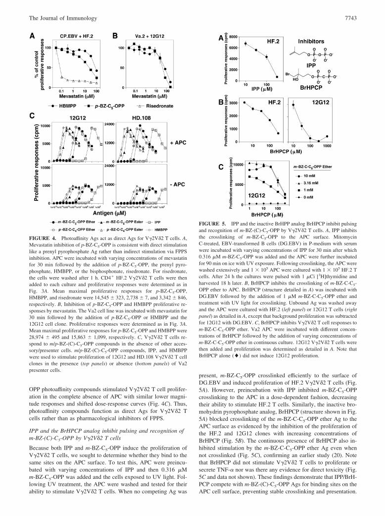
IPP and the BrHPCP analog inhibit pulsing and recognition ofm-BZ-(C)-C5-OPP by V�2V�2 T cells
Because both IPP and m-BZ-C5-OPP induce the proliferation ofV�2V�2 T cells, we sought to determine whether they bind to thesame sites on the APC surface. To test this, APC were preincu-bated with varying concentrations of IPP and then 0.316 �Mm-BZ-C5-OPP was added and the cells exposed to UV light. Fol-lowing UV treatment, the APC were washed and tested for theirability to stimulate V�2V�2 T cells. When no competing Ag was
present, m-BZ-C5-OPP crosslinked efficiently to the surface ofDG.EBV and induced proliferation of HF.2 V�2V�2 T cells (Fig.5A). However, preincubation with IPP inhibited m-BZ-C5-OPPcrosslinking to the APC in a dose-dependent fashion, decreasingtheir ability to stimulate HF.2 T cells. Similarly, the inactive bro-mohydrin pyrophosphate analog, BrHPCP (structure shown in Fig.5A) blocked crosslinking of the m-BZ-C-C5-OPP ether Ag to theAPC surface as evidenced by the inhibition of the proliferation ofthe HF.2 and 12G12 clones with increasing concentrations ofBrHPCP (Fig. 5B). The continuous presence of BrHPCP also in-hibited stimulation by the m-BZ-C-C5-OPP ether Ag even whennot crosslinked (Fig. 5C), confirming an earlier study (20). Notethat BrHPCP did not stimulate V�2V�2 T cells to proliferate orsecrete TNF-� nor was there any evidence for direct toxicity (Fig.5C and data not shown). These findings demonstrate that IPP/BrH-PCP compete with m-BZ-(C)-C5-OPP Ags for binding sites on theAPC cell surface, preventing stable crosslinking and presentation.
FIGURE 4. Photoaffinity Ags act as direct Ags for V�2V�2 T cells. A,Mevastatin inhibition of p-BZ-C5-OPP is consistent with direct stimulationlike a prenyl pyrophosphate Ag rather than indirect stimulation via FPPSinhibition. APC were incubated with varying concentrations of mevastatinfor 30 min followed by the addition of p-BZ-C5-OPP, the prenyl pyro-phosphate, HMBPP, or the bisphosphonate, risedronate. For risedronate,the cells were washed after 1 h. CD4� HF.2 V�2V�2 T cells were thenadded to each culture and proliferative responses were determined as inFig. 3A. Mean maximal proliferative responses for p-BZ-C5-OPP,HMBPP, and risedronate were 14,545 � 323, 2,738 � 7, and 3,342 � 846,respectively. B, Inhibition of p-BZ-C5-OPP and HMBPP proliferative re-sponses by mevastatin. The Va2 cell line was incubated with mevastatin for30 min followed by the addition of p-BZ-C5-OPP or HMBPP and the12G12 cell clone. Proliferative responses were determined as in Fig. 3A.Mean maximal proliferative responses for p-BZ-C5-OPP and HMBPP were28,974 � 495 and 15,863 � 1,099, respectively. C, V�2V�2 T cells re-spond to m/p-BZ-(C)-C5-OPP compounds in the absence of other acces-sory/presenter cells. m/p-BZ-(C)-C5-OPP compounds, IPP, and HMBPPwere used to stimulate proliferation of 12G12 and HD.108 V�2V�2 T cellclones in the presence (top panels) or absence (bottom panels) of Va2presenter cells.
FIGURE 5. IPP and the inactive BrHPP analog BrHPCP inhibit pulsingand recognition of m-BZ-(C)-C5-OPP by V�2V�2 T cells. A, IPP inhibitsthe crosslinking of m-BZ-C5-OPP to the APC surface. MitomycinC-treated, EBV-transformed B cells (DG.EBV) in P-medium with serumwere incubated with varying concentrations of IPP for 30 min after which0.316 �M m-BZ-C5-OPP was added and the APC were further incubatedfor 90 min on ice with UV exposure. Following crosslinking, the APC werewashed extensively and 1 � 105 APC were cultured with 1 � 105 HF.2 Tcells. After 24 h the cultures were pulsed with 1 �Ci [3H]thymidine andharvested 18 h later. B, BrHPCP inhibits the crosslinking of m-BZ-C-C5-OPP ether to APC. BrHPCP (structure detailed in A) was incubated withDG.EBV followed by the addition of 1 �M m-BZ-C-C5-OPP ether andtreatment with UV light for crosslinking. Unbound Ag was washed awayand the APC were cultured with HF.2 (left panel) or 12G12 T cells (rightpanel) as detailed in A, except that background proliferation was subtractedfor 12G12 with DG.EBV. C, BrHPCP inhibits V�2V�2 T cell responses tom-BZ-C-C5-OPP ether. Va2 APC were incubated with different concen-trations of BrHPCP followed by the addition of varying concentrations ofm-BZ-C-C5-OPP ether in continuous culture. 12G12 V�2V�2 T cells werethen added and proliferation was determined as detailed in A. Note thatBrHPCP alone (�) did not induce 12G12 proliferation.
7743The Journal of Immunology

Photoaffinity Ags can be presented by a broad array ofhematopoietic and nonhematopoietic tumor cell lines
The ability to covalently attach the prenyl pyrophosphate analog toa molecule on the APC surface allowed us to test tumor cells froma variety of different lineages for expression of the presenting mol-ecule without the complicating possibility of self-presentation byV�2V�2 T cells to each other. We found that virtually any humantumor cell line, irrespective of tissue origin or developmentalstage, was able to present the m-BZ-C5-OPP Ag to V�2V�2 T cells(Table I). In contrast, none of the six murine hematopoietic celllines tested were able to present HMBPP or the bisphosphonaterisedronate to V�2V�2 T cells (Table I). This suggests that theputative Ag-presenting molecule for prenyl pyrophosphate Ags isbroadly distributed like classical MHC class I molecules but isfunctionally nonpolymorphic.
Prenyl pyrophosphate Ags can be pulsed onto APC withoutcrosslinking
During our survey of different tumor cell lines, we noted some,such as Va2, SH-5YSY, and HT-1080, that presented pulsedm-BZ-C5-OPP without UV crosslinking (Fig. 6A). When pulsed inRPMI 1640 with FCS, the Va2 cell line was able to present m-BZ-C5-OPP without UV crosslinking but required a 200-fold higherconcentration during pulsing to elicit a similar half-maximal re-
sponse to that observed in the continuous presence of the Ag (0.21vs 42 �M; Fig. 6B). IPP could also be pulsed onto Va2 but re-quired a 100-fold higher concentration to elicit a similar level ofresponse to the continuous presence of the Ag (Fig. 6B). Thus,prenyl pyrophosphate Ags can be pulsed onto APC without cova-lent linkage, albeit inefficiently under standard conditions.
In earlier attempts at pulsing prenyl pyrophosphates, we usedEBV-transformed B cells or PBMC as APC with moderate po-tency Ags such as IPP, mono-methyl-phosphate, EPP, or low con-centrations of HMBPP in bacterial lysates (24). Because APC maydiffer in their ability to present prenyl pyrophosphates, we com-pared the ability of three tumor cell lines (the EBV-transformed Bcell line, DG.EBV, Va2, and SH-5YSY) to present m-BZ-C5-OPP(without UV crosslinking) to the NKG2D�CD8��� V�2V�2 Tcell clone DG.SF68 (Fig. 6C).
DG.EBV B cells stimulated V�2V�2 T cell proliferative re-sponses similar to or higher than those of the other presenter celllines. However, compared with Va2, DG.EBV cells required Agconcentrations for half-maximal responses (at the optimal APCnumber) that were 17-fold higher for mono-ethyl phosphate (Expt.1) and 12-fold higher for IPP (Expt. 2) (Fig. 6C). The concentra-tions required by SH-5YSY were intermediate between the twocell lines. Thus, the Va2 and SH-5YSY cell lines were more ef-fective presenter cells than was DG.EBV.
Table I. Human but not murine tumor cells from a variety of cell lineages serve as antigen presenting cells form-BZ-C5-OPP/prenyl pyrophosphates
Cell Line/Lineage Designation Tumor Typem-BZ-C5-OPP/Prenyl
Pyrophosphate Presentation
Human Cell Linesa
HematopoieticB cell DG.EBV EBV transformed ��B cell 721 EBV transformed ��B cell Raji Burkitt’s lymphoma ��T cell J.RT3-T3.5 Thymoma ��Myeloid cell K562 Erythroleukemia ��Myeloid cell U-937 Myelomonocytic Leukemia ��Myeloid cell THP-1 Monocytic Leukemia �
NonhematopoieticEpithelial cell T84 Colon Adenocarcinoma �/�Epithelial cell HT-29 Colon Adenocarcinoma �Epithelial cell HuTu 80 Duodenal Adenocarcinoma ��Epithelial cell JAR Choriocarcinoma �Epithelial cell HeLa Cervical carcinoma (Epitheloid) ��Epithelial cell A-431 Skin (Epidermoid) �Melanocyte FO-1 Melanoma ��Melanocyte SK-MEL-28 Melanoma ��Neural crest SH-5YSY Neuroblastoma ��Fibroblast HT-1080 Fibrosarcoma ���Fibroblast Va2 Transformed fibroblast ���Fibroblast IMR-90 Fibroblast �Squamous epithelium FADU Squamous cell carcinoma ��Squamous epithelium SCC24a Squamous cell carcinoma ��Squamous epithelium CAL 27 Squamous cell carcinoma ��Squamous epithelium DV Squamous cell carcinoma ��
Murine cell linesb
HematopoieticB cell A20 B cell Lymphoma �T cell EL4 T cell lymphoma �Dendritic cell DC2.4 Immortalized dendritic cell �Monocyte/macrophage J774 Sarcoma �Macrophage RAW 264.7 A-MuLV transformed �Mast cell P815 Mastocytoma �
a Assessed by stimulation of proliferation of V�2V�2 T cell clones after UV crosslinking.b Assessed by pulsing or culturing murine cell lines with m-BZ-C5-OPP, IPP, HMBPP, and/or risedronate and using as APCs to stimulate the
proliferation of V�2V�2T cell clones.
7744 PHOTOAFFINITY Ags FOR V�2V�2 T CELLS

Va2 are more effective presenter cells, in part because theyexpress the MICA, ULBP2, and ULBP3 ligands that bind to theNKG2D receptors expressed on the surface of the CD8 ���
V�2V�2 DG.SF68 T cell clone. We have previously shown thatNKG2D binding to its ligands enhances V�2V�2 T cell re-sponses to prenyl pyrophosphates (12) and that DG.EBV lackssuch NKG2D ligands (data not shown). However, there arelikely additional accessory molecule interactions or other fac-tors that enhance recognition, because the CD4� V�2V�2 T cellclone HF.2 (which lacks NKG2D) also requires 16-fold higherHMBPP concentrations with DG.EBV as compared with Va2(half-maximal proliferation at 0.53 vs 0.033 nM for DG.EBV vsVa2; Fig. 6D). Thus, prenyl pyrophosphate Ags can be pulsedonto APC, although this is difficult to demonstrate using IPPand B cells.
m-BZ-C5-OPP pulsing is inhibited by serum and mediumcomponents
Because prenyl pyrophosphates can pulse onto APC, albeit inef-ficiently, even without UV crosslinking (Fig. 6), we sought to op-timize conditions for the pulsing of m-BZ-C5-OPP onto APC. Un-like earlier experiments where medium with serum was used (Fig.6B), we pulsed m-BZ-C5-OPP onto APC in PBS either with orwithout serum in the presence or absence of UV light to crosslinkthe Ag (Fig. 7). Following pulsing, the APC were washed andresuspended in medium with serum and cultured with the 12G12clone. To rule out changes in the APC due to UV exposure or thelack of serum, control APC were treated as above in the absence
of m-BZ-C5-OPP and then assessed for their ability to presentm-BZ-C5-OPP added with the T cells (Fig. 7A, bottom panels). Inthe absence of serum, pulsing of m-BZ-C5-OPP was very efficientfollowing crosslinking to the APC (Fig. 7A, top right panel). Im-portantly, even without UV crosslinking, m-BZ-C5-OPP could bepulsed onto the APC with similar efficiency (0.33 vs 0.20 �Mrespectively; Fig. 7A, top left panel vs top right panel). Thus, puls-ing was more efficient in the absence of serum, and this effect wasmore pronounced for m-BZ-C5-OPP that had not been crosslinked(21-fold for uncrosslinked vs 5-fold for UV-crosslinked). Theseresults suggest that an unknown serum component inhibits the ef-ficient pulsing of m-BZ-C5-OPP onto APC.
Because pulsing of m-BZ-C5-OPP onto APC was most efficientwhen the pulsing reaction was conducted without serum, we ex-tended these observations to natural prenyl pyrophosphate Ags.We incubated HMBPP in either medium with or without serum orin PBS without serum (Fig. 7B). When HMBPP was continuouslypresent, serum had little effect on V�2V�2 T cell proliferation(Fig. 7B, left bottom panel). However, pulsing of HMBPP ontoAPC was better in medium without serum and better still in PBSwithout serum (Fig. 7B, top panels). Thus, pulsing of both naturaland synthetic prenyl pyrophosphates onto APC is inhibited by se-rum and by other medium components.
Prenyl pyrophosphate Ags pulse rapidly onto APC
Because we found that prenyl pyrophosphate Ags could be pulsedefficiently onto APC, we sought to determine the kinetics of non-peptide Ag pulsing. Mitomycin C-treated or glutaraldehyde-fixed
FIGURE 6. Prenyl pyrophosphate Ags can be pulsedonto APC without UV crosslinking. A, Some cell linesstably associate with m-BZ-C5-OPP without UV treat-ment. Various mitomycin C-treated tumor cell lineswere incubated for 90 min with medium alone or me-dium with 40 �M m-BZ-C5-OPP in the presence or ab-sence of 350 nm UV light on ice. The APC were thenwashed three times with PBS at 4°C and 1 � 105 APCwere cultured with 1 � 105 12G12 T cells in medium.After 24 h the cultures were pulsed with 1 �Ci of[3H]thymidine and harvested 18 h later. Note that theVa2, SH-5YSY, HT-1080, and HeLa cell lines pulsedwith m-BZ-C5-OPP stimulate the 12G12 T cells evenwithout UV crosslinking with varying efficiency. B, Ef-ficient presentation by the Va2 cell line reveals pulsingof prenyl pyrophosphates. For continuous exposure,m-BZ-C5-OPP (F) or IPP (Œ) were added directly toculture. For pulsing, the transformed fibroblast line Va2was treated with mitomycin C and incubated withm-BZ-C5-OPP (E) or IPP (‚) for 1 h in P-medium withFCS in the absence of UV treatment on ice. The APCwere washed seven times in PBS at 4°C and then incu-bated with the CD8��� NKG2D� 12G12 T cell clone.C, Efficient presentation of prenyl pyrophosphate Agsby some tumor cell lines to the CD8��� NKG2D�
DG.SF68 T cell clone. Varying concentrations of theVa2, SH-5YSY, and DG.EBV cells were continuouslycultured with DG.SF68 T cells with varying dilutions ofmono-ethyl-phosphate (MEP) or IPP. D, Efficient pre-sentation of prenyl pyrophosphate Ags by the Va2 cellline to the CD4� NKG2D HF.2 T cell clone. MitomycinC-treated Va2 or CP.EBV cells were cultured with theCD4� HF.2 T cell clone in the presence of HMBPP. Toassess IFN-� levels, supernatants were harvested at 24 hand IFN-� levels were determined by ELISA. Prolifer-ation was determined as in A.
7745The Journal of Immunology

Va2 cells were incubated with HMBPP for varying lengths of time(5–120 min) in PBS without serum after which the APC werewashed extensively. The pulsed APC were then incubated withthe CD4� HF.2 clone and �� T cell proliferation (Fig. 8) andrelease of TNF-� (data not shown) were measured. In agreementwith our previous observations (24), fixing APC with glutaralde-hyde had no affect on pulsing of HMBPP. In fact, glutaraldehyde-fixed APC were better than mitomycin C-treated APC at present-ing nonpeptide Ags to V�2V�2 T cells (Fig. 8). Within 5 min ofincubation with the prenyl pyrophosphate Ag HMBPP, �75% ofthe antigenic activity of HMBPP was already associated with theAPC. Pulsing of HMBPP onto glutaraldehyde-fixed APC peakedat 45 min of incubation, whereas mitomycin C-treated APC re-quired 60–90 min of incubation. These results demonstrate that theputative Ag-presenting molecule on the APC associates with pre-nyl pyrophosphates very rapidly (�5 min). These data are consis-tent with the rapid activation of �� T cells that we and others haveobserved (24–26) and with our IPP binding results (data notshown).
Recognition of prenyl pyrophosphates does not require APCexpression of classical MHC class I, MHC class II, �2Mdependent, or CD1 molecules
We earlier demonstrated that V�2V�2 T cells do not require prenylpyrophosphates to be internalized or processed for presentationand do not require professional APC (24). To determine whether aknown Ag-presenting molecule was required for presentation ofprenyl pyrophosphates, we tested mutant APC (24) that lackedthese molecules and found their absence on the APC had no effecton prenyl pyrophosphate recognition and that mAbs to these mol-ecules did not inhibit prenyl pyrophosphate recognition (55). How-ever, because V�2V�2 T cells are as efficient as dendritic cells atpresenting peptide Ags to �� T cells (56) and can present prenylpyrophosphates to each other (24), we could not exclude that aknown Ag-presenting molecule on the V�2V�2 T cells themselveswas presenting to daughter T cells in these experiments. To ex-clude presentation by V�2V�2 T cells to each other, we directlycrosslinked m-BZ-C5-OPP to different APC cell lines that lack
expression of known Ag-presenting molecules and used thecrosslinked APC to stimulate V�2V�2 T cells. In this way, wecould avoid any possible presentation by V�2V�2 T cells, becausethe Ags are covalently linked to the APC. We found that MHCclass II-negative APC (24), including the transcription factor mu-tant RAJI Burkitt’s lymphoma cell line, RJ-2.2.5, the J.RT3-T3.5
FIGURE 7. Stable association of m-BZ-C5-OPP and other prenyl pyrophosphates with APC is impaired by serum. A, m-BZ-C5-OPP pulsing is impairedby a serum component. Mitomycin C-treated Va2 cells were resuspended in PBS with (E) or without FCS (F) and incubated with or without m-BZ-C5-OPPin the presence (�) or absence (�) of UV light for 90 min on ice. The APC were then washed three times with PBS at 4°C. Va2 cells (4 � 104) that hadbeen pulsed with Ag (top panels) or not pulsed (bottom panels) were then cultured with 1 � 105 DG.SF68 V�2V�2 T cells. For the nonpulsed APC,m-BZ-C5-OPP was continuously present. To assess proliferation, the cultures were pulsed with 1 �Ci of [3H]thymidine at 24 h and harvested 18 h later.B, HMBPP pulsing is impaired by compounds in serum and RPMI 1640 medium. Va2 was treated with mitomycin C, resuspended in PBS (f) or RPMI1640 medium without (F) or with (E) serum and incubated with varying concentrations of HMBPP for 1 h at 37°C. The APC were washed three timesin PBS at room temperature, resuspended in medium with serum, and incubated with the 12G12 CD8��� V�2V�2 T cell clone. As a control, HMBPPwas added back to the APC for continuous culture with �� T cells.
FIGURE 8. Prenyl pyrophosphate Ags pulse rapidly onto the APC cellsurface. The Va2 cell line was treated with mitomycin C (Mito. C) (F) orfixed with glutaraldehyde (Glut.) (E) and incubated at 37°C with HMBPPfor the indicated time period. The cells were then washed three times inPBS at room temperature, resuspended in medium with serum, and incu-bated with the CD8��� V�2V�2 T cell clone 12G12 for 48 h. The highestproliferative response at 120 min was normalized to 100% maximal pro-liferative activity. Note that HMBPP pulses onto APC very rapidly in PBS,with �75% of maximal antigenic activity associating with the APC at theearliest time point (5 min).
7746 PHOTOAFFINITY Ags FOR V�2V�2 T CELLS

thymoma, and the erythroleukemia K-562, were able to presentcrosslinked m-BZ-C5-OPP to V�2V�2 T cells (Fig. 9). Similarly,721.221, which lacks HLA-A, -B, and -C expression, the erythro-leukemia cell line K-562, which lacks MHC class I, as well as themelanoma cell line FO-1, which lacks �2M expression, all pre-sented m-BZ-C5-OPP to V�2V�2 T cells (Fig. 9). Also, expressionof CD1a, CD1b, CD1c, and CD1d molecules was not requiredbecause the EBV-transformed B cell line DG.EBV and the Bur-kitt’s cell line RAJI, both lack these molecules (57) yet still presentm-BZ-C5-OPP. These data clearly demonstrate, therefore, that rec-ognition of prenyl pyrophosphate Ags by V�2V�2 T cells does notrequire classical MHC class I, MHC class II, �2M, or CD1 ex-pression by the APC.
DiscussionThe molecular basis for prenyl pyrophosphate recognition by hu-man V�2V�2 T cells is poorly understood due to the inability toidentify an Ag-presenting molecule or to measure binding of theV�2V�2 TCR to these compounds. We sought photoaffinity prenylpyrophosphate Ags that would stably crosslink to the APC surfaceto aid in these studies. To achieve this goal, we used m/p-BZ-(C)-C5-OPP ester- and ether-linked photoaffinity analogues of FPP andHMBPP (40). We had previously found that m-BZ-C5-OPP andp-BZ-C5-OPP were substrates for three bacterial prenyl trans-ferases and underwent efficient chain elongation to polyprenyldiphosphates (40). We report here that these compounds also stim-ulate V�2V�2 T cells at lower concentrations than IPP. Recogni-tion of the m/p-BZ-(C)-C5-OPP ester- and ether-linked photoaf-
finity compounds was greatly affected by the type of linkage andthe spacing from the BZ moiety and required the presence of thepyrophosphate moiety. Based on statin sensitivity and APC inde-pendence, recognition of m/p-BZ-(C)-C5-OPP was clearly due totheir direct antigenic activity rather than any ability to inhibitFPPS. Importantly, m/p-BZ-(C)-C5-OPP Ags retained immunoge-nicity even after UV crosslinking to the APC surface. IPP and anonstimulatory pyrophosphonate analog of BrHPP, BrHPCP,blocked the covalent crosslinking of m-BZ-(C)-C5-OPP to theAPC cell surface, suggesting that they bind to the same sites on theAPC as do the m-BZ-(C)-C5-OPP Ags. m-BZ-C5-OPP was able tostably associate with the cell surface of human hematopoietic andnonhematopoietic cell lines, including ones lacking known Ag-presenting molecules, for stimulation of V�2V�2 T cells. Thus, themolecule(s) that the photoaffinity Ags bind to are broadly distrib-uted, functionally nonpolymorphic, and not a known Ag-present-ing molecule.
Photoaffinity derivatives of Ags, GTP, and other ligands havebeen used to dissect various aspects of cellular functions and todefine the binding of antigenic peptides to MHC class I. For in-stance, photoreactive derivatives of cyclosporins have been used todemonstrate the binding of cyclosporins to cyclophilins and thesubsequent complex formation with calcineurin (58). Photoaffinityderivatives of antigenic peptides have been used to demonstratethat cell surface MHC class I glycoproteins do bind peptide Agsand that this interaction takes place even in the absence of the ��
TCR (59). Further, a photoreactive derivative of the Plasmodiumberghei antigenic peptide, P.b. CS 249–260, bound to cell-asso-ciated MHC class I molecules (60) and was used to determine thepeptide binding motif for the H-2Kd molecule (61). The same pho-toreactive peptide has also been used to demonstrate that the avid-ity of TCR-ligand interactions is strengthened by CD8 on T cells(62) and that CD8� (but not CD8�) was involved in p56 bindingin lipid rafts. Recently, it was used to demonstrate that the �-chainof the �� TCR is involved not just in binding to the ligand but isalso involved in enhancing the CD8-TCR interaction (63). Otherphotoreactive probes have been used to identify the nucleotidebinding sites in human IL-2 (64), GTP binding proteins that arebiologically active in the T lymphocyte and thymocyte plasmamembranes (65), and the active sites of enzymes. These studiesdemonstrate the usefulness of photoaffinity ligands/Ags to identifyand isolate interacting or binding proteins.
For our study, we used BZ compounds that were originally de-veloped as analogues of FPP (40). These compounds are photo-activatable substrates for isoprenoid pathway enzymes such asFPPS, farnesyl transferase, GPP synthase, and undecaprenyl py-rophosphate synthase and can label these enzymes. Because wehad shown that V�2V�2 T cells recognize FPP (5, 24), we rea-soned that these analogues might also be recognized. Indeed,m-BZ-C5-OPP stimulated V�2V�2 T cells to proliferate like otherprenyl pyrophosphates, even after photocrosslinking to the cellsurface. This stimulation by m-BZ-C5-OPP (which has a large ar-omatic BZ moiety at the end of the five-carbon alkenyl chain (Fig.1)) is consistent with our finding that the carbon chain closest tothe pyrophosphate moiety plays the critical role in determiningV�2V�2 T cell stimulation (21). The type of linkage and spacingfrom the BZ group was very important in determining bioactivity,with the highest activity noted with ester linkage of the alkenylpyrophosphate spaced one carbon from the BZ group. In manycases, V�2V�2 T cells could also distinguish between the m and pisomers of BZ-(C)-C5-OPP compounds, similar to their ability todistinguish between the (R)- and (S)-stereoisomers of the chiral
FIGURE 9. Expression of classical MHC class I molecules, �2M-de-pendent molecules, MHC class II molecules, and CD1 molecules is notrequired for the presentation of prenyl pyrophosphate Ags to V�2V�2T cells. The HF.2 V�2V�2 T cell clone was cultured with (Expt. 1) theEBV-transformed B cell line DG.EBV, the Burkitt’s lymphoma cell lineRaji and its mutant derivative line RJ-2.2.5 (lacking MHC class II), theerythroleukemia cell line K562 (lacking MHC class I and class II),the TCR�� thymoma cell line J.RT3-T3.5 (lacking MHC class II), andthe melanoma cell line FO-1, (lacking �2M) or with (Expt. 2) the parentEBV-transformed B cell line 721 and its mutant 721.221 (lacking clas-sical HLA-A, -B, and -C MHC class I) in the absence (open bars) orpresence (filled bars) of UV-crosslinked m-BZ-C5-OPP Ag.
7747The Journal of Immunology
phosphoantigens, BrHPP, and 3,4-epoxy-3-methyl-1-butyl pyro-phosphate (66), and the (E)- and (Z)- forms of HMBPP (67). Rec-ognition of m-BZ-C5-OPP also requires the pyrophosphate moiety,because the BZ photophore and the 4-maleimide derivative of BZ(both lacking the pyrophosphate moiety) failed to stimulateV�2V�2 T cell proliferation. Thus, like synthetic and natural phos-phoantigens (21), recognition of m/p-BZ-(C)-C5-OPP Ags is crit-ically dependent on the phosphate moiety and the adjacent alkenylchain. Large moieties such as a BZ attached to the alkenyl chain ora ribonucleotide phosphate attached to the pyrophosphate group donot interfere with recognition if spaced sufficiently far away fromthe C5-OPP structure.
Although, like bisphosphonates, m-BZ-C5-OPP binds to FPPS,it has only very low activity as an inhibitor, requiring 250 �M for20% inhibition of FPPS activity as compared with 20% stimulationof V�2V�2 T cells at a 2,500-fold lower concentration of 0.1 �M.Thus, it is likely to function as a direct Ag. Supporting this mech-anism of stimulation of V�2V�2 T cells, the response of V�2V�2T cells to m-BZ-C5-OPP is highly resistant to mevastatin inhibi-tion. This is identical to prenyl pyrophosphate responses (Fig. 4, Aand B) and unlike bisphosphonate and alkylamine responses,which are very sensitive to statin inhibition (Fig. 4A and H. Wangand C. T. Morita, manuscript in preparation). The photoaffinityAgs can also stimulate V�2V�2 T cells in the absence of additionalAPC like prenyl pyrophosphates (Fig. 4C and Ref. 24). Thus, m/p-BZ-(C)-C5-OPP Ags function as direct Ags for V�2V�2 T cellsrather than as indirect stimulators through pharmacological inhi-bition of FPPS.
Although recognition of m-BZ-C5-OPP by V�2V�2 T cells wasspecific and direct, it was not clear whether the BZ photophore wascrosslinking specifically or nonspecifically to the cell surface. Toaddress this question, we used IPP and the biologically inactivepyrophosphonate analog of BrHPP, BrHPCP, to compete withm-BZ-(C)-C5-OPP Ags for binding to the APC surface. As ex-pected if m-BZ-(C)-C5-OPP and IPP/BrHPCP were competing forthe same specific binding sites, the stimulatory activity of pho-tocrosslinked m-BZ-(C)-C5-OPP for �� T cells was diminished ina dose-dependent manner by the presence of IPP or BrHPCP dur-ing UV crosslinking. This result strongly suggests that IPP andm-BZ-C5-OPP compete for the same binding sites on the APCsurface. Our results also would suggest an alternative explanationfor the specific inhibitory activity of pyrophosphonate (methylenediphosphonate) and difluorodiphosphonate analogues of bromohy-drin and iodohydrin pyrophosphate (20). Because BrHPCP pre-vents the crosslinking of m/p-BZ-(C)-C5-OPP compounds (Fig. 5),it likely competes for the same binding sites on the cell surface asIPP. We speculate that rather than blocking dephosphorylation ofphosphoantigens due to their nonhydrolyzable phosphonate bonds,these phosphonate compounds compete for binding with prenylpyrophosphate Ags to the proposed presenting molecule. Unlikephosphoantigens, bound pyrophosphonate compounds are not rec-ognized by the V�2V�2 TCR because of their structural differ-ences from pyrophosphate compounds. Such inhibition of bindingwould be predicted to result in Ag-specific antagonism but to notaffect V�2V�2 T cell mitogen responsiveness, identical to whatwas observed (20).
Prenyl pyrophosphates may bind to a plasma protein before theirpresentation at the APC cell surface. A soluble protein could bindIPP or HMBPP and inhibit presentation to limit V�2V�2 T cellresponses. Alternatively, a soluble protein could enhance presen-tation by binding IPP or HMBPP and then transferring them to cellsurface molecules for presentation. For example, apolipoprotein Ebinds the exogenous �-galactosyl ceramide lipid Ag for uptake andpresentation by CD1d to �� NKT cells (68). In this study, we
found that binding of m-BZ-C5-OPP to the APC, as measured bystimulation of �� T cell proliferation, was inhibited by serum andby nonprotein components of RPMI 1640 medium. In the absenceof serum and medium components, natural prenyl pyrophosphateAgs, which were earlier reported not to associate with the APC cellsurface (24), could be shown to stably associate with APC. How-ever, this association is not very efficient, because it required 100-to 1,000-fold more Ag during pulsing to achieve the same stimu-lation as that observed when the Ag was continuously present.These results suggest that unknown components of serum and me-dium can diminish the binding of the negatively charged prenylpyrophosphate Ags to the APC surface. Apolipoprotein A1 hasbeen proposed to bind to the V�2V�2 TCR to enhance recognitionof the F1 ATPase � subunit (69). It is possible that this lipoproteininterferes with prenyl pyrophosphate Ag binding to the putativepresenting molecule. Serum albumin binding of the hydrophobicalkenyl chain of prenyl pyrophosphate could also compete forbinding. Alternatively, this inhibition could be due to dephosphor-ylation of the Ags by the alkaline phosphatase that is present in theserum, because incubation of BrHPP with cells resulted in hydro-lysis of the pyrophosphate moiety presumably through the actionof cell surface alkaline phosphatase (20). RPMI 1640 medium con-tains divalent cations, amino acids, and other compounds that areabsent in PBS and that might interfere with the binding of pyro-phosphate Ags to the APC surface.
In the absence of serum and medium components, we found thatthe binding of prenyl pyrophosphate Ags with APC was rapid,being detectable within 5 min (the least amount of time requiredfor experimental manipulation) (Fig. 8). This binding of pyrophos-phate Ags with the APC likely takes seconds because we foundthat [14C]IPP binding with APC was extremely rapid, taking only30 s (minimum time required for experimental manipulation) toachieve near-maximal binding. Although rapid, IPP bindingshowed very low affinity and was difficult to accurately measure(data not shown). It is unlikely that the prenyl pyrophosphate Agsrequire internalization for presentation, because they can be pulsedonto APC that have been fixed with glutaraldehyde, supporting ourprevious observations (24).
Most tumor cells of human origin can present prenyl pyrophos-phate Ags to V�2V�2 T cells (Table I). These results, taken to-gether with previous studies, might suggest that prenyl pyrophos-phates associate nonspecifically with the APC surface forrecognition. However, we and others have found that only APC ofhuman origin can present nonpeptide prenyl pyrophosphate Ags to�� T cells, because APC from mice and other species fail to stim-ulate V�2V�2 T cells (Table I and Refs. 28 and 30). Moreover, wenow demonstrate that IPP and HMBPP can be pulsed onto theAPC cell surface. Although the lack of presentation by xenogeneiccells could reflect species differences in accessory and/or costimu-latory molecules (28), these results rule out the simple modelwhere prenyl pyrophosphate Ags associate with the APC cell sur-face nonspecifically to stimulate V�2V�2 T cells.
Earlier studies could not rule out that V�2V�2 T cells werepresenting nonpeptide Ags to daughter V�2V�2 T cells, becauserecognition required the continuous presence of Ag. Because wecould covalently link m-BZ-C5-OPP to the APC surface, humancell lines lacking known Ag-presenting molecules could be testedfor presentation of m-BZ-C5-OPP to V�2V�2 T cells in the ab-sence of soluble Ag, thus ruling out Ag presentation by theV�2V�2 T cells. Using the m-BZ-C5-OPP photoaffinity Ag, wefind that V�2V�2 T cells do not require classical MHC class I(HLA-A, HLA-B, and HLA-C), MHC class II, or CD1a, CD1b,CD1c, or CD1d molecules on APC for prenyl pyrophosphate rec-ognition. These findings suggest that a novel cell surface molecule
7748 PHOTOAFFINITY Ags FOR V�2V�2 T CELLS

is functioning to present these Ags. However, this putative pre-senting molecule would be predicted to be widely distributed andnonpolymorphic, given that most tumor cells (except for thoselacking accessory molecules) can present Ag to V�2V�2 T cellsdespite coming from different tissues and different individuals.
Further supporting the existence of a presenting molecule is therestriction of recognition of prenyl pyrophosphate Ags to V�2V�2T cells. We have shown that recognition is TCR mediated becausetransfection of V�2V�2 TCR cDNAs into the TCR� mutant of the�� T cell tumor, Jurkat, confers responsiveness to prenyl pyro-phosphate Ags (11) and because recognition is blocked by mAbsto the �� TCR (39, 70). Moreover, only V�2V�2 �� T cell clonesrespond to the prenyl pyrophosphate Ags (39, 71, 72). Mutation ofthe V�2V�2 TCR in the V�2 and V�2 CDR3 regions and otherCDR can abolish prenyl pyrophosphate recognition while preserv-ing anti-TCR mAb responses (H. Wang and C. T. Morita, manu-script in preparation and Refs. 73–75). However, there is no evi-dence for direct binding to prenyl pyrophosphates to solubleV�2V�2 TCR (data not shown and 19). Also, unlike murine ��TCR recognition of T22 MHC class Ib molecules (76), there is noconserved amino acid motif in the V�2 CDR3 region that couldmediate Ag binding. These results, coupled with the small size ofphosphoantigens (minimum recognition unit is methyl phosphate(21)), support the existence of an Ag-presenting molecule.
Among the various stimulating compounds for �� T cells, wehypothesize that only prenyl pyrophosphates are directly presentedon the APC cell surface to the V�2V�2 TCR. Supporting thisassertion, prenyl pyrophosphate recognition can be extremelyrapid (10 s) (24, 25) and is not abolished by glutaraldehyde fixationof the APC (24). In contrast, we and others have found that stim-ulation of human V�2V�2 T cells by bisphosphonates (14–16),alkylamines (77, 78), and certain tumor cells (16) is indirect andmediated by the intracellular accumulation of IPP. However, it isunclear how this intracellular IPP is detected at the cell surface.We speculate that there exists an intracellular pathway wherethe putative Ag-presenting molecule encounters IPP (and per-haps HMBPP from intracellular pathogens) in the cell, leadingto their transport to the cell surface. Evidence that this pathwayuses transport by multidrug-related protein 5 transport has re-cently been reported (79). The ability to covalently attach aprenyl pyrophosphate analog to a molecule on the APC surfaceusing photocrosslinking is, therefore, a significant advance andshould assist in identifying this putative Ag-presenting mole-cule for V�2V�2 T cells.
AcknowledgmentsWe thank Dr. David Leslie for technical assistance. We thank ChenggangJin, Grafechew Workelamahu, Diana Colgan, Kristin Ness, MasashiSuzuki, and Amy Raker for critical review of the manuscript.
DisclosuresThe authors have no financial conflict of interest.
References1. Brenner, M. B., J. McLean, D. P. Dialynas, J. L. Strominger, J. A. Smith,
F. L. Owen, J. G. Seidman, S. Ip, F. Rosen, and M. S. Krangel. 1986. Identifi-cation of a putative second T-cell receptor. Nature 322: 145–149.
2. Morita, C. T., C. Jin, G. Sarikonda, and H. Wang. 2007. Nonpeptide antigens,presentation mechanisms, and immunological memory of human V�2V�2 Tcells: discriminating friend from foe through the recognition of prenyl pyrophos-phate antigens. Immunol. Rev. 215: 59–76.
3. Puan, K.-J., C. Jin, H. Wang, G. Sarikonda, A. M. Raker, H. K. Lee,M. I. Samuelson, E. Marker-Hermann, L. Pasa-Tolic, E. Nieves, et al. 2007.Preferential recognition of a microbial metabolite by human V�2V�2 T cells. Int.Immunol. 19: 657–673.
4. Hintz, M., A. Reichenberg, B. Altincicek, U. Bahr, R. M. Gschwind, A.-K.Kollas, E. Beck, J. Wiesner, M. Eberl, and H. Jomaa. 2001. Identification of
(E)-4-hydroxy-3-methyl-but-2-enyl pyrophosphate as a major activator for hu-man �� T cells in Escherichia coli. FEBS Lett. 509: 317–322.
5. Tanaka, Y., C. T. Morita, Y. Tanaka, E. Nieves, M. B. Brenner, and B. R. Bloom.1995. Natural and synthetic non-peptide antigens recognized by human �� Tcells. Nature 375: 155–158.
6. Kato, Y., Y. Tanaka, F. Miyagawa, S. Yamashita, and N. Minato. 2001. Targetingof tumor cells for human �� T cells by nonpeptide antigens. J. Immunol. 167:5092–5098.
7. Liu, Z., B. L. Guo, B. C. Gehrs, L. Nan, and R. D. Lopez. 2005. Ex vivo expandedhuman V�2V�2� ��-T cells mediate innate antitumor activity against humanprostate cancer cells in vitro. J. Urol. 173: 1552–1556.
8. Kabelitz, D., D. Wesch, E. Pitters, and M. Zoller. 2004. Potential of human �� Tlymphocytes for immunotherapy of cancer. Int. J. Cancer 112: 727–732.
9. Wrobel, P., H. Shojaei, B. Schittek, F. Gieseler, B. Wollenberg, H. Kalthoff,D. Kabelitz, and D. Wesch. 2007. Lysis of a broad range of epithelial tumourcells by human �� T cells: involvement of NKG2D ligands and T-cell receptor-versus NKG2D-dependent recognition. Scand. J. Immunol. 66: 320–328.
10. Tanaka, Y. 2006. Human �� T cells and tumor immunotherapy. J. Clin. Exp.Hematop. 46: 11–23.
11. Bukowski, J. F., C. T. Morita, Y. Tanaka, B. R. Bloom, M. B. Brenner, andH. Band. 1995. V�2V�2 TCR-dependent recognition of non-peptide antigens andDaudi cells analyzed by TCR gene transfer. J. Immunol. 154: 998–1006.
12. Das, H., V. Groh, C. Kuijl, M. Sugita, C. T. Morita, T. Spies, and J. F. Bukowski.2001. MICA engagement by human V�2V�2 T cells enhances their antigen-dependent effector function. Immunity 15: 83–93.
13. Rincon-Orozco, B., V. Kunzmann, P. Wrobel, D. Kabelitz, A. Steinle, andT. Herrmann. 2005. Activation of V�9V�2 cells by NKG2D. J. Immunol. 175:2144–2151.
14. Sanders, J. M., S. Ghosh, J. M. W. Chan, G. Meints, H. Wang, A. M. Raker,Y. Song, A. Colantino, A. Burzynska, P. Kafarski, et al. 2004. Quantitative struc-ture-activity relationships for �� T cell activation by bisphosphonates. J. Med.Chem. 47: 375–384.
15. Thompson, K., and M. J. Rogers. 2004. Statins prevent bisphosphonate-induced��-T-cell proliferation and activation in vitro. J. Bone Miner. Res. 19: 278–288.
16. Gober, H. J., M. Kistowska, L. Angman, P. Jeno, L. Mori, and G. De Libero.2003. Human T cell receptor �� cells recognize endogenous mevalonate metab-olites in tumor cells. J. Exp. Med. 197: 163–168.
17. Wilhelm, M., V. Kunzmann, S. Eckstein, P. Reimer, F. Weissinger, T. Ruediger,and H.-P. Tony. 2003. �� T cells for immune therapy of patients with lymphoidmalignancies. Blood 102: 200–206.
18. Dieli, F., D. Vermijlen, F. Fulfaro, N. Caccamo, S. Meraviglia, G. Cicero,A. Roberts, S. Buccheri, M. D’Asaro, N. Gebbia, et al. 2007. Targeting human ��T cells with zoledronate and interleukin-2 for immunotherapy of hormone-re-fractory prostate cancer. Cancer Res. 67: 7450–7457.
19. Allison, T. J., C. C. Winter, J. J. Fournie, M. Bonneville, and D. N. Garboczi.2001. Structure of a human �� T-cell antigen receptor. Nature 411: 820–824.
20. Belmant, C., E. Espinosa, F. Halary, Y. Tang, M.-A. Peyrat, H. Sicard,A. Kozikowski, R. Buelow, R. Poupot, M. Bonneville, and J.-J. Fournie. 2000. Achemical basis for selective recognition of nonpeptide antigens by human �� Tcells. FASEB J. 14: 1669–1670.
21. Morita, C. T., H. K. Lee, H. Wang, H. Li, R. A. Mariuzza, and Y. Tanaka. 2001.Structural features of nonpeptide prenyl pyrophosphates that determine their an-tigenicity for human �� T cells. J. Immunol. 167: 36–41.
22. Gossman, W., and E. Oldfield. 2002. Quantitative structure-activity relations for�� T cell activation by phosphoantigens. J. Med. Chem. 45: 4868–4874.
23. Tanaka, Y., H. Kobayashi, T. Terasaki, H. Toma, A. Aruga, T. Uchiyama,K. Mizutani, B. Mikami, C. T. Morita, and N. Minato. 2007. Synthesis of pyro-phosphate-containing compounds that stimulate V�2V�2 T cells: application tocancer immunotherapy. Med. Chem. 3: 85–99.
24. Morita, C. T., E. M. Beckman, J. F. Bukowski, Y. Tanaka, H. Band, B. R. Bloom,D. E. Golan, and M. B. Brenner. 1995. Direct presentation of nonpeptide prenylpyrophosphate antigens to human �� T cells. Immunity 3: 495–507.
25. Espinosa, E., C. Belmant, F. Pont, B. Luciani, R. Poupot, F. Romagne, H. Brailly,M. Bonneville, and J. J. Fournie. 2001. Chemical synthesis and biological activityof bromohydrin pyrophosphate, a potent stimulator of human �� T cells. J. Biol.Chem. 276: 18337–18344.
26. Lang, F., M. A. Peyrat, P. Constant, F. Davodeau, J. David-Ameline, Y. Poquet,H. Vie, J. J. Fournie, and M. Bonneville. 1995. Early activation of humanV�9V�2 T cell broad cytotoxicity and TNF production by nonpeptidic myco-bacterial ligands. J. Immunol. 154: 5986–5994.
27. LaSalle, J. M., F. Toneguzzo, M. Saadeh, D. E. Golan, R. Taber, andD. A. Hafler. 1993. T-cell presentation of antigen requires cell-to-cell contact forproliferation and anergy induction: differential MHC requirements for superan-tigen and autoantigen. J. Immunol. 151: 649–657.
28. Kato, Y., Y. Tanaka, H. Tanaka, S. Yamashita, and N. Minato. 2003. Require-ment of species-specific interactions for the activation of human �� T cells bypamidronate. J. Immunol. 170: 3608–3613.
29. Kato, Y., Y. Tanaka, M. Hayashi, K. Okawa, and N. Minato. 2006. Involvementof CD166 in the activation of human �� T cells by tumor cells sensitized withnonpeptide antigens. J. Immunol. 177: 877–884.
30. Green, A. E., A. Lissina, S. L. Hutchinson, R. E. Hewitt, B. Temple, D. James,J. M. Boulter, D. A. Price, and A. K. Sewell. 2004. Recognition of nonpeptideantigens by human V�9V�2 T cells requires contact with cells of human origin.Clin. Exp. Immunol. 136: 472–482.
31. Schnyder, B., D. Mauri-Hellweg, M. Zanni, F. Bettens, and W. J. Pichler. 1997.Direct, MHC-dependent presentation of the drug sulfamethoxazole to human ��T cell clones. J. Clin. Invest. 100: 136–141.
7749The Journal of Immunology

32. Zanni, M. P., S. von Greyerz, B. Schnyder, K. A. Brander, K. Frutig, Y. Hari,S. Valitutti, and W. J. Pichler. 1998. HLA-restricted, processing- and metabo-lism-independent pathway of drug recognition by human �� T lymphocytes.J. Clin. Invest. 102: 1591–1598.
33. Pichler, W. J., A. Beeler, M. Keller, M. Lerch, S. Posadas, D. Schmid, Z. Spanou,A. Zawodniak, and B. Gerber. 2006. Pharmacological interaction of drugs withimmune receptors: the p-i concept. Allergol. Int. 55: 17–25.
34. Manolova, V., M. Kistowska, S. Paoletti, G. M. Baltariu, H. Bausinger,D. Hanau, L. Mori, and G. De Libero. 2006. Functional CD1a is stabilized byexogenous lipids. Eur. J. Immunol. 36: 1083–1092.
35. Shamshiev, A., A. Donda, T. I. Prigozy, L. Mori, V. Chigorno, C. A. Benedict,L. Kappos, S. Sonnino, M. Kronenberg, and G. De Libero. 2000. The �� T cellresponse to self-glycolipids shows a novel mechanism of CD1b loading and arequirement for complex oligosaccharides. Immunity 13: 255–264.
36. Thompson, K., M. J. Rogers, F. P. Coxon, and J. C. Crockett. 2006. Cytosolicentry of bisphosphonate drugs requires acidification of vesicles after fluid-phaseendocytosis. Mol. Pharmacol. 69: 1624–1632.
37. Miyagawa, F., Y. Tanaka, S. Yamashita, and N. Minato. 2001. Essential require-ment of antigen presentation by monocyte lineage cells for the activation ofprimary human �� T cells by aminobisphosphonate antigen. J. Immunol. 166:5508–5514.
38. Giner, J.-L. 2002. A convenient synthesis of (E)-4-hydroxy-3-methyl-2-butenylpyrophosphate and its [4-13C]-labeled form. Tetrahedron Lett. 43: 5457–5459.
39. Tanaka, Y., S. Sano, E. Nieves, G. De Libero, D. Roca, R. L. Modlin,M. B. Brenner, B. R. Bloom, and C. T. Morita. 1994. Nonpeptide ligands forhuman �� T cells. Proc. Natl. Acad. Sci. USA 91: 8175–8179.
40. Marecak, D. M., Y. Horiuchi, H. Arai, M. Shimonaga, Y. Maki, T. Koyama,K. Ogura, and G. D. Prestwich. 1997. Benzoylphenoxy analogs of isoprenoiddiphosphates as photoactivatable substrates for bacterial prenyltransferases.Bioorg. Med. Chem. Lett. 7: 1973–1978.
41. Turek, T. C., I. Gaon, M. D. Distefano, and C. L. Strickland. 2001. Synthesis offarnesyl diphosphate analogues containing ether-linked photoactive benzophe-nones and their application in studies of protein prenyltransferases. J. Org. Chem.66: 3253–3264.
42. Turek, T. C., I. Gaon, and M. D. Distefano. 1996. Analogs of farnesyl pyrophos-phate incorporating internal benzoylbenzoate esters: synthesis, inhibition kinet-ics, and photoinactivation of yeast protein farnesyltransferase. Tetrahedron Lett.37: 4845–4848.
43. Gaon, I., T. C. Turek, and M. D. Distefano. 1996. Farnesyl and geranylgeranylpyrophosphate analogs incorporating benzoylbenzyl ethers: synthesis and inhi-bition of yeast protein farnesyltransferase. Tetrahedron Lett. 37: 8833–8836.
44. Ponten, J., F. Jensen, and H. Koprowski. 1963. Morphological and virologicalinvestigation of human tissue cultures transformed with SV40. J. Cell. Comp.Physiol. 61: 145–163.
45. Weiss, M. C., B. Ephrussi, and L. J. Scaletta. 1968. Loss of T-antigen fromsomatic hybrids between mouse cells and SV40-transformed human cells. Proc.Natl. Acad. Sci. USA 59: 1132–1135.
46. Stiles, C. D., W. Desmond, Jr., G. Sato, and M. H. Saier, Jr. 1975. Failure ofhuman cells transformed by simian virus 40 to form tumors in athymic nudemice. Proc. Natl. Acad. Sci. USA 72: 4971–4975.
47. Accolla, R. S. 1983. Human B cell variants immunoselected against a single Iaantigen subset have lost expression of several Ia antigen subsets. J. Exp. Med.157: 1053–1058.
48. Shimizu, Y., and R. DeMars. 1989. Production of human cells expressing indi-vidual transferred HLA-A,-B,- C genes using an HLA-A,-B,-C null human cellline. J. Immunol. 142: 3320–3328.
49. D’Urso, C. M., Z. Wang, Y. Cao, R. Tatake, R. A. Zeff, and S. Ferrone. 1991.Lack of HLA class I antigen expression by cultured melanoma cells FO-1 due toa defect in �2M gene expression. J. Clin. Invest. 87: 284–292.
50. Rajagopalan, S., and M. B. Brenner. 1994. Calnexin retains unassembled majorhistocompatibility complex class I free heavy chains in the endoplasmic reticu-lum. J. Exp. Med. 180: 407–412.
51. Morita, C. T., S. Verma, P. Aparicio, C. Martinez, H. Spits, and M. B. Brenner.1991. Functionally distinct subsets of human �/� T cells. Eur. J. Immunol. 21:2999–3007.
52. Spits, H., X. Paliard, Y. Vandekerckhove, P. van Vlasselaer, and J. E. de Vries.1991. Functional and phenotypic differences between CD4� and CD4� T cellreceptor-�� clones from peripheral blood. J. Immunol. 147: 1180–1188.
53. Morita, C. T., C. M. Parker, M. B. Brenner, and H. Band. 1994. T cell receptorusage and functional capabilities of human �� T cells at birth. J. Immunol. 153:3979–3988.
54. Kabelitz, D., A. Bender, S. Schondelmaier, B. Schoel, and S. H. E. Kaufmann.1990. A large fraction of human peripheral blood �/�� T cells is activated byMycobacterium tuberculosis but not by its 65-kD heat shock protein. J. Exp. Med.171: 667–679.
55. Morita, C. T., H. Li, J. G. Lamphear, R. R. Rich, J. D. Fraser, R. A. Mariuzza,and H. K. Lee. 2001. Superantigen recognition by �� T cells: SEA recognitionsite for human V�2 T cell receptors. Immunity 14: 331–344.
56. Brandes, M., K. Willimann, and B. Moser. 2005. Professional antigen-presenta-tion function by human �� T cells. Science 309: 264–268.
57. Wang, B., T. Chun, I. C. Rulifson, M. Exley, S. P. Balk, and C. R. Wang. 2001.Human CD1d functions as a transplantation antigen and a restriction element inmice. J. Immunol. 166: 3829–3836.
58. Ryffel, B., G. Woerly, M. Murray, H. P. Eugster, and B. Car. 1993. Binding ofactive cyclosporins to cyclophilin A and B, complex formation with calcineurinA. Biochem. Biophys. Res. Commun. 194: 1074–1083.
59. Phillips, M. L., C. C. Yip, E. M. Shevach, and T. L. Delovitch. 1986. Photoaf-finity labeling demonstrates binding between Ia molecules and nominal antigenon antigen-presenting cells. Proc. Natl. Acad. Sci. USA 83: 5634–5638.
60. Luescher, I. F., P. Romero, J. C. Cerottini, and J. L. Maryanski. 1991. Specificbinding of antigenic peptides to cell-associated MHC class I molecules. Nature351: 72–74.
61. Romero, P., G. Corradin, I. F. Luescher, and J. L. Maryanski. 1991. H-2Kd-restricted antigenic peptides share a simple binding motif. J. Exp. Med. 174:603–612.
62. Luescher, I. F., E. Vivier, A. Layer, J. Mahiou, F. Godeau, B. Malissen, andP. Romero. 1995. CD8 modulation of T-cell antigen receptor-ligand interactionson living cytotoxic T lymphocytes. Nature 373: 353–356.
63. Naeher, D., I. F. Luescher, and E. Palmer. 2002. A role for the �-chain connectingpeptide motif in mediating TCR-CD8 cooperation. J. Immunol. 169: 2964–2970.
64. Campbell, S., H. Kim, M. Doukas, and B. Haley. 1990. Photoaffinity labeling ofATP and NAD� binding sites on recombinant human interleukin 2. Proc. Natl.Acad. Sci. USA 87: 1243–1246.
65. Pessa-Morikawa, T., T. Mustelin, and L. C. Andersson. 1990. Functional matu-ration of human T lymphocytes is accompanied by changes in the G-proteinpattern. J. Immunol. 144: 2690–2695.
66. Song, Y., Y. Zhang, H. Wang, A. M. Raker, J. M. Sanders, E. Broderick,A. Clark, C. T. Morita, and E. Oldfield. 2004. Synthesis of chiral phosphoanti-gens and their activity in �� T cell stimulation. Bioorg. Med. Chem. Lett. 14:4471–4477.
67. Boedec, A., H. Sicard, J. Dessolin, G. Herbette, S. Ingoure, C. Raymond,C. Belmant, and J.-L. Kraus. 2008. Synthesis and biological activity of phospho-nate analogues and geometric isomers of the highly potent phosphoantigen (E)-1-hydroxy-2-methylbut-2-enyl 4-diphosphate. J. Med. Chem. 51: 1747–1754.
68. van den Elzen, P., S. Garg, L. Leon, M. Brigl, E. A. Leadbetter, J. E. Gumperz,C. C. Dascher, T. Y. Cheng, F. M. Sacks, P. A. Illarionov, et al. 2005. Apoli-poprotein-mediated pathways of lipid antigen presentation. Nature 437:906–910.
69. Scotet, E., L. O. Martinez, E. Grant, R. Barbaras, P. Jeno, M. Guiraud,B. Monsarrat, X. Saulquin, S. Maillet, J. P. Esteve, et al. 2005. Tumor recognitionfollowing V�9V�2 T cell receptor interactions with a surface F1-ATPase-relatedstructure and apolipoprotein A-I. Immunity 22: 71–80.
70. Munk, M. E., A. J. Gatrill, and S. H. E. Kaufmann. 1990. Target cell lysis andIL-2 secretion by �/� T lymphocytes after activation with bacteria. J. Immunol.145: 2434–2439.
71. De Libero, G., G. Casorati, C. Giachino, C. Carbonara, N. Migone, P. Matzinger,and A. Lanzavecchia. 1991. Selection by two powerful antigens may account forthe presence of the major population of human peripheral �/� T cells. J. Exp.Med. 173: 1311–1322.
72. Davodeau, F., M.-A. Peyrat, M.-M. Hallet, J. Gaschet, I. Houde, R. Vivien,H. Vie, and M. Bonneville. 1993. Close correlation between Daudi and myco-bacterial antigen recognition by human �� T cells and expression of V9JPC1�/V2DJC�-encoded T cell receptors. J. Immunol. 151: 1214–1223.
73. Bukowski, J. F., C. T. Morita, H. Band, and M. B. Brenner. 1998. Crucial role ofTCR� chain junctional region in prenyl pyrophosphate antigen recognition by ��T cells. J. Immunol. 161: 286–293.
74. Miyagawa, F., Y. Tanaka, S. Yamashita, B. Mikami, K. Danno, M. Uehara, andN. Minato. 2001. Essential contribution of germline-encoded lysine residues inJ�1.2 segment to the recognition of nonpeptide antigens by human �� T cells.J. Immunol. 167: 6773–6779.
75. Yamashita, S., Y. Tanaka, M. Harazaki, B. Mikami, and N. Minato. 2003. Rec-ognition mechanism of non-peptide antigens by human �� T cells. Int. Immunol.15: 1301–1307.
76. Adams, E. J., Y. H. Chien, and K. C. Garcia. 2005. Structure of a �� T cellreceptor in complex with the nonclassical MHC T22. Science 308: 227–231.
77. Bukowski, J. F., C. T. Morita, and M. B. Brenner. 1999. Human �� T cellsrecognize alkylamines derived from microbes, edible plants, and tea: implicationsfor innate immunity. Immunity 11: 57–65.
78. Thompson, K., J. Rojas-Navea, and M. J. Rogers. 2006. Alkylamines causeV�9V�2 T-cell activation and proliferation by inhibiting the mevalonate path-way. Blood 107: 651–654.
79. Kistowska, M. 2007. Antigen recognition and thymic maturation of human TCRV�9-V�2 cells. Doctoral dissertation, Basel University, Basel, Switzerland.
7750 PHOTOAFFINITY Ags FOR V�2V�2 T CELLS
Copyright © 2022 FDOKUMEN