Diffuse Intralobular Liver Fibrosis in Dogs Naturally Infected with Leishmania (Leishmania) chagasi
Upload
independentCategory
view
0download
0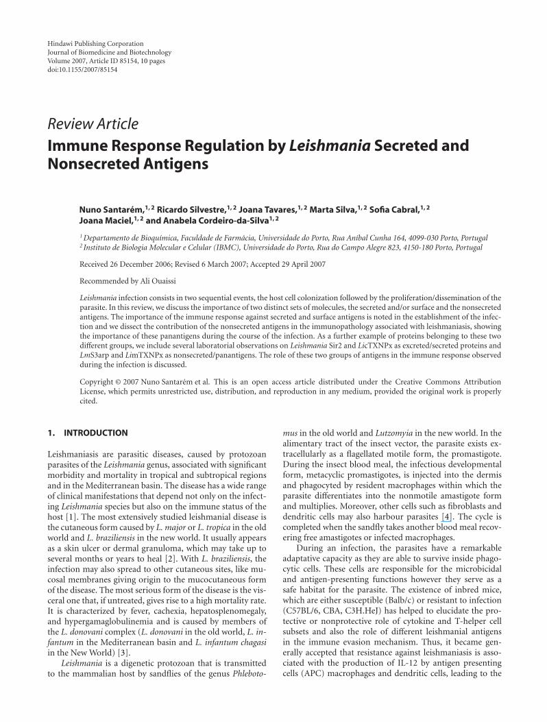
Hindawi Publishing CorporationJournal of Biomedicine and BiotechnologyVolume 2007, Article ID 85154, 10 pagesdoi:10.1155/2007/85154
Review ArticleImmune Response Regulation by Leishmania Secreted andNonsecreted Antigens
Nuno Santarem,1, 2 Ricardo Silvestre,1, 2 Joana Tavares,1, 2 Marta Silva,1, 2 Sofia Cabral,1, 2
Joana Maciel,1, 2 and Anabela Cordeiro-da-Silva1, 2
1 Departamento de Bioquımica, Faculdade de Farmacia, Universidade do Porto, Rua Anıbal Cunha 164, 4099-030 Porto, Portugal2 Instituto de Biologia Molecular e Celular (IBMC), Universidade do Porto, Rua do Campo Alegre 823, 4150-180 Porto, Portugal
Received 26 December 2006; Revised 6 March 2007; Accepted 29 April 2007
Recommended by Ali Ouaissi
Leishmania infection consists in two sequential events, the host cell colonization followed by the proliferation/dissemination of theparasite. In this review, we discuss the importance of two distinct sets of molecules, the secreted and/or surface and the nonsecretedantigens. The importance of the immune response against secreted and surface antigens is noted in the establishment of the infec-tion and we dissect the contribution of the nonsecreted antigens in the immunopathology associated with leishmaniasis, showingthe importance of these panantigens during the course of the infection. As a further example of proteins belonging to these twodifferent groups, we include several laboratorial observations on Leishmania Sir2 and LicTXNPx as excreted/secreted proteins andLmS3arp and LimTXNPx as nonsecreted/panantigens. The role of these two groups of antigens in the immune response observedduring the infection is discussed.
Copyright © 2007 Nuno Santarem et al. This is an open access article distributed under the Creative Commons AttributionLicense, which permits unrestricted use, distribution, and reproduction in any medium, provided the original work is properlycited.
1. INTRODUCTION
Leishmaniasis are parasitic diseases, caused by protozoanparasites of the Leishmania genus, associated with significantmorbidity and mortality in tropical and subtropical regionsand in the Mediterranean basin. The disease has a wide rangeof clinical manifestations that depend not only on the infect-ing Leishmania species but also on the immune status of thehost [1]. The most extensively studied leishmanial disease isthe cutaneous form caused by L. major or L. tropica in the oldworld and L. braziliensis in the new world. It usually appearsas a skin ulcer or dermal granuloma, which may take up toseveral months or years to heal [2]. With L. braziliensis, theinfection may also spread to other cutaneous sites, like mu-cosal membranes giving origin to the mucocutaneous formof the disease. The most serious form of the disease is the vis-ceral one that, if untreated, gives rise to a high mortality rate.It is characterized by fever, cachexia, hepatosplenomegaly,and hypergamaglobulinemia and is caused by members ofthe L. donovani complex (L. donovani in the old world, L. in-fantum in the Mediterranean basin and L. infantum chagasiin the New World) [3].
Leishmania is a digenetic protozoan that is transmittedto the mammalian host by sandflies of the genus Phleboto-
mus in the old world and Lutzomyia in the new world. In thealimentary tract of the insect vector, the parasite exists ex-tracellularly as a flagellated motile form, the promastigote.During the insect blood meal, the infectious developmentalform, metacyclic promastigotes, is injected into the dermisand phagocyted by resident macrophages within which theparasite differentiates into the nonmotile amastigote formand multiplies. Moreover, other cells such as fibroblasts anddendritic cells may also harbour parasites [4]. The cycle iscompleted when the sandfly takes another blood meal recov-ering free amastigotes or infected macrophages.
During an infection, the parasites have a remarkableadaptative capacity as they are able to survive inside phago-cytic cells. These cells are responsible for the microbicidaland antigen-presenting functions however they serve as asafe habitat for the parasite. The existence of inbred mice,which are either susceptible (Balb/c) or resistant to infection(C57BL/6, CBA, C3H.HeJ) has helped to elucidate the pro-tective or nonprotective role of cytokine and T-helper cellsubsets and also the role of different leishmanial antigensin the immune evasion mechanism. Thus, it became gen-erally accepted that resistance against leishmaniasis is asso-ciated with the production of IL-12 by antigen presentingcells (APC) macrophages and dendritic cells, leading to the

2 Journal of Biomedicine and Biotechnology
differentiation and proliferation of the Th1-subset of CD4+
T-cells producers of IFN-γ. This will ultimately lead to the ac-tivation of parasite-infected macrophages that, through theinduction of effector molecules as nitrogen and oxygen re-active species, will kill the intracellular parasites [5]. In con-trast, failure to control the infection has been associated withthe production of anti-inflammatory cytokines as IL-4, IL-10, IL-13 and TGF-β [6]. Given the ancient evolutionary di-vergence in Leishmania species, it is not surprising that thecontrol of the different Leishmania driven diseases is relatedto different immunological properties. Hence, while in cuta-neous leishmaniasis, IL-4 has been implicated in disease pro-gression, in visceral leishmaniasis its importance has beenruled out [7]. In the latter, IL-10 has been shown to bethe major immunosuppressive cytokine along with TGF-β.Overall, it suggests that it is the overshadowing of the Th2response by a Th1 cell associated response that leads to thecontrol of the infection [8]. Moreover, the real contributionof the humoral response is still under debate, however stud-ies in different intracellular pathogens have shown that an-tibodies can also have a function in restricting the infectionwhen the parasite is exposed to the extracellular milieu [9].Consequently, in leishmaniasis, the induction of specific hu-moral responses to parasite antigens would, theoretically, beable to neutralize the parasite whether as free promastigotes,after the inoculum, or as amastigotes, when released fromthe infected macrophages, contributing to develop a protec-tive response [10]. However, until now, no effective vaccineagainst human leishmaniasis is available for clinical use [3].
Leishmania parasites inside their hosts do not behaveinertly. Rather, the virulence related to their pathology seemsto be linked to an induced lack of immune response control.The parasite actively secretes proteases and other moleculesthat affect host immune system (cells and cytokines) facil-itating the infection process. In addition, the parasite pos-sesses intracellular nonsecreted antigens, members of con-served protein families, which are believed to contributeto the chronic immunopathology, observed in leishmania-sis. Here, we review these two groups of relevant parasitemolecules, illustrated with laboratory observations of pro-teins belonging to the secreted and nonsecreted groups ofantigens. Finally, we discuss their differential role in Leish-mania infection and persistence as well in the developmentof a protective immune response.
1.1. The importance of the secreted versusnonsecreted antigens
Leishmania virulence has been explained using two differentgroups of parasite molecules, the secreted and surface and theintracellular molecules [11]. This model proposes that thesecreted and surface molecules will be mostly important forthe establishment of infection, protecting the parasite fromthe early action of the host immune system, acting as inva-sive/evasive determinants. According to this model, the in-tracellular molecules will be ultimately responsible for thedisease phenotype [11].
1.2. Surface and secreted molecules
The secreted proteins have distinct functions during Leish-mania infection. First, they play a role in the establishmentof the infection [12] in conjunction with important elementsexistent in the saliva of the sandfly vector [13, 14]. In a secondphase, they contribute to the maintenance of the infectionby interfering with the macrophagic microbicidal functions,cytokine production, antigen presentation, and effector cellsactivation. This is achieved by repression of gene expression,post-translation protein modification or degradation, and byactivation of suppressive pathways and molecules [15]. Thismacrophagic anergy enables the continuous multiplicationof the amastigote form. The bulk of the knowledge on sur-face and secreted molecules of Leishmania is focused on ly-pophosphoglycan (LPG), on the promastigote surface pro-tease named glycoprotein 63 (gp63), glycosylinositol phos-pholipids (GIPLs), cysteine peptidases and on a few oth-ers like β-mercaptoethanol activated proteases, acid phos-phatases and chitinases. The importance of some of thesemolecules in the establishment of the infection is well doc-umented [15, 16], but the real contribution of the secretedmolecules remains elusive due to the difficulty of the intra-macrophagic studies.
After entrance into a susceptible mammalian host, theLeishmania promastigotes are targeted by the host immunesystem. Serum components, like the complement system rep-resent the first challenge following entrance into the blood-stream. Procyclic promastigotes are highly susceptible tocomplement action, unlike the metacyclic that can avoidcomplement mediated lysis [17]. This remarkable differenceis mostly due to the surface molecule in Leishmania, the LPG.Composed mainly of repetitive units of a disaccharide anda phosphate, LPG is linked to the membrane by a glyco-sylphosphatidylinositol anchor [18]. The LPG is longer inmetacyclic promastigotes preventing the attachment of C5b-C9 subunits of the complement complex avoiding its lyticaction [17]. The relevance of LPG is not limited to com-plement resistance. Its importance is stated by several stud-ies using either purified LPG or mutant strains. The LPGis implicated in several processes including the binding tothe epithelial cells of the sandfly midgut [19], receptor me-diated phagocytosis of macrophages through the CR3/CR1ligand or the manose-fucose receptor (in conjunction withgp63) [20, 21], toll-like receptor 2 signalling [22], stimula-tion of NK cells [23], inhibition of phagosome-endosome fu-sion [24–26], and inhibition of phagosome-derived superox-ide [27]. Several attempts to use LPG to confer protectionwere unproductive [28, 29]. Constitutively shed by severalLeishmania species, the LPG is the paradigm molecule re-ferred to as evasive and invasive. After the initial steps ofinfection, LPG is downregulated being almost absent fromamastigotes [30].
Another molecule implicated in the invasive and eva-sive mechanisms is gp63. This protein is the most abun-dant in the parasite surface, although 10 fold less abundantthan LPG [30]. In the promastigote form, gp63 is in thesurface of the parasite under the LPG coat and is involved

Nuno Santarem et al. 3
175
115
84.9
62.2
55.6
35.2
30.9
MW
(kD
)
Pre-immuneserum
LicTXNPx immuneserum
LiTXN1 immuneserum
ES PL ES PL ES PL
Figure 1: The LicTXNPx and LiTXN1 are excreted/secreted pro-teins. Autoradiography of [35S] methionine labelled L. infantumpromastigotes lysate (PL) and excreted/secreted antigens (ES), af-ter 3 hours of incubation experiments, immunoprecipitated in thepresence of immune anti-LicTXNPx or anti-LiTXN1 sera or with apreimmune serum.
in L. donovani promastigote multiplication [31]. Like LPG,gp63 was shown to be implicated in complement resistance,in L. major and L. amazonensis, by mediating the intercon-vertion of C3b to C3bi [32]. This interconvertion favoursthe internalization via CR3 avoiding the oxidative burst. Thebinding of gp63 to fibronectin receptors favours the parasiteuptake into the macrophage [33]. Furthermore, gp63 is anendopeptidase with the potential to degrade immunoglobu-lins, complement factors, and lysosomal proteins [34]. Theoptimal proteolitic activity of gp63 is at pH 4 that may indi-cate some active proteolitic function in the amastigote stage[34, 35]. Despite this, gp63 expression is downregulated inamastigotes [36]. In spite of being a virulence factor in mostLeishmania species, immunization trials with gp63 were un-able to protect mice from infectious challenge [37]. More-over, gp63 mutation in L. major did not impair in vitro in-tramacrophagic survival [38]. So the importance of gp63 inthe course of the infection remains elusive. The GIPLs aremolecules 10 times more abundant than LPG on the para-site surface, although like gp63 they are physically under theLPG coat [39]. The GIPLs were described in L. major as hav-ing a protective role at the parasite surface by modulating theexpression of nitric oxide synthetase in murine macrophages[40, 41]. Another interesting group of proteins are the cys-teine proteases. In L. mexicana, this family of proteins seemsto be associated with disease progression [42]. Cysteine pro-tease activity can be found at the parasite surface or inside
the macrophage endoplasmatic reticulum, probably associ-ated with proteases released in the phagolysosome by Leish-mania. The inhibition of major histocompatibility complexclass II molecules in macrophages seems to involve, in L.amazonensis, the direct sequestering of these molecules fol-lowing cysteine-peptidase-dependent degradation [43, 44].Also, cysteine peptidase activity was demonstrated in L. mex-icana to induce IL-12 repression and degradation of NF-kB[45]. It is still worthy to mention some other secreted pro-teins described as virulence factors, like the L. mexicana chiti-nase [46] and the L. donovani acid phosphatases [47–50]. Anin depth study of the Leishmania secretome is missing. Themost remarkable effort was done by Chenik and colleaguesthat were able to screen 33 different proteins using an L. ma-jor cDNA library and a rabbit immune sera raised against thesecreted proteins [51]. Nine of them were already describedas excreted/secreted proteins in Leishmania or other species,11 corresponded to known proteins but not characterized assecreted and the other 13 were completely new and unchar-acterized proteins [51]. This shows how little is known aboutthe Leishmania secretome since only a few proteins are exten-sively characterized [52–56]. It is already known that total L.major secreted molecules, described as highly immunogenic[54, 57–59], can confer protection from infectious challenge[57, 59]. So it is obvious that somewhere among the Leish-mania secreted proteins exist future candidates for vaccinedesign and drug targets. Nonetheless, one of the problems invaccine design using surface or secreted/excreted proteins isthe fact that these proteins are naturally exposed to the im-mune system. Chang et al suggest that these secreted/excretedproteins were evolutionarily selected becoming immunolog-ically “silent” [60]. This fact implies that secreted proteinsthat have a specific function in the establishment of the in-fection will be “silent,” allowing them to perform their vitalfunctions unchecked by the host immune system [11, 12].This will be more significant for the proteins involved in thefirst steps of infection, while the parasite is still exposed tothe extracellular environment. As an example of this fact,we present three distinct proteins: a cytosolic tryparedoxinperoxidase of L. infantum (LicTXNPx) [61], the Leishma-nia silent information regulator 2 (Sir2) [52], and a try-
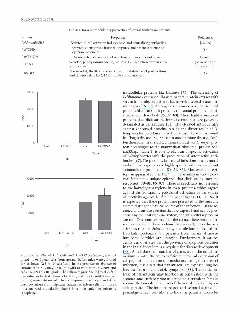
paredoxin of L. infantum (LiTXN1) [62]. All are Leishma-nia secreted proteins (Figure 1) [52], that show distinct im-munological properties. A high antibody titre against theLicTXNPx was detected in children [63]. This antibody titreis maintained during the Leishmania infection and decreasesafter its resolution [63]. Despite its high immunogenicitywhen tested in vitro or in vivo using the Balb/c model, thisexcreted/secreted protein did not show immunomodulatoryproperties (Figures 4, 5, and Table 1) and provided no pro-tection against the infectious challenge (data not shown).On the other hand, the Leishmania Sir2 is a typical poorlyimmunogenic secreted antigen (Figure 2) characterized as avirulence factor [64]. Infectious challenge after LeishmaniaSir2 immunization results in a decreased infectivity in theacute phase (Figure 3). This could be partially due to theproduction of lytic and neutralizing antibodies [65]. Theimmunization leads to a significant decrease of the spleen

4 Journal of Biomedicine and Biotechnology
0 2 8 11 15 17 19
Weeks after L. infantum infection
Figure 2: Antibodies against Leishmania SIR2 protein in the sera ofchronically L. infantum infected Balb/c mice. Sera from 108 intraperi-toneal (i.p.) L. infantum promastigotes infected Balb/c mice after 2,8, 11, 15, 17, and 19 weeks, were used in a western blot against 1 μgof rLiSIR2 at two different dilutions, 1 : 200 and 1 : 50 (left andright lanes, respectively, for each different serum). A 0 weeks serumwas obtained from noninfected mice.
and liver parasite load at two weeks post infection (Figure 3)[65]. However, it is incapable by itself of resolving the in-fection, as seen six weeks after infection, where there is nosignificant difference between the immunized infected groupand the infected control group (Figure 3). Certain secretedproteins seem to function as immunomodulatory compo-nents, acting as host immune evasive proteins. As an exam-ple, another excreted/secreted Leishmania protein, LiTXN1(Figure 1), is capable to increase IL-10 splenocyte secretion(Table 1), a major immunosuppressive cytokine (manuscriptin preparation). LiTXN1 can be among the proteins respon-sible for a transient immunosuppressive state that can favourthe parasite internalization and colonization of the host cells.These examples show that among the secreted proteins wecan find proteins naturally immunogenic, albeit nonprotec-tive, like LicTXNPx while others less immunogenic show in-teresting properties in terms of protection probably due tothe disruption of their in vivo functions, Leishmania Sir2, orby their immunomodulatory properties, LiTXN1. Unfortu-nately, the reduced immunogenicity of the most interestingsecreted proteins probably will prevent their identification byserological based approaches [51].
The reduction of the secreted/excreted proteins to thegiven examples is an oversimplification. However, it is ob-vious that much more work is needed in this area, especiallyin the huge black hole of knowledge that concerns the inter-action between host cell and Leishmania at a molecular level.Since most of the studies have been done using infection-phenotype approaches, little is known about the true agentsinvolved in macrophagic disruption [16, 58, 68, 69]. We sug-gest that amastigote secreted proteins will be more immuno-genic and can have interesting immunomodulatory proper-ties since they have not been under the selective pressure asthe promastigote secreted proteins. The selective pressure ofthe host immune system is a powerful driving force in evolu-tion, as demonstrated in the case of Schistosoma mansoni thathas the ability to completely evade the host immune systemrendering itself “invisible” [70].
1.3. Panantigens—nonsecreted proteins
Human visceral leishmaniasis, unlike cutaneous leishmani-asis is characterized by high anti-Leishmania antibody titres[71, 72]. The role of these antibodies is still unclear as there
6
5
4
30.5
02 6
Weeks
∗
Log
(par
asit
esg−
1)
Non-immunizedrLiSIR2-immunized
Spleen
(a)
6
5
4
30.5
02 6
Weeks
∗
∗Lo
g(p
aras
itesg−
1)
Non-immunizedrLiSIR2-immunized
Liver
(b)
Figure 3: The recombinant Leishmania SIR2 immunization reducesthe parasite load in an acute phase of L. infantum Balb/c mice infec-tion. The immunized mice (�) received 3 i.p. injections of recom-binant Leishmania SIR2 (50 μg) once a week and infected 2 weeksafter the last immunization with 108 L. infantum stationary phasepromastigotes. The nonimmunized mice (�) were subjected to thesame protocol but received PBS instead of recombinant LeishmaniaSIR2. The mice were sacrificed after 2 and 6 weeks of infection andthe parasite load in the spleen and liver determined by the organlimiting dilution method [66]. The data represent means and stan-dard deviations for three mice and are representative of two inde-pendent experiments. Statistical analysis was performed using Stu-dent t-test. Statistically, significant differences between immunizedand nonimmunized mice are indicated. ∗P < .05.
seems to be no relation with the progression or resolutionof the infection[58, 73, 74]. This exuberant humoral re-sponse against promastigote and amastigote antigens (frac-tions or total protein extract or specific Leishmania proteins)has been exploited for serodiagnosis with different degreesof success [58, 63, 74, 75]. Interestingly, one of the mostsensitive techniques using recombinant Leishmania proteinsdoes not involve surface molecules like LPG or gp63 but
Nuno Santarem et al. 5
Table 1: Immunomodulatory properties of several Leishmania proteins
Protein Properties References
Leishmania Sir2 Secreted, B-cell activator, induces lytic, and neutralizing antibodies [64, 65]
LicTXNPx Secreted, elicits strong humoral response and has no influence oncytokine production
[63]
LimTXNPx Nonsecreted, decreases IL-4 secretion both in vitro and in vivo Figure 3
LiTXN1 Secreted, poorly immunogenic, induces IL-10 secretion both in vitroand in vivo
(Manuscript inpreparation)
LmS3arp Nonsecreted, B-cell polyclonal activator, inhibits T-cell proliferation,and downregulate IL-2, 12 and IFN-γ in splenocytes
[67]
30000
20000
10000
0Ctrl LicTXNPx Ctrl LicTXNPx
ConA
CP
M
(a)
30000
20000
10000
0
CP
M
Ctrl LimTXNPx Ctrl LimTXNPx
ConA
(b)
Figure 4: No effect of rLicTXNPx and rLimTXNPx (a) on spleen cellproliferation. Spleen cells from normal Balb/c mice were culturedfor 48 hours (2.5 × 10⁵ cells/well) in the presence or absence ofconcanavalin A (ConA) (5 μg/ml) with or without rLicTXNPx andrLimTXNPx (b) (10 μg/ml). The cells were pulsed with [methyl-3H]thymidine in the last 8 hours of culture, and cpm (scintillations perminute) were determined. The data represent mean cpm and stan-dard deviations from triplicate cultures of spleen cells from threemice analyzed individually. One of three independent experimentsis depicted.
intracellular proteins like histones [75]. The screening ofLeishmania expression libraries or total protein extract withserum from infected patients has unveiled several major im-munogens [76–79]. Among these immunogens, nonsecretedproteins like heat shock proteins, ribosomal proteins and hi-stones were described [76, 77, 80]. These highly-conservedproteins that elicit strong immune responses are generallydesignated as panantigens [81]. The elevated antibody titreagainst conserved proteins can be the direct result of B-lymphocytes polyclonal activation similar to what is foundin Chagas disease [82, 83] or in autoimmune diseases [84].Furthermore, in the Balb/c mouse model, an L. major pro-tein homologue to the mammalian ribosomal protein S3a,LmS3arp, (Table 1) is able to elicit an unspecific activationof B-lymphocytes with the production of autoreactive anti-bodies [67]. Despite this, in natural infections, the humoraland cellular responses are highly specific with no significantautoantibody production [80, 81, 85]. Moreover, the epi-tope mapping of several Leishmania panantigens tends to re-veal Leishmania unique epitopes that elicit strong immuneresponses [79–81, 86, 87]. There is practically no responseto the homologous regions in these proteins, which arguesagainst the nonspecific polyclonal activation as the sourceof reactivity against Leishmania panantigens [11, 81]. So, itis expected that these proteins are presented to the immunesystem during the natural course of the infection. Unlike se-creted and surface proteins that are exposed and can be pro-cessed by the host immune system, the intracellular proteinsare not. One must expect that the contact between the im-mune system and these proteins happens only upon the par-asite destruction. Subsequently, one obvious source of in-tracellular proteins is the parasites from the initial inocu-lum some of which are destroyed. Furthermore, it was re-cently demonstrated that the presence of apoptotic parasitesin the initial inoculum is a requisite for disease development[88]. Albeit the small number of parasites in the initial in-oculum is not sufficient to explain the physical expansion ofcell populations and immune mediators during the course ofinfection, it is a fact that panantigens are exposed long be-fore the onset of any visible symptoms [88]. This initial re-lease of panantigens may function in conjugation with thesecreted and surface proteins acting as a transient “smokescreen” that enables the onset of the initial infection by vi-able parasites. The immune response developed against thepanantigens may contribute to hide the parasite molecules
6 Journal of Biomedicine and Biotechnology
0.45
0.4
0.35
0.3
0.25
0.2
0.15
0.1
0.05
0
∗
Ctrl Ctrl LimTXNPx LicTXNPx
ConA
IL-4
(μg/
ml)
(a)
0.45
0.4
0.35
0.3
0.25
0.2
0.15
0.1
0.05
0
∗
Ctrl LimTXNPx LicTXNPx Ctrl
ConA
LimTXNPx LicTXNPx
IL-4
(μg/
ml)
∗∗
(b)
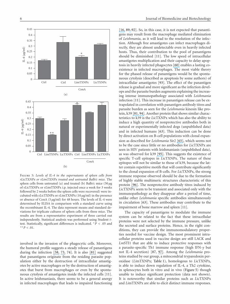
Figure 5: Levels of IL-4 in the supernatants of spleen cells fromrLicTXNPx or rLimTXNPx treated and untreated Balb/c mice. Thespleen cells from untreated (a) and treated (b) Balb/c mice (50 μgof rLicTXNPx or rLimTXNPx i.p. injected once a week for 3 weeksfollowed by 2 weeks before the spleen cells were recovered) were in-cubated with rLicTXNPx or rLimTXNPx (10 μg/ml) in the presenceor absence of ConA (5 μg/ml) for 48 hours. The levels of IL-4 weredetermined by ELISA in comparison with a standard curve usingthe recombinant IL-4. The data represent means and standard de-viations for triplicate cultures of spleen cells from three mice. Theresults are from a representative experiment of three carried outindependently. Statistical analysis was performed using Student t-test. Statistically, significant differences is indicated. ∗P < .05 and∗∗P < .01.
involved in the invasion of the phagocytic cells. Moreover,the humoral profile suggests a steady release of panantigensduring the infection [58, 73, 74]. It is also [81] suggestedthat panantigens originate from the residing parasite pop-ulation either by the destruction of intracellular amastig-otes by active macrophages or by the destruction of amastig-otes that burst from macrophages or even by the sponta-neous cytolysis of amastigotes inside the infected cells [11].In active leishmaniasis, there seems to be a general anergyin infected macrophages that leads to impaired functioning
[16, 89–92]. So, in this case, it is not expected that pananti-gens may result from the macrophage mediated eliminationof Leishmania, as it will lead to the resolution of the infec-tion. Although free amastigotes can infect macrophages di-rectly, they are almost undetectable even in heavily infectedhosts. Thus, their contribution to the pool of panantigensshould be diminished [11]. The low speed of intracellularamastigotes multiplication and their capacity to delay apop-tosis in heavily infected phagocytes [60] enables a lasting co-existence in infected macrophages. The most viable theoryfor the phased release of panantigens would be the sponta-neous cytolysis (described as apoptosis by some authors) ofintracellular amastigotes [93]. The effect of the panantigenrelease is gradual and more significant as the infection devel-ops and the parasite burden augments explaining the increas-ing intense immunopathology associated with Leishmaniainfection [11]. This increase in panantigen release can be ex-trapolated in correlation with panantigen antibody titres andparasite burden as seen for the Leishmania kinesin like pro-tein, k39 [81, 94]. Another protein that shows similar charac-teristics to k39 is the LicTXNPx which has also the ability toinduce a high quantity of nonprotective antibodies both innatural or experimentally infected dogs (unpublished data)and in infected humans [63]. This induction can be doneby direct activation on B-cell populations with clonal expan-sion as described for Leishmania Sir2 [65], which seems notto be the case since little or no antibodies for LicTXNPx areseen in HIV patients with leishmaniasis (unpublished data),as was observed for k39 [95]. This suggests the existence ofspecific T-cell epitopes in LicTXNPx. The nature of theseepitopes will not be similar to those of k39, because the lat-ter contain repetitive motifs that will contribute significantlyto the clonal expansion of B-cells. For LicTXNPx, the strongimmune response observed should be due to the formationof highly stable multimeric structures characteristic of thisprotein [96]. The nonprotective antibody titres induced byLicTXNPx seem to be transient and associated only with theimmunopathology as they disappear after a period of time,unlike other Leishmania specific antibodies simultaneouslyin circulation [63]. These antibodies may contribute to theimpairment of bone marrow and spleen [11].
The capacity of panantigens to modulate the immunesystem can be related to the fact that these intracellularproteins were not selected by the immune pressure, unlikethe secreted and surface proteins. Hence, in the right con-ditions, they can provide the immunomodulatory proper-ties needed for vaccine design. The most prominent intra-cellular proteins used in vaccine design are still LACK andLmSTI1 that are able to induce protective responses witha parasite-specific Th1 immune response (high IFN-γ butnot IL-4 secretion) [87, 97]. Among the Leishmania pro-teins studied by our group, a mitocondrial tryparedoxin per-oxidase (LimTXNPx; Table 1), homologous to LicTXNPx,is able to induce down regulation of IL-4, a Th2 cytokine,in splenocytes both in vitro and in vivo (Figure 5) thoughunable to induce significant protection (data not shown).It is noteworthy that similar proteins such as LicTXNPxand LimTXNPx are able to elicit distinct immune responses.

Nuno Santarem et al. 7
LicTXNPx is secreted inducing only the production of non-protective antibodies, while its related intracellular counter-part LimTXNPx has immunomodulatory properties inter-fering with cytokine production (Figure 5). This can be agood example of the type of evolutionary pressure inducedby the immune system, in which two related proteins havedistinct immunomodulatory properties (Figures 4, 5). It sug-gests that the host immune system selects characteristics inthe exposed proteins that are either innocuous or nondelete-rious to the parasite. Since this does not occur in the intra-cellular proteins they can retain distinct immunoregulatoryproperties that could be useful in vaccine design.
2. CONCLUDING REMARKS
Taken altogether, these observations support the idea that se-creted and surface proteins tend to be poor or nonprotec-tive immune modulators, like LicTXNPx. Nonetheless, theiruse in vaccine could induce short-lived protection probablydue to the disruption of their biological activity or by pro-duction of lytic antibodies, as seen with Leishmania Sir2. In-tracellular components like LmS3arp and LimTXNPx tendto have defined immunomodulatory properties. LmS3arp isable to induce polyclonal activation of B lymphocytes whileLiLimTXNPxTXNPx confers a nonprotective dowregulationof IL-4 secretion by splenocytes.
Using the basic knowledge acquired in the study of theimmune response against Leishmania in different murinemodels, one can look for proteins that induce the immuno-logical phenotype needed for protection. Therefore, our datasuggests that in vaccine development, the conjugation of se-creted and surface proteins with intracellular componentsshould provide a more efficient protection. Hence, the im-pairment of the parasite entrance in the host cells, eitherby lytic antibodies or by the disruption of protein function,will delay the onset of the immune suppression associatedwith Leishmania. The parasite elimination could be achievedthrough a protective cellular response, induced by the intra-cellular parasite components present in the vaccine.
ACKNOWLEDGMENTS
This work was supported by FCT and Programa Operacional Ciencia e Inovacao 2010 (POCI 2010) and FEDER inthe projects: POCI/SAU-FCF/59837/2005 and POCI/CVT/59840/2004. R.S. and J.T. are supported by fellowshipsfrom FCT and FEDER number SFRH/BD/13120/2003and SFRH/BD/18137/2004, respectively. The proteins,LicTXNPx, LimTXNPx, and LiTXN1, used in this workswere purified from clones provided by A. Tomas.
REFERENCES
[1] R. Badaro, T. C. Jones, E. M. Carvalho, et al., “New perspec-tives on a subclinical form of visceral leishmaniasis,” Journal ofInfectious Diseases, vol. 154, no. 6, pp. 1003–1011, 1986.
[2] S. M. Salman, N. G. Rubeiz, and A.-G. Kibbi, “Cutaneousleishmaniasis: clinical features and diagnosis,” Clinics in Der-matology, vol. 17, no. 3, pp. 291–296, 1999.
[3] World Health Organization, “Leishmaniasis and Leishmania-HIV co-infection,” in WHO Report on Global Surveillance ofEpidemic-Prone Infectious Diseases, WHO, Geneva, Switzer-land, 2000.
[4] W. Solbach and T. Laskay, “The host response to Leishma-nia infection,” Advances in Immunology, vol. 74, pp. 275–317,2000.
[5] C. Bogdan, A. Gessner, S. Werner, and M. Rollinghoff, “Inva-sion, control and persistence of Leishmania parasites,” CurrentOpinion in Immunology, vol. 8, no. 4, pp. 517–525, 1996.
[6] J. Alexander and K. Bryson, “T helper (h)1/Th2 and Leish-mania: paradox rather than paradigm,” Immunology Letters,vol. 99, no. 1, pp. 17–23, 2005.
[7] A. Satoskar, H. Bluethmann, and J. Alexander, “Disruptionof the murine interleukin-4 gene inhibits disease progressionduring Leishmania mexicana infection but does not increasecontrol of Leishmania donovani infection,” Infection and Im-munity, vol. 63, no. 12, pp. 4894–4899, 1995.
[8] G. D. Miralles, M. Y. Stoeckle, D. F. McDermott, F. D. Finkel-man, and H. W. Murray, “Th1 and Th2 cell-associated cy-tokines in experimental visceral Leishmaniasis,” Infection andImmunity, vol. 62, no. 3, pp. 1058–1063, 1994.
[9] A. Casadevall, “Antibody-mediated protection against intra-cellular pathogens,” Trends in Microbiology, vol. 6, no. 3, pp.102–107, 1998.
[10] R. Ravindran and N. Ali, “Progress in vaccine research andpossible effector mechanisms in visceral leishmaniasis,” Cur-rent Molecular Medicine, vol. 4, no. 6, pp. 697–709, 2004.
[11] K. P. Chang, S. G. Reed, B. S. McGwire, and L. Soong, “Leish-mania model for microbial virulence: the relevance of parasitemultiplication and pathoantigenicity,” Acta Tropica, vol. 85,no. 3, pp. 375–390, 2003.
[12] M. G. Rittig and C. Bogdan, “Leishmania-host-cell interac-tion: complexities and alternative views,” Parasitology Today,vol. 16, no. 7, pp. 292–297, 2000.
[13] M. C. de Almeida, V. Vilhena, A. Barral, and M. Barral-Netto,“Leishmanial infection: analysis of its first steps. A review,”Memorias do Instituto Oswaldo Cruz, vol. 98, no. 7, pp. 861–870, 2003.
[14] S. Kamhawi, “The biological and immunomodulatory proper-ties of sand fly saliva and its role in the establishment of Leish-mania infections,” Microbes and Infection, vol. 2, no. 14, pp.1765–1773, 2000.
[15] D. J. Gregory and M. Olivier, “Subversion of host cell sig-nalling by the protozoan parasite Leishmania,” Parasitology,vol. 130, supplement 1, pp. S27–S35, 2005.
[16] M. Olivier, D. J. Gregory, and G. Forget, “Subversion mecha-nisms by which Leishmania parasites can escape the host im-mune response: a signaling point of view,” Clinical Microbiol-ogy Reviews, vol. 18, no. 2, pp. 293–305, 2005.
[17] S. M. Puentes, D. M. Dwyer, P. A. Bates, and K. A. Joiner,“Binding and release of C3 from Leishmania donovani pro-mastigotes during incubation in normal human serum,” Jour-nal of Immunology, vol. 143, no. 11, pp. 3743–3749, 1989.
[18] M. J. McConville, J. E. Thomas-Oates, M. A. J. Ferguson,and S. W. Homans, “Structure of the lipophosphoglycan fromLeishmania major,” Journal of Biological Chemistry, vol. 265,no. 32, pp. 19611–19623, 1990.
[19] C. R. Davies, A. M. Cooper, C. Peacock, R. P. Lane, and J. M.Blackwell, “Expression of LPG and gp63 by different develop-mental stages of Leishmania major in the sandfly Phlebotomuspapatasi,” Parasitology, vol. 101, no. 3, pp. 337–343, 1990.
[20] D. L. Sacks, “Metacyclogenesis in Leishmania promastigotes,”Experimental Parasitology, vol. 69, no. 1, pp. 100–103, 1989.

8 Journal of Biomedicine and Biotechnology
[21] D. M. Mosser and L. A. Rosenthal, “Leishmania-macrophageinteractions: multiple receptors, multiple ligands and diversecellular responses,” Seminars in Cell Biology, vol. 4, no. 5, pp.315–322, 1993.
[22] M. J. de Veer, J. M. Curtis, T. M. Baldwin, et al., “MyD88 isessential for clearance of Leishmania major: possible role forlipophosphoglycan and toll-like receptor 2 signaling,” Euro-pean Journal of Immunology, vol. 33, no. 10, pp. 2822–2831,2003.
[23] I. Becker, N. Salaiza, M. Aguirre, et al., “Leishmania lipophos-phoglycan (LPG) activates NK cells through toll-like receptor-2,” Molecular and Biochemical Parasitology, vol. 130, no. 2, pp.65–74, 2003.
[24] L. Miao, A. Stafford, S. Nir, S. J. Turco, T. D. Flanagan, and R.M. Epand, “Potent inhibition of viral fusion by the lipophos-phoglycan of Leishmania donovani,” Biochemistry, vol. 34,no. 14, pp. 4676–4683, 1995.
[25] M. Desjardins and A. Descoteaux, “Inhibition of phagolyso-somal biogenesis by the Leishmania lipophosphoglycan,” Jour-nal of Experimental Medicine, vol. 185, no. 12, pp. 2061–2068,1997.
[26] J. F. Dermine, S. Scianimanico, C. Prive, A. Descoteaux, andM. Desjardins, “Leishmania promastigotes require lipophos-phoglycan to actively modulate the fusion properties ofphagosomes at an early step of phagocytosis,” Cellular Micro-biology, vol. 2, no. 2, pp. 115–126, 2000.
[27] R. Lodge, T. O. Diallo, and A. Descoteaux, “Leishmania dono-vani lipophosphoglycan blocks NADPH oxidase assembly atthe phagosome membrane,” Cellular Microbiology, vol. 8,no. 12, pp. 1922–1931, 2006.
[28] W. K. Tonui, S. S. Mpoke, A. S. Orago, S. J. Turco, P. A. Mbati,and G. M. Mkoji, “Leishmania donovani-derived lipophospho-glycan plus BCG induces a Th1 type immune response butdoes not protect Syrian golden hamsters (Mesocricetus aura-tus) and BALB/c mice against Leishmania donovani,” Onder-stepoort Journal of Veterinary Research, vol. 70, no. 4, pp. 255–263, 2003.
[29] W. K. Tonui, “Vaccination of BALB/c mice with Leishmaniadonovani derived lipophosphoglycan does not conver cross-protection to L. major infections,” East African Medical Jour-nal, vol. 80, no. 5, pp. 260–263, 2003.
[30] P. F. P. Pimenta, E. M. B. Saraiva, and D. L. Sacks, “The com-parative fine structure and surface glycoconjugate expressionof three life stages of Leishmania major,” Experimental Para-sitology, vol. 72, no. 2, pp. 191–204, 1991.
[31] S. Pandey, P. Chakraborti, R. Sharma, S. Bandyopadhyay, D.Sarkar, and S. Adhya, “Involvement of Leishmania donovanimajor surface glycoprotein gp63 in promastigote multiplica-tion,” Journal of Biosciences, vol. 29, no. 1, pp. 15–22, 2004.
[32] A. Brittingham, C. J. Morrison, W. R. McMaster, B. S. McG-wire, K. P. Chang, and D. M. Mosser, “Role of the Leishmaniasurface protease gp63 in complement fixation, cell adhesion,and resistance to complement-mediated lysis,” Journal of Im-munology, vol. 155, no. 6, pp. 3102–3111, 1995.
[33] A. Brittingham, G. Chen, B. S. McGwire, K. P. Chang, and D.M. Mosser, “Interaction of Leishmania gp63 with cellular re-ceptors for fibronectin,” Infection and Immunity, vol. 67, no. 9,pp. 4477–4484, 1999.
[34] G. Chaudhuri and K. P. Chang, “Acid protease activity of amajor surface membrane glycoprotein (gp63) from Leishma-nia mexicana promastigotes,” Molecular and Biochemical Par-asitology, vol. 27, no. 1, pp. 43–52, 1988.
[35] G. Chaudhuri, M. Chaudhuri, A. Pan, and K. P. Chang,“Surface acid proteinase (gp63) of Leishmania mexicana. A
metalloenzyme capable of protecting liposome-encapsulatedproteins from phagolysosomal degradation by macrophages,”Journal of Biological Chemistry, vol. 264, no. 13, pp. 7483–7489, 1989.
[36] P. Schneider, J. P. Rosat, J. Bouvier, J. Louis, and C. Bordier,“Leishmania major: differential regulation of the surface met-alloprotease in amastigote and promastigote stages,” Experi-mental Parasitology, vol. 75, no. 2, pp. 196–206, 1992.
[37] E. Handman, L. L. Button, and R. W. McMaster, “Leishmaniamajor: production of recombinant gp63, its antigenicity andimmunogenicity in mice,” Experimental Parasitology, vol. 70,no. 4, pp. 427–435, 1990.
[38] P. B. Joshi, D. L. Sacks, G. Modi, and W. R. McMaster, “Tar-geted gene deletion of Leishmania major genes encoding de-velopmental stage-specific leishmanolysin (gp63),” MolecularMicrobiology, vol. 27, no. 3, pp. 519–530, 1998.
[39] M. A. J. Ferguson, “The surface glycoconjugates of trypanoso-matid parasites,” Philosophical Transactions of the Royal Societyof London B: Biological Sciences, vol. 352, no. 1359, pp. 1295–1302, 1997.
[40] L. Proudfoot, P. Schneider, M. A. J. Ferguson, and M. J. Mc-Conville, “Biosynthesis of the glycolipid anchor of lipophos-phoglycan and the structurally related glycoinositolphospho-lipids from Leishmania major,” Biochemical Journal, vol. 308,part 1, pp. 45–55, 1995.
[41] L. Proudfoot, A. V. Nikolaev, G. J. Feng, et al., “Regulationof the expression of nitric oxide synthase and leishmanicidalactivity by glycoconjugates of Leishmania lipophosphoglycanin murine macrophages,” Proceedings of the National Academyof Sciences of the United States of America, vol. 93, no. 20, pp.10984–10989, 1996.
[42] J. C. Mottram, G. H. Coombs, and J. Alexander, “Cysteine pep-tidases as virulence factors of Leishmania,” Current Opinion inMicrobiology, vol. 7, no. 4, pp. 375–381, 2004.
[43] S. De Souza Leao, T. Lang, E. Prina, R. Hellio, and J. C. An-toine, “Intracellular Leishmania amazonensis amastigotes in-ternalize and degrade MHC class II molecules of their hostcells,” Journal of Cell Science, vol. 108, part 10, pp. 3219–3231,1995.
[44] N. Courret, C. Frehel, E. Prina, T. Lang, and J. C. Antoine, “Ki-netics of the intracellular differentiation of Leishmania ama-zonensis and internalization of host MHC molecules by theintermediate parasite stages,” Parasitology, vol. 122, no. 3, pp.263–279, 2001.
[45] P. Cameron, A. McGachy, M. Anderson, et al., “Inhibition oflipopolysaccharide-induced macrophage IL-12 production byLeishmania mexicana amastigotes: the role of cysteine pepti-dases and the NF-κB signaling pathway,” Journal of Immunol-ogy, vol. 173, no. 5, pp. 3297–3304, 2004.
[46] M. B. Joshi, M. E. Rogers, A. M. Shakarian, et al., “Molecu-lar characterization, expression, and in vivo analysis of Lmex-Cht1: the chitinase of the human pathogen, Leishmania mexi-cana,” Journal of Biological Chemistry, vol. 280, no. 5, pp. 3847–3861, 2005.
[47] A. M. Shakarian, M. B. Joshi, M. Yamage, S. L. Ellis, A. Debra-bant, and D. M. Dwyer, “Members of a unique histidine acidphosphatase family are conserved amongst a group of primi-tive eukaryotic human pathogens,” Molecular and Cellular Bio-chemistry, vol. 245, no. 1-2, pp. 31–41, 2003.
[48] J. K. Lovelace and M. Gottlieb, “Comparison of extracellularacid phosphatases from various isolates of Leishmania,” Amer-ican Journal of Tropical Medicine and Hygiene, vol. 35, no. 6,pp. 1121–1128, 1986.
Nuno Santarem et al. 9
[49] S. L. Ellis, A. M. Shakarian, and D. M. Dwyer, “Leishmania:amastigotes synthesize conserved secretory acid phosphatasesduring human infection,” Experimental Parasitology, vol. 89,no. 2, pp. 161–168, 1998.
[50] P. S. Doyle and D. M. Dwyer, “Leishmania: immunochemicalcomparison of the secretory (extracellular) acid phosphatasesfrom various species,” Experimental Parasitology, vol. 77, no. 4,pp. 435–444, 1993.
[51] M. Chenik, S. Lakhal, N. Ben Khalef, L. Zribi, H. Louzir, andK. Dellagi, “Approaches for the identification of potential ex-creted/secreted proteins of Leishmania major parasites,” Para-sitology, vol. 132, no. 4, pp. 493–509, 2006.
[52] B. Yahiaoui, A. Taibi, and A. Ouaissi, “A Leishmania major pro-tein with extensive homology to silent information regulator 2of Saccharomyces cerevisiae,” Gene, vol. 169, no. 1, pp. 115–118,1996.
[53] M. Wiese, T. Ilg, F. Lottspeich, and P. Overath, “Ser/Thr-richrepetitive motifs as targets for phosphoglycan modifications inLeishmania mexicana secreted acid phosphatase,” The EMBOJournal, vol. 14, no. 6, pp. 1067–1074, 1995.
[54] J. R. Webb, A. Campos-Neto, P. J. Ovendale, et al., “Humanand murine immune responses to a novel Leishmania ma-jor recombinant protein encoded by members of a multicopygene family,” Infection and Immunity, vol. 66, no. 7, pp. 3279–3289, 1998.
[55] F. M. Symons, P. J. Murray, H. Ji, et al., “Characterization ofa polymorphic family of integral membrane proteins in pro-mastigotes of different Leishmania species,” Molecular and Bio-chemical Parasitology, vol. 67, no. 1, pp. 103–113, 1994.
[56] D. Sereno, L. Vanhille, B. Vergnes, A. Monte-Allegre, andA. Ouaissi, “Experimental study of the function of the ex-creted/secreted Leishmania LmSIR2 protein by heterologousexpression in eukaryotic cell line,” Kinetoplastid Biology andDisease, vol. 4, p. 1, 2005.
[57] W. K. Tonui, J. S. Mejia, L. Hochberg, et al., “Immunizationwith Leishmania major exogenous antigens protects suscepti-ble BALB/c mice against challenge infection with L. major,”Infection and Immunity, vol. 72, no. 10, pp. 5654–5661, 2004.
[58] J. R. Ryan, A. M. Smithyman, G.-H. Rajasekariah, L.Hochberg, J. M. Stiteler, and S. K. Martin, “Enzyme-linkedimmunosorbent assay based on soluble promastigote antigendetects immunoglobulin M (IgM) and IgG antibodies in serafrom cases of visceral and cutaneous leishmaniasis,” Journal ofClinical Microbiology, vol. 40, no. 3, pp. 1037–1043, 2002.
[59] J. L. Lemesre, P. Holzmuller, M. Cavaleyra, R. B. Goncalves, G.Hottin, and G. Papierok, “Protection against experimental vis-ceral leishmaniasis infection in dogs immunized with purifiedexcreted secreted antigens of Leishmania infantum promastig-otes,” Vaccine, vol. 23, no. 22, pp. 2825–2840, 2005.
[60] K. P. Chang, S. G. Reed, B. S. McGwire, and L. Soong, “Leish-mania model for microbial virulence: the relevance of parasitemultiplication and pathoantigenicity,” Acta Tropica, vol. 85,no. 3, pp. 375–390, 2003.
[61] H. Castro, C. Sousa, M. Santos, A. Cordeiro-da-Silva, L. Flohe,and A. M. Tomas, “Complementary antioxidant defense bycytoplasmic and mitochondrial peroxiredoxins in Leishmaniainfantum,” Free Radical Biology and Medicine, vol. 33, no. 11,pp. 1552–1562, 2002.
[62] H. Castro, C. Sousa, M. Novais, et al., “Two linked genes ofLeishmania infantum encode tryparedoxins localised to cy-tosol and mitochondrion,” Molecular and Biochemical Para-sitology, vol. 136, no. 2, pp. 137–147, 2004.
[63] N. Santarem, A. Tomas, A. Ouaissi, et al., “Antibodies againsta Leishmania infantum peroxiredoxin as a possible marker fordiagnosis of visceral leishmaniasis and for monitoring the ef-ficacy of treatment,” Immunology Letters, vol. 101, no. 1, pp.18–23, 2005.
[64] B. Vergnes, D. Sereno, N. Madjidian-Sereno, J. L. Lemesre,and A. Ouaissi, “Cytoplasmic SIR2 homologue overexpressionpromotes survival of Leishmania parasites by preventing pro-grammed cell death,” Gene, vol. 296, no. 1-2, pp. 139–150,2002.
[65] R. Silvestre, A. Cordeiro-da-Silva, J. Tavares, D. Sereno, andA. Ouaissi, “Leishmania cytosolic silent information regula-tory protein 2 deacetylase induces murine B-cell differentia-tion and in vivo production of specific antibodies,” Immunol-ogy, vol. 119, no. 4, pp. 529–540, 2006.
[66] B. Vergnes, D. Sereno, J. Tavares, et al., “Targeted disruption ofcytosolic SIR2 deacetylase discloses its essential role in Leish-mania survival and proliferation,” Gene, vol. 363, no. 1-2, pp.85–96, 2005.
[67] A. Cordeiro-da-Silva, M. C. Borges, E. Guilvard, and A.Ouaissi, “Dual role of the Leishmania major ribosomal pro-tein S3a homologue in regulation of T- and B-cell activation,”Infection and Immunity, vol. 69, no. 11, pp. 6588–6596, 2001.
[68] M. A. Vannier-Santos, A. Martiny, and W. de Souza, “Cell biol-ogy of Leishmania spp.: invading and evading,” Current Phar-maceutical Design, vol. 8, no. 4, pp. 297–318, 2002.
[69] S. Saha, S. Mondal, A. Banerjee, J. Ghose, S. Bhowmick, and N.Ali, “Immune responses in kala-azar,” Indian Journal of Medi-cal Research, vol. 123, no. 3, pp. 245–266, 2006.
[70] A. W. Cheever, K. F. Hoffmann, and T. A. Wynn, “Im-munopathology of Schistosomiasis mansoni in mice and men,”Immunology Today, vol. 21, no. 9, pp. 465–466, 2000.
[71] A. B. Neogy, A. Nandy, B. Ghosh Dastidar, and A. B. Chowd-hury, “Antibody kinetics in kala-azar in response to treat-ment,” Annals of Tropical Medicine and Parasitology, vol. 81,no. 6, pp. 727–729, 1987.
[72] R. D. Pearson and A. Q. Sousa, “Clinical spectrum of leish-maniasis,” Clinical Infectious Diseases, vol. 22, no. 1, pp. 1–13,1996.
[73] A. K. Ghosh, S. Dasgupta, and A. C. Ghose, “ImmunoglobulinG subclass-specific antileishmanial antibody responses in In-dian kala-azar and post-kala-azar dermal leishmaniasis,” Clin-ical and Diagnostic Laboratory Immunology, vol. 2, no. 3, pp.291–296, 1995.
[74] K. Anam, F. Afrin, D. Banerjee, et al., “Differential declinein Leishmania membrane antigen-specific immunoglobulin G(IgG), IgM, IgE, and IgG subclass antibodies in Indian kala-azar patients after chemotherapy,” Infection and Immunity,vol. 67, no. 12, pp. 6663–6669, 1999.
[75] I. A. Maalej, M. Chenik, H. Louzir, et al., “Comparative eval-uation of ELISAs based on ten recombinant or purified Leish-mania antigens for the serodiagnosis of Mediterranean visceralleishmaniasis,” American Journal of Tropical Medicine and Hy-giene, vol. 68, no. 3, pp. 312–320, 2003.
[76] M. Soto, J. M. Requena, M. Garcia, L. C. Gomez, I. Navarrete,and C. Alonso, “Genomic organization and expression of twoindependent gene arrays coding for two antigenic acidic ribo-somal proteins of Leishmania,” Journal of Biological Chemistry,vol. 268, no. 29, pp. 21835–21843, 1993.
[77] J. MacFarlane, M. L. Blaxter, R. P. Bishop, M. A. Miles, and J.M. Kelly, “Identification and characterisation of a Leishmaniadonovani antigen belonging to the 70-kDa heat-shock proteinfamily,” European Journal of Biochemistry, vol. 190, no. 2, pp.377–384, 1990.

10 Journal of Biomedicine and Biotechnology
[78] M. A. Dea-Ayuela, S. Rama-Iniguez, and F. Bolas-Fernandez,“Proteomic analysis of antigens from Leishmania infantumpromastigotes,” Proteomics, vol. 6, no. 14, pp. 4187–4194,2006.
[79] J. M. Burns Jr., W. G. Shreffler, D. R. Benson, H. W. Ghalib,R. Badaro, and S. G. Reed, “Molecular characterization of akinesin-related antigen of Leishmania chagasi that detects spe-cific antibody in African and American visceral leishmaniasis,”Proceedings of the National Academy of Sciences of the UnitedStates of America, vol. 90, no. 2, pp. 775–779, 1993.
[80] M. Soto, J. M. Requena, L. Quijada, et al., “Antigenicity of theLeishmania infantum histones H2B and H4 during canine vis-cerocutaneous leishmaniasis,” Clinical and Experimental Im-munology, vol. 115, no. 2, pp. 342–349, 1999.
[81] J. M. Requena, C. Alonso, and M. Soto, “Evolutionarily con-served proteins as prominent immunogens during Leishma-nia infections,” Parasitology Today, vol. 16, no. 6, pp. 246–250,2000.
[82] P. Minoprio, O. Burlen, P. Pereira, et al., “Most B cells in acuteTrypanosoma cruzi infection lack parasite specificity,” Scan-dinavian Journal of Immunology, vol. 28, no. 5, pp. 553–561,1988.
[83] A. Cordeiro-da-Silva, A. G. Espinoza, A. Taibi, A. Ouaissi, andP. Minoprio, “A 24,000 MW Trypanosoma cruzi antigen is a B-cell activator,” Immunology, vol. 94, no. 2, pp. 189–196, 1998.
[84] S. Avrameas, “Natural autoantibodies: from ‘horror autotoxi-cus’ to ‘gnothi seauton’,” Immunology Today, vol. 12, no. 5, pp.154–159, 1991.
[85] L. Quijada, J. M. Requena, M. Soto, and C. Alonso, “Duringcanine viscero-cutaneous leishmaniasis the anti-Hsp70 anti-bodies are specifically elicited by the parasite protein,” Para-sitology, vol. 112, no. 3, pp. 277–284, 1996.
[86] M. Soto, J. M. Requena, L. Quijada, et al., “Mapping of thelinear antigenic determinants from the Leishmania infantumhistone H2A recognized by sera from dogs with leishmaniasis,”Immunology Letters, vol. 48, no. 3, pp. 209–214, 1995.
[87] E. Mougneau, F. Altare, A. E. Wakil, et al., “Expression cloningof a protective Leishmania antigen,” Science, vol. 268, no. 5210,pp. 563–566, 1995.
[88] G. van Zandbergen, A. Bollinger, A. Wenzel, et al., “Leishma-nia disease development depends on the presence of apop-totic promastigotes in the virulent inoculum,” Proceedings ofthe National Academy of Sciences of the United States of Amer-ica, vol. 103, no. 37, pp. 13837–13842, 2006.
[89] O. Bacellar, C. Brodskyn, J. Guerreiro, et al., “Interleukin-12restores interferon-γ production and cytotoxic responses invisceral leishmaniasis,” Journal of Infectious Diseases, vol. 173,no. 6, pp. 1515–1518, 1996.
[90] S. Buates and G. Matlashewski, “General suppression ofmacrophage gene expression during Leishmania donovani in-fection,” Journal of Immunology, vol. 166, no. 5, pp. 3416–3422, 2001.
[91] H. W. Ghalib, J. A. Whittle, M. Kubin, et al., “IL-12 en-hances Th1-type responses in human Leishmania donovani in-fections,” Journal of Immunology, vol. 154, no. 9, pp. 4623–4629, 1995.
[92] N. E. Reiner, W. Ng, and W. R. McMaster, “Parasite-accessorycell interactions in murine leishmaniasis. II. Leishmania dono-vani suppresses macrophage expression of class I and class IImajor histocompatibility complex gene products,” Journal ofImmunology, vol. 138, no. 6, pp. 1926–1932, 1987.
[93] N. Lee, S. Bertholet, A. Debrabant, J. Muller, R. Duncan, andH. L. Nakhasi, “Programmed cell death in the unicellular pro-tozoan parasite Leishmania,” Cell Death and Differentiation,vol. 9, no. 1, pp. 53–64, 2002.
[94] S. Singh, A. Gilman-Sachs, K. P. Chang, and S. G. Reed, “Di-agnostic and prognostic value of K39 recombinant antigen inIndian leishmaniasis,” Journal of Parasitology, vol. 81, no. 6, pp.1000–1003, 1995.
[95] J. Alvar, C. Canavate, B. Gutierrez-Solar, et al., “Leishmaniaand human immunodeficiency virus coinfection: the first 10years,” Clinical Microbiology Reviews, vol. 10, no. 2, pp. 298–319, 1997.
[96] L. Flohe, H. J. Hecht, and P. Steinert, “Glutathione and trypan-othione in parasitic hydroperoxide metabolism,” Free RadicalBiology and Medicine, vol. 27, no. 9-10, pp. 966–984, 1999.
[97] J. R. Webb, D. Kaufmann, A. Campos-Neto, and S. G. Reed,“Molecular cloning of a novel protein antigen of Leishmaniamajor that elicits a potent immune response in experimentalmurine leishmaniasis,” Journal of Immunology, vol. 157, no. 11,pp. 5034–5041, 1996.
Copyright © 2022 FDOKUMEN