Ia-like antigens on freshly explanted human melanoma
10
CLINICAL IMMUNOLOGY AND IMMUNOPATHOLOGY 19, 250-259 (1981) la-Like Antigens on Freshly Explanted Human Melanoma P. G. NATALI, P. CORDIALI-FEI, R. CAVALIERE, F. DI FILIPPO, V. QUARANTA, M. A. PELLEGRINO, AND S. FERRONE Regina Elena Cancer Institute, Rome. Italy, and Department oj’ Molecular Immunology, Scripps Clinic and Research Foundation, La Jolla, California 92037 Received August 25, 1980 Human Ia-like antigens have unexpectedly been found on human melanoma cells maintained in long-term culture. Since these antigens may play a role in the biology of melanoma and in the interaction of the tumor with the host’s immune system, tissues surgically removed from 16 patients in different stages of the disease were tested in indirect immunofluorescence with monoclonal antibodies and operationally specific xenoantisera to framework determinants of Ia-like antigens. All of the samples which included primary, metastatic, and recurrent lesions specifically reacted with anti Ia-like antigen antibodies. Melanoma cells can synthesize Ia-like antigens since they reappeared on cells cultured in vitro following treatment with pronase. The number of Ia-like antigen-positive cells in the melanoma lesions ranged from 15 to 90% and was unrelated to the presence of melanine and to the clinical stage of the disease; however, a low number of Ia-like antigen-bearing cells was found in primary tumors with high levels of invasivity (Clark’s staging) and in lesions from patients with massive metastatic spread- ing. Intradermic nevi surgically removed from 14 patients did not react with anti la-like antigen xenoantibodies. If these results are confirmed with a large number of lesions then testing of tumor biopsies for Ia-like antigens may aid to solve controversial diagnosis of melanoma. INTRODUCTION Human Ia-like antigens, a set of histocompatibility antigens with functional and structural properties similar to those of murine la E/C subregion antigens, are believed to be mainly expressed on cells associated with immune functions (for review see Ref. (1)). However, these antigens have been unexpectedly detected on human melanoma cells maintained in long-term culture (2, 3). Whether this finding reflects the in vitro expansion of a limited number of Ia-like antigen- bearing cells within the tumor, and has biological and clinical implications is presently unknown. Because Ia-like antigens are believed to mediate immune recognition processes (4) and to regulate cell proliferation (5), we have tested whether (i) Ia-like antigens are expressed on primary, metastatic, and recurrent melanoma; (ii) the level of expression of Ia-like antigens correlates with the clini- cal stage of the disease; and (iii) the expression of Ia-like antigens on cells of the melanocyte lineage is confined to those undergoing malignant transformation. This paper summarizes the results of these investigations in which melanomas and nevi have been tested with monoclonal antibodies and operationally specific xenoantisera to framework determinants of Ia-like antigens in indirect immuno- fluorescence. 250 0090-1229/81/050250-10$01.00/O Copyright 6 1981 by Academic Press, Inc. All rights of reproductron in any form reserved.
-
Upload
independent -
Category
Documents
-
view
3 -
download
0
Transcript of Ia-like antigens on freshly explanted human melanoma

CLINICAL IMMUNOLOGY AND IMMUNOPATHOLOGY 19, 250-259 (1981)
la-Like Antigens on Freshly Explanted Human Melanoma
P. G. NATALI, P. CORDIALI-FEI, R. CAVALIERE, F. DI FILIPPO, V. QUARANTA, M. A. PELLEGRINO, AND S. FERRONE
Regina Elena Cancer Institute, Rome. Italy, and Department oj’ Molecular Immunology,
Scripps Clinic and Research Foundation, La Jolla, California 92037
Received August 25, 1980
Human Ia-like antigens have unexpectedly been found on human melanoma cells maintained in long-term culture. Since these antigens may play a role in the biology of melanoma and in the interaction of the tumor with the host’s immune system, tissues surgically removed from 16 patients in different stages of the disease were tested in indirect immunofluorescence with monoclonal antibodies and operationally specific xenoantisera to framework determinants of Ia-like antigens. All of the samples which included primary, metastatic, and recurrent lesions specifically reacted with anti Ia-like antigen antibodies. Melanoma cells can synthesize Ia-like antigens since they reappeared on cells cultured in vitro following treatment with pronase. The number of Ia-like antigen-positive cells in the melanoma lesions ranged from 15 to 90% and was unrelated to the presence of melanine and to the clinical stage of the disease; however, a low number of Ia-like antigen-bearing cells was found in primary tumors with high levels of invasivity (Clark’s staging) and in lesions from patients with massive metastatic spread- ing. Intradermic nevi surgically removed from 14 patients did not react with anti la-like antigen xenoantibodies. If these results are confirmed with a large number of lesions then testing of tumor biopsies for Ia-like antigens may aid to solve controversial diagnosis of melanoma.
INTRODUCTION
Human Ia-like antigens, a set of histocompatibility antigens with functional and structural properties similar to those of murine la E/C subregion antigens, are believed to be mainly expressed on cells associated with immune functions (for review see Ref. (1)). However, these antigens have been unexpectedly detected on human melanoma cells maintained in long-term culture (2, 3). Whether this finding reflects the in vitro expansion of a limited number of Ia-like antigen- bearing cells within the tumor, and has biological and clinical implications is presently unknown. Because Ia-like antigens are believed to mediate immune recognition processes (4) and to regulate cell proliferation (5), we have tested whether (i) Ia-like antigens are expressed on primary, metastatic, and recurrent melanoma; (ii) the level of expression of Ia-like antigens correlates with the clini- cal stage of the disease; and (iii) the expression of Ia-like antigens on cells of the melanocyte lineage is confined to those undergoing malignant transformation. This paper summarizes the results of these investigations in which melanomas and nevi have been tested with monoclonal antibodies and operationally specific xenoantisera to framework determinants of Ia-like antigens in indirect immuno- fluorescence.
250 0090-1229/81/050250-10$01.00/O Copyright 6 1981 by Academic Press, Inc. All rights of reproductron in any form reserved.

la-LIKE ANTIGENS ON HUMAN MELANOMA 251
MATERIALS AND METHODS
Tissue samples. Melanoma and nevic tissues were obtained surgically from untreated patients and from patients treated with different therapeutic regimens. Clinical stage of the tumors was evaluated according to Sugarbaker and McBride (6). The samples were immediately snap frozen in liquid nitrogen. Two consecu- tive cryostat sections of l-pm thickness were obtained for each lesion. One was stained with 0.1% toluidine blue and the other one was processed for indirect immunofluorescence. When a sufficient amount of tissue was available a cell suspension was prepared by carefully teasing the tissue with forceps and gently forcing it through a fine mesh metal net. The contamination of the cell suspension with nonmelanoma cells was assessed microscopically on cells sedimented on glass slides by cytocentrifuge and stained by the Papanicolau method. In all tumor cell suspensions the contamination with peripheral blood white cells was less than 10%.
Lymphoid cells. The human lymphoblastoid T-cell line MOLT 4, which does not express Ia-like antigens (7), was grown in RPM1 1640 medium with 10% fetal calf serum (FCS). Lymphocytes were isolated from the heparinized peripheral blood of patients with chronic lymphocytic leukemia (CLL) by plasmagel sedimentation and centrifugation on a Ficoll- Hypaque gradient (8).
Operationally specific xenoantisera and monoclonal antibodies to framework determinants of la-like antigens. The xenoantiserum No. 8802 was from a rabbit immunized with Ia-like antigens bound to immunoadsorbents composed of Staphylococcus aureus Cowan I bacteria and anti-Ia-like antigen xenoantibodies (9). In complement-dependent cytotoxicity and in binding assays the antiserum reacted specifically with cells carrying Ia-like antigens; in indirect immuno- precipitation experiments with intrinsically labeled B lymphoid cell glycoproteins the antiserum reacted with components which upon polyacrylamide gel electro- phoresis in the presence of sodium dodecyi sulfate showed molecular weights of 34,000 and 29,000 characteristic of the LY and /3 chain of Ia-like antigens (9). The preparation as well as the serological immunochemical characterization of the monoclonal antibody QY13 to framework determinants of Ia-like antigens have been described elsewhere (10). Before use in indirect immunofluorescence (IIF) the xenoantiserum No. 8802 was extensively absorbed with ABRh+ red blood cells and MOLT 4 lymphoblastoid cells. Fluorescein isothiocyanate (FITC) rabbit anti-mouse Ig2a antiserum (Meloy laboratories, Springfield, Va.) and goat anti- rabbit IgG (Cappel, Cochranville, PA) antisera had a fluorescein/protein ratio of 3 and were used at a protein concentration of 1.5 mg/ml. Both antisera were exten- sively absorbed with ABRh+ red blood cells and lymphocytes of CLL patients.
Indirect immunofluorescence test. IIF staining on 4-p cryostat tissue sections was done using a standard procedure after fixation of the specimens in absolute acetone at 4°C for 10 min. When cells in suspension were the substrate, 2 x lo6 freshly explanted melanoma cells were incubated with 50 ~1 of antibody source at 4°C for 30 min. Following three washings the cell suspension was incubated with the FITC-labeled second antibody for an additional 30 min. After three washings the cells were resuspended in 100 ~1 of RPM1 1640 medium, mounted on tissue slides, and observed both with light and ultraviolet microscopy in a Seitz Ortholux

252 NATAL1 ET AL.
II type microscope. As control of the specificity of the staining each substrate was reacted in direct staining with FITC-labeled antisera and in IIF with anti- Ia-like antigen antibodies extensively absorbed with MOLT 4 cells and lympho- cytes of CLL patients.
Treatment of melanoma cells with pronase. Following two washings, 2 x lo6 melanoma cells were resuspended in 10 ml of RPM1 1640 medium containing 50 Kg/ml of pronase (Calbiochem Behring., San Diego, Calif.) and incubated at 37°C for 30 min. Control cells were incubated in RPM1 1640 medium. The enzymatic digestion was stopped by adding an excess of cold RPM1 1640 medium supplemented with 10% FCS. After repeated washings one aliquot of cells was tested for expression of Ia-like antigens; one aliquot was resuspended in RPM1 1640 medium containing 10% FCS and incubated overnight at 37°C. These cells were then washed and used as targets in IIF assays.
RESULTS
Tissue specimens from 16 melanoma patients reacted with monoclonal an- tibodies and/or operationally specific xenoantisera to framework determinants of Ia-like antigens in indirect immunofluorescence (Table 1). The staining is specific since it was not detected when the cryostat sections or the cell suspensions were reacted with FITC rabbit anti-mouse Ig antisera or FITC goat anti-rabbit Ig anti- sera in direct fluorescence. Furthermore, prior absorption of sources of anti-Ia- like antigen antibodies with CLL cells, which express Ia-like antigens, but not with MOLT 4, which lack these antigens (7), abolished the staining of melanoma tissues. The intensity of the cell surface or tissue fluorescence was not reduced by
TABLE 1 EXPRESSION OF Ia-LIKE ANTIGENS ON SURGICALLY REMOVED HUMAN MELANOMA TISSUES
Patient Type of lesion, Clark’s level (CL) Cell suspension Cryostat
(Clinical stage) Previous therapy (% positive) section
D.M. Recurrent (IIIA) C.F. Metastasis (n.d.) L.L. Metastasis (IIIAB) C.A. Recurrent (n.d.) P.I. Primary IV C.L., (IIIAB) P.E. Primary V C.L., (IIIAB) M.M. Metastasis amelonotic (IIIA) G.E. Primary III C.L., (IIIAB) T.M. Primary III C.L., (1) F.E. Primary 111 C.L., (I) D.N. Metastasis (IV)
M.R.
G.L.
D.C. L.A. L.A.
Metastasis (IIIAB)
Metastasis (IV)
Primary melanoma II C.L. (I) Primary (I) Metastasis amelanotic
Surgery Surgery Chemotherapy Surgery None None Surgery None None None Hyperthermia Chemotherapy Hyperthermia
BCG Chemotherapy Radiotherapy None None None
60 n.t. 15 n.t. 90 n.t. 50 n.t. 63 n.t. 35 + 80 + 35 + 21 +
n.t. + n.t. +
n.t. t
35 t
n.t. + n.t. + n.t. +
n.t. = Not Tested.

la-LIKE ANTIGENS ON HUMAN MELANOMA 253
prolonged washing of the substrate with phosphate-buffered saline suggesting that Ia-like antigens are not loosely bound to the plasma membrane of melanoma cells.
To prove that Ia-like antigens are not absorbed from the milieu, the ability of freshly explanted melanoma cells to synthesize Ia-like antigens in vitro was tested; melanoma cells treated with pronase for 30 min at 37°C showed a significantly reduced expression of Ia-like antigens. Following three washings, the melanoma cells were cultured at 37°C for 16 hr and then tested for reactivity with the anti- la-like antigen monoclonal antibody Q5/13. The percentage of Ia-like antigen- bearing cells in the pronase-treated melanoma cell suspension was similar to that found in the control culture indicating that melanoma cells can synthesize Ia-like antigens in vitro (Fig. 1).
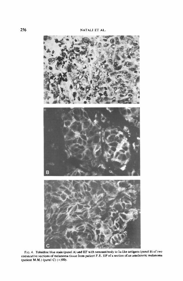
The fluorescent pattern of the membrane of melanoma cells in suspension was patchy (Fig. 2) and showed a tendency to segregate under the form of a cap in a minority of cells (Fig. 3). In most specimens the fluorescent staining of the melanoma cell membrane was uniformly bright independently of the melanine content of the cells (Fig. 4); however, in some patients, such as patient G.L., a variable intensity of the stain was observed in the tumor tissue.
Ia-like antigen-bearing cells were detected in primary, recurrent, and metastatic lesions; in the patient L.A. the primary and metastatic lesions were surgically removed at the same time. Both types of lesions were stained by xenoantibodies to Ia-like antigens.
The percentage of Ia-like antigen-bearing melanoma cells in cell suspensions obtained from 10 patients was determined by parallel observation by ultraviolet light and light microscopy: criteria to identify melanoma cells were cell dimension, nuclear morphology, and presence of characteristic pigment granules, the latter being especially useful to differentiate melanoma cells from pigment-loaded mac- rophages. The percentage of Ia-like antigen-bearing cells in each melanoma cell suspension showed wide variations ranging from 15 to 90%. The lack of staining of a certain number of melanoma cells in each tissue specimen studied occurred with both monoclonal antibodies and operationally specific xenoantisera to Ia-like
FIG. I. Ability of melanoma cells to synthesize in vitro Ia-like antigens following their removal by treatment with pronase. Enzyme treatment (30 mitt at 37°C) of a melanoma cell suspension prepared from patient P.I. reduces the percentage of Ia-like-bearing cells from 27% in the untreated cell suspension (H) to 6% in the enzyme-treated cell suspension (0) (panel A). Following removal of pronase and three washings, cells were cultured at 37°C for 16 hr. The percentage of Ia-like antigens bearing cells in the pronase-treated melanoma cell suspension (0) is similar to that in the untreated cell suspension (m).

NATAL1 ET AL.
FIG. 2. Distribution of la-like antigens on melanoma cells isolated from a primary lesion (I P.I.). A single melanoma cell which by light microscopy shows characteristic cytoplasmic granules (panel A), shows a tine-speckled fluorescence of the plasma membrane when reacted with anti-Ia-like antigen xenoantibodies (panel B) (x800).
antigens, even if concentrated or diluted antibodies were used. The percenta ,ge of Ia-like antigen-bearing cells did not correlate with the stage of the neopl lastic lesion, since primary, recurrent and metastatic tumors contained variable per- centages of Ia-like antigen-bearing cells without any significant pattern o f dis- tribution. However, a low percentage of Ia-like antigen-bearing cells (ranging from
)atient black in IIF

of 1 ZIG 3. Antibody induced capping of Ia-like antigens (white arrow: insert) on the membrane :hn ee melanoma cells with cytoplasmic dark granules (black arrow) (x600).
Ia-LIKE ANTIGENS ON HUMAN MELANOMA 255
oft me
27 to 63%) was found in primary lesions with high degree of invasivity as judged by Clark’s staging, and in lesions from patients with a massive spreading.
None of the 14 nevic lesions studied was stained by the monoclonal antibodies and by the operationally specific xenoantisera to Ia-like antigens; only isolated cells in the dermis were fluorescent and a representative example is given in Fig. 5.
DISCUSSION
This study has shown that freshly explanted melanoma cells can specifically react with operationally specific xenoantisera and monoclonal antibodies to framework antigenic determinants of Ia-like antigens in indirect immunofluores- cence. The reaction is specific since it was observed with monoclonal antibodies to Ia-like antigens, which exclude the possible interference of contaminating an- tibodies one has to take into account when using conventional polyclonal antisera. Furthermore no staining of melanoma tissue was observed when anti Ia-like anti- gen antibodies had been specifically removed from their source by absorption with Ia-like antigen-bearing,cells.
Ia-like antigens are present in plasma in an immunologically functional form (9); therefore one has to take into account that Ia-like antigens detected on melanoma cells may be passively absorbed from plasma. However, two lines of evidence argue against this possibility: Ia-like antigens on melanoma cell membrane under- went cap formation following reaction with antibodies, suggesting that these gly- coproteins are integral components of the melanoma cell membrane. Melanoma
256 NATAL1 ET AL.
FIG. 4. To11 consec :uti\ re SC (patier lt M [.M.
” :c idine blue stain (panel A) and IIF with xenoantibody to Ia-like antigens :tions of melanoma tissue from patient F.E. IIF of a section of an amek (panel C) (x 500).
(P lane1 B) of two m( Xic melanoma

Ia-LIKE ANTIGENS ON HUMAN MELANOMA 257
FIG. 5. Toluidine blue staining (panel A) and IIF with xenoantibody to Ia-like antigens (panel B) of two consecutive sections of an intradermal nevus.
cells can synthesize Ia-like antigens, as they reappeared on melanoma cells cul- tured in vitro following removal of antigens by treatment with pronase.
In each tumor studied a certain percentage of cells did not express la-like antigens: this finding does not reflect a prozone effect (12) or an insufficient amount of antibody, since no staining of melanoma cells was detected using dif- ferent concentrations of anti-Ia-like antigen antibodies. The melanoma cells

NATAL1 ET AL.
showing no reaction with anti-Ia-like antigen antibodies in each lesion may repre- sent a set of cells which are unable to synthesize these antigens or may be cells in a phase of their growth cycle associated with low expression of Ia-like antigens, if their expression is cell cycle dependent.
The expression of Ia-like antigens on melanoma cells is not correlated with the clinical stage of the tumor; however, a role of Ia-like antigens expressed by melanoma cells in the biology of the tumor is suggested by three lines of evidence: (i) a low number of Ia-like antigen-bearing cells was found in a limited number of primary lesions with high degree of invasivity and in lesions from patients with massive spreading of the disease; (ii) the antigenicity of leukemia-associated anti- gens in the guinea pig system is dependent on expression of Ia-like antigens on tumor cells (13) and (iii) immune response to melanoma-associated antigens is believed to effect the clinical course of the disease (14).
Detection of Ia-like antigens on melanoma cells may have a clinical relevance since all the surgically removed melanoma lesions tested contained a variable number of Ia-like antigen-bearing cells and the appearance of Ia-like antigens on cells of the melanocyte lineage is restricted to those which have undergone malig- nant transformation. If this finding is confirmed by testing a large number of benign and malignant tissue samples, then testing of lesions of dubious histologic classification for Ia-like antigens may aid to solve controversial diagnoses which still occur in about 7% of the cases (15).
ACKNOWLEDGMENTS
This work was supported by Italian National Research Council Progetto Finalizzato “Controllo della Crescita Neoplastica” No. 780285296, No. 800151096, and by the National Institutes of Health Grants Al 13154, CA 16069, CA 16071, and CA 24329, a Fellowship of the Leukemia Society of America (V. Quaranta), a Research Career Development Award (M. A. Pellegrino), and an American Heart Association Established Investigatorship Award (S. Ferrone). The authors wish to thank Dr. G. Balivi of the ID1 Institute, Rome, for providing biopsies of nevic lesions, and Professor A. Bigotti for helpful discussions; to acknowledge the excellent technical assistance of Ms. M. R. Nicotra, the excellent photography work of Mr. M. Rosati, and the skilled secretarial assistance of Mr. D. Trinko. This is publication No. 2053 from the Department of Molecular Immunology and Scripps Clinic and Research Foundation.
REFERENCES
1. Ferrone, S., Allison, J. P., and Pellegrino, M. A.. Contemp. Top. Mol. Immunol. 7, 239, 1978. 2. Winchester, R. J., Wang, C. I.. Gibofsky. A.. Kunkel, H. G.. Lloyd, K. 0.. and Old. L. J., Proc.
Nat. Acad. SC;. USA 75, 6255, 1978. 3. Wilson, B. S., Indiveri. F.. Pellegrino, M. A., and Ferrone, S., J. Exp. Med. 149. 658. 1979. 4. Bergholtz, B. 0.. and Thorsby, E., Stand. J. Immunol. 8, 63, 1978. 5. Winchester, R. J., Ross, G. D., Jarowski, C. I., Wang, C. I.. Halper. J.. and Broxmeyer, H. E..
Proc. Nat. Acad. Sci. USA 74, 4012, 1977. 6. Sugarbaker, E. V.. and McBride, G. M., Cancer 37, 188, 1976. 7. Wilson, B. S., Indiveri, F., Pellegrino, M. A., and Ferrone, S., Transplant. Proc. 11. 712, 1979. 8. Pellegrino, M. A., Ferrone, S.. Dierich, M. P.. and Reisfeld, R. A., Clin. Immunol. Im-
munopathol. 3. 324. 1975. 9. Wilson, B. S.. Indiveri, F.. Pellegrino, M. A., and Ferrone, S., J. Immunol. 122, 1967, 1979.

la-LIKE ANTIGENS ON HUMAN MELANOMA 259
10. Quaranta, V., Walker, L. E.. Pellegrino, M. A., and Ferrone, S., J. Immunol. 125, 1421, 1980. 11. Clark, W. H. Jr., From, L., Bernardino, E. A., and Hihm, H. C., Cancer Res. 29. 705, 1969. 12. Linder, E., and Mieltimen, A., Scond. J. Immunol. 5, 513, 1976. 13. Fomi, G., Shevach, E. H., and Green, I. J. Exp. Med. 143, 1067, 1975. 14. Ferrone, S., and Pellegrino, M. A., In “Handbook of Cancer Immunodiagnosis” (H. Waters,
Ed.), Vol. 3, pp. 291-327, Garland STPM Press, New York/London. 15. Truax, H.. Barnett, R. N., Hukill, P. B., Campbell, P. C., and Eisenberg, H., Cancer 19, 1543.
1976.


![Colec]ia PLURAL](https://static.fdokumen.com/doc/165x107/63165d23c32ab5e46f0dbd8a/colecia-plural.jpg)