Ecdysteroid 7,9(11)-dien-6-ones as potential photoaffinity labels for ecdysteroid binding proteins
10
Journal of Insect Science insectscience.org Schal C, Sevala V, Capurro ML, Snyder TE, Blomquist GJ, Bagnères AG. 2001. Tissue distribution and lipophorin transport of hydrocarbons and sex pheromones in the house fly, Musca domestica. 11 pp. Journal of Insect Science, 1:12. Available online: insectscience.or g/1.12 Ecdysteroid 7,9(11)-dien-6-ones as potential photoaffinity labels for ecdysteroid binding proteins Pauline C. Bourne 1 , Pensri Whiting 1 , Tarlochan S. Dhadialla 2a , Robert E. Hormann 2b , Jean- Pierre Girault 3 , Juraj Harmatha 4 , René Lafont 5 and Laurence Dinan 1 1 Department of Biological Sciences, University of Exeter, Hatherly Laboratories, Prince of Wales Road, Exeter, Devon, EX4 4PS, U.K. 2 Rohm & Haas Co., Research Laboratories, 727 Norristown Road, Spring House, PA 19477-0904, U.S.A 3 Equipe de RMN et Modélisation Moléculaire, Université René Descartes, Laboratoire de Chimie et Biochimie Pharmacologiques et Toxicologiques, Unité Mixte de Recherche CNRS, UMR 8601, 45 rue des Saints-Pères, 75270 Paris Cedex 06, France 4 Institute of Organic Chemistry and Biochemistry, Academy of Sciences, Flemingovo nám. 2, 166 10 Prague, Czech Republic. 5 Laboratoire d’Endocrinolgie Moléculaire et Evolution, Université Pierre et Marie Curie, 7 Quai St. Bernard, 75252 Paris 05, France a Current address: Biochemistry, Dow AgroSciences LLC, 9330 Zionsville Road, Indianapolis, IN 46268, U.S.A. b Current address: RHeogene Inc., Research Labs., 727, Norristown Road, PO Box 949, Spring House, PA 19477-0904, U.S.A. [email protected] Received 2 March 2002, Accepted 10 June 2002, published ????? Abstract Three ecdysteroid 7,9(11)-dien-7-ones (dacryhainansterone, 25-hydroxydacryhainansterone and kaladasterone) were prepared by dehydration of the corresponding 11a-hydroxy ecdysteroids (ajugasterone C, turkesterone and muristerone A, respectively). The biological activities of the dienones in the Drosophila melanogaster B II cell bioassay, which reflect the affinity for the ecdysteroid receptor complex, showed that the dienones retain high biological activity. Irradiation at 350 nm of the ecdysteroid dienones (100 nM) with bacterially- expressed dipteran and lepidopteran ecdysteroid receptor proteins (DmEcR/DmUSP or CfEcR/CfUSP), followed by loading with [ 3 H]ponasterone A revealed that irradiation of dacryhainansterone or kaladasterone resulted in blocking of >70% of the specific binding sites. Thus, ecdysteroid dienones show considerable potential as photoaffinity analogues for ecdysteroid binding proteins. Keywords: ecdysteroid receptor, affinity labelling, Ultraspiracle, Quantitative Structure-Activity Relationship, Comparative Molecular Field Analysis, 20-hydroxyecdysone, dacryhainansterone Abbreviations: 20E 20-hydroxyecdysone ax axial Cf Choristoneura fumiferana CI-MS chemical ionization mass spectrometry CoMFA comparative molecular field analysis DCC dextran-coated charcoal Dm Drosophila melanogaster EcR ecdysteroid receptor protein eq equatorial FAB-MS fast-atom bombardment mass-spectrometry HPLC high-performance liquid chromatography HR-MS high-resolution mass-spectrometry LBD ligand-binding domain LSIMS liquid secondary-ion mass-spectrometry MS mass-spectrometry NMR nuclear magnetic resonance ponA ponasterone A QSAR quantitative structure-activity relationship RI relative intensity USP Ultraspiracle protein UV ultra-violet. Introduction Affinity labelling is an important method in elucidating signal transduction mechanisms (Dorman, 2000) and has proved to be a powerful method for the characterisation of vertebrate steroid binding proteins, including intracellular receptors, transport proteins and enzymes (Katzenellenbogen and Katzenellenbogen, 1984; Gronemeyer and Govindan, 1986; Gronemeyer, 1988). These studies have provided information about the points of attachment, the orientation of the ligand in the binding site and guidance to direct
-
Upload
independent -
Category
Documents
-
view
0 -
download
0
Transcript of Ecdysteroid 7,9(11)-dien-6-ones as potential photoaffinity labels for ecdysteroid binding proteins

Journalof
Insect
Science
insectscience.org
Schal C, Sevala V, Capurro ML, Snyder TE, Blomquist GJ, Bagnères AG. 2001. Tissue distribution and lipophorintransport of hydrocarbons and sex pheromones in the house fly, Musca domestica. 11 pp. Journal of Insect Science,1:12. Available online: insectscience.org/1.12
Ecdysteroid 7,9(11)-dien-6-ones as potential photoaffinity labels for ecdysteroidbinding proteins
Pauline C. Bourne1, Pensri Whiting1, Tarlochan S. Dhadialla2a, Robert E. Hormann2b, Jean-Pierre Girault3, Juraj Harmatha4, René Lafont5 and Laurence Dinan1
1Department of Biological Sciences, University of Exeter, Hatherly Laboratories, Prince of Wales Road, Exeter, Devon, EX4 4PS, U.K.2Rohm & Haas Co., Research Laboratories, 727 Norristown Road, Spring House, PA 19477-0904, U.S.A
3Equipe de RMN et Modélisation Moléculaire, Université René Descartes, Laboratoire de Chimie et Biochimie Pharmacologiques etToxicologiques, Unité Mixte de Recherche CNRS, UMR 8601, 45 rue des Saints-Pères, 75270 Paris Cedex 06, France
4Institute of Organic Chemistry and Biochemistry, Academy of Sciences, Flemingovo nám. 2, 166 10 Prague, Czech Republic.5Laboratoire d’Endocrinolgie Moléculaire et Evolution, Université Pierre et Marie Curie, 7 Quai St. Bernard, 75252 Paris 05, France
aCurrent address: Biochemistry, Dow AgroSciences LLC, 9330 Zionsville Road, Indianapolis, IN 46268, U.S.A.bCurrent address: RHeogene Inc., Research Labs., 727, Norristown Road, PO Box 949, Spring House, PA 19477-0904, U.S.A.
Received 2 March 2002, Accepted 10 June 2002, published ?????
Abstract
Three ecdysteroid 7,9(11)-dien-7-ones (dacryhainansterone, 25-hydroxydacryhainansterone and kaladasterone) were prepared bydehydration of the corresponding 11a-hydroxy ecdysteroids (ajugasterone C, turkesterone and muristerone A, respectively). The biologicalactivities of the dienones in the Drosophila melanogaster B
II cell bioassay, which reflect the affinity for the ecdysteroid receptor complex,
showed that the dienones retain high biological activity. Irradiation at 350 nm of the ecdysteroid dienones (100 nM) with bacterially-expressed dipteran and lepidopteran ecdysteroid receptor proteins (DmEcR/DmUSP or CfEcR/CfUSP), followed by loading with[3H]ponasterone A revealed that irradiation of dacryhainansterone or kaladasterone resulted in blocking of >70% of the specific bindingsites. Thus, ecdysteroid dienones show considerable potential as photoaffinity analogues for ecdysteroid binding proteins.
Keywords: ecdysteroid receptor, affinity labelling, Ultraspiracle, Quantitative Structure-Activity Relationship, Comparative MolecularField Analysis, 20-hydroxyecdysone, dacryhainansterone
Abbreviations:20E 20-hydroxyecdysoneax axialCf Choristoneura fumiferanaCI-MS chemical ionization mass spectrometryCoMFA comparative molecular field analysisDCC dextran-coated charcoalDm Drosophila melanogasterEcR ecdysteroid receptor proteineq equatorialFAB-MS fast-atom bombardment mass-spectrometry
HPLC high-performance liquid chromatographyHR-MS high-resolution mass-spectrometryLBD ligand-binding domainLSIMS liquid secondary-ion mass-spectrometryMS mass-spectrometryNMR nuclear magnetic resonanceponA ponasterone AQSAR quantitative structure-activity relationshipRI relative intensityUSP Ultraspiracle proteinUV ultra-violet.
Introduction
Affinity labelling is an important method in elucidating signaltransduction mechanisms (Dorman, 2000) and has proved to be apowerful method for the characterisation of vertebrate steroid
binding proteins, including intracellular receptors, transport proteinsand enzymes (Katzenellenbogen and Katzenellenbogen, 1984;Gronemeyer and Govindan, 1986; Gronemeyer, 1988). These studieshave provided information about the points of attachment, theorientation of the ligand in the binding site and guidance to direct

Schal C, Sevala V, Capurro ML, Snyder TE, Blomquist GJ, Bagnères AG. 2001. Tissue distribution and lipophorin transport of hydrocarbons andsex pheromones in the house fly, Musca domestica. 11 pp. Journal of Insect Science, 1:12. Available online: insectscience.org/1.12
2
site-directed mutagenesis studies. In contrast, the steroid hormonereceptor of arthropods has received far less attention in respect tothe identification of potential affinity labels. This is remarkableconsidering that arthropods comprise 95% of all animal species andthat insights into ecdysteroid binding would provide usefulinformation for understanding ecdysteroid-regulated gene controland identifying new possibilities for insect pest control.
Ecdysteroid receptor genes have been characterised from insects(e.g. Saleh et al., 1998, and references cited therein), crustaceans(Durica et al., 1999) and ticks (Palmer et al., 1999). The receptorcomplex consists of two major polypeptides, an EcR protein and aUSP/RXR protein. Both are members of the nuclear receptorsuperfamily. The best characterised proteins are those of Drosophilamelanogaster, where 3 isoforms of EcR are present which arise fromdifferent promoters (isoforms A and B) and alternative splicing(isoforms B1 and B2) (Koelle et al., 1991). There may also beisoforms of USP (Vögtli et al., 1999). The natural ligand ofecdysteroid receptors is generally assumed to be 20-hydroxyecdysone (1), but other ligands may be important in certainspecies or at particular stages of development (Wang et al., 2000).The ecdysteroid binds to the ligand-binding domain of the EcRprotein, but only with high affinity when this is complexed withUSP (Yao et al., 1993). It is currently not known with any certaintywhether USP recognises a ligand, although it has been suggestedthat juvenile hormone may bind to this protein (Jones and Jones,2000). In spite of extensive structure-activity relationship studiesfor ecdysteroids (Dinan et al., 1999; Ravi et al., 2001), the orientationand interactions of the ligand within the LBD of EcR remainunknown. The necessity for interaction between EcR and USP forhigh-affinity ligand binding complicates crystallographic studies onthe ligand binding domain of EcR, although it has recently beenpossible to determine the crystal structure of the LBD of USP fromHeliothis virescens (Billas et al., 2001). Recent modelling and virtualdocking experiments (Wurtz et al., 2000) with 20E and RH5849 (anon-steroidal ecdysteroid agonist; Dhadialla et al., 1998) with modelsof the Chironomus tentans EcR ligand binding domain based on thecrystal structures of the human vitamin D receptor and the humanretinoic acid receptor LBDs provide conflicting information aboutthe orientation of the ecdysteroid in the binding pocket.Consequently, the generation of an efficient ecdysteroid affinity labelwould provide important information on the orientation of the ligandand perhaps also facilitate crystallographic studies.
Previous approaches to affinity labelling of ecdysteroid receptors/binding proteins have comprised photoaffinity labelling based onactivation of the natural 14α-hydroxy-7-en-6-one (λ
max = 242nm)
of ecdysteroids (Gronemeyer and Pongs, 1980; Schaltmann andPongs, 1982) or electrophilic labelling based on brominated side-chain analogues (Strangmann-Diekmann et al., 1990). The formermethod has the advantage of using natural arthropod ecdysteroids(such as 1), but appears to suffer the disadvantage of poor cross-linking yield (ca. 5%), probably as a consequence of irradiation at awavelength (254 nm) at which the receptor proteins also absorb.No further reports of the application of the electrophilic affinityecdysteroid analogues have appeared. The ideal affinity labellingreagent should possess i) a high affinity for the binding protein, ii)
a high cross-linking yield, iii) low non-specific binding and iv) thecapacity to be detected (e.g. by radiolabelling) without detriment tothe other three criteria. Extension of the natural chromophore ofecdysteroids to a dien-6-one (7,9(11) or 7,14) would shift the λ
max
to longer wavelength (ca. 300 nm). The natural occurrence of 11α-hydroxy ecdysteroids in certain plant species (especially Ajuga spp.;Imai et al., 1969; Canonica et al., 1973; Usmanov et al., 1975),provides precursors for the generation of 7,9(11)-dien-6-ones bydehydration (Canonica et al., 1977; Qui and Nie, 1983). Here wereport the simple synthesis in good yield of ecdysteroid 7,9(11)-dien-6-ones from the corresponding 11α-hydroxy ecdysteroid,demonstrate that the dienones retain high biological activity andprovide indirect evidence that they are capable of covalently linkingto ecdysteroid receptors in high yield on irradiation with longwavelength UV light (300 or 350 nm).
Experimental
General
Ajugasterone C (2) was isolated from Leuzea carthamoides (Píš etal., 1994). Muristerone A (3) was purchased from Simes spa, Milan,Italy. Turkesterone (4) was a generous gift from Ziyadilla Saatov.[24,25,26,27-3H]Ponasterone A (25-deoxy-20-hydroxyecdysone;150 Ci/mmol) was obtained from American Radiochemicals Inc.(www.arc-inc.com).
Ultra-violet spectra were obtained in methanol using a Shimadzu(www.shimadzu.com) UV-2401 PC, UV-VIS dual beamspectrophotometer. Nuclear magnetic resonance (NMR)spectroscopy was performed i) on a Bruker (http://www.Bruker.com)AVANCE DRX400 instrument (in Exeter, UK) using standard Brukermicroprograms with samples dissolved in CD3OD or ii) on a BrukerAMX500 instrument (in Paris, France) at 300 K, where sampleswere lyophilised and dissolved in D2O. TSPd4, 3-(trimethylsilyl)[2,2,3,3-d4] propionic acid, sodium salt, was usedas internal reference for the proton and carbon shifts. Chemical shiftsare expressed in ppm. Sep-Pak (0.5 g) C
18 cartridges (Millipore,
www.waters.com) were used for pre-HPLC purification of thesamples. HPLC: (1) semipreparative, Gilson (www.gilson.com)model 806 HPLC coupled with Gilson UV-Visible detector, (2)analytical, Gilson model 811 HPLC coupled with Gilson 160 diodearray detector and using Gilson Unipoint computer program; C
18
Spherisorb ODS-2 semipreparative and Spherisorb 5 ODS-2analytical C
18 columns were used (both 5 µm particle size; Jones
Chromatography,(www.jones-chrom.co.uk). Chromatographicseparations were monitored at 242 and 300 nm. HPLC-grade solventswere from BDH (www.bdh.com).
The LSIMS spectrum of 6 was recorded by Mark Prescott and HuwRees at the University of Liverpool on a VG Quattro triplequadrupole mass spectrometer (VG Biotech, www.micromass.co.uk)using a Cs+ primary beam and glycerol matrix. CI-MS spectra wererecorded on a Jeol MS 700 spectrometer equipped with a directinlet probe. Spectra were recorded in the chemical ionization/desorption mode using ammonia as the reagent gas.

Schal C, Sevala V, Capurro ML, Snyder TE, Blomquist GJ, Bagnères AG. 2001. Tissue distribution and lipophorin transport of hydrocarbons andsex pheromones in the house fly, Musca domestica. 11 pp. Journal of Insect Science, 1:12. Available online: insectscience.org/1.12
3
Prediction of biological activities
The biological activities of the ecdysteroid dienones were predictedbased on the CoMFA QSAR models developed from the agonistactivities of a training set of 71 ecdysteroid analogues in the B
II
bioassay (Dinan et al., 1999).
Conversion of 11α-hydroxyecdysteroids to dienones
The ecdysteroid was dissolved in 5% w/v NaOH methanolic solution(1 ml/mg ecdysteroid reacted, typically 5 mg) and left to react atroom temperature. The reaction was quenched after 40 minutes withthe addition of 0.8 M HCl (same volume as NaOH methanolicsolution). The sample was diluted to 10% MeOH by the addition ofH
20 and this was applied to a C
18 Sep-Pak cartridge (0.5 g). The
sample was eluted with 10% and 100% MeOH (5 ml of each). The100% MeOH fraction was analysed on a C
18 analytical column
(ODS2, 25 cm x 4.6 mm i.d., 5 µm particle size, 1:1 v/v MeOH:H20,
1ml/min), initially with the photodiode-array detector monitoringat 242 nm and 300 nm. This indicated further purification wasnecessary and this was carried out using a C
18 semi-preparative
column (ODS2, 25 cm x 9.6 mm i.d., 5 µm particle size, MeOH:H20,
1:1 v/v, 2 ml/min) with detection at 300 nm. The purified productwas dried and subjected to 1H and 13C NMR. In this way, ajugasteroneC (2) was converted to dacryhainansterone (5), muristerone A (3)to kaladasterone (6) and turkesterone (4) to 25-hydroxydacryhainansterone (7) (Fig. 1).
Dacryhainansterone (5): UV: MeOH (λmax = 299 nm: ε = 14200L/mol/cm), H2O (λmax = 307 nm: ε = 12900 L/mol/cm);
1H NMR:
δH(CD3OD): 6.28 (1H, apparent d, H-11); 5.75 (1H, s, H-7); 3.84(1H, m, H-3); 3.73 (1H, m, H-2); 3.35 (1H, d,d, 11, 2, H-22); 2.75(1H, broad d, 18, H-12ax); 2.5 (1H, H-12eq); 2.48 (1H, H-17); 2.45(1H, H-5); 2.1 (1H, H-1eq); 2.05 (1H, H-16α); 2.0 (1H, H-15 α);1.8 (1Η, Η−4eq); 1.75 (1H, H-16β); 1.7 (1H, H-1ax); 1.6 (1H, H-4ax); 1.55 (1H, H-15β); 1.55 (1H, H-23b); 1.55 (1H, H-25); 1.46(1H, H-24a); 1.30 (1H, H-23a); 1.24 (1H, H-24b); 1.18 (3H, s,21Me); 1.10 (3H, s, 19-Me); 0.94 (3H, d, 6.5, 27-Me); 0.91 (3H, d,6.5, 26-Me); 0.89 (3H, s, 18-Me);
13C NMR: δc(CD3OD): 205.5
(C-6); 155 (C-8); 134.7 (C-9); 132.4 (C-11); 117.8 (C-7); 83.1 (C-14); 76.6 (C-22); 76.2 (C-20); 67.4 (C-3/2); 66.9 (C-2/3); 50.2 (C-5); 49.1 (C-17); 48.0 (C-13); 46.5 (C-10); 39.4 (C-12); 37.7 (C-24); 36.2 (C-1); 36.0 (C-4); 30.2 (C-19); 29.9 (C-23 ); 29.1 (C-15);27.8 (C-25); 22.0 (C-27); 21.3 (C-26); 20.3 (C-16); 19.3 (C-21);16.7 (C-18); CI-MS m/z (% RI): 480.7 ([M+NH
4]+, 11%), 463.6
([M+H]+, 100%), 447.6 ([M-CH3]+, 51%), 445.6 ([M+H-H
2O]+,
32%), 427 ([M+H-2H2O]+, 21%), 362.5 (74%), 345.5 ([M-C-20 →
C-27]+, 61%).
Kaladasterone (6): UV: MeOH (λmax = 299 nm: ε = 10700 L/mol/cm);
1H NMR: δH(CD3OD): 6.3 (1H, apparent d, H-11); 5.80 (1H,
s, H-7); 3.88 (1H, m, H-3); 3.83 (1H, m, H-2); 3.35 (1H, d,d, 11, 2,H-22); 2.75 (1H, broad d, 18, H-12ax); 2.46 (1H, H-12eq); 2.40(1H, H-17); 2.1 (1H, H-1eq); 2.05 (1Η, Η−4eq); 2.02 (1H, H-16α);1.95 (1H, H-15 α); 1.8 (1H, H-4ax); 1.75 (1H, H-16β); 1.7 (1H, H-1ax); 1.55 (1H, H-15β); 1.55 (1H, H-23b); 1.55 (1H, H-25); 1.45(1H, H-24a); 1.29 (1H, H-23a); 1.24 (1H, H-24b); 1.18 (3H, s, 21-
Me); 1.05 (3H, s, 19-Me); 0.94 (3H, d, 6.5, 27-Me); 0.91 (3H, d,6.5, 26-Me); 0.89 (3H, s, 18-Me);
13C NMR: δc(CD3OD): 201.3
(C-6); 155 (C-8); 136.3 (C-9); 132.7 (C-11); 116.3 (C-7); 82.9 (C-14); 78.97 (C-5); 76.6 (C-22); 76.2 (C-20); 68.7 (C-3/2); 67.0 (C-2/3); 49.0 (C-17); 46.5 (C-10); 44.8 (C-12); 37.7 (C-24-); 36.2 (C-1);33.0 (C-4); 29.94 (C-23); 29.1 (C-15); 27.8 (C-25); 24.97 (C-19);22.0 (C-27); 21.3 (C-26); 20.3 (C-16); 19.3 (C-21); 16.7 (C-18);LSIMS (-ve ion-mode) m/z (%RI): 477 ([M-H]
-, 80%), 460 ([M-
H2O]-, 36%), 445 ([M-H2O-CH3]
-, 25%), 183 (100%).
25-Hydroxydacryhainansterone (7): UV: MeOH (λmax = 299 nm:ε = 10700 L/mol/cm);
1H NMR: δH(CD3OD): 6.3 (1H, apparent t,
H-11); 5.75 (1H, s, H-7); 3.84 (1H, m, H-3); 3.71 (1H, m, H-2);3.35 (1H, d,d, 11, 2, H-22); 2.71 (1H, broad d, 18, H-12ax); 2.5(1H, H-12eq); 2.46 (1H, H-17); 2.06 (1H, H-1eq); 2.05 (1H, H-16α); 1.98 (1H, H-15 α); 1.83 (1H, H-24a); 1.78 (1Η, Η−4eq);1.75 (1H, H-16β); 1.73 (1H, H-1ax); 1.67 (1H, H-23a); 1.65 (1H,H-4ax); 1.60 (1H, H-15β); 1.45 (1H, H-24b); 1.29 (1H, H-23b);1.21 (3H, s , 26-Me); 1.20 (3H, s , 27-Me); 1.19 (3H, s, 21-Me);1.10 (3H, s, 19-Me); 0.90 (3H, 18-Me);
13C NMR: δc(CD3OD):
205.5 (C-6); 155 (C-8); 134.7 (C-9); 132.3 (C-11); 117.8 (C-7); 83.1(C-14); 77.1 (C-22); 76.3 (C-20); 69.9 (C-25); 67.4 (C-3/2); 66.9(C-2/3); 50.17 (C-5); 49.1 (C-17); 46.6 (C-10); 40.96 (C-24); 39.4(C-12); 35.97 (C-1); 34.3 (C-4); 30.2 (C-19); 29.99 (C-15); 28.3(C-27); 27.6 (C-26); 26.0 (C-23); 20.3 (C-16); 19.4 (C-21); 16.7(C-18); CI-MS m/z (% RI): 496.7 ([M+NH
4]+, 1%), 479 ([M+H]+,
9%), 463.6 ([M-CH3]+, 15%), 461.6 ([M+H-H
2O]+, 12%), 445 ([M-
CH3-H
2O]+, 7%), 443 ([M+H-2H
2O]+, 6%), 427 ([M+H-3H
2O]+, 8%),
362.5 (64%), 345.5 ([M-C-20 → C-27]+, 45%), 116.2 ([C-22 → C-27]+, 100%).
Preparation of podecdysone B
Podecdysone B (8) was prepared by thermal decomposition of 20E(100 mg) adsorbed on basic alumina (Woelm B, activity grade I; 5g), heated at 140 °C for 3 hours. The reaction product was elutedfrom the alumina with methanol and then the solvent evaporated.The residue was separated by column chromatography on silica gelwith chloroform/methanol (95:5 v/v). The major fraction was furtherpurified by NP-HPLC on a Separon (Tessek) column (7 µm particlesize; 250 mm x 8 mm i.d.) eluted with n-hexane/ethanol/water(938:60:2 v/v/v) at a flow-rate of 2 ml/min. Compound 8 (40 mg)was obtained as an amorphous solid.
Podecdysone B (8): IR (KBr), νmax
(cm-1): 3415 (OH), 1711 (C=O),1652 (C=C), 1056 (C-OH); UV (MeOH), λ
max: 244 nm, 302 nm;
1H
NMR: δH (D2O): 1.02 (3H, s, 18-CH3); 1.03 (3H, s, 19-CH3); 1.21(3H, s, 26-CH3); 1.23 (3H, s, 27-CH3); 1.29 (3H, s, 21-CH3); 1.33(1H, 23Ha); 1.49 (1H, 24-Hb); 1.53 (1H, 1-Hax); 1.58 (1H, 12-Hax); 1.61 (1H, 23-Hb); 1.74 (1H, d,t,13.6; 3.7, 4-Heq); 1.78 (1H,24-Ha); 1.86 (1H, t,d,13.6; 2.5, 4-Hax); 2.01 (1H, dd, 13.7, 3.4, 1-Heq); 2.09 (1H, d,d, 11; 7.6, 17-H); 2.23 (1H, d,d, 16; 11, 16-H);2.26 (1H, 11-H); 2.31 (1H, d,d, 13; 4.9, 12-Heq); 2.40 (1H, broadm, 11-H); 2.42 (1H, d,d, 13.2; 3.8, 2.85-Hb); 2.54 (1H, d,d, 16; 11,16-H); 2.82 (1H, d,d, 22; 2.8, 7-Ha); 3.46 (1H, d, 9.4, 22-H); 3.42(1H, d, 22., 7-Hb); 3.61 (1H, dt,12.5; 3.5, 2-Hax); 4.04 (1H, m w1/2 = 8, 3-Heq); 5.55 (1H, broad m, w1/2 = 6, 15-H);
13C NMR: δC
Schal C, Sevala V, Capurro ML, Snyder TE, Blomquist GJ, Bagnères AG. 2001. Tissue distribution and lipophorin transport of hydrocarbons andsex pheromones in the house fly, Musca domestica. 11 pp. Journal of Insect Science, 1:12. Available online: insectscience.org/1.12
4
OH
OH
OH
HO
HO
OH
stachysterone�B�(9)
OH
HO
HO
OHOH
OH
podecdysone�B�(8)
OH
OH
OH
OH
HO
HO
OH
20-hydroxyecdysone�(1)
OR'
R''
OH
OH
HO
HO
OH
HO
OR'
R''
OH
OH
HO
HO
OH
ajugasterone�C�(2:�R'=H,�R''=H)
muristerone�A�(3:�R'�=OH,�R''=�H)
turkesterone�(4:�R'=H,�R''�=OH)
dacryhainansterone�(5:�R'=H,�R''=H)
kaladasterone�(6:�R'=OH,�R''=H)
25-hydroxydacryhainansterone�(7:�R'=H,�R''=OH)
OH
OH
OH
H
HO
HO
OH
(14α-H)2β,3β,20R,22R,25-pentahydroxy-5β-cholest-7,9(11)-dien-6-one�(10)
(14β-H)2β,3β,20R,22R,25-pentahydroxy-5β-cholest-7,9(11)-dien-6-one�(11)
�
20-hydroxyecdysone(1)���������podecydsone�B�(8)�����������stachysterone�B�(9)�
ajugasterone�C�(2)�R=’H,�R”=H� � dacryhainansterone�(5)�R’=H,�R”=H��muristerone�A�(3)�R’=OH,�R”=H�� kaladasterone�(6)�R’=OH,�R”=H��turkesterone�(4)�R’=H,�R”=OH� � 25-hydroxycacryhainansterone�(7)�R’=H,�R”=OH�
(14α-H)2β,3β,20�R,22R,25-pentahydroxy-5β-cholest-7-9(11)-dien-6-one�(10)��(14α-H)2β,3β,20�R,22R,25-pentahydroxy-5β-cholest-7-9(11)-dien-6-one�(11)��
Figure 1: Ecdysteroid structures and generation of ecdysteroid 7,9(11)-dien-6-ones.

Schal C, Sevala V, Capurro ML, Snyder TE, Blomquist GJ, Bagnères AG. 2001. Tissue distribution and lipophorin transport of hydrocarbons andsex pheromones in the house fly, Musca domestica. 11 pp. Journal of Insect Science, 1:12. Available online: insectscience.org/1.12
5
(D2O): 19.7 (C-18); 21.6 (C-21); 25.2 (C-11); 28.5 (C-23); 29.8(C-26); 31.1 (C-27); 30.8 (C-19); 33.2 (C-16); 33.5 (C-4); 38.9 (C-1); 39.5 (C-12); 41.2 (C-7); 43.1 (C-24); 46.0 (C-10); 48.8 (C-13);55.1 (C-5); 58.6 (C-17); 69.9 (C-3); 71.3 (C-2); 74.4 (C-25); 80.1(C-22); 80.1 (C-20); 122.1 (C-15); 139.3 (C-9); 151.5 (C-14);FAB-MS, m/z (% RI): 485 ([M+Na]+, 35%), 463 ([M+H]+, 41%), 445([M-H
2O+H]+, 21%), 427 ([M-2H
2O+H]+, 30%), 367 (12%), 343
(14%), 329 (16%), 299 (29%), 279 (38%), 181 (100%); HR-MS forC
27H
43O
6 [M+H]+ found 463.30363, calculated 463.30597. These
data are in accord with those previously published (Nishimoto etal., 1988).
Preparation of stachysterone B
Stachysterone B (9) was prepared by acid treatment of 20E (1)(Harmatha et al., 2002). 20E (20 mg) were dissolved in 2 ml EtOH,and 100 µl 10N HCl was added. The mixture was left overnight atroom temperature under an atmosphere of argon, then the samplewas dried under a nitrogen flow and purified by NP-HPLC on asemi-preparative column (Zorbax-SIL, 250 mm long, 9.4 mm i.d.,flow-rate 4 ml/min) eluted with cyclohexane/isopropanol/water,100:40:3 v/v/v). Three peaks were collected (peak 1: Rt = 12.4 min;peak 2: Rt = 14.8 min; peak 3 = 16.0 min). These three peaks werefurther purified using dichloromethane/isopropanol/water,125:30:1.5 v/v/v). Peak 1 (Rt = 11.6 min; 4.5 mg) was identified aspodecdysone B (8). Peak 2 separated into 2 compounds: Peak 2-1(Rt = 14.4 min; 10; (14α-H)2β,3β,20R,22R,25-pentahydroxy-5β-cholest-7,9(11)-dien-6-one; 0.75 mg) and Peak 2-2 (Rt = 17.6 min;11; (14β-H)2β,3β,20R,22R,25-pentahydroxy-5β-cholest-7,9(11)-dien-6-one; ca. 0.75 mg). Peak 3 yielded stachysterone B (9: Rt =14.5 min; 0.75 mg).
Stachysterone B (9): UV: MeOH (λmax = 298 nm); 1
H NMR : δH(D2O); 1.00 (3H, s, 19-CH3); 1.10 (3H, s, 18-CH3); 1.21 (3H, s,26-CH3); 1.23 (3H, s, 27-CH3); 1.28 (3H, s, 21-CH3); 1.34 (1H,23-Ha); 1.41 (1H, t,13.4, 1-Hax); 1.50 (1H, td,13, 3.5, 24-Ha); 1.62(1H, 12-H); 1.62 (1H, 23-Hb); 1.72 (1H, 4-H
ax); 1.77 (1H, 4-H
eq);
1.78 (1H, 24-Hb); 1.80 (1H, 11-Hax
); 1.87 (1H, 11-Heq
); 1.88 (1H,1-H
eq); 2.24 (1H, dd,11, 8, 17-H); 2.30 (1H, dd,16, 8, 16-Hβ); 2.31
(1H, 12-Heq
); 2.32 (1H, 5-H); 2.53 (1H, dd, 16, 11, 16-Hα); 2.77(1H, broad m, w
1/2 = 23, 9-H); 3.44 (1H, dd, 10.6, 1.4, 22-H); 3.95
(1H, dt, 11.7, 3.5, 2-Hax
); 4.05 (1H, broad m, w1/2
= 8, 3-Heq
); 6.23(1H, broad s, w
1/2 = 6.3, 7-H); 6.23 (1H, broad s, w
1/2 = 6.3, 15-H);
13C NMR: δC (D2O); 21.6 (C-21); 21.8 (C-18); 25.4 (C-19); 28.4
(C-23); 30.0 (C-26); 30.9 (C-27); 31.8 (C-16); 33.7 (C-4); 37.7 (C-1); 41.0 (C-9); 41.5 (C-10); 41.8 (C-12); 43.0 (C-24); 50.2 (C-13);52.7 (C-5); 59.5 (C-17); 69.8 (C-2); 69.9 (C-3); 74.4 (C-25); 80.0(C-20); 80.0 (C-22); 122.0 (C-7); 133.1 (C-15); 152.0 (C-14); LSIMS(-ve-ion mode, glycerol as matrix) m/z (% RI): 553 (3%,[M+glycerol-H]
-), 461 (8%, [M-H]
-), 183 (100%); CI-MS m/z (%
RI): 480 ([M+NH4]+, 1%), 463.6 ([M+H]+, 6%), 447.6 ([M-CH
3]+,
2%), 445 ([M+H-H2O]+, 1%), 429.6 ([M+H-2H
2O]+, 2%), 116.2 ([C-
22 → C-27]+, 100%).
(14α-H)2β,3β,20R,22R,25-Pentahydroxy-5β-cholest-7,9(11)-dien-6-one (10): UV: MeOH (λmax = 296 nm);
1H NMR: δH (D2O):
0.79 (3H, s, 18-CH3); 1.13 (3H, s, 19-CH3); 1.22 (3H, s, 27-CH3);
1.23 (3H, s, 26-CH3); 1.25 (3H, s, 21-CH3); 1.32 (1H, 23Hb); 1.49(1H, 24-Hb); 1.54 (1H, 4-H
ax); 1.60 (1H, 15-Hβ); 1.63 (1H, 23-
Ha); 1.65 (1H, t,13.4, 1-Hax
); 1.79 (1H, 16-Hα); 1.77 (1H, 24-Ha);1.81 (1H, 4-H
eq); 1.85 (1H, 16-Hβ); 1.95 (1H, 15-Hα); 1.96 (1H,
broad t,10, 17-H); 2.14 (1H, dd,13.4, 3.4, 1-Heq
); 2.37 (1H, dd,13.3,3.8, 5-Hβ); 2.49 (1H, broad d,18.3, 12-H
ax); 2.62 (1H, broad d, d,10,
8, 14-Hα); 2.72 (1H, dd, 18.3, 6.7, 12-Heq
); 3.42 (1H, broad d, 10.5,22-H); 3.77 (1H, dt,12.2, 3.4, 2-H
ax); 3.94 (1H, broad m, w
1/2 = 8.3,
3-Heq
); 5.71 (1H, broad s, w1/2
= 4, 7-H); 6.45 (1H, broad d, w1/2
=6.7, 11-H);
13C NMR: δC (D2O): 13.9 (C-1); 15.0 (C-18); 21.7
(C-21); 24.0 (C-16); 24.5 (C-15); 28.3 (C-23); 29.9 (C-27); 30.8(C-26); 32.2 (C-4); 32.9 (C-19); 41.8 (C-10); 43.0 (C-24); 45.2 (C-12); 52.2 (C-17); 52.8 (C-5); 54.5 (C-14); 69.4 (C-3); 70.0 (C-2);74.1 (C-25); 79.6 (C-22); 80.3 (C-20); 120.2 (C-7); 137.8 (C-11);138.5 (C-9); LSIMS (-ve-ion mode, glycerol as matrix) m/z (% RI):553 (1%, [M+glycerol-H]
-), 461 (10%, [M-H]
-), 183 (100%); CI-
MS m/z (% RI): 480.7 ([M+NH4]+, 0.3%), 463.6 ([M+H]+, 4%),
445.5 ([M+H-H2O]+, 1%), 346.5 (9%), 344.5 (5%), 116.2 ([C-22 →
C-27]+, 100%).
(14β-H)2β,3β,20R,22R,25-Pentahydroxy-5β-cholest-7,9(11)-dien-6-one (11): UV: EtOH (λmax = 298 nm);
1H NMR: δH (D2O): 1.11
(3H, s, 19-CH3); 1.180 (3H, s, 18-CH3); 1.184 (3H, s, 26-CH3);1.20 (3H, s, 27-CH3); 1.245 (3H, s, 21-CH3); 1.31 (1H, 23Ha);1.43 (1H, 4-Hax); 1.46 (1H, 24-Hb); 1.53 (1H, 23-Hb); 1.64 (1H,d,d, 13.7, 12.5, 1-Hax); 1.76 (1H, 24-Ha); 1.78 (1H, 15-Hβ); 1.80(2H, 16-Hα ,Hβ); 1.80 (1H, 4-Heq); 1.98 (1H, m, 15-Hα); 2.01(1H, t, 8.6, 17-H); 2.13 (1H, dd, 13.7, 4.2, 1-Heq); 2.33 (1H, dd,19.3, 5.2, 12-Heq); 2.38 (1H, dd, 13.2, 3.7, 5-Hβ); 2.61 (1H, m,14-Hβ); 2.64 (1H, dd, 19.3, 3.4, 12-Hax); 3.53 (1H, broad d, 10,22-H); 3.79 (1H, dt,12.5, 3.5, 2-Hax); 3.96 (1H, m w1/2 = 8, 3-Heq); 5.89 (1H, broad s, w1/2 = 4, 7-H); 6.39 (1H, t, broad, 4, 11-H);
13C NMR: δC (D2O): 21.7 (C-21); 25.5 (C-18); 26.8 (C-16);
28.4 (C-23); 29.7 (C-26); 30.6 (C-27); 31.8 (C-15); 31.9 (C-19);36.9 (C-4); 37.8 (C-1); 40.9 (C-12); 42.3 (C-10); 42.8 (C-24); 46.1(C-13); 52.4 (C-5); 52.8 (C-17); 55.2 (C-14); 69.4 (C-3); 70.0 (C-2); 74.0 (C-25); 79.2 (C-22); 80.9 (C-20); 122.5 (C-7); 137.5 (C-11); 136.3 (C-9); CI-MS m/z (% RI): 480.7 ([M+NH
4]+, 10%), 463.6
([M+H]+, 100%), 445.5 ([M+H-H2O]+, 10%), 427.5 (18%), 364.5
(70%), 345.4 (65%), 116.2 ([C-22 → C-27]+, 100%).
The cholest-7,9(11)-dien-6-one structures of compounds 10 and 11were established thanks to 1D and 2D 1H correlations (COSY andTOCSY) and 2D 1H-13C 2D PFG-HMQC and PFG-HMBCcorrelation (Girault, 1998; Girault and Lafont, 1988). For compounds10 and 11 one notes only minor modifications for the A-ring andthe side-chain with respect to 20-hydroxyecdysone and the presenceof two protons signals in the ethylenic region. The 18-CH3 signalshows in 2D 1H-13C HMBC a long-range coupling which allowsthe assignment of 12-C and consequently of 12-H which are J-coupled with the ethylenic proton signal at 6.45 ppm (1H, broad d,w1/2 = 6.7, 11-H) for 10 or 6.39 ppm (1H, t, broad, 4, 11-H) for 11.The 18-CH3 signals do not show correlation with ethylenic orhydroxylated carbons, but the presence of an additional couplingwith an sp3 carbon (assigned to 14-C) leads to the exclusion of adouble bond at positions 14-15 or 8-14. Moreover, the 19-CH3 signalshows a long-range correlation in 2D 1H-13C HMBC with an

Schal C, Sevala V, Capurro ML, Snyder TE, Blomquist GJ, Bagnères AG. 2001. Tissue distribution and lipophorin transport of hydrocarbons andsex pheromones in the house fly, Musca domestica. 11 pp. Journal of Insect Science, 1:12. Available online: insectscience.org/1.12
6
ethylenic carbon assigned to 9-C. All this evidence is in agreementwith the cholest-7,9(11)-dien-6-one structures proposed forcompounds 10 and 11.
The configuration at 14-C was established from the following points.The 14α-H configuration of 10 was established thanks to a 2DNOESY experiment where there is a strong Noe correlation of the17-H signal (α−face) with the 14-H signal and a strong Noecorrelation of the 18-CH3 of the β−face with one 12-H, which wasconsequently assigned as 12β-H (eq). Subsequently, 12β-H (eq) ledto the assignment of the 12α-H (ax) signal. (α−face) which presentsa strong Noe correlation with the 14-H signal. Moreover, no Noeare observed between the 14-H signal and the 18-CH3 signal. The14β-H configuration for 11 was established thanks to a 2D NOESYexperiment where one observes a strong Noe for the 18-CH3 and15β−H signals (both on the β−face) with the 14-H signal. Moreover,no Noe is observed between the 14-H signal and 17-H signal of theα−face.
Moreover, the chemical shifts observed for the 18-CH3 of 10 (0.79
ppm, αH) and 11 (1.18 ppm 14β-H) are in good agreement with thedownfield shift observed for 18-CH
3 of 5β,14α-androstane (0.692
ppm) with respect to 5β,14β-androstane (0.992 ppm) (Jackman andSternell, 1969).
Bioassay and receptor assays
The agonist and antagonist biological activities of the products weredetermined with a microplate-based bioassay using the Drosophilamelanogaster B
II cell line as described previously (Clément et al.,
1993). Prior to bioassay, purity of all ecdysteroid samples wasassessed by HPLC and, if necessary, purified to >98%. Theecdysteroid receptor affinities of the three 7,9(11)-dien-6-ones wereassessed in an in vitro receptor assay using crude extracts ofbacterially-expressed DmEcR/DmUSP and CfEcR/CfUSP, asdescribed previously (Dinan et al., 2001).
UV-Irradiation
Samples were irradiated in quartz vessels using a Rayonet Mini-Photochemical reactor (RMR 600; Southern New EnglandUltraviolet Co., Branford, CT, U.S.A). The dienones 5 – 7 (100nM) were incubated with the EcR and USP proteins (as above) at 7oC and allowed to reach equilibrium (3 hours for CfEcR/CfUSP and16 hours DmEcR/DmUSP). Samples were transferred to quartz tubesand placed in a merry go-round to ensure even exposure to the UVlight sources. The irradiation apparatus was operated in a refrigeratedroom (temperature maintained at 7 oC) and the use of an internalcooling fan ensured a large rise in temperature did not occur duringthe irradiation process. Samples were irradiated for 2.5 minutes at300 nm or 20 minutes at 350 nm. Fatty-acid-free bovine serumalbumin (0.4 mg) and dextran-coated charcoal (25 µl: 500 mg acid-washed charcoal + 50 mg T70 dextran in 20 ml 10 mM Tris/HClbuffer, pH 7.2) was added to each sample and incubated for 5 minutesin ice-cold water before centrifugation at 13,000 x g for 5 minutes.The supernatant from each sample was incubated with [3H]pon A(2.6 nM) at 7 oC and allowed to reach equilibrium. A second DCC
assay was completed and aliquots of the supernatant (100 µl) wereradioassayed after the addition of 1 ml scintillation cocktail(Emulsifier-Safe; Packard Bioscience,www.lifesciences.perkinelmer.com).
Results and Discussion
CoMFA predictions
Biological activity predictions for a range of ecdysteroid dienoneswere obtained using two CoMFA models based on a training set of71 ecdysteroids (Dinan et al., 1999). Both models utilised the region-focussed electrostatic indicator field, but Model B also used theregion-focussed steric indicator field. There is very good correlationbetween the predicted and observed EC
50 values for the training set
in both models (A: r2 = 0.903; B: r2 = 0.892), while the predictioncapacity of models was found to be good (A: q2 = 0.631; B: q2 =0.694; Dinan et al., 1999). EC
50 predictions are presented in Table
1. For the 7,9(11)-dien-6-ones (5 - 7), the models are not inagreement, in terms of either the level or order of activity. However,both models predict that the activities of these dienones will behigher than that of 20E (1), which has an EC
50 of 7.5 x 10⁻⁹M. Both
models predict a similarly high activity for the 8,14-dien-6-one,podecdysone B (8), but in reality this compound has a very lowactivity. The UV-spectrum of this compound (λ
max = 244 nm) also
precludes it from consideration as a photoaffinity ligand. Bothmodels predict that the activity of stachysterone B (9), a 7,14-dien-6-one, would be similar to those of the 7,9(11)-dien-6-ones.
The dehydration reaction
The three ecdysteroids used for the dehydration reaction (2 - 4)have all been isolated from plant sources (Usmanov et al., 1975;
� � � � � � Predicted�EC50� � Determined�EC50��Ecdysteroid�(λmax�nm#)� � � Model�A� Model�B� �1:�20-hydroxyecdysone�(242)�� � � � � � � 7.5x10-
9M*�2:�ajugasterone�C�(243)� � � � � � � � 8.0x10-
8M*�3:�muristerone�A�(236)� � � � � � � � 2.2x10-
8M*�4:�turkesterone�(244)� � � � � � � � � 1.3x10-
6M*�5:�dacryhainansterone�(235�&�299)� � 1.8x10⁻⁹M� 5.6x10⁻¹¹M� � 5.2x10-
9M�6:�kaladasterone�(299)�� � � 1.2x10⁻⁹M� 2.6x10⁻¹⁰M� � 3.4x10-
7M�7:�25-hydroxydacryhainansterone�(299)� 3.0x10⁻⁹M� 5.9x10⁻¹¹M� � 2.6x10-
7M�8:�podecdysone�B�(244)� � � 4.6x10⁻¹¹M� 1.1x10⁻¹¹M� � 1.2x10-
5M�9:�stachysterone�B�(298)� � � 2.0x10⁻⁹M� 2.2x10⁻¹⁰M� � 8.2x10-
8M�10:�(14α-H)7,9(11)-dien-6-one�(296)�� � � � � � 2.2x10-
7M�11:�(14β-H)7,9(11)-dien-6-one�(298)�� � � � � � 1.3x10-
6M��*�Data�taken�from�Dinan�et�al.,�1999�#�in�methanol��
Table 1: Predicted and determined biological activities (EC50
values) ofrelevant ecdysteroids in the ecdysteroid agonist version of the Drosophila
melanogaster BII cell bioassay.

Schal C, Sevala V, Capurro ML, Snyder TE, Blomquist GJ, Bagnères AG. 2001. Tissue distribution and lipophorin transport of hydrocarbons andsex pheromones in the house fly, Musca domestica. 11 pp. Journal of Insect Science, 1:12. Available online: insectscience.org/1.12
7
Canonica et al., 1972; Imai et al., 1969), as have two of the intendeddehydration products (5 [Hou et al., 1982] and 6 [Canonica et al.,1973]). Dacryhainansterone (5) has also been synthesised previouslyin a dehydration reaction from ajugasterone C (Qui and Nie, 1983;Szendrei et al., 1988). Dehydration reactions were monitored byUV-spectrophotometry and a shift in the λ
max from 242 nm to 300
nm indicated the formation of the conjugated dienone. Optimumyields occurred after 40 min and no further increase was noted upto 2 h. HPLC analysis indicated the presence of small quantities ofunreacted starting material remained, so purification was necessary.Overall, the three reactions proceeded cleanly and produced yieldsin excess of 60%.
Spectroscopic identification of the 14α-hydroxy conjugated dienones
In addition to the characteristic UV-spectra, the dienone productswere identified unambiguously by 1H and 13C NMR. NMR data forthe three parent ecdysteroids have been published previously alongwith 1H and 13C NMR data for 5 (Lafont and Wilson, 1996; Volodinet al., 1998), allowing direct comparisons with the data for thedehydration products. The introduction of the 9(11)-double bondresulted in a loss of the signal for H-9 (δ3.1) and a large downfieldshift for the H-11 signal (from δ4.1 to δ6.3) in the 1H spectra.Similarly, large shifts in the 13C spectra were observed for both C-9(from δ43 to δ135) and C-11 (from δ69 to δ132). NMR data aresummarised in the Experimental section.
Acid treatment of 20-hydroxyecdysone
In addition to the expected products of podecdysone B andstachysterone B, this reaction yielded the previously unknownecdysteroids 10 and 11. We hypothesize these compounds arise bysequential rearrangements of the diene following dehydration at C-14: 7,14-dien-6-one (stachysterone B; 9) → 8(9),14-dien-6-one(podecdysone B; 8) → 8(14),9(11)-dien-6-one (hypotheticalintermediate) → (14α-H)7,9(11)-dien-6-one (10) + (14β-H)7,9(11)-dien-6-one (11).
Biological activity
BII bioassay data (Table 1) demonstrate that the three 14α-hydroxy
conjugated dienones all displayed high levels of agonist activity inthe sequence dacryhainansterone > 25-hydroxydacryhainansterone> kaladasterone. Both dacryhainansterone and 25-hydroxydacryhainansterone displayed higher activities than the 11α-hydroxyecdysteroids from which they were derived, but the conversewas true for kaladasterone. The activity curve for kaladasteroneappears bi-phasic when compared to the other ecdysteroids (Fig.2). Although the activity levels were lower than those predicted byboth CoMFA models, the order of activity was correctly predictedby model B and dacryhainansterone (5) had a slightly higher activitythan 20E (1). It is interesting to note that earlier structure-activityrelationship studies suggested the presence of a 5β-hydroxyl groupand the absence of a C-25 hydroxyl group should potentiatebiological activity (Dinan, 1989), and the CoMFA data from ModelA predicted that kaladasterone should display the highest biologicalactivity. However, kaladasterone actually exhibited the lowest EC
50
value, suggesting that the presence of a 5β-hydroxyl together with
the absence of a 25-hydroxyl somehow result in an interaction, whichis detrimental to activity. Further evidence for this can be seen with25-deoxypolypodine B (Dinan et. al., 1999). Polypodine B containsboth 5β and C-25 hydroxyl groups and has an EC
50 in the B
II bioassay
of 1.0 x 10⁻⁹M. 25-Deoxypolypodine B lacks the C-25 hydroxylgroup, but is otherwise structurally identical to polypodine B andsubsequently a higher EC
50 could be assumed. However the EC
50 of
this compound was actually 10-fold lower (1.0 x 10⁻⁸M). Thediscovery of such an interaction may also explain the bi-phasic effectseen in the biological activity curve for kaladasterone. Absence of a14α-hydroxyl group (compound 10) did not affect the biologicalactivity significantly (compare 7 and 10), which is in accord withrecent findings (Harmatha et al., 2002) which showed that theconfiguration at C-14 is more important than the substituent (14-H,14-OH or 14-OOH; compare 10 and 11).
Figure 2: Concentration-dependence of agonist activity of selectedecdysteroids in the Drosophila melanogaster B
II cell bioassay: 20-
hydroxyecdysone (1: ), dacryhainansterone (5: à), kaladasterone (6: +), 25-hydroxydacryhainansterone (7: *), podecdysone B ( 8: ´) and stachysterone B
(9: ∆). Data points are the means of 4 replicates.
Both CoMFA models dramatically overpredict the activity ofpodecdysone B, and also overpredict the activity of stachysteroneB. This probably arises from the lack of structural variety in theregion of C-8 to C-11 amongst the members of the CoMFA trainingset.
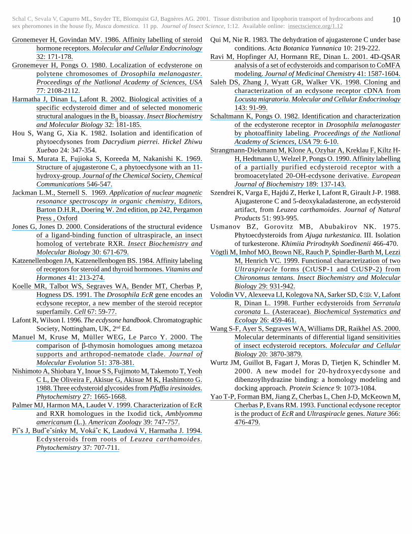
Stability of receptor complexes to irradiation with UV lightEcdysteroid receptor complexes (CfEcR/CfUSP) were irradiatedwith UV light at 300 or 350 nm and the level of specific bindingcapacity for [3H]pon A (2.6 nM) remaining was determined (Fig. 3).Irradiation at 350 nm for 20 min left 87% of the specific bindingcapacity intact, while irradiation at 300 nm resulted in substantialdegradation even by 5 min. Subsequent experiments involvedreceptor irradiation for 20 min at 350 nm or 2.5 min at 300 nm.
Stability of dacryhainansterone to irradiation with UV light
Samples of 5 (50 µg) were dissolved in water (100 µl) and irradiatedat 350 nm for up to 3 h or at 300 nm for up to 1 h. Aliquots of thesolutions (10 µl) were assessed by gradient RP-HPLC withmonitoring by photo-diode array detector. Dacryhainansterone wasunaffected by irradiation under these conditions (data not shown).
Irradiation of receptor proteins in the presence of ecdysteroiddienones
Schal C, Sevala V, Capurro ML, Snyder TE, Blomquist GJ, Bagnères AG. 2001. Tissue distribution and lipophorin transport of hydrocarbons andsex pheromones in the house fly, Musca domestica. 11 pp. Journal of Insect Science, 1:12. Available online: insectscience.org/1.12
8
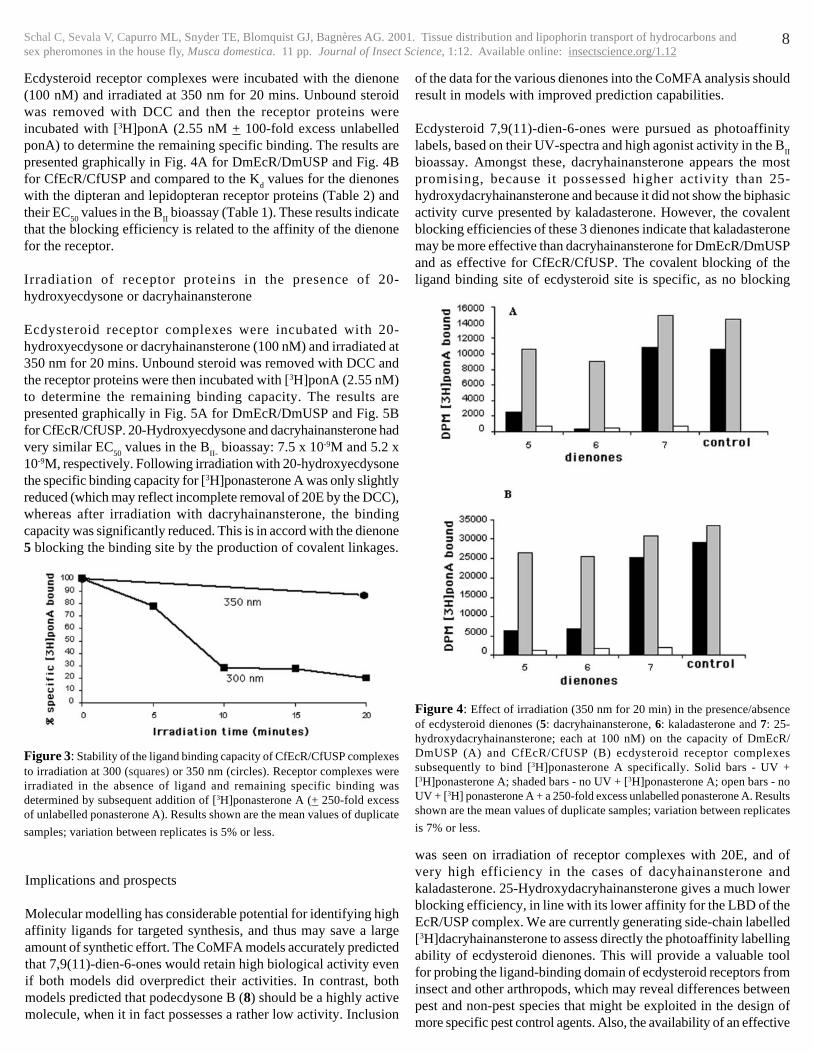
Figure 4: Effect of irradiation (350 nm for 20 min) in the presence/absenceof ecdysteroid dienones (5: dacryhainansterone, 6: kaladasterone and 7: 25-hydroxydacryhainansterone; each at 100 nM) on the capacity of DmEcR/DmUSP (A) and CfEcR/CfUSP (B) ecdysteroid receptor complexessubsequently to bind [3H]ponasterone A specifically. Solid bars - UV +[3H]ponasterone A; shaded bars - no UV + [3H]ponasterone A; open bars - noUV + [3H] ponasterone A + a 250-fold excess unlabelled ponasterone A. Resultsshown are the mean values of duplicate samples; variation between replicates
is 7% or less.
Implications and prospects
Molecular modelling has considerable potential for identifying highaffinity ligands for targeted synthesis, and thus may save a largeamount of synthetic effort. The CoMFA models accurately predictedthat 7,9(11)-dien-6-ones would retain high biological activity evenif both models did overpredict their activities. In contrast, bothmodels predicted that podecdysone B (8) should be a highly activemolecule, when it in fact possesses a rather low activity. Inclusion
Figure 3: Stability of the ligand binding capacity of CfEcR/CfUSP complexesto irradiation at 300 (squares) or 350 nm (circles). Receptor complexes wereirradiated in the absence of ligand and remaining specific binding wasdetermined by subsequent addition of [3H]ponasterone A (+ 250-fold excessof unlabelled ponasterone A). Results shown are the mean values of duplicate
samples; variation between replicates is 5% or less.
Ecdysteroid receptor complexes were incubated with the dienone(100 nM) and irradiated at 350 nm for 20 mins. Unbound steroidwas removed with DCC and then the receptor proteins wereincubated with [3H]ponA (2.55 nM + 100-fold excess unlabelledponA) to determine the remaining specific binding. The results arepresented graphically in Fig. 4A for DmEcR/DmUSP and Fig. 4Bfor CfEcR/CfUSP and compared to the K
d values for the dienones
with the dipteran and lepidopteran receptor proteins (Table 2) andtheir EC
50 values in the B
II bioassay (Table 1). These results indicate
that the blocking efficiency is related to the affinity of the dienonefor the receptor.
Irradiation of receptor proteins in the presence of 20-hydroxyecdysone or dacryhainansterone
Ecdysteroid receptor complexes were incubated with 20-hydroxyecdysone or dacryhainansterone (100 nM) and irradiated at350 nm for 20 mins. Unbound steroid was removed with DCC andthe receptor proteins were then incubated with [3H]ponA (2.55 nM)to determine the remaining binding capacity. The results arepresented graphically in Fig. 5A for DmEcR/DmUSP and Fig. 5Bfor CfEcR/CfUSP. 20-Hydroxyecdysone and dacryhainansterone hadvery similar EC
50 values in the B
II- bioassay: 7.5 x 10⁻⁹M and 5.2 x
10-9M, respectively. Following irradiation with 20-hydroxyecdysonethe specific binding capacity for [3H]ponasterone A was only slightlyreduced (which may reflect incomplete removal of 20E by the DCC),whereas after irradiation with dacryhainansterone, the bindingcapacity was significantly reduced. This is in accord with the dienone5 blocking the binding site by the production of covalent linkages.
of the data for the various dienones into the CoMFA analysis shouldresult in models with improved prediction capabilities.
Ecdysteroid 7,9(11)-dien-6-ones were pursued as photoaffinitylabels, based on their UV-spectra and high agonist activity in the B
II
bioassay. Amongst these, dacryhainansterone appears the mostpromising, because it possessed higher activity than 25-hydroxydacryhainansterone and because it did not show the biphasicactivity curve presented by kaladasterone. However, the covalentblocking efficiencies of these 3 dienones indicate that kaladasteronemay be more effective than dacryhainansterone for DmEcR/DmUSPand as effective for CfEcR/CfUSP. The covalent blocking of theligand binding site of ecdysteroid site is specific, as no blocking
was seen on irradiation of receptor complexes with 20E, and ofvery high efficiency in the cases of dacyhainansterone andkaladasterone. 25-Hydroxydacryhainansterone gives a much lowerblocking efficiency, in line with its lower affinity for the LBD of theEcR/USP complex. We are currently generating side-chain labelled[3H]dacryhainansterone to assess directly the photoaffinity labellingability of ecdysteroid dienones. This will provide a valuable toolfor probing the ligand-binding domain of ecdysteroid receptors frominsect and other arthropods, which may reveal differences betweenpest and non-pest species that might be exploited in the design ofmore specific pest control agents. Also, the availability of an effective

Schal C, Sevala V, Capurro ML, Snyder TE, Blomquist GJ, Bagnères AG. 2001. Tissue distribution and lipophorin transport of hydrocarbons andsex pheromones in the house fly, Musca domestica. 11 pp. Journal of Insect Science, 1:12. Available online: insectscience.org/1.12
9
� � � � � CfEcR/CfUSP� � DmEcR/DmUSP�Dienone� � � � CBE� � Kd� � CBE� � Kd�dacryhainansterone�(5)� � 78%� � 5x10⁻⁹M� 77%� � 1.3x10-
9M�kaladasterone�(6)� � � 79%� � 1.3x10⁻⁹M� 97%� � 3.6x10-
10M�25-hydroxydacryhainansterone�(7)� 13%� � 2.7x10⁻⁷M� 0%� � 2.3x10-
7M��
Table 2: Comparison of the apparent irradiation-induced covalent blockingefficiency (CBE) of ecdysteroid dienones with the K
d values for each com-
pound with DmEcR/DmUSP and CfEcR/CfUSP.
Figure 5: Effect of irradiation (350 nm for 20 min) in the presence of 100 nM20-hydroxyecdysone (1) or dacryhainansterone (5) on the capacity of DmEcR/DmUSP (A) and CfEcR/CfUSP (B) ecdysteroid receptor complexessubsequently to bind [3H]ponasterone A; Filled bars, samples irradiated at 350nm for 20 minutes; open bars samples not irradiated. Results shown are themean values of duplicate samples; variation between replicates is 5% or less.
ACKNOWLEDGEMENTS
PCB was supported by a BBSRC CASE studentship (in conjunctionwith Rohm & Haas Co.). Research in JH’s lab. was supported bythe Grant Agency of the Czech Republic (grant No. 203/01/183).
ecdysteroid affinity label could be used to search for ecdysteroidreceptor proteins in invertebrate species of other Classes (e.g.
nematodes), where previous physiological, biochemical andmolecular biological investigations on the hormonal role ofecdysteroids have been inconclusive. This could shed further lighton whether arthropods and nematodes form a Ecdysozoa clade(Manuel et al., 2000).
We thank Ziyadilla Saatov (Institute of Plant Chemistry, Tashkent,Uzbekistan) for the kind provision of turkesterone, Dr. VladimirˆSik (Dept. Chemistry, University of Exeter) for his assistance withNMR and Mark Prescott and Prof. Huw Rees (School of BiologicalSciences, University of Liverpool, U.K.) for LSIMS analyses.
REFERENCES
Billas IM, Moulinier L, Rochel N, Moras D. 2001. Crystal structureof the ligand-binding domain of the Ultraspiracle proteinUSP, the ortholog of retinoid X receptors in insects. Journalof Biological Chemistry 276: 7465-7474.
Canonica L, Danieli B, Weisz-Vincze I. 1972. Stucture ofmuristerone A, a new phytoecdysone. Journal of theChemical Society, Chemical Communications 1060-1061.
Canonica L, Danieli B, Ferrari G, Haimova MA, Krepinsky J. 1973.The structure of a new phytoecdysone kaladasterone: anapplication of 13C magnetic resonance spectroscopy tostructural problems. Experientia 29: 1062-1063.
Canonica L, Danieli B, Ferrari G, Krepinsky J. 1977. Newphytoecdysone from Kaladana. I. Structure of muristeroneA and kaladasterone. Gazzetta Chimica Italiana 107: 123-130.
Clément CY, Bradbrook DA, Lafont R, Dinan L. 1993. Assessmentof a microplate-based bioassay for the detection ofecdysteroid-like or antiecdysteroid activities. InsectBiochemistry and Molecular Biology 23: 187-193.
Dhadialla TS, Carlson GR, Le DP. 1998. New insecticides withecdysteroidal and juvenile hormone activity. AnnualReviews of Entomology 43: 545-469.
Dinan L. 1989. Ecdysteroid structure and hormonal activity. In:Koolman J (Ed.) Ecdysone: From Chemistry to Mode ofAction, Thieme Verlag, Stuttgart, 345-354.
Dinan L, Hormann RE, Fujimoto T. 1999. An extensive ecdysteroidCoMFA. Journal of Computer-aided Molecular Design 13:185-207.
Dinan L, Bourne PC, Meng Y, Sarker SD, Tolentino RB, Whiting P.2001. Assessment of natural products in the Drosophilamelanogaster B
II cell bioassay for ecdysteroid and
antagonist activities. Cellular and Molecular Life Sciences58: 321-342.
Dormán G. 2000. Photoaffinity labeling in biological signaltransduction. Topics in Current Chemistry 211: 171-225.
Durica DS, Chung AC-K, Hopkins PM. 1999. Characterization ofEcR and RXR gene homologs and receptor expression duringthe molt cycle in the crab, Uca pugilator. AmericanZoologist 39: 758-773.
Girault J.-P. 1998. Determination of ecdysteroids structureby 1D and 2D NMR. Russian Journal of PlantPhysiology 45: 306-309 (translated from FiziologiyaRastenii 1998; 45: 360-364).
Girault J.-P., Lafont R. 1988. The complete 1H-NMR assignmentof ecdysone and 20-hydroxyecdysone. Journal of InsectPhysiology 34: 701-706.
Gronemeyer H (Ed.) 1988. Affinity labelling and cloning of steroidand thyroid hormone receptors. VCH/Ellis Horwood,Chichester, UK.
Schal C, Sevala V, Capurro ML, Snyder TE, Blomquist GJ, Bagnères AG. 2001. Tissue distribution and lipophorin transport of hydrocarbons andsex pheromones in the house fly, Musca domestica. 11 pp. Journal of Insect Science, 1:12. Available online: insectscience.org/1.12
10
Gronemeyer H, Govindan MV. 1986. Affinity labelling of steroidhormone receptors. Molecular and Cellular Endocrinology32: 171-178.
Gronemeyer H, Pongs O. 1980. Localization of ecdysterone onpolytene chromosomes of Drosophila melanogaster.Proceedings of the Natlional Academy of Sciences, USA77: 2108-2112.
Harmatha J, Dinan L, Lafont R. 2002. Biological activities of aspecific ecdysteroid dimer and of selected monomericstructural analogues in the B
II bioassay. Insect Biochemistry
and Molecular Biology 32: 181-185.Hou S, Wang G, Xia K. 1982. Isolation and identification of
phytoecdysones from Dacrydium pierrei. Hickel ZhiwuXuebao 24: 347-354.
Imai S, Murata E, Fujioka S, Koreeda M, Nakanishi K. 1969.Structure of ajugasterone C, a phytoecdysone with an 11-hydroxy-group. Journal of the Chemical Society, ChemicalCommunications 546-547.
Jackman L.M., Sternell S. 1969. Application of nuclear magneticresonance spectroscopy in organic chemistry, Editors,Barton D.H.R., Doering W. 2nd edition, pp 242, PergamonPress , Oxford
Jones G, Jones D. 2000. Considerations of the structural evidenceof a ligand-binding function of ultraspiracle, an insecthomolog of vertebrate RXR. Insect Biochemistry andMolecular Biology 30: 671-679.
Katzenellenbogen JA, Katzenellenbogen BS. 1984. Affinity labelingof receptors for steroid and thyroid hormones. Vitamins andHormones 41: 213-274.
Koelle MR, Talbot WS, Segraves WA, Bender MT, Cherbas P,Hogness DS. 1991. The Drosophila EcR gene encodes anecdysone receptor, a new member of the steroid receptorsuperfamily. Cell 67: 59-77.
Lafont R, Wilson I. 1996. The ecdysone handbook. ChromatographicSociety, Nottingham, UK, 2nd Ed.
Manuel M, Kruse M, Müller WEG, Le Parco Y. 2000. Thecomparison of β-thymosin homologues among metazoasupports and arthropod-nematode clade. Journal ofMolecular Evolution 51: 378-381.
Nishimoto A, Shiobara Y, Inoue S S, Fujimoto M, Takemoto T, YeohC L, De Oliveira F, Akisue G, Akisue M K, Hashimoto G.1988. Three ecdysteroid glycosides from Pfaffia iresinoides.Phytochemistry 27: 1665-1668.
Palmer MJ, Harmon MA, Laudet V. 1999. Characterization of EcRand RXR homologues in the Ixodid tick, Amblyommaamericanum (L.). American Zoology 39: 747-757.
Píˆs J, Budˆeˆsínky M, Vokáˆc K, Laudová V, Harmatha J. 1994.Ecdysteroids from roots of Leuzea carthamoides.Phytochemistry 37: 707-711.
Qui M, Nie R. 1983. The dehydration of ajugasterone C under baseconditions. Acta Botanica Yunnanica 10: 219-222.
Ravi M, Hopfinger AJ, Hormann RE, Dinan L. 2001. 4D-QSARanalysis of a set of ecdysteroids and comparison to CoMFAmodeling. Journal of Medicinal Chemistry 41: 1587-1604.
Saleh DS, Zhang J, Wyatt GR, Walker VK. 1998. Cloning andcharacterization of an ecdysone receptor cDNA fromLocusta migratoria. Molecular and Cellular Endocrinology143: 91-99.
Schaltmann K, Pongs O. 1982. Identification and characterizationof the ecdysterone receptor in Drosophila melanogasterby photoaffinity labeling. Proceedings of the NatlionalAcademy of Sciences, USA 79: 6-10.
Strangmann-Diekmann M, Klone A, Ozyhar A, Kreklau F, Kiltz H-H, Hedtmann U, Welzel P, Pongs O. 1990. Affinity labellingof a partially purified ecdysteroid receptor with abromoacetylated 20-OH-ecdysone derivative. EuropeanJournal of Biochemistry 189: 137-143.
Szendrei K, Varga E, Hajdú Z, Herke I, Lafont R, Girault J-P. 1988.Ajugasterone C and 5-deoxykaladasterone, an ecdysteroidartifact, from Leuzea carthamoides. Journal of NaturalProducts 51: 993-995.
Usmanov BZ, Gorovitz MB, Abubakirov NK. 1975.Phytoecdysteroids from Ajuga turkestanica. III. Isolationof turkesterone. Khimiia Prirodnykh Soedinenii 466-470.
Vögtli M, Imhof MO, Brown NE, Rauch P, Spindler-Barth M, LezziM, Henrich VC. 1999. Functional characterization of twoUltraspiracle forms (CtUSP-1 and CtUSP-2) fromChironomus tentans. Insect Biochemistry and MolecularBiology 29: 931-942.
Volodin VV, Alexeeva LI, Kolegova NA, Sarker SD, ¢Sik V, LafontR, Dinan L. 1998. Further ecdysteroids from Serratulacoronata L. (Asteraceae). Biochemical Systematics andEcology 26: 459-461.
Wang S-F, Ayer S, Segraves WA, Williams DR, Raikhel AS. 2000.Molecular determinants of differential ligand sensitivitiesof insect ecdysteroid receptors. Molecular and CellularBiology 20: 3870-3879.
Wurtz JM, Guillot B, Fagart J, Moras D, Tietjen K, Schindler M.2000. A new model for 20-hydroxyecdysone anddibenzoylhydrazine binding: a homology modeling anddocking approach. Protein Science 9: 1073-1084.
Yao T-P, Forman BM, Jiang Z, Cherbas L, Chen J-D, McKeown M,Cherbas P, Evans RM. 1993. Functional ecdysone receptoris the product of EcR and Ultraspiracle genes. Nature 366:476-479.