Antigens and Alternatives for Control of Anaplasma marginale Infection in Cattle
15
CLINICAL MICROBIOLOGY REVIEWS, Oct. 2003, p. 698–712 Vol. 16, No. 4 0893-8512/03/$08.000 DOI: 10.1128/CMR.16.4.698–712.2003 Copyright © 2003, American Society for Microbiology. All Rights Reserved. Antigens and Alternatives for Control of Anaplasma marginale Infection in Cattle Katherine M. Kocan, 1 * Jose ´ de la Fuente, 1 Alberto A. Guglielmone, 2 and Roy D. Mele ´ndez 3 Department of Veterinary Pathobiology, College of Veterinary Medicine, Oklahoma State University, Stillwater, Oklahoma 74078 1 ; Instituto Nacional de Tecnologı ´a Agropecuaria (INTA), Rafaela 2300, Argentina 2 ; and Area de Parasitologia, decanato del Ciencias Veterinarias, Universidad Centroccidental “Lisandro Alvarado,” Barquisimeto, Lara State 3001-A, Venezuela 3 INTRODUCTION .......................................................................................................................................................698 Bovine Anaplasmosis ..............................................................................................................................................698 Geographic Distribution of Bovine Anaplasmosis .............................................................................................700 Economic Impact of Bovine Anaplasmosis .........................................................................................................701 TAXONOMY ...............................................................................................................................................................702 Present Classification and Phylogenetic Relationships of A. marginale with Respect to the Organisms in the Family Anaplasmataceae ........................................................................................................................702 A. marginale Genome and Major Surface Protein Genes..................................................................................703 Gene Regulation in A. marginale Multigene Families .......................................................................................703 Phylogenetic Relationships of Geographic Isolates of A. marginale ................................................................704 ANAPLASMOSIS VACCINES ..................................................................................................................................706 Live Vaccines ...........................................................................................................................................................706 Infection-treatment method ...............................................................................................................................706 Vaccination with attenuated strains of A. marginale. ....................................................................................706 Live A. centrale vaccine .......................................................................................................................................706 Possible side effects ............................................................................................................................................707 Killed Vaccines ........................................................................................................................................................707 Prospects for Development of New and More Effective Vaccines ....................................................................707 Development of a cell culture-derived killed vaccine.....................................................................................707 Development of novel vaccines ..........................................................................................................................708 ACKNOWLEDGMENTS ...........................................................................................................................................709 REFERENCES ............................................................................................................................................................709 INTRODUCTION Bovine Anaplasmosis Bovine anaplasmosis is an arthropod-borne hemolytic dis- ease of cattle that is caused by the rickettsia Anaplasma mar- ginale (Rickettsiales: Anaplasmataceae) (28, 57, 80, 150). Clin- ical disease is most notable in cattle, but other ruminants including water buffalo, bison, African antelopes, and mule deer can become persistently infected with A. marginale (88). Sir Arnold Theiler first described A. marginale infection in erythrocytes of South African cattle as “marginal points” (161). A similar report was published in the United States by Salmon and Smith in 1896, which described the presence of a point-like pathogen in blood smears of cattle as “very minute roundish body which is stained blue to bring it into view. The body as a rule is situated near the edge of the corpuscle” (148). Theiler subsequently described a subspecies of A. marginale, A. cen- trale, which appeared to be less pathogenic and for which Anaplasma inclusions were more often found in the center of erythrocytes rather than in a marginal location (162). Erythrocytes are the only known site of infection of A. mar- ginale in cattle (Fig. 1A) Within these cells the membrane- bound inclusions (also called initial bodies) contain four to eight rickettsia (Fig. 1B), and 70% or more of the erythrocytes may become infected during acute infection (137, 140). The incubation period of infection (prepatent period) varies with the number of organisms in the infective dose and ranges from 7 to 60 days, with an average of 28 days. After erythrocytic infection is detected, the number of parasitized erythrocytes increases geometrically. Infected erythrocytes are subsequently phagocytized by bovine reticuloendothelial cells, resulting in the development of mild to severe anemia and icterus without hemoglobinemia and hemoglobinuria. Clinical symptoms may include fever, weight loss, abortion, lethargy, icterus, and often death in animals older than 2 years (138). Cattle that survive acute infection develop persistent infections characterized by cyclic low-level rickettsemia (64, 65, 77) (Fig. 2). Persistently infected or “carrier” cattle have lifelong immunity and are resistant to clinical disease on challenge exposure. However, persistently infected cattle serve as reservoirs of A. marginale because they provide a source of infective blood for both me- chanical and biological transmission by ticks. Bos taurus breeds (i.e., Holstein, Brown Swiss, or Hereford) are more likely to develop acute anaplasmosis than are crossbred Zebu or Creole cattle (2, 3). * Corresponding author. Mailing address: Department of Veteri- nary Pathobiology, College of Veterinary Medicine, Oklahoma State University, Stillwater, OK 74078-2007. Phone: (405) 744-7271. Fax (405) 744-5275. E-mail: [email protected]. 698 on July 8, 2016 by guest http://cmr.asm.org/ Downloaded from
-
Upload
independent -
Category
Documents
-
view
0 -
download
0
Transcript of Antigens and Alternatives for Control of Anaplasma marginale Infection in Cattle

CLINICAL MICROBIOLOGY REVIEWS, Oct. 2003, p. 698–712 Vol. 16, No. 40893-8512/03/$08.00�0 DOI: 10.1128/CMR.16.4.698–712.2003Copyright © 2003, American Society for Microbiology. All Rights Reserved.
Antigens and Alternatives for Control of Anaplasma marginaleInfection in Cattle
Katherine M. Kocan,1* Jose de la Fuente,1 Alberto A. Guglielmone,2and Roy D. Melendez3
Department of Veterinary Pathobiology, College of Veterinary Medicine, Oklahoma State University, Stillwater,Oklahoma 740781; Instituto Nacional de Tecnologıa Agropecuaria (INTA), Rafaela 2300, Argentina2; and
Area de Parasitologia, decanato del Ciencias Veterinarias, Universidad Centroccidental“Lisandro Alvarado,” Barquisimeto, Lara State 3001-A, Venezuela3
INTRODUCTION .......................................................................................................................................................698Bovine Anaplasmosis..............................................................................................................................................698Geographic Distribution of Bovine Anaplasmosis .............................................................................................700Economic Impact of Bovine Anaplasmosis .........................................................................................................701
TAXONOMY ...............................................................................................................................................................702Present Classification and Phylogenetic Relationships of A. marginale with Respect to the Organisms
in the Family Anaplasmataceae........................................................................................................................702A. marginale Genome and Major Surface Protein Genes..................................................................................703Gene Regulation in A. marginale Multigene Families .......................................................................................703Phylogenetic Relationships of Geographic Isolates of A. marginale ................................................................704
ANAPLASMOSIS VACCINES ..................................................................................................................................706Live Vaccines ...........................................................................................................................................................706
Infection-treatment method...............................................................................................................................706Vaccination with attenuated strains of A. marginale. ....................................................................................706Live A. centrale vaccine.......................................................................................................................................706Possible side effects ............................................................................................................................................707
Killed Vaccines........................................................................................................................................................707Prospects for Development of New and More Effective Vaccines ....................................................................707
Development of a cell culture-derived killed vaccine.....................................................................................707Development of novel vaccines..........................................................................................................................708
ACKNOWLEDGMENTS ...........................................................................................................................................709REFERENCES ............................................................................................................................................................709
INTRODUCTION
Bovine Anaplasmosis
Bovine anaplasmosis is an arthropod-borne hemolytic dis-ease of cattle that is caused by the rickettsia Anaplasma mar-ginale (Rickettsiales: Anaplasmataceae) (28, 57, 80, 150). Clin-ical disease is most notable in cattle, but other ruminantsincluding water buffalo, bison, African antelopes, and muledeer can become persistently infected with A. marginale (88).Sir Arnold Theiler first described A. marginale infection inerythrocytes of South African cattle as “marginal points” (161).A similar report was published in the United States by Salmonand Smith in 1896, which described the presence of a point-likepathogen in blood smears of cattle as “very minute roundishbody which is stained blue to bring it into view. The body as arule is situated near the edge of the corpuscle” (148). Theilersubsequently described a subspecies of A. marginale, A. cen-trale, which appeared to be less pathogenic and for whichAnaplasma inclusions were more often found in the center oferythrocytes rather than in a marginal location (162).
Erythrocytes are the only known site of infection of A. mar-ginale in cattle (Fig. 1A) Within these cells the membrane-bound inclusions (also called initial bodies) contain four toeight rickettsia (Fig. 1B), and 70% or more of the erythrocytesmay become infected during acute infection (137, 140). Theincubation period of infection (prepatent period) varies withthe number of organisms in the infective dose and ranges from7 to 60 days, with an average of 28 days. After erythrocyticinfection is detected, the number of parasitized erythrocytesincreases geometrically. Infected erythrocytes are subsequentlyphagocytized by bovine reticuloendothelial cells, resulting inthe development of mild to severe anemia and icterus withouthemoglobinemia and hemoglobinuria. Clinical symptoms mayinclude fever, weight loss, abortion, lethargy, icterus, and oftendeath in animals older than 2 years (138). Cattle that surviveacute infection develop persistent infections characterized bycyclic low-level rickettsemia (64, 65, 77) (Fig. 2). Persistentlyinfected or “carrier” cattle have lifelong immunity and areresistant to clinical disease on challenge exposure. However,persistently infected cattle serve as reservoirs of A. marginalebecause they provide a source of infective blood for both me-chanical and biological transmission by ticks. Bos taurus breeds(i.e., Holstein, Brown Swiss, or Hereford) are more likely todevelop acute anaplasmosis than are crossbred Zebu or Creolecattle (2, 3).
* Corresponding author. Mailing address: Department of Veteri-nary Pathobiology, College of Veterinary Medicine, Oklahoma StateUniversity, Stillwater, OK 74078-2007. Phone: (405) 744-7271. Fax(405) 744-5275. E-mail: [email protected].
698
on July 8, 2016 by guesthttp://cm
r.asm.org/
Dow
nloaded from

Calves are less susceptible to infection with A. marginaleand, when infected, are less susceptible to clinical disease. Thisphenomenon is not well understood, but removal of the spleenrenders calves fully susceptible to infection, and anaplasmosisin splenectomized calves is often more severe than that ob-served in older cattle. However, once calves become infected,they develop persistent infections and lifelong immunity toanaplasmosis.
Transmission of A. marginale can be effected both mechan-ically by biting flies or blood-contaminated fomites and biolog-ically by ticks (56, 60, 78). Mechanical transmission frequentlyoccurs via blood-contaminated fomites, including needles, de-horning saws, nose tongs, tattooing instruments, ear-taggingdevices, and castration instruments. Mechanical transmissionby arthropods has been reported for bloodsucking diptera ofthe genera Tabanus, Stomoxys, and mosquitoes (60, 63, 132).This form of mechanical transmission is considered to be themajor route of dissemination of A. marginale in areas of Cen-
tral and South America and Africa where tick vectors do notoccur (60, 63) and where Boophilus microplus, the tropicalcattle tick, does not appear to be a biological vector of A.marginale (42, 61). In areas of the United States where geo-graphic isolates of A. marginale are not infective for ticks orwhere ticks have been eradicated by fire ants, mechanicaltransmission appears to be the major mode of A. marginaletransmission (47, 156, 172).
In addition to mechanical and biological transmission, A.marginale can be transmitted from cow to calf transplacentallyduring gestation (111, 176, 177). For example, a 15.6% prev-alence rate of in utero transmission of Anaplasma infectionswas reported in South Africa (135). Transplacental transmis-sion of anaplasmosis may therefore contribute to the epidemi-ology of this disease in some regions.
Biological transmission of A. marginale is effected by ticks,and approximately 20 species of ticks have been incriminatedas vectors worldwide (56, 60). Tick transmission can occurfrom stage to stage (transstadial) or within a stage (intrasta-dial), while transovarial transmission from one tick generationto the next does not appear to occur (158). Interstadial trans-mission of A. marginale has been demonstrated by the three-host ticks Dermacentor andersoni and D. variabilis in the UnitedStates (78, 79, 83, 159) and by Rhipicephalus simus in SouthAfrica (131, 133, 134). The one-host tick B. annulatus transmitsA. marginale in Israel, Central America, South America, andMexico (68, 149).
Intrastadial transmission of A. marginale is effected by maleticks. Recent studies have demonstrated that male Dermacen-tor ticks may play an important role in the biological transmis-sion of A. marginale because they become persistently infectedwith A. marginale and can transmit A. marginale repeatedlywhen they transfer among cattle (82, 85). Male ticks thereforealso serve as a reservoir of A. marginale along with persistentlyinfected cattle (67, 80, 82, 85). Transmission of A. marginale bymale ticks may be an important mechanism of transmission ofA. marginale by one-host ticks, including Boophilus spp. and D.albipictus. However, it was shown recently that the cofeeding of
FIG. 1. Bovine erythrocytes infected with A. marginale. (A) Inclusion bodies (arrowheads) are located at the periphery of the erythrocyte in astained blood film. (B) Electron micrograph of an A. marginale inclusion that contains three organisms. Bar, 10 �m (A) and 0.5 �m (B).
FIG. 2. The high A. marginale levels in acute rickettsemia (�109
ml�1) are resolved after the development of a primary immune re-sponse, but the emergence of antigenic variants results in persistentinfection. Persistence is characterized by sequential rickettsemic cy-cles, occurring at approximately 5-week intervals, in which new MSP2variants replicate to a peak of �106 ml�1 and are then controlled by avariant-specific immune response. Variants arising in three sequentialrickettsemic cycles are shown and are designated V1, V2, and V3. Thepoints of variant emergence and variant control are designated for V2.(Reprinted from reference 125 with permission of the publisher.)
VOL. 16, 2003 CONTROL OF A. MARGINALE INFECTION IN CATTLE 699
on July 8, 2016 by guesthttp://cm
r.asm.org/
Dow
nloaded from

adult Dermacentor spp. does not appear to influence the dy-namics of A. marginale transmission (81).
The developmental cycle of A. marginale in ticks is complexand coordinated with the tick feeding cycle (78, 82, 85) (Fig. 3).Infected erythrocytes taken into ticks with the blood mealprovide the source of A. marginale infection for tick gut cells(Fig. 4). After development of A. marginale in tick gut cells,many other tick tissues become infected, including the salivaryglands (Fig. 5), from where the rickettsiae are transmitted tovertebrates during feeding (67, 78, 82, 85). At each site ofinfection in ticks, A. marginale develops within membrane-bound vacuoles or colonies. The first form of A. marginale seenwithin the colony is the reticulated (vegetative) form, whichdivides by binary fission (Fig. 6A), forming large colonies thatmay contain hundreds of organisms. The reticulated form thenchanges into the dense form (Fig. 6B), which is the infectiveform and can survive outside the host cells. Cattle becomeinfected with A. marginale when the dense form is transmittedduring tick feeding via the salivary glands.
Geographic Distribution of Bovine Anaplasmosis
Anaplasmosis occurs in tropical and subtropical areasthroughout the world and is a major constraint to the cattle
production in many countries. In the United States, anaplas-mosis is enzootic throughout the southern Atlantic states, GulfCoast states, and several of the Midwestern and Western states(98). However, anaplasmosis has been reported in almost everystate in the United States, and this widening distribution maybe due to increased transportation of cattle and hence theopportunity for mechanical transmission from asymptomaticpersistently infected cattle.
Bovine anaplasmosis is also endemic in Mexico, Central andSouth America, and the Caribbean Islands. It is enzootic inmost Latin American countries, with the exception of desertareas or mountain ranges such as the Andes (68). The sero-prevalence of A. marginale varies widely among countries inthe Americas (Table 1), and this variability contributes to thedevelopment of geographically stable or unstable enzootic re-gions.
The distribution of anaplasmosis may continue to changedue to the trend of global warming, which may influence themovement of the tick hosts (N. N. Jonsson and S. W. J. Reid,Guest Editorial Vet. J. 160:87–89, 2000). An example of thisprediction is the confirmation of anaplasmosis in a bison herdin Saskatchewan, Canada, during the summer of 2000 (136).The first outbreak of anaplasmosis occurred in Canada in 1971(25), but this outbreak was determined to be due to mechanical
FIG. 3. Schematic of the development cycle of A. marginale in cattle and ticks. Infected erythrocytes are ingested by ticks (Dermacentor spp.,Rhipicephalus spp., or Boophilus spp.) with the blood meal. The first site of infection of A. marginale in ticks is the gut cells. When the ticks feeda second time, many tick tissues become infected, including salivary gland cells, from where the rickettsia is transmitted back to cattle. Two formsof A. marginale, reticulated and dense forms, are found in infected tick cells. Reticulated forms appear first and are the vegetative stage that dividesby binary fission. The reticulated form changes into the dense form, which is the infective form and can survive extracellularly. (Reprinted fromreference 125 with permission from the publisher.)
700 KOCAN ET AL. CLIN. MICROBIOL. REV.
on July 8, 2016 by guesthttp://cm
r.asm.org/
Dow
nloaded from
transmission from imported carrier cattle. Protocols proposedin the Sanitary and Phytosanitary Measures and the Risk As-sessment Methodology may be important to control the spreadof diseases such as anaplasmosis in the future global tradingmarket (152).
Economic Impact of Bovine Anaplasmosis
Bovine anaplasmosis causes important economic loss inmost countries, mainly due to the high morbidity and mortalityin susceptible cattle herds. The losses due to anaplasmosis aremeasured through several parameters: low weight gain, reduc-tion in milk production, abortion, the cost of anaplasmosistreatments, and mortality. However, few controlled studieshave been carried out to determine the exact annual loss
caused by anaplasmosis in a country, since in general loss isreported as high, tremendous, or enormous. Nonetheless, thecurrent annual losses in beef cattle in the United States as aresult of anaplasmosis morbidity and mortality are estimatedto be over $300 million per year (98), whereas in Latin Amer-ica those losses were calculated to be approximately $800 mil-lion (95). More recently, it was reported that bovine anaplas-mosis and babesiosis were responsible for causing an economicloss of $875 millions in Latin American nations (33). However,the most important economic constraint of anaplasmosis tocattle production in the tropics is on public or private programsfor genetic improvement of cattle. Imported Bos taurus cattlebrought from temperate nations to the tropics for breed im-provement are highly susceptible to tick-borne diseases andoften do not survive to become part of planned reproduction
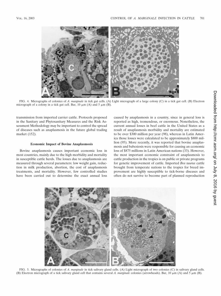
FIG. 4. Micrographs of colonies of A. marginale in tick gut cells. (A) Light micrograph of a large colony (C) in a tick gut cell. (B) Electronmicrograph of a colony in a tick gut cell. Bar, 10 �m (A) and 5 �m (B).
FIG. 5. Micrographs of colonies of A. marginale in tick salivary gland cells. (A) Light micrograph of two colonies (C) in salivary gland cells.(B) Electron micrograph of a tick salivary gland cell that contains several A. marginale colonies (arrowheads). Bar, 10 �m (A) and 5 �m (B).
VOL. 16, 2003 CONTROL OF A. MARGINALE INFECTION IN CATTLE 701
on July 8, 2016 by guesthttp://cm
r.asm.org/
Dow
nloaded from

programs. This constraint is a notable reality for programs forthe improvement of cattle in most Latin American countries(105).
TAXONOMY
Present Classification and Phylogenetic Relationships ofA. marginale with Respect to the Organisms in the
Family Anaplasmataceae
The organisms in the order Rickettsiales were recently re-classified based on biological characteristics and genetic anal-yses of 16S rRNA genes, groESL, and surface protein genes(57). These phylogenetic analyses consistently supported theformation of four distinct genera within the family Anaplas-mataceae: (i) Anaplasma, with a 96.1% minimum similarity; (ii)Ehrlichia, with a 97.7% similarity; (iii) Wolbachia, with a 95.6%similarity; and (iv) Neorickettsia, with a 94.9% similarity (57)(Table 2). Organisms classified within the family Rickettsiaceae(genera Rickettsia and Orientia) are all obligate intracellularbacteria that grow freely within the cytoplasm of eukaryotic
cells. While organisms placed in the family Anaplasmataceaeare also obligate intracellular organisms, they are found exclu-sively within membrane-bound vacuoles in the host cell cyto-plasm. Furthermore, most all organisms in the family Anaplas-mataceae multiply in both vertebrates and invertebrates(primarily ticks and trematodes).
The genus of interest in this review, Anaplasma, includesthree species that infect ruminants: A. marginale (the type
FIG. 6. Electron micrographs of the two developmental stages of A. marginale within colonies in tick cells. (A) Reticulated forms within acolony, dividing by binary fission (arrowhead). (B) Dense forms within a colony in an infected tick cell. Bars, 1 �m.
TABLE 1. Geographic distribution and seroprevalence ofanaplasmosis in countries of the Americas
Country Prevalence (%) Techniquea Reference(s)
United States(Louisiana)
5.6 CT 73
United States(Oklahoma)
4.7–17.6 CFb 143
Costa Rica 61–90 cELISA, PCR 72Venezuela 57.7 IFA 74, 106Colombia 64–100 IFA 117Brazil 67.3 IFA 167Paraguay 92 CT 128Argentina 7–61 Blood smears 93Jamaica 69.9 CT 102Lesser Antilles 18–71 Dot ELISA 41
a CF, complement fixation test; CT, card test; cELISA, competitive enzyme-linked immunosorbent assay; IFA, immunofluorescence assay.
b The results of the complement fixation test provide minimum estimates ofseropositive cattle and are likely to underreport the prevalence of A. marginale(27).
TABLE 2. Current classification of the order Rickettsiales
Family Rickettsiaceae (obligate intracellular bacteria that growfreely in the cytoplasm of their eukaryotic host cells)
Genus Rickettsia
Genus Orientia
Family Anaplasmataceae (obligate intracellular bacteria thatreplicate with membrane-derived vacuoles in the cytoplasm ofeukaryotic host cells)
Genus AnaplasmaAnaplasma marginale (type species)Anaplasma centraleAnaplasma ovisAnaplasma bovis (formerly Ehrlichia bovis)Anaplasma phagocytophilum (formerly Ehrlichia
phagocytophilum, E. equi, HGE agent)Anaplasma platys (formerly Ehrlichia platys)Aegyptianella (genus incertae sedis due to lack of sequence
information)
Genus EhrlichiaEhrlichia chaffeensisEhrlichia ruminantium (formerly Cowdria ruminantium)Ehrlichia ewingiiEhrlichia ovisEhrlichia canisEhrlichia muris
Genus NeorickettsiaNeorickettsia helminthoecaNeorickettsia risticii (formerly Ehrlichia risticii)Neorickettsia sennetsu (formerly Ehrlichia sennetsu)
Genus WolbachiaWolbachia pipientis
702 KOCAN ET AL. CLIN. MICROBIOL. REV.
on July 8, 2016 by guesthttp://cm
r.asm.org/
Dow
nloaded from

species). A. marginale subsp. centrale (referred to in this reviewas A. centrale), and A. ovis (57). Bovine anaplasmosis is causedprimarily by A. marginale. While A. centrale is less pathogenicfor cattle and has been used as a live vaccine in Israel, Aus-tralia, Africa, and South America, infection with this organismcan, on occasion, cause clinical disease. A. ovis is a pathogen ofsheep and does not establish persistent infection in cattle.
The genus Anaplasma also includes A. phagocytophilum (for-merly Ehrlichia equi, E. phagocytophila, and the agent of hu-man granulocytic ehrlichiosis [HGE], now recognized as syn-onymous), A. bovis (formerly E. bovis), and A. platys (formerlyE. platys). Aegyptianella was retained in this genus because ofphenotypic similarities to the species of Anaplasma and wasdesignated a genus incertae sedis due to lack of sequenceinformation.
The organisms classified within the other three genera of thefamily Anaplasmataceae (Ehrlichia, Neorickettsia, and Wolba-chia) are listed in Table 2 (57). Note that E. ruminantium(formerly Cowdria ruminantium), the tick-borne pathogen thatcauses heartwater disease in cattle, was found to be moreclosely related to the organisms in the genus Ehrlichia, whichincludes organisms that infect a variety of vertebrate hostsranging from humans to rodents.
A. marginale Genome and Major Surface Protein Genes
The small genome of A. marginale is circular, and the size isestimated at 1.2 to 1.6 Mb (6, 104). Research during the past 20years has focused on identification of the major surface pro-teins (MSPs) of A. marginale. Six MSPs, MSP1a, MSP1b, MSP2,MSP3, MSP4, and MSP5, have been identified on erythrocyte-derived organisms, and information about the gene sequences,recombinant protein, monospecific and monoclonal antibod-ies, isolate variability, and potential value in diagnostic assaysand vaccines is available. MSP1a, MSP4, and MSP5 are en-coded by single genes, while MSP1b, MSP2, and MSP3 areencoded by multigene families.
MSP1a and MSP1b form the MSP1 complex. MSP1a is vari-able in molecular weight among geographic isolates because ofdifferent numbers of tandem 28- or 29-amino-acid repeats lo-cated in the amino-terminal portion of the protein (8, 46, 52,53, 54). Because of the variation in the repeated portion of theMSP1a gene, it has been used as a stable genetic marker foridentification of A. marginale geographic isolates (8, 14, 17, 53).The gene, msp1a, that encodes MSP1a is conserved during themultiplication of the rickettsia in cattle and ticks (26, 126). Aneutralization-sensitive epitope was demonstrated on theMSP1a tandem repeats (127) and was found to be conservedamong A. marginale isolates (46, 47, 52, 53, 114, 127). MSP1awas shown to be an adhesin for bovine erythrocytes and bothnative and cultured tick cells by using recombinant Escherichiacoli expressing MSP1a in microtiter hemagglutination and ad-hesion recovery assays and by microscopy (45, 46, 100, 101).The portion of MSP1a with the tandem repeats was found tobe necessary and sufficient to effect adhesion to bovine eryth-rocytes and tick cells (46). MSP1a was shown to be involved ininfection and transmission of A. marginale by Dermacentorticks (44) and to contribute to immunity to A. marginale infec-tion in cattle (34, 35, 118).
MSP1b, encoded by at least two genes, msp1�1 and msp1�2,
is polymorphic among geographic isolates of A. marginale (17,26, 40, 169). Although MSP1b is encoded by a multigene fam-ily, only small variations in protein sequences of MSP1b1 andMSP1b2 were observed during the life cycle of the rickettsia incattle and ticks (26). This protein, which forms a complex withMSP1a, is an adhesin for bovine erythrocytes (100, 101). How-ever, MSP1b was recently demonstrated to be an adhesin onlyfor bovine erythrocytes and did not prove to be an adhesin fortick cells (45).
MSP2 and MSP3 are both encoded by large polymorphic,multigene families (7, 123). The MSP2 sequence and antigeniccomposition varies during cyclic rickettsemia in cattle (15, 64,65) and in persistently infected ticks (49). MSP2 is encoded ona polycistronic mRNA. The msp2 gene within the expressionsite is polymorphic. msp2 encodes numerous amino acid se-quence variants selected in bovine erythrocytic and tick sali-vary gland populations of A. marginale (16, 29, 49, 64, 65, 104).MSP3 also varies in antigenic properties and structure amonggeographic isolates (5). MSP2 and MSP3 are involved in theinduction of a protective bovine immune response to A. mar-ginale (125). MSP4 and MSP5 are encoded by single-copygenes. Although MSP4 is highly conserved (52, 53, 54, 112),information about its function is not available. MSP5 is also ahighly conserved surface protein that has been proven effectiveas a diagnostic antigen and used in a competitive enzyme-linked immunosorbent assay (ELISA) commercially availablein the United States (163). The function of MSP5 is alsounknown. The msp2 operon-associated genes OpAG1, OpAG2,and OpAG3, have been identified recently in A. marginale andmay encode for surface proteins (94).
Despite the advances in characterizing major MSPs in A.marginale, our knowledge of these proteins is limited and willbe greatly enhanced by the completion of the genome se-quence presently under way at Washington State University(http://www.vetmed.wsu.edu/research_vmp/anagenome/index.html), which will further facilitate the analysis of sequenceinformation.
Gene Regulation in A. marginale Multigene Families
At least two transcriptionally active copies of msp1� havebeen identified in the genome of A. marginale (26). However,although small variations were observed in the MSP1b1 andMSP1b2 protein sequences, recombination does not seem to bean important mechanism in msp1� regulation and the expres-sion from different loci appears to play the major role (26). Themsp2 gene is estimated to have 10 or more copies in thegenome of A. marginale (30). However, all but one of theidentified msp2 copies are pseudogenes, and the operon con-taining the expressed msp2 is a single copy (30). The msp2transcripts are polycistronic and linked to the MSP2-encodingopen reading frame (16). The pseudogenes recombine into themsp2 gene to generate new hypervariable sequences and newantigenic variants during the multiplication of the bacterium(30). Partial pseudogene cassettes are also present for themsp3 gene family, and the pseudogenes for the two gene fam-ilies often appear close together (29, 104). There is increasingevidence that for pathogenic microbial species, loss of genefunction or genome decay increases with adaptation to the host(174). The msp2 and msp3 pseudogenes may be remnants of
VOL. 16, 2003 CONTROL OF A. MARGINALE INFECTION IN CATTLE 703
on July 8, 2016 by guesthttp://cm
r.asm.org/
Dow
nloaded from
functional genes from a host-adapted pathogen in the processof down-sizing its genome content with a more efficient use ofgenome information: a process of reductive convergent evolu-tion caused by prolonged intracellular life (119, 146). The twogene families have the same 5� sequence, suggesting that theycould use similar mechanisms to regulate recombination intothe expression site; this specificity is guaranteed by the respec-tive 3� recombinatorial site in the coding region of each gene(29, 104). The coordinated control of the recombination ofthese genes contributes to the evasion of the host immuneresponse by the pathogen. However, variation must arise spon-taneously and frequently to allow selection of variants thatescape the host immune system (30). Genome decay and vari-ation of gene expression have been reported for other patho-genic bacteria including Rickettsia prowazekii (174).
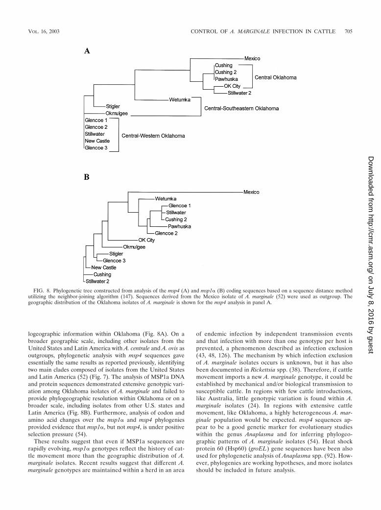
Phylogenetic Relationships ofGeographic Isolates of A. marginale
Phylogenetic analysis of A. marginale geographic isolatesfrom the United States was performed using the single-copygenes msp1� and msp4 (53). The results of these analysesstrongly support a southeastern clade of A. marginale com-posed of isolates from Virginia and Florida. Analysis of 16Sribosomal DNA fragment sequences from the tick vector of A.marginale, D. variabilis, from various areas of the United Stateswas performed and suggested coevolution of the vector andpathogen (53).
Phylogenetic studies were also done using New World iso-lates of A. marginale from the United States, Mexico, Brazil,and Argentina. Seventeen isolates of A. marginale plus twooutgroup taxa (A. centrale and A. ovis) were included for the
analysis of MSP4 sequences (52) (Fig. 7). Maximum-parsimonyanalysis of MSP4 sequences provided phylogenetic informa-tion about the evolution of A. marginale isolates. Strong boot-strap support was detected for a Latin American clade of A.marginale isolates. Moreover, within this Latin Americanclade, strong bootstrap support was detected for Mexican andSouth American clades. Isolates of A. marginale from theUnited States also grouped into two clades, a southern cladeconsisting of isolates from Florida, Mississippi, and Virginia,and a west-central clade consisting of isolates from California,Idaho, Illinois, Oklahoma, and Texas. Although little phylo-geographic resolution was detected within any of these higherclades, msp4 sequences appear to be a good genetic marker forinferring phylogeographic patterns of isolates of A. marginaleon a broad geographic scale. In contrast to the phylogeo-graphic resolution provided by MSP4, DNA and protein se-quence variation from MSP1a representing 20 New Worldisolates of A. marginale failed to provide phylogeographic res-olution (52). Most variation in MSP1a sequences appearedunique to a given isolate. In fact, similar DNA sequence vari-ation in MSP1a was detected within isolates from Idaho andFlorida and from Idaho and Argentina. These results suggestthat the MSP1a sequence may be rapidly evolving and that themsp1� gene may provide phylogeographic information onlywhen numerous MSP1a sequences from a given area are in-cluded in the analysis.
Eleven A. marginale isolates isolated from cattle with ana-plasmosis in Oklahoma during 2001, plus two previous isolatesfrom Wetumka (Oklahoma isolate [52, 53]) and Pawhuskaidentified in 1997 and the 1960s, respectively, were analyzedfor the msp1� and msp4 gene and protein sequences (54). Onlythe phylogenetic analysis with msp4 sequences provided phy-
FIG. 7. Maximum-parsimony analysis of MSP4 sequences. The topology of 1 of 11 equally most-parsimonious trees based on DNA sequencevariation in the msp4 gene is shown. The tree was rooted with A. centrale and A. ovis. The solid portion of each branch represents the minimumbranch length; the dashed portion of each branch represents the maximum branch length as determined by PAUP*4.0b4a. Numbers below thebranches represent the percentage of 500 bootstrap iterations in which each clade was detected, and numbers with asterisks indicate bootstrapsupport based on a phylogenetic analysis of deduced amino acid residues. Vertical lines show synapomorphic amino acid changes documenting themonophyly of A. marginale and the Latin American clade of A. marginale isolates along with the amino acid residue and its position in the sequence.(Adapted from reference 52 with permission of the publisher.)
704 KOCAN ET AL. CLIN. MICROBIOL. REV.
on July 8, 2016 by guesthttp://cm
r.asm.org/
Dow
nloaded from
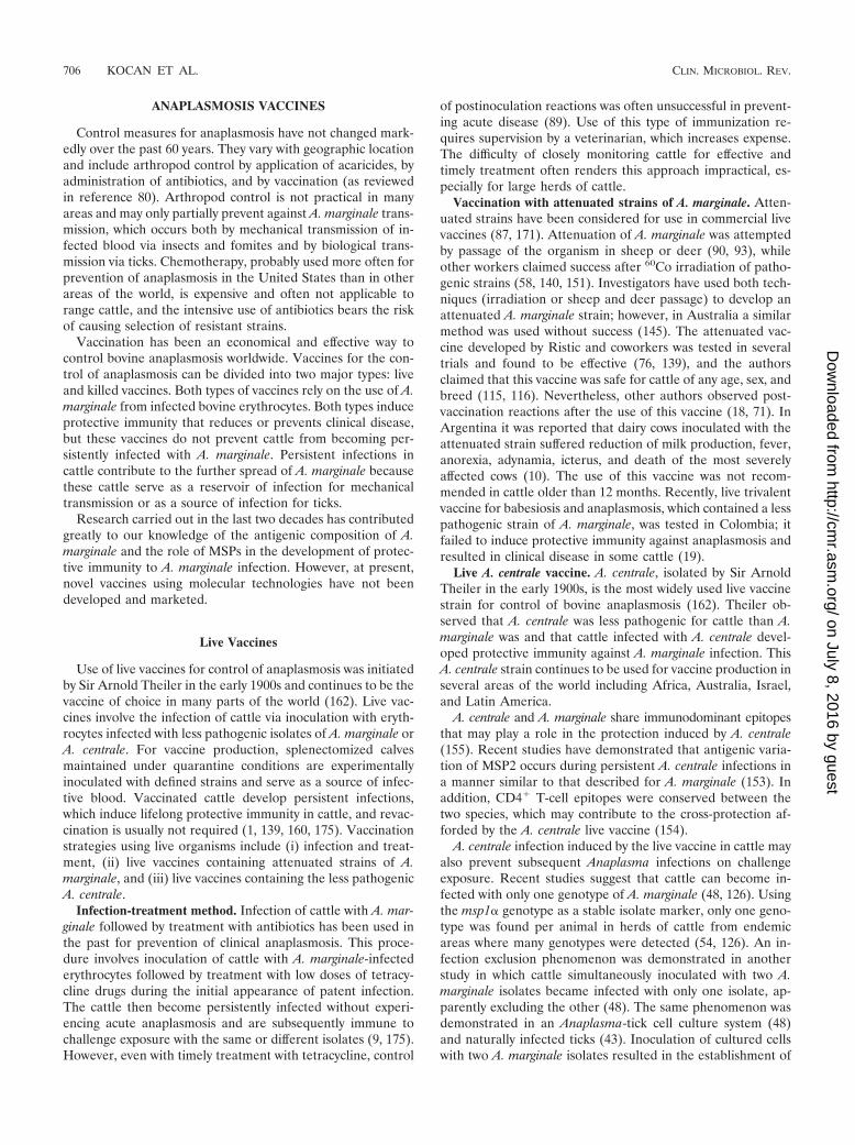
logeographic information within Oklahoma (Fig. 8A). On abroader geographic scale, including other isolates from theUnited States and Latin America with A. centrale and A. ovis asoutgroups, phylogenetic analysis with msp4 sequences gaveessentially the same results as reported previously, identifyingtwo main clades composed of isolates from the United Statesand Latin America (52) (Fig. 7). The analysis of MSP1a DNAand protein sequences demonstrated extensive genotypic vari-ation among Oklahoma isolates of A. marginale and failed toprovide phylogeographic resolution within Oklahoma or on abroader scale, including isolates from other U.S. states andLatin America (Fig. 8B). Furthermore, analysis of codon andamino acid changes over the msp1� and msp4 phylogeniesprovided evidence that msp1�, but not msp4, is under positiveselection pressure (54).
These results suggest that even if MSP1a sequences arerapidly evolving, msp1� genotypes reflect the history of cat-tle movement more than the geographic distribution of A.marginale isolates. Recent results suggest that different A.marginale genotypes are maintained within a herd in an area
of endemic infection by independent transmission eventsand that infection with more than one genotype per host isprevented, a phenomenon described as infection exclusion(43, 48, 126). The mechanism by which infection exclusionof A. marginale isolates occurs is unknown, but it has alsobeen documented in Rickettsia spp. (38). Therefore, if cattlemovement imports a new A. marginale genotype, it could beestablished by mechanical and/or biological transmission tosusceptible cattle. In regions with few cattle introductions,like Australia, little genotypic variation is found within A.marginale isolates (24). In regions with extensive cattlemovement, like Oklahoma, a highly heterogeneous A. mar-ginale population would be expected. msp4 sequences ap-pear to be a good genetic marker for evolutionary studieswithin the genus Anaplasma and for inferring phylogeo-graphic patterns of A. marginale isolates (54). Heat shockprotein 60 (Hsp60) (groEL) gene sequences have been alsoused for phylogenetic analysis of Anaplasma spp. (92). How-ever, phylogenies are working hypotheses, and more isolatesshould be included in future analysis.
FIG. 8. Phylogenetic tree constructed from analysis of the msp4 (A) and msp1� (B) coding sequences based on a sequence distance methodutilizing the neighbor-joining algorithm (147). Sequences derived from the Mexico isolate of A. marginale (52) were used as outgroup. Thegeographic distribution of the Oklahoma isolates of A. marginale is shown for the msp4 analysis in panel A.
VOL. 16, 2003 CONTROL OF A. MARGINALE INFECTION IN CATTLE 705
on July 8, 2016 by guesthttp://cm
r.asm.org/
Dow
nloaded from
ANAPLASMOSIS VACCINES
Control measures for anaplasmosis have not changed mark-edly over the past 60 years. They vary with geographic locationand include arthropod control by application of acaricides, byadministration of antibiotics, and by vaccination (as reviewedin reference 80). Arthropod control is not practical in manyareas and may only partially prevent against A. marginale trans-mission, which occurs both by mechanical transmission of in-fected blood via insects and fomites and by biological trans-mission via ticks. Chemotherapy, probably used more often forprevention of anaplasmosis in the United States than in otherareas of the world, is expensive and often not applicable torange cattle, and the intensive use of antibiotics bears the riskof causing selection of resistant strains.
Vaccination has been an economical and effective way tocontrol bovine anaplasmosis worldwide. Vaccines for the con-trol of anaplasmosis can be divided into two major types: liveand killed vaccines. Both types of vaccines rely on the use of A.marginale from infected bovine erythrocytes. Both types induceprotective immunity that reduces or prevents clinical disease,but these vaccines do not prevent cattle from becoming per-sistently infected with A. marginale. Persistent infections incattle contribute to the further spread of A. marginale becausethese cattle serve as a reservoir of infection for mechanicaltransmission or as a source of infection for ticks.
Research carried out in the last two decades has contributedgreatly to our knowledge of the antigenic composition of A.marginale and the role of MSPs in the development of protec-tive immunity to A. marginale infection. However, at present,novel vaccines using molecular technologies have not beendeveloped and marketed.
Live Vaccines
Use of live vaccines for control of anaplasmosis was initiatedby Sir Arnold Theiler in the early 1900s and continues to be thevaccine of choice in many parts of the world (162). Live vac-cines involve the infection of cattle via inoculation with eryth-rocytes infected with less pathogenic isolates of A. marginale orA. centrale. For vaccine production, splenectomized calvesmaintained under quarantine conditions are experimentallyinoculated with defined strains and serve as a source of infec-tive blood. Vaccinated cattle develop persistent infections,which induce lifelong protective immunity in cattle, and revac-cination is usually not required (1, 139, 160, 175). Vaccinationstrategies using live organisms include (i) infection and treat-ment, (ii) live vaccines containing attenuated strains of A.marginale, and (iii) live vaccines containing the less pathogenicA. centrale.
Infection-treatment method. Infection of cattle with A. mar-ginale followed by treatment with antibiotics has been used inthe past for prevention of clinical anaplasmosis. This proce-dure involves inoculation of cattle with A. marginale-infectederythrocytes followed by treatment with low doses of tetracy-cline drugs during the initial appearance of patent infection.The cattle then become persistently infected without experi-encing acute anaplasmosis and are subsequently immune tochallenge exposure with the same or different isolates (9, 175).However, even with timely treatment with tetracycline, control
of postinoculation reactions was often unsuccessful in prevent-ing acute disease (89). Use of this type of immunization re-quires supervision by a veterinarian, which increases expense.The difficulty of closely monitoring cattle for effective andtimely treatment often renders this approach impractical, es-pecially for large herds of cattle.
Vaccination with attenuated strains of A. marginale. Atten-uated strains have been considered for use in commercial livevaccines (87, 171). Attenuation of A. marginale was attemptedby passage of the organism in sheep or deer (90, 93), whileother workers claimed success after 60Co irradiation of patho-genic strains (58, 140, 151). Investigators have used both tech-niques (irradiation or sheep and deer passage) to develop anattenuated A. marginale strain; however, in Australia a similarmethod was used without success (145). The attenuated vac-cine developed by Ristic and coworkers was tested in severaltrials and found to be effective (76, 139), and the authorsclaimed that this vaccine was safe for cattle of any age, sex, andbreed (115, 116). Nevertheless, other authors observed post-vaccination reactions after the use of this vaccine (18, 71). InArgentina it was reported that dairy cows inoculated with theattenuated strain suffered reduction of milk production, fever,anorexia, adynamia, icterus, and death of the most severelyaffected cows (10). The use of this vaccine was not recom-mended in cattle older than 12 months. Recently, live trivalentvaccine for babesiosis and anaplasmosis, which contained a lesspathogenic strain of A. marginale, was tested in Colombia; itfailed to induce protective immunity against anaplasmosis andresulted in clinical disease in some cattle (19).
Live A. centrale vaccine. A. centrale, isolated by Sir ArnoldTheiler in the early 1900s, is the most widely used live vaccinestrain for control of bovine anaplasmosis (162). Theiler ob-served that A. centrale was less pathogenic for cattle than A.marginale was and that cattle infected with A. centrale devel-oped protective immunity against A. marginale infection. ThisA. centrale strain continues to be used for vaccine production inseveral areas of the world including Africa, Australia, Israel,and Latin America.
A. centrale and A. marginale share immunodominant epitopesthat may play a role in the protection induced by A. centrale(155). Recent studies have demonstrated that antigenic varia-tion of MSP2 occurs during persistent A. centrale infections ina manner similar to that described for A. marginale (153). Inaddition, CD4� T-cell epitopes were conserved between thetwo species, which may contribute to the cross-protection af-forded by the A. centrale live vaccine (154).
A. centrale infection induced by the live vaccine in cattle mayalso prevent subsequent Anaplasma infections on challengeexposure. Recent studies suggest that cattle can become in-fected with only one genotype of A. marginale (48, 126). Usingthe msp1� genotype as a stable isolate marker, only one geno-type was found per animal in herds of cattle from endemicareas where many genotypes were detected (54, 126). An in-fection exclusion phenomenon was demonstrated in anotherstudy in which cattle simultaneously inoculated with two A.marginale isolates became infected with only one isolate, ap-parently excluding the other (48). The same phenomenon wasdemonstrated in an Anaplasma-tick cell culture system (48)and naturally infected ticks (43). Inoculation of cultured cellswith two A. marginale isolates resulted in the establishment of
706 KOCAN ET AL. CLIN. MICROBIOL. REV.
on July 8, 2016 by guesthttp://cm
r.asm.org/
Dow
nloaded from
only one of the isolates. In addition, infection of the culturedcells with a second species, A. ovis, prevented the establish-ment of A. marginale in cell culture. However, recent results byShkap et al. (154) show that cattle vaccinated with A. centralelater became infected with A. marginale, suggesting that thephenomenon of infection exclusion may not operate for allAnaplasma spp. or occurs in cattle infected by isolates of thesame species (i.e., A. marginale) only. As discussed above, themechanism of infection exclusion of A. marginale isolates isunknown and may be different in infected cattle and ticks.Therefore, more research is needed to study the phenomenonof infection exclusion and to determine whether A. centraleinfection, established in cattle via live vaccines, prevents cattlefrom subsequently becoming infected with A. marginale. If so,protection of cattle against subsequent challenge exposurewith other A. marginale isolates would be an important advan-tage of live vaccines.
Field and laboratory failures of A. centrale vaccines havebeen reported and are not uncommon (31, 69, 173), and thevaccine strain has been reported to cause severe anaplasmosisin splenectomized and adult cattle (86, 130). High-perfor-mance milking cows appear to be most severely affected afterA. centrale infection (129), and the vaccine has been mostsuccessfully used in young cattle (130). When live vaccines forA. centrale and Babesia bovis were administered together, thegrowth rate of calves was not affected (4, 157). In a recent studyconducted in Australia (24), use of the live A. centrale vaccineappeared to be justified because although the bovine responsewas variable, protection against challenge exposure was ade-quate to prevent disease in most cases. However, the authorscautioned that this vaccine may not provide protection againstantigenically diverse and highly virulent stocks of A. marginalein other parts of the world. Nevertheless, A. centrale vaccineshave been used for almost a century, and it is apparent thatherdsmen and veterinarians promote the use of this live vac-cine for prevention of anaplasmosis outbreaks under field con-ditions. This is especially evident in countries such as Argen-tina, Australia, Brazil, South Africa, and Uruguay, whereseveral hundreds of thousands doses are sold yearly, and use ofthe vaccine will most probably continue until more effectivevaccines become available.
Possible side effects. The repeated usage of live vaccines incows may result in the production of erythrocytic isoantibodieswhich, when ingested by calves in colostrum, may cause hemo-lytic anemia (55). Live vaccines, if administered correctly,should induce persistent infection in cattle. Therefore, subse-quent vaccinations should not be required, and the use of onlyone inoculation would minimize the development of erythro-cytic antibodies and the associated risk of hemolytic anemia incalves (39).
Another drawback of live, blood-derived vaccines is the riskof transmitting other pathogens that persistently infect cattle.The spread of bovine leukosis virus by live vaccines has beenreported (144). Emerging infectious agents may infect andcause disease in cattle, which may increase the risk of intro-ducing contaminating pathogens via live vaccines. It is recom-mended that the use of these blood-derived live vaccines berestricted to the area where they were produced.
Killed Vaccines
Killed vaccines developed in the United States in the 1960swere marketed until 1999, when they were withdrawn from themarketplace due to company restructuring. Killed vaccinescontinue to be tested (141, 142) and may still be used in someareas. They have several advantages over to live vaccines. Therisk of contamination with undesirable infectious agents is low,storage is inexpensive, and postinoculation reactions are ofminimal clinical relevance. Disadvantages of killed vaccinesinclude the need for yearly boosters, the higher cost of purifi-cation of A. marginale from erythrocytes, and the lack of cross-protection among isolates from widely separated geographicareas. In addition, the protective immunity afforded by killedvaccines is usually lower than that of live vaccines. However,despite the advantages of killed vaccines, these vaccines arenot used worldwide as frequently as live vaccines.
The first commercial killed vaccine for the control ofanaplasmosis used A. marginale from hemolyzed erythrocytesas an antigen that was lyophilized and combined with an oil-based adjuvant at the time of vaccination (32). Two vaccinedoses administered 4 weeks apart were required during thefirst year, followed by one booster immunization per year. Thisoriginal vaccine was heavily contaminated with erythrocytestroma, which resulted in the development of erythrocyticisoantibodies in vaccinated cattle. Hemolytic anemia occurredin calves after they ingested colostrum from cows with highantibody titers (55). This problem was subsequently overcomeby purification of A. marginale from erythrocytes (70). In ad-dition, vaccination was not recommended for cows in the latterpart of pregnancy, thus ensuring that calves would not beexposed to high levels of erythrocytic isoantibodies. A tech-nique for large-scale production of A. marginale antigen frominfected bovine blood was developed (99). This vaccine wasused effectively until it was removed from the market in 1999(96).
Some killed vaccines were effective for the prevention ofanaplasmosis (32, 107), while others showed protection failures(3, 62, 91). It was demonstrated that some A. marginale isolateswere not cross-protective, and it appears the vaccines are mosteffective when made from local isolates (91). A killed erythro-cyte-derived vaccine which was tested in Mexico containedthree isolates which provided complete protection against oneisolate and partial protection against a second; protectioncould not be evaluated for the third isolate (141).
Prospects for Development of New andMore Effective Vaccines
Development of a cell culture-derived killed vaccine. Re-cently, a cell culture system was developed for A. marginale inwhich the rickettsia was propagated in a continuous culture ina cell line, IDE8, derived from embryos of the tick Ixodesscapularis (108, 109). The developmental cycle of A. marginalein cultured tick cells was similar to that described previously innaturally infected ticks (22). A. marginale isolates harvestedfrom cell culture were infective for both cattle and ticks (20,108). The six MSPs characterized on A. marginale from bovineerythrocytes were found to be conserved on the cell culture-derived organisms, and the antigenic composition of A. margi-
VOL. 16, 2003 CONTROL OF A. MARGINALE INFECTION IN CATTLE 707
on July 8, 2016 by guesthttp://cm
r.asm.org/
Dow
nloaded from

nale remained the same after successive passage in cell culture(14) or after passage through ticks (15). The antigenic identityof the A. marginale isolate, as determined by the molecularweight of the MSP1a, was retained in culture (20, 26, 47, 108).A. marginale derived from the cultured tick cell was tested asan immunogen for cattle. In two trials, cattle immunized withthe cell culture-derived A. marginale isolate developed protec-tive immunity and did not develop clinical signs of anaplasmo-sis after challenge exposure by infected blood or by feedinginfected ticks (50, 84). Nevertheless, the protection was partialand the disease was not prevented. The main effect of thevaccine was similar to the effect observed with erythrocyte-derived A. marginale, resulting predominantly in a less pro-nounced reduction in the levels of packed cell volume, whichdirectly correlate with the anemia produced by A. marginaleinfection. These studies were conducted by the group at Okla-homa State University and the vaccine licensee, Grand Labo-ratories (Larchwood, Iowa), now part of Novartis Animal Vac-cines Inc. (Larchwood, Iowa).
A differential immune response to MSP1a and MSP1b wasobserved in cattle immunized with erythrocyte- or cell culture-derived A. marginale (50, 84; J. C. Garcia-Garcia, J. de laFuente, E. F. Blouin, and K. M. Kocan, Conf. Research Work-ers Anim. Dis., abstr. 199, 2002); Cattle immunized with eryth-rocyte-derived organisms had a preferential antibody responseto MSP1a, while cattle immunized with cell culture-derivedorganisms produced antibodies predominantly to MSP1b.These findings suggest that the expression of MSP1 may varyduring multiplication of the rickettsia in the tick and cattlehosts. These differences appear to correlate with the differen-tial function of MSP1a and MSP1b in bovine erythrocytes andtick cells. MSP1a is an A. marginale adhesin for both bovineerythrocytes and tick cells, while MSP1b is an adhesin only forbovine erythrocytes (45, 100, 101).
Recent phylogenetic studies of U.S. geographic isolates of A.marginale demonstrated two clades: one from the southeasternUnited States and the other from the central and westernUnited States (53). The inclusion of A. marginale isolates fromgeographic regions of the United States into a cell culture-derived vaccine may enhance the efficacy of the vaccine. Todate, three isolates of A. marginale (from Virginia, Oklahoma,and Oregon) have been propagated in the cell culture system(20, 21, 108). Use of the cell culture-derived vaccine wouldavoid problems associated with previous erythrocyte-derivedvaccines. This vaccine would be easily standardized, would befree of bovine erythrocyte stroma and contaminating patho-gens, and, importantly, would not require the use of cattle forantigen production. The cell culture-derived antigen is beingused for the development of a new killed vaccine in the UnitedStates and is projected to be marketed within the next 2 years.This cell culture-derived vaccine should fill a void in the UnitedStates, where vaccines for anaplasmosis are currently not avail-able. The same approach could be used for other countries aswell, using local isolates.
Nevertheless, despite the advances in vaccine developmentand the improvements introduced by use of the cell culture-derived vaccine, this preparation requires further research toevaluate the effect of combinations with recombinant antigensin order to improve the efficacy of the vaccine, to confer pro-
tection to A. marginale infection, and to block the biologicaltransmission of the pathogen.
Development of novel vaccines. The success of novel vac-cines for anaplasmosis by using molecular technologies willdepend on their ability to either mimic or redirect the hostresponse during natural infections or block infection of hostcells. Recent research, as reviewed by Palmer (118) and Palmeret al. (125), has provided much information about the natureof the immune response of cattle to A. marginale infection, aswell as the definition of key A. marginale antigens that appearto play a role in the immune response (120). A model forvaccine-induced immunity to A. marginale was proposed inwhich pathogen clearance is effected by antibody against sur-face epitopes in combination with macrophage activation forenhanced phagocytosis and killing. The centerpiece of thismodel is the CD4� T lymphocyte expressing gamma inter-feron, which enhances the synthesis of the predominant opso-nizing bovine immunoglobulin G (IgG) subclass, IgG2, andconcomitantly activates macrophages to increase receptor ex-pression, phagocytosis, phagolysomosal fusion, and release ofrickettsiacidal nitric oxide. Brown et al. (36) demonstrated thatinduction of these responses using purified outer membraneproteins prevented A. marginale rickettsemia on challenge ex-posure. T-lymphocyte clones from protectively immunized cat-tle were found to be diverse, and several clones responded toMSP2 and MSP3 (37). Interleukin-12, when used as an adju-vant, promoted IgG and type 1 cytokine recall responses toMSP2 (164). Highly conserved regions of MSP2 were found tobe rich in naturally derived CD4� T-lymphocyte epitopes.These immunodominant peptides induced the high levels ofgamma interferon required for rapid generation of variant-specific IgG2 (34). MSP1a was also recognized by CD4� Tlymphocytes. The carboxyl terminus of MSP1a, which is con-served among A. marginale isolates, was preferentially recog-nized by these immune cells (35). However, in a recent study,thymectomized calves were able to control acute anaplasmosisafter their CD4� T lymphocytes were selectively depleted bytreatment with an anti-CD4 monoclonal antibody (165).Therefore, although CD4� T lymphocytes may play a role incontrolling A. marginale infection, the antibody response ap-pears to be essential.
Limited vaccine trials have been conducted with recombi-nant MSPs (as reviewed in references 40, 118, and 124) andrecombinant vaccinia virus expressing A. marginale antigens(103) or with naked DNA (11). Thus far, only partial protec-tion has been obtained with recombinant antigens used forvaccination, indicating that a combination of several antigenswill probably be required to attain a strong protective immuneresponse. DNA vaccines, as reviewed in reference 166, showpromise for vaccine development because they may producelong-lived immunity and a broad spectrum of immune re-sponses (both humoral and cell-mediated) and may be used forsimultaneous vaccination against multiple pathogens (11). Inaddition, the magnitude and direction of the immune responseby coadministration of plasmid-encoded cytokines and anti-gens may be modulated. These novel vaccine approaches showpromise, but considerable research and development are re-quired before new vaccines using DNA as a delivery system aredeveloped and marketed.
708 KOCAN ET AL. CLIN. MICROBIOL. REV.
on July 8, 2016 by guesthttp://cm
r.asm.org/
Dow
nloaded from
The ideal vaccine for anaplasmosis would be one that pre-vents infection, as well as inducing protective immunity. Cur-rent vaccines do not prevent infection, and persistently in-fected cattle are a major reservoir of A. marginale, serving as asource of infection for mechanical transmission and biologicaltransmission by ticks. At present, development of a vaccine forinduction of protective immunity appears to be a realistic goal.Additionally, the possibility of blocking the biological trans-mission of A. marginale is an important goal of vaccines foranaplasmosis. Although no transmission-blocking antigenshave been identified from the tick vector or the pathogen,recent results suggest that antibodies to recombinant MSP1areduce infectivity for D. variabilis (J. de la Fuente, K. M.Kocan, J. C. Garcia-Garcia, E. F. Blouin, T. Halbur, and V.Onet, submitted for publication), in accordance with resultsobtained in neutralization studies in vitro (21, 23). However,further research is needed to more fully understand the devel-opment cycle of A. marginale in cattle and ticks in order todesign a vaccine that will prevent the infection of both hosts.
ACKNOWLEDGMENTS
Preparation of this review was supported by the project 1669 of theOklahoma Agricultural Experiment Station, the Endowed Chair forFood Animal Research (K. M. Kocan, College of Veterinary Medi-cine, Oklahoma State University), and the Oklahoma Center for theAdvancement of Science and Technology (OCAST), Applied Re-search Program grant AR02(1)-037.
Joy Yoshioka (Department of Veterinary Pathobiology, OklahomaState University) is gratefully acknowledged for critical review andediting of the manuscript.
REFERENCES
1. Abdala, A. A., E. Pipano, D. H. Aguirre, A. B. Gaido, M. A. Zurbriggen, A. J.Mangold, and A. A. Guglielmone. 1990. Frozen and fresh Anaplasma cen-trale vaccines in the protection of cattle against Anaplasma marginale in-fection. Rev. Elev. Med. Vet. Pays Trop. 43:155–158.
2. Aguirre, D. H., A. C. Bermudez, A. J. Mangold, and A. A. Guglielmone.1988. Infeccion natural con Anaplasma marginale en bovines de raza Here-ford, Criolla and Nelore en Tucuman, Argentina. Rev. Latinoam. Micro-biol. 30:37–42.
3. Aguirre, D. H., A. B. Gaido, A. A. Abdala, L. G. de Rıos, A. J. Mangold, andA. A. Guglielmone. 1988. Evaluacion de la proteccion conferida contraAnaplasma marginale por una vacuna de A. marginale muerto, una vacunade Anaplasma centrale vivo y una combinacion de ambas en bovinos Ho-lando Argentino. Rev. Med. Vet. (Buenos Aires) 69:13–19.
4. Aguirre, D. H., A. J. Mangold, L. G. de Rıos, and A. A. Guglielmone. 1991.Respuesta clınica y evolucion del peso corporal (Bos taurus) vacunadassimultaneamente contra babesiosis y anaplasmosis con inmunogenos vivos.Med. Vet. (Barcelona) 8:95–101.
5. Alleman, A. R., and A. F. Barbet. 1996. Evaluation of Anaplasma marginalemajor surface protein 3 (MSP3) as a diagnostic test antigen. J. Clin. Mi-crobiol. 34:270–276.
6. Alleman, A. R., S. M. Kamper, N. Viseshakul, and A. F. Barbet. 1993.Analysis of the Anaplasma marginale genome by pulsed-field electrophore-sis. J. Gen. Microbiol. 139:2439–2444.
7. Alleman, A. R., G. H. Palmer, T. C. McGuire, T. F. McElwain, L. E.Perryman, and A. F. Barbet. 1997. Anaplasma marginale major surfaceprotein 3 is encoded by a polymorphic, multigene family. Infect. Immun.65:156–163.
8. Allred, D. R., T. C. McGuire, G. H. Palmer, S. R. Leib, T. M. Harkins, T. F.McElwain, and A. F. Barbet. 1990. Molecular basis for surface antigen sizepolymorphisms and conservation of a neutralization-sensitive epitope inAnaplasma marginale. Proc. Natl. Acad. Sci. USA 87:3220–3224.
9. Anziani, O. S., C. A. Ford, H. D. Tarabla, and A. Hadani. 1982. Inoculacionde vaquillonas con cepas de campo de Anaplasma marginale. Evaluacion dela inmunidad conferida a traves del desafıo experimental. Rev. Med. Vet.(Buenos Aires) 63:249–256.
10. Anziani, O. S., A. Hadani, C. A. Ford, A. A. Guglielmone, A. C. Bermudez,A. J. Mangold, C. M. Suarez, and D. H. Tarabla. 1981. Observaciones decampo y laboratorio sobre la inoculacion de bovinos Holando Argentinocon una cepa de Anaplasma marginale. Gac. Vet. 43:962–974.
11. Arulkanthan, A., W. C. Brown, T. C. McGuire, and D. P. Knowles. 1999.
Biased immunoglobulin G1 isotype responses induced in cattle with DNAexpressing msp1a of Anaplasma marginale. Infect. Immun. 67:3481–3487.
12. Barbet, A. F. 1995. Recent developments in the molecular biology ofanaplasmosis. Vet. Parasitol. 57:43–49.
13. Barbet, A. F., and D. R. Allred. 1991. The msp1 beta multigene family ofAnaplasma marginale: nucleotide sequence analysis of an expressed copy.Infect. Immun. 59:971–976.
14. Barbet, A. F., R. Blentlinger, Jooyoung Yi, A. M. Lundgren, E. F. Blouin,and K. M. Kocan 1999. Comparison of surface proteins of Anaplasmamarginale grown in tick cell culture, tick salivary glands, and cattle. Infect.Immun. 67:102–107.
15. Barbet, A. F., Jooyoung Yi, A. Lundgren, B. R. McEwen, E. F. Blouin, andK. M. Kocan. 2001. Antigenic variation of Anaplasma marginale: majorsurface protein 2 diversity during cyclic transmission between ticks andcattle. Infect. Immun. 69:3057–3066.
16. Barbet, A. F., A. Lundgren, Jooyoung Yi, F. R. Rurangirwa, and G. H.Palmer. 2000. Antigenic variation of Anaplasma marginale by expression ofMSP2 mosaics. Infect. Immun. 68:6133–6138.
17. Barbet, A. F., G. H. Palmer, P. J. Myler, and T. C. McGuire. 1987. Char-acterization of an immunoprotective protein complex of Anaplasma mar-ginale by cloning and expression of the gene coding for polypeptide AM105L. Infect. Immun. 55:2428–2435.
18. Barbosa Riberio, M. F., R. Reis, and J. H. Patarroyo Salcedo. 1980. Avali-acao da vacina atenuada de Anaplasma marginale em bezerros mantidos empiquetes. Arq. Esc. Vet. Univ. Fed. Minas Gerais 32:251–258.
19. Benavides, E., O. Vizcaino, C. M. Britto, A. Romero, and A. Rubio. 2000.Attenuated trivalent vaccine against babesiosis and anaplasmosis in Colom-bia. Ann. N. Y. Acad. Sci. 916:613–616.
20. Blouin, E. F., A. F. Barbet, Jooyoung Yi, and K. M. Kocan. 1999. Estab-lishment and characterization of an Oklahoma isolate of Anaplasma mar-ginale in cultured Ixodes scapularis cells. Vet. Parasitol. 87:301–313.
21. Blouin, E. F., J. de la Fuente, J. C. Garcia-Garcia, J. R. Sauer, J. T. Saliki,and K. M. Kocan. 2002. Use of a cell culture system for studying theinteraction of Anaplasma marginale with tick cells. Anim. Health Res. Rev.3:57–68.
22. Blouin, E. F., and K. M. Kocan. 1998. Morphology and development ofAnaplasma marginale (Rickettsiales: Anaplasmatacea) in cultured Ixodesscapularis (Acari: Ixodidae) cells. J. Med. Entomol. 35:788–797.
23. Blouin, E. F., J. T. Saliki, J. de la Fuente, J. C. Garcia-Garcia, and K. M.Kocan. 2003. Antibodies to Anaplasma marginale major surface proteins 1aand 1b inhibit infectivity for cultured tick cells. Vet. Parasitol. 111:247–260.
24. Bock, R. E., and A. J. de Vos. 2001. Immunity following use of Australiantick fever vaccine: a review of the evidence. Aust. Vet. J. 79:832–839.
25. Boulanger, P., G. M. Ruckerbauer, G. L. Bannister, R. R. McKay, and N. Y.Peter. 1971. Anaplasmosis: control of the first outbreak in Canada byserologic identification and slaughter. Can. J. Comp. Med. 35:429–432.
26. Bowie, M. V., J. de la Fuente, K. M. Kocan, E. F. Blouin, and A. F. Barbet.2002. Conservation of major surface protein 1 genes of Anaplasma margi-nale during cyclic transmission between ticks and cattle. Gene 282:95–102.
27. Bradway, D. S., S. Torioni de Eschaide, D. P. Knowles, S. G. Hennager, andT. F. McElwain. 2001. Sensitivity and specificity of the complement fixationtest for detection of cattle persistently infected with Anaplasma marginale.J. Vet. Diagn. Investig. 13:79–81.
28. Bram, R. A. 1975. Tick-borne livestock diseases and their vectors. 1. Theglobal problem. World Anim. Rev. 6:1–5.
29. Brayton, K. A., D. P. Knowles, T. C. McGuire, and G. H. Palmer. 2001.Efficient use of a small genome to generate antigenic diversity in tick-borneehrlichial pathogens. Proc. Natl. Acad. Sci. USA 98:4130–4135.
30. Brayton, K. A., G. H. Palmer, A. Lundgren, J. Yi, and A. F. Barbet. 2002.Antigenic variation of Anaplasma marginale msp2 occurs by combinatorialgene conversion. Mol. Microbiol. 43:1151–1159.
31. Brizuela, C. M., C. A. Ortellado, A. Sanabria, A. Torres, and D. Ortigosa.1998. The safety and efficacy of Australian tick-borne disease vaccinestrains in cattle in Paraguay. Vet. Parasitol. 76:27–41.
32. Brock, W. E., I. O. Kliewer, and C. C. Pearson. 1965. A vaccine for anaplas-mosis. J. Am. Vet. Med. Assoc. 147:948–951.
33. Brown, D. C. G. 1997. Dynamic and impact of tick-borne diseases of cattle.Trop. Anim. Health Prod. 29:1S–3S.
34. Brown, W. C., T. C. McGuire, D. Zhu, H. A. Lewin, J. Sosnow, and G. H.Palmer. 2001. Highly conserved regions of the immunodominant majorsurface protein 2 of the genogroup II ehrlichial pathogen Anaplasma mar-ginale are rich in naturally derived CD4�T lymphocyte epitopes that elicitstrong recall responses. J. Immunol. 166:1114–1124.
35. Brown, W. C., G. H. Palmer, H. A. Lewin, and T. C. McGuire. 2001. CD4�
T lymphocytes from calves immunizied with Anaplasma marginale majorsurface protein 1 (MSP1), a heteromeric complex of MSP1a and MSP1b,preferentially recognize the MSP1a carboxyl terminus that is conservedamong strains. Infect. Immun. 69:6853–6862.
36. Brown, W. C., V. Shkap, D. Zhu, T. C. McGuire, W. Tuo, T. F. McElwain,and G. H. Palmer. 1998. CD4� T-lymphocyte and immunoglobulin G2responses in calves immunized with Anaplasma marginale outer membranesand protected against homologous challenge. Infect. Immun. 66:5406–5413.
VOL. 16, 2003 CONTROL OF A. MARGINALE INFECTION IN CATTLE 709
on July 8, 2016 by guesthttp://cm
r.asm.org/
Dow
nloaded from

37. Brown, W. C., D. Zhu, V. Shkap, T. C. McGuire, E. F. Blouin, K. M. Kocan,and G. H. Palmer. 1998. The repertoire of Anaplasma marginale antigensrecognized by CD4� T-lymphocyte clones from protectively immunizedcattle is diverse and includes major surface protein 2 (MSP-2) and MSP-3.Infect. Immun. 66:5414–5422.
38. Burgdorfer, W., S. F. Hayes, and A. J. Mavros. 1981. Nonpathogenic rick-ettsia in Dermacentor andersoni: a limiting factor for the distribution ofRickettsia rickettsii, p. 585–594. In W. Burgdorfer and R. J. Anacker (ed.),Rickettsiae and rickettsial diseases. Academic Press, Inc., New York, N.Y.
39. Callow, L. L., and R. J. Dalgliesh. 1980. The development of effective, safevaccination against babesiosis and anaplasmosis in Australia, p. 4–8. InL. A. Y. Johnson and M. G. Cooper (ed.), Tick and tick-borne diseases.Proceedings of the Symposium of the 56th Annual Conference of theAustralian Veterinary Association.
40. Camacho Nuez, M., M. L. Mun�oz, C. E. Suarez, T. C. McGuire, W. C.Brown, and G. H. Palmer. 2000. Expression of polymorphic msp1� genesduring acute Anaplasma marginale rickettsemia. Infect. Immun. 68:1946–1952.
41. Camus, E., and S. Montenegro-James. 1994. Bovine anaplasmosis andbabesiosis in the Lesser Antilles: risk assessment of an unstable epidemio-logic situation. Vet. Res. 25:313–317.
42. Coronado, A. 2001. Is Boophilus microplus the main vector of Anaplasmamarginale?. Technical note. Rev. Cient. FCV-LUZ/XI:408–411.
43. de la Fuente, J., E. F. Blouin, and K. M. Kocan. 2003. Infection exclusionof the rickettsial pathogen, Anaplasma marginale, in the tick vector, Der-macentor variabilis. Clin. Diagn. Lab. Immunol. 10:182–184.
44. de la Fuente, J., J. C. Garcia-Garcia, E. F. Blouin, and K. M. Kocan. 2001.Major surface protein 1a effects tick infection and transmission of theehrlichial pathogen Anaplasma marginale. Int. J. Parasitol. 31:1705–1714.
45. de la Fuente, J., J. C. Garcia-Garcia, E. F. Blouin, and K. M. Kocan. 2001.Differential adhesion of major surface proteins 1a and 1b of the ehrlichialcattle pathogen Anaplasma marginale to bovine erythrocytes and tick cells.Int. J. Parasitol. 31:145–153.
46. de la Fuente, J., J. C. Garcia-Garcia, E. F. Blouin E. F., and K. M. Kocan.2003. Characterization of the functional domain of major surface protein 1ainvolved in adhesion of the rickettsia Anaplasma marginale to host cells.Vet. Microbiol. 91:265–283.
47. de la Fuente J., J. C. Garcia-Garcia, E. F. Blouin, S. D. Rodriguez, M. A.Garcia, and K. M. Kocan. 2001. Evolution and function of tandem repeatsin the major surface protein 1a of the ehrlichial pathogen Anaplasmamarginale. Anim. Health Res. Rev. 2:163–173.
48. de la Fuente, J., J. C. Garcia-Garcia, E. F. Blouin, J. T. Saliki, and K. M.Kocan. 2002. Infection of tick cells and bovine erythrocytes with one ge-notype of the intracellular ehrlichia Anaplasma marginale excludes infectionwith other genotypes. Clin. Diagn. Lab. Immunol. 9:658–668.
49. de la Fuente, J., and K. M. Kocan. 2001. Expression of Anaplasma margi-nale major surface protein 2 variants in persistently infected ticks. Infect.Immun. 69:5151–5156.
50. de la Fuente, J., K. M. Kocan, J. C. Garcia-Garcia, E. F. Blouin, P. L.Claypool, and J. T. Saliki. 2002. Vaccination of cattle with Anaplasmamarginale derived from tick cell culture and bovine erythrocytes followed bychallenge-exposure by infected ticks. Vet. Microbiol. 89:239–251.
51. Reference deleted.52. de la Fuente, J., R. A. Van Den Bussche, J. C. Garcia-Garcia, S. D. Ro-
driquez, M. A. Garcia, A. A. Guglielmone, A. J. Mangold, L. M. FrichePassos, M. F. Barbosa Ribeiro, E. F. Blouin, and K. M. Kocan. 2002.Phylogeography of New World isolates of Anaplasma marginale (Rickett-siaceae: Anaplasmataceae) based on major surface protein sequences. Vet.Microbiol. 88:275–285.
53. de la Fuente, J., R. A. Van Den Bussche, and K. M. Kocan. 2001. Molecularphylogency and biogeography of North American isolates of Anaplasmamarginale (Rickettsiaceae: Ehrlichieae). Vet. Parasitol. 97:65–76.
54. de la Fuente, J., R. A. Van Den Bussche, T. Prado, and K. M. Kocan. 2003.Anaplasma marginale major surface protein 1� genotypes evolved underpositive selection pressure but are not a marker for geographic isolates.J. Clin. Microbiol. 41:1609–1616.
55. Dennis, R. A., P. J. O’Hara, M. F. Young, and K. D. Dorris. 1970. Neonatalimmunohemolytic anemia and icterus of calves. J. Am. Vet. Med. Assoc.156:1861–1869.
56. Dikmans, G. 1950. The transmission of anaplasmosis. Am. J. Vet. Res.11:5–16.
57. Dumler, J. S., A. F. Barbet, C. P. J. Bekker, G. A. Dasch, G. H. Palmer, S. C.Ray, Y. Rikihisa, and F. R. Rurangirwa. 2001. Reorganization of the generain the families Rickettsiaceae and Anaplasmataceae in the order Rickett-siales: unification of some species of Ehrlichia with Anaplasma, Cowdriawith Ehrlichia and Ehrlichia with Neorickettsia, descriptions of six newspecies combinations and designation of Ehrlichia equi and “HGE agent” assubjective synonyms of Ehrlichia phagocytophila. Int. J. Syst. Evol. Micro-biol. 51:2145–2165.
58. Edds, G. T., C. F. Simpson, F. C. Neal, and F. H. White. 1966. Irradiationof Anaplasma marginale for vaccine production, p. 242–251. In Proceedingsof the 5th Panamerican Congress of Veterinary Medicine.
59. Eid, G., D. M. French, A. M. Lundgren, A. F. Barbet, T. F. McElwain, andG. H. Palmer. 1996. Expression of major surface protein 2 antigenic vari-ants during acute Anaplasma marginale rickettsemia. Infect. Immun. 64:836–841.
60. Ewing, S. A. 1981. Transmission of Anaplasma marginale by arthropods. p.395–423. In R. J. Hidalgo and E. W. Jones (ed.), Proceedings of the 7thNational Anaplasmosis Conference. Mississippi State University, Missis-sippi State.
61. Figueroa, J. V., J. A. Alvarez, J. A. Ramos, E. E. Rojas, C. Santiago, J. J.Mosqueda, C. A. Vega, and G. M. Buening. 1998. Bovine babesiosis andanaplasmosis follow-up on cattle relocated in an endemic area for hemo-parasitic diseases. Ann. N. Y. Acad. Sci. 849:1–10.
62. Figueroa, J. V., G. J. Canto, J. A. Ramos, E. E. Rojas, C. S. Valencia, G. G.Colin, M. A. Garcıa, and F. Parrodi. 1999. Evaluacion en condiciones decampo de la vacuna inactivada de Anaplasma marginale denominada Plaz-vax. Vet. Mex. 30:221–225.
63. Foil, L. D. 1989. Tabanids as vectors of disease agents. Parasitol. Today5:88–96.
64. French, D. M., W. C. Brown, and G. H. Palmer. 1999. Emergence ofAnaplasma marginale antigenic variants during persistent rickettsemia. In-fect. Immun. 67:5834–5840.
65. French, D. M., T. F. McElwain, T. C. McGuire, and G. H. Palmer. 1998.Expression of Anaplasma marginale major surface protein 2 variants duringpersistent cyclic rickettsemia. Infect. Immun. 66:1200–1207. (Erratum, 66:2400.)
66. Reference deleted.67. Ge, N. L., K. M. Kocan, E. F. Blouin, and G. L. Murphy. 1996. Develop-
mental studies of Anaplasma marginale (Rickettsiales: Anaplasmataceae) inmale Dermacentor andersoni (Acari: Ixodidae) infected as adult using non-radioactive in situ hybridization. J. Med. Entomol. 33:911–920.
68. Guglielmone, A. A. 1995. Epidemiology of babesiosis and anaplasmosis inSouth and Central America. Vet. Parasitol. 57:109–119.
69. Guglielmone, A. A., and V. R. Vanzini. 1999. Analisis de fracasos en laprevencion de la anaplasmosis y la babesiosis en bovinos inoculados convacunas vivas. Rev. Med. Vet. (Buenos Aires) 80:66–68.
70. Hart, L. T., A. D. Larson, J. L. Decker, J. P. Weeks, and P. L. Clancy. 1981.Preparation of intact Anaplasma marginale devoid of host cell antigen.Curr. Microbiol. 5:95–100.
71. Henry, E. T., B. B. Norman, D. E. Fly, R. W. Wichmann, and S. M. York.1983. Effects and use of modified live Anaplasma marginale vaccine in beefheifers in California. J. Am. Vet. Med. Assoc. 183:66–69.
72. Herrero, M. V., E. Perez, W. L. Goff, S. Torioni de Eschaide, D. P. Knowles,T. F. McElwain, V. Alverez, A. Alvarez, and G. M. Buening. 1998. Prospec-tive study for the detection of Anaplasma marginale Theiler, 1911 (Rick-ettsiales: Anaplasmataceae) in Costa Rica. Ann. N. Y. Acad. Sci. 849:226–233.
73. Hugh-Jones, M. E., D. Busch, C. Raby, and F. Jones. 1988. Seroprevalencesurvey for Anaplasma card-test reactors in Louisiana, USA, cattle. Prev.Vet. Med. 6:143–153.
74. James, M. A., A. Coronado, W. Lopez, R. Melendez, and M. Ristic. 1985.Seroepidemiology of bovine anaplasmosis and babesiosis in Venezuela.Trop. Anim. Health Prod. 17:9–18.
75. Reference deleted.76. Kessler, R. H., A. M. S. Sacco, C. R. Madruga, M. Muller, and M. Miguita.
1998. Teste crıtico de vacinas atenuadas de Babesia bovis, B. bigemina eAnaplasma marginale em novilhas da raca Holandesa. Rev. Bras. Parasitol.7:1–5.
77. Kieser, S. T., I. E. Eriks, and G. H. Palmer. 1990. Cyclic rickettsemia duringpersistent Anaplasma marginale infection in cattle. Infect. Immun. 58:1117–1119.
78. Kocan, K. M. 1986. Development of Anaplasma marginale in ixodid ticks:coordinated development of a rickettsial organism and its tick host, p.472–505. In J. R. Sauer and J. A. Hair (ed.), Morphology, physiology andbehavioral ecology of ticks. Ellis Horwood Ltd., Chichester, United King-dom.
79. Kocan, K. M., S. J. Barron, S. A. Ewing, and J. A. Hair. 1985. Transmissionof Anaplasma marginale by adult Dermacentor andersoni during feedingcalves. Am. J. Vet. Res. 46:1565–1567.
80. Kocan, K. M., E. F. Blouin, and A. F. Barbet. 2000. Anaplasmosis control:past, present and future. Ann. N. Y. Acad. Sci. 916:501–509.
81. Kocan, K. M., and J. de la Fuente. 2003. Co-feeding of tick infected withAnaplasma marginale. Vet. Parasitol. 112:295–305.
82. Kocan, K. M., W. L. Goff, D. Stiller, P. L. Claypool, W. Edwards, S. A.Ewing, J. A. Hair, and S. J. Barron. 1992. Persistence of Anaplasma mar-ginale (Rickettsiales: Anaplasmataceae) in male Dermacentor andersoni(Acari: Ixodidae) transferred successively from infected to susceptible cat-tle. J. Med. Entomol. 29:657–668.
83. Kocan, K. M., J. A. Hair, S. A. Ewing, and L. G. Stratton. 1981. Transmis-sion of Anaplasma marginale Theiler by Dermacentor andersoni Stiles andDermacentor variabilis Say. Am. J. Vet. Res. 42:15–18.
84. Kocan, K. M., T. Halbur, E. F. Blouin, V. Onet, J. de la Fuente, J. C.Garcia-Garcia, and J. T. Saliki. 2001. Immunization of cattle with
710 KOCAN ET AL. CLIN. MICROBIOL. REV.
on July 8, 2016 by guesthttp://cm
r.asm.org/
Dow
nloaded from
Anaplasma marginale derived from tick cell culture. Vet. Parasitol. 102:151–161.
85. Kocan, K. M., D. Stiller, W. L. Goff, P. L. Claypool, W. Edwards, S. A.Ewing, T. C. McGuire, J. A. Hair, and S. J. Barron. 1992. Development ofAnaplasma marginale in male Dermacentor andersoni transferred from in-fected to susceptible cattle. Am. J. Vet. Res. 53:499–507.
86. Kuttler, K. L. 1966. Clinical and hematologic comparison of Anaplasmamarginale and Anaplasma centrale infections in cattle. Am. J. Vet. Res.27:941–946.
87. Kuttler, K. L. 1967. A study of the immunological relationship ofAnaplasma marginale and Anaplasma centrale. Res. Vet. Sci. 8:467–471.
88. Kuttler, K. L. 1984. Anaplasma infections in wild and domestic ruminants:a review. J. Wildl. Dis. 20:12–20.
89. Kuttler, K. L., and R. A. Todorovic. 1973. Techniques of premunization forthe control of Anaplasmosis, p. 106–112. In E. W. Jones (ed.), Proceedingsof the 6th National Anaplasmosis Conference. Heritage Press, Stillwater,Okla.
90. Kuttler, K. L., and J. L. Zaugg. 1988. Characteristics of an attenuatedAnaplasma marginale of deer origin as an anaplasmosis vaccine. Trop.Anim. Health Prod. 20:85–91.
91. Kuttler, K. L., J. L. Zaugg, and L. W. Johnson. 1984. Serologic and clinicalresponses of premunized vaccinated and previously infected cattle to chal-lenge exposure by two different Anaplasma marginale isolates. Am. J. Vet.Res. 45:2223–2226.
92. Lew, A. F., K. R. Gale, C. M. Minchin, V. Shkap, and T. D. de Waal. 2003.Phylogenetic analysis of the erythrocytic Anaplasma species based on 16SrDNA and GroEl (HSP60) sequences of A. marginale, A. centrale, and A.ovis and the specific detection of A. centrale vaccine strain. Vet. Microbiol.20:145–160.
93. Lignieres, J. 1928. Sur la vaccination des bovides contre la piroplasmose, lebabesiellose et l’anaplasmose. Comparison des procedes employes en Ar-gentine et en Algerie. Bull. Soc. Pathol. 21:371–378.
94. Lohr, C. V., K. A. Brayton, V. Shkap, T. Molda, A. F. Barbet, W. C. Brown,G. H. Palmer. 2002. Expression of Anaplasma marginale major surfaceprotein 2 operon-associated proteins during mammalian and arthropodinfection. Infect. Immun. 70:6005–6012.
95. Lonibardo, R. A. 1976. Socioeconomic importance of the tick problem inthe Americas. PAHO Sci. Publ. 316:79.
96. Luther, D. G., L. T. Hart, W. J. Todd, N. G. Morris, N. D. Taylor, and J.McRae. 1989. Field study of an experimental anaplasmosis vaccine onpregnant cows and neonatal isoerythrolisis, p. 559–562. In Proceedings ofthe 8th National Veterinary Hemoparasite Disease Conference.
97. Mangold, A., S. Torioni de Eschaide, and C. Lugaresi. 1999. Situacion de laanaplasmosis en bovinos para leche en la region pampeana. Rev. Prod. XXI8:33–37.
98. McCallon, B. R. 1973. Prevalence and economic aspects of anaplasmosis, p.1–3. In E. W. Jones (ed.), Proceedings of the 6th National AnaplasmosisConference. Heritage Press, Stillwater, Okla.
99. McCorkle-Shirley, S., L. T. Hart, A. D. Larson, W. J. Todd, and J. D.Myhand. 1985. High-yield preparation of purified Anaplasma marginalefrom infected bovine red blood cells. Am. J. Vet. Res. 46:1745–1747.
100. McGarey, D. J., and D. R. Allred. 1994. Characterization of hemaggluti-nating components on the Anaplasma marginale initial body surface andidentification of possible adhesins. Infect. Immun. 62:4587–4593.
101. McGarey, D. J., A. F. Barbet, G. H. Palmer, T. C. McGuire, and D. R.Allred. 1994. Putative adhesins of Anaplasma marginale: major surfacepolypeptides 1a and 1b. Infect Immun. 62:4594–4601.
102. McGinnis, B., J. Zingeser, G. Grant, and M. E. Hugh-Jones. 1988. Bovineanaplasmosis in Jamaica. Trop. Anim. Health Prod. 20:42–44.
103. McGuire, T. C., E. B. Stephens, G. H. Palmer, T. F. McElwain, C. A.Leichtensteiger, S. R. Leib, and A. F. Barbet. 1994. Recombinant vacciniavirus expression of Anaplasma marginale surface protein MSP-1a: effect ofpromoters, leader sequences and GPI anchor sequence on antibody re-sponse. Vaccine 12:465–471.
104. Meeus, P. F. M., and A. F. Barbet. 2001. Ingenious gene generation. TrendsMicrobiol. 9:353–355.
105. Melendez, R. D. 2000. Future perspectives on veterinary hemoparasiteresearch in the tropics at the start of this century. Ann. N. Y. Acad. Sci.916:253–258.
106. Melendez, R. D., and M. Forlano. 1997. Seroprevalence and incidence ofbabesiosis and anaplasmosis in a Carora breed herd from Venezuela. Rev.Bras. Parasitol. Vet. 6:105–109.
107. Montenegro-James, S., M. A. James, M. Toro Benıtez, E. Leon, B. K. Baek,and A. T. Guillen. 1991. Efficacy of purified Anaplasma marginale initialbodies as a vaccine against anaplasmosis. Parasitol. Res. 77:93–101.
108. Munderloh, U. G., E. F. Blouin, K. M. Kocan, N. L. Ge, W. E. Edwards, andT. J. Kurtii. 1996. Establishment of the tick (Acari: Ixodidae) borne cattlepathogen Anaplasma marginale (Rickettsiales: Anaplasmatacae) in tick cellculture. J. Med. Entomol. 33:656–664.
109. Munderloh, U. G., Y. L. M. Wang, C. Chen, and T. J. Kurtti. 1994. Estab-lishment, maintenance and description of cell lines from the tick Ixodesscapularis. J. Parasitol. 80:533–543.
110. Mundozana, D., T. F. McElwain, D. P. Knowles, and G. H. Palmer. 1998.Conformational dependence of Anaplasma marginale major surface protein5 surface-exposed B-cell epitopes. Infect. Immun. 66:2619–2624.
111. Norton, J. H., R. J. Parker, and J. C. Forbes-Faulkner. 1983. Neonatalanaplasmosis in a calf. Aust. Vet. J. 60:348.
112. Oberle, S. M., and A. F. Barbet. 1993. Derivation of the complete msp4gene sequence of Anaplasma marginale without cloning. Gene 136:291–294.
113. Oberle, S. M., G. H. Palmer, and A. F. Barbet. 1993. Expression andimmune recognition of the conserved MSP4 outer membrane protein ofAnaplasma marginale. Infect. Immun. 61:5245–5251.
114. Oberle, S. M., G. H. Palmer, A. F. Barbet, and T. C. McGuire. 1988.Molecular size variations in an immunoprotective protein complex amongisolates of Anaplasma marginale. Infect. Immun. 56:1567–1573.
115. Osorno, B. M., and M. Ristic. 1977. Anaplasmosis bovina con enfasis encontrol, diagnostico, distribucion de la enfermedad, en Mexico y uso de unavacuna atenuada de Anaplasma marginale. Vet. Mex. 8:85–98.
116. Osorno, M. B., P. M. Solana, J. M. Perez, and T. R. Lopez. 1975. Study ofan attenuated Anaplasma marginale vaccine in Mexico. Natural challenge ofimmunity in an enzootic area. Am. J. Vet. Res. 36:631–633.
117. Otte, E. 1992. Anaplasmosis y babesiosis bovina en Colombia, p. 241. InInforme Tecnico 12. Proyecto Colombo-Aleman ICA-GTZ. Santa Fe deBogota, Colombia.
118. Palmer, G. H. 1989. Anaplasma vaccines, p. 2–29. In I. G. Wright (ed.),Veterinary protozoan and hemoparasite vaccines. CRC Press, Boca Raton,Fla.
119. Palmer, G. H. 2002. The highest priority: what microbial genomes aretelling us about immunity. Vet. Immunol. Immunopathol. 85:1–8.
120. Palmer, G. H., A. F. Barbet, G. H. Cantor, and T. C. McGuire. 1989.Immunization of cattle with the MSP-1 surface protein complex inducesprotection against a structurally variant Anaplasma marginale isolate. In-fect. Immun. 57:3666–3669.
121. Palmer, G. H., A. F. Barbet, W. C. Davis, and T. C. McGuire. 1986.Immunization with an isolate-common surface protein protects cattleagainst anaplasmosis. Science 231:1299–1302.
122. Palmer, G. H., A. F. Barbet, K. L. Kuttler, and T. C. McGuire. 1986.Detection of an Anaplasma marginale common surface protein present inall stages of infection. J. Clin. Microbiol. 23:1078–1083.
123. Palmer, G. H., G. Eid, A. F. Barbet, T. C. McGuire, and T. F. McElwain.1994. The immunoprotective Anaplasma marginale major surface protein 2is encoded by a polymorphic multigene family. Infect. Immun. 62:3808–3816.
124. Palmer, G. H., and T. F. McElwain. 1995. Molecular basis for vaccinedevelopment against anaplasmosis and babesiosis. Vet. Parasitol. 57:233–253.
125. Palmer, G. H., F. R. Rurangirwa, K. M. Kocan, and W. C. Brown. 1999.Molecular basis for vaccine development against the ehrlichial pathogenAnaplasma marginale. Parasitol. Today 15:253–300.
126. Palmer, G. H., F. R. Rurangirwa, and T. F. McElwain. 2001. Strain com-position of the ehrlichia Anaplasma marginale within persistently infectedcattle, a mammalian reservoir for tick transmission. J. Clin. Microbiol.39:631–635.
127. Palmer, G. H., S. D. Waghela, A. F. Barbet, W. C. Davis, and T. C.McGuire. 1987. Characterization of a neutralization-sensitive epitope onthe Am 105 surface protein of Anaplasma marginale. Int. J. Parasitol.17:1279–1285.
128. Payne, R. C., and O. Osorio. 1990. Tick-borne diseases of cattle in Para-guay. I. Seroepidemiological studies of anaplasmosis and babesiosis. Trop.Anim. Health Prod. 22:53–60.
129. Pipano, E. 1976. Control of bovine theileriosis and anaplasmosis in Israel.Bull. Off. Int. Epizootiol. 86:55–59.
130. Pipano, E., E. Mayer, and M. Frank. 1985. Comparative response of Frie-sian milking cows and calves to Anaplasma centrale vaccine. Br. Vet. J.141:174–178.
131. Potgieter, F. T., K. M. Kocan, R. W. McNew, and S. A. Ewing. 1983.Demonstration of colonies of Anaplasma marginale in the midgut of Rhi-picephalus simus. Am. J. Vet. Res. 44:2256–2261.
132. Potgieter, F. T., B. Sutherland, and H. C. Biggs. 1981. Attempts to transmitAnaplasma marginale with Hippobosca rufipes and Stomoxys calcitrans.Onderstepoort J. Vet. Res. 48:119–122.
133. Potgieter, F. T., and L. Van Rensburg. 1980. Isolation of Anaplasma mar-ginale from Rhipicephalus simus males. Onderstepoort J. Vet. Res. 47:285–286.
134. Potgieter, F. T., and L. Van Rensburg. 1982. The effect of incubation andprefeeding of infected Rhipicephalus simus nymphae and adults on thetransmission of Anaplasma marginale. Onderstepoort J. Vet. Res. 49:99–101.
135. Potgieter, F. T., and L. Van Rensburg. 1987. The persistence of colostralAnaplasma antibodies and incidence of in utero transmission of Anaplasmainfections in calves, under laboratory conditions. Onderstepoort J. Vet.Res. 54:557–560.
136. Reference deleted.137. Richey, E. J. 1981. Bovine anaplasmosis, p. 767–772. In R. J. Howard (ed.),
VOL. 16, 2003 CONTROL OF A. MARGINALE INFECTION IN CATTLE 711
on July 8, 2016 by guesthttp://cm
r.asm.org/
Dow
nloaded from
Current veterinary therapy food animal practice. The W. B. Saunders Co.,Philadelphia, Pa.
138. Ristic, M. 1977. Bovine anaplasmosis, p. 235–249. In J. Kreier (ed.), Para-sitic Protozoa, vol. 4. Academic Press, Inc., New York, N.Y.
139. Ristic, M., and C. A. Carson. 1977. Methods of immunoprophylaxis againstbovine anaplasmosis with emphasis on use of the attenuated Anaplasmamarginale vaccine, p. 151–188. In L. H. Miller, J. A. Pino, and J. J. McKelvey(ed.), Immunity to blood parasites of animals and man. Plenum PublishingCo., New York, N.Y.
140. Ristic, M., S. Sibinovic, and C. J. Welter. 1968. An attenuated Anaplasmamarginale vaccine, p. 56–69. In Proceedings of the 72nd Annual Meeting ofthe United States Livestock Sanitary Association.
141. Rodriguez Camarilla, S. D., M. A. Garcia Ortiz, G. J. Canto Alarcon, G.Hernandez Salgado, N. Santos Cerda, and R. Aboytes Torres. 1999. Ensayode un immunogeno experimental inactivado contra Anaplasma marginale.Tec. Pecu. Mex. 37:1–12.
142. Rodriguez Camarilla, S. D., M. A. Garcia Ortiz, G. Hernandez Salgado,N. A. Santos Cerda, R. Aboytes Torres, and C. G. Canto Alarcon. 2000.Anaplasma marginale inactivated vaccine: dose titration against a homolo-gous challenge. Comp. Immunol. Microbiol. Infect. Dis. 23:239–252.
143. Rodgers, S. J., R. D. Welsh, and M. E. Stebbins. 1994. Seroprevalence ofbovine anaplasmosis in Oklahoma from 1977 to 1991. J. Vet. Diagn. Inves-tig. 6:200–206.
144. Rogers, R. J., C. K. Dimmock, A. J. De Vos, and B. J. Rodwell. 1988. Bovineleucosis virus contamination of a vaccine produced in vivo against bovinebabesiosis and anaplasmosis. Aust. J. Vet. Res. 65:285–287.
145. Rogers, R. J., and I. A. Shiels. 1979. Epidemiology and control of anaplas-mosis in Australia. J. S. Afr. Vet. Assoc. 50:363–366.
146. Rurangirwa, F. R., K. A. Brayton, T. C. McGuire, D. P. Knowles, G. H.Palmer. 2002. Conservation of the unique rickettsial rRNA gene arrange-ment in Anaplasma. Int. J. Syst. Evol. Microbiol. 52:1405–1409.
147. Saitou, N., and M. Nei. 1987. The neighbor-joining method: a new methodfor reconstructing phylogenetic trees. Mol. Biol. Evol. 4:406–425.
148. Salmon, D. E., and T. Smith. 1896. Infectious diseases of cattle: southerncattle fever (Texas fever), p. 428–438. In Special report on diseases of cattleand on cattle feeding. USDA Bureau of Animal Industry. GovernmentPrinting Office, Washington.
149. Samish, M., E. Pipano, and A. Hadani. 1993. Intrastadial and interstadialtransmission of Anaplasma marginale by Boophilus annulatus ticks in cattle.Am. J. Vet. Res. 54:411–414.
150. Schmidt, H. E. 1937. Anaplasmosis in cattle. J. Am. Vet. Med. Assoc.90:723–736.
151. Sharma, S. P., and G. C. Bansal. 1986. Immune responses in cattle vacci-nated with gamma-irradiated Anaplasma marginale. Indian J. Anim. Sci.56:490–493.
152. Sheesley, D. J., and J. K. Greifer. 1996. Implications of international tradeagreements for global health. Ann. N. Y. Acad. Sci. 791:296–302.
153. Shkap, V., T. Molad, K. A. Brayton, W. C. Brown, and G. H. Palmer. 2002.Expression of major surface protein 2 variants with conserved T-cellepitopes in Anaplasma centrale vaccinates. Infect. Immun. 70:642–648.
154. Shkap, V., T. Molad, L. Fish, and G. H. Palmer. 2002. Detection of theAnaplasma centrale vaccine strain and specific differentiation fromAnaplasma marginale in vaccinated and infected cattle. Parasitol. Res. 88:546–552.
155. Shkap, V., E. Pipano, T. C. McGuire, and G. H. Palmer. 1991. Identificationof immunodominant polypeptides common between Anaplasma centraleand Anaplasma marginale. Vet. Immunol. Immunopathol. 29:31–40.
156. Smith, R. D., M. G. Levy, M. S. Kuhlenschmidt, J. H. Adams, D. L.Rzechula, T. A. Hardt, and K. M. Kocan. 1986. Isolate of Anaplasmamarginale not transmitted by ticks. Am. J. Vet. Res. 47:127–129.
157. Solari, M. A., H. Cardozo, P. Zerbino, and J. M. Etchevarne. 1989. Inci-dencia de la inmunizacion con cepas no patogenas de Babesia bovis, Babesiabigemina y Anaplasma centrale sobre la ganancia de peso en toros decabana, p. 1–6. In Actas de la 17a Jornadas Uruguayas de Buiatrıa.
158. Stich, R. W., K. M. Kocan, G. H. Palmer, S. A. Ewing, J. A. Hair, and S. J.
Barron. 1989. Transstadial and attempted transovarial transmission ofAnaplasma marginale by Dermacentor variabilis. Am. J. Vet. Res. 50:1377–1380.
159. Stiller, D., K. M. Kocan, W. Edwards, S. A. Ewing, J. A. Hair, and S. J.Barron. 1989. Demonstration of colonies of Anaplasma marginale Theilerin salivary glands of three Dermacentor spp. infected as either nymphs ofadults. Am. J. Vet. Res. 50:1386–1391.
160. Tebele, N., and G. H. Palmer. 1991. Crossprotective immunity between theFlorida and a Zimbabwe stock of Anaplasma marginale. Trop. Anim.Health Prod. 23:197–202.
161. Theiler, A. 1910. Anaplasma marginale (gen. spec. nov.). The marginalepoints in the blood of cattle suffering from a specific disease, p. 7–64. In A.Theiler (ed.), Report of the government veterinary bacteriologist, 1908–9.Transvaal, South Africa.
162. Theiler, A. 1911. Further investigations into anaplasmosis of South Africancattle, p. 7–46. In 1st Report of the Director of Veterinary Research,Department of Agriculture of the Union of South Africa.
163. Torioni de Eschaide, S., D. P. Knowles, T. C. McGuire, G. H. Palmer, C. E.Suarez, and T. F. McElwain. 1998. Detection of cattle naturally infectedwith Anaplasma marginale in a region of endemicity by nested PCR and acompetitive enzyme-lined immunosorbent assay using recombinant majorsurface protein 5. J. Clin. Microbiol. 36:777–782.
164. Tuo, W., G. H. Palmer, T. C. McGuire, D. Zhu, and W. C. Brown. 2000.Interleukin-12 as an adjuvant promotes immunoglobulin G and type 1cytokine recall responses to major surface protein 2 of the ehrlichial patho-gen Anaplasma marginale. Infect. Immun. 68:270–280.
165. Valdez, R. A., T. C. McGuire, W. C. Brown, W. C. Davis, J. M. Jordan, andD. P. Knowles. 2002. Selective in vivo depletion of CD4� T lymphocyteswith anti-CD4 monoclonal antibody during acute infection of calves withAnaplasma marginale. Clin. Diagn. Lab. Immunol. 9:417–424.
166. Van Drunen Little-van den Hurk, S., B. I. Loehr, and L. A. Babiuk. 2001.Immunization of livestock with DNA vaccines: current studies and futurepropects. Vaccine 19:2474–2479.
167. Vidotto, O., G. M. Andrade, C. H. S. Amaral, C. S. Barbosa, R. L. Freire,M. A. Rocha, and M. C. Vidotto. 1997. Frequencia de anticorpos contraBabesia bigemina, B. bovis e Anaplasma marginale em rebanhos leiteiros daregiao de Londrina, Parana. Arq. Bras. Med. Vet. Zootec. 49:655–659.
168. Vidotto, M. C., T. C. McGuire, T. F. McElwain, G. H. Palmer, and D. P.Knowles. 1994. Intermolecular relationships of major surface proteins ofAnaplasma marginale. Infect. Immun. 62:2940–2946.
169. Viseshakul, N., S. Kamper, M. V. Bowie, and A. F. Barbet. 2000. Sequenceand expression analysis of a surface antigen gene family of the rickettsiaAnaplasma marginale. Gene 253:45–53.
170. Visser, E. S., T. C. McGuire, G. H. Palmer, W. C. Davis, V. Shkap, E.Pipano, and D. P. Knowles. 1992. The Anaplasma marginale msp5 geneencodes a 19-kilodalton protein conserved in all recognized Anaplasmaspecies. Infect. Immun. 60:5139–5144.
171. Vizcaıno, G. O., and A. Betancourt. 1983. Anaplasma marginale: evaluacionde dosis minima infectiva. Rev. Inst. Colomb. Agropecu. 18:329–334.
172. Wickwire, K. B., K. M. Kocan, S. J. Barron, S. A. Ewing, and R. D. Smith.1987. Infectivity of three Anaplasma marginale isolates for Dermacentorandersoni. Am. J. Vet. Res. 48:96–99.
173. Wilson, A. J., R. Parker, and K. F. Trueman. 1980. Experimental immuni-zation of calves against Anaplasma marginale infection: observation on theuse of living A. centrale and A. marginale. Vet. Parasitol. 7:305–311.
174. Wren, B. W. 2000. Microbial genome analysis: insights into virulence, hostadaptation and evolution. Nat. Rev. Genet. 1:30–39.
175. Wright, I. G. 1990. Immunodiagnosis and immunoprophylaxis against thehaemoparasites Babesia sp. and Anaplasma sp. in domestic animals. Rev.Sci. Tech. Off. Int. Epizool. 9:345–356.
176. Zaugg, J. L. 1985. Bovine anaplasmosis: transplacental transmission as itrelates to stage of gestation. Am. J. Vet. Res. 46:570–572.
177. Zaugg, J. L., and K. L. Kuttler. 1984. Anaplasmosis: in utero transmissionand the immunological significance of ingested colostral antibodies. Am. J.Vet. Res. 45:440–443.
712 KOCAN ET AL. CLIN. MICROBIOL. REV.
on July 8, 2016 by guesthttp://cm
r.asm.org/
Dow
nloaded from