Regulation of Hepatitis B Virus Infection by Rab5, Rab7, and the Endolysosomal Compartment
14
Published Ahead of Print 27 March 2013. 2013, 87(11):6415. DOI: 10.1128/JVI.00393-13. J. Virol. Florian and Norica Branza-Nichita Alina Macovei, Catalina Petrareanu, Catalin Lazar, Paula Compartment Rab5, Rab7, and the Endolysosomal Regulation of Hepatitis B Virus Infection by http://jvi.asm.org/content/87/11/6415 Updated information and services can be found at: These include: REFERENCES http://jvi.asm.org/content/87/11/6415#ref-list-1 at: This article cites 66 articles, 34 of which can be accessed free CONTENT ALERTS more» articles cite this article), Receive: RSS Feeds, eTOCs, free email alerts (when new http://journals.asm.org/site/misc/reprints.xhtml Information about commercial reprint orders: http://journals.asm.org/site/subscriptions/ To subscribe to to another ASM Journal go to: on June 10, 2014 by guest http://jvi.asm.org/ Downloaded from on June 10, 2014 by guest http://jvi.asm.org/ Downloaded from
-
Upload
independent -
Category
Documents
-
view
0 -
download
0
Transcript of Regulation of Hepatitis B Virus Infection by Rab5, Rab7, and the Endolysosomal Compartment
Published Ahead of Print 27 March 2013. 2013, 87(11):6415. DOI: 10.1128/JVI.00393-13. J. Virol.
Florian and Norica Branza-NichitaAlina Macovei, Catalina Petrareanu, Catalin Lazar, Paula CompartmentRab5, Rab7, and the Endolysosomal Regulation of Hepatitis B Virus Infection by
http://jvi.asm.org/content/87/11/6415Updated information and services can be found at:
These include:
REFERENCEShttp://jvi.asm.org/content/87/11/6415#ref-list-1at:
This article cites 66 articles, 34 of which can be accessed free
CONTENT ALERTS more»articles cite this article),
Receive: RSS Feeds, eTOCs, free email alerts (when new
http://journals.asm.org/site/misc/reprints.xhtmlInformation about commercial reprint orders: http://journals.asm.org/site/subscriptions/To subscribe to to another ASM Journal go to:
on June 10, 2014 by guesthttp://jvi.asm
.org/D
ownloaded from
on June 10, 2014 by guest
http://jvi.asm.org/
Dow
nloaded from
Regulation of Hepatitis B Virus Infection by Rab5, Rab7, and theEndolysosomal Compartment
Alina Macovei,a Catalina Petrareanu,a,b Catalin Lazar,a Paula Florian,c Norica Branza-Nichitaa
Institute of Biochemistry, Department of Viral Glycoproteins, Bucharest, Romaniaa; Department of Analytical Chemistry and Environmental Engineering, Faculty ofApplied Chemistry and Materials Science, Polytehnica University of Bucharest, Bucharest, Romaniab; Institute of Biochemistry, Department of Ligand-ReceptorInteractions, Bucharest, Romaniac
Despite important progress toward deciphering human hepatitis B virus (HBV) entry into host cells, many aspects of the earlysteps of the life cycle remained completely obscure. Following endocytosis, HBV must travel through the complex network of theendocytic pathway to reach the cell nucleus and initiate replication. In addition to guiding the viral particles to the replicationsite, the endosomal vesicles may play a crucial role in infection, providing the appropriate environment for virus uncoating andnucleocapsid release. In this work, we investigated the trafficking of HBV particles internalized in permissive cells. Expression ofkey Rab proteins, involved in specific pathways leading to different intracellular locations, was modulated in HepaRG cells, us-ing a stable and inducible short hairpin RNA (shRNA) expression system. The trafficking properties of the newly developed cellswere demonstrated by confocal microscopy and flow cytometry using specific markers. The results showed that HBV infectionstrongly depends on Rab5 and Rab7 expression, indicating that HBV transport from early to mature endosomes is required for astep in the viral life cycle. This may involve reduction of disulfide bond-linked envelope proteins, as alteration of the redox po-tential of the endocytic pathway resulted in inhibition of infection. Subcellular fractionation of HBV-infected cells showed thatviral particles are further transported to lysosomes. Intriguingly, infection was not dependent on the lysosomal activity, suggest-ing that trafficking to this compartment is a “dead-end” route. Together, these data add to our understanding of the HBV-hostcell interactions controlling the early stages of infection.
Chronic hepatitis B virus (HBV) infections can lead to life-threatening liver diseases, such as cirrhosis and hepatocellular
carcinoma, the third cause of cancer deaths worldwide (1). Infec-tious viral particles consist of an icosahedral nucleocapsid made ofthe core protein and a lipid bilayer embedding the small (S), mid-dle (M), and large (L) envelope proteins (2). The nucleocapsidsurrounds the relaxed circular, partially double-stranded DNAgenome, to which the viral polymerase is covalently attached (3).Apart from the crucial roles played in virus morphogenesis andinfection, the envelope proteins bear the intriguing property toself-assemble into spherical and filamentous subviral particles(SVP), which are released from cells in vast excess over virions,accounting for more than 90% of the total particles in the serum ofinfected patients (4).
Studies regarding the early events of the HBV life cycle havebeen particularly problematic, as human primary hepatocytes, thephysiological host, are difficult to procure and maintain in tissueculture and are refractory to genetic manipulation. The develop-ment of alternative infectivity models for HBV, represented byproliferating liver-derived cell lines able to differentiate and sup-port HBV infection in vitro, has considerably overcome many ofthese drawbacks (5–7). Moreover, the suitability of differentiatedumbilical cord matrix stem cells to investigate the early steps ofHBV infection was also demonstrated (8). Thus, important prog-ress in understanding viral entry has been made within the lastyears; the molecular determinants of HBV attachment to targetcells have been characterized for both host cells (9, 10) and theviral particle (11), and new cellular factors such as caveolin-1,clathrin heavy chain, and clathrin adaptor protein AP-2 have beendescribed to play a role in HBV endocytosis in different cell lines(6, 12). Very recently, sodium taurocholate cotransporting poly-peptide (NTCP), a multiple-domain-spanning transmembrane
transporter, has been proposed as a functional receptor for HBVand hepatitis D virus (HDV) (13). Following cell attachment andendocytosis, virus particles must travel along the endocytic routeto reach the replication site. Despite these recent discoveries, theintracellular trafficking events, which are critical for the initiationof a productive infection by providing the appropriate environ-ment for virus uncoating and nucleocapsid release, have remainedcompletely obscure for HBV.
The dynamics of the complex network of vesicles of the endo-cytic pathway is regulated by Rab proteins and their effectors (14).These are small guanosine triphosphatases (GTPases) of the Rassuperfamily involved in selection of vesicle cargos, budding, tar-geting, and fusion (15–17). Owing to a restricted expression pat-tern in specific endocytic compartments and their ability to recruitdistinctive effector proteins, Rab proteins are powerful tools todiscriminate between pathways leading to different intracellularlocations (16, 18, 19).
In this work we investigated the trafficking of HBV particles atpostentry stages, by modulating expression of endogenous Rab5,Rab7, Rab9, and Rab11 in differentiated HepaRG cells, which arepermissive for HBV infection. These proteins regulate individualsteps in the transport of newly endocytosed vesicles, from theplasma membrane to early endosomes (Rab5) (20) and further tolate endosomes and lysosomes (Rab7) (21) or the trans-Golgi net-
Received 11 February 2013 Accepted 21 March 2013
Published ahead of print 27 March 2013
Address correspondence to Norica Branza-Nichita, [email protected].
Copyright © 2013, American Society for Microbiology. All Rights Reserved.
doi:10.1128/JVI.00393-13
June 2013 Volume 87 Number 11 Journal of Virology p. 6415–6427 jvi.asm.org 6415
on June 10, 2014 by guesthttp://jvi.asm
.org/D
ownloaded from
work (Rab9) (22) and are also involved in vesicle recycling(Rab11) (23, 24). We demonstrate that silencing of either Rab5 orRab7 expression results in significant inhibition of the early stagesof HBV infection, while Rab9 and Rab11 have no effect. The re-sults indicate that transport of viral particles to late endosomes isrequired for a crucial step of the life cycle, most probably thenucleocapsid release. Subcellular fractionation of HBV-infectedcells showed that an important amount of viral particles are fur-ther transported to late endosomes/endolysosomes, in a time-de-pendent manner. Intriguingly, interference with the lysosomal ac-tivity by pH neutralization or treatment with protease inhibitorshad no effect on infection, suggesting that HBV transport into thedegradative branch of the endocytic pathway is not required toinitiate this process. Together, these data build up a model for thecellular mechanisms of HBV trafficking controlling the earlystages of the viral life cycle.
MATERIALS AND METHODSCell lines. HepaRG cells were routinely maintained in William’s E me-dium (Gibco) supplemented with 10% fetal bovine serum, 50 U/ml pen-icillin, 50 �g/ml streptomycin, 2 mM Glutamax (Invitrogen), 5 �g/mlinsulin, and 5 � 105 M hydrocortisone hemisuccinate (Sigma). Cell dif-ferentiation was induced by addition of 2% dimethyl sulfoxide (DMSO)to the medium, as previously described (5). HepG2.2.2.15 cells, stablytransfected with two copies of the HBV genome, were grown in RPMI1640 medium (Euroclone) containing 10% fetal bovine serum (FBS), 50U/ml penicillin, 50 �g/ml streptomycin, and 2 mM Glutamax (Invitro-gen), supplemented with 200 �g/ml of G418 (Gibco).
Generation of HepaRG cell lines stably expressing tetracycline-in-ducible short hairpin RNA (shRNA) specific for Rab5, Rab7, Rab9, andRab11. All short hairpin oligonucleotide sequences (Invitrogen) (Table 1)were cloned into pENTR/H1/TO entry vector (Invitrogen) following themanufacturer’s instructions, and the correct insertion was confirmed by
sequencing. Of the three known Rab5 isoforms, Rab5a was targeted forsilencing, due to its ubiquitous expression and established function inregulating trafficking through the early endocytic pathway. The shRNAexpression vector pLenti4/H1/TO was generated by recombinationpENTR/H1/TO entry vector with the pLenti4/BLOCK-iT-DEST vectorvia a standard Gateway LR recombination reaction (Invitrogen). Theplasmid confers zeocin resistance and expression is controlled by a tetra-cycline (Tet)-regulated promoter.
The HepaRGtetR cell line stably overexpressing the Tet repressor(tetR) was obtained by transduction with a lentivirus produced frompLenti6-TR plasmid (BLOCK-iT inducible H1 lentiviral RNA interference[RNAi] system; Invitrogen). Transduced cells were treated with 2 �g/mlblasticidin S, and resistant clones were selected following serial dilution.HepaRGtetR cells were transduced with the pLenti4/H1/TO lentivirus con-taining a specific shRNA sequence, cloned above. HepaRG clones expressingshRNA specific for Rab5 (HepaRGtetR/shRab5), Rab7 (HepaRGtetR/shRab7),Rab9 (HepaRGtetR/shRab9), and Rab11 (HepaRGtetR/shRab11) were selected byserial dilution in the presence of 400 �g/ml zeocin. In control experiments,HepG2.2.2.15tetR/shRab5 and HepG2.2.2.15tetR/shRab7 cell lines were also gen-erated following the same procedure as described for HepaRG. For furthermaintenance and induction experiments, the new cell lines were grown intheir corresponding medium supplemented with 1 �g/ml blasticidin S and200 �g/ml zeocin.
SDS-PAGE and Western blotting. Cells were grown in six-well plates,and shRNA expression was induced with 1 �g/ml Tet for 72 h. Cells werefurther lysed as described previously (25), and lysates were subjected tocentrifugation at 10,000 � g for 10 min. The proteins in the supernatantwere quantified using the bicinchoninic acid method (Pierce), and equalamounts were loaded for sodium dodecyl sulfate-polyacrylamide gel elec-trophoresis (SDS-PAGE). The proteins were transferred to nitrocellulosemembranes using a semidry blotter (Millipore) and detected by incuba-tion with either rabbit anti-Rab5, -Rab7, and -Rab9 antibodies (Abs) (CellSignaling Technology, diluted 1:1,000) or mouse anti-Rab11 antibody(BD Transduction Laboratories, diluted 1:1,000) followed by horseradish
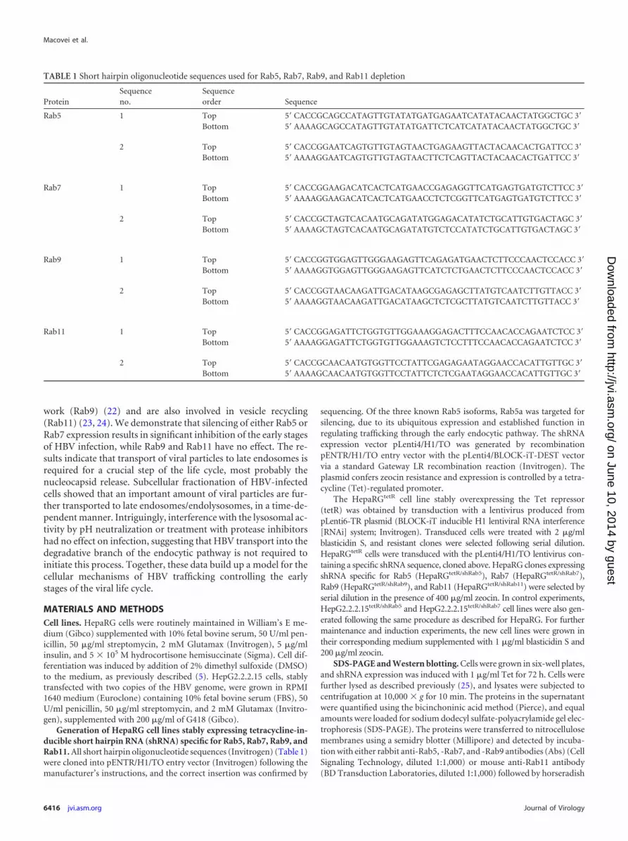
TABLE 1 Short hairpin oligonucleotide sequences used for Rab5, Rab7, Rab9, and Rab11 depletion
ProteinSequenceno.
Sequenceorder Sequence
Rab5 1 Top 5= CACCGCAGCCATAGTTGTATATGATGAGAATCATATACAACTATGGCTGC 3=Bottom 5= AAAAGCAGCCATAGTTGTATATGATTCTCATCATATACAACTATGGCTGC 3=
2 Top 5= CACCGGAATCAGTGTTGTAGTAACTGAGAAGTTACTACAACACTGATTCC 3=Bottom 5= AAAAGGAATCAGTGTTGTAGTAACTTCTCAGTTACTACAACACTGATTCC 3=
Rab7 1 Top 5= CACCGGAAGACATCACTCATGAACCGAGAGGTTCATGAGTGATGTCTTCC 3=Bottom 5= AAAAGGAAGACATCACTCATGAACCTCTCGGTTCATGAGTGATGTCTTCC 3=
2 Top 5= CACCGCTAGTCACAATGCAGATATGGAGACATATCTGCATTGTGACTAGC 3=Bottom 5= AAAAGCTAGTCACAATGCAGATATGTCTCCATATCTGCATTGTGACTAGC 3=
Rab9 1 Top 5= CACCGGTGGAGTTGGGAAGAGTTCAGAGATGAACTCTTCCCAACTCCACC 3=Bottom 5= AAAAGGTGGAGTTGGGAAGAGTTCATCTCTGAACTCTTCCCAACTCCACC 3=
2 Top 5= CACCGGTAACAAGATTGACATAAGCGAGAGCTTATGTCAATCTTGTTACC 3=Bottom 5= AAAAGGTAACAAGATTGACATAAGCTCTCGCTTATGTCAATCTTGTTACC 3=
Rab11 1 Top 5= CACCGGAGATTCTGGTGTTGGAAAGGAGACTTTCCAACACCAGAATCTCC 3=Bottom 5= AAAAGGAGATTCTGGTGTTGGAAAGTCTCCTTTCCAACACCAGAATCTCC 3=
2 Top 5= CACCGCAACAATGTGGTTCCTATTCGAGAGAATAGGAACCACATTGTTGC 3=Bottom 5= AAAAGCAACAATGTGGTTCCTATTCTCTCGAATAGGAACCACATTGTTGC 3=
Macovei et al.
6416 jvi.asm.org Journal of Virology
on June 10, 2014 by guesthttp://jvi.asm
.org/D
ownloaded from
peroxidase (HRP)-conjugated goat anti-rabbit (Cell Signaling Technol-ogy, diluted 1:2,000) or sheep anti-mouse (GE Healthcare, diluted1:2,000) Abs. Detection of calreticulin (Crt) expression with goat anti-Crt(Santa Cruz Biotechnology, diluted 1:500) followed by donkey anti-goatHRP-conjugated Abs (Santa Cruz Biotechnology, diluted 1:200) was usedas a control. The proteins were visualized using the ECL enhanced chemi-luminescence detection system (GE Healthcare).
Confocal fluorescence microscopy. Cells were seeded on cover glassand maintained for 3 days in the presence or absence of 1 �g/ml Tet.When human transferrin (hTfn) internalization was monitored, the cellswere washed with phosphate-buffered saline (PBS) and incubated for 1 hin serum-free medium before addition of 50 �g/ml hTfn-Alexa Fluor 488(Invitrogen), for 30 min at 4°C. The medium containing the fluorescentmarker was removed, and the cells were washed three times with PBS,followed by 30-min incubation in complete medium at 37°C. For micros-copy analysis cells were fixed with 4% paraformaldehyde, washed threetimes with PBS, permeabilized with 0.2% Triton X-100 in PBS, and incu-bated for 30 min with specific primary Abs. Rabbit anti-Rab5, -Rab7, and-Rab9 or mouse anti-Rab11 was used at a 1:100 dilution. Mouse anti-Lamp1 (Santa Cruz Biotechnology) and cation-independent mannose6-phosphate receptors (CI-MPRs) (Santa Cruz Biotechnology) were di-luted 1:1,000 and 1:250, respectively. Cells were washed three times withPBS and further incubated for 30 min with secondary Alexa Fluor 488-conjugated goat anti-mouse or Alexa Fluor 594-conjugated goat-anti rab-bit Abs (Invitrogen) at 1:400 dilutions. The samples were mounted withVectashield mounting medium (Invitrogen) containing DAPI (4=,6-di-amidino-2-phenylindole) to visualize the nuclei and analyzed under aZeiss LSM 710 laser-scanning confocal microscope using a 63� objective.Images were processed with ZEN software.
Flow cytometry analysis. Cells were seeded in six-well plates andmaintained for 3 days in the presence or absence of 1 �g/ml Tet. Toinvestigate the internalization of hTfn, cells were starved in serum-freemedium for 1 h at 37°C, followed by incubation with 50 �g/ml hTfn-Alexa Fluor 488 for 30 min at 4°C. Unbound hTfn was removed by wash-ing with PBS, and cells were further incubated for 30 min at 37°C incomplete medium. Noninternalized ligand was removed by treatmentwith 0.05% trypsin–EDTA followed by washing with PBS, before the flu-orescence-activated cell sorter (FACS) analysis. For LysoTracker internal-ization, the cells were rapidly detached with 0.05% trypsin–EDTA beforeincubation with 75 nM LysoTracker Green Dnd-26 (Invitrogen) for 3min. Duplicate samples from two independent experiments were ana-lyzed using the FACS Calibur and the Cell Quest Pro software.
HBV infection assay. HepaRG cells were differentiated as described inreference 5. A 300-fold-concentrated supernatant of HepG2.2.2.15 cellscontaining 104 HBV genome equivalents (Geq)/cell was used as viral in-oculum. Infection was performed in the presence of 4% polyethyleneglycol 8000 for 24 h at 37°C as described previously (26). In experimentsaddressing the effect of Rab proteins silencing on HBV life cycle, 1 �g/mlTet was added at day 3 prior to viral inoculation and maintained to day 3postinfection (p.i.); in a different setting, Tet treatment was applied fromdays 7 to 14 p.i., as indicated in the figure legends. When the effects ofdifferent inhibitors were monitored, the cells were incubated with me-dium containing 400 �M buthionine sulfoximine (BSO), 50 nM bafilo-mycin A (Baf), 50 mM NH4Cl or 100 �M E64d. The drugs were addedeither after removal of the HBV inoculum (BSO) or 24 h later (BSO, Baf,NH4Cl, and E64d) and were maintained for 24 h. Infected cells wereharvested 14 days p.i., and the encapsidated viral DNA was quantified byreal-time PCR. All infection experiments were performed with duplicatesamples and repeated at least three times.
Quantification of HBV DNA by real-time PCR. HBV-infectedHepaRG cells were harvested, and intracellular encapsidated viral DNAwas purified by phenol-chloroform extraction, as described elsewhere(27). The DNA was quantified using a Corbett Rotor-Gene 6000 real-timePCR system and Maxima SYBR green quantitative PCR (qPCR) mastermix (Fermentas), following amplification of a 279-bp HBV-specific frag-
ment. The number of viral Geq was determined using a calibration curvecontaining serial dilutions of known amounts of a pTriEx-HBV1.1 vector.
Quantification of HBV replication by Southern blotting.HepG2.2.2.15tetR/shRab5 and HepG2.2.2.15tetR/shRab7 cells were inducedwith 1 �g/ml Tet for 3 days or maintained untreated as a control. Treat-ment with 10 �M lamivudine (3TC; Moravek Biochemicals) was alsoincluded as a control of inhibition of viral replication. An equal number ofcells were used to extract nucelocapsid viral DNA, following the sameprocedure as for real-time PCR. The purified DNA was separated on a1.2% agarose gel and transferred to a Hybond-N membrane (GE Health-care), using a vacuum transfer blotter (Bio-Rad). The blot was hybridizedwith a fluorescein-labeled probe obtained by random priming using theHBV DNA genome as the template. Detection of the HBV-specific DNAbands was described previously (27).
Subcellular fractionation of HBV-infected cells. HepaRG cells weredifferentiated in collagen-coated T75 flasks and infected with 104 HBVGeq/cell, for 24 h. Following removal of the viral inoculum, the cells werewashed extensively with PBS containing 0.05% trypsin–EDTA to removeany bound and noninternalized virions. The cells were further incubatedfor either 24 or 48 h, extensively washed with PBS, and harvested with0.05% trypsin–EDTA. The pellet obtained following centrifugation ofcells for 5 min at 2,000 � g was washed twice with TS buffer (0.25 Msucrose, 10 mM Tris-Cl [pH 7.4]) then resuspended in 3 ml TS buffer onice. Samples were homogenized with 40 strokes in a Douncer on ice,followed by centrifugation at 1,000 � g for 20 min at 4°C. The supernatantwas centrifuged at 6,000 � g for 20 min at 4°C. The resulting supernatantwas resolved in a 30% Percoll gradient by centrifugation in a 70Ti rotor ofthe OptimaXPN-100 Beckman ultracentrifuge at 28,000 rpm for 45 min at4°C. A first fraction of 9 ml was collected from the top, followed by 20fractions of 0.6 ml. The Percoll was removed following 3-fold dilution ofeach fraction in TS buffer and centrifugation at 25,000 � g for 45 min at4°C. The pellets were split in two and used to quantify the amount encap-sidated viral DNA by real-time PCR or to determine the distribution ofintracellular markers by SDS-PAGE and Western blotting using anti-Lamp1 (dilution, 1:200), anti-Crt, and anti-cathepsin D (dilution 1:2,000)Abs (Abcam).
RESULTSCharacterization of HepaRG cell lines with tetracycline-induc-ible, downregulated Rab5, Rab7, Rab9, or Rab11 expression. In-terference with expression of key regulators of the endocytic path-way is widely used to investigate viral entry, the Rab proteins beingvery good candidates for such a task (28–31). However, geneticmanipulation is very problematic for primary hepatic or differen-tiated HepaRG cells, mainly because of the very low transfectionefficiency. Therefore, to investigate the trafficking events follow-ing HBV endocytosis, newly HepaRG cell lines were established,stably and controllably expression of shRNA molecules targetingRab5, Rab7, Rab9, and Rab11. This inducible gene expression sys-tem brings an enormous advantage over other approaches, byeliminating the discrepancies due to clonal variation and has beensuccessfully applied for HepaRG to investigate the role of the HBxprotein in HBV replication (32).
The new HepaRG clones were analyzed for the expression ofthe corresponding Rab proteins, in the absence and presence ofthe inducer, by Western blotting using specific Abs. As shown inFig. 1, addition of Tet to the cell medium resulted in a significantreduction of Rab5 biosynthesis in HepaRGtetR/shRab5 cells but notin the cell lines bearing shRNA targeted to either Rab7, Rab9, orRab11 (Fig. 1A). Similarly, Rab7 expression was downregulated inHepaRGtetR/shRab7 cells; however, it was unaffected in the remain-ing cell lines (Fig. 1B). The same behavior was observed for Rab9and Rab11 when their expression was analyzed in all four HepaRG
HBV Trafficking in HepaRG Cells
June 2013 Volume 87 Number 11 jvi.asm.org 6417
on June 10, 2014 by guesthttp://jvi.asm
.org/D
ownloaded from
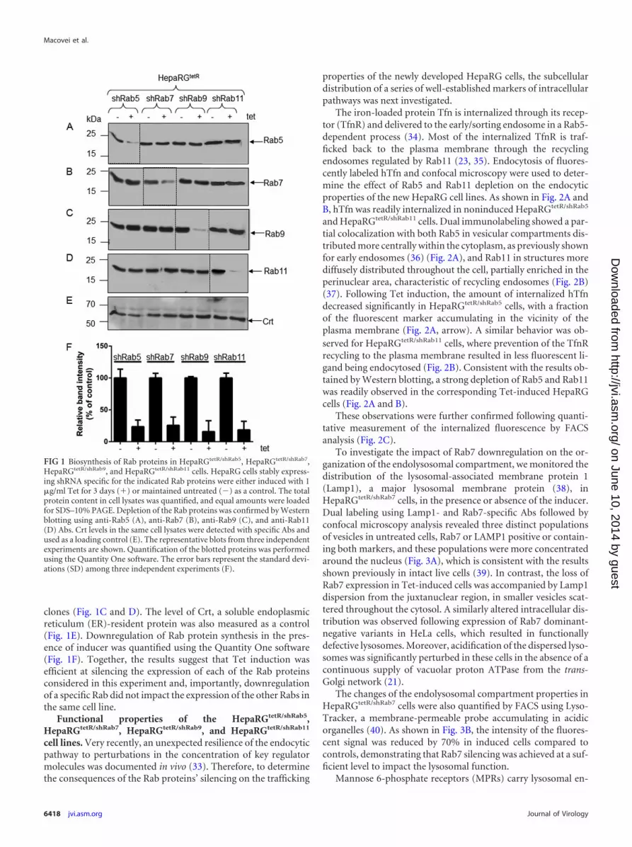
clones (Fig. 1C and D). The level of Crt, a soluble endoplasmicreticulum (ER)-resident protein was also measured as a control(Fig. 1E). Downregulation of Rab protein synthesis in the pres-ence of inducer was quantified using the Quantity One software(Fig. 1F). Together, the results suggest that Tet induction wasefficient at silencing the expression of each of the Rab proteinsconsidered in this experiment and, importantly, downregulationof a specific Rab did not impact the expression of the other Rabs inthe same cell line.
Functional properties of the HepaRGtetR/shRab5,HepaRGtetR/shRab7, HepaRGtetR/shRab9, and HepaRGtetR/shRab11
cell lines. Very recently, an unexpected resilience of the endocyticpathway to perturbations in the concentration of key regulatormolecules was documented in vivo (33). Therefore, to determinethe consequences of the Rab proteins’ silencing on the trafficking
properties of the newly developed HepaRG cells, the subcellulardistribution of a series of well-established markers of intracellularpathways was next investigated.
The iron-loaded protein Tfn is internalized through its recep-tor (TfnR) and delivered to the early/sorting endosome in a Rab5-dependent process (34). Most of the internalized TfnR is traf-ficked back to the plasma membrane through the recyclingendosomes regulated by Rab11 (23, 35). Endocytosis of fluores-cently labeled hTfn and confocal microscopy were used to deter-mine the effect of Rab5 and Rab11 depletion on the endocyticproperties of the new HepaRG cell lines. As shown in Fig. 2A andB, hTfn was readily internalized in noninduced HepaRGtetR/shRab5
and HepaRGtetR/shRab11 cells. Dual immunolabeling showed a par-tial colocalization with both Rab5 in vesicular compartments dis-tributed more centrally within the cytoplasm, as previously shownfor early endosomes (36) (Fig. 2A), and Rab11 in structures morediffusely distributed throughout the cell, partially enriched in theperinuclear area, characteristic of recycling endosomes (Fig. 2B)(37). Following Tet induction, the amount of internalized hTfndecreased significantly in HepaRGtetR/shRab5 cells, with a fractionof the fluorescent marker accumulating in the vicinity of theplasma membrane (Fig. 2A, arrow). A similar behavior was ob-served for HepaRGtetR/shRab11 cells, where prevention of the TfnRrecycling to the plasma membrane resulted in less fluorescent li-gand being endocytosed (Fig. 2B). Consistent with the results ob-tained by Western blotting, a strong depletion of Rab5 and Rab11was readily observed in the corresponding Tet-induced HepaRGcells (Fig. 2A and B).
These observations were further confirmed following quanti-tative measurement of the internalized fluorescence by FACSanalysis (Fig. 2C).
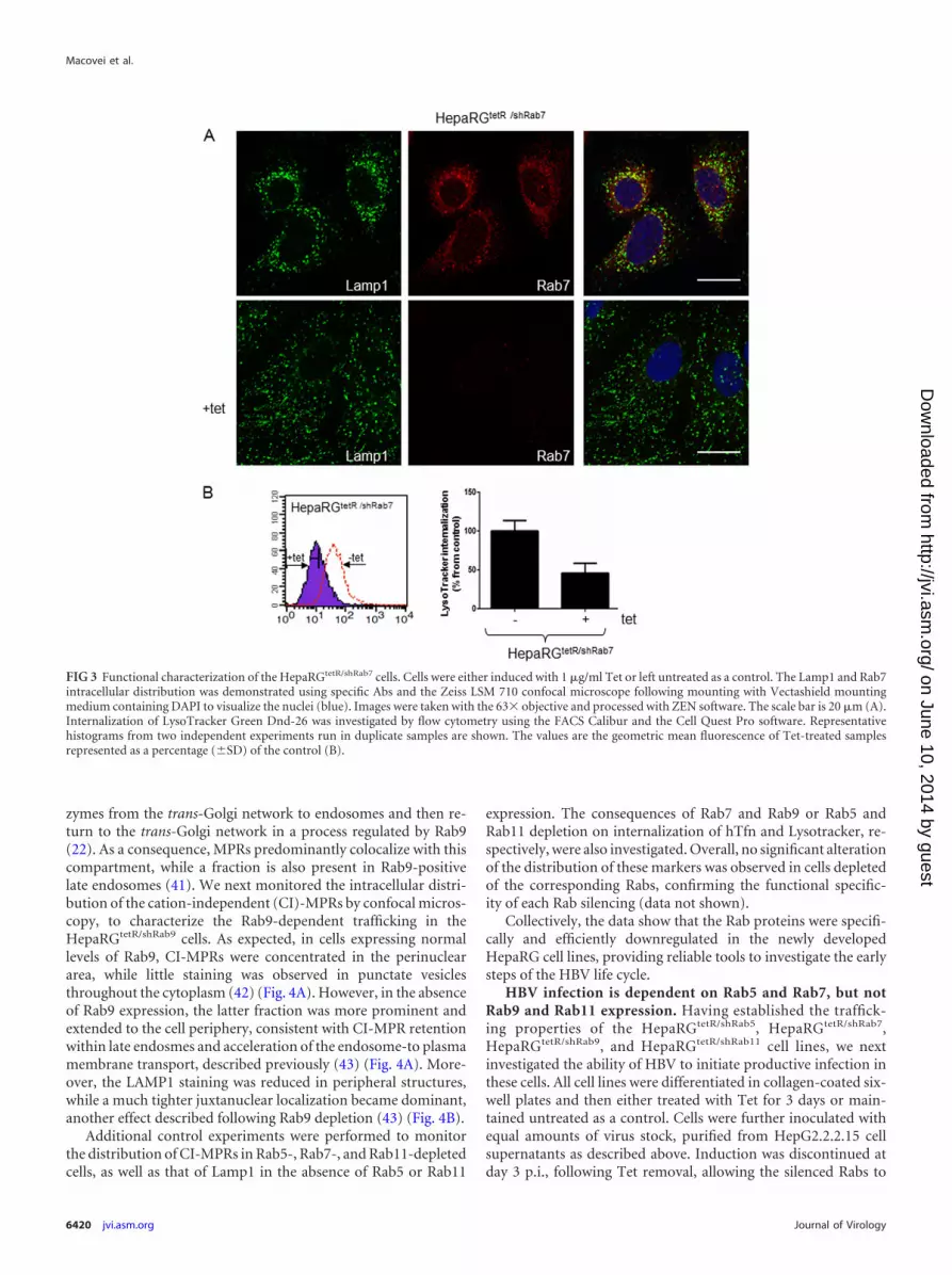
To investigate the impact of Rab7 downregulation on the or-ganization of the endolysosomal compartment, we monitored thedistribution of the lysosomal-associated membrane protein 1(Lamp1), a major lysosomal membrane protein (38), inHepaRGtetR/shRab7 cells, in the presence or absence of the inducer.Dual labeling using Lamp1- and Rab7-specific Abs followed byconfocal microscopy analysis revealed three distinct populationsof vesicles in untreated cells, Rab7 or LAMP1 positive or contain-ing both markers, and these populations were more concentratedaround the nucleus (Fig. 3A), which is consistent with the resultsshown previously in intact live cells (39). In contrast, the loss ofRab7 expression in Tet-induced cells was accompanied by Lamp1dispersion from the juxtanuclear region, in smaller vesicles scat-tered throughout the cytosol. A similarly altered intracellular dis-tribution was observed following expression of Rab7 dominant-negative variants in HeLa cells, which resulted in functionallydefective lysosomes. Moreover, acidification of the dispersed lyso-somes was significantly perturbed in these cells in the absence of acontinuous supply of vacuolar proton ATPase from the trans-Golgi network (21).
The changes of the endolysosomal compartment properties inHepaRGtetR/shRab7 cells were also quantified by FACS using Lyso-Tracker, a membrane-permeable probe accumulating in acidicorganelles (40). As shown in Fig. 3B, the intensity of the fluores-cent signal was reduced by 70% in induced cells compared tocontrols, demonstrating that Rab7 silencing was achieved at a suf-ficient level to impact the lysosomal function.
Mannose 6-phosphate receptors (MPRs) carry lysosomal en-
FIG 1 Biosynthesis of Rab proteins in HepaRGtetR/shRab5, HepaRGtetR/shRab7,HepaRGtetR/shRab9, and HepaRGtetR/shRab11 cells. HepaRG cells stably express-ing shRNA specific for the indicated Rab proteins were either induced with 1�g/ml Tet for 3 days (�) or maintained untreated (�) as a control. The totalprotein content in cell lysates was quantified, and equal amounts were loadedfor SDS–10% PAGE. Depletion of the Rab proteins was confirmed by Westernblotting using anti-Rab5 (A), anti-Rab7 (B), anti-Rab9 (C), and anti-Rab11(D) Abs. Crt levels in the same cell lysates were detected with specific Abs andused as a loading control (E). The representative blots from three independentexperiments are shown. Quantification of the blotted proteins was performedusing the Quantity One software. The error bars represent the standard devi-ations (SD) among three independent experiments (F).
Macovei et al.
6418 jvi.asm.org Journal of Virology
on June 10, 2014 by guesthttp://jvi.asm
.org/D
ownloaded from
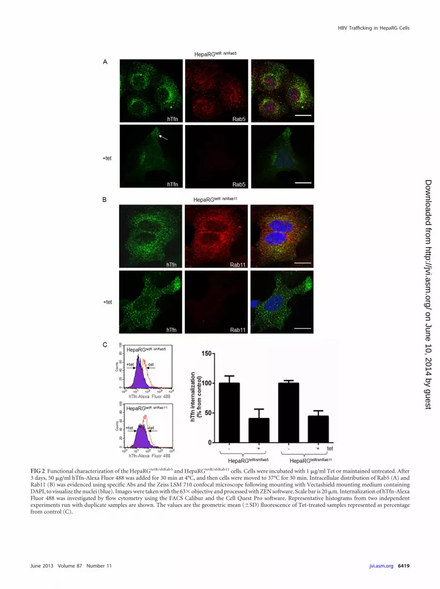
FIG 2 Functional characterization of the HepaRGtetR/shRab5 and HepaRGtetR/shRab11 cells. Cells were incubated with 1 �g/ml Tet or maintained untreated. After3 days, 50 �g/ml hTfn-Alexa Fluor 488 was added for 30 min at 4°C, and then cells were moved to 37°C for 30 min. Intracellular distribution of Rab5 (A) andRab11 (B) was evidenced using specific Abs and the Zeiss LSM 710 confocal microscope following mounting with Vectashield mounting medium containingDAPI, to visualize the nuclei (blue). Images were taken with the 63� objective and processed with ZEN software. Scale bar is 20 �m. Internalization of hTfn-AlexaFluor 488 was investigated by flow cytometry using the FACS Calibur and the Cell Quest Pro software. Representative histograms from two independentexperiments run with duplicate samples are shown. The values are the geometric mean (�SD) fluorescence of Tet-treated samples represented as percentagefrom control (C).
HBV Trafficking in HepaRG Cells
June 2013 Volume 87 Number 11 jvi.asm.org 6419
on June 10, 2014 by guesthttp://jvi.asm
.org/D
ownloaded from
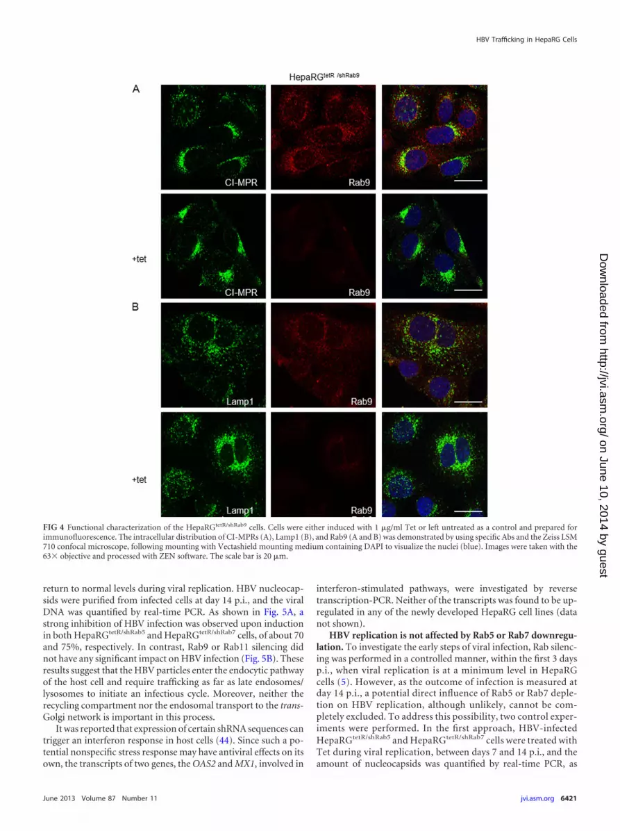
zymes from the trans-Golgi network to endosomes and then re-turn to the trans-Golgi network in a process regulated by Rab9(22). As a consequence, MPRs predominantly colocalize with thiscompartment, while a fraction is also present in Rab9-positivelate endosomes (41). We next monitored the intracellular distri-bution of the cation-independent (CI)-MPRs by confocal micros-copy, to characterize the Rab9-dependent trafficking in theHepaRGtetR/shRab9 cells. As expected, in cells expressing normallevels of Rab9, CI-MPRs were concentrated in the perinucleararea, while little staining was observed in punctate vesiclesthroughout the cytoplasm (42) (Fig. 4A). However, in the absenceof Rab9 expression, the latter fraction was more prominent andextended to the cell periphery, consistent with CI-MPR retentionwithin late endosmes and acceleration of the endosome-to plasmamembrane transport, described previously (43) (Fig. 4A). More-over, the LAMP1 staining was reduced in peripheral structures,while a much tighter juxtanuclear localization became dominant,another effect described following Rab9 depletion (43) (Fig. 4B).
Additional control experiments were performed to monitorthe distribution of CI-MPRs in Rab5-, Rab7-, and Rab11-depletedcells, as well as that of Lamp1 in the absence of Rab5 or Rab11
expression. The consequences of Rab7 and Rab9 or Rab5 andRab11 depletion on internalization of hTfn and Lysotracker, re-spectively, were also investigated. Overall, no significant alterationof the distribution of these markers was observed in cells depletedof the corresponding Rabs, confirming the functional specific-ity of each Rab silencing (data not shown).
Collectively, the data show that the Rab proteins were specifi-cally and efficiently downregulated in the newly developedHepaRG cell lines, providing reliable tools to investigate the earlysteps of the HBV life cycle.
HBV infection is dependent on Rab5 and Rab7, but notRab9 and Rab11 expression. Having established the traffick-ing properties of the HepaRGtetR/shRab5, HepaRGtetR/shRab7,HepaRGtetR/shRab9, and HepaRGtetR/shRab11 cell lines, we nextinvestigated the ability of HBV to initiate productive infection inthese cells. All cell lines were differentiated in collagen-coated six-well plates and then either treated with Tet for 3 days or main-tained untreated as a control. Cells were further inoculated withequal amounts of virus stock, purified from HepG2.2.2.15 cellsupernatants as described above. Induction was discontinued atday 3 p.i., following Tet removal, allowing the silenced Rabs to
FIG 3 Functional characterization of the HepaRGtetR/shRab7 cells. Cells were either induced with 1 �g/ml Tet or left untreated as a control. The Lamp1 and Rab7intracellular distribution was demonstrated using specific Abs and the Zeiss LSM 710 confocal microscope following mounting with Vectashield mountingmedium containing DAPI to visualize the nuclei (blue). Images were taken with the 63� objective and processed with ZEN software. The scale bar is 20 �m (A).Internalization of LysoTracker Green Dnd-26 was investigated by flow cytometry using the FACS Calibur and the Cell Quest Pro software. Representativehistograms from two independent experiments run in duplicate samples are shown. The values are the geometric mean fluorescence of Tet-treated samplesrepresented as a percentage (�SD) of the control (B).
Macovei et al.
6420 jvi.asm.org Journal of Virology
on June 10, 2014 by guesthttp://jvi.asm
.org/D
ownloaded from
return to normal levels during viral replication. HBV nucleocap-sids were purified from infected cells at day 14 p.i., and the viralDNA was quantified by real-time PCR. As shown in Fig. 5A, astrong inhibition of HBV infection was observed upon inductionin both HepaRGtetR/shRab5 and HepaRGtetR/shRab7 cells, of about 70and 75%, respectively. In contrast, Rab9 or Rab11 silencing didnot have any significant impact on HBV infection (Fig. 5B). Theseresults suggest that the HBV particles enter the endocytic pathwayof the host cell and require trafficking as far as late endosomes/lysosomes to initiate an infectious cycle. Moreover, neither therecycling compartment nor the endosomal transport to the trans-Golgi network is important in this process.
It was reported that expression of certain shRNA sequences cantrigger an interferon response in host cells (44). Since such a po-tential nonspecific stress response may have antiviral effects on itsown, the transcripts of two genes, the OAS2 and MX1, involved in
interferon-stimulated pathways, were investigated by reversetranscription-PCR. Neither of the transcripts was found to be up-regulated in any of the newly developed HepaRG cell lines (datanot shown).
HBV replication is not affected by Rab5 or Rab7 downregu-lation. To investigate the early steps of viral infection, Rab silenc-ing was performed in a controlled manner, within the first 3 daysp.i., when viral replication is at a minimum level in HepaRGcells (5). However, as the outcome of infection is measured atday 14 p.i., a potential direct influence of Rab5 or Rab7 deple-tion on HBV replication, although unlikely, cannot be com-pletely excluded. To address this possibility, two control exper-iments were performed. In the first approach, HBV-infectedHepaRGtetR/shRab5 and HepaRGtetR/shRab7 cells were treated withTet during viral replication, between days 7 and 14 p.i., and theamount of nucleocapsids was quantified by real-time PCR, as
FIG 4 Functional characterization of the HepaRGtetR/shRab9 cells. Cells were either induced with 1 �g/ml Tet or left untreated as a control and prepared forimmunofluorescence. The intracellular distribution of CI-MPRs (A), Lamp1 (B), and Rab9 (A and B) was demonstrated by using specific Abs and the Zeiss LSM710 confocal microscope, following mounting with Vectashield mounting medium containing DAPI to visualize the nuclei (blue). Images were taken with the63� objective and processed with ZEN software. The scale bar is 20 �m.
HBV Trafficking in HepaRG Cells
June 2013 Volume 87 Number 11 jvi.asm.org 6421
on June 10, 2014 by guesthttp://jvi.asm
.org/D
ownloaded from

above. As shown in Fig. 6A, neither Rab5, nor Rab7 silencing hadany significant effect on the viral DNA level. In a second approach,Rab5 and Rab7 depletion was achieved in HepG2.2.2.15 cells,which are not permissive for HBV infection, using the same Tet-controlled shRNA expression system employed for the HepaRGcells. The decreased amount of Rab5 and Rab7 proteins in inducedcells was confirmed by Western blotting (Fig. 6B). Southern blot-ting was further used to monitor the HBV replication in the newlyestablished HepaG2.2.2.15tetR/shRab5 and HepaG2.2.2.15tetR/shRab7
cells, in the presence or absence of inducer. Lamivudine (3TC), apotent HBV replication inhibitor was also included in the exper-iment as a control. As shown in Fig. 6C, Tet treatment had noimpact on the HBV replication forms (RF) in either HepG2.2.2.15cell line, while 3TC inhibited replication almost completely, asexpected.
Together, the results demonstrate that the inhibitory effects ofRab5 and Rab7 described in the section above clearly address theearly stages of viral entry.
HBV is trafficked to the degradative branch of the endocyticpathway. To further characterize the compartments traversed byHBV following internalization, a subcellular fractionation exper-iment was performed with HepaRG cells at 24 and 48 h p.i. byultracentrifugation in a 30% Percoll gradient. This type of gradi-ent has been used before to separate endocytic vesicles in twodistinct populations: a light fraction consisting of early endosomesand heterogeneous secretory vesicles and a heavier fraction mainlycomposed of late endosomes/lysosomes (45).
The postnuclear fractions were collected from top to bottomand characterized for the presence of different endocytic markersby Western blotting and viral nucleocapsids by real-time PCR.Lamp1 was concentrated in fractions 2 to 6, which were also pos-itive for the ER-resident protein Crt, and fractions 15 to 20, whichwere heavily loaded with the 34-kDa, mature cathepsin D chaincharacteristic of the lysosomal compartment (46) (Fig. 7A). Theseresults are consistent with Lamp1 being shuttled between lyso-somes and endosomes and confirm the successful separation ofthe endolysosome heavy fraction from the remaining heteroge-nous vesicles. Interestingly, purification of the HBV particles fromthe collected fractions showed that the virus is distributed alongthe endocytic pathway during 24 h p.i., mainly in the light endo-somal vesicles. In contrast, the major pool of HBV particles is
FIG 6 HBV replication in Rab-depleted cells. Differentiated HepaRGtetR/shRab5 and
HepaRGtetR/shRab7 cells were infected with 104 HBV Geq/cell and then treatedwith 1 �g/ml Tet from day 7 to day 14 p.i. (�) or maintained untreated (�) asthe control. Infected cells were harvested 14 days p.i., and the amount ofencapsidated viral DNA was quantified by real-time PCR. The results are ex-pressed as percentage of HBV infection from untreated controls. The errorbars represent the standard deviations among three independent experi-ments, each run in duplicate samples (A). HepaG2.2.2.15tetR/shRab5 andHepaG2.2.2.15tetR/shRab7 cells were induced with 1 �g/ml Tet for 3 days ormaintained untreated as a control. Depletion of Rab5 and Rab7 was confirmedby Western blotting using specific Abs and Crt expression as a loading control(B). HepG2.2.2.15tetR/shRab5 and HepG2.2.2.15tetR/shRab7 cells were treated with1 �g/ml Tet or 10 �M 3TC for 3 days or maintained untreated as a control.HBV nucleocapsids were extracted from an equal number of cells, and thepurified DNA was analyzed by Southern blotting. The HBV replication forms(RF) were detected with a fluorescein-labeled probe obtained by randompriming using the HBV DNA genome as the template (C).
FIG 5 HBV infection in Rab-depleted cells. Differentiated cells were treated with 1 �g/ml Tet for 3 days (�) or maintained untreated (�) before infection with104 HBV Geq/cell. Treatment was discontinued at day 3 p.i. Infected cells were harvested 14 days p.i., and the amount of encapsidated viral DNA was quantifiedby real-time PCR. The results are expressed as the percentage of HBV infection from untreated controls. The error bars represent the SD between threeindependent experiments, each run in duplicate samples (A and B).
Macovei et al.
6422 jvi.asm.org Journal of Virology
on June 10, 2014 by guesthttp://jvi.asm
.org/D
ownloaded from
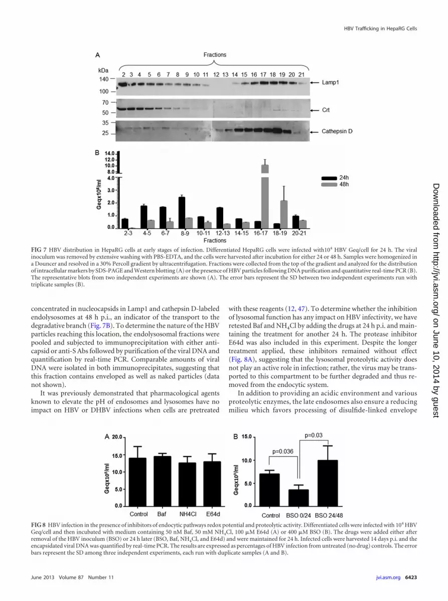
concentrated in nucleocapsids in Lamp1 and cathepsin D-labeledendolysosomes at 48 h p.i., an indicator of the transport to thedegradative branch (Fig. 7B). To determine the nature of the HBVparticles reaching this location, the endolysosomal fractions werepooled and subjected to immunoprecipitation with either anti-capsid or anti-S Abs followed by purification of the viral DNA andquantification by real-time PCR. Comparable amounts of viralDNA were isolated in both immunoprecipitates, suggesting thatthis fraction contains enveloped as well as naked particles (datanot shown).
It was previously demonstrated that pharmacological agentsknown to elevate the pH of endosomes and lysosomes have noimpact on HBV or DHBV infections when cells are pretreated
with these reagents (12, 47). To determine whether the inhibitionof lysosomal function has any impact on HBV infectivity, we haveretested Baf and NH4Cl by adding the drugs at 24 h p.i. and main-taining the treatment for another 24 h. The protease inhibitorE64d was also included in this experiment. Despite the longertreatment applied, these inhibitors remained without effect(Fig. 8A), suggesting that the lysosomal proteolytic activity doesnot play an active role in infection; rather, the virus may be trans-ported to this compartment to be further degraded and thus re-moved from the endocytic system.
In addition to providing an acidic environment and variousproteolytic enzymes, the late endosomes also ensure a reducingmilieu which favors processing of disulfide-linked envelope
FIG 7 HBV distribution in HepaRG cells at early stages of infection. Differentiated HepaRG cells were infected with104 HBV Geq/cell for 24 h. The viralinoculum was removed by extensive washing with PBS-EDTA, and the cells were harvested after incubation for either 24 or 48 h. Samples were homogenized ina Douncer and resolved in a 30% Percoll gradient by ultracentrifugation. Fractions were collected from the top of the gradient and analyzed for the distributionof intracellular markers by SDS-PAGE and Western blotting (A) or the presence of HBV particles following DNA purification and quantitative real-time PCR (B).The representative blots from two independent experiments are shown (A). The error bars represent the SD between two independent experiments run withtriplicate samples (B).
FIG 8 HBV infection in the presence of inhibitors of endocytic pathways redox potential and proteolytic activity. Differentiated cells were infected with 104 HBVGeq/cell and then incubated with medium containing 50 nM Baf, 50 mM NH4Cl, 100 �M E64d (A) or 400 �M BSO (B). The drugs were added either afterremoval of the HBV inoculum (BSO) or 24 h later (BSO, Baf, NH4Cl, and E64d) and were maintained for 24 h. Infected cells were harvested 14 days p.i. and theencapsidated viral DNA was quantified by real-time PCR. The results are expressed as percentages of HBV infection from untreated (no drug) controls. The errorbars represent the SD among three independent experiments, each run with duplicate samples (A and B).
HBV Trafficking in HepaRG Cells
June 2013 Volume 87 Number 11 jvi.asm.org 6423
on June 10, 2014 by guesthttp://jvi.asm
.org/D
ownloaded from

proteins and could potentially facilitate virus release from theendocytic compartment (48). To test if this hypothesis appliesto HBV, we depleted the cells of glutathione (GSH), by usingBSO, a membrane-permeable inhibitor that irreversibly inhib-its �-glutamylcysteine synthetase, the enzyme required forGSH biosynthesis (49). Interestingly, BSO treatment within thefirst 24 h p.i. resulted in about 55% reduction of HBV infection,while the inhibitory effect was abolished when the drug wasadded at later points p.i. (Fig. 8B). This suggests that a reduc-tion step along the endocytic pathway is required for initiationof a productive infection.
DISCUSSION
Although important advances have been made in recent years to-ward understanding HBV entry into host cells, many issues re-main unclear. Very recently, NTCP, a liver bile acid transporter,was proposed as a potential receptor for HBV (13). NTCP over-expression in cell lines that are not permissive for HBV infection
rendered them susceptible; however, the efficiency of the infectionremained intriguingly low, despite these cells being able to ensurehigh levels of virus replication when the entry steps are bypassedby transfection of the viral genome. This strongly suggests thatplasma membrane receptors are not the only limiting factors ofinfection, other yet unidentified cellular factors may significantlycontribute to this process at its very early steps.
This study addressed the unexplored HBV fate, postinternal-ization in HepaRG cells. By depleting the cells of several key Rabproteins in a controlled manner, we were able to monitor theintracellular trafficking of HBV particles following endocytosis.The data show that HBV requires Rab5- and Rab7-dependenttransport through the endosomal compartment to initiate a suc-cessful infection, while neither the recycling endosomes nor thevesicles en route to trans-Golgi network are involved in this pro-cess (summarized in Fig. 9). The use of early endocytic pathway bythe HBV particles is not in disagreement with our previous resultsreporting a dependence on functional caveolin-1 for productive
FIG 9 Schematic representation of HBV trafficking following internalization in HepaRG cells. HBV infection depends on Rab5 and Rab7 transportthrough the endocytic pathway, while neither the recycling endosomes nor the vesicles en route to the trans-Golgi network play a role in this process. Viraluncoating occurs in a compartment preceding the lysosomes, in a redox-potential-dependent, pH- and proteolytic activity-independent manner. Furthertransport to lysosomes has no influence on infection, indicating a role in viral disposal. Other proteins shown to play a function in HBV infection aremarked with an asterisk.
Macovei et al.
6424 jvi.asm.org Journal of Virology
on June 10, 2014 by guesthttp://jvi.asm
.org/D
ownloaded from
infection (12). Recent studies have linked caveolin to conven-tional endocytic trafficking pathways and the existence of thecaveosome, the unique transport organelle involved in caveola-mediated endocytosis, is now doubted (50).
The results of this study imply that viral particles enter earlyendosomes that progress to late endosomes where the appropriateenvironment may provide the signal for the release of the viralgenome into the cytoplasm, leading to infection. Traffickingthrough endosomes is known to be important for viruses thatdepend on low pH or endosomal proteases to facilitate uncoating(51, 52). However, acidification does not appear to be an essentialfactor for HBV infection, as extended cell treatment with Baf orNH4Cl did not affect this process; similarly, inhibition of endo-somal cathepsin activity remained without having any conse-quences on the infection outcome. This intriguing behavior is notsingular to HBV; other viruses, such as echovirus 7, use the endo-cytic trafficking to initiate infection in a pH- and protease activity-independent manner (53). One possible interpretation is that traf-ficking through endosomes, albeit essential, may only represent anintermediate step leading to a cellular organelle where the viralgenome is released. For instance, it has been shown that canineparvovirus and mouse polyomavirus use the recycling endosomesto finally reach the lysosomes and the ER, respectively (54, 55);however, the recycle compartment was clearly shown to be dis-pensable for HBV infection. Alternatively, the late endosomesmay host another factor essential for HBV uncoating. The struc-ture of the HBV envelope is stabilized by extensive intra- andintermolecular disulfide cross-linking of the component polypep-tides (56), thus the disulfide bond-reducing activity of the endo-cytic pathway is a good candidate for such a function (57, 58).Using fluorescence resonance energy transfer (FRET)-based im-aging, it was shown that the disulfide reduction machinery is ac-tivated upon internalization of a folate-FRET reporter into endo-somes, independent of the interaction of this compartment withthe lysosomes or the Golgi apparatus (59). Such activity requiresdeprotonation of thiols, which is not favored thermodynamicallyby the acidic environment of the endosome, suggesting that mostlikely, an enzymatic catalysis is involved (59). Interestingly, entryof other enveloped viruses, such as the human immunodeficiencyvirus (HIV), depends on a reduction step involving a cell-surfaceprotein disulfide isomerase (PDI)-related protein (60, 61). How-ever, this does not appear to be the case for HBV, since treatmentof cells with either membrane-impermeable compounds that alterthe redox state of surface-exposed cysteine residues or PDI inhib-itors had no effect on infection (62). In our study, the kinetics ofHBV infection in the presence of the membrane-permeable GSHbiosynthesis inhibitor favors the hypothesis of thiol-disulfide ex-change occurring in an internal cellular compartment, not at thecell surface. Indeed, BSO efficiently inhibited HBV early in infec-tion but not at later points, implying that the reduction step isrestricted to a particular location along the endocytic pathway. Weare currently employing extensive proteomic analysis to identifyadditional host factors associated with the redox machinery of theendocytic pathway with a potential role in infection, using an ap-proach described before for the plasma membrane proteins (63).
Monitoring of viral entry by single-particle tracking has notbeen possible for HBV, as infection efficiency is very low in vitroand attempts to label functional HBV virions have failed so far,despite successful approaches being described for SVP (64, 65). Asan alternative, here we have employed subcellular fractionation
and amplification of encapsidated viral DNA to further examinethe intracellular distribution of internalized virions. After uptakeby differentiated HepaRG cells, the viral particles were found dis-persed into a fraction of heterogeneous endocytic vesicles, consist-ing of early and late endosomes, while they later were concen-trated into lysosomes, suggesting that viral nucleocapsids remainintact in the endocytic pathway for at least several days. This isconsistent with the observation made during the first character-ization of the HepaRG cell line, showing that the input viral par-ticle level is relatively stable, being detected within the cells for atleast 2 days (5). Moreover, enveloped virus particles were immu-noprecipitated in the lysosomal fraction, which is an indicationthat uncoating in a previous compartment is a rather inefficientprocess, in competition with trafficking further down the degra-dation pathway. The HBV transport to lysosomes remains in-triguing, as no evidence for a role of this compartment in promot-ing infection could be demonstrated. Interestingly, it has beenshown that a fraction of HIV particles bound to target cells is takenup by endocytosis but remains entrapped in the endocytic path-way and fails to initiate infection, eventually being targeted tolysosomes for degradation (66). Following cell treatment withpharmacological agents that increase the pH of endosomes andlysosomes, HIV-1 infection was greatly enhanced suggesting thatthe virus is able to escape from lysosomes when rescued fromdegradation (66). This behavior was not observed for HBV;rather, the trafficking into the endocytic degradative branch ap-pears to be a dead-end pathway, possibly as a part of the cell de-fense mechanisms to limit the number of viral particles availableto initiate productive infection. This hypothesis is supported bythe observation that internalized HBV core particles are trans-ported to lysosomes and further disassembled as a result of endo-proteolytic cleavage within the arginine-rich domain of the corepolypeptide, by cysteine proteases (67).
Taken together, these studies indicate that HBV transportthrough early and late endosomal compartments is a viable path-way leading to productive infection in HepaRG cells. Future ex-periments should establish whether the lysosomal trafficking andaccumulation of the viral particles in this compartment are con-tributing factors to the unusual low efficiency of HBV infection invitro, characterized by low kinetics and requiring a high concen-tration of virus inoculum (26).
ACKNOWLEDGMENTS
This work was supported by the Romanian Academy Project 3 of theInstitute of Biochemistry. Alina Macovei and Catalin Lazar were sup-ported by the Sectoral Operational Programme Human Resources Devel-opment POSDRU/89/1.5/S/60746 grant. Catalina Petrareanu was sup-ported by the Sectoral Operational Programme Human ResourcesDevelopment 2007–2013 of the Romanian Ministry of Labor, Family andSocial Protection through Financial Agreement POSDRU/107/1.5/S/76903.
REFERENCES1. Ganem D, Prince AM. 2004. Hepatitis B virus infection—natural history
and clinical consequences. N. Engl. J. Med. 350:1118 –1129.2. Bruss V. 2007. Hepatitis B virus morphogenesis. World J. Gastroenterol.
13:65–73.3. Zuckerman AJ. 1996. Hepatitis viruses. Chapter 70. In Baron S (ed),
Medical microbiology, 4th ed. University of Texas Medical Branch,Galveston, TX.
4. Heermann KH, Goldmann U, Schwartz W, Seyffarth T, Baumgarten H,Gerlich WH. 1984. Large surface proteins of hepatitis B virus containingthe pre-s sequence. J. Virol. 52:396 – 402.
HBV Trafficking in HepaRG Cells
June 2013 Volume 87 Number 11 jvi.asm.org 6425
on June 10, 2014 by guesthttp://jvi.asm
.org/D
ownloaded from
5. Gripon P, Rumin S, Urban S, Le Seyec J, Glaise D, Cannie I, GuyomardC, Lucas J, Trepo C, Guguen-Guillouzo C. 2002. Infection of a humanhepatoma cell line by hepatitis B virus. Proc. Natl. Acad. Sci. U. S. A.99:15655–15660.
6. Huang HC, Chen CC, Chang WC, Tao MH, Huang C. 2012. Entry ofhepatitis B virus into immortalized human primary hepatocytes by clath-rin-dependent endocytosis. J. Virol. 86:9443–9453.
7. Aly HH, Watashi K, Hijikata M, Kaneko H, Takada Y, Egawa H,Uemoto S, Shimotohno K. 2007. Serum-derived hepatitis C virus infec-tivity in interferon regulatory factor-7-suppressed human primary hepa-tocytes. J. Hepatol. 46:26 –36.
8. Paganelli M, Dallmeier K, Nyabi O, Scheers I, Kabamba B, Neyts J,Goubau P, Najimi M, Sokal EM. 2013. Differentiated umbilical cordmatrix stem cells as a new in vitro model to study early events duringhepatitis B virus infection. Hepatology 57:59 – 69.
9. Schulze A, Gripon P, Urban S. 2007. Hepatitis B virus infection initiateswith a large surface protein-dependent binding to heparan sulfate pro-teoglycans. Hepatology 46:1759 –1768.
10. Leistner CM, Gruen-Bernhard S, Glebe D. 2008. Role of glycosamino-glycans for binding and infection of hepatitis B virus. Cell. Microbiol.10:122–133.
11. Sureau C, Salisse J. 2013. A conformational heparan sulfate-binding siteessential to infectivity overlaps with the conserved hepatitis B virus A-de-terminant. Hepatology 57:985–994.
12. Macovei A, Radulescu C, Lazar C, Petrescu S, Durantel D, Dwek RA,Zitzmann N, Nichita NB. 2010. Hepatitis B virus requires intact caveo-lin-1 function for productive infection in HepaRG cells. J. Virol. 84:243–253.
13. Yan H, Zhong G, Xu G, He W, Jing Z, Gao Z, Huang Y, Qi Y, Peng B,Wang H, Fu L, Song M, Chen P, Gao W, Ren B, Sun Y, Cai T, Feng X,Sui J, Li W. 2012. Sodium taurocholate cotransporting polypeptide is afunctional receptor for human hepatitis B and D virus. eLife 1:e00049.doi:10.7554/eLife.00049.
14. Martinez O, Goud B. 1998. Rab proteins. Biochim. Biophys. Acta 1404:101–112.
15. Zerial M, McBride H. 2001. Rab proteins as membrane organizers. Nat.Rev. Mol. Cell Biol. 2:107–117.
16. Somsel Rodman J, Wandinger-Ness A. 2000. Rab GTPases coordinateendocytosis. J. Cell Sci. 113:183–192.
17. Smythe E. 2002. Regulating the clathrin-coated vesicle cycle by AP2 sub-unit phosphorylation. Trends Cell Biol. 12:352–354.
18. Vonderheit A, Helenius A. 2005. Rab7 associates with early endosomes tomediate sorting and transport of Semliki forest virus to late endosomes.PLoS Biol. 3:e233. doi:10.1371/journal.pbio.0030233.
19. Rink J, Ghigo E, Kalaidzidis Y, Zerial M. 2005. Rab conversion as amechanism of progression from early to late endosomes. Cell 122:735–749.
20. Gorvel JP, Chavrier P, Zerial M, Gruenberg J. 1991. rab5 controls earlyendosome fusion in vitro. Cell 64:915–925.
21. Bucci C, Thomsen P, Nicoziani P, McCarthy J, van Deurs B. 2000. Rab7:a key to lysosome biogenesis. Mol. Biol. Cell 11:467– 480.
22. Lombardi D, Soldati T, Riederer MA, Goda Y, Zerial M, Pfeffer SR.1993. Rab9 functions in transport between late endosomes and the transGolgi network. EMBO J. 12:677– 682.
23. Ullrich O, Reinsch S, Urbe S, Zerial M, Parton RG. 1996. Rab11regulates recycling through the pericentriolar recycling endosome. J. CellBiol. 135:913–924.
24. Urbe S, Huber LA, Zerial M, Tooze SA, Parton RG. 1993. Rab11, a smallGTPase associated with both constitutive and regulated secretory path-ways in PC12 cells. FEBS Lett. 334:175–182.
25. Macovei A, Zitzmann N, Lazar C, Dwek RA, Branza-Nichita N. 2006.Brefeldin A inhibits pestivirus release from infected cells, without affectingits assembly and infectivity. Biochem. Biophys. Res. Commun. 346:1083–1090.
26. Schulze A, Mills K, Weiss TS, Urban S. 2012. Hepatocyte polarization isessential for the productive entry of the hepatitis B virus. Hepatology55:373–383.
27. Lazar C, Durantel D, Macovei A, Zitzmann N, Zoulim F, Dwek RA,Branza-Nichita N. 2007. Treatment of hepatitis B virus-infected cells withalpha-glucosidase inhibitors results in production of virions with alteredmolecular composition and infectivity. Antiviral Res. 76:30 –37.
28. Krishnan MN, Sukumaran B, Pal U, Agaisse H, Murray JL, Hodge TW,
Fikrig E. 2007. Rab 5 is required for the cellular entry of dengue and WestNile viruses. J. Virol. 81:4881– 4885.
29. Meertens L, Bertaux C, Dragic T. 2006. Hepatitis C virus entry requiresa critical postinternalization step and delivery to early endosomes viaclathrin-coated vesicles. J. Virol. 80:11571–11578.
30. Sieczkarski SB, Whittaker GR. 2003. Differential requirements of Rab5and Rab7 for endocytosis of influenza and other enveloped viruses. Traffic4:333–343.
31. Wolf M, Deal EM, Greenberg HB. 2012. Rhesus rotavirus traffickingduring entry into MA104 cells is restricted to the early endosome com-partment. J. Virol. 86:4009 – 4013.
32. Lucifora J, Arzberger S, Durantel D, Belloni L, Strubin M, Levrero M,Zoulim F, Hantz O, Protzer U. 2011. Hepatitis B virus X protein isessential to initiate and maintain virus replication after infection. J. Hepa-tol. 55:996 –1003.
33. Zeigerer A, Gilleron J, Bogorad RL, Marsico G, Nonaka H, Seifert S,Epstein-Barash H, Kuchimanchi S, Peng CG, Ruda VM, Del Conte-Zerial P, Hengstler JG, Kalaidzidis Y, Koteliansky V, Zerial M. 2012.Rab5 is necessary for the biogenesis of the endolysosomal system in vivo.Nature 485:465– 470.
34. Maxfield FR, McGraw TE. 2004. Endocytic recycling. Nat. Rev. Mol. CellBiol. 5:121–132.
35. Ren M, Xu G, Zeng J, De Lemos-Chiarandini C, Adesnik M, SabatiniDD. 1998. Hydrolysis of GTP on rab11 is required for the direct delivery oftransferrin from the pericentriolar recycling compartment to the cell sur-face but not from sorting endosomes. Proc. Natl. Acad. Sci. U. S. A. 95:6187– 6192.
36. Vandenbulcke F, Nouel D, Vincent JP, Mazella J, Beaudet A. 2000.Ligand-induced internalization of neurotensin in transfected COS-7 cells:differential intracellular trafficking of ligand and receptor. J. Cell Sci. 113:2963–2975.
37. Trischler M, Stoorvogel W, Ullrich O. 1999. Biochemical analysis ofdistinct Rab5- and Rab11-positive endosomes along the transferrin path-way. J. Cell Sci. 112:4773– 4783.
38. Marsh M, Schmid S, Kern H, Harms E, Male P, Mellman I, Helenius A.1987. Rapid analytical and preparative isolation of functional endosomesby free flow electrophoresis. J. Cell Biol. 104:875– 886.
39. Szymanski CJ, Humphries WH, IV, Payne CK. 2011. Single particletracking as a method to resolve differences in highly colocalized proteins.Analyst 136:3527–3533.
40. Wubbolts R, Fernandez-Borja M, Oomen L, Verwoerd D, Janssen H,Calafat J, Tulp A, Dusseljee S, Neefjes J. 1996. Direct vesicular transportof MHC class II molecules from lysosomal structures to the cell surface. J.Cell Biol. 135:611– 622.
41. Barbero P, Bittova L, Pfeffer SR. 2002. Visualization of Rab9-mediatedvesicle transport from endosomes to the trans-Golgi in living cells. J. CellBiol. 156:511–518.
42. Lin SX, Mallet WG, Huang AY, Maxfield FR. 2004. Endocytosed cation-independent mannose 6-phosphate receptor traffics via the endocytic re-cycling compartment en route to the trans-Golgi network and a subpop-ulation of late endosomes. Mol. Biol. Cell 15:721–733.
43. Ganley IG, Carroll K, Bittova L, Pfeffer S. 2004. Rab9 GTPase regulateslate endosome size and requires effector interaction for its stability. Mol.Biol. Cell 15:5420 –5430.
44. Bridge AJ, Pebernard S, Ducraux A, Nicoulaz AL, Iggo R. 2003. Induc-tion of an interferon response by RNAi vectors in mammalian cells. Nat.Genet. 34:263–264.
45. Weaver DJ, Jr, Voss EW, Jr. 1999. Kinetics and intracellular pathwaysrequired for major histocompatibility complex II-peptide loading andsurface expression of a fluorescent hapten-protein conjugate in murinemacrophage. Immunology 96:557–568.
46. Laurent-Matha V, Derocq D, Prebois C, Katunuma N, Liaudet-Coopman E. 2006. Processing of human cathepsin D is independent of itscatalytic function and auto-activation: involvement of cathepsins L and B.J. Biochem. 139:363–371.
47. Rigg RJ, Schaller H. 1992. Duck hepatitis B virus infection of hepatocytesis not dependent on low pH. J. Virol. 66:2829 –2836.
48. Collins DS, Unanue ER, Harding CV. 1991. Reduction of disulfide bondswithin lysosomes is a key step in antigen processing. J. Immunol. 147:4054 – 4059.
49. Sinnathamby G, Maric M, Cresswell P, Eisenlohr LC. 2004. Differentialrequirements for endosomal reduction in the presentation of two H2-Ed-
Macovei et al.
6426 jvi.asm.org Journal of Virology
on June 10, 2014 by guesthttp://jvi.asm
.org/D
ownloaded from
restricted epitopes from influenza hemagglutinin. J. Immunol. 172:6607–6614.
50. Hayer A, Stoeber M, Ritz D, Engel S, Meyer HH, Helenius A. 2010.Caveolin-1 is ubiquitinated and targeted to intralumenal vesicles in en-dolysosomes for degradation. J. Cell Biol. 191:615– 629.
51. Modis Y, Ogata S, Clements D, Harrison SC. 2004. Structure of thedengue virus envelope protein after membrane fusion. Nature 427:313–319.
52. Smith AE, Helenius A. 2004. How viruses enter animal cells. Science304:237–242.
53. Kim C, Bergelson JM. 2012. Echovirus 7 entry into polarized intestinalepithelial cells requires clathrin and Rab7. mBio 3(2):e00304-11. doi:10.1128/mBio.00304-11.
54. Mannova P, Forstova J. 2003. Mouse polyomavirus utilizes recyclingendosomes for a traffic pathway independent of COPI vesicle transport. J.Virol. 77:1672–1681.
55. Suikkanen S, Saajarvi K, Hirsimaki J, Valilehto O, Reunanen H, Vihi-nen-Ranta M, Vuento M. 2002. Role of recycling endosomes and lyso-somes in dynein-dependent entry of canine parvovirus. J. Virol. 76:4401–4411.
56. Mangold CM, Streeck RE. 1993. Mutational analysis of the cysteine res-idues in the hepatitis B virus small envelope protein. J. Virol. 67:4588 –4597.
57. Feener EP, Shen WC, Ryser HJ. 1990. Cleavage of disulfide bonds inendocytosed macromolecules. A processing not associated with lysosomesor endosomes. J. Biol. Chem. 265:18780 –18785.
58. Ohgami RS, Campagna DR, Greer EL, Antiochos B, McDonald A, ChenJ, Sharp JJ, Fujiwara Y, Barker JE, Fleming MD. 2005. Identification of
a ferrireductase required for efficient transferrin-dependent iron uptakein erythroid cells. Nat. Genet. 37:1264 –1269.
59. Yang J, Chen H, Vlahov IR, Cheng JX, Low PS. 2006. Evaluation ofdisulfide reduction during receptor-mediated endocytosis by using FRETimaging. Proc. Natl. Acad. Sci. U. S. A. 103:13872–13877.
60. Fenouillet E, Barbouche R, Jones IM. 2007. Cell entry by envelopedviruses: redox considerations for HIV and SARS-coronavirus. Antioxid.Redox Signal. 9:1009 –1034.
61. Ryser HJ, Fluckiger R. 2005. Progress in targeting HIV-1 entry. DrugDiscov. Today 10:1085–1094.
62. Abou-Jaoude G, Sureau C. 2007. Entry of hepatitis delta virus requiresthe conserved cysteine residues of the hepatitis B virus envelope proteinantigenic loop and is blocked by inhibitors of thiol-disulfide exchange. J.Virol. 81:13057–13066.
63. Sokolowska I, Dorobantu C, Woods AG, Macovei A, Branza-Nichita N,Darie CC. 2012. Proteomic analysis of plasma membranes isolated fromundifferentiated and differentiated HepaRG cells. Proteome Sci. 10:47.
64. Hao X, Shang X, Wu J, Shan Y, Cai M, Jiang J, Huang Z, Tang Z, WangH. 2011. Single-particle tracking of hepatitis B virus-like vesicle entry intocells. Small 7:1212–1218.
65. Lambert C, Thome N, Kluck CJ, Prange R. 2004. Functional incorpo-ration of green fluorescent protein into hepatitis B virus envelope parti-cles. Virology 330:158 –167.
66. Fredericksen BL, Wei BL, Yao J, Luo T, Garcia JV. 2002. Inhibition ofendosomal/lysosomal degradation increases the infectivity of human im-munodeficiency virus. J. Virol. 76:11440 –11446.
67. Cooper A, Shaul Y. 2006. Clathrin-mediated endocytosis and lysosomalcleavage of hepatitis B virus capsid-like core particles. J. Biol. Chem. 281:16563–16569.
HBV Trafficking in HepaRG Cells
June 2013 Volume 87 Number 11 jvi.asm.org 6427
on June 10, 2014 by guesthttp://jvi.asm
.org/D
ownloaded from