Two-compartment basophil cell trafficking model for methylprednisolone pharmacodynamics
16
Journal of Pharmacokinetics and Biopharmaceutics, Vol. 19, No. 5, 1991 Two-Compartment Basophil Cell Trafficking Model for Methylprednisolone Pharmacodynamics Jeffrey A. Wald, 1 Daniel E. Salazar, 1 Haiyung Cheng, 1 and William J. Jusko 1"2 Received September 18, 1990--Final May 31, 1991 A two-compartment closed model was used to characterize the movement of basophils between blood and extravascular sites resulting from methylprednisolone (MP) exposure. This model is consistent with the view that corticosteroids cause a decrease in the recirculation of these cells from peripheral compartments. Methylprednisolone (Solu-Medrol) was given to healthy males at doses of 10, 25, and 40 rag. Blood samples were collected and assayed for MP by HPLC for pharmacokinetic analysis. Whole blood histamine, an index of circulating basophils, was assessed by RIA over 32 hr. Nonlinear least-squares analysis was carried out to solve for the model parameters reflecting cell movement between compartments and sensitivity (IC5o) to the steriod. This model quantitates the fall and return pattern of biologic response to corticosteroids with a minimal number of parameters which jointly fit several dose/response curves and yields a mean ICso value of 8.1 ng/ml similar to receptor binding of MP. Properties of the temporal and integrated response curve and model extrapolations over a wide dose range were explored with simulations. Because corticosteroids exert similar effects on other cells in blood, this model may be applicable to various regulatory and immunosuppressive effects. KEY WORDS: methylprednisolone; pharmacodynamics; cell trafficking; corticosteroids; glucocorticoid receptors; modeling. INTRODUCTION Corticosteroids such as methylprednisolone (MP) are widely utilized for their diverse array of effects. Clinically evident immunosuppression and anti-inflammation are the sum of multifarious corticosteroid activities. In This work was supported in part by Grants GM 24211 and 150-1885-0 from the National Institute of General Medical Sciences, NIH. L Department of Pharmaceutics, School of Pharmacy, State University of New York at Buffalo, Buffalo, New York 14260. 2To whom all correspondence should be addressed. 521 o090-466x/91/1oo0.05215o6.50/o ~ 1991PlenumPublishing Corporation
-
Upload
eliwidoyoretno50rocketmail -
Category
Documents
-
view
0 -
download
0
Transcript of Two-compartment basophil cell trafficking model for methylprednisolone pharmacodynamics

Journal o f Pharmacokinetics and Biopharmaceutics, Vol. 19, No. 5, 1991
Two-Compartment Basophil Cell Trafficking Model for Methylprednisolone Pharmacodynamics
Jeffrey A. Wald, 1 Daniel E. Salazar, 1 Haiyung Cheng, 1 and William J. Jusko 1"2
Received September 18, 1990--Final May 31, 1991
A two-compartment closed model was used to characterize the movement of basophils between blood and extravascular sites resulting from methylprednisolone (MP) exposure. This model is consistent with the view that corticosteroids cause a decrease in the recirculation o f these cells from peripheral compartments. Methylprednisolone (Solu-Medrol) was given to healthy males at doses o f 10, 25, and 40 rag. Blood samples were collected and assayed for M P by HPLC for pharmacokinetic analysis. Whole blood histamine, an index of circulating basophils, was assessed by R I A over 32 hr. Nonlinear least-squares analysis was carried out to solve for the model parameters reflecting cell movement between compartments and sensitivity (IC5o) to the steriod. This model quantitates the fall and return pattern of biologic response to corticosteroids with a minimal number of parameters which jointly fit several dose/response curves and yields a mean ICso value of 8.1 ng/ml similar to receptor binding of MP. Properties o f the temporal and integrated response curve and model extrapolations over a wide dose range were explored with simulations. Because corticosteroids exert similar effects on other cells in blood, this model may be applicable to various regulatory and immunosuppressive effects.
KEY WORDS: methylprednisolone; pharmacodynamics; cell trafficking; corticosteroids; glucocorticoid receptors; modeling.
INTRODUCTION
Corticosteroids such as methylprednisolone (MP) are widely utilized for their diverse array of effects. Clinically evident immunosuppression and anti-inflammation are the sum of multifarious corticosteroid activities. In
This work was supported in part by Grants GM 24211 and 150-1885-0 from the National Institute of General Medical Sciences, NIH. L Department of Pharmaceutics, School of Pharmacy, State University of New York at Buffalo, Buffalo, New York 14260.
2To whom all correspondence should be addressed.
521
o090-466x/91/1oo0.05215o6.50/o ~ 1991 Plenum Publishing Corporation

522 Wald, Salazar, Cheng, and Jusko
addition to decreased synthesis and release of arachidonic acid metabolites (1) and cytokines (2), corticosteroids alter the circulating patterns of leukocytes such as basophils (3), generally resulting in a net movement of cells, out of their functional domain in blood, to extravascular sites.
Basophils, constituting a relatively small fraction of total leukocytes, are major contributors to the development of allergic and anaphylactic reactions. Basophilic granules contain histamine, slow-reacting substance of anaphylaxis, platelet activating factor, eosinophil attracting factor, kalli- krein, and large amounts of heparin (4). Degranulation occurs upon exposure to antigens, releasing these substances into the bloodstream and at sites of inflammation. Because at least 98% of circulating histamine is contained within basophils (3,5), decreases in circulating basophils are directly paralleled by decreases in whole blood histamine concentrations ( W B H ) (3,5-8).
Kong et al. (8) used an approximate pharmacodynamic model to characterize MP pharmacokinetics and its effect on W B H in six human subjects. The present basophil cell trafficking model is thus a second- generation pharmacodynamic model; it has been developed to more accur- ately define and extrapolate corticosteroid-induced alterations in the distri- bution of circulating cells. Furthermore, it provides parameters for sensitivity to steroid and apparent cell movement. Because many other leukocytes exhibit similar responses to MP and other corticosteroids, this model is potentially applicable to a variety of perturbations in steady state cell recirculation phenomena.
THEORETICAL
Methylprednisolone-induced basopenia is paradigmatic for a variety of cell trafficking phenomena seen immediately following corticosteroid exposure. Lymphocytes, monocytes, and eosinophils all exhibit cortico- steroid-induced cytopenia (9-11). Leonard and Skeel (6) and Fauci and Dale (10) have indicated that a transient selective depletion of basophils and lymphocytes from blood to other body compartments accounts for corticosteroid effects. Characteristics of this effect are a rapid diminution of circulating cells, a nadir occurring several hours after steroid administra- tion, followed by repopulation of the bloodstream well after plasma con- centrations of drug have declined to subdetectable levels.
The basophil cell trafficking model, shown in Fig. 1, is a pharmaco- dynamic model structured with physiological analogy, which characterizes corticosteroid-induced basopenia over a range of doses using a minimum number of parameters. It is assumed that W BH is directly proportional to the number of blood basophils and that this ratio remains constant. In this

Basophil Cell Trafficking Model for Methylprednisolone 523
Fig. I. Diagrammatic representation of the basophil cell trafficking model in which A B is the number of basophils in the blood measured as whole blood histamine ( WBH); kout is a first-order rate constant for the move- ment of basophils from blood to extravascular sites; A E is the number of circulating basophils in the extravascular pool; ki~ is the apparent zero-order return rate constant; ICso is the MP concentration producing a 50% decrease in k~ CMp is the plasma MP con- centration, and k~ is the first-order decay rate constant for MP.
Basophil Cell Trafficking Model
.."CMp"'kel ~
two-compartment closed model, the total number of basophils is assumed to remain constant.
Under normal conditions the circulating pool of basophils can be divided into two subsets, cells in the blood and cells at extravascular sites, the amount of each remaining essentially constant. Evidence from leukapheresis studies (6) indicates that the proportion of basophils occupy- ing the blood is very small when compared to extravascular basophils. Assuming instantaneous transfer of extravascular basophils to the blood during leukapheresis, a maximal estimate of the fraction of the total cell pool contained in the blood is about 16%. In reality this number is likely to be smaller because repopulation of the blood is not instantaneous.
The movement of basophils from the blood to extravascular sites is governed by the rate constant, kout, and is directly proportional to the amount of basophils in the blood, AB. Likewise, the return of basophils to the blood is governed by a rate constant, kin, and is directly proportional to the amount of basophils in the extravascular pool, AE. Exposure to MP produces a concentration-dependent decrease in kin resulting in a net decrease in AB. Equations describing the above patterns are as follow:
dA._._~s= I(t) . k~n" AE--ko~t" As (1) dt
d A E = - I ( t ) , ki." AE+ kout" As (2) dt
with initial conditions of AB(0)=A ~ and A E ( 0 ) = A ~ and where I(t) represents the fractional inhibition function (8)
CMp+ IC5o] (3)

524 Wald, Salazar, Cheng, and Jusko
The 1(75o equals the concentration of MP producing a 50% inhibition of the rate constant for return of basophils to the blood (kin).
Because the total circulating pool of basophils (AT) is very large relative to AB, AE remains essentially constant with the addition of basophils from the blood. Therefore, the product of ki~ and AE generates an apparent zero-order rate constant, ki ~ Thus Eqs. (1) and (2) can be combined as
dAn = I ( t ) . ki ~ ko~t" AB (4) dt
As observed by Kong et al. (8), the pharmacokinetics of MP after intravenous bolus administration may be described by
CMp = COp e -k~ (5)
where C~ = CMp at time ( t )= 0 and k~, is the first-order elimination rate constant for MP (~ = 0.29 h-~). In characterizing MP pharmacodynamics with Eqs. (3) and (4), W B H is substituted for AB and the time pattern for response is used to generate the rate constants kout , ki~ and IC5o.
The initial estimates of some of the parameter values can be obtained by inspection of data curves. Figure 2 is representative of the basopenia and diminution of W B H observed following steroid exposure. This curve can be divided into four distinct regions, each providing insight to the model.
The initial slope of the cell number vs. time curve, when the first part of Eq. (1) is negligible (i.e., when CMp>> IC5o), yields an approximation of kout
dAB lim - - = - kout" A ~ t-,o d t
(6)
As steroid concentrations decline to low values (CMp <(IC5o) providing little or no effect, the slope approaches zero at the nadir (t = tmi.)- The curve characterizing the return to baseline is a complex relationship of kout, /C~
R~tur= Eiit'
o L_ Nadir
Time -9
Fig. 2. Depiction of blood basophil cell number as a function of time after methylprednisolone administration. The four major phases of the curve are marked where A B E C is the area between the baseline and effect curve.

Basophil Cell Trafficking Model for Methylprednisolone 525
IC5o, and the disposition of drug. However, as shown by Kong et al. (8), it can be approximated by
(AB)t>tmj ~ A ~ I ( t ) (7)
Because the steady state flux of basophils is zero at baseline or time 0, the product of kou t and baseline W B H supplies an initial estimate of k~
At the nadir (train) of the cell number vs. time curve a pseudo steady state is achieved, hence
dAB lim - - = Imin" k ~ ko.t" AB = 0
t=tmi,, dt (8)
where /rnin, the inhibitory fraction at the nadir, is calculated from MP concentrations and estimates of IC5o (as will be described) according to Eq. (3). In turn, this equation can be rearranged to
koUt 1rain = kiOn " AB (9)
Thus a plot of Imin VS. W B H m i n values (i.e., A B at the nadir) should yield a straight line with a slope of kout/k~ Thus k~ can be estimated after obtaining the initial slope estimate of kout [Eq. (6)].
A summary parameter used to characterize the overall effect of the corticosteroid at each dosage level is the area between the baseline and the effect curve ( A B E C ) which was defined as
A B E C = W B H . ta - AUHCo_,~ (10)
where W B H is the average baseline blood histamine concentration and A U H C is the area under the W B H vs. time curve after various doses of MP over the time interval of 0 to tl (see Fig. 2). In the present case, ta = 32. Integration of Eq. (4) (see Appendix) yields the following relationship:
k~ In [ ICso+ C~ �9 e-%C'~ ] (11) A B E C = A ~ t, k~l" ko~t L ( ~ COP) . e-k~
When A B E C is plotted as a function of log A UC of the drug, it can be noted that the slope of the graph at high doses is 0.434k~ . kou t.
METHODS
The data fitted to the basophil cell trafficking model were originally reported by Kong et al. (8). Briefly, six healthy young males were given

526 Wald, Salazar, Cheng, and Jusko
intravenous injections of MP succinate (10, 20, and 40 mg of MP). Addi- tionally, a control phase in which no steroid was given was included to assess the baseline values for WBH and cortisol. Blood samples were drawn at selected times over a 32-hr period. Plasma was analyzed for steroids by HPLC. Aliquots of whole blood were assayed for WBH by radioim- munoassay (BIOMERICA, Newport Beach, CA). It was necessary to omit the data from one subject because his WBH, before and after steroid, were typically near or below the minimum detectable concentration.
The model was fitted to the data using PCNONLIN (Statistical Consul- tants Inc, Lexington, KY). Initial parameter estimates and initial conditions were obtained from WBH vs. time plots. The values of k~ kout, and ICso were then obtained in the regression analysis. Simulations of model expecta- tions were also carried out using the PCNONLIN program.
The general linear models procedure in SAS was utilized to detect significant variances in pharmacodynamic parameters estimated for different doses. The Tukey Multiple Comparison test was used to test for significance in the differences between means. All statistical procedures were performed with the p < 0.05 level indicating significant differences.
RESULTS
As discussed previously (8), the pharmacokinetics of MP were well described with a monoexponential equation [Eq. (5)]. The average values of Vss and CL calculated by noncompartmental analysis were 1.23 (SD = 0.24) L/kg and 353 (53) ml. hr/kg resulting in a tl/2 of 2.37 (0.23) hr. The plasma protein binding of MP was constant and averaged 78% bound. There were no apparent dose-dependent differences in pharmacokinetic par- ameters for MP over this dose range.
The suppression of WBH at three dosage levels of MP along with the model characterization of the data is shown in Fig. 3 for the average data and for a representative subject. Simultaneous curve-fitting of all three dosage levels were carried out. The curves show the classic suppression pattern as described in Fig. 2. The larger doses of MP produced a more profound and prolonged effect. Parameters of the pharmacodynamic model are listed in Table I. The values of kout, k~ and IC5o were similar for the 3 dosage levels and for the simultaneous fitting. No statistically significant differences in these parameters were detected between these groups. The quality of the fitting (lack of systematic deviations between actual and fitted data) and the lack of differences in parameter values between doses indicates that the model successfully recreated the overall behavior of this biological response. However, with only 5 subjects it may not be possible to detect subtle differences in individual model parameters with dose. Most important,

Basophil Cell Trafficking Model for Methylprednisolone 527
3o
2 0
"-• 10 �84
c
3o
20~
10 rng.
10 ~ ~ n Qnn@ n Q I
o-- ,'o 2'o Jo
20 rag.
i i u
40 rng.
,'o 2'0
TIME, hr
e
�9 1 0 i I 10 20 30
Fig. 3. Composite graph showing whole blood histamine (WBH) vs. time for 3 dosage levels of MP with depiction of the average data (+1 SD) for 5 subjects (top), and one individual subject (bottom). Solid lines were generated by using nonlinear least-squares regression to fit the model to data from all dosage levels simultaneously.
Table L Pharmacodynamic Model Parameters for WBH after Three Dose Levels of Methylprednisolone
Individual means (n = 5) ~ Methylprednisolone (mg)
Parameter 10 20 40 Simultaneous b
kom (hr -1) 0.243 0.311 0.358 0.277 (0.096) (0.132) (0.079) (0.052)
k~ (ng/ml hr) 5.55 7.55 8.25 6.10 (1.29) (4.05) (2.54) (1.22)
IC5o (ng/ml) 5.96 C 8.85 6.62 8.06 c (6.89) (5.58) (6.31) (5.76)
~Individual means (SD) were found by fitting the dosage groups individually. bAll data from each subject were fitted simultaneously�9 clCso from Subjects 2 and 3 excluded for 10 mg dose. dDifferences between the fitting groups were not statistically significant�9

528 Waid, Salazar, C h e n g , a n d J u s k o
only 3 dynamic and 2 kinetic parameters were needed to reasonably charac- terize the effects from all dosage levels simultaneously; i.e., kout, ki ~ IC5o, Vss, and CL.
Figure 4 is a correlation plot relating Imi,, the suppression factor, to the WBH at the nadir. In principle if the IC5o is known or can be estimated in order to calculate Imin, a linear relationship is found with the slope approximately equal to kout/k~ . In the present case, the slope of the plot (0.0414) agrees well with the ratio obtained by simultaneous curve fitting (kout/k~ The graph also provides a method of assessing the general dynamic behavior of individual data from a group of subjects. Our subjects showed reasonably consistent responses.
The basophil cell trafficking model was used to extrapolate the net pharmacodynamic effects of MP over a broad range of doses (Fig. 5). When very low doses of steroid were simulated, the initial decline of WBH was not monoexponential as seen with higher doses. Because the IC5o is not exceeded for appreciable lengths of time with small doses, the initial slope is a hybrid of kou t and ki ~ I(t). With larger doses the duration of the monoexponential phase and the time until the nadir occurs (tmi.) both
0.4 �9149
E
0.2 �9
/ e �9 �9
0.0 I I I I I I 0 2 4 6 8 10 12
WBHmi n, n g / m l
Fig. 4. The relationship of Imi,, and WBHmi n at the nadir of basopenia following M P administration. This plot was derived from Eq. (9) and the least-squares slope of the plot equals 0.0414.

Basophil Cell Trafficking Model for Methylprednisolone 529
E
r-
:s rn
25-
15"
5"
SUPPRESSION of WBH
10000
E lOOO
c- 100
a_- lO
t I 0 10 20 30
TIME, hr
Fig. 5. Simulations of WBH concentrations vs. time (top), and methylpred- nisolone plasma concentrations (bottom) following doses of 1, 10, 20, 40, and 100 mg. Curves were generated from the basophil cell trafficking model using typical values for cell movement, IC5o , and M P pharmacokinetics. The triangles mark the time of occurrence of the IC5o value for each dosage level.
increase. There is also a dose-related decrease in the WBH at the nadir. It can be seen that the trough occurs at or somewhat before MP concentrations fall to the IC5o value. This provides a means of estimating the IC5o for use in Eq. (9). Very high doses will cause a maximum diminution of WBH to undetectable values with a broad and extended nadir; however, the duration of the response is prolonged until the steroid concentrations fall well below the IC5o and sufficient time elapses for k~ to be fully operative.
The overall response measured as ABEC [Eqs. (10) and (11)] versus the AUC of MP (equivalent to C~ is shown in Fig. 6. In each case t~ allowed for return of cells to baseline. The ABEC exhibited an initial curved portion with an apparent threshold. Subsequently, ABEC increased as a linear function of the logarithm of MP AUC. There is no plateau in the response curve: larger steroid doses simply cause a more prolonged effect (see Fig. 5).
The mathematical definition of ABEC was confirmed using differential o equations to simulate the response pattern as a function of dose or CMp.

530 Wald, Salazar, Cheng, and Jusko
0 400
CI3
20O
0 ~ I, I I
10 100 1000 10,000
AUC MP, ng hr/ml Fig. 6. The area between the baseline and effect curve (ABEC) a measure of net response, is plotted vs. the log of MP AUC. The solid line depicts the model expectations for ABEC over a broad range of MP doses. Symbols show average ( • data from two studies: O (ref. 8) and A (ref. 21).
In turn, the upper linear slope of Fig. 6 (plotted as log AUC) was identified as 0.434. k~ " kout, and is thus a function of two intrinsic parameters of the cells and two of the drug (i.e., kel = CL/Vss).
DISCUSSION
The basophil cell trafficking model was devised to quantitatively assess the time course and extent of corticosteroid-induced basopenia and define parameters of cell movement and steroid sensitivity following MP administration. The model has flexibility in potentially allowing any dosing or disposition function to be used as the pharmacokinetic input. Thus, it can be used to characterize the effects of complicated dosing regimens, other corticosteroids, or changes in pharmacokinetics caused by disease states and drug interactions. However, such applications require use of the differential Eq. (4) with insertion of the appropriate pharmacokinetic func- tion in the I(t) relationship [Eq. (3)]. An integrated relationship such as Eq. (A3) is cumbersome to use and would require rederivation for non-iv bolus administration of drug.
The teleological derivation of this model predicts that if the plasma steroid concentration reflects the free (cytosolic) drug concentration at the receptor site then the IC5o value will reflect the receptor dissociation constant (KD). The average IC5o value from simultaneous fittings (Table I) is about

Basophil Cell Tratficking Model for Methyiprednisolone 531
22 nM. Literature values for MP KD in human tissues are 9.8 and 43 nM (12,13). This close correspondence between IC5o and KD indicates that the physiological response to corticosteroids in the basophil cell trafficking model may reflect intrinsic receptor binding.
The mechanism by which corticosteroids affect basophil trafficking is not known (4). However, the first level of traffic control for other leukocytes involves binding to venular endothelium (14). The extravasation process is highly specialized for different cell types, The processes involved with respect to lymphocytes include the presence of a number of adhesion molecules on the cell and the occurrence of tissue address signals termed "vascular addressins." In addition, other cells express homing receptors for peripheral lymph nodes. We assume that corticosteroid binding to basophils results in some type of altered affinity of the cells for extravascular tissues causing a transient retention. Schleimer (15) hypothesized that gluco- corticoids act by preventing the release of factors that initiate basophil recruitment rather than the adhesive response per se. However, this does not explain the present responses that occur in the absence of inflammation. In any case, the therapeutic benefit may include a reduction in histamine release by prevention of basophil access to inflammatory sites such as the lung in asthmatic patients.
Simulations using this model can be a useful tool for predicting the response to corticosteroids under a variety of conditions; however, extrapol- ating to larger or chronic doses requires caution. Considerations such as synthesis and/or loss of cells from the circulating pool, down-regulation of steroid receptor content, and the contribution of endogenous steroids need to be more closely examined. Interpretation of the model parameters must also be done with caution, particularly the significance of ki~ and kout. The value of kout most likely reflects the major rate-limiting process for cells exiting the blood. Presumably, k~ is a hybrid consisting of cells returning to the bloodstream from a collection of nonhomogeneous tissue compart- ments, the sum of which constitutes the extravascular compartment in the model. Individual tissue sites may exhibit unique characteristics.
Also, reports that the total circulating pool of basophils consists of two subsets containing differing amounts of histamine (6) and different sensitivities to prednisolone (7) should be addressed in further studies.
Oosterhuis et al. (16) used a variation of the Sheiner hypothetical effect compartment model to characterize the lymphopenic effect of prednisolone. In this approach, the hysteresis of the effect versus drug concentration curve was accounted for as the time required for diffusion of drug into an effect compartment. The time at which peak concentrations in this compartment occurs is the same independent of dose, hence the maximum effect (nadir) should occur at the same time regardless of dose. This is not consistent
532 Wald, Salazar, Cheng, and Jusko
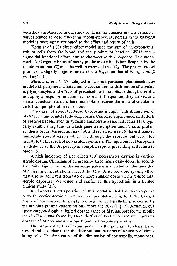
with the data observed in our study or theirs; the changes in their parameter values related to dose reflect this inconsistency. Hysteresis in the basophil model is more aptly attributed to the efflux and return of cells.
Kong et al.'s (8) direct effect model used the sum of an exponential exit of cells from the blood and the product of baseline W B H and a sigmoidal fractional effect term to characterize this response. This model works for larger iv bolus of methylprednisolone but is handicapped by the requirement that C O must be well in excess of the IC5o. The present model produces a slightly larger estimate of the ICso than that of Kong et al. (8 vs. 3 ng/ml).
Bloemena et al. (17) adapted a two-compartment pharmacokinetic model with peripheral elimination to account for the distribution of circulat- ing lymphocytes and effects of prednisolone in rabbits. Although they did not apply a response function such as our I ( t ) equation, they arrived at a similar conclusion to ours that prednisolone reduces the influx of circulating cells from peripheral sites to blood.
The onset of steroid-induced basopenia is rapid with diminution of W B H seen immediately following dosing. Conversely, gene-mediated effects of corticosteroids, such as tyrosine aminotransferase induction (18), typi- cally exhibit a lag time in which gene transcription and de novo protein synthesis occur. Various authors (19, and reviewed in ref. 8) have discussed immediate steroid effects which act through the receptor but occur too rapidly to be the result of new protein synthesis. The rapid onset ofbasopenia is attributed to the drug-receptor complex rapidly preventing cell return to blood (8).
A high incidence of side effects (20) necessitates caution in cortico- steroid dosing. Clinicians often prescribe large single daily doses. In accord- ance with Figs. 5 and 6, the response pattern is dictated by the time that MP plasma concentrations exceed the IC5o. A steroid dose-sparing effect may also be achieved from two or more smaller doses which reduce total steroid exposure. We tested and confirmed this hypothesis in a limited clinical study (21).
An important extrapolation of this model is that the dose-response curve for corticosteroid effects has no upper plateau (Fig. 6). Indeed, larger doses of corticosteroids simply prolong the cell trafficking response by maintaining plasma concentrations above the IC5o (Fig. 5). Although our study employed only a limited dosage range of MP, support for the profile seen in Fig. 6 was found by Derendorf et al. (22) who used much greater dosages of MP to assess various blood cell response patterns.
The proposed cell trafficking model has the potential to characterize steroid-induced changes in the distributional patterns of a variety of circu- lating cells. The time course of the diminution of eosinophils, monocytes,

Basophil Cell Trafficking Model for Methylprednisolone 533
and lymphocytes is identical in pattern to basopenia. Neutrophils, which increase in circulating numbers, might be described by an altered version of the model in which the movement of cells into the blood is enhanced by corticosteroids.
The basophil cell trafficking model presents equations, estimation techniques, and extrapolations to evaluate the non-gene-mediated dose/response relationship of corticosteroids. This approach of physio- logical pharmacodynamic modeling may be used to evaluate alterations in the kinetics and dynamics of various steroids in the presence of disease states and drug interactions. We have extended this work to other cell types such as T-helper (CD4 +) lymphocytes, incorporating their baseline circadian patterns in the model (23).
SYMBOLS
AB AE AT ABEC A UHC AUC CMp CL I(t) Imln IC5o kin, k~n ko~t k~l t tl train
WBH
Number of cells in blood compartment Number of cells in extravascular compartment Total number of cells (AB + AE) Area between the baseline and decline in cell number curve Area under the WBH versus time curve Area under the drug concentration versus time curve Plasma concentration of drug Clearance of drug Fractional inhibition function Inhibitory fraction at the nadir Concentration of drug causing 50% inhibition of kin Rate constant for return of basophils to blood Rate constant for efflux of basophils from blood Rate constant for drug elimination Time Time at completion of cell trafficking response Time at occurrence of nadir in blood cell number Volume of distribution of drug Whole blood histamine concentration
ACKNOWLEDGMENTS
We appreciate the participation of Prof. Richard Slaughter and Drs. Ah-Ng Kong, Elizabeth A. Ludwig, Providence M. DiStefano, James DeMasi, William G. Reiss, and Elliott Middleton, Jr. in the original clinical

534 Wald, Salazar, Cheng, and Jusko
pharmacodynamic studies (refs. 8 and 21). Ms. Nancy Pyszczynski provided technical assistance.
A P P E N D I X
An integrated solution to define the determinants of A B E C can be found as follows. Combining Eqs. (3) to (5) yields
dAB( t )= IC5o" ki ~ " �9 Aa(t) (A1) dt ICso + c O p e -k~lt out
Multiplying both sides of Eq. (A1) with dt yields
IC5o" ki ~ dt dAB( t )=ICso + CM P0 e-k,to kout" AB(t ) dt (A2)
Integrating both sides of Eq. (A2) from time 0 to t yields
k 0 AB (t) - AB(0) = :~2.~. In IC5o+ C ~ e -k~', kout" A s ( t ) dt (A3)
[IC50+ cOp] �9 e-~'
Since AB(t) = AB(0) at t = tl, from Eq. (A3) it follows that
fo ki ~ . IC5o"4- COp e -kelt, (A4) " AB(t) dt = ke," kou-----~t'm[ICso+----~C~ - - - - ] ] e -k~
Thus, by definition
f0 A B E C = AB(O) " t l - AB(0) dt
k~ , IC5o+ COp . e -k~ - - - m 0 (AS) =AB(0) �9 tl kel. kout [iCsoq_CMp]. e_ke/1
In the above In quotient, IC5o >> COp. e - k ~ t ' in the numerator at late times when the trafficking change is back to baseline. Also, for large doses of drug, o . . CMp>> IC5o in the denominator. Further, C ~ kel This allows Eq. (A5) to simplify to
k o A B E C ~- AB(0) �9 tl + In IC5o+ In kel - kel �9 tl + - - " In A U C (A6)
ke~" kou~

Basophil Cell Trafficking Model for Methylprednisolone 535
While the combination of constants constituting the intercept is not meaning- ful, the slope of a plot of A B E C vs. log A U C (for larger doses) yields an estimate of 0.434. k~ �9 kout-
REFERENCES
1. T. R. Cnpps and A. S. Fauci. Corticosteroid-mediated immunoregulation in man. Immunol. Rev. 65:133-155 (1982).
2. P. M. Guyre, M. T. Girard, P. M. Morganelli, and P. D. Mangaliello. Glucocorticoid effects on the production and actions of immune cytokines. J. Steroid Biochern. 30:89-93 (1988).
3. A. P. Saavedra-Delgado, K. P. Mathews, P. M. Pan, D. R. Kay, and M. L. Muilenberg. Dose-response studies of the suppression of whole blood histamine and basophil counts by prednisone. J. Allergy Clin. ImmunoL 66:464-471 (1980).
4. R. P. Schleimer. The effects of glucocorticoids on mast cells and basophils. In R. P. Schleimer, H. N. Claman, and A. L. Oronsky (eds.), Anti-Inflammatory Steroid Action-Basic and Clinical Aspects, Academic Press, San Diego, CA, pp. 226-258.
5. E. H. Dunsky, B. Zweiman, E. Fischler, and D. A. Levy. Early effects of corticosteroids on basophils, leukocyte histamine, and tissue histamine. J. Allergy Clin. lmmunol. 63:426- 432 (1979).
6. E.J. Leonard and A. SkeeL Separation of human basophils into two fractions with different density and histamine content. J. Allergy Clin. lmrnunol. 76:556-562 (1985).
7. E. J. Leonard. Two populations of human blood basophils: Effect of prednisone on circulating numbers. J. Allergy Clin. Immunol. 79:775-780 (1987).
8. A.-N. Kong, E. A. Ludwig, R. L. Slaughter, P. M. DiStefano, J. DeMasi, E. Middleton Jr., and W. J. Jusko. Pharmacokinetics and pharmacodynamic modeling of direct sup- pression effects of methylprednisolone on serum cortisol and blood histamine in human subjects. Clin. Pharmacol. Ther. 46:616-628 (1989).
9. C. W. Parker, M. G. Huber, and M. L. Baumann. Alterations in cyclic AMP metabolism in human bronchial asthma. III. Leucocyte and lymphocyte responses to steroids. J. Clin. Invest. 52:1342-1348 (1973).
10. A. S. Fauci and D. C. Dale. The effect of in vivo hydrocortisone on subpopulations of human lymphocytes. J. Clin. lnvest. 53:240-246 (1974).
I 1. A. S. Fauci and D. C. Dale. The effect of hydrocortisone on the kinetics of normal human lymphocytes. Blood 46"235-243 (1975).
12. J. M. Engler, R. Y. Chestnut, G. C. Borst, and C. Eil. The effects of triacetyloleandomycin and oleandomycin phosphate on the glucocorticoid receptors in clutured skin fibroblasts. J. Allergy Clin. Immunol. 75:395-400 (1985).
13. P. Rohdewald, H. W. Mollman, and G. Hochhaus. Affinities of glucocorticoids for gluco- corticoid receptors in the human lung. Agents Actions 17:290-291 (1989).
14. E. C. Butcher. Cellular and molecular mechanisms that direct leukocyte traffic. Am. J. Pathol. 136:3-11 (1990).
15. R. P. Schleimer. Effects of glucocorticosteroids on inflammatory cells relevant to their therapeutic applications in asthma. Am. Rev. Resp. Dis. 141:$59-$69 (1990).
16. B. Oosterhuis, I. J. M. TenBerge, P. T. A. Schellekens, R. P. Koopmans, and C. J. van Boxtel. Prednisolone concentration-effect relations in humans and the influence of plasma hydrocortisone. J. Pharmacol. Exp. Ther. 239:919-926 (1986).
17. E. Bloemena, S. Weinreich, and P. T. A. Schellekens. The influence of prednisolone on the recirculation of peripheral blood lymphocytes in vivo. Clin. Exp. lmmunol. 80:460-466 (1990).
18. A. I. Nichols, F. D. Boudinot, and W. J. Jusko. Second generation model for prednisolone pharmacodynamics in the rat. J. Pharmacokin. Biopharrn. 17:209-227 (1989).

536 Wald, Salazar, Cheng, and Jusko
19. L. K. Johnson, J. P. Longnecker, J. D. Baxter, M. F. Dallman, E. P. Widmaier, and N. L. Eberhardt. Glucocorticoid action: A mechanism involving nuclear and non-nuclear path- ways. Br. J. Dermatol. 107(Suppl. 23):6-23 (1982).
20. G. P. Lewis, W. J. Jusko, C. W. Burke, L. Graves, and the Boston Collaborative Drug Surveillance Program. Prednisone side effects and serum protein levels. Lancet 2:778-781 (1971).
21. W. G. Reiss, R. L. Slaughter, E. A. Ludwig, E. Middleton Jr., and W. J. Jusko. Steroid dose sparing: Pharmacodynamic responses to single versus divided doses of methyl- prednisolone in man. J. Allergy Clin. Immunol. 85:1058-1066 (1990).
22. H. Derendorf, H. M611mann, M. Krieg, S. Tunn, C. M611mann, M. Krieg, S. Tunn, J. Barth, and H. J. R~thig. Pharmacodynamics of methylprednisolone phosphate after single intravenous administration to healthy volunteers. Pharm. Res. 8:263-268 (1991).
23. T. E. Dunn, E. A. Ludwig, R. L. Slaughter, D. S. Camara, and W. J. Jusko. Pharmacokinetics and pharmacodynamics of methylprednisolone in obese and non-obese men. Clin. Phar- macol. Ther. 50:536-549 (1991).