Evaluating Antibacterial Efficacy and Biocompatibility of PAN ...
Upload
independentCategory
view
1download
0
Eur J Clin Microbiol Infect Dis (2003) 22:203–221DOI 10.1007/s10096-003-0907-5
R E V I E W
A. Dalhoff · F.-J. Schmitz
In Vitro Antibacterial Activity and Pharmacodynamicsof New Quinolones
Published online: 1 April 2003� Springer-Verlag 2003
Abstract This synopsis of published literature summaris-es data on the in vitro antibacterial activity and pharma-codynamics of fluoroquinolones. Data were compiled forciprofloxacin, levofloxcin, moxifloxacin, gatifloxacin,grepafloxacin, gemifloxacin, trovafloxacin, sitafloxacinand garenoxacin. All of these quinolones are almostequipotent against gram-negative bacteria but demon-strate improved activity against gram-positive species.The new quinolones are uniformly active against gram-positive species except Streptococcus pneumoniae;against which gemifloxacin, sitafloxacin and garenoxacinare one to two dilution steps more active than moxi-floxacin. All of the new quinolones except gemifloxacindemonstrate enhanced activity against anaerobes. Sinceall the new quinolones show similar activity against themajor respiratory tract pathogens except Streptococcuspneumoniae and members of the family Enterobacteri-aceae, their pharmacokinetics and pharmacodynamicswill be clinically relevant differentiators and determinantsof their overall activity and efficacy. In vitro simulationsof serum concentrations revealed that (i) gemifloxacinand levofloxacin were significantly and gatifloxacinmoderately less active than moxifloxacin against Strep-tococcus pneumoniae and Staphylococcus aureus, and (ii)resistant subpopulations emerged following exposure tolevofloxacin and gatifloxacin (gemifloxacin not yetpublished) but not to moxifloxacin. The emergence ofresistance is a function of drug concentrations achievablein vivo and the susceptibility pattern of the targetorganisms. Therefore, the use of less potent fluoro-
quinolones with borderline or even suboptimal pharma-cokinetic/pharmacodynamic surrogate parameters willinadvertently foster the development of class resistance.Drugs with the most favourable pharmacokinetic/phar-macodynamic characteristics should be used as first-lineagents in order to preserve the potential of this drug classand, most importantly, to provide the patient with anoptimally effective regimen.
Introduction
Streptococcus pneumoniae, Haemophilus influenzae andMoraxella catarrhalis are the most important causes ofinfective bacterial exacerbation in patients with chronicobstructive pulmonary disease. The percentage of beta-lactam-resistant strains among all three species is contin-uously increasing in Europe and the USA. Production ofbeta-lactamases – usually of the TEM-1 type but some-times of the ROB-1 type – by Haemophilus influenzae isincreasing, approaching >40% in some countries [1, 2, 3,4]. Resistance to cephalosporins and tetracyclines isuncommon at present but is emerging in some patientgroups or geographical areas. Beta-lactamase-negativebut otherwise ampicillin-resistant (BLNAR) strains havebeen reported since the early 1980s [5, 6, 7, 8]. Theirincidence remains low in various countries, but in Japanmore than 25% of all beta-lactamase-negative strains areBLNAR, and the frequency seems to be increasing [1, 4,9, 10, 11, 12].
Before the 1970s, Moraxella catarrhalis was suscep-tible to penicillin G and aminopenicillins. In the 1970sand 1980s, the susceptibility patterns began to change,and today between 70 and 90% of Moraxella catarrhalisisolates are reported to be beta-lactamase producers [13,14]. Moraxella catarrhalis produces a constitutivelyexpressed beta-lactamase. The enzymes BRO-1 andBRO-2 are transposon mediated, which explains theirrapid spread [15].
Penicillin has long been the treatment of choice forrespiratory tract infections caused by Streptococcus
A. Dalhoff ())Institute for Medical Microbiology and Virology,Universit�tsklinikum Schleswig-Holstein,Brunswiker Strasse 4, 24105 Kiel, Germanye-mail: [email protected].: +49-431-5973300Fax: +49-431-5972216
F.-J. SchmitzInstitute for Medical Microbiology and Virology,Universit�tsklinikum D�sseldorf,Universit�tsstrasse 1, Geb. 22.21, 40225 D�sseldorf, Germany

pneumoniae. The first penicillin-resistant pneumococciwere isolated in Papua New Guinea in 1967 from healthycarriers [16, 17]. Since then, however, there have beenincreasing numbers of reports of penicillin-resistantisolates from many countries throughout the world [18].A multicentre, collaborative study in Europe and Asiaduring the winter of 1997–1998 revealed that penicillinresistance (MIC, �2 mg/l) amongst pneumococci rangedfrom 0.7% in Germany to 43.5% and 33.9% in Spain andFrance; penicillin resistance in Japan and China was10.1% and 3.2%, respectively. However, macrolideresistance amounted to 48% in Japan and 73–74% inChina; a 49% difference in macrolide resistance wasdetected between France and Germany (58% and 9%,respectively) [19]. A dramatic increase in macrolideresistance among invasive pneumococci isolated in Ger-many was noted (from 3% in 1992 to 13% in 1998 up to15.3% in 2000) [20]. The past 3 years in particular havewitnessed a dramatic increase worldwide in the incidenceof pneumococcal strains that are resistant not only topenicillin but also to macrolides and other antimicrobialagents [21, 22]. The problem is exacerbated by thetendency of penicillin-resistant clones to spread fromcountry to country and from continent to continent [18].This penicillin resistance has emerged as a majorproblem, creating a need for alternative non-beta-lactamtherapies.
In an effort to respond to these problems, the pastdecade has seen the search for and development of manynew quinolones. Monofluorinated quinolones, like cipro-floxacin, are predominantly active against gram-negativepathogens; di- and tri-halogenated quinolones, on theother hand, exhibit greatly enhanced antibacterial activityand pharmacokinetics in humans. However, the develop-ment of many of these promising di- and tri-halogenatedagents was discontinued due to adverse events, includingphototoxic and other unexpected adverse reactions. Oth-ers, like trovafloxacin, are still commercially available,but their use is limited to a very small number of specificindications only.
This review examines the in vitro activities of newfluoroquinolones and their propensities for selection ofresistance.
Problems Encountered in Comparing SusceptibilityData
During recent years it became increasingly difficult togenerate a representative synopsis of published data onthe antibacterial activity of new quinolones against thetarget pathogens because the scope of susceptibilitystudies varies significantly, thus impacting the data beinggenerated. The following points outline the nature of theproblem inherent in comparing the results of susceptibil-ity data.
1. The resistance problem in general is in the focus of thepublic and scientific interest; authors may therefore
have selected resistant strains from strain collections ormay have used laboratory-derived strains with specificresistance mechanisms. Such specific subpopulationsor genetically defined strains with specific resistancemechanisms do need to be studied to fully exploit thepotential of a new agent. However, such straincollections do not represent the clinically relevantpathogens found in the target population.
2. Similar to the quandary outlined above, drugs inclinical use for long periods of time or those consid-ered to be gold standards have been studied exten-sively; in particular, mechanisms of resistance againstthese drugs and the activities of these drugs againststrains with selected and specific resistance patternsare of special interest but do not mirror susceptibilitypatterns of common pathogens.
3. To meet the EMEA (European Agency for theEvaluation of Medical Products) requirements, resis-tant strains must be selected for susceptibility studies.In the “Note for Guidance” in the PharmacodynamicSection of the SPC (Summary of Product Character-istics) of Antibacterial Medicinal Products, the EMEArequests that “strains without known acquired resis-tance mechanisms and, when available, harbouringknown resistance mechanisms to other antibioticswithin the same or related classes of antibiotics shouldbe tested (e.g. a new quinolone should be tested againstnalidixic-sensitive Escherichia colibut also againstnalidixic acid-resistant and ciprofloxacin-resistant Es-cherichia coli, and a new macrolide should be testedagainst erythromycin-resistant organisms).”
4. The epidemiology of resistance varies locally, region-ally, nationally and internationally, and sometimesstrains included in studies may have been collectedfrom pockets where atypical resistance characteristicspredominate. Because resistance profiles differ dra-matically, it is important that the strain collectionsstudied include substantial numbers of recent clinicalisolates from diverse areas in order to reflect the “realworld scenario”.
5. Strains are most frequently collected during a definedstudy period; consequently, such collections mayinclude strains isolated during an outbreak situation,so that such collections probably include strains ofclonal origin. If resistant strains of clonal origin areoverrepresented within a strain collection studied, thedistribution of MICs determined for these strains willlikely not reflect the susceptibility pattern of commonpathogens.
6. Many studies originate from hospital-based laborato-ries, so that isolates studied may derive predominantlyfrom hospitalised patients. Although the newest andmost active antibacterial agents are used in hospi-talised patients, particularly those at risk, manyhospitalised patients acquire resistant and even mul-tiresistant bacteria. Therefore, neither the epidemiolo-gy of resistance nor the susceptibility pattern of suchstrains mirrors the bacterial etiology in the outpatientsetting.
204

Similarly, strains obtained in the outpatient setting arealso likely to include resistant subpopulations. Mostbacterial infections in outpatients are treated adequate-ly and effectively by the general practitioner, withoutany bacteriological assessment. Most frequently, sam-ples are withdrawn and analysed only in thoseoutpatients in whom the initial therapy failed and inwhom resistant subpopulations may already have beenselected. Thus, such isolates do not reflect the “true”resistance epidemiology of community-acquired infec-tions.Consequently, new community-based surveillanceprogrammes are being pursued that differentiatebetween community- and hospital-acquired infections.However, at present the database originating fromthese programmes is too small to allow a comprehen-sive review of the in vitro activities of new quinolones.
7. Another scenario in which the selection of resistantstrains is favoured is that of chronic disease. In patientsinfected by species that may cause chronic disease(e.g. Pseudomonas aeruginosainfections in cysticfibrosis), samples will be withdrawn repeatedly overlong periods of time. Because these patients receivelong-term therapy, the causative pathogens will likelyhave acquired resistance. Such isolates have a signif-icant impact on the susceptibility pattern of the straincollections studied, as the resistant subpopulation willbe over-represented if isolates from patients withshort-term versus long-term exposure are not reportedseparately.
The factors mentioned above have a significant impact onstudies of the in vitro activity of antibacterial agents. Theevaluation of a drug’s activity against specifically select-ed resistant strains or against isolates from hospitalisedpatients will result in an activity profile different fromthat of isolates causing community-acquired infections.Higher minimal inhibitory concentrations (MICs) ofantimicrobial agents will be reported as compared tostudies in which the most recent unselected isolates havebeen used, particularly if the isolates originate fromoutpatients. For example, in one study the level ofmacrolide resistance in invasive pneumococci is 50%higher (15.3%) than the level of macrolide resistance incommunity-acquired respiratory tract pathogens [20].Consequently, the modal MICs, the MIC50s and MIC90sof all drug classes other than quinolones differ forunselected surveillance isolates and invasive pneumococ-ci [20, 23].
Other examples of broad differences in the datareported for a given drug can be cited. For example,MIC90 values of ciprofloxacin against Escherichia colivary from 0.03 mg/l for nalidixic-acid- and ciprofloxacin-susceptible isolates to 1 mg/l for nalidixic-acid-resistantbut ciprofloxacin-susceptible isolates; MICs reached up to128 mg/l for the nalidixic-acid-resistant as well as theciprofloxacin-resistant subgroup [24]. King et al. [24]have mentioned explicitly that the organisms they studiedwere collected over a 10-year period and were selected to
include those known to be resistant. They also reportedthe data for every subgroup per species separately, thusdemonstrating clearly that the data described do notreflect the clinical isolation rates of resistant or suscep-tible bacteria and do not reflect the activities of the agentsstudied against unselected bacteria. Usually, however, theinclusion of quinolone-resistant strains into the studypopulation is not mentioned specifically. Instead, only thevery broad range of individual MICs for the pathogensstudied indicates that test strains known to be quinoloneresistant may have been selected. Consequently, thecompilation of all published data on the in vitro activity ofquinolones against bacterial isolates will result in a veryheterogeneous data set, even for one specific agent, andwill not be informative.
In conclusion, many of the strain collections used toevaluate the antibacterial activity of a new agent do notrepresent the resistance epidemiology of those strains thatpredominate in the target population. Rather, resistantstrains are likely to be over-represented.
Susceptibility Data for New Quinolones
Data on the susceptibilities of gram-positive and gram-negative isolates reported below originate from a very fewpublications in which almost all new quinolones havebeen compared with each other on the basis of directdrug-to-drug comparisons. Milatovic et al. [25] comparedthe activities of sitafloxacin, ciprofloxacin, trovafloxacin,levofloxacin, clinafloxacin, gatifloxacin and moxifloxacinagainst a total of 8,796 bacterial strains that were isolatedbetween April 1997 and February 1999 from patients in24 university hospitals in 14 European countries, 3hospitals in South Africa and 1 hospital in Israel. In thisreport, the MIC90 values are not subgrouped according toquinolone susceptibility or resistance of the bacteriaisolated. For example, the MICs of ciprofloxacin forEscherichia coli range from �0.008 up to �16 mg/l, thusillustrating the points mentioned above.
King et al. [24] studied ciprofloxacin, moxifloxacin,gemifloxacin, trovafloxacin, grepafloxacin, clinafloxacinand ofloxacin against strains that were specificallyscreened for ciprofloxacin and/or nalidixic acid resis-tance. If necessary, data on the susceptibility ofquinolones to gram-positive or gram-negative aerobicbacteria were complemented by results from other studies[26, 27].
The data on the investigational des-fluoro(6) quinoloneBMS 284756 (garenoxacin) are derived from recentpublications [28, 29, 30, 31, 32, 33]. Fung-Tomc et al.[28] compared garenoxacin with trovafloxacin, moxi-floxacin, levofloxacin, ofloxacin and ciprofloxacin, andthe SENTRY group compared garenoxacin with cipro-floxacin, gatifloxacin, levofloxacin and trovafloxacin aswell as with 11 non-quinolone agents.
In general, the new quinolones demonstrate improvedactivity against gram-positive microorganisms and anaer-obes as compared to previous quinolones such as
205

ciprofloxacin and levofloxacin, which are active predom-inantly against gram-negative pathogens. The improvedactivity of the new quinolones against gram-positiveaerobes is exemplified by the low MIC90 values againststreptococci, ranging from 0.25 mg/l (moxifloxacin) and0.5 mg/l (gatifloxacin, grepafloxacin) to 0.06 mg/l(sitafloxacin, gemifloxacin) (Table 1).
Although ciprofloxacin-resistant pneumococci are ex-tremely rare and, consequently, the numbers tested aresmall, the improved activity of the newer quinolones isreflected by the low MIC values, even against highlyciprofloxacin-resistant pneumococci, for which MIC90values of 64 mg/l have been reported [24]. The MIC90s ofmoxifloxacin and garenoxacin against 53 selected clinicalstrains of Streptococcus pneumoniae demonstrating ef-flux-mediated fluoroquinolone resistance were within onedilution step (i.e. 0.5 and 0.12 mg/l, respectively) of theMIC90s reported for wild-type strains, thus indicating thatboth quinolones are unaffected by PmrA-mediated efflux[31].
Highly ciprofloxacin-resistant isolates will be resistant(based on breakpoint definition) to all the new quino-lones. Borderline ciprofloxacin-susceptible pneumococciare almost as susceptible as wild-type strains to the newerquinolones [33, 34, 35]. Thus, new quinolones withenhanced activity against streptococci and other gram-positive organisms may offer a useful therapeutic alter-native for pneumococcal infections, provided (i) theyretain activity against gram-positive bacteria resistant tothe early fluoroquinolones, and (ii) the development offluoroquinolone resistance can be minimised and con-trolled.
Activity of New Quinolones Against Pneumococciwith Genetic Mutations
In order to assess the potential of new fluoroquinolones astreatment alternatives for pneumococci resistant to theearly fluoroquinolones, their in vitro activity against
genetically defined resistant isolates was examined. TheMIC ranges for first-step mutants, i.e. isolates with singlealterations in gyrase or topoisomerase IV, generallyoverlapped those for wild-type isolates. In contrast,mutations in both loci increased the MICs of almost allnew fluoroquinolones up to levels of intermediate or high-level resistance, except for gemifloxacin and cli-nafloxacin [36, 37, 38]. However, no difference in thefrequency of selection of mutations in wild-type versusfirst-step mutants was detected [36]. Thus, fluoro-quinolone resistance and cross-resistance are consistentonly among the subset of double mutants with mutationsin both gyrase and topoisomerase IV.
Although, on the one hand, mutations in gyrA andparC, alone or in combination, most frequently contributeto fluoroquinolone resistance, mutations are, on the otherhand, much more heterogeneous than previously thought.Numerous in vitro studies (for summary, see [39]) haveindicated that a single amino acid change in either of thetwo targets results in an almost negligible increase inMICs of the new fluoroquinolones. However, a recentgenetic analysis of mutations contributing to fluoro-quinolone resistance in clinical isolates of Streptococcuspneumoniae revealed that mutations in the quinoloneresistance determining region (QRDR) were much moreheterogeneous [38]. The positions of amino acid changeswithin the genes gyrA, gyrB, parC and parE affectedresistance more than did the total number of QRDRmutations. The effect of a specific mutation on the MIC ofthat specific isolate varied significantly, depending on theagent tested. It seems to be conceivable that the hetero-geneity of mutations conferring resistance to fluoro-quinolones may increase, since pneumococci will beexposed to an increasing number of different fluoro-quinolones. The heterogeneity of mutations will compli-cate the prediction of cross-resistance amongst thefluoroquinolones.
Table 1 Comparative antibacterial activity (MIC90, mg/l) of quinolones against gram-positive bacteria (modified according to [24, 25, 26,27, 28, 29, 30, 31, 32, 33, 34])
Agent S. pneumoniae Alpha-haemolyticstreptococci
Beta-haemolyticstreptococci
S. agalactiae MSSA
cipS cipRa cipS cipR
CIP 1 64/2* 2 1 2 0.5 >128LEV 1 –/1 – 1 1 0.25 >32MFX 0.25 4/0.25 0.5 0.25 0.5 0.06 4GAT 0.5 –/0.5 – 0.5 – 0.12 4GRE 0.5 8/– 1 1 0.5 0.12 32GEM 0.06 1/– 0.12 0.06 0.06 0.03 8TRO 0.25 4/0.25 0.25 0.25 0.5 0.06 2SIT 0.06 –/0.06 – 0.03 – 0.03 0.25GAR 0.12 –/0.12 – 0.25 0.12 0.03 1.4
CIP, ciprofloxacin; LEV, levofloxacin; MFX, moxifloxacin; GAT, gatifloxacin; GRE, grepafloxacin; GEM, gemifloxacin; TRO,trovafloxacin; SIT, sitafloxacin; GAR, garenoxacin (BMS-284756); MSSA, methicillin-susceptible Staphylococcus aureus; cipS,ciprofloxacin suscepitible; cipR; ciprofloxacin resistanta Data quoted from references [24/25], respectively
206
Activity of Newer Quinolones AgainstStaphylococcus aureus
The newer quinolones also exhibit improved activityagainst highly ciprofloxacin-resistant but methicillin-susceptible Staphylococcus aureus isolates [33, 36, 40,41]. Although the markedly improved activity of all thenew quinolones against ciprofloxacin- and levofloxacin-resistant gram-positive organisms will not translate intoclinical efficacy, these data demonstrate that (i) the newquinolones exhibit markedly improved activity againstgram-positive aerobes in general due to high targetaffinity to both targets, i.e. gyrase as well as topoiso-merase IV, and (ii) quinolone resistance mechanisms ingram-positive bacteria have a differential effect on boththe older and the newer quinolones (see below).
Methicillin-resistant Staphylococcus aureus (MRSA)sensu strictu are as susceptible to moxifloxacin and,probably, the other new quinolones as methicillin-sus-ceptible Staphylococcus aureus (MSSA) [42, 43, 44, 45].The decreased susceptibility of MRSA to fluoro-quinolones is not the result of increased mutation rates;MSSA have been shown to have the same mutation ratesas MRSA [46]. It seems likely that an association existsbetween the action of fluoroquinolones on mec(A)-positive Staphylococcus aureus on the one hand, andthe increase in the proportion of colony-forming units permillilitre expressing methicillin resistance (i.e. the resis-tance index) on the other hand. Growth in the presence ofvarious fluoroquinolones increased the proportion of themec(A)-positive heteroresistant Staphylococcus aureusthat developed methicillin resistance (oxacillin MIC,�128 mg/l). Ciprofloxacin exhibited a >1,000-foldgreater effect than newer quinolones. The resultantoxacillin-resistant strains also showed a 1.5- to 3-foldincrease in fluoroquinolone MICs. This phenomenon isnot caused by the fluoroquinolone acting as a mutagen butrather to the selection of more fluoroquinolone-resistantsubpopulations, which were also predominantly moreresistant to oxacillin [47].
Resistance to methicillin emerged as early as 8 h afterexposure to the fluoroquinolone. Similarly, the 8-chlo-rine-substituted fluoroquinolone BAY y 3118 selected forquinolone resistance in MRSA strains as early as 6 h aftercommencement of exposure to BAY y 3118. Moxi-floxacin did not select for quinolone resistance in theseMRSA strains at all [48]. In the latter two studies [47, 48],no resistance emerged in MSSA strains. These two studiesindicate that quinolones differ in their potential to selectfor quinolone as well as methicillin resistance. Themechanisms of resistance to the two drug classes areunrelated, but their selective emergence tends to beassociated.
Since most of the clinical MRSA isolates are almostalways multidrug resistant, they are resistant to quino-lones, too. Therefore, quinolones in general, includingsitafloxacin and garenoxacin, do not offer a therapeuticalternative for treatment of MRSA infections.
Activity of Newer Quinolones AgainstMajor Pathogens
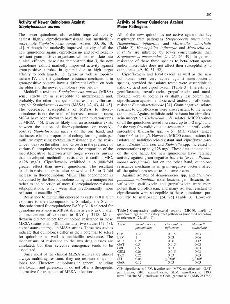
All of the new quinolones are active against the keyrespiratory tract pathogens Streptococcus pneumoniae,Haemophilus influenzae and Moraxella catarrhalis(Table 2). Haemophilus influenzae and Moraxella ca-tarrhalis are inhibited by lower concentrations thanStreptococcus pneumoniae [24, 25, 26, 49]. In general,resistance of these three species to beta-lactam agentsand/or macrolides does not affect their susceptibility toquinolones [49, 50, 51, 52].
Ciprofloxacin and levofloxacin as well as the newquinolones were very active against enterobacterialspecies, provided the isolates tested were susceptible tonalidixic acid and ciprofloxacin (Table 3). Interestingly,gemifloxacin, trovafloxacin, grepafloxacin and moxi-floxacin were as potent as or slightly less potent thanciprofloxacin against nalidixic-acid- and/or ciprofloxacin-resistant Enterobacteriaceae [24]. Gram-negative isolatesresistant to ciprofloxacin were also resistant to the newerquinolones. Against nalidixic-acid-resistant but ciproflox-acin-susceptible Escherichia coli isolates, MIC90 valuesof all the quinolones tested increased up to 1–2 mg/l, and,for the very few nalidixic-acid-resistant but ciprofloxacin-susceptible Klebsiella spp. (n=5), MIC values rangedfrom 0.06 to 1 mg/l. However, MIC90 concentrations forisolates of nalidixic-acid-resistant and ciprofloxacin-re-sistant Escherichia coli and Klebsiella spp. increased toconcentrations up to �128 mg/l. These data indicate that,on the one hand, the new quinolones have retainedactivity against gram-negative bacteria (except Pseudo-monas aeruginosa), but on the other hand, quinoloneresistance mechanisms in gram-negative bacteria affectall the quinolones tested to the same extent.
Against isolates of Acinetobacter spp. and Stenotro-phomonas maltophilia, moxifloxacin, gemifloxacin, tro-vafloxacin, gatifloxacin and grepafloxacin were morepotent than ciprofloxacin, and many isolates resistant tociprofloxacin were susceptible to these quinolones, par-ticularly to sitafloxacin [24, 25] (Table 3). However,
Table 2 Comparative antibacterial activity (MIC90, mg/l) ofquinolones against respiratory tract pathogens (modified accordingto references [24, 25, 49])
Agent Streptococcuspneumoniae
Haemophilusinfluenzae
Moraxellacatarrhalis
CIP 1–2 0.015 0.03LEV 1 0.03 0.06MFX 0.25 0.06 0.12GAT 0.5 0.015 0.03GRE 0.5 0.03 0.03GEM 0.06 0.015 0.03TRO 0.25 0.03 0.03SIT 0.06 �0.008 �0.008GAR 0.12 0.03 0.03
CIP, ciprofloxacin; LEV, levofloxacin; MFX, moxifloxacin; GAT,gatifloxacin; GRE, grepafloxacin; GEM, gemifloxacin; TRO,trovafloxacin; SIT, sitafloxacin; GAR, garenoxacin (BMS-284756)
207

against Pseudomonas aeruginosa, ciprofloxacin is still themost potent quinolone; sitafloxacin tended to be onetitration step more active. However, assuming the samebreakpoints for ciprofloxacin and sitafloxacin as proposedby Jones et al. [53], almost complete cross-resistancebetween ciprofloxacin and sitafloxacin was observed.
King et al. [24] subgrouped the Pseudomonas aerug-inosa strains studied according to their ciprofloxacinsusceptibility pattern: the MIC90 of ciprofloxacin was0.5 mg/l for the susceptible strains and 128 mg/l for theresistant strains. Milatovic et al. [25] did not subgroup thePseudomonas aeruginosa strains studied, so the MIC90values exceed 16 mg/l. On the basis of the outcome of thetwo detailed studies mentioned below, it seems to belikely that the isolates evaluated by Milatovic et al. [25]or the ciprofloxacin-resistant subgroup studied by King etal. [24] may have originated from chronically infectedpatients. These examples illustrate the impact of asampling bias on the characterisation of the antibacterialactivities of both new and established drugs.
Bauernfeind et al. [54] recently evaluated the cipro-floxacin resistance pattern of Pseudomonas aeruginosaisolates from cystic fibrosis patients. Overall, 66.4% ofthe strains were ciprofloxacin susceptible. However,subgrouping the strains according to the age of patientsfrom whom Pseudomonas aeruginosa was isolated
revealed that, in the age groups �14 years and 15–19years (i.e. patients who either have not been or wererarely treated with ciprofloxacin), the resistance rateswere 14.8% and 15.4%, respectively. In patients olderthan 30 years (who were frequently treated withciprofloxacin), the resistance rate was as high as 48.6%.On average, ciprofloxacin resistance amounted to 28.7%.Surprisingly, in patients less than 14 years old – who mayvery rarely have been treated with a quinolone, if at all –ciprofloxacin resistance amounted to 14.8%. Although itcould have been anticipated that the ciprofloxacin resis-tance rate should have been much lower, this phenom-enon may likely result from the transmission of strainsfrom patients of older age groups (in whom resistanceranged from 30–49%) to younger patients.
The strains studied by Bauernfeind et al. [54] were notgenotyped, so it cannot be ruled out that isolates of clonalorigin were evaluated. Worlitzsch et al. [55] used for theirsusceptibility studies genotyped isolates only from youngcystic fibrosis patients, thus excluding copy strains. Forcomparison, isolates from healthy individuals and envi-ronmental sources (neonatal intensive care unit) werestudied. The mean MIC of ciprofloxacin for Pseudomonasaeruginosa isolates was 0.6 mg/l, independent of theorigin of the isolates; no ciprofloxacin resistance could bedetected.
Table 3 Comparative antibacterial activity (MIC90, mg/l) of quinolones (modified according to references [24, 25, 26, 27, 28, 29, 30, 31,32, 33, 49])
Agent Escherichia coli Klebsiella pneumoniea Acinetobacter spp. Stenotrophomonasmaltophilia
Pseudomonasaeruginosaa
CIP 0.03 6 0.06 0.25/0.5* 1/2 >16 2/8 2/0.25*LEV 0.03 8 0.13 0.25/0.5 –/1 16 1/2 4/2MFX 0.03 16 0.13 0.5/1 0.25/1 16 0.5/1 8/8GAT 0.03 8 0.13 –/1 –/– 8 1/– –/2GRE 0.03 – 0.13 –/– 0.12/– – 0.5/– –/1GEM 0.03 – 0.13 –/– 0.12/– – 0.5/– –/1TRO 0.03 >16 0.13 0.5/0.5 0.06/0.25 16 0.5/1 4/2SIT – 1 – –/0.12 – 2 0.12/– –/0.25GAR 0.06 – – 0.5/– –/0.25 – –/4 16/–
CIP, ciprofloxacin; LEV, levofloxacin; MFX, moxifloxacin; GAT, gatifloxacin; GRE, grepafloxacin; GEM, gemifloxacin; TRO,trovafloxacin; SIT, sitafloxacin; GAR, garenoxacin (BMS-284756)a Data quoted from references [24/25], respectively
Table 4 Comparative antibacterial activity (MIC90, mg/l) of quinolones against anaerobes (modified according to references [24, 25, 26,56, 59])
Agent Peptococcusspp.
Peptostreptococcusspp.
Fusobacteriaspp.
Bacteroidesfragilis
Porphyromonasspp.
Prevotellaspp.
CIP 2–4 2–4 2–8 8–32 1 2–>16LEV 4 4 8 2–8 0.5 4MFX 0.25–1 0.5–1 1–4 1–2 0.5 0.5–2GAT 2 2 8 1 0.12 2GRE 1 1 8 16 4 4GEM 0.12 0.06–4 0.25–>8 0.5–2 0.12–1 2–16TRO 0.5–1 0.5–1 0.5–4 0.25–1 0.25 1SIT 0.12 0.12 0.5 0.25 0.03 0.25GAR – 0.25 0.25–8 1 – 0.25
CIP, ciprofloxacin; LEV, levofloxacin; MFX, moxifloxacin; GAT, gatifloxacin; GRE, grepafloxacin; GEM, gemifloxacin; TRO,trovafloxacin; SIT, sitafloxacin; GAR, garenoxacin (BMS-284756)
208

In conclusion, by excluding copy strains from thesusceptibility study, almost all isolates remained fluoro-quinolone susceptible. These patient isolates were assusceptible as the environmental strains collected on aneonatal intensive care unit, where quinolones are not beused for treatment. These detailed studies by Bauernfeindet al. [54], as well as those of Worlitzsch et al. [55],clearly indicate that the evaluation of drug activities isvery much dependent on the origin of the strains studied.
Activity of Quinolones Against Anaerobesand Atypical Bacteria
In general, data on the activity of quinolones againstanaerobes are rather less homogeneous than data availablefor aerobic bacteria, probably reflecting the differentmethods used by the various authors [24, 25, 26, 27, 56,57, 58, 59]. Despite this variability, published dataindicate that the newer quinolones are more active againstanaerobes than the older ones, with the exception ofgemifloxacin (Table 4). The activity of gemifloxacinagainst anaerobes appears to be more variable; anaerobicgram-positive bacteria are more susceptible than anaer-obic gram-negative bacteria. A significant intraspeciesvariability of gemifloxacin MICs for anaerobes wasnoted. Due to the variable activity of gemifloxacin,Goldstein [56] concluded that efficacy is unpredictable.Therefore, the susceptibilities of the isolates should betested before gemifloxacin is used clinically in thetreatment of anaerobic infections.
Against atypical bacteria in general and, in particular,Chlamydia spp. Mycoplasma spp. and Ureaplasma ure-alyticum, the newer quinolones are more active than theolder ones (Table 5) [27, 30, 60, 61, 62, 63, 64, 65, 66].
Summary of Activity of Newer Quinolones
In summary, the antibacterial activity of the newerquinolones is characterised by a retained activity againstgram-negative bacteria—with the exception of Pseudo-monas aeruginosa, against which ciprofloxacin is still the
most active quinolone – but a markedly enhanced activityagainst gram-positive bacteria, with sitafloxacin being themost active. Against most of the gram-positive species, allthe new quinolones are almost equipotent except againstStreptococcus pneumoniae, against which sitafloxacin,gemifloxacin and garenoxacin are more active thanmoxifloxacin by one to two dilution steps. Since all thenew quinolones show similar activity against the majorrespiratory tract pathogens (except Streptococcus pneu-moniae) and the Enterobacteriaceae, their pharmacody-namics, propensity for resistance selection, pharmaco-kinetics and toxicity/safety profile will be clinicallyrelevant differentiators and determinants of their overallactivity and efficacy. Thus, studies on the pharmacody-namics of quinolones will generate useful informationthat contributes to the differentiation of the various newquinolones.
Pharmacodynamics of New Quinolones
MICs of antibacterial agents provide a quantitativeparameter of the inhibition of bacterial growth at anendpoint of 18 h of incubation following exposure to aconstant antibiotic concentration. The endpoints are bynature discrete, so that MICs do not mirror an antibiotic’sbactericidal activity as a function of time and/or concen-tration. Antibacterial efficacy in vivo, however, is afunction of drug concentration at the focus of infectionversus time. Therefore, models that interpret both phar-macokinetics and pharmacodynamics provide more clin-ically meaningful information about the potential of anantibiotic than static endpoints. However, in vitro studiesassessing the antibacterial effects of fluctuating drugconcentrations that simulate serum- or “tissue” concen-tration versus time are cumbersome and therefore notroutinely performed.
Although not the optimal method for determiningefficacy, antibacterial effects in preclinical models ofinfection are most frequently correlated with one of threepharmacokinetic parameters: (i) time (T) of exposure of abacterium to serum concentrations exceeding the MIC(T>MIC); (ii) the ratio of peak concentration (Cmax) of an
Table 5 Comparative antibacterial activity (MIC90, mg/l) of quinolones (modified according to references [27, 30, 60, 66])
Agent Legionellapneumophila
Chlamydiapneumoniae
Chlamydiatrachomatis
Mycoplasmapneumoniae
Ureaplasmaurealyticum
CIP 0.03 1 2 1–4 4LEV 0.016 0.5–1 0.5 0.5–1 1MFX 0.016 0.06 0.06 0.12 1GAT 0.03 0.25 0.12 0.25 0.5GRE 0.016 0.5 NA 0.12 1GEM 0.016 0.25 NA 0.12 0.25TRO �0.004 1 NA 0.25 0.12SIT NA NA NA NA NAGAR 0.06 0.008 0.016 0.06 0.25
CIP, ciprofloxacin; LEV, levofloxacin; MFX, moxifloxacin; GAT, gatifloxacin; GRE, grepafloxacin; GEM, gemifloxacin; TRO,trovafloxacin; SIT, sitafloxacin; GAR, garenoxacin (BMS-284756); NA, information not available
209

agent to its MIC for the bacterium (Cmax/MIC); and (iii)the ratio of the area under the concentration-versus-timecurve (AUC) to the MIC (AUC/MIC). Antimicrobialagents can be categorised by the best fit between thesecomposite parameters and their efficacy [67, 68]. Theparameters used for these calculations (Cmax, AUC andMIC90) are almost always the mean; usually, proteinbinding is not considered. Nevertheless, traditionalcalculations based on the means of pharmacodynamicparameters do not take into account their variabilities.Ideally, pharmacodynamic calculations should include allpossible drug exposures for standard dosage regimens andall MIC values likely to be found for the clinical isolates.Therefore, it would be more informative to publish theMIC distribution pattern for a large number of microor-ganisms. Similarly, maximal serum concentrations (Cmax)and AUC values demonstrate a Gaussian distribution ofthe individual values.
The integration of variability into both pharmacoki-netic parameters (Cmax and AUC) and of MIC distribu-tions into pharmacodynamic parameters can be achievedby various procedures. One approach could be to base thepharmacodynamic modelling on the Monte Carlo simu-lation. However, the information needed to integratevariability into pharmacokinetics as well as MIC distri-butions into pharmacodynamic modelling is, unfortunate-ly, usually not published. Another concept was developedby Schentag et al. [69], who, more than 15 years ago,suggested the concept of dual individualisation, whichintegrates patient-specific data on pharmacokinetics andsusceptibility of the causative pathogen into the dosageoptimisation specifically for that individual patient.Although this concept was a major advance, this approachwould have required real-time drug quantitation, MICtesting (qualitative disk diffusion would be inadequate)and computer modelling. As it was almost unrealistic toapply the dual individualisation concept in daily routinepatient care, the pharmacodynamics of intravenousciprofloxacin in seriously ill patients was analysedretrospectively in order to identify a drug exposuremeasure significantly associated with outcome. Based onthe retrospective study of Forrest et al. [70], in which thecorrelation between pharmacokinetics/pharmacodynam-ics outcomes and clinical efficacy was investigated, anAUC/MIC ratio of �125 is considered to be predictive foroptimal clinical and microbiological outcome. Thus, theAUC/MIC theory as defined by Schentag et al. [69]describes the probability of clinical and microbiologicalresponse to therapy.
Furthermore, it was demonstrated that reduced drugexposure and thus low AUC/MIC ratios correlateddirectly with the emergence of resistance [71]. With anMIC ratio below 100, there was a 50% probability thatresistance would emerge. Overall, 83% of bacteriadeveloped resistance if the initial AUC/MIC ratio was�100. However, if the initial AUC/MIC ratio was �101,resistant pathogens could be isolated from only 9% of thepatients [71].
In the study of Forrest et al. [70], elderly hospitalisedpatients suffering almost exclusively from gram-negativepneumonia were studied. Unfortunately, the target AUC/MIC ratio of �125 has been inappropriately applied toother patient groups and other causative pathogens. Thepharmacodynamic goal of an AUC/MIC ratio of �125 fortherapy of respiratory tract infections due to gram-negative bacteria in hospitalised patients appears to bedifferent from the target AUC/MIC ratio for gram-positive organisms, which cause community-acquiredrespiratory tract infections. Multiple data from in vitroand animal models of infection and recent human clinicaltrials provide strong evidence that the pharmacokinetics/pharmacodynamics parameter for quinolones that corre-lates to clinical outcome in the treatment of community-acquired pneumococcal infections is an AUC/MIC ratioof �30 [68, 72, 73, 74, 75, 76]. In addition, the Cmax wasfound to be linked to the propensity for selection ofresistant subpopulations and also antibacterial effects invitro and in animal models of infection [77, 78, 79, 80,81]; the pharmacodynamic goal of therapy should be apeak Cmax/MIC ratio of �10.
The most common approach to link pharmacokinetic/pharmacodynamic to the probability of response totherapy is to calculate the AUC/MIC and Cmax/MICratios by using the mean AUC and Cmax values and theMIC90 values of the relevant bacterial species. Thepharmacokinetic parameters used are summarised inTables 6 and 7. In addition, protein binding is taken intoaccount because only the unbound fraction is freelydiffusible and antibacterially active [82, 83]; Escherichiacoli and Streptococcus pneumoniae were used as repre-sentative indicator organisms. Since all fluoroquinolonesexhibit pronounced activity against ciprofloxacin-suscep-tible Enterobacteriaceae, they are all characterised byfavourable pharmacokinetic/pharmacodynamic surrogateparameters: for example, ciprofloxacin achieves the mostoptimal Cmax/MIC as well as AUC/MIC ratio (data notshown) for Enterobacteriaceae.
Lower Cmax and AUC values (as, for example, forgrepafloxacin, gemifloxacin or sitafloxacin) and highprotein binding (in the case of sitafloxacin, gemifloxacinand, in particular, garenoxacin) (Tables 6 and 7) have a
Table 6 Minimal inhibitory concentrations (MICs, mg/l) ofquinolones for wild type (WT) and single-step gyrase andtopoisomerase IV mutants of Streptococcus pneumoniae (MICsare derived from references [34, 84, 85]), used for the calculation ofpharmacokinetic/pharmacodynamic surrogate parameters
Agent WT GyrA ParC
Ciprofloxacin 1 8 4Levofloxacin 1 4 2Trovafloxacin 0.25 0.5 1Grepafloxacin 0.5 0.5 1Gatifloxacin 0.5 1 2Moxifloxacin 0.25 0.5 0.25Sitafloxacin 0.06 0.25 0.06Gemifloxacin 0.06 0.12 0.12Garenoxacin 0.06 0.25 0.12
210

significant impact on the pharmacodynamics of the newquinolones against gram-positive bacteria. The improvedactivity of the new quinolones against gram-positivebacteria is only in part mirrored by their pharmacody-namics. Although garenoxacin, sitafloxacin and gemi-floxacin exhibit the highest in vitro activity against wildtype Streptococcus pneumoniae, their low MICs arecounterbalanced by either low serum concentrations and/or high protein binding. The ratios between the unboundand thus antibacterially active fraction of the AUC andthe corresponding MICs for wild type Streptococcuspneumoniae are highest for garenoxacin, followed inorder by those for sitafloxacin and moxifloxacin (almost
identical to those of sitafloxacin), but are lower forgatifloxacin and still lower for gemifloxacin. The un-bound Cmax/MIC ratios for the last two quinolones arealmost identical (Table 8, Table 9).
Although fluoroquinolone-resistant pneumococci arestill rare, isolates of Streptococcus pneumoniae withdecreased susceptibility to these agents can occasionallybe isolated. Thus, it is of interest to evaluate pharmaco-dynamic surrogate parameters for these strains, too. parCand gyrA mutations, alone and in combination, contributesignificantly to fluoroquinolone resistance [34]. Amongstthe gyrA mutations, Ser-81!Phe or Tyr and Ser-79!Phein parC were the most common and were not found in
Table 7 Fluoroquinolone phar-macokinetic constants used forthe calculation of pharmacoki-netic/pharmacodynamic surro-gate parameters
Agent Dose (mg) Cmax (mg/l) AUC 0–24 h(mg x h/l)
Protein binding(%)
Ref.
Ciprofloxacin 500 b.i.d. 2.9 28.0 35 [86]Ciprofloxacin 750 b.i.d. 4.1 44.14 35 [86]Levofloxacin 500 q.d. 5.2 47.7 26 [87]Trovafloxacin 200 q.d. 2.2 30.4 76 [88]Grepafloxacin 400 q.d. 0.9 11.4 50 [89]Grepafloxacin 600 q.d. 1.4 19.7 50 [89]Gatifloxacin 400 q.d. 3.4 32.4 20 [90]Moxifloxacin 400 q.d. 3.5 35.0 37 [91]Sitafloxacin 100 b.i.d. 1.0 11.1 50 [92]Gemifloxacin 320 q.d. 1.5 9.82 60 [93]Garenoxacin 400 q.d. 5.0 60 75, 87 [94, 95]
Table 8 Pharmacokinetic/phar-macodynamic surrogate param-eter AUC/MIC (=AUIC) forwild type (WT) and single-stepgyrase (GyrA) or topoisomeraseIV (ParC) mutants, differentiat-ed into total and protein un-bound fractions
Agent WT GyrA ParC
Total Unbound AUIC Total Unbound
Total Unbound
Ciprofloxacin 500 b.i.d. 28.0 18.2 3.5 2.3 7.0 4.5Ciprofloxacin 750 b.i.d. 44.2 28.6 5.5 3.6 11.0 7.2Levofloxacin 500 q.d. 47.6 35.2 11.9 8.8 23.8 17.6Trovafloxacin 200 q.d. 121.6 29.2 60.8 14.6 30.4 7.3Grepafloxacin 400 q.d. 22.8 11.4 22.8 11.4 11.4 5.8Grepafloxacin 600 q.d. 39.4 19.7 39.4 19.7 19.7 9.8Gatifloxacin 400 q.d. 64.8 51.8 32.4 25.9 16.2 12.9Moxifloxacin 400 q.d. 140.0 88.2 70.0 44.1 140.0 88.2Sitafloxacin 100 b.i.d. 185.0 92.5 44.4 22.2 185.0 92.5Gemifloxacin 320 q.d. 163.3 65.4 81.7 32.7 81.7 32.7Garenoxacin 400 q.d. 1,000.0 130.0 240.0 31.1 500.0 65.0
Table 9 Pharmacokinetic/phar-macodynamic surrogate param-eter Cmax/MIC for wild type(WT) and single-step gyrase(GyrA) or topoisomerase IV(ParC) mutants, differentiatedinto total and protein unboundfractions
Agent WT GyrA ParC
Total Unbound Cmax/MIC Total Unbound
Total Unbound
Ciprofloxacin 500 b.i.d. 2.8 1.8 0.3 0.2 0.7 0.4Ciprofloxacin 750 b.i.d. 4.0 2.6 0.5 0.3 1.0 0.6Levofloxacin 500 q.d. 5.2 3.8 1.3 0.9 2.6 1.9Trovafloxacin 200 q.d. 8.8 2.1 4.4 1.0 2.2 0.5Grepafloxacin 400 q.d. 1.8 0.9 1.8 0.9 0.9 0.4Grepafloxacin 600 q.d. 2.8 1.4 2.8 1.4 1.4 0.7Gatifloxacin 400 q.d. 6.8 5.5 1.7 1.4 3.4 2.7Moxifloxacin 400 q.d. 14.0 8.8 7.0 4.4 14.0 8.8Sitafloxacin 100 b.i.d. 16.8 8.4 4.0 2.0 16.7 8.4Gemifloxacin 320 q.d. 25.0 10.0 12.5 5.0 12.5 5.0Garenoxacin 400 q.d. 83.4 10.8 41.7 5.4 20.8 2.7
211

quinolone-susceptible isolates. Therefore, the MIC90s ofthe new quinolones for gyrA and parC mutants, respec-tively, were used to calculate the pharmacokinetic/phar-macodynamic surrogate parameters in order to assesswhether the improved in vitro activity of the newfluoroquinolones against single-step, quinolone-resistant,gram-positive organisms may translate into improvedprobability of clinical efficacy.
The pharmacokinetic/pharmacodynamic surrogate pa-rameters (for the unbound fraction) calculated for single-step mutants are most favourable for moxifloxacin, sincefor both the gyrA as well as the parC mutant the AUC/MIC ratio exceeds the threshold value of 30 significantly.In contrast, the gyrA mutation reduces the AUC/MICratios below (sitafloxacin) or to borderline values,whereas the parC mutation affects the AUC/MIC ratiosless significantly (Table 8). The unbound Cmax/MIC ratiosare almost identical for sitafloxacin, moxifloxacin, gemi-floxacin and garenoxacin (Table 9). Least favourablesurrogate parameters were obtained for levofloxacin,trovafloxacin, grepafloxacin and gatifloxacin. Clearly,both parameters are suboptimal for levofloxacin. On thebasis of the above-mentioned threshold values of theCmax/MIC and AUC/MIC ratios, this finding indicates thatresistant subpopulations are likely to occur as a conse-quence of exposure to levofloxacin, trovafloxacin orgrepafloxacin (see below).
Pharmacodynamic in vitro models simulating kineticprofiles provide more detailed information about dosingregimens of fluoroquinolones than the calculation ofAUC/MIC or Cmax/MIC ratios; these models can also beused to assess which parameter is associated withmaximising antibacterial effects or minimising the emer-gence of resistance. Data generated thus far by usingpharmacodynamic models of infection were summarisedrecently [96]. Only those in vitro pharmacodynamicstudies in which the emergence of quinolone resistanceduring drug exposure was evaluated are summarisedbelow. The synopsis provided in Table 10 clearlyindicates that (i) the antibacterial effect is dose and thusconcentration dependent, i.e. a once-daily regimen provedto be more efficacious than a twice-daily treatment of thesame total daily dose, (ii) resistance emerged as a functionof drug exposure, i.e. a Cmax/MIC ratio �10 and/or anAUC/MIC ratio �40–30 resulted in the emergence ofresistant bacteria. Therefore, if drug concentrations aretoo low as a consequence of inappropriate dosing and/or ifthe causative pathogen is borderline susceptible, resis-tance is likely to occur. These findings, derived from invitro pharmacodynamic models, were confirmed bystudies in experimental animals. The in vivo studies haveclearly demonstrated that emergence of resistance is afunction of drug concentrations at the focus of infection(for summary, see [76]).
On the basis of the current dosage regimens ofgatifloxacin and levofloxacin and the susceptibility ofthe predominant pathogen of community-acquired respi-ratory tract infections, i.e. Streptococcus pneumoniae, it islikely that these two quinolones will select for quinolone
resistance within this species as opposed to, e.g. moxi-floxacin or, probably, gemifloxacin or garenoxacin,whose propensity for resistance development, however,has not yet been studied in pharmacodynamic models.
Four studies are discussed in greater detail as pars prototo in order to support the conclusions drawn from thesynopsis presented in Table 9. Lister [103] compared thepharmacodynamics of moxifloxacin and levofloxacinagainst three strains each of Staphylococcus aureus andStaphylococcus epidermidis; two isolates from each groupwere methicillin and ciprofloxacin resistant, which re-sulted in moxifloxacin MICs ranging from 0.12 to 1 mg/land levofloxacin MICs ranging from 2 to 8 mg/l.Moxifloxacin was significantly bactericidal against allthe isolates, eradicating three strains and reducing viablecounts of three first-step mutants by 2–5 logs below theinoculum. Levofloxacin was initially as bactericidal asmoxifloxacin. However, resistant subpopulations wereselected from one wild type and three first-step mutantstrains that grew up to control viable counts. The selectionand outgrowth of resistant mutant subpopulations withlevofloxacin was observed only if Cmax/MIC ratios were4:1 or less.
No resistant subpopulations were observed followingexposure of wild type strains or first-step mutants tomoxifloxacin. It was of interest that no mutants wereselected with moxifloxacin, even if the peak Cmax/MICratios were 5:1. This may indicate that, in addition to thepharmacodynamic threshold values, structural elements ofthe drug molecule may contribute to either a lower or ahigher propensity for development of resistance (seebelow).
Bauernfeind et al. [104] evaluated the bactericidaleffect of gatifloxacin by exposing three different Strep-tococcus pneumoniae strains for which MICs ranged from0.25 to 1 mg/l to simulated serum concentrationsfollowing gatifloxacin doses of 100–800 mg p.o. oncedaily. Initially, all regimens reduced viable countssignificantly. However, after an incubation period ofapproximately 4 h, Streptococcus pneumoniae strainsbegan to regrow, provided both the Cmax/MIC and theAUC/MIC ratios were suboptimal (i.e. below 5 and 30,respectively). MICs of gatifloxacin for the regrownsubpopulations were 4–8 times higher than the initialMICs.
The pharmacodynamics and the impact of the C-8substitution of the fluoroquinolone core structure on theemergence of resistance were studied by Dalhoff [105].Moxifloxacin and the experimental fluoroquinolone BAYy 3118 differ structurally from each other only in the C-8substitution, moxifloxacin carrying a methoxy substituentand BAY y 3118 a chlorine substituent. Exposure of twociprofloxacin-susceptible MSSA strains to both agentsresulted in rapid elimination of the inoculum within 8 h,and regrowth did not occur. The two MRSA strains werekilled by moxifloxacin almost as effectively as the MSSAstrains, and, consequently, resistance did not emerge.
The two MRSA strains were, however, marginallyaffected by a single oral dose of BAY y 3118. Therefore,
212

Table 10 Synopsis of published data on the use of in vitro pharmacodynamic models to evaluate the emergence of fluoroquinoloneresistance as a function of dosing regimens
Quinolone Dose Organism Effect Resistance Ref.
Ciprofloxacin 500 mgp.o., b.i.d.
Staphylococus aureus almost complete eradicationby ofloxacin and pefloxacin asopposed to a transitorybactericidal effect ofciprofloxacin and norfloxcacindue to 2–3 times lower Cmaxand ~2 times shorter t1/2
4- to 8-fold increase in MICsof ciprofloxacin and nor-floxacin in regrowing bacteria
[97]
Norfloxacin 400 mgp.o., b.i.d.
Pefloxacin 400 mgp.o., b.i.d.
Ofloxacin 400 mgp.o., b.i.d
Ciprofloxacin Cmax=1/2 xMIC
Pseudomonasaeruginosa(4 strains for whichMICs were different)
transient bactericidal effectfollowed by regrowth due tolow Cmax
up to 64-fold increase in MICs [98]
Enoxacin 500 mgp.o., b.i.d.
Pseudomonas aeruginosa,Klebsiella pneumoniae,Escherichia coli,Staphylococcus aureus
q.d. regimen more potent andbactericidal if Cmax/MIC �8,otherwise regrowth
4- to 8-fold increase in MICsin regrowing bacteria
[78]
Levofloxacin 200 mgi.v., b.i.d.
MSSA, MRSA in general, q.d. regimen morepotent and more rapid kill thanb.i.d. treatment
16- and 64-fold increase inMIC after low-dose cipro-floxacin only; however,ciprofloxacin t1/2 was 2.7 h vs.ofloxacin/levoflxacin t1/2=6.5 h
[99]
Ofloxacin 400 mgi.v., b.i.d.
Ciprofloxacin 400 mgi.v., q.d.
Ciprofloxacin 200 mgi.v., b.i.d.
Klebsiella pneumoniae,Pseudomonas aeruginosa,Enterococcus faecalis,Staphylococcus aureus
dose-dependent kill 4-fold increase in MIC onlywith lowest dose or in border-line susceptible bacteria(MIC=0.25–1.0 mg/l)
[100]
400 mgi.v., b.i.d.600 mgi.v., b.i.d.400 mgi.v., t.i.d.
Ciprofloxacin 400 mgi.v., b.i.d.
Streptococcus pneumoniae(4 strains for which MICswere different)
effective AUC/MIC ratiosranged from 30 to 55
2- to 32-fold increase inciprofloxacin MICs, as allAUICs were below 28
[73]
Levofloxacin 500 mgi.v., b.i.d.
Ciprofloxacin 500 mgp.o., b.i.d.
Streptococcus pneumoniae ciprofloxacin was more rapidlybactericidal than sparfloxacindespite lower PK/PD surrogateparameters
4-fold increase in ciprofloxacinMICs only if Cmax/MIC �4, notcorrelated with AUIC; 2- to4-fold increase in sparfloxacinMICs despite a Cmax/MIC �12
[101]
Sparfloxacin 200 mgp.o., q.d.
Trovafloxacin 200 mgp.o., q.d.
Streptococcus pneumoniae(8 strains for which MICswere different)
�3 log kill was achieved onlyif the AUIC was �22
no resistant mutants weredetected
[75]
Ofloxacin 400 mgp.o., q.d.
Ciprofloxacin 750 mgp.o., q.d.
Moxifloxacin 400 mgp.o., q.d.
Streptococcus pneumoniae(10 strains for whichMICs were different)
a Cmax/MIC �10 and an AUIC�40 resulted in long times toor never-achieved eradication
no resistant mutants weredetected
[102]
Levofloxacin 500 mgp.o., q.d.
Sparfloxacin 200 mgp.o., q.d.
Moxifloxacin 400 mgp.o., q.d.
Staphylococcus aureus(n=3), Staphylococcusepidermidis (n=3)
regrowth and selection ofresistance if levofloxacinCmax/MIC �10 and AUIC�30; moxifloxacin wasbactericidal against all 6 strains
no moxifloxacin-resistantmutants; levofloxacin-resistantmutants in 4 strains if Cmax/MICwas 4 or less
[103]Levofloxacin
500 mgp.o., q.d.
Gatifloxacin 100 mgp.o., q.d.
Streptococcus pneumoniae,3 strains for which MICswere 0.25, 0.5 and1.0 mg/l
�3 log was achieved only ifAUIC was �30 and Cmax/MICwas >5
4- to 8-fold increase in MIC ifCmax/MIC was �5 and AUICwas <30
[104]
200 mgp.o., q.d.400 mgp.o., q.d.600 mgp.o., q.d.800 mgp.o., q.d.
213

these two strains were exposed to fluctuating BAY y 3118concentrations, simulating a twice-daily dosing regimen(200 mg each, 12 h apart). Under these experimentalconditions, regrowth was not as rapid and marked asdescribed above, but regrowth still did occur. In parallelto a continuous increase in total viable counts, resistanceemerged rapidly.
Exposure of both Staphylococcus aureus strains tofluctuating BAY y 3118 concentrations simulating aregimen of 200 mg twice daily caused a rapid andextensive selection of resistance, independent from theMICs of the test strains. Resistance emerged within 6 hafter commencement of exposure to BAY y 3118 andcontinued to increase throughout the incubation period.
These data clearly demonstrate that the single substi-tution at the C-8 position significantly lowered thepropensity for mutant selection; that is, the methoxymoiety at the C-8 position, as in moxifloxacin, selectedmutants much less frequently than did the chlorine-substituted quinolone BAY y 3118. These in vitro datawere corroborated by studies in experimental animals[106, 107]. Analogue data were generated for gati-floxacin, another 8-methoxy quinolone.
The contribution of the 8-methoxy group to resistanceselectivity, target preference and antibacterial activityagainst Streptococcus pneumoniae was analysed byFukuda et al. [108]. Gatifloxacin selected mutant strainsat a low frequency, whereas the respective 8-H counter-part selected mutants at a high frequency (3.7�10–9 vs.>2.4�10–6). The 8-methoxy group contributed to en-hancement of antibacterial activity against target alteredmutant strains as well as the wild type strain. The 8-methoxy group also increased the level of target inhibi-tion, especially DNA gyrase, to the almost same extent asfor topoisomerase IV: thus, the 8-methoxy group leads topotent antibacterial activity and a low level of resistanceselectivity [108].
The emergence of multistep resistance to gatifloxacinwas compared with development of resistance to grepa-floxacin, which differs from gatifloxacin by a hydrogensubstitution at position C8 (and a methyl substitution at
position C5); ofloxacin and levofloxacin served ascontrols. Four strains each of Staphylococcus aureusand Streptococcus pneumoniae were exposed to subin-hibitory concentrations of fluoroquinolones for 6 days.The mean increase in MICs from baseline to day 6 forStaphylococcus aureus and Streptococcus pneumoniaewas 2 and 4 mg/l, respectively, for moxifloxacin, 32 and16 mg/l for BAY y 3118, 4 mg/l for gatifloxacin, 32 mg/lfor grepafloxacin, 16 and 32 mg/l for ofloxacin and 32and 64 mg/l for levofloxacin [109, 110, 111, 112, 113].As these numbers were based on small sample sizes,Schmitz et al. [114] compared the rates of fluoro-quinolone resistance development in 70 clinical isolatesof Streptococcus pneumoniae. The rate of resistancedevelopment was lowest for moxifloxacin and gati-floxacin, 50% higher for levofloxacin and approximatelytwice as high for gemifloxacin. These data indicate thatthe development of resistance amongst quinolones isdissociated. The C-8 methoxy group significantly de-creases the propensity for development of resistance toquinolones.
The possible contribution of efflux of fluoroquinolonesto the probability of resistance development was studiedby Madaras-Kelly et al. [115]. By exposing efflux andQRDR mutants to simulated moxifloxacin, sparfloxacinand levofloxacin dosing regimens, the ability of fluoro-quinolones to select out stepwise mutations was eval-uated. In general, moxifloxacin exhibited greaterantimicrobial effects against the wild type strain andagainst the mutants than the comparator agents. Effluxwas generally associated with partial loss of antimicrobialeffects, and sparfloxacin as well as levofloxacin retainedno effect against the parC-expressing mutant. Moxi-floxacin exhibited a relatively pronounced effect againstall isolates. Only levofloxacin selected out an effluxmutant from one of the two wild type strains. Amongstthe post-exposure isolates of the efflux mutant, one offour exposed to moxifloxacin, four of four exposed tosparfloxacin and two of five exposed to levofloxacindeveloped high-level fluoroquinolone resistance andcorresponding mutations in both parC and gyrA. Ana-
Table 10 (continued)
Quinolone Dose Organism Effect Resistance Ref.
Moxifloxacin 100–600 mgp.o., q.d.
Streptococcus pneumoniae;for which MICs were0.12–1 mg/l
as compared to gatifloxacin,moxifloxacin showed anenhanced rate of killing
no moxifloxain-resistantmutants; 4-fold increase ingatifloxacin MICs if Cmax/MIC�4 and AUIC �25
[105]
Gatifloxacin 100–800 mgp.o., q.d.
Moxifloxacin 100–600 mgp.o., q.d.
Staphylococcusaureus; 4 strains: 2 MSSA;1 MRSA ciprofloxacinsensitive; 1 MRSAciprofloxacin resistant
rapid initial bactericidal effectachieved with both drugs, butregrowth following exposureto 3118
no moxifloxain-resistantmutants; 4- to 8-fold increasein 3118 MICs for the 2 MRSAstrains, as Cmax/MIC �3.5
[106]
BAY y 3118 200 mgp.o., q.d.200 mgp.o., b.i.d.
PK/PD, pharmacokinetic/pharmacodynamic; AUIC, pharmacokinetic/pharmacodynamic surrogate parameter AUC/MIC
214

logue results were obtained with the parC-expressingefflux mutant. Post-exposure high-level fluoroquinolone-resistant mutants had mutations in both loci, the parC andgyrA.
The results of this study indicate that (i) differencesexist between the antimicrobial effects of the quinolonesagainst wild type strains, efflux and first-step mutants; (ii)overall genotypes of the selected mutants did not differ;and (iii) once organisms with a target mutation or thoseexhibiting phenotypic fluoroquinolone efflux appear, afurther selection of double mutants occurs. In conclusion,these results [115] support the notion that the probabilityof avoiding efflux reduces the potential for selection offurther resistance [116].
Efflux may permit short-term survival that then leadsto adaptive fluoroquinolone resistance via mutations inthe QRDR. Structural differences among fluoro-quinolones like overall molecular hydrophobicity and/orbulkiness of the C-7 substituent influence the efficiencyof efflux [116, 117]. Of the quinolones tested by Madras-Kelly et al. [115] and Beyer et al. [116], levofloxacin hasthe least bulky C-7 substituent, sparfloxacin a slightlylarger derivative and moxifloxacin the bulkiest C-7substituent. On the other hand, sparfloxacin is a ratherhydrophobic drug, while levofloxacin and moxifloxacinare much more hydrophilic. Moxifloxacin and sparflox-acin were relatively unaffected by pmrA- or norA-medicated efflux, whereas levofloxacin was highlyaffected. These data demonstrate that the bulk at C-7appears to be the key structural characteristic for avoid-ance of efflux. The ease of selection of fluoroquinolone-resistant mutants correlated with efflux susceptibility[116].
In summary, bulk at C-7 and the C-8 methoxy grouphas been shown to correlate with the prevention ofemergence of fluoroquinolone-resistant mutants. Previousfluoroquinolones such as ciprofloxacin and levofloxacinhave the least bulky C-7 substituent and a hydrogensubstitution at C-8, whereas, amongst the C-8 methoxy-quinolones, gatifloxacin has a slightly larger C-7 sub-stitution than levofloxacin; moxifloxacin has the bulkiestC-7 substitution, in addition to its C-8 methoxy group.These structural differences amongst the fluoroquinolones
provide a rationale for the dissociated propensity fordevelopment of resistance amongst gram-positive bacte-ria. On the basis of this theory, previous fluoroquinolonesshould have higher resistance selectivity in gram-positivebacteria than the “new” quinolones.
Apart from these structural specificities, the datasummarised in Table 8 convincingly support the theorythat (i) unfavourable pharmacokinetic/pharmacodynamicparameters, i.e. a Cmax/MIC ratio of �5–10 and/or anAUC/MIC ratio of �25–30 are linked to the emergence ofresistance in gram-positive bacteria, and (ii) activity in invitro models of infection is correlated with these param-eters, too. The clinical relevance of a potentially lowerpropensity to develop resistance should be analysed inanimal studies simulating human pharmacokinetics aswell as in surveillance studies to investigate increases influoroquinolone MICs for Streptococcus pneumoniae orMSSA as a consequence of the clinical use of differentfluoroquinolones.
Most recently, Boswell et al. [118] tried to link thepropensity for development of resistance in Streptococcuspneumoniae to focal drug concentrations of either mox-ifloxacin or levofloxacin. Four Streptococcus pneumoniaestrains (1 clinical isolate susceptible to fluoroquinolonesand 1 strain each of a ParC mutant, a GyrA mutant, and aParC/GyrA double mutant) were studied. These fourstrains were first exposed to drug concentrations quanti-tated in epithelial lining fluid (ELF) or lung mucosa 24 hafter oral administration of either 400 mg moxifloxacin or500 mg levofloxacin. First-step moxifloxacin-resistantmutants were elicited with a frequency ranging from1.3�10–10 to 1.4�10–8, while levofloxacin mutation ratesranged from 2.0�10–1 to 7.3�10–2, irrespective of whetherELF or mucosa concentrations were mimicked. Thesefirst-step mutants were transferred into fresh mediumcontaining maximal ELF or mucosal concentrations ofeither moxifloxacin or levofloxacin. Second-step mutantswere elicited with different frequencies that directlycorrelated to the absolute focal concentrations mimickedto which the first-step mutants were exposed (Fig. 1).Exposure to the highest concentrations studied, i.e.moxifloxacin ELF levels, gave rise to the lowest numbersof second-step mutants, which is in contrast to the high
Fig. 1 Propensity for resistancedevelopment in Streptococcuspneumoniae (mean of 3–4 ex-periments) as determined byusing focal drug concentrations(modified according to [118])
215

numbers of mutation frequencies observed followingexposure to low focal concentrations of levofloxacin(Fig. 1).
These data corroborate the notion that emergence ofresistance is a function of drug concentrations achievablein vivo and the susceptibility pattern of the targetorganisms. The closer the MIC90 values or the modalMIC is to clinically relevant fluoroquinolone concentra-tions, the more likely resistant subpopulations willemerge. Once first-step mutants have been selected, theyare prone to mutate further.
These conclusions, drawn on the basis of in vitropharmacodynamic studies, suggest first that publishedclinical trials may lag behind the actual situation, as thesusceptibility of the causative pathogens may diminishbetween the time of the last patient/last visit and thepublication of study results. However, MICs of drugs forthe causative pathogens may increase in the meantime, sothat pharmacokinetic/pharmacodynamic surrogate param-eters become less favourable and resistance becomesmore likely to emerge. Therefore, drugs without any“comfort zone” at the time of the execution of the studiesmay, in the meantime, have converted into suboptimallyactive agents. Secondly, the use of weaker fluoro-quinolones with borderline or even suboptimal pharma-cokinetic/pharmacodynamic surrogate parameters willinadvertently foster the development of class resistance.
Consequently, it is tempting to speculate that fluoro-quinolone resistance should have emerged, particularly ingram-positive bacteria. The most frequently used fluoro-quinolones such as ofloxacin and ciprofloxacin wereintroduced in the mid 1980s, while levofloxacin wasintroduced in 1996 and 1998 in the USA and Europe,respectively. Although these agents are most frequentlyused to treat gastrointestinal disorders and urinary tractinfections, gram-positive bacteria in general and strepto-cocci in particular have been exposed to fluoroquinolonesunintentionally, as these microorganisms are commensalsof the oropharyngeal tract. In principle, oropharyngealstreptococci could have acquired and could be a reservoirof fluoroquinolone resistance. Thus, fluoroquinoloneresistance in Streptococcus pneumoniae could, in theory,have arisen from horizontal transfer between viridansgroup streptococci and Streptococcus pneumoniae. Nev-ertheless, the prevalence of fluoroquinolone-resistantStreptococcus pneumoniae is still very low [119, 120,121]. Hypothetically, various factors—either separatelyor in combination—may have contributed to the low ratesof resistance of streptococci to the fluoroquinolones, asdiscussed below.
Insufficient Exposure of Streptococci to Quinolones
Similar to the emergence of penicillin resistance inStreptococcus pneumoniae, the exposure of streptococcito fluoroquinolones may not have been sufficiently long.It took more than 25 years from the introduction ofpenicillin until the first moderately penicillin-resistant
pneumococci were isolated from healthy carriers [16, 17].A decade later the first multiresistant strains were isolatedfrom infected patients in South Africa [122, 123]. Sincethen, penicillin resistance has developed into a globalproblem.
Possible Existence of a Plasmid That ConfersResistance to Quinolones
Resistance to many antibacterial agents is plasmidmediated. Bacterial resistance of this type often precedesthe clinical use of a new antibiotic, the use of which inturn favors selection and, in particular, dissemination ofplasmid-mediated resistance. For several antibacterialagents, however, no plasmid-mediated resistance mech-anisms are known; chromosomal mutations confer resis-tance to, e.g., nitrofurans, novobiocin, polypeptides,rifampin and quinolones. Still, two reports [124, 125]indicated that quinolone resistance in Enterobacteriaceaemay originate from a transferable plasmid.
In the late 1980s there was a single report of plasmid-mediated resistance to nalidixic acid in Shigella dysen-teriae type 1. This strain caused an outbreak in southernBangladesh. In the first series of laboratory studies, thisstrain was found to harbour a 30-kilobase plasmidconferring resistance to nalidixic acid only [124]. Rein-spection of the data, however, revealed that the presumedtransconjugants were resistant mutants because they wereselected with nalidixic acid and exhibited no otherplasmid-mediated resistance [126]. The finding thatnalidixic-acid-resistant mutants rather than authentictransconjugants were obtained is attributable to the factthat the putative donor strain contained gyrA mutations[127] and that a plasmid was not directly involved [128].In the experiments performed by Munshi et al. [124], noserial transfers of the plasmid were attempted, and itselimination with acridine orange was unsuccessful. Allthese findings confirm that nalidixic acid resistance in theShigella dysenteriae isolate studied by Munshi et al. [124]was due to chromosomal mutation. The plasmid itselfdoes not code for nalidixic acid resistance but offers asurvival advantage to its host under nalidixic acid stress.
The second report [125] describes the transfer of amultiresistance plasmid (pMG252) from a clinical isolateof Klebsiella pneumoniae into (i) strains of Klebsiellapneumoniae deficient in outer membrane porins, and (ii)into Klebsiella pneumoniae or Escherichia coli withnormal porins. Ciprofloxacin resistance was high in thetwo membrane-deficient transconjugants (increase from 4to 32 mg/l and from 0.5 to 4 mg/l, respectively), whereasthe increase in MICs was much lower in the transconju-gants with normal porins (from 0.004 to 0.125 mg/l).Transconjugants were obtained by selection with a beta-lactam agent, so that there was no risk of selectingquinolone-resistant host mutations.
The findings described by Martinez-Martinez et al.[125], however, must be put into perspective. First,although this plasmid originated from a ciprofloxacin-
216

resistant strain of Klebsiella pneumoniae, it mediatesbroad resistance to beta-lactam antibiotics and severalother classes of antibacterial agents, but not to quinolones.The plasmid conferred resistance to aztreonam, ceftazi-dime, cefotaxime, cefoxitin, cefotetan, chloramphenicol,kanamycin, gentamicin, streptomycin, tobramycin, sulfa-furazole, trimethoprim and mercuric chloride. Second, thepresence of the plasmid in the transconjugants exerts anindirect effect on susceptibility to quinolones. Themechanism of “plasmid-mediated quinolone resistance”is due to the protection of the gyrase from inhibition byciprofloxacin. The plasmid quinolone resistance gene qnrcodes for a protein belonging to the pentapeptide repeatfamily. This protein shares sequence homology with theimmunity protein Mcb G, which is thought to protectDNA gyrase from the action of microcin B17. Protectionof topoisomerase IV was not evident [129].
By itself, the qnr gene causes only a moderate increasein ciprofloxacin MICs, so that the transconjugants are stillclassified as susceptible. Its clinical importance lies in theaugmenting effect it produces between itself and otherresistance mutations. At present, the qnr determinant isvery uncommon among clinical isolates of ampC beta-lactamase-producing Klebsiella pneumoniae and wasundetectable in Escherichia coli [130]. So far, the closestrelatives to the qnr gene have been found in Caulobactercrescentus, Mycobacterium smegmatis, Bacillus subtilis,Enterococcus faecalis and Mycobacterium tuberculosis(nucleotide identity decreasing from 44.7 to 18.9%) butnot in other gram-positive bacteria [129].
Minimal Interspecies Horizontal Gene TransferBetween Streptococci
The horizontal transfer of genes between streptococcalspecies is well known for genes encoding penicillin-binding proteins and has contributed to the emergence ofbeta-lactam resistance [131]. The horizontal transfer ofparC and gyrA genes in fluoroquinolone-resistant clinicalisolates of Streptococcus pneumoniae has been suggested,assuming that viridans group streptococci could act as areservoir for fluoroquinolone resistance [132]. In princi-ple, the nucleotide sequences of the gyrase and topoiso-merase IV genes show high identity, and viridansstreptococci and Streptococcus pneumoniae share thesame quinolone resistance mechanisms [133, 134]. How-ever, the contribution of horizontal interspecies genetransfer to fluoroquinolone resistance has been disputed,as strains sharing such chimeric quinolone target genesare rarely found [135, 136]. Therefore, the horizontalinterspecies transfer of fluoroquinolone resistance genesdoes not appear to be a major mechanism by whichStreptococcus pneumoniae gains fluoroquinolone resis-tance.
Conclusions
Although it is an inevitable consequence of any antibac-terial therapy that resistance will emerge, it may be morelikely that fluoroquinolone resistance in streptococcioccurs sporadically through acquired point mutationsrather than by interspecies gene transfer that favours theemergence and rapid spread of resistance to other drugclasses. The above-mentioned factors—either separatelyor in combination—likely have contributed to the gener-ally low prevalence of fluoroquinolone resistanceamongst streptococcal species despite extremely highexposure rates. However, once first-step mutants havearisen, these strains are the progenitors of fully resistantstrains with dual mutations. Similar factors are probablyoperative for staphylococci [137]. Therefore, strategies toreduce resistance will become ever more important. Thenew fluoroquinolones offer demonstrable advantage overother agents. Those fluoroquinolones with the mostfavourable pharmacokinetic/pharmacodynamic character-istics should be used as first-line agents in order topreserve the potential of this drug class and, mostimportantly, to provide the patient with an optimallyeffective regimen and to reduce the likelihood of resis-tance selection.
Acknowledgement The assistance of Paul Higgins in the carefulpreparation of the manuscript is gratefully acknowledged.
References
1. Dabernat H, Delmas C (1998) Activite due Centre National deReference des Haemophilus influenzae, annees 1996–1997: ledeclin du type b. Med Maladies Infectieuses 28:940–946
2. Jacobs MR, Bajaksouzian S (1997) Evaluation of Haemophi-lus influenzae isolates with elevated MICs to amoxicillin/clavulanic acid. Diagn Microbiol Infect Dis 28:105–112
3. Jorgensen JH (1992) Update on mechanisms and prevalence ofantimicrobial resistance in Haemophilus influenzae. ClinInfect Dis 14:1119–1123
4. Scriver SR, Walmsley SL, Kau CL, Hoban DJ, Brunton J,McGeer A, Moore TC, Witwicki E, the Canadian Haemophi-lus Study Group, Low DE (1994) Determination of antimi-crobial susceptibilities of Canadian isolates of Haemophilusinfluenzae and characterization of their beta-lactamases. JAntimicrob Agents Chemother 38:1678–1680
5. Bell SM, Plowman D (1980) Mechanisms of ampicillinresistance in Haemophilus influenzae from respiratory tract.Lancet i:279–280
6. Markowitz SM (1980) Isolation of an ampicillin-resistant,non-beta-lactamase-producing strain of Haemophilus influen-zae. J Antimicrob Agents Chemother 17:80–83
7. Mendelman PM, Chaffin DO, Stull TL, Rubens CE, MackKD, Smith AL (1984) Characterization of non-beta-lactamase-mediated ampicillin resistance in Haemophilus influenzae. JAntimicrob Agents Chemother 26:235–244
8. Offit PA, Campos JM, Plotkin SA (1982) Ampicillin-resistant,beta-lactamase-negative Haemophilus influenzae type b. Pe-diatrics 69:230–231
9. Doern GV, Jones RN, Pfaller MA, Kugler K, and theSENTRY Participants Group (1997) Haemophilus influenzaeand Moraxella catarrhalis from patients with community-acquired respiratory tract infections: antimicrobial suscepti-bility patterns from the SENTRY antimicrobial surveillance
217

program (United States and Canada 1997). J AntimicrobAgents Chemother 43:385–389
10. Clairoux N, Picard M, Brochu A, Rousseau N, Gourde P,Beauchamp D, Parr TR, Bergeron MG, Malouin F (1992)Molecular basis of the non-beta-lactamase-mediated resis-tance to beta-lactam antibiotics in strains of Haemophilusinfluenzae isolated in Canada. J Antimicrob Agents Chemo-ther 36:1504–1513
11. Seki H, Asahara Y, Ohta K, Ohta K, Saikawa Y, Sumita R,Yachie A, Fujita SI, Koizumi S (1999) Increasing prevalenceof ampicillin-resistant, non-beta-lactamase-producing strainsof Haemophilus influenzae in children in Japan. Chemother-apy 45:15–21
12. Ubukata K, Shibasaki Y, Yamamoto K, Chiba N, HasegawaK, Takeuchi Y, Sunakawa K, Inoue M, Konno M (2001)Association of amino acid substitutions in penicillin-bindingprotein 3 with beta-lactam resistance in beta-lactamase-negative ampicillin-resistant Haemophilus influenzae. J An-timicrob Agents Chemother 45:1693–1699
13. Hoi-Dang AB, Brive-Le Bouguenec C, Barthelemy M, LabiaR (1978) Novel beta-lactamase from Branhamella catarrhalis.Annu Rev Microbiol 129B:397–406
14. Felmingham D, Washington J (1999) Trends in the antimi-crobial susceptibility of bacterial respiratory tract pathogens—findings of the Alexander Project 1992–1996. J Chemother 11[Suppl 1]:5–21
15. Wallace JR, Steingrube VA, Nash DR, Hollis DG, FlanaganC, Brown BA, Labidi A, Weaver RE (1989) BRO beta-lactamases of Branhamella catarrhalis and Moraxella subge-nus Moraxella, including evidence for chromosomal beta-lactamase transfer by conjugation in Branhamella catarrhalis,M. nonliquefaciens and M. lacunata. J Antimicrob AgentsChemother 33:1845–1854
16. Hausman D, Bullen MM (1967) A resistant pneumococcus.Lancet ii:264–265
17. Hausman D, Glasgow H, Sturb J, Devitt L, Douglas R (1971)Increased resistance to penicillin in pneumococci isolatedfrom man. N Engl J Med 284:175–177
18. Tenover FC (2001) Development and spread of bacterialresistance to antimicrobial agents: an overview. Clin InfectDis 33 [Suppl 3]:108–115
19. Sahm DF, Jones ME, Hickey ML, Diakun DR, Mani SV,Thornsberry C (2000) Resistance surveillance of Streptococ-cus pneumoniae, Haemophilus influenzae and Moraxellacatarrhalis isolated in Asia and Europe, 1997–1998. JAntimicrob Agents Chemother 45:457–466
20. Reinert RR, Al-Lahham A, Lemperle M, Tenholte C, Briefs C,Haupts S, Gerards HH, L�tticken R (2002) Emergence ofmacrolide and penicillin resistance among invasive pneumo-coccal isolates in Germany. J Antimicrob Agents Chemother49:61–68
21. Whitney GG, Fawley MM, Hadler J, Harrison LH, Lexau C,Renigold A, Lefkowitz L, Cieslak PR, Cetron M, Zell ER,Jorgensen JH, Schuchat A (2000) Increasing prevalence ofmultidrug-resistant Streptococcus pneumoniae in the UnitedStates. N Engl J Med 343:1917–1924
22. Foster DA, Heller ST, Young JK (2001) Multi-resistantStreptococcus pneumoniae. N Engl J Med 344:1329
23. Johnson AZ, Warner M, George RC, Livermore DM (2001)Activity of moxifloxacin against clinical isolates of Strepto-coccus pneumoniae from England and Wales. J AntimicrobChemother 47:411–415
24. King A, May J, French G, Phillips I (2000) Comparative invitro activity of gemifloxacin. J Antimicrob Chemother 45[Suppl S1]:1–12
25. Milatovic D, Schmitz F-J, Brisse S, Verhoef J, Fluit AC(2000) In vitro activities of sitafloxacin (DU-6859a) and sixother fluoroquinolones against 8796 clinical bacterial isolates.Antimicrob Agents Chemother 44:1102–1107
26. Bauernfeind A (1997) Comparison of the antimicrobialactivities of the quinolones BAY 12-8039, gatifloxacin (AM
1155), trovafloxacin, clinafloxacin, levofloxacin and cipro-floxacin. J Antimicrob Chemother 40:639–651
27. Dalhoff A (1999) In vitro activities of quinolones. Exp OpinInvest Drugs 8:123–137
28. Fung-Tomc JC, Minassian B, Kolek B, Huczko E, AleksunesL, Stickle T, Washo T, Gradelski E, Valera L, Bonner DP(2000) Antibacterial spectrum of a novel des-fluoro (6)quinolone, BMS-284756. J Antimicrob Agents Chemother44:3351–3356
29. Fix AM, Pfaller MA, Biedenbach DJ, Beach ML, Jones RN(2001) Comparative antimicrobial spectrum and activity ofBMS-284756 (T-3811, a desfluoroquinolone) tested against656 Enterobacteriaceae, including preliminary in vitro sus-ceptibility test comparisons and development. Int J Antimi-crob Agents 18:141–145
30. Takahata M, Shimakura M, Hori R, Kizawa K, Todo Y,Minami S, Watanabe Y, Narita H (2001) In vitro and in vivoefficacies of T-3811ME (BMS-284756) against Mycoplasmapneumoniae. Antimicrob Agents Chemother 45:312–315
31. Boswell FJ, Andrews JM, Wise R (2000) Comparison of the invitro activities of BMS-284756 and four fluoroquinolonesagainst Streptococcus pneumoniae. J Antimicrob Chemother48:446–447
32. Bassetti M, Dembry LM, Farrel PA, Collan DA, Andriole VT(2002) Antimicrobial activities of BMS-284756 comparedwith those of fluoroquinolones and beta-lactams against gram-positive clinical isolates. Antimicrob Agents Chemother46:234–238
33. Dalhoff A, Petersen U, Endermann R (1996) In vitro activityof BAY 12-8039, a new 8-methoxy quinolone. Chemotherapy42:410–425
34. Jones ME, Sahm DF, Martin N, Scheuring S, Heisig P,Thornsberry C, K�hrer K, Schmitz F-J (2000) Prevalence ofgyrA, gyrB, parC and parE mutations in clinical isolates ofStreptococcus pneumoniae with decreased susceptibilities todifferent fluoroquinolones and originating from worldwidesurveillance studies during the 1997–1998 respiratory season.Antimicrob Agents Chemother 44:462–466
35. Perez-Trallero E, Garcia-Rey C, Martin-Sanchez AM, AguilarL, Garcia-de-Lomas J, Ruiz J, and the Spanish SurveillanceGroup for Respiratory Pathogens (SAUCE program) (2002)Activities of six different quinolones against clinical respira-tory isolates of Streptococcus pneumoniae with reducedsusceptibility to ciprofloxacin in Spain. Antimicrob AgentsChemother 46:2665–2667
36. Jones M, Critchley IA, Karlowsky JA, Blosser-Middeton RS,Schmitz FJ, Thornsberry C, Sahm DF (2002) In vitro activitiesof novel nonfluorinated quinolones PGE 9262932 and PGE950994 against clinical isolates of Staphylococcus aureus andStreptococcus pneumoniae with defined mutations in DNAgyrase and topoisomerase IV. Antimicrob Agents Chemother46:1651–1657
37. Schmitz FJ, Milatovic D, Boos M, Mayer S, Fluit AC (2002)In vitro activitiy of the novel des-F(6) quinolone BMS-284756against genetically characterised clinical isolates includingisolates with reduced quinolone susceptibility. J AntimicrobChemother 49:698–701
38. Weigel LM, Anderson GJ, Facklam RR, Tenover FC (2001)Genetic analysis of mutations contributing to fluoroquinoloneresistance in clinical isolates of Streptococcus pneumoniae.Antimicrob Agents Chemother 45:3517–3523
39. Schmitz F-J, Higgins PG, Mayer S, Fluit AC, Dalhoff A(2002) Activity of quinolones against gram-positive cocci:mechanisms of drug action and bacterial resistance. Eur J ClinMicrobiol Infect Dis 21:647–659
40. Low DE, M�ller M, Duncan CL, Willey BM, de Azavedo JC,McGeer A, Kreiswirth BM, Pong-Porter S, Bast DJ (2002)Activity of BMS-284756, a novel des-fluoro(6) quinolone,against Staphylococcus aureus, including contributions ofmutations to quinolone resistance. Antimicrob AgentsChemother 46:1119–1121
218

41. Schmitz F-J, Fluit C, Hafner D, Beeck A, Perdikouli M, BoosM, Scheurig S, Verhoef J, K�hrer K, von Eiff C (2000)Development of resistance to ciprofloxacin, rifampin andmupirocin in methicillin-susceptible and -resistant Staphylo-coccus aureus isolates. Antimicrob Agents Chemother44:3229–3231
42. Dalhoff A (1999) Antistaphylococcal activity of moxifloxacin.Drugs 58 [Suppl 2]:8–9
43. Schedletzky H, Wiedemann B, Heisig P (1999) The effect ofmoxifloxacin on its target topoisomerases from Escherichiacoli and Staphylococcus aureus. J Antimicrob Chemother 43[Suppl B]:31–37
44. MacGowan AP (1999) Moxifloxacin (BAY 12-8039): a newmethoxy quinolone antibacterial. Exp Opin Invest Drugs8:181–199
45. Mzali F, Nice C, Dalhoff A, Hawkey P (1999) Differentialselection of quinolone resistance by new quinolones inmethicillin-sensitive (MSSA) and -resistant (MRSA) Staphy-lococcus aureus. Clin Microbiol Infect 5 [Suppl 3]:119–120
46. Schmitz F-J, Fluit AC, Hafner D, Beek A, Perdikouli M, BoosM (2000) Development of resistance to ciprofloxacin, rifam-pin and mupirocin in methicillin-susceptible and -resistantStaphylococcus isolates. Antimicrob Agents Chemother44:3229–3231
47. Venezia RA, Domaracki BE, Evans AM, Preston KE,Graffunder EM (2001) Selection of high-level oxacillinresistance in heteroresistant Staphylococcus aureus by fluo-roquinolone exposure. J Antimicrob Chemother 48:375–381
48. Dalhoff A (2001) Comparative in vitro and in vivo activity ofthe C-8 methoxy quinolone moxifloxacin and the C-8 chlorinequinolone BAY y 3118. Clin Infect Dis 32 [Suppl 1]:16–22
49. SENTRY Participants Group (Latin America), Gales A, SaderH, Jones RN (2001) Activities of BMS-284756 (T-3811)against Haemophilus influenzae, Moraxella catarrhalis, andStreptococcus pneumoniae isolates from SENTRY antimicro-bial surveillance program medical centers in Latin America(1999). Antimicrob Agents Chemother 45:1463–1466
50. Biedenbach DJ, Jones RN, Pfaller MA (2001) Activity ofBMS284756 against 2,681 recent clinical isolates of Haemopinfluenzae and Moraxella catarrhalis: Report from TheSENTRY Antimicrobial Surveillance Program (2000) inEurope, Canada and the United States. Diagn Microbiol InfectDis 39:245–250
51. Fuchs PC, Barry AL, Brown SD (1997) Susceptibility ofmulti-resistant Streptococcus pneumoniae to ciprofloxacin,ofloxacin and levofloxacin. J Antimicrob Chemother 39:671–673
52. Blondeau JM (1999) A review of the comparative in-vitroactivities of 12 antimicrobial agents, with a focus on five new“respiratory quinolones”. J Antimicrob Chemother 43 [SupplB]:1–11
53. Jones RN, Barrett MS, Biedenbach DJ (1994) Interpretativecriteria for susceptibility tests with DU-6859a and FK-037tested against Haemophilus influenzae and Neisseria gonor-rhoeae. Diagn Microbiol Infect Dis 19:93–99
54. Bauernfeind A, Jungerwirth R, Ratjen F (1999) Susceptibilityof Pseudomonas aeruginosa isolates from cystic fibrosispatients to ciprofloxacin. Drugs 58 [Suppl 2]:209–210
55. Worlitzsch D, Dalhoff A, D�ring G (1999) In vitro activity offluoroquinolones against Staphylococcus aureus isolates fromcystic fibrosis patients and Pseudomonas aeruginosa environ-mental strains. Drugs 58 [Suppl 2]:147
56. Goldstein EJC (2000) Review of the in vitro activity ofgemifloxacin against gram-positive and gram-negative anaer-obic pathogens. J Antimicrob Chemother 45 [Suppl S1]:55–65
57. Kleinkauf N, Ackermann G, Schaumann R, Rodloff AC(2001) Comparative in vitro activities of gemifloxacin, otherquinolones, and nonquinolone antimicrobials against obligate-ly anaerobic bacteria. Antimicrob Agents Chemother45:1896–1899
58. Ackermann G, Schaumann R, Pless B, Claros MC, GoldsteinEJC, Rodloff AC (2000) Comparative activity of moxifloxacin
in vitro against obligately anaerobic bacteria. Eur J ClinMicrobiol Infect Dis 19:228–232
59. Hoellmann DB, Kelly LM, Jacobs MR, Appelbaum PC (2001)Comparative anti-anaerobic activity of BMS 284756. Antimi-crob Agents Chemother 45:589–592
60. Hammerschlag MR (2000) Activity of gemifloxacin and othernew quinolones against Chlamydia pneumoniae: a review. JAntimicrob Chemother 45 [Suppl S1]:35–39
61. Dubois J, St-Pierre C (2000) Comparative in vitro activity andpost-antibiotic effect of gemifloxacin against Legionella spp. JAntimicrob Chemother 45 [Suppl S1]:41–46
62. Rittenhouse S, McCloskey L, Broskey J, Niconovich N,Jakielaszek C, Poupard J, Coleman K (2000) In vitroantimicrobial activity of gemifloxacin and comparator com-pounds against common respiratory pathogens. J AntimicrobChemother 45 [Suppl S1]:23–27
63. Pendland SL, Losnedahl KJ, Schriever CA (1999) In vitroactivity of gatifloxacin, a novel fluoroquinolone, comparedwith that of ciprofloxacin against Legionella spp. J AntimicrobChemother 44:295–297
64. Ullmann U, Schubert S, Krausse R (1999) Comparative invitro activity of levofloxacin, other fluoroquinolones, doxy-cycline and erythromycin against Ureaplasma urealyticumand Mycoplasma hominis. J Antimicrob Chemother 43 [SupplC]:33–36
65. Woodcock JM, Andrews JM, Boswell FJ, Brenwald NP, WiseR (1997) In vitro study of BAY 12-8039, a new fluoro-quinolone. Antimicrob Agents Chemother 40:639–651
66. Weller TMA, Andrews JM, Jevons G, Wise R (2002) The invitro activity of BMS-284756, a new des-fluorinated quino-lone. J Antimicrob Chemother 49:177–184
67. Dalhoff A, Ullmann U (1990) Correlation between pharma-cokinetics, pharmacodynamics and efficacy of antibacterialagents in animal models. Eur J Clin Microbiol Infect Dis9:479–487
68. Craig WA (1998) Pharmacokinetics/pharmacodynamics pa-rameters: rationale for antibacterial dosing in mice and men.Clin Infect Dis 26:1–12
69. Schentag JJ, Smith JL, Swanson DJ, De Angelis C, Vari A,Vance JW (1994) Role for dual individualization withcefmenoxime. Am J Med 77 [Suppl 6A]:43–50
70. Forrest A, Nix DE, Ballow CH, Goss TF, Birmingham MC,Schentag JJ (1993) Pharmacodynamics of intravenous cipro-floxacin in seriously ill patients. Antimicrob Agents Chemo-ther 37:1073–1081
71. Thomas JK, Forrest A, Bhavnami SM, Hyatt JM, Cheng A,Ballow CH, Schentag JJ (1998) Pharmacodynamic evaluationof factors associated with the development of bacterialresistance in acutely ill patients during therapy. AntimicrobAgents Chemother 42:521–527
72. Dudley MN, Ambrose PG (2000) Pharmacodynamics in thestudy of drug resistance and establishing in vitro susceptibilitybreakpoints ready for primetime. Curr Opin Microbiol 3:515–521
73. Preston SL, Drusano GL, Berman AL, Fowler CL, Chow AT,Dornseif B, Reichl V, Natarajan J, Corrado M (1998)Pharmacodynamics of levofloxacin; a new paradigm for earlyclinical trials. JAMA 279:125–129
74. Lacy ML, Lu W, Xu X, Niolau DP, Quintiliani R, NightingaleCH (1999) Pharmacodynamic comparisons of levofloxacinand ciprofloxacin against Streptococcus pneumoniae in an invitro model of infection. Antimicrob Agents Chemother43:79–86
75. Ambrose PG, Grasela DM, Grasela TH, Passarell J, MayerHB, Pierce PF (2001) Pharmacodynamics of fluoroquinolonesagainst Streptococcus pneumoniae: analysis of phase IIIclinical trials. Antimicrob Agents Chemother 45:2793–2797
76. Lister PD, Sanders CC (1999) Pharmacodynamics of trova-floxacin, ofloxacin and ciprofloxacin against Streptococcuspneumoniae in an in vitro pharmacokinetic model. AntimicrobAgents Chemother 43:1118–1123
219

77. Craig W, Dalhoff A (1998) Pharmacodynamics of fluoro-quinolones in experimental animals. In: Kuhlmann J, DalhoffA, Zeiler HJ (eds) Quinolone antibacterials. Springer, BerlinHeidelberg New York, pp 207–232
78. Drusano GL, Johnson DE, Rosen M, Standiford HC (1993)Pharmacodynamics of a fluoroquinolone antimicrobial agentin a neutropenic rat model of Pseudomonas sepsis. AntimicrobAgents Chemother 37:483–490
79. Blaser J, Stone BB, Groner MC, Zinner SH (1987) Compar-ative study with enoxacin and netilmicin in a pharmacody-namic model to determine importance of ratio of antibioticpeak concentration to MIC for bactericidal activity andemergence of resistance. Antimicrob Agents Chemother31:1054–1060
80. Moore RD, Lietman PS, Smith CR (1987) Clinical response toaminoglycoside therapy: importance of the ratio of peakconcentration to minimum inhibitory concentration. J InfectDis 155:93–99
81. Dalhoff A (1999) Pharmacodynamics of fluoroquinolones. JAntimicrob Chemother 43 [Suppl B]:51–59
82. Wise R (1983a) Protein binding of beta-lactams: the effects onactivity and pharmacology, particularly tissue penetration, partI. J Antimicrob Chemother 12:1–18
83. Wise R (1983b) Protein binding of beta-lactams: the effects onactivity and pharmacology, particularly tissue penetration, partII. J Antimicrob Chemother 12:105–118
84. Pankuch GA, Nagai K, Davies TA, Jacobs MR, AppelbaumPC (2002) Antipneumococcal activity of BMS 284756compared to those of six other agents. Antimicrob AgentsChemother 46:251–254
85. Nagai K, Davies TA, Dewasse BE, Jacobs MR, AppelbaumPC (2001) Single- and multistep resistance selection study ofgemifloxacin compared with trovafloxacin, ciprofloxacin,gatifloxacin and moxifloxacin in Streptococcus pneumoniae.J Antimicrob Chemother 48:365–374
86. Gonzalez MA, Uribe F, Duran Moisen S, Pichardo Fuster A,Selen A, Welling PG, Painter B (1984) Multiple-dosepharmacokinetics and safety of ciprofloxacin in normalvolunteers. Antimicrob Agents Chemother 26:741–744
87. Fish DN, Chow AT (1997) The clinical pharmacokinetics oflevofloxacin. Clin Pharmacokinet 32:101–119
88. Teng R, Harris SC, Nix DE, Schentag JJ, Foulds G, Liston TE(1995) Pharmacokinetics and safety of trovafloxacin (CP-99,219), a new quinolone antibiotic, following administrationof single oral doses to healthy male volunteers. J AntimicrobChemother 36:385–394
89. Efthymiopoulos C (1997) Pharmacokinetics of grepafloxacin.J Antimicrob Chemother 40 [Suppl A]:35–43
90. Nakashima M, Uematsu T, Kosuge K, Kusajima H, Ooie T,Masuda Y, Ishida R, Uchida H (1995) Single- and multiple-dose pharmacokinetics of AM-1155, a new 6-fluoro-8-me-thoxy quinolone in humans. Antimicrob Agents Chemother39:2635–2640
91. Stass H, Proeve A (1999) Pharmacokinetic/pharmacodynamicestimates for the treatment of community-acquired respiratorytract infections with moxifloxacin (MXF). Clin MicrobiolInfect 5 [Suppl 3]:291–292
92. Nakashima M, Uematsu T, Kosuge K, Umemura K, HakusuiH, Tanaka M (1995) Pharmacokinetics and tolerance of DU-6859a, a new fluoroquinolone, after single and multiple oraldoses in healthy volunteers. Antimicrob Agents Chemother39:170–174
93. Allen A, Bygate E, Oliver S, Johnson M, Ward C, Cheon AJ,Choo YS, Kim IC (2000) Pharmacokinetics and tolerability ofgemifloxacin (SB-265805) after administration of single oraldoses to healthy volunteers. Antimicrob Agents Chemother44:1604–1608
94. Bello A, Hollenbaugh D, Gajjar D, Christopher L, Grasela D(2001) Ex vivo serum protein binding of BMS-284756: impacton the unbound AUC/MIC ratio for Streptococcus pneumo-niae. In: Program and abstracts, 41st Interscience Conference
on Antimicrobial Agents and Chemotherapy, Chicago, ab-stract no. A-45
95. Wise R, Gee T, Marshall G, Andrews JM (2002) Single-dosepharmacokinetics and penetration of BMS 284756 intoinflammatory exudate. Antimicrob Agents Chemother46:242–244
96. MacGowan A, Rogers C, Bowker K (2000) The use of in vitropharmacodynamic models of infection to optimise fluoro-quinolone dosing regimens. J Antimicrob Chemother 46:163–170
97. Bauernfeind A, Peterm�ller C, Heinrich B, Gablac R (1986)Abh�ngigkeit von Bakterizide und Mutantenselektion bei 4-Chinolonen von deren Serumkonzentrationsverl�ufen. Infec-tion 14 [Suppl 1]:26–30
98. Dalhoff A (1991) Clinical perspectives of quinolone resistancein Pseudomonas aeruginosa. Antimicrob Agents Chemother44:221–239
99. Kang SL, Rybak MJ, McGrath BJ, Kaatz GW, Seo SM (1994)Pharmacodynamics of levofloxacin, ofloxacin and ciproflox-acin, alone and in combination with rifampin, againstmethicillin-susceptible and -resistant Staphylococcus aureusin an in vitro infection model. Antimicrob Agents Chemother38:2702–2709
100. Bauernfeind A (1993) Questioning dosing regimens ofciprofloxacin. J Antimicrob Chemother 31:789–798
101. Thorburn CE, Edwards DI (2001) The effect of pharmacoki-netics on the bactericidal activity of ciprofloxacin andsparfloxacin against Streptococcus pneumoniae and theemergence of resistance. J Antimicrob Chemother 48:15–22
102. Lister PD, Sanders CC (2001) Pharmacodynamics of moxi-floxacin, levofloxacin and sparfloxacin against Streptococcuspneumoniae. J Antimicrob Chemother 47:811–818
103. Lister PD (2001) Pharmacodynamics of moxifloxacin andlevofloxacin against Staphylococcus aureus and Staphylococ-cus epidermidis in an in vitro pharmacodynamic model. ClinInfect Dis 32 [Suppl 1]:33–38
104. Bauernfeind A, Eberlein E, Schneider I (1998) Comparativebactericidal kinetics of gatifloxacin (AM1155) at variousdosages in a pharmacodynamic model. In: Program andabstracts, 2nd European Congress on Chemotherapy and 7th
BICON, Hamburg, abstract no. T135105. Dalhoff A (2001) Comparative pharmacodynamics of moxi-
floxacin and gatifloxacin against Streptococcus pneumoniae.In: Program and abstracts, 7th International Symposium onNew Quinolones, Denver, abstract no. P104
106. Dalhoff A (2001) Comparative in vitro and in vivo activity ofthe C-8 methoxy quinolone moxifloxacin and the C-8 chlorinequinolone BAY y 3118. Clin Infect Dis 32 [Suppl 1]:16–22
107. Dalhoff A (1999) In vivo lack of emergence of resistance tomoxifloxacin in Staphylococcus aureus and Streptococcuspneumoniae. In: Adam D, Finch R (eds) Moxifloxacin inpractice. Maxim Medical, Oxford, pp 81–89
108. Fukuda H, Kishii R, Takei M, Hosaka M (2001) Contributionsof the 8-methoxy group of gatifloxacin to resistance selec-tivity, target preference and antibacterial activity againstStreptococcus pneumoniae. Antimicrob Agents Chemother45:1649–1653
109. Dalhoff A, Heidtmann M, Obertegger S, Hesse D (1998) Lackof in vivo emergence of resistance against BAY 12-8039 inStaphylococcus aureus and Streptococcus pneumoniae. In:Program and abstracts, 8th International Congress of InfectiousDiseases, Boston, abstract no. 47.003
110. Dalhoff A (1999) The C-8 methoxy-group of moxifloxacindecreases the propensity for quinolone resistance developmentin Staphylococcus aureus. In: Program and abstracts, 37th
Annual Meeting of the Infectious Diseases Society ofAmerica, Philadelphia, abstract no. 102
111. Dalhoff A (2000) Dissociated resistance among fluoro-quinolones. In: Program and abstracts, 1st InternationalSymposium on Resistant Gram-Positive Infections, San An-tonio, abstract no. I-9
220

112. Dalhoff A (2001) Dissociated resistance among fluoro-quinolones. In: Program and abstracts, 7th InternationalSymposium on New Quinolones, Denver, abstract no. P67
113. Boos M, Mayer S, Fischer A, K�hrer K, Scheuring S, HeisigP, Verhoef J, Fluit AC, Schmitz FJ (2001) In vitro develop-ment of resistance to six fluoroquinolones in Streptococcuspneumoniae and Staphylococcus aureus. Antimicrob AgentsChemother 45:938–942
114. Schmitz FJ, Boos M, Meyer S, Hafner D, Jagusch H, VerhoefJ, Fluit AC (2001) Propensity of fluoroquinolones withdifferent moieties at position 8 to cause resistance develop-ment in clinical isolates of Streptococcus pneumoniae.Antimicrob Agents Chemother 45:2666–2667
115. Madaras-Kelly KJ, Daniels C, Haegbloom M, Thompson M(2002) Pharmacodynamic characterisation of efflux andtopoisomerase IV mediated fluoroquinolone resistance inStreptococcus pneumoniae. J Antimicrob Chemother50:211–218
116. Beyer R, Pestova E, Millichap JJ, Stosor V, Noskin GA,Peterson LR (2000) A convenient assay for estimating thepossible involvement of efflux of fluoroquinolones by Strep-tococcus pneumoniae and Staphylococcus aureus: evidencefor diminished moxifloxacin, sparfloxacin and trovafloxacinefflux. Antimicrob Agents Chemother 44:798–801
117. Takenouchi T, Tabata F, Iwata Y, Hanzawa H, Sugawara M,Ohya S (1996) Hydrophilicity of quinolones is not anexclusive factor for decreased activity in efflux-mediatedresistant mutants of Staphylococcus aureus. AntimicrobAgents Chemother 40:1835–1842
118. Boswell FJ, Andrews JM, Jevons G, Wise R (2002) Compar-ison of in vitro activities of several new quinolones againstrespiratory pathogens and their abilities to select fluoro-quinolone resistance. J Antimicrob Chemother 50:495–502
119. Karlowsky JA, Draghi DC, Thornsberry C, Jones ME,Critchley IA, Sahm DF (2002) Antimicrobial susceptibilitiesof Streptococcus pneumoniae, Haemophilus influenzae andMoraxella catarrhalis isolated in two successive respiratoryseasons in the U.S. Int J Antimicrob Agents 20:76–85
120. Hoban DJ, Doern GV, Fluit AC, Roussel-Delvallez M, JonesRN (2001) Worldwide prevalence of antimicrobial resistancein Streptococcus pneumoniae, Haemophilus influenzae andMoraxella catarrhalis in the SENTRY antimicrobial surveil-lance program, 1997–1999. Clin Infect Dis 32:81–93
121. Yamaguchi K, Miyazaki S, Kashitani F, Iwata M, and the L-SGroup (2000) Activities of antimicrobial agents against 5,180clinical isolates obtained from 26 medical institutions during1998 in Japan. Jpn J Antibiot 53:387–408
122. Appelbaum PC, Bhamjee A, Scragg JN, Hallett AF, BowenAJ, Cooper RC (1977) Penicillin and chloramphenicol-resis-tant Streptococcus pneumoniae from clinical specimens.Lancet ii:995–997
123. Jacobs MR, Koornhof HJ, Robins-Porowne RM (1977)Emergence of multiply resistant pneumococci. N Engl JMed 299:735–740
124. Munshi MH, Haider K, Rahaman MM, Sack DA, Ahmed ZU(1987) Plasmid-mediated resistance to nalidixic acid inShigella dysenteriae type 1. Lancet i:419–421
125. Martinez-Martinez L, Pascual A, Jacoby GA (1998) Quino-lone resistance from a transferable plasmid. Lancet i:797–799
126. Courvalin P (1990) Plasmid-mediated 4-quinolone resistance:a real or apparent absence? Antimicrob Agents Chemother34:681–684
127. Rahman M, Mauff G, Levy J, Couturier M, Pulverer G,Glasdorff N, B�tzler JP (1994) Detection of 4-quinoloneresistance mutation in gyrA gene of Shigella dysenteriae type1 by PCR. Antimicrob Agents Chemother 38:2488–2491
128. Ashraf MM, Ahmed ZU, Sack DA (1991) Unusual associationof a plasmid with nalidixic acid resistance in an epidemicstrain of Shigella dysenteriae type 1 from Asia. Can JMicrobiol 37:59–63
129. Tran JH, Jacoby GA (2002) Mechanism of plasmid-mediatedquinolone resistance. Proc Natl Acad Sci USA 99:5638–5642
130. Rodriguez J, Pacual A, Garaa I, Martinez-Martinez L (2002)Detection of the quinolone-resistance determinant qnr amongclinical isolates of Klebsiella pneumoniae producing AmpC-type beta-lactamase. In: Program and abstracts, 42nd Intersci-ence Conference on Antimicrobial Agents and Chemotherapy,San Diego, abstract no. C1-26
131. Hakenbeck R (1999) Beta-lactam-resistant Streptococcuspneumoniae: epidemiology and evolutionary mechanism.Chemotherapy 45:83–94
132. Ferr�ndiz MJ, Fenoll A, Linares J, De La Campa AG (2000)Horizontal transfer of parC and gyrA in fluoroquinolone-resistant clinical isolates of Streptococcus pneumoniae. An-timicrob Agents Chemother 44:840–847
133. Ferr�ndiz MJ, Oteo J, Aracil B, G�mez-Garc�s JL, De LaCampa AG (1999) Drug efflux and parC mutations areinvolved in fluoroquinolone resistance in viridans groupstreptococci. Antimicrob Agents Chemother 43:2520–2523
134. Gonz�lez I, Georgiou M, Alcaide F, Balas D, Linares J, De LaCampa AG (1998) Fluoroquinolone resistance mutations inthe parC, parE and gyrA genes of clinical isolates of viridansgroup streptococci. Antimicrob Agents Chemother 42:2792–2798
135. Bast DJ, De Azavedo JC, Tam TY, Kilburn L, Duncan C,Mandell LA, Davidson RJ, Low DE (2001) Interspeciesrecombination contributes minimally to fluoroquinolone re-sistance in Streptococcus pneumoniae. Antimicrob AgentsChemother 45:2631–2634
136. Yokota S, Sato K, Kuwahara O, Habadera S, Tsukamoto N,Ohuchi H, Akizawa H, Himi T, Fujii N (2002) Fluoro-quinolone-resistant Streptococcus pneumoniae strains occurfrequently in elderly patients in Japan. Antimicrob AgentsChemother 46:3311–3315
137. Hooper D (2002) Fluoroquinolone resistance among gram-positive cocci. Lancet Infect Dis 2:530–538
221
Copyright © 2022 FDOKUMEN