Carbon nanomaterials as antibacterial and antiviral alternatives
150
THESE EN COTUTELLE présentée à L’UNIVERSITE DE LILLE Ecole Doctorale Régionale Sciences Pour l’Ingénieur Lille Nord-de-France Et RUHR-UNIVERSITÄT BOCHUM Graduate school of Chemistry and Biochemistry Pour obtenir le grade de DOCTEUR EN SCIENCES Dans la spécialité Micro et Nano Technologies, Acoustique et Télécommunications par ALEKSANDRA ŁOCZECHIN LES NANOMATERIAUX EN CARBONE: DES ALTERNATIVES ANTIBACTERIENNES ET ANTIVIRALES Soutenue le 16 DÉCEMBRE 2019 devant le jury composé de : Prof. Chantal PICHON Rapportrice CNRS & Université d’Orléans Prof. Kevin BRAECKMANS Rapporteur Ghent University Prof. Nils METZLER-NOLTE Co-Directeur de thèse Ruhr Universität Bochum Prof. Sabine SZUNERITS Co-Directrice de these // Président du jury Université de Lille Prof. Axel ROSENHAHN Dr. Emerson GIOVANELLI Invité Invité Ruhr Universität Bochum Université de Lille
-
Upload
khangminh22 -
Category
Documents
-
view
0 -
download
0
Transcript of Carbon nanomaterials as antibacterial and antiviral alternatives

THESE EN COTUTELLE
présentée à
L’UNIVERSITE DE LILLE
Ecole Doctorale Régionale Sciences Pour l’Ingénieur Lille Nord-de-France
Et
RUHR-UNIVERSITÄT BOCHUM
Graduate school of Chemistry and Biochemistry
Pour obtenir le grade de
DOCTEUR EN SCIENCES
Dans la spécialité
Micro et Nano Technologies, Acoustique et Télécommunications
par
ALEKSANDRA ŁOCZECHIN
LES NANOMATERIAUX EN CARBONE:
DES ALTERNATIVES ANTIBACTERIENNES ET
ANTIVIRALES
Soutenue le 16 DÉCEMBRE 2019 devant le jury composé de :
Prof. Chantal PICHON Rapportrice CNRS & Université d’Orléans
Prof. Kevin BRAECKMANS Rapporteur Ghent University
Prof. Nils METZLER-NOLTE Co-Directeur de thèse Ruhr Universität Bochum
Prof. Sabine SZUNERITS Co-Directrice de these //
Président du jury
Université de Lille
Prof. Axel ROSENHAHN
Dr. Emerson GIOVANELLI
Invité
Invité
Ruhr Universität Bochum
Université de Lille

COTUTELLE THESIS
Presented at
University of Lille
Ecole Doctorale Régionale Sciences Pour l’Ingénieur Lille Nord-de-France
and
RUHR-UNIVERSITÄT BOCHUM
Graduate school of Chemistry and Biochemistry
For the degree of
DOCTOR OF SCIENCE
In specialization
Micro and Nano Technologies, Acoustique and Telecommunications
by
ALEKSANDRA ŁOCZECHIN
CARBON NANOMATERIALS AS ANTIBACTERIAL AND
ANTIVIRAL ALTERNATIVES
Defended on the 16th of December 2019 before the following PhD committee :
Prof. Chantal PICHON Reviewer CNRS & Orleans University
Prof. Kevin BRAECKMANS Reviewer Ghent University
Prof. Nils METZLER-NOLTE Co-director Ruhr University Bochum
Prof. Sabine SZUNERITS Co-director //
President
University of Lille
Prof. Axel ROSENHAHN
Dr. Emerson GIOVANELLI
Invited
Invited
Ruhr University Bochum
University of Lille

ACKNOWLEDGEMENT
ACKNOWLEDGEMENT
Preparing my PhD thesis has been a wonderful experience. During this period, I have had the
opportunity to meet a lot of remarkable people. I have received support and encouragement
from a great number of individuals that made my stay in France and Germany an unforgettable
experience.
First, I would like to express my sincere gratitude to my PhD supervisors.
Prof. Sabine Szunerits, for her support, guidance and scientific discussions throughout my study
period and also for the suggestions during the writing process of this thesis and the patience
while correcting it. Prof. Nils Metzler- Nolte, also, for his guidance, scientific ideas and
discussions for the past three years. I am especially grateful for your confidence and the freedom
you gave me to do this work. I would never have come this far without your support,
encouragement and possibility you both created initiating this co-tutelle project.
My sincere thanks go to Dr. Rabah Boukherroub for his suggestions and sharpening my
scientific thinking. To Dr. Alexandre Barras for his valuable advice during my research work
and guidance while exploring new areas that I was no familiar with and to Dr. Emerson
Giovanelli for his useful suggestions and corrections throughout the process of writing this
thesis.
A special thank goes to Dr. Jean Dubuisson for his generosity and letting me experience the
research of human coronavirus and helping me to develop my background in this field. I would
also like to express my special thanks to Dr. Karin Seron, who had helped me with the viral
assays experiments.
I want to thank all the NanoBioInterfaces group (NBI, France) and Inorganic chemistry I group
(AC1, Germany) for providing a stimulating research environment and for their friendship,
support and company during the past three years.
My heartfelt thanks go to Ioana Silvia Hosu and Milica Budimir for always being there during
the good and bad days, listening to my problems and providing possible solutions. Without you,
life in Lille would not have been the same. I will always cherish all those humour, stupid jokes
and meaningful discussions. To Vedi without whom we would be just the fantastic trio instead
of magnificent four. Anna, Vlad and Mathias thank you for being a great company (Slavic

ACKNOWLEDGEMENT
power), especially during the karaoke evenings. Many thanks to Quentin without whom my
coffee breaks would not be the same.
I would like to thank Sugina for always remembering me when buying a coffee. Without you,
my mornings would be miserable, especially when the day was starting earlier than usual. Also,
many thanks to all the girls who were sharing the lab space with me for maintaining the
ambience and peaceful working environment in the lab. Especially to Nicole for always
entertaining me throughout the day. I would also like to express my thanks to Carsten and Matt
who had helped me a lot with HPLC, often sacrificing their free time.
I would like to thank all the lab members from both groups, which I did not mention for
stimulating research environment. I truly appreciate the nature of sharing the ideas among lab
mates.
I extend my sincere thanks to all members of the Department of Chemistry at Ruhr University
Bochum, all members of the NBI team and all those who contributed directly or indirectly to
the dissertation. Especially to Dr. Laura Chambre and Prof. Amitav Sanyal for providing the
cryogels.
Special thanks go to Université de Lille and Ruhr Universität Bochum for funding my PhD
fellowship.
I would like to thank all the people I met on my way during the past three years who believed
in me and make my PhD experience even more valuable as well as all my friends who supported
my decision of studying abroad even if that meant being miles away from them.
I have no words to explain my gratitude to Antoine for his encouragement, continuous support
and help he had offered me for the past 3 years.
Finally, I owe my deepest gratitude to my mother, grandmother and my two sisters for their
unconditional love, encouragement and support in my decision for studying miles away from
my hometown.
Aleksandra Łoczechin
October 2019

TABLE OF CONTENTS
i
TABLE OF CONTENTS
ACRONYMS a
RESUME/ABSTRACT/ZUSAMMENFASSUNG I
OBJECTIVES III
CHAPTER 1 Nanoparticles for biomedical application 1
1.1. Introduction 1
1.2. Inorganic nanoparticles 5
1.3. Organic nanoparticles 10
1.4. Carbon nanoparticles 12
1.4.1. Carbon quantum dots- CQDs 12
1.4.2 Nanodiamonds 17
1.4.3. Graphene, graphene oxide and reduced graphene oxide 20
1.5. References 22
CHAPTER 2 Carbon nanomaterials as antibacterial alternatives 34
2.1. Introduction 34
2.2. Synthesis of short synthetic antimicrobial peptides (synAMPs) and their antibacterial activity 38
2.3. Synthesis and surface modifications of nanoparticles prior to formation of bioconjugates 40
2.4. Subsequent post functionalization of carbon nanostructures:
Nanoparticles- peptide bioconjugates 46
2.5. Antibacterial activity of carbon- based conjugates 50
2.6. Conclusions 53
2.7. References 54
CHAPTER 3 On-demand antimicrobial releasing platform triggered by
near-infrared light 57
3.1. Introduction 57
3.2. Fabrication and photothermal properties of furan- modified cryogels with and without 47
embedded rGO 59
3.3. Loading of cryogels with antimicrobial agents 62
3.3.1. Loading of the antimicrobial drug via absorption and subsequent release 62
3.3.2. Loading of the antibacterial agent via Diels-Alder reaction and subsequent release 63

TABLE OF CONTENTS
ii
3.4. Conclusions 67
3.5. References 68
CHAPTER 4 Functional carbon quantum dots as a medical countermeasure to
human coronavirus (HCoV) 70
4.1. Introduction 70
4.2. First- generation of CQDs inhibitors of host cell infections by HCoV-229E coronavirus:
Boronic acid- modified CQDs 72
4.2.1. Formation, functionalization and characterisation 72
4.2.2. Cytotoxicity assay 77
4.2.3. Antiviral assay of first- generation CQDs 79
4.3. Second- generation of CQDs inhibitors of host cell infections by HCoV-229E coronavirus 80
4.3.1. Formation and characterisation of CQDs-6, -7, -8 80
4.3.2. Antiviral assay of seconds generation CQDs-6, -7, -8 87
4.4. Mechanism of action 89
4.5. Conclusions 91
4.6. References 92
CHAPTER 5 Conclusions and perspectives 94
APPENDIX 97
EXPERIMENTAL PART 97
6.1. Materials 97
6.2. Synthesis 98
6.2.1. General procedure for solid-phase peptide synthesis (SPPS) 98
6.2.1.1. Synthesis of H-RWRWRW-NH2: 1a 99
6.2.1.2. Synthesis of H-WRWRWR-NH2: 1b 100
6.2.1.3. Synthesis of FcCO-WRWRW-NH2: 2a 101
6.2.1.4. Synthesis of FcCO-RWRWR-NH2: 2b 102
6.2.1.5. Synthesis of RcCO-WRWRW-NH2: 3a 103
6.2.1.6. Synthesis of RcCO-RWRWR-NH2: 3b 104
6.2.1.7. Synthesis of CHCCH2CH2C(O)-RWRWR-NH2: 4a 105
6.2.1.8. Synthesis of CHCCH2CH2C(O)-WRWRW-NH2: 4b 106
6.2.1.9. Synthesis of H-RWRWRWG(CH2CCH)-NH2: 5a 107
6.2.1.10. Synthesis of H-WRWRWRG(CH2CCH)-NH2: 5b 108

TABLE OF CONTENTS
iii
6.2.1.11. Synthesis of FcCO-WRWRWG(CH2CCH)-NH2: 6a 109
6.2.1.12. Synthesis of FcCO-RWRWRG(CH2CCH)-NH2: 6b 110
6.2.1.13. Synthesis of RcCO-WRWRWG(CH2CCH)-NH2: 7a 111
6.2.1.14. Synthesis of RcCO-RWRWRG(CH2CCH)-NH2: 7b 112
6.2.1.15. Synthesis of H-WRWRWRC-NH2: 8 113
6.2.2. Organic compounds 114
6.2.2.1. Synthesis of H-WRWRWRC-maleimide 114
6.2.2.2. Ruthenocenecarboxylic acid 114
6.2.2.3. 4-[(1-oxo-4-pentyn-1-yl)amino]phenylboronic acid 115
6.2.3. Nanoparticles 116
6.2.3.1. Carbon dots: CQDs 116
6.2.3.1.1. CQDs-1: NH2-rich CQDs 116
6.2.3.1.2. CQDs-2: CQDs-COOH 116
6.2.3.1.3. CQDs-3: CQDs-N3 116
6.2.3.1.4. CQDs-4: CQDs-BA 116
6.2.3.1.5. CQDs-5 117
6.2.3.1.6. CQDs-6 117
6.2.3.1.7. CQDs-7 117
6.2.3.1.8. CQDs-8 118
6.2.3.1.9. Fluorescently labelled CQDs-7 118
6.2.3.1.10. CQDs-peptide conjugates: CQDs-2.1a; -2.1b; -2.2a; -2.2b; -2.3a; -2.3b 118
6.2.3.1.11. CQDs-peptide conjugates: CQDs-3.4a; -3.4b; -3.5a; -3.6b; -3.7a; -3.7b 119
6.2.3.2. Nanodiamonds: ND 119
6.2.3.2.1. ND-3: ND-N3 119
6.2.3.2.2. Nanodiamonds-peptide conjugates: ND-1.1a; -1.1b; -1.2a; -1.2b; -1.3a; -1.3b 119
6.2.3.2.3. Nanodiamonds- peptide conjugates: ND-3.4a; -3.4b; -3.5a; -3.5b; -3.6a; -3.6b;
-3.7a; -3.7b 120
6.2.3.3. Reduced graphene oxide: rGO 120
6.2.4. Preparation of flexible patch photothermal heaters 120
6.2.5. Fabrication of furfuryl- containing cryogels 120
6.2.6. Synthesis of rGO embedded furfuryl containing cryogels 121
6.3. Swelling of cryogels 121

TABLE OF CONTENTS
iv
6.4. Drug loading 121
6.4.1. Non- covalent interaction with ampicillin 121
6.4.2. Diels-Alder reaction 121
6.4.2.1. Conjugation of N-(5-fluoresceinyl)maleimide 121
6.4.2.2. Conjugation of maleimide-functionalized antibacterial peptides 122
6.5. High- Performance Liquid Chromatography for quantification 122
6.5.1. AMP loading and release 122
6.5.2. Quantification of peptides incorporated into the nanostructures of carbon nanoparticles 122
6.6. Fluorescent plate reader for quantification of dye loading and release 122
6.7. Purification and purity assessment of peptides: 1a-8 123
6.8. Quantification of the amino group by modified Keiser test 123
6.9. Quantum yield measurements 124
6.10. Photothermal release studies 125
6.11. Biological assays 125
6.11.1. Cytotoxicity assay – Huh-7 cells 125
6.11.2. Cytotoxicity assay – HeLa cells 125
6.11.3. Uptake mechanism: fluorescently labelled CQDs-7 126
6.11.4. Antiviral assay: HCoV-229E-Luc 126
6.11.5. Time-of-addition assay 127
6.11.6. Virus-nanoparticles interaction assay 127
6.11.7. Competitive assay with mannose 127
6.11.8. Susceptibility testing – MIC values determination 128
6.12. Instrumentation 129
6.12.1. NMR Spectroscopy 129
6.12.2. Mass Spectrometry 129
6.12.3. High-Performance Liquid Chromatography (HPLC) 129
6.12.4. Fourier Transform Infrared (FTIR) Spectroscopy 130
6.12.5. X-ray Photoelectron Spectroscopy (XPS) 130
6.12.6. Raman Spectroscopy 130
6.12.7. UV-Vis Spectroscopy 130
6.12.8. X-Ray Diffraction (XRD) 130
6.12.9. Transmission Electron Microscopy (TEM) 131

TABLE OF CONTENTS
v
6.12.10. Scanning Electron Microscopy (SEM) 131
6.12.11. Emission fluorescence Spectroscopy 131
6.12.12. Particle Size and Zeta Potential Measurements 131
6.12.13. Photothermal Effect Measurements 131
6.13. References 132
SCIENTIFIC ACTIVITY 133

ACRONYMS
a
ACRONYMS
ACN Acetonitrile
AMP Antimicrobial peptide
APBA 4-aminophenylboronic acid
ARG, R Arginine
ATP Adenosine triphosphate
BA Boronic acid
CDCl3 Chloroform
CG Cryogel
CCK-8 Cell counting kit 8
CuAAC Copper(I)-catalysed azide-alkyne cycloaddition
CQDs Carbon dots
DA Diels-alder
DAPI 4’-6-diamidino-phenylindole
DCC N,N’-dicyclohexylcarbodimide
DCM Dichlotomethane
DIC N,N′-Diisopropylcarbodiimide
DiPEA N,N-Diisopropylethylamine
DMAP 4-Dimethylaminopyridine
DMEM Dulbecco modified Eagle medium
DMF Dimethylformamide
EC50 Half maximal effective concentration
E.coli Escherichia coli
EDTA Ethylenediaminetetraacetic acid
EDC N-(3-dimethylaminopropyl)-N′-ethylcarbodiimide
EtOAc Ethyl acetate
ESI Electron spray ionisation
FA Formic acid
FBS Fetal bovine serum
Fc Ferrocene
FITC Fluorescein isothiocyanate

ACRONYMS
b
fluorescein- NHS 5/6-carboxyfluorescein succinimidyl ester
FTIR Fourier-transform infrared spectroscopy
FMOC Fluorenylmethyloxycarbonyl
GO Graphene oxide
HCoV Human corona virus
HCoV 229E Human corona virus 229E
HOBt 1-Hydroxybenzotriazole hydrate
Huh-7 Hepatocyte derived cellular carcinoma cell line
HPLC High performance liquid chromatography
IPA Isopropanol
KCN Potassium cyanide
KOtBu Potassium tert-butoxide
LB Luria-bertani
MERS Middle east respiratory syndrome
MHA Muller Hinton agar
MHB Muller Hinton Broth
MIC Minimal inhibitory concentration
MOI Multiplicity of infection
MRSA Methicillin-resistant Staphylococcus aureus
MWCO Molecular weight cut off
ND, NDs Nanodiamonds
NHS N-hydroxysuccinimide
NIR Near-infrared
NP, NPs Nanoparticles
NMR Nuclear magnetic resonance
OD600 Optical density at 600nm
PBA Phenylboronic acid
PBS Phosphate buffer saline
PEG Polyethylene glycol
Rc Ruthenocene
rDA Retro-Diels-Alder
rGO Reduced graphene oxide
PDI Polydispersity index

ACRONYMS
c
Ppm Part per million
PTT Photothermal therapy
ROS Reactive oxygen species
RPM Revolutions per minute
tR Retention time
S.aureus Staphylococcus aureus
SARS Severe acute respiratory syndrome
SEM Scanning electron microscope
SPPS Solid phase peptide synthesis
synAMP Synthetic antimicrobial peptide
TBTU 2-(1H-Benzotriazole-1-yl)-1,1,3,3-tetramethylaminiumtetrafluoroborate
TEM Transmission electron microscopy
TFA Trifluoroacetic acid
TIS Triisopropylsilane
TRP,W Tryptophan
VISA Vancomycin resistant Staphylococcus aureus
WHO World Health Organisation
XPS X-ray photoelectron spectroscopy
XRD X-ray powder diffraction
QY Quantum yield

RESUME/ABSTRACT/ZUSAMMENFASSUNG
I
RESUME
La résistance croissante aux antibiotiques et les limitations dans le développement de
nouveaux médicaments nécessitent la recherche de stratégies alternatives afin d’éradiquer les
infections bactériennes. Des problèmes semblables apparaissent dans le développement de
thérapeutiques antivirales, en raison de l’émergence constante de nouveaux virus et leur
capacité à contourner les thérapies par des mutations génétiques.
Ce travail de recherche examine la potentielle activité antibactérienne et/ou antivirale
de nanostructures à base de carbone telles que les nanoparticules de diamant et les points
quantiques carbonés (carbon quantum dots, CQDs), ainsi que l’oxyde de graphène réduit
(reduced graphene oxide, rGO) combiné à des cryogels. Les CQDs produits par synthèse
hydrothermale à partir de l’acide 4-aminophénylboronique comme précurseur carboné se sont
montrés efficace en tant qu’inhibiteurs de l’attachement du coronavirus humain HCoV-229E-
Luc aux cellules avec une EC50 de 5,2±0.7 µg mL-1. Les études mécanistiques suggèrent que
les CQDs agissent lors des tout premiers stades de l’infection virale ainsi que lors de l’étape de
réplication du virus. En parallèle, nous avons tiré parti du caractère multivalent des CQDs et
des nanodiamants pour les modifier en y fixant de courts peptides synthétiques antimicrobiens
(antimicrobial peptides, AMPs). Ces nanostructures ont été testées contre des bactéries
pathogènes à Gram positif Staphylococcus aureus et à Gram négatif Escherichia coli et ont
montré une activité antibactérienne plus élevée que celle des AMPs seuls. Dans le cas du rGO
combiné à des cryogels chargés en AMPs, l’éradication des bactéries a été réalisée efficacement
et à la demande en utilisant une irradiation infrarouge comme activateur externe permettant le
relargage des AMPs.
ABSTRACT
Increasing antibiotic resistance and limited development of new drugs necessitate the
search for alternative strategies to eradicate bacterial infections. Similar problems are faced in
the development of antiviral therapeutics, due to the constant emergence of new viruses and
their ability to escape therapy by genetic mutations.
This work investigates the potential antibacterial and/or antiviral activity of carbon-
based nanostructures such as diamond nanoparticles and carbon quantum dots (CQDs) as well
as reduced graphene oxide (rGO) in combination with cryogels. CQDs formed by hydrothermal
synthesis from 4-aminophenylboronic acid as the carbon precursor showed to be efficient in the
inhibition of the viral attachment of human coronavirus HCoV-229E-Luc to cells with an EC50
of 5.2±0.7 µg mL-1. Mechanistic studies suggest that the CQDs are acting at the early stage of
virus infection as well at the viral replication step. In parallel, we took advantage of the
multivalent character of CQDs as well as nanodiamonds and modified them with short synthetic
antimicrobial peptides (AMPs). Tests of these nanostructures against Gram-positive
Staphylococcus aureus and Gram-negative Escherichia coli pathogens showed increased
antibacterial activity when compared to AMPs alone. In the case of rGO combined with
cryogels loaded with AMPs, bacterial eradication was achieved efficiently and on-demand
using near-infrared light as external trigger to release AMPs.

RESUME/ABSTRACT/ZUSAMMENFASSUNG
II
ZUSAMMENFASSUNG
Die zunehmende Antibiotikaresistenz und die begrenzte Entwicklung neuer
Medikamente erfordern die Suche nach alternativen Strategien zur Ausrottung bakterieller
Infektionen. Ähnliche Probleme stellen sich bei der Entwicklung antiviraler Therapeutika, da
stetig neue Viren auftauchen und diese durch genetische Mutationen der Therapie entgehen
können.
In dieser Arbeit wurde die potenzielle antibakterielle und/oder antivirale Aktivität von
Nanostrukturen auf Kohlenstoffbasis wie Diamantnanopartikeln und Kohlenstoff-
Quantenpunkten (CQDs) sowie reduziertem Graphenoxid (rGO) in Kombination mit
Kryogelen untersucht. CQDs, die durch hydrothermale Synthese aus 4-
Aminophenylboronsäure als Kohlenstoffvorläufer gebildet wurden, erwiesen sich als effizient
bei der Hemmung der viralen Anheftung des humanen Coronavirus HCoV-229E-Luc an Zellen
mit einem EC50 von 5,2±0,7μg mL-1. Mechanistische Studien deuten darauf hin, dass die CQDs
sowohl im frühen Stadium der Virusinfektion als auch im viralen Replikationsschritt wirken.
Parallel dazu nutzten wir den multivalenten Charakter der CQDs sowie der Nanodiamanten und
modifizierten diese mit kurzen synthetischen antimikrobiellen Peptiden (AMPs). Analysen
dieser Nanostrukturen gegen gram-positive Staphylococcus aureus und gram-negative
Escherichia coli-Erreger zeigten eine erhöhte antibakterielle Aktivität im Vergleich zu AMPs allein. Im Falle von rGO in Kombination mit AMPs beladenen Kryogelen wurde die bakterielle
Eradikation effizient erreicht. Bei Bedarf wurde Nahes Infrarot Licht als externer Auslöser für
die Freisetzung von AMPs verwendet.

OBJECTIVES
III
OBJECTIVES
Both, the increasing antibiotic resistance and the limited development of new
antibacterial drugs, make imperative the search for alternative strategies to eradicate bacterial
infections. Similar problems are faced in the development of antiviral therapeutics, due to the
continued emergence of new viruses and their ability to escape therapy by genetic mutations.
Nanoparticles have attracted tremendous interest for the treatment of bacterial and viral
infections mainly due to either their intrinsic antibacterial properties (e.g. silver nanoparticles)
or their multivalent nature. Indeed, multivalent interactions are omnipresent in biology and
confer on biological systems dramatically enhanced affinities towards determined receptors.
Such multivalent binding interactions have lately been considered for the development of new
therapeutic strategies against bacterial and viral infections. While a high synthetic effort is often
needed for the development of such therapeutics, the integration of several molecules
(otherwise named ligands) onto nanostructures turned out to be a viable alternative. Particles
modified with numerous ligands have the additional advantage of giving rise to high local
concentrations of the corresponding bound molecules.
In this thesis, different surface modification strategies have been applied and developed
with the goal of obtaining nanostructures with antibacterial and antiviral properties. Carbon-
based nano scaffolds, notably diamond nanoparticles (often also referred to as nanodiamonds),
carbon quantum dots (CQDs) and 2D carbon nanomaterials such a reduced graphene oxide
(rGO), were chosen herein as they possess unique biological and physicochemical properties.
Each nanostructure exhibits inimitable features and can be exploited in diverse biological
applications, including antiviral and antibacterial applications.
The thesis is divided into two parts: the first part is devoted to the screening of a large
number of peptide nanostructures and nanostructures against Gram-positive Staphylococcus
aureus and Gram-negative Escherichia coli; the second part is an in-depth study of the
possibility of functionalised CQDs to work as inhibitors for the interaction of the human
coronavirus HCoV-229E with the cell membrane. After a general overview of the use of
nanoparticles for biological applications, a brief description of the surface modification
strategies of carbon nanomaterials towards biomedical applications is provided in Chapter 1.

OBJECTIVES
IV
Chapter 2 reports the design of effective carbon-based nano scaffolds working as
nanobiotics with inhibitory concentrations below that of synthetic antibacterial peptides. The
synthesis, characterisation and functionalisation of CQDs and nanodiamonds with short
synthetic antimicrobial peptides (synAMP) are described first. The antibacterial activity of
these particles, conjugated with or without organometallic moieties, against Staphylococcus
aureus and Escherichia coli is then investigated. We show that the multivalent character of the
treatment when combined with synAMP, has a positive effect on the eradication of bacterial
cells. Chapter 3 describes another nanotechnological concept aimed at killing bacteria. It is
based on the development of an on-demand antimicrobial peptide delivery platform and consists
in incorporation synAMP into photothermally active cryogels, from which the drugs can be
released as a result of an external light trigger.
Chapter 4 is dedicated to the study of the potential of CQDs as inhibitors of host cells
infections caused by HCoV-229E. Therapeutic options for the highly pathogenic human
coronavirus (HCoV) infections are urgently needed. Anti-coronavirus therapy is, however,
challenging, as coronaviruses are biologically diverse and mutate rapidly. The antiviral activity
of seven different CQDs for the treatment of human coronavirus HCoV-229E infections is
investigated in this chapter. The first generation of antiviral CQDs derives from the
hydrothermal carbonisation of ethylenediamine/citric acid as carbon precursors and subsequent
modification with ligands bearing a boronic acid function. These nanostructures show a
concentration-dependent virus inactivation with an estimated EC50 of 52±8 µg mL-1. CQDs
derived from 4-aminophenylboronic acid without any further modification resulted in the
second generation of anti-HCoV nanomaterials with an EC50 lowered down to
5.2±0.7 µg mL-1. The underlying mechanism of action of these CQDs proved to be the
inhibition of HCoV-229E entry which can be explained by a favourable interaction of the
functional groups of the CQDs towards HCoV-229E entry receptors; surprisingly, an equally
significant inhibition activity was observed at the viral replication step.
Chapter 5 summarises the results and gives some perspectives on the work.

CHAPTER 1 Nanoparticles for biomedical application
1
CHAPTER 1
Nanoparticles for biomedical application
1.1. Introduction
Nanotechnology, introduced almost half a century ago, is an active research area with
both novel science and useful applications that has gradually established itself in the past two
decades. Much of nanoscience and nanotechnology is concerned with producing new or
enhanced materials in the form of nanoparticles (NPs) and nanostructured materials and
represents an active area of research and a techno-economic sector with full expansion in many
application domains. These nanostructures have gained prominence in technological
advancements due to their tunable physicochemical characteristics (Figures 1.1) such as
melting point, wettability, electrical and thermal conductivity, catalytic activity, light
absorption and scattering resulting in enhanced performance over their bulk counterparts.
Figure 1.1: Design strategies and properties of nanoparticles for biomedical applications (image is taken from ref
[1]).

CHAPTER 1 Nanoparticles for biomedical application
2
The implementation and rapid development of chemical functionalization strategies on
nanomaterials have, in addition, greatly increased the variety of available nanomaterials and
their application. Through the manipulation of size, surface characteristics and the material
used, the nanoparticles can be developed into smart systems, encasing therapeutic and imaging
agents as well as bearing stealth property. Nanometre-sized materials are thus gaining massive
attention for various application areas ranging from their use in energy devices and as catalytic
scaffolds to their interest for biomedical applications(Figure 1.2) [2-4]. This is due to their
unique physical and chemical properties which are barely found in bulk materials.
Figure 1.2: Different classes of the properties and functions of NPs utilised for biomedical applications (image is
taken from ref [5]).
In the biomedical field, the large surface-to-volume ratio of NPs is of value when
engineering drug delivery systems, diagnostic platforms and nanotechnology-based imaging
strategies. The high surface-to-volume ratio allows the integration of different molecules
allowing the target delivery of drugs [6, 7] and is responsible for the multivalent character of
these nanostructures. A series of experiments and theoretical studies have also shown that the
multivalent nature of nanoparticles allows distinguishing surfaces (cells) on the basis of their
receptor concentration, rather than just on the basis of the presence of a suitable receptor [8, 9].
The use of multivalent particles coated with a single type of ligand is very effective, provided
that a cognate receptor has been identified that is sufficiently overexpressed in targeted cells.
There are various other reasons why using nanoparticles for therapeutic and diagnostic agents,
as well as the advancement of drug delivery, is important and much needed. One of them is that
that hydrophobic character of many drugs limits their oral or injectable administration, as they
are not manufactured as the optimal formulation for each product. Products containing proteins
or nucleic acids require a more innovative type of carrier system to enhance their efficacy and

CHAPTER 1 Nanoparticles for biomedical application
3
protect them from unwanted degradation. It is notable that the efficiency of most drug delivery
systems is directly related to particle size (excluding intravenous and solution). Due to their
small size and large surface area, drug nanoparticles show increase solubility and thus enhanced
ability to cross the blood-brain barrier (BBB), enter the pulmonary system and be absorbed
through the tight junctions of endothelial cells of the skin [10-14]. Additionally, the use of
multifunctional theranostic agents with targeted delivery can solve problems associated with an
increase in the drug doses and the resulting toxicity [15-17].
While a high surface-to-volume ratio is a fundamental characteristic of NPs for drug
delivery, the ability of some NPs (e.g. gold nanorods, rGO) to convert light energy into heat
also allows the remote manipulation of their environment. Such a capacity can cause intentional
damages to the targeted tissues and/or favour the interaction with determined bioactive
compounds. For example, thermosensitive drug delivery vehicles can be activated by
photothermal therapy (PTT), allowing the on-demand drug release at a target site [18-20].
Understanding how the body handles the exogenous particulate matter is warranted.
Once, they enter systemic circulation, particle-protein interaction is the first phenomenon taking
place before distribution into various organs. Absorption from the blood capillaries allows the
lymphatic system to further distribute and eliminate the particles. If something is recognised as
foreign, macrophages will engulf and clear it from the body. This tends to be the struggle with
nanoparticle-based nanomedicines; however, clearance can be influenced by the size and
surface characteristics of particles.
Currently, several therapeutic agents and technologies based on nanotechnology have
been approved for clinical use (Table 1.1) or are under clinical trials, which underlines the
already high interest in this field.

CHAPTER 1 Nanoparticles for biomedical application
4
Table 1.1: Clinically approved intravenous nanoparticle therapies and diagnostics. [21] Name Particle type Approved application Year of approval
Doxil/Caelyx Liposomal doxorubicin
(PEGyleated) Cancer treatment
1995 (FDA); 1996
(EMA)
Abraxane Albumin-particle bound paclitaxel Cancer treatment 2005 (FDA); 2008
(EMA)
Onivyde MM-398 Liposomal irinotecan (PEGylated) Cancer treatment 2015 (FDA)
Injectafter Iron carboxymaltose colloid Iron- replacement
nanoparticle therapies 2013 (FDA)
Definity Perflutren lipid microspheres Imaging agents 2001 (FDA)
Epaxal Liposome with hepatitis A virus Nano vaccines Some of
Europe(Discontinued)
Inflexal V Liposome with trivalent- influenza Nano vaccines Some of Europe
(Discontinued)
AmBisome Liposomal amphotericin B Fungal treatment 1997 (FDA); Most of
Europe
Diprivan Liposomal propofol anaesthetics 1989 (FDA)
NPs typically used for biomedical applications fall into three categories (Figure 1.3):
i) inorganic nanoparticles (gold, silica, iron oxide, metal quantum dots); ii) organic
nanoparticles (polymeric liposomes, micelles, dendrimers); iii) carbon nanoparticles (fullerene,
nanodiamonds, carbon nanotubes, carbon dots, graphene).
Figure 1.3: Classification of nanomaterials commonly used in nanomedicine. Sketch inspired from ref [22].

CHAPTER 1 Nanoparticles for biomedical application
5
In this thesis work, we have exploited different strategies for the surface modification
of carbon nanoparticles. Our goal was to obtain nanostructures with antibacterial and antiviral
properties. Therefore, carbon-based nanoparticles, notably carbon dots, nanodiamonds, and 2D-
materials such a graphene oxide were chosen. Before discussing our results on the application
of these nanomaterials, a general outline about a different class of nanomaterials focusing on
carbon based materials is given below.
1.2. Inorganic nanoparticles
Inorganic particles comprise metal, metal oxides and semiconductors. One of the main
reasons for their use in biomedicine is their intrinsic physical properties. As a few examples,
we can cite iron oxide magnetic NPs, gold NPs exhibiting surface plasmon resonance,
luminescent semiconductor quantum dots, and upconverting luminescent lanthanide-doped NPs
[23-28]. All of these properties can be fine-tuned during the synthesis by engineering the size,
shape, composition and structure of the inorganic core.
Basic requirements for biomedical applications of such nanostructures include water
solubility and colloidal stability under physiological conditions while keeping their
physicochemical properties steady. Another critical factor is their biocompatibility [29].
Biocompatible surface modifications enable water solubility and endow NPs with stability and
biocompatibility. Surface modifications can modulate the pharmacokinetics and medical
functions of NPs aiming at different tissue or organ lesions [30].
Synthetic strategies to obtain biocompatible NPs include in situ “one-pot” synthesis of
hydrophilic particles [31, 32] or post surface modification of hydrophobic NPs. Most common
coating materials are polymers, zwitterionic molecules and biomolecules [33-35]. For
improving solubility and biocompatibility among polymers, the most commonly used is PEG
and PEG-copolymers [36]. PEG-based modifications can be roughly categorized into
i) encapsulation strategies [36] ii) ligand exchange by coordinating with the anchoring groups
strategies [37]. Apart from PEG molecules, zwitterions are another class of materials often used
for coating of inorganic particles to increase their biocompatibility. The advantage compared
to the PEG molecule is little apparent increase of hydrodynamic size under physiological
conditions [38, 39]. Zwitterions and other biomolecules such as proteins, nucleic acids, peptides
can be coated onto the particle surface upon covalent or non-covalent interactions (Figure 1.4).

CHAPTER 1 Nanoparticles for biomedical application
6
Figure 1.4: Schematic summary of bioconjugation strategies for inorganic nanoparticles [40].
The covalent coupling can be achieved by creating the bond directly or through cross-
linker coupled with biomolecules with functional groups like -COOH, -NH2, -SH, maleimide
[41, 42]. The non-covalent attachment includes electrostatic interactions [43]which depend on
the affinity of two differently charged species, biotin-avidin system [44] allowing incorporation
of biomolecules as well as non-biomolecules such as DNA, antibodies, fluorescent dyes,
hydrophobic interactions and base-pairing interactions [45, 46].
Inorganic nanoparticles found their application in many different biomedical fields.
They have been extensively studied for their potential use as antimicrobial agents due to their
direct and indirect antimicrobial properties (Table 1.2). Their mode of action varies between
particles, but they exhibit bactericidal properties mostly through the generation of reactive
oxygen species (ROS) although some are effective due to their physical structure and metal ion
release (Table 1.2).
One of the major types of NPs with antimicrobial properties that have been utilized for
decades is metals/metal oxides NPs [47, 48]. This group of NPs include silver (Ag), iron oxide
(Fe3O4), titanium oxide (TiO2), copper oxide (CuO), zinc oxide (ZnO), and gold NPs.

CHAPTER 1 Nanoparticles for biomedical application
7
Silver nanoparticles possess unique physicochemical properties that have a significant
impact on their biological properties. It has been shown that silver nanoparticles can effectively
prevent the growth of bacteria on the surface of agar plates but are becoming less effective in a
liquid medium [49]. The difference is associated with the aggregation of colloidal silver in the
presence of high salts concentration and other media components. These results suggest that
the antimicrobial efficacy depends on the surface oxidation state of NPs and particles dispersion
[49]. Next to that, size and shape play a significant role in their ability to interact with the
bacterial surface [50, 51]. Small and spherical NPs show higher affinity towards the surface of
a cell membrane [52, 53]. Although silver NPs are known to researchers for decades and have
been extensively studied. Their bactericidal mechanism of action is not yet fully understood. In
AgNPs there might be three sources of bactericidal activity: Ag, Ag+, AgNPs. All three of them
can interlacing, making it difficult to determine what effect comes from each of them [52, 54].
Impregnation of medical devices and wound dressing significantly reduced the infections rate
in hospital settings [55], but the toxicity of the particles on mammalian cells is still in dispute
[56-58].
Titanium oxide, similarly to silver NPs, has been studied for its antibacterial and
antiviral activities [59, 60]. Their optical, dielectric, and photo-catalytic properties resulting
from size quantization makes them promising structures which can be utilized as a self-cleaning
and self-disinfecting material for surface coating in many application, e.g. self-cleaning or self-
sterilizing surfaces for places such as medical centres. Their bactericidal properties are
associated mostly with the formation of ROS [61].
Zinc oxide NPs show broad-spectrum bactericidal activity. The particles show to
successfully inhibit the growth of methicillin-sensitive S. aureus (MSSA), methicillin-resistant
S. aureus (MRSA), and methicillin-resistant S. epidermidis (MRSE) strains and proved to be
effective bactericidal agents that were not affected by the drug-resistant mechanisms of MRSA
and MRSE [62, 63]. These inexpensive NPs proved to be effective in size dependency against
a wide range of bacteria species. They possess the highest photocatalytic efficiency among all
inorganic photocatalytic materials and are more biocompatible than TiO2 [64]. Their white
colour and ability to absorb UV light enhances its conductivity which significantly activates the
interaction with bacteria. It has been proven that zinc oxide NPs have the ability to prevent the
biofilm formation [65] making them suitable coating material designated for medical and other
application like, e.g. potential antibacterial agent in food preservation and packaging [66].

CHAPTER 1 Nanoparticles for biomedical application
8
Iron oxide in its bulk form is considered inert and has lack of any antimicrobial
properties. However, when synthesized as nanosized particles, iron oxide NPs demonstrate anti-
adherent properties and significantly reduce both Gram-negative and Gram-positive bacterial
colonization [67]. For biomedical applications, iron and iron oxide magnetic NPs with
nanocrystalline magnetite (Fe3O4) cores is preferable because of its biocompatibility profile,
biodegradability and facile synthesis. Roughly iron oxide can be divided into two classes,
superparamagnetic iron oxide nanoparticles (SPIONs) and ultra-small superparamagnetic iron
oxide nanoparticles (USPIONs), each with different mean hydrodynamic diameters, as well as
different relaxometry properties. It has been shown that FeO-NPs can improve the stability and
solubility of the encapsulated drug [68], and no toxicity effect has been observed after cell
uptake. Additionally, nano-therapy with smartly coated MPs possess a broad scope in
diagnostic and therapeutic applications [69-72].
Gold NPs possess effective antimicrobial properties due to its large surface area, high
chemical stability, optical properties, oxidation resistance and biocompatibility [73-75]. The
key characteristics that distinguish AuNPs from other nanostructures are their unique optical
properties resulting from a physical phenomenon known as localised surface plasmon
resonance (LSPR). LSPR is known to be highly sensitive to the geometric parameter (e.g. size,
shape, symmetry) and material composition. In the case of spherical gold NPs, LSPR occurs in
the visible region and can be shifted to the NIR region. As a result, the optical properties of
gold nanostructures can be tuned and adjust to suit specific biomedical application [76]. Besides
their surface can be easily modified by a wide variety of ligands via thiol or amine functions
which allows the incorporation of biomolecules that can tailor their properties as well as
introduce new functionalities to NPs [77, 78]. AuNPs, when decorated with antibiotics, show
bactericidal properties to many multidrug- drug-resistant pathogens [79]. Additionally, their
activity can be enhanced by binding to nonantibiotic molecules and together with light energy
can induce ROS production and mutation extensively use in therapy against cancer cells [80,
81].
Another group of inorganic particles which thanks to their properties found an
application in the diverse biomedical field are quantum dots (QDs). They possess superior
photo-stability, bright photoluminescence, narrow emission and broad absorption spectra. They
are nanometer-scale semiconductor crystals composed of groups II to VI or III to V elements.
Their ability to absorb white light and then remit specific colour depends on the bandgap of the

CHAPTER 1 Nanoparticles for biomedical application
9
material. QDs usually consist of a semiconductor core, overcoated by a shell to improve optical
properties and a cap enabling improved solubility in aqueous buffers. Their use for the
biological application has cast some doubts due to their potential toxicity. There have been
several studies reported that size, charge, coating ligands, oxidative, photolytic and mechanical
stability each could contribute to the toxicity of cadmium containing QDs [82]. In addition, the
potential leakage of heavy metal ions from the core of the particles caused by photolysis and
oxidation has a significant impact on the toxicity of QDs [82, 83]. The additional layer on the
surface of QDs (usually ZnS or CdS) not only protect the core from photo-oxidation minimizing
cytotoxicity but also enhancing photostability [84]. However, even with advances in synthesis,
obtaining medically useful QDs is still problematic due to differences in optical properties from
batch to batch. Moreover, additional functionalization is required for incorporation of
biologically active molecules [85, 86].
Table 1.2: Examples of antimicrobial inorganic nanoparticles and their mechanism of action.
Nanoparticles Targeted microorganism Mechanism of action Ref.
Ag
E. coli, B. subtilis, S. aureus Release of Ag+ ions [87]
C. albicans Cell membrane disruption and electron
transport, DNA damage [88]
TiO2
E. coli, B. megaterium Production of reactive oxygen species
Cell membrane and cell wall damage [89]
C. albicans Generation of electron–hole pairs by visible
light excitation with low recombination rate [90]
E. coli, S. aureus
B. cinerea, P. expansum
Intracellular accumulation of NPs
Cell membrane damage
H2O2 production
Release of Zn2+ ions
[91-93]
Au
P. aeruginosa, E. coli Sequestration of Mg2+ or Ca2+ ions to disrupt
bacterial cell membrane [94]
E. coli, K.pneumonia,
S. aureus, and B.subtilis
Changed the membrane potential and reduced
adenosine triphosphate (ATP) synthase
activities
Declined the subunit of the ribosome for tRNA
binding
[95]
QDs E.coli Increased antibacterial activity,
higher therapeutic indices [96]

CHAPTER 1 Nanoparticles for biomedical application
10
1.3. Organic nanoparticles
Another class of nanomaterials used for biomedical purposes includes organic NPs.
They consist of a wide range of different materials like liposomes, polymers, lipids, capsules,
gels [97, 98]. These kinds of NPs are mostly made from organic matter, and their synthesis and
design rely on non-covalent interactions that convert them into desired structures via self-
assembly. The most important feature of these nanostructures is their ability to encapsulate
other materials, such as hydrophobic drugs. The material used for their fabrication is by nature
biodegradable, which makes them an attractive system for drug delivery.
Methods used for the preparation of organic nanoparticles can be divided into three
categories [99, 100]: i) procedure based emulsification ii) nano-precipitation iii) drying process.
The first method is based on two steps process. The first step involves the preparation of
nanodroplets of specific size wherein organic compound are formerly solubilized. In the second
step, nanoparticles are formed by a different mechanism such as precipitation, gelation or
polymerization. The second method is based on the nanoprecipitation where the organic
compound is precipitating out of the solution through solvent displacement, self-assembly
mechanism induced by ionic gelation or by the formation of polyelectrolyte complexes.
One of the most extensively used organic nanoparticles for drug delivery is liposomes.
Liposomes are spherical in shape particles that contain one or more lipid or phospholipids
bilayer with the spacing in between the bilayers. Their structure contains lipophilic as well as
hydrophobic and amphiphilic molecules which makes them suitable for incorporation of many
different molecules with different solubility [101].
Another NPs that consists of the lipid-based formulation are nanoparticles/
nanoemulsions, solid-lipid nanoparticles (SLNs), and nanostructured lipid carriers (NLCs).
Solid lipid nanoparticles (SLNs) consists of the lipid solid core such as glycerides, which can
solubilize lipophilic molecules. SLNs are physiologically well tolerated, protect labile drugs
from chemical degradation, control the release of the drug due to the solid state of the lipid
matrix. Nanostructured lipid carriers consist of a matrix composed of solid and liquid lipids
stabilized by a surfactant solution. Other than that, we can distinguish lipid, protein and
polysaccharides based systems that belong to the group called “generally recognized as safe
(GRAS) nanoparticles” [102]. These systems are often made from natural products and
therefore are much safer than their chemically synthesized counterpart.

CHAPTER 1 Nanoparticles for biomedical application
11
Micelles and dendrimers are another class of organic nanoparticles which are used as
drug delivery vehicles. Micelles are polymeric structures that consist of monomer joined
together; it is the bipolarity of the monomer molecules that causes the formation of micelles.
Dendrimers, on the other hand, are highly branched molecules that combine the properties of
polymers as well as small discrete entities. In comparison to micelles, their structure does not
have a completely hollow core, but the polymeric network of repeating units is expanding
inside-out. Drugs can be incorporated into the dendrimer structure by three different ways:
i) covalent linkage to outer boundary ii) ionically joined to functional groups on the periphery
iii) encapsulated inside the dendrimer. Some examples of different drug delivery systems
together with delivered substance and targeted microorganism are collected in the Table 1.3.
Table 1.3: Organic nanoparticle-based systems for drug delivery.
NPs Drug Targeted
microorganism Activity and features Ref.
Liposomes
- S.epidermidis Inhibition of biofilm formation [103]
Vancomycin S.aureus Inhibition of biofilm formation [104]
Amikacin P.aeruginosa Inhibition of biofilm formation [105]
Tobramycin P.aeruginosa Liposome fusion with bacterial cells [106]
Vancomycin/
rifampin S.epidermidis Enhanced biofilm eradication [107]
Gentamycin S.aureus
Significantly increased efficacy as
compared to liposomal gentamicin;
complete sterilization of bone tissues;
prolonged drug release
[108]
Diclofenac
sodium - Improved drug delivery through skin [109]
Peptides Cancer cells Improved pharmacokinetics and
pharmacodynamics of the drug
[110,
111]
Dox Cancer cells Enhanced antitumor efficacy [112,
113]
Micelles
Vancomycin P.aeruginosa Decrease bacterial colonization [114]
Piperacillin/
Tazobactam P.aeruginosa
Enhanced inhibition of biofilm
formation
increase the antibacterial activity
[115]
Hypocrellin A S.aureus Enhanced antibacterial activity for
non-resistant bacterial strain [116]
Rifampicin Mycobacterium
tuberculosis Enhanced antibacterial activity [117]
PTX Cancer cells
Enhanced accumulation in tumor
tissue and significant tumor weight
decrease
[118]
Dox/ cRGDfK
Cancer cells
(human Kaposi’s
sarcoma)
Enhanced internalization in tumor
endothelial cells [119]
Dox Cancer cells
Increased therapeutic activity
better cytotoxicity in vitro and low
systemic toxicity with superior anti-
tumor metastasis activity in vivo
[120]

CHAPTER 1 Nanoparticles for biomedical application
12
Table 1.3: Continued
NPs Drug Targeted
microorganism Activity and features Ref.
Dendrimers
1-hexadecyl-
azoniabicylo
[2.2.2]
octane
(C16-DABCO)
E.coli
B.cereus
P.aeruginosa
S.aureus
S.oralis
Increased antibacterial activity
against gram positive bacteria [121]
PAMAM
dendrimer/
nadifloxacin and
prulifloxacin
E.coli Increased solubility of quinolones
and antibacterial activity [122]
PEI/silver NPs - Used as imaging agent, antimicrobial
agent
[123,
124]
SLNs
Hyaluronic
acid/PTX
Cervical breast
tumor
Sustained drug release profiles,
increased tumor drug concentration [125]
Floxuridine Cancer cells Higher efficacy on cancer cells [126]
Curcumin Skin disorders Controlled drug delivery [127]
Squalane Yiest vaccines
squalene-based adjuvants with
excellent biocompatibility and
provided immune stimulating
properties comparable to Freund's
adjuvant.
Tretinoin Propionibacterium
acnes, S. aureus
Improved drug loading capacity
Greater stability [128]
Tobramycin P.aeruginosa Increased drug bioavailability [129]
Atazanavir HIV Improved drug loading capacity [130]
penciclovir - Improved targeting [131]
SLN/NLC clotrimazole fungi
Greater stability
Improved drug loading capacity
Improved drug penetration through
stratum corneum
[132]
1.4. Carbon nanoparticles
In the field of biomedicine, carbon-based materials are becoming more and more
attractive [133-137]. The existence of different carbon allotropes, from well-known and
characterised allotropic phases such as amorphous carbon, graphite and diamonds to more lately
discovered carbon nanotubes, graphene (and graphene oxide) or carbon dots, has recently
attracted a lot of attention. Each of these structures exhibits distinctive features and has been
extensively exploited in diverse biological applications [138, 139]. NPs from this group show
unique mechanical, electrical and structural characteristics which find their application in
sensing, medical diagnosis, and disease treatment [140, 141].
1.4.1. Carbon quantum dots- CQDs
Carbon quantum dots (CQDs) have attracted significant attention since their accidental
discovery in 2004 [142]. They show an overall size diameter below 10 nm and with [143] and

CHAPTER 1 Nanoparticles for biomedical application
13
can exist as amorphous or crystalline nanostructures commonly with an sp2 carbon
hybridisation, even if cases of sp3 hybridisation have also been reported [144]. The large
number of oxygen-based functional groups they bear is responsible for their excellent water
dispersibility and fluorescent properties [143]. In addition, the presence of these different
reactive groups on their surface makes further functionalization possible. The diversity of
surface functional groups on CQDs is related to the carbon precursor used for the synthesis as
well as to the synthesis method itself. In general, the synthetic methods for the development of
CQDs can be classified into two main categories commonly found in all nanotechnological
approaches, namely top-down and bottom-up. Top-down processes are based on breaking down
larger carbon structures while bottom-up processes rely on synthesis from small molecular
precursors. Table 1.4 shows different methods corresponding to each approach. Depending on
the synthesis route chosen for the preparation of the carbon nanomaterial, their properties may
differ significantly.
Table 1.4: Top-down and bottom-up methods for the synthesis of CQDs.
Top-down Bottom-up
Laser ablation [145] Hydrothermal/Solvothermal [146, 147]
Ultrasonic synthesis [148] Microwave-hydrothermal
Electrochemical/chemical oxidation [149] Plasma hydrothermal [150]
Arc discharge [142] Chemical Vapor Deposition [151]
Ball milling [152] Pyrolysis [153]
In biomedical application, the optical properties of CQDs are of great importance. Next to the
absorption and fluorescence properties, size-dependent photoluminescence is an exciting
feature of the particles [154, 155]. This phenomenon can be explained by the core-state
emission induced by perfect carbon crystal with fewer defects, surface- state emission-
determined by the hybridisation of the carbon backbone and/or molecular fluorescence- induced
by fluorescence impurities. The quantum confinement effect occurs when the particles decrease
in size, and their radius approaches the exciton Bohr radius. The quantisation of the energy
levels occurs according to Pauli’s exclusion principle (Figure 1.5A), which relates them more
closely to the atoms than in the bulk material. A decrease in size results in an increase in the
energy difference between the top of the valence band and the bottom of the conduction band
so that more energy is needed to excite the dot. Consequently, more energy is released when it
returns to its ground state (Figure 1.5B). In terms of application, it means that changing the
size of the dots (controlling the size during the synthesis), any emission wavelength can be

CHAPTER 1 Nanoparticles for biomedical application
14
obtained from the same material (in a determined wavelength range depending on the material)
[156], which is a very useful property especially for bioimaging [157].
Figure 1.5: A) Schematic representation of quantum confinement effects (image is taken from ref [158]); B)
Jablonski diagram (image is taken from ref [159]).
However, not only the size but also the surface functionalization can tailor the optical
properties of carbon based quantum dots: modifying the nanoparticles with either electron-
donating or electron-accepting molecules will cause the photoluminescence wavelength to shift.
Another important feature of CQDs for biomedical application is their cell cytotoxicity
[160] which is, in contrast to metallic nanoparticles for example, significantly lower due to their
hydrophilic character and to the “green” reagents involved in their process of fabrication which
does not include any metals. Comprehensive studies of in vitro cytotoxicity have already been
conducted by many research groups [161]. As for in vivo studies, Figure 1.6c shows a
biodistribution of CQDs used in this thesis labelled with a NIR-fluorescent dye. It shows that
the fluorescence signal can be observed in the bladder after already 2.5 h after injection,
indicating that the CQDs are removed from the body of mice through an excretion pathway (via
urine) which is in accordance to other studies carried out in this direction [162, 163]. Additional
tests of hemolytic activity of such nanostructures still need to be performed as there is only a
little information available up to date. These properties of make CQDs promising candidates in
the biomedical field (Figure 1.6).

CHAPTER 1 Nanoparticles for biomedical application
15
Figure 1.6. Schematic illustration of the properties and applications of CQDs (image a is taken from [164]; image
b is taken from [165]; c bio-distribution of CF-790-CQDs after intravenous injection (via tail) in five-week-old
swiss nude mice; image d is taken from [166]; image e is taken from [167]; image f is taken from [168]; image g
is taken from [169]).
The choice of the precursor or passivation agent used for the synthesis already defines
which functional groups will (potentially) be exhibited at their surface. Carboxylic acid,
hydroxyl or amino groups give us a possibility to covalently link biologically active material.
Next to covalent functionalization, non-covalent modification based on π-π interaction as well
as van der Waals forces have been reported [170]. Figure 1.7 shows some of the most common
functionalization approaches used in biomedicine for CQDs. The majority of CQDs contain
abundant carboxyl or amino groups on their surface, which allow grafting the molecules via
condensation reactions either via an amine- or a carboxy-group-containing agent. Condensation
reactions (mainly peptide-coupling-like reactions) usually use EDC/NHS chemistry due to the
high solubility of the material in aqueous media. Next to carboxylic and amino functions,
hydroxyl groups allow modifications by silylation which is the reaction between silane and
active hydrogen at the surface. The resulting alkoxysilane derivative is a biocompatible and
nontoxic material which can act as a protective silicon shell, encapsulating the CQDs inside.
Other classes of widely used covalent modifications are the esterification and sulfonylation
reactions. The second reaction introduces a sulphur atom close to the surface which may

CHAPTER 1 Nanoparticles for biomedical application
16
provide energy or emissive trap states for a photoexcited electron capture. As a consequence, it
can potentially alter the electronic structure of CQDs. Another kind of reaction that is becoming
increasingly attractive for biological purposes is the copper(I) azide-alkyne cycloaddition
reaction. It relies on the reaction between an azide function which can be grafted onto the
surface of the NPs by EDC/NHS chemistry and an alkyne group born by the molecule to be
linked to the CQDs. An example, recently described by us, utilisation of the click reaction for
the inhibition of viral infection [171]. Carbon dots rich in amino groups prepared from citric
acid and ethylenediamine were first functionalized with an azide moiety and further reacted
with the boronic acid molecule, as prepared conjugate show a moderate activity towards Human
coronavirus-229E (HCoV-229E). A group of Huang [172] reported the synthesis of carboxyl
rich CQDs and further amidation with propargylamine, which in consequence, was further
reacted with azido beacon DNA, as prepared conjugate was then bonded to gold nanoparticles
and applied to the recognition of single-base-mismatched target. The advantage of such a
“click” reaction is its high selectivity and shows great potential in the fields of biomedicine.
Apart from covalent modifications, non-covalent functionalization is another approach.
Compared to covalent bonding, π-π interactions, complexation or non-covalent interactions
have a lesser impact on the surface of NPs and also provide new targeting molecules.
Figure 1.7: Covalent and non-covalent approaches for the surface functionalization of CQDs (image is taken from
ref [173]).

CHAPTER 1 Nanoparticles for biomedical application
17
1.4.2. Nanodiamonds
In contrast to CQDs, the carbon atoms in diamond all possess sp3 hybridisation. The
average size of these structures is 4-5 nm, and their quality and surface chemistry can be
influenced by the considered synthetic approaches. The most popular synthesis conditions or
processes include the detonation (D-NDs), the high pressure/temperature (HPHT-NDs) and the
chemical vapour deposition (CVD-NDs). All NDs share the same core structure, which confers
them properties similar to bulk diamond which are: excellent stability in harsh conditions, high
refractive index and excellent thermal conductivity. They also provide versatile surface
chemistry which allows to tailor them as a function of the desired application. In addition, they
can undergo processes which can modify their sp3 core. Bombarding them with high-energy
ions implants defects inside the diamond lattice [174]. These defects are commonly called
“coloured centres” because the creation of vacancies in the lattice of the material leads to
fluorescence properties. This makes the corresponding NDs especially suitable for bioimaging.
Like previously mentioned for CQDs, nanodiamonds can be produced by a variety of
bottom-up and top-down methods [175]. Bottom-up processes include the formation of
detonated NDs: there, the carbon lattice is built from a molecular precursor. During this process,
a mixture of 2,4,6-trinitrotoluene (TNT) and 1,3,5-trinitroperhydro-1,3,5-triazine (hexogen) is
exploded, and nanodroplets of liquid carbon are converted into D-NDs by fast cooling
(Figure 1.8). Detonated NDs contain metal impurities and sp2 carbon; therefore, the material is
subjected to an intense oxidative purification process [175]. During the pre-
treatment/purification process, the surface of particles covers itself with various functional
groups which are further oxidised and eventually lead to hydroxyl, anhydride or carboxylic
groups. Such NDs subjected to strong acid oxidation exhibit a significant affinity for proteins,
easily form stable conjugates under different conditions through adsorption [176].
Figure 1.8: Illustration of the synthesis and post-synthetic treatment in the preparation of D-NDs (image is taken
from ref [85]).

CHAPTER 1 Nanoparticles for biomedical application
18
Even after the intense purification process, sp2 carbon impurities might be still present
in the cleaned D-NDs, causing a significant tendency to agglomeration [175]. Therefore, prior
to utilisation, they need to undergo deagglomeration and fractionation. This step is usually
achieved by high energy ball milling [177] followed by a fractionation which separates the
different agglomerates according to their size.
To make nanodiamonds applicable, they very often need to undergo additional surface
functionalization. These modifications will tune their future interaction with the surrounding
media. The presence of hydroxyls, carboxylic acids, epoxides, as well as lactones, has been
identified at the surface of NDs [178]. Various kinds of NDs have been commercialised up to
date. However, we must not forget that the composition might differ as a function of the
producer.
The main strategies used to modify of NDs’ surface can be divided into covalent and
non-covalent approaches (Figure 1.9). Non-covalent functionalisation includes mostly
electrostatic (highly negatively charged surface of NDs) and hydrophobic interactions
(adsorption onto the surface via apolar interactions) [179]. This approach found a successful
application in drug delivery, where an adsorbed molecule can be released upon a specific
stimulus, e.g. the pH [180, 181]. The group of Ho and co-workers applied non-covalent
adsorption of doxorubicin (DOX) onto the surface of NDs. The composite led to a significant
increase in cell apoptosis compared to conventional treatment [182, 183]. Another example of
successful non-covalent adsorption is the adsorption of cell-penetrating peptide TAT onto NDs
improving its delivery [184]. It is worth to notice that incorporating a peptide or protein can
provide additional binding sites for other molecules to be linked covalently, leading to improved
and more effective penetration and delivery abilities [185].
Main covalent modifications are similar to the one described for carbon dots. The
presence of abundant carboxylic groups onto the surface allows condensation-type reactions
between the NPs and the drug. Another popular response is the esterification reaction between
the carboxylic groups on the particles and molecules bearing an alcohol moiety (or the other
way around). Barras and co-workers created azido- or alkynyl-modified NDs by coupling 4-
azidobenzoic acid or 4-pentynoic acid with hydroxyl groups present at the ND surface [186].
This connection creates another possibility for grafting molecules by applying the copper(I)
catalysed azide-alkyne cycloaddition. The esterification reaction is commonly used for grafting

CHAPTER 1 Nanoparticles for biomedical application
19
biopolymers with the aim of improving colloidal stability and blood circulation [187]. Another
approach consists in the reaction with siloxanes which exhibits several advantages such as the
use of mild reaction conditions and an extended versatility. The product after the reaction is
covered with a uniform shell, the thickness of which depends on the amount of precursor. This
reaction is often used to enhance the surface area, hence to increase the loading capacity or to
introduce amine functions which later will be used in a condensation reaction with a carboxylic-
group-bearing biomolecule [186]. The derivatisation of the hydroxyl group has been found to
be an excellent method for modification and has been intensively explored. Barras and co-
workers reported the direct functionalisation of NDs-OH with dopamine derivatives bearing an
azide function [188]. Such an approach creates new possibilities for preparing multifunctional
particles. In addition, Turcheniuk and co-workers created pre-modified NDs with
perfluorophenylazide ligands and linked them photochemically with unmodified
polysaccharides to obtain NDs showing a pronounced affinity towards lectins in pathogenic
UTI E. coli [189]. The group conducted further studies in the direction of biofilm disruption
and the particles were tested against S. aureus and E. coli [190]. It has also been reported that
NDs upon thiolation can be successfully conjugated with gold nanoparticles which provide a
new platform for NDs applications [191].
Figure 1.9: Common surface functionalization methods used in the chemistry of NDs: A) non-covalent
electrostatic interactions; B) non-covalent hydrophobic interactions; C) covalent functionalization via carboxylic
acid groups; D) covalent functionalization via hydroxyl groups. The image is taken from ref [192].

CHAPTER 1 Nanoparticles for biomedical application
20
NDs can be used as co-adjuvants, stimulating the pro-inflammatory or anti-
inflammatory signalling pathway [193, 194]. They found their application not only as a
platform for the delivery of antibodies but also as enhancers of host immune response. Recently
it has been reported that NDs have been used in an influenza vaccine in conjugation with
trimeric H-7 antigen (viral protein). This nanoconjugate resulted in a virus-like particle vaccine
suspension which showed increased efficiency in vitro [194]. In addition, NDs can show an
intrinsic bactericidal activity [195, 196]. Ong and co-workers demonstrated that these
bactericidal properties depend on the bacteria type, concentration, size and structure [197, 198].
The toxicity of nanodiamonds is doubtful [199]. Indeed, NDs undergo various cleaning
processes, and each of them depends on the considered producer. This induces multiple variants
as far as the surface modifications are concerned. In vivo and in vitro studies have been carried
out in order to test their features such as cell vitality, in vivo physiological and mechanistic
behaviour [200]. It has been reported that even at high doses, NDs conjugates did not increase
systemic immune response nor increase or modify the serum indicators of liver toxicity [201].
Nanodiamonds, due to their excellent properties, biocompatible nature and their
easy-to-modify surface, are excellent candidates as nanocarriers for biomolecules. In addition,
nitrogen-vacancy defects centre in NDs makes them fluorescent. Therefore, they found their
application as a non-photobleaching fluorescent probe for bioimaging experiments [202].
1.4.3. Graphene, Graphene oxide and reduced graphene oxide
Graphene and its derivatives such as graphene oxide (GO) and reduced graphene oxide
(rGO) (Figure 1.10) have also attracted attention for biomedical applications [203, 204].
Graphene is a two-dimensional material, consisting of carbon atoms with the sp2 hybridisation.
Ever since its discovery in 2004, a vast number of studies has been performed followed up its
exceptional properties [205-208]. Structurally graphene is considered the basic material with
the ability to fold in a different way or aggregate thus resulting in carbon allotropes like 0D-
fullerene, 1D- carbon nanotubes, 2D- graphene itself, 3D- graphite [209]. It exists as few layers
of graphite with the crystal structure of a tightly packed honeycomb-like structure [210]. The
family of graphene includes members such as graphene oxide (GO) and its reduced form
reduced graphene oxide (rGO) (Figure 1.10).

CHAPTER 1 Nanoparticles for biomedical application
21
Figure 1.10: Schematic illustration of graphene (left), graphene oxide (middle), reduced graphene oxide (right).
Graphene can be formed in a bottom-up or top-down approaches, the most widely used
being chemical vapour deposition (CVD) on catalytic interfaces such as Ni or Cu. The main
drawback of using this graphene for biomedical applications is that it is a time-consuming
process of low yield and high cost. An alternative method scalable, cost-effective with an
excessive yield is the chemical exfoliation of graphite resulting in graphene oxide (GO) [211].
The most widely applied method used for fabrication of GO is a Hummer’s method, which
consists of oxidation of graphite accomplished by use of sulfuric acid and potassium
permanganate [212]. GO bears a variety of oxygen-containing functional groups like
carboxylic, hydroxyl, carbonyl or epoxy groups. Their presence makes GO a chemically
complex inhomogeneous 2D system of hydrophilic character [213]. Reduction of GO by
thermal, chemical, microwave or chemical means reduces the oxygen amount on its surface
results in reduced graphene oxide (rGO) nanosheets of hydrophobic character, with good
electrical conductivity and possibility to convert absorbed light into heat. Typically to improve
the hydrophilic character of rGO, poly(ethylene glycol) (PEG) chains are integrated [214].
The possibility of non-covalent functionalization via van der Waals forces and π-π
interactions with organic molecules or polymers is an easy manner to modify these nanosheets.
The first one applied to molecules with high hydrophobic character while the other to the
molecules with short or highly extended π system. In addition to that, the presence of oxygen
groups on the surfaces and edges of GO allow ionic interactions and hydrogen bonding.
Electrostatic interactions employ two opposite charged materials commonly used as a
convenient tool in the preparation of composites via self-assembly as such GO/rGO composites
with biocompatible materials have been reported. For example, successful anchoring of
lactoferrin, lactoferrin protected gold nanoclusters, chitosan has been reported and as prepared
material enhances the antibacterial property of GO/rGO [215]. Non-covalent occurs without
interrupting the extended π system of graphenic nanostructures. π-π stacking can be comparable
to covalent interactions where two aromatic rings have overlapping π orbitals. They provide
more stable alternatives to above mention interactions. One of the strategies incorporates

CHAPTER 1 Nanoparticles for biomedical application
22
modification of molecules with pyrene which is a π-orbital rich group [216]. Recently a group
of Liu and co-workers synthesised thermoresponsive graphene/polymer by first preparing well
defined thermoresponsive pyrene- poly(N-isopropylacrylamide) followed by the attachment
onto the basal plane of graphene sheets [217]. In addition to that, Zhou et al. recently showed
that the solubility of a very poor soluble drug could be increased by its interaction with GO
surface. They incorporated hypocrellins onto the surface via π-π interaction/hydrogen bonds
and the in vitro studies showed an effective uptake of the hybrid into the tumour, which
subsequently was irradiated and damaged [218]. Another example of improving the solubility
of the molecule is the incorporation of quercetin which resulted in stable hybrid in physiological
solution with no toxicity in vitro [219]. Another example of successful surface loading of the
drugs is doxorubicin [220]. Recently described by some of us, showed the potential of drug
loading onto the surface of rGO with the combination of photothermal therapy. rGO was
incorporated into the matrix of a hydrogel and further loaded with insulin. The release was
achieved upon near-infrared irradiation thanks to the good photothermal properties of rGO.
Another example developed by our group was the non-covalent functionalization of rGO with
maleimide containing dopamine which further can undergo “click” reaction with sulphur
containing molecules like the one used in the studies- c(RGDfC) peptide (cancer cell targeting
peptide). It was shown that such a construct could work as a targeted drug delivery system for
doxorubicin [221]
1.5. References
1. Chou, L.Y.T., K. Ming, and W.C.W. Chan, Strategies for the intracellular delivery of
nanoparticles. Chemical Society Reviews, 2011. 40(1): p. 233-245.
2. Aricò, A.S., et al., Nanostructured materials for advanced energy conversion and
storage devices. Nature Materials, 2005. 4(5): p. 366-377.
3. Talapin, D.V., et al., Prospects of Colloidal Nanocrystals for Electronic and
Optoelectronic Applications. Chemical Reviews, 2010. 110(1): p. 389-458.
4. Pelaz, B., et al., The State of Nanoparticle-Based Nanoscience and Biotechnology:
Progress, Promises, and Challenges. ACS Nano, 2012. 6(10): p. 8468-8483.
5. Kim, D., et al., Synthesis and Biomedical Applications of Multifunctional
Nanoparticles. Advanced Materials, 2018. 30(49): p. 1802309.
6. Byrne, J.D., T. Betancourt, and L. Brannon-Peppas, Active targeting schemes for
nanoparticle systems in cancer therapeutics. Advanced Drug Delivery Reviews, 2008.
60(15): p. 1615-1626.
7. Choi, H.S., et al., Design considerations for tumour-targeted nanoparticles. Nature
Nanotechnology, 2010. 5(1): p. 42-47.
8. Turcheniuk, V., et al., Affinity of Glycan-Modified Nanodiamonds towards Lectins and
Uropathogenic Escherichia Coli. ChemNanoMat, 2016. 2.

CHAPTER 1 Nanoparticles for biomedical application
23
9. Khanal, M., et al., Inhibition of type 1 fimbriae-mediated Escherichia coli adhesion
and biofilm formation by trimeric cluster thiomannosides conjugated to diamond
nanoparticles. Nanoscale, 2015.
10. Shilo, M., et al., The effect of nanoparticle size on the probability to cross the blood-
brain barrier: an in-vitro endothelial cell model. Journal of nanobiotechnology, 2015.
13: p. 19-19.
11. Barua, S. and S. Mitragotri, Challenges associated with Penetration of Nanoparticles
across Cell and Tissue Barriers: A Review of Current Status and Future Prospects.
Nano today, 2014. 9(2): p. 223-243.
12. Doroudian, M., et al., Nanotechnology based therapeutics for lung disease. Thorax,
2019. 74(10): p. 965.
13. Teleanu, D.M., et al., Blood-Brain Delivery Methods Using Nanotechnology.
Pharmaceutics, 2018. 10(4): p. 269.
14. Zhou, Y., et al., Crossing the blood-brain barrier with nanoparticles. Journal of
Controlled Release, 2018. 270: p. 290-303.
15. Miller, M.A., et al., Predicting therapeutic nanomedicine efficacy using a companion
magnetic resonance imaging nanoparticle. Science translational medicine, 2015.
7(314): p. 314ra183-314ra183.
16. Danhier, F., To exploit the tumor microenvironment: Since the EPR effect fails in the
clinic, what is the future of nanomedicine? Journal of Controlled Release, 2016. 244:
p. 108-121.
17. Mura, S. and P. Couvreur, Nanotheranostics for personalized medicine. Advanced
Drug Delivery Reviews, 2012. 64(13): p. 1394-1416.
18. Biesso, A., et al., Gold Nanoparticles Surface Plasmon Field Effects on the Proton
Pump Process of the Bacteriorhodopsin Photosynthesis. Journal of the American
Chemical Society, 2009. 131(7): p. 2442-2443.
19. Lee, H., et al., An endoscope with integrated transparent bioelectronics and
theranostic nanoparticles for colon cancer treatment. Nature Communications, 2015.
6(1): p. 10059.
20. Di Corato, R., et al., Combining Magnetic Hyperthermia and Photodynamic Therapy
for Tumor Ablation with Photoresponsive Magnetic Liposomes. ACS Nano, 2015.
9(3): p. 2904-2916.
21. Anselmo, A.C. and S. Mitragotri, Nanoparticles in the clinic. Bioengineering &
translational medicine, 2016. 1(1): p. 10-29.
22. Tang, L. and J. Cheng, Nonporous silica nanoparticles for nanomedicine application.
Nano Today, 2013. 8(3): p. 290-312.
23. Lee, N. and T. Hyeon, Designed synthesis of uniformly sized iron oxide nanoparticles
for efficient magnetic resonance imaging contrast agents. Chemical Society Reviews,
2012. 41(7): p. 2575-2589.
24. Lynch, J., et al., Gas-Bubble Effects on the Formation of Colloidal Iron Oxide
Nanocrystals. Journal of the American Chemical Society, 2011. 133(32): p. 12664-
12674.
25. Cheng, X., et al., Light-Triggered Assembly of Gold Nanoparticles for Photothermal
Therapy and Photoacoustic Imaging of Tumors In Vivo. Advanced Materials, 2017.
29(6): p. 1604894.
26. Zhao, E., et al., Surface engineering of gold nanoparticles for in vitro siRNA delivery.
Nanoscale, 2012. 4(16): p. 5102-5109.
27. Ding, K., et al., Magnetically engineered Cd-free quantum dots as dual-modality
probes for fluorescence/magnetic resonance imaging of tumors. Biomaterials, 2014.
35(5): p. 1608-1617.

CHAPTER 1 Nanoparticles for biomedical application
24
28. Gao, J., et al., Ultrasmall near-infrared non-cadmium quantum dots for in vivo tumor
imaging. Small (Weinheim an der Bergstrasse, Germany), 2010. 6(2): p. 256-261.
29. Giner-Casares, J.J., et al., Inorganic nanoparticles for biomedicine: where materials
scientists meet medical research. Materials Today, 2016. 19(1): p. 19-28.
30. Bruns, O.T., et al., Next-generation in vivo optical imaging with short-wave infrared
quantum dots. Nature Biomedical Engineering, 2017. 1(4): p. 0056.
31. Reiss, P., et al., Synthesis of Semiconductor Nanocrystals, Focusing on Nontoxic and
Earth-Abundant Materials. Chemical Reviews, 2016. 116(18): p. 10731-10819.
32. Bilan, R., et al., Quantum Dot Surface Chemistry and Functionalization for Cell
Targeting and Imaging. Bioconjugate Chemistry, 2015. 26(4): p. 609-624.
33. Li, Z., et al., One-Pot Reaction to Synthesize Biocompatible Magnetite Nanoparticles.
Advanced Materials, 2005. 17(8): p. 1001-1005.
34. Kroll, A.V., R.H. Fang, and L. Zhang, Biointerfacing and Applications of Cell
Membrane-Coated Nanoparticles. Bioconjugate Chemistry, 2017. 28(1): p. 23-32.
35. Fang, R.H., et al., Cell membrane-derived nanomaterials for biomedical applications.
Biomaterials, 2017. 128: p. 69-83.
36. Palui, G., et al., Strategies for interfacing inorganic nanocrystals with biological
systems based on polymer-coating. Chemical Society Reviews, 2015. 44(1): p. 193-
227.
37. Huang, J., et al., Chemical Spacer Design for Engineering the Relaxometric
Properties of Core–Shell Structured Rare Earth Nanoparticles. Chemistry of
Materials, 2015. 27(23): p. 7918-7925.
38. Wang, W., et al., Multifunctional and High Affinity Polymer Ligand that Provides Bio-
Orthogonal Coating of Quantum Dots. Bioconjugate Chemistry, 2016. 27(9): p. 2024-
2036.
39. García, K.P., et al., Zwitterionic-Coated “Stealth” Nanoparticles for Biomedical
Applications: Recent Advances in Countering Biomolecular Corona Formation and
Uptake by the Mononuclear Phagocyte System. Small, 2014. 10(13): p. 2516-2529.
40. Jiao, M., et al., Recent advancements in biocompatible inorganic nanoparticles
towards biomedical applications. Biomaterials Science, 2018. 6(4): p. 726-745.
41. Sapsford, K.E., et al., Functionalizing Nanoparticles with Biological Molecules:
Developing Chemistries that Facilitate Nanotechnology. Chemical Reviews, 2013.
113(3): p. 1904-2074.
42. Hou, Y., et al., Protease-Activated Ratiometric Fluorescent Probe for pH Mapping of
Malignant Tumors. ACS Nano, 2015. 9(3): p. 3199-3205.
43. Miao, X., et al., Sensitive detection of miRNA by using hybridization chain reaction
coupled with positively charged gold nanoparticles. Scientific Reports, 2016. 6(1): p.
32358.
44. Jain, A. and K. Cheng, The principles and applications of avidin-based nanoparticles
in drug delivery and diagnosis. Journal of Controlled Release, 2017. 245: p. 27-40.
45. Tiwari, A.P., S.J. Ghosh, and S.H. Pawar, Biomedical applications based on magnetic
nanoparticles:DNA interactions. Analytical Methods, 2015. 7(24): p. 10109-10120.
46. Li, L.-L., et al., An Exceptionally Simple Strategy for DNA-Functionalized Up-
Conversion Nanoparticles as Biocompatible Agents for Nanoassembly, DNA Delivery,
and Imaging. Journal of the American Chemical Society, 2013. 135(7): p. 2411-2414.
47. Zhao, G. and S.E. Stevens, Multiple parameters for the comprehensive evaluation of
the susceptibility of Escherichia coli to the silver ion. Biometals, 1998. 11(1): p. 27-
32.

CHAPTER 1 Nanoparticles for biomedical application
25
48. Ansari, M., et al., Evaluation of antibacterial activity of silver nanoparticles against
MSSA and MRSA on isolates from skin infections. Biology and Medicine, 2011. 3: p.
141-146.
49. Lok, C.-N., et al., Silver nanoparticles: partial oxidation and antibacterial activities.
JBIC Journal of Biological Inorganic Chemistry, 2007. 12(4): p. 527-534.
50. Pal, S., Y.K. Tak, and J.M. Song, Does the antibacterial activity of silver
nanoparticles depend on the shape of the nanoparticle? A study of the Gram-negative
bacterium Escherichia coli. Applied and environmental microbiology, 2007. 73(6): p.
1712-1720.
51. Sondi, I. and B. Salopek-Sondi, Silver nanoparticles as antimicrobial agent: a case
study on E. coli as a model for Gram-negative bacteria. Journal of Colloid and
Interface Science, 2004. 275(1): p. 177-182.
52. Morones, J.R., et al., The bactericidal effect of silver nanoparticles. Nanotechnology,
2005. 16(10): p. 2346-2353.
53. Yamanaka, M., K. Hara, and J. Kudo, Bactericidal Actions of a Silver Ion Solution on
<em>Escherichia coli</em>, Studied by Energy-Filtering Transmission
Electron Microscopy and Proteomic Analysis. Applied and Environmental
Microbiology, 2005. 71(11): p. 7589.
54. He, W., et al., Mechanisms of the pH dependent generation of hydroxyl radicals and
oxygen induced by Ag nanoparticles. Biomaterials, 2012. 33(30): p. 7547-7555.
55. Politano, A.D., et al., Use of silver in the prevention and treatment of infections: silver
review. Surgical infections, 2013. 14(1): p. 8-20.
56. Carlson, C., et al., Unique Cellular Interaction of Silver Nanoparticles: Size-
Dependent Generation of Reactive Oxygen Species. The Journal of Physical Chemistry
B, 2008. 112(43): p. 13608-13619.
57. Xia, T., et al., Comparison of the Abilities of Ambient and Manufactured
Nanoparticles To Induce Cellular Toxicity According to an Oxidative Stress
Paradigm. Nano Letters, 2006. 6(8): p. 1794-1807.
58. Lekamge, S., et al., The Toxicity of Silver Nanoparticles (AgNPs) to Three Freshwater
Invertebrates With Different Life Strategies: Hydra vulgaris, Daphnia carinata, and
Paratya australiensis. Frontiers in Environmental Science, 2018. 6(152).
59. Akhtar, S., et al., Antibacterial and antiviral potential of colloidal Titanium dioxide
(TiO2) nanoparticles suitable for biological applications. Materials Research Express,
2019. 6.
60. Baek, S., et al., Effects of coating materials on antibacterial properties of industrial
and sunscreen-derived titanium-dioxide nanoparticles on Escherichia coli.
Chemosphere, 2018. 208: p. 196-206.
61. Huang, Z., et al., Bactericidal mode of titanium dioxide photocatalysis. Journal of
Photochemistry and Photobiology A: Chemistry, 2000. 130(2): p. 163-170.
62. Ansari, M.A., et al., Characterization of clinical strains of MSSA, MRSA and MRSE
isolated from skin and soft tissue infections and the antibacterial activity of ZnO
nanoparticles. World Journal of Microbiology and Biotechnology, 2012. 28(4): p.
1605-1613.
63. Kadiyala, U., et al., Unexpected insights into antibacterial activity of zinc oxide
nanoparticles against methicillin resistant Staphylococcus aureus (MRSA). Nanoscale,
2018. 10(10): p. 4927-4939.
64. Fan, Z. and J. Lu, Zinc Oxide Nanostructures: Synthesis and Properties. Journal of
nanoscience and nanotechnology, 2005. 5: p. 1561-73.
65. Mahamuni-Badiger, P.P., et al., Biofilm formation to inhibition: Role of zinc oxide-
based nanoparticles. Materials Science and Engineering: C, 2020. 108: p. 110319.

CHAPTER 1 Nanoparticles for biomedical application
26
66. Espitia, P.J.P., C.G. Otoni, and N.F.F. Soares, Chapter 34 - Zinc Oxide Nanoparticles
for Food Packaging Applications, in Antimicrobial Food Packaging, J. Barros-
Velázquez, Editor. 2016, Academic Press: San Diego. p. 425-431.
67. Gabrielyan, L., et al., Antibacterial effects of iron oxide (Fe3O4) nanoparticles:
distinguishing concentration-dependent effects with different bacterial cells growth
and membrane-associated mechanisms. Applied Microbiology and Biotechnology,
2019. 103(6): p. 2773-2782.
68. Cheng, K.-W. and S.-H. Hsu, A facile method to prepare superparamagnetic iron
oxide and hydrophobic drug-encapsulated biodegradable polyurethane nanoparticles.
International journal of nanomedicine, 2017. 12: p. 1775-1789.
69. Ebrahimi, E., et al., Novel drug delivery system based on doxorubicin-encapsulated
magnetic nanoparticles modified with PLGA-PEG1000 copolymer. Artificial Cells,
Nanomedicine, and Biotechnology, 2016. 44(1): p. 290-297.
70. Ji, F., et al., A Dual pH/Magnetic Responsive Nanocarrier Based on PEGylated Fe₃O₄
Nanoparticles for Doxorubicin Delivery. Journal of Nanoscience and Nanotechnology,
2018. 18: p. 4464-4470.
71. Attia, M.F., et al., One-step synthesis of iron oxide polypyrrole nanoparticles
encapsulating ketoprofen as model of hydrophobic drug. International Journal of
Pharmaceutics, 2016. 508(1): p. 61-70.
72. Ivashchenko, O., et al., Synthesis and characterization of magnetite/silver/antibiotic
nanocomposites for targeted antimicrobial therapy. Materials Science and
Engineering: C, 2015. 55: p. 343-359.
73. Giljohann, D.A., et al., Gold Nanoparticles for Biology and Medicine. Angewandte
Chemie International Edition, 2010. 49(19): p. 3280-3294.
74. Dykman, L. and N. Khlebtsov, Gold nanoparticles in biomedical applications: recent
advances and perspectives. Chemical Society Reviews, 2012. 41(6): p. 2256-2282.
75. Kim, H., et al., Multifunctional Photonic Nanomaterials for Diagnostic, Therapeutic,
and Theranostic Applications. Advanced Materials, 2018. 30(10): p. 1701460.
76. Wiley, B., Y. Sun, and Y. Xia, Synthesis of Silver Nanostructures with Controlled
Shapes and Properties. Accounts of Chemical Research, 2007. 40(10): p. 1067-1076.
77. Singh, R., et al., Dual functionality nanobioconjugates targeting intracellular bacteria
in cancer cells with enhanced antimicrobial activity. Scientific Reports, 2017. 7(1): p.
5792.
78. Rattanata, N., et al., Gallic acid conjugated with gold nanoparticles: antibacterial
activity and mechanism of action on foodborne pathogens. International journal of
nanomedicine, 2016. 11: p. 3347-3356.
79. Silvero C, M.J., et al., Selective Photoinduced Antibacterial Activity of Amoxicillin-
Coated Gold Nanoparticles: From One-Step Synthesis to in Vivo Cytocompatibility.
ACS omega, 2018. 3(1): p. 1220-1230.
80. Farooq, M.U., et al., Gold Nanoparticles-enabled Efficient Dual Delivery of
Anticancer Therapeutics to HeLa Cells. Scientific Reports, 2018. 8(1): p. 2907.
81. Singh, P., et al., Gold Nanoparticles in Diagnostics and Therapeutics for Human
Cancer. International journal of molecular sciences, 2018. 19(7): p. 1979.
82. Zhang, T., et al., Cellular Effect of High Doses of Silica-Coated Quantum Dot Profiled
with High Throughput Gene Expression Analysis and High Content Cellomics
Measurements. Nano Letters, 2006. 6(4): p. 800-808.
83. Fang, T.-T., et al., Toxicity evaluation of CdTe quantum dots with different size on
Escherichia coli. Toxicology in Vitro, 2012. 26(7): p. 1233-1239.
84. Vasudevan, D., et al., Core–shell quantum dots: Properties and applications. Journal
of Alloys and Compounds, 2015. 636: p. 395-404.

CHAPTER 1 Nanoparticles for biomedical application
27
85. Medintz, I.L., H. Mattoussi, and A.R. Clapp, Potential clinical applications of
quantum dots. International journal of nanomedicine, 2008. 3(2): p. 151-167.
86. Matea, C.T., et al., Quantum dots in imaging, drug delivery and sensor applications.
International journal of nanomedicine, 2017. 12: p. 5421-5431.
87. Jung, W.K., et al., Antibacterial Activity and Mechanism of Action of the Silver Ion in
<em>Staphylococcus aureus</em> and <em>Escherichia
coli</em>. Applied and Environmental Microbiology, 2008. 74(7): p. 2171.
88. Vazquez-Muñoz, R., M. Avalos-Borja, and E. Castro-Longoria, Ultrastructural
analysis of Candida albicans when exposed to silver nanoparticles. PloS one, 2014.
9(10): p. e108876-e108876.
89. Darbari, S., et al., Investigating the antifungal activity of TiO2 nanoparticles deposited
on branched carbon nanotube arrays. Journal of Physics D: Applied Physics, 2011.
44: p. 245401.
90. Gopalu, K., et al., Impact of nano and bulk ZrO2, TiO2 particles on soil nutrient
contents and PGPR. Journal of nanoscience and nanotechnology, 2013. 13: p. 678-85.
91. He, L., et al., Antifungal activity of zinc oxide nanoparticles against Botrytis cinerea
and Penicillium expansum. Microbiological Research, 2011. 166(3): p. 207-215.
92. Liu, Y., et al., Antibacterial activities of zinc oxide nanoparticles against Escherichia
coli O157:H7. Journal of Applied Microbiology, 2009. 107(4): p. 1193-1201.
93. Jones, N., et al., Antibacterial activity of ZnO nanoparticle suspensions on a broad
spectrum of microorganisms. FEMS Microbiology Letters, 2008. 279(1): p. 71-76.
94. Zhao, Y., et al., Small Molecule-Capped Gold Nanoparticles as Potent Antibacterial
Agents That Target Gram-Negative Bacteria. Journal of the American Chemical
Society, 2010. 132(35): p. 12349-12356.
95. Shamaila, S., et al., Gold Nanoparticles: An Efficient Antimicrobial Agent against
Enteric Bacterial Human Pathogen. Nanomaterials (Basel, Switzerland), 2016. 6(4):
p. 71.
96. Williams, D.N., et al., Poly(oxanorbornene)-Coated CdTe Quantum Dots as
Antibacterial Agents. ACS Applied Bio Materials, 2020. 3(2): p. 1097-1104.
97. Doshi, N. and S. Mitragotri, Designer Biomaterials for Nanomedicine. Advanced
Functional Materials, 2009. 19(24): p. 3843-3854.
98. Mattu, C., G. Brachi, and G. Ciardelli, 2 - Smart Polymeric Nanoparticles, in Smart
Nanoparticles for Biomedicine, G. Ciofani, Editor. 2018, Elsevier. p. 15-29.
99. Emulsion: Theory and practice. By Paul Becher. Reinhold Publishing Corporation, N.
Y., 1957. ix + 392 pp. 15 × 23 cm. Price $12.50. Journal of the American
Pharmaceutical Association, 1958. 47(10): p. 762-762.
100. Grumezescu, A., Nano- and microscale drug delivery systems: Design and
fabrication. 2017. 1-491.
101. Jesorka, A. and O. Orwar, Liposomes: Technologies and Analytical Applications.
Annual Review of Analytical Chemistry, 2008. 1(1): p. 801-832.
102. Amini, S.M., Safety of Nanotechnology in Food Industries. Electronic physician,
2014. 6: p. 964-968.
103. Sanderson, N.M., et al., The interaction of cationic liposomes with the skin-associated
bacterium Staphylococcus epidermidis: effects of ionic strength and temperature.
Biochimica et Biophysica Acta (BBA) - Biomembranes, 1996. 1283(2): p. 207-214.
104. Kim, H.-J., E.L. Michael Gias, and M.N. Jones, The adsorption of cationic liposomes
to Staphylococcus aureus biofilms. Colloids and Surfaces A: Physicochemical and
Engineering Aspects, 1999. 149(1): p. 561-570.

CHAPTER 1 Nanoparticles for biomedical application
28
105. Meers, P., et al., Biofilm penetration, triggered release and in vivo activity of inhaled
liposomal amikacin in chronic Pseudomonas aeruginosa lung infections. Journal of
Antimicrobial Chemotherapy, 2008. 61(4): p. 859-868.
106. Sachetelli, S., et al., Demonstration of a fusion mechanism between a fluid
bactericidal liposomal formulation and bacterial cells. Biochimica et Biophysica Acta
(BBA) - Biomembranes, 2000. 1463(2): p. 254-266.
107. Moghadas-Sharif, N., et al., The effect of nanoliposomal formulations on
Staphylococcus epidermidis biofilm. Drug Development and Industrial Pharmacy,
2015. 41(3): p. 445-450.
108. Hui, T., et al., Treatment of osteomyelitis by liposomal gentamicin-impregnated
calcium sulfate. Archives of Orthopaedic and Trauma Surgery, 2009. 129(10): p.
1301-1308.
109. Manconi, M., et al., Ex vivo skin delivery of diclofenac by transcutol containing
liposomes and suggested mechanism of vesicle–skin interaction. European Journal of
Pharmaceutics and Biopharmaceutics, 2011. 78(1): p. 27-35.
110. Chang, D.-K., et al., Antiangiogenic targeting liposomes increase therapeutic efficacy
for solid tumors. The Journal of biological chemistry, 2009. 284(19): p. 12905-12916.
111. Lee, T.-Y., et al., Peptide-Mediated Targeting to Tumor Blood Vessels of Lung Cancer
for Drug Delivery. Cancer Research, 2007. 67(22): p. 10958.
112. Pastorino, F., et al., Enhanced Antitumor Efficacy of Clinical-Grade Vasculature-
Targeted Liposomal Doxorubicin. Clinical Cancer Research, 2008. 14(22): p. 7320.
113. Sugano, M., et al., Antibody Targeting of Doxorubicin-loaded Liposomes Suppresses
the Growth and Metastatic Spread of Established Human Lung Tumor Xenografts in
Severe Combined Immunodeficient Mice. Cancer Research, 2000. 60(24): p. 6942.
114. Chen, M., et al., Antibacterial Micelles with Vancomycin-Mediated Targeting and
pH/Lipase-Triggered Release of Antibiotics. ACS Applied Materials & Interfaces,
2018. 10(43): p. 36814-36823.
115. Morteza, M., et al., Synthesis and evaluation of polymeric micelle containing
piperacillin/tazobactam for enhanced antibacterial activity. Drug delivery, 2019.
26(1): p. 1292-1299.
116. Guo, L.-Y., et al., Evaluation of hypocrellin A-loaded lipase sensitive polymer
micelles for intervening methicillin-resistant Staphylococcus Aureus antibiotic-
resistant bacterial infection. Materials Science and Engineering: C, 2020. 106: p.
110230.
117. Tripodo, G., et al., Drug delivery of rifampicin by natural micelles based on inulin:
Physicochemical properties, antibacterial activity and human macrophages uptake.
European Journal of Pharmaceutics and Biopharmaceutics, 2019. 136: p. 250-258.
118. Torchilin, V.P., et al., Immunomicelles: Targeted pharmaceutical carriers for poorly
soluble drugs. Proceedings of the National Academy of Sciences, 2003. 100(10): p.
6039.
119. Nasongkla, N., et al., cRGD-Functionalized Polymer Micelles for Targeted
Doxorubicin Delivery. Angewandte Chemie International Edition, 2004. 43(46): p.
6323-6327.
120. Wei, X., et al., Stability Influences the Biodistribution, Toxicity, and Anti-tumor
Activity of Doxorubicin Encapsulated in PEG-PE Micelles in Mice. Pharmaceutical
Research, 2012. 29(7): p. 1977-1989.
121. VanKoten, H.W., et al., Synthesis and Biological Activity of Highly Cationic
Dendrimer Antibiotics. Molecular Pharmaceutics, 2016. 13(11): p. 3827-3834.

CHAPTER 1 Nanoparticles for biomedical application
29
122. Cheng, Y., et al., Polyamidoamine (PAMAM) dendrimers as biocompatible carriers of
quinolone antimicrobials: An in vitro study. European Journal of Medicinal
Chemistry, 2007. 42(7): p. 1032-1038.
123. Aymonier, C., et al., Hybrids of silver nanoparticles with amphiphilic hyperbranched
macromolecules exhibiting antimicrobial properties. Chemical Communications,
2002(24): p. 3018-3019.
124. Tülü, M. and A.S. Ertürk, Dendrimers as Antibacterial Agents. 2012.
125. Wang, F., et al., Hyaluronic acid decorated pluronic P85 solid lipid nanoparticles as
a potential carrier to overcome multidrug resistance in cervical and breast cancer.
Biomedicine & Pharmacotherapy, 2017. 86: p. 595-604.
126. Chirio, D., et al., Lipophilic Prodrug of Floxuridine Loaded into Solid Lipid
Nanoparticles: In Vitro Cytotoxicity Studies on Different Human Cancer Cell Lines.
Journal of Nanoscience and Nanotechnology, 2018. 18: p. 556-563.
127. Goncalez, M.L., et al., Curcumin-loaded cationic solid lipid nanoparticles as a
potential platform for the treatment of skin disorders. Pharmazie, 2017. 72: p. 721-
727.
128. Ridolfi, D.M., et al., Chitosan-solid lipid nanoparticles as carriers for topical delivery
of tretinoin. Colloids and Surfaces B: Biointerfaces, 2012. 93: p. 36-40.
129. Cavalli, R., et al., Solid lipid nanoparticles (SLN) as ocular delivery system for
tobramycin. International Journal of Pharmaceutics, 2002. 238(1): p. 241-245.
130. Chattopadhyay, N., et al., Solid Lipid Nanoparticles Enhance the Delivery of the HIV
Protease Inhibitor, Atazanavir, by a Human Brain Endothelial Cell Line.
Pharmaceutical Research, 2008. 25(10): p. 2262-2271.
131. Lv, Q., et al., Development and evaluation of penciclovir-loaded solid lipid
nanoparticles for topical delivery. International Journal of Pharmaceutics, 2009.
372(1): p. 191-198.
132. Souto, E.B., et al., Development of a controlled release formulation based on SLN and
NLC for topical clotrimazole delivery. International Journal of Pharmaceutics, 2004.
278(1): p. 71-77.
133. Cha, C., et al., Carbon-Based Nanomaterials: Multifunctional Materials for
Biomedical Engineering. ACS Nano, 2013. 7(4): p. 2891-2897.
134. Wang, L., et al., Carbon-Dot-Based Two-Photon Visible Nanocarriers for Safe and
Highly Efficient Delivery of siRNA and DNA. Advanced Healthcare Materials, 2014.
3(8): p. 1203-1209.
135. Tiwari, J.N., et al., Engineered Carbon-Nanomaterial-Based Electrochemical Sensors
for Biomolecules. ACS Nano, 2016. 10(1): p. 46-80.
136. Lin, G., et al., Inorganic Nanocarriers Overcoming Multidrug Resistance for Cancer
Theranostics. Advanced science (Weinheim, Baden-Wurttemberg, Germany), 2016.
3(11): p. 1600134-1600134.
137. Zhang, D.-Y., et al., Graphene Oxide Decorated with Ru(II)–Polyethylene Glycol
Complex for Lysosome-Targeted Imaging and Photodynamic/Photothermal Therapy.
ACS Applied Materials & Interfaces, 2017. 9(8): p. 6761-6771.
138. Hong, G., et al., Carbon Nanomaterials for Biological Imaging and Nanomedicinal
Therapy. Chemical Reviews, 2015. 115(19): p. 10816-10906.
139. Maiti, D., et al., Carbon-Based Nanomaterials for Biomedical Applications: A Recent
Study. Frontiers in pharmacology, 2019. 9: p. 1401-1401.
140. Kumar, S., et al., Graphene, carbon nanotubes, zinc oxide and gold as elite
nanomaterials for fabrication of biosensors for healthcare. Biosensors and
Bioelectronics, 2015. 70: p. 498-503.

CHAPTER 1 Nanoparticles for biomedical application
30
141. Wu, H.-C., et al., Chemistry of carbon nanotubes in biomedical applications. Journal
of Materials Chemistry, 2010. 20(6): p. 1036-1052.
142. Xu, X., et al., Electrophoretic Analysis and Purification of Fluorescent Single-Walled
Carbon Nanotube Fragments. Journal of the American Chemical Society, 2004.
126(40): p. 12736-12737.
143. Yuan, F., et al., Shining carbon dots: Synthesis and biomedical and optoelectronic
applications. Nano Today, 2016. 11(5): p. 565-586.
144. Zheng, X.T., et al., Glowing Graphene Quantum Dots and Carbon Dots: Properties,
Syntheses, and Biological Applications. Small, 2015. 11(14): p. 1620-1636.
145. Reyes, D., et al., Laser Ablated Carbon Nanodots for Light Emission. Nanoscale
Research Letters, 2016. 11(1): p. 424.
146. Barras, A., et al., High Efficiency of Functional Carbon Nanodots as Entry Inhibitors
of Herpes Simplex Virus Type 1. ACS Applied Materials & Interfaces, 2016. 8(14): p.
9004-9013.
147. Qian, Z., et al., Highly Luminescent N-Doped Carbon Quantum Dots as an Effective
Multifunctional Fluorescence Sensing Platform. Chemistry – A European Journal,
2014. 20(8): p. 2254-2263.
148. Li, H., et al., Synthesis of fluorescent carbon nanoparticles directly from active carbon
via a one-step ultrasonic treatment. Materials Research Bulletin, 2011. 46(1): p. 147-
151.
149. Wang, C.-I., et al., Electrochemical synthesis of photoluminescent carbon nanodots
from glycine for highly sensitive detection of hemoglobin. Green Chemistry, 2014.
16(5): p. 2509-2514.
150. Wang, J., C.-F. Wang, and S. Chen, Amphiphilic Egg-Derived Carbon Dots: Rapid
Plasma Fabrication, Pyrolysis Process, and Multicolor Printing Patterns.
Angewandte Chemie International Edition, 2012. 51(37): p. 9297-9301.
151. Yan, L., et al., Synthesis of carbon quantum dots by chemical vapor deposition
approach for use in polymer solar cell as the electrode buffer layer. Carbon, 2016.
109: p. 598-607.
152. Wang, L., et al., Carbon quantum dots displaying dual-wavelength photoluminescence
and electrochemiluminescence prepared by high-energy ball milling. Carbon, 2015.
94: p. 472-478.
153. Pan, D., et al., Observation of pH-, solvent-, spin-, and excitation-dependent blue
photoluminescence from carbon nanoparticles. Chemical Communications, 2010.
46(21): p. 3681-3683.
154. Zhu, S., et al., The photoluminescence mechanism in carbon dots (graphene quantum
dots, carbon nanodots, and polymer dots): current state and future perspective. Nano
Research, 2015. 8(2): p. 355-381.
155. Lim, S.Y., W. Shen, and Z. Gao, Carbon quantum dots and their applications.
Chemical Society Reviews, 2015. 44(1): p. 362-381.
156. Gao, D., et al., Exploring of multicolor emissive carbon dots with novel double
emission mechanism. Sensors and Actuators B: Chemical, 2018. 277: p. 373-380.
157. Boakye-Yiadom, K.O., et al., Carbon dots: Applications in bioimaging and
theranostics. International Journal of Pharmaceutics, 2019. 564: p. 308-317.
158. Berends, A.C. and C. de Mello Donega, Ultrathin One- and Two-Dimensional
Colloidal Semiconductor Nanocrystals: Pushing Quantum Confinement to the Limit.
The journal of physical chemistry letters, 2017. 8(17): p. 4077-4090.
159. Lee, J., Bioluminescence, the Nature of the Light. 2017.

CHAPTER 1 Nanoparticles for biomedical application
31
160. Havrdova, M., et al., Toxicity of carbon dots – Effect of surface functionalization on
the cell viability, reactive oxygen species generation and cell cycle. Carbon, 2016. 99:
p. 238-248.
161. Yan, J., et al., The effect of surface charge on the cytotoxicity and uptake of carbon
quantum dots in human umbilical cord derived mesenchymal stem cells. Colloids and
Surfaces B: Biointerfaces, 2018. 171: p. 241-249.
162. Yang, S.-T., et al., Carbon dots for optical imaging in vivo. Journal of the American
Chemical Society, 2009. 131(32): p. 11308-11309.
163. Choi, H.S., et al., Renal clearance of quantum dots. Nature biotechnology, 2007.
25(10): p. 1165-1170.
164. Wang, Q., et al., Hollow luminescent carbon dots for drug delivery. Carbon, 2013. 59:
p. 192-199.
165. Ge, J., et al., Red‐Emissive Carbon Dots for Fluorescent, Photoacoustic, and Thermal
Theranostics in Living Mice. Advanced Materials, 2015. 27(28): p. 4169-4177.
166. Zheng, M., et al., Integrating Oxaliplatin with Highly Luminescent Carbon Dots: An
Unprecedented Theranostic Agent for Personalized Medicine. Advanced Materials,
2014. 26(21): p. 3554-3560.
167. Weng, C.-I., et al., One-step synthesis of biofunctional carbon quantum dots for
bacterial labeling. Biosensors and Bioelectronics, 2015. 68: p. 1-6.
168. Chandra, S., et al., One-step synthesis of amikacin modified fluorescent carbon dots
for the detection of Gram- negative bacteria like Escherichia coli. RSC Adv., 2016. 6.
169. Zhong, D., et al., Employing carbon dots modified with vancomycin for assaying
Gram-positive bacteria like Staphylococcus aureus. Biosensors and Bioelectronics,
2015. 74: p. 546-553.
170. Yan, F., et al., Surface modification and chemical functionalization of carbon dots: a
review. Microchimica Acta, 2018. 185(9): p. 424.
171. Loczechin, A., et al., Functional Carbon Quantum Dots as Medical Countermeasures
to Human Coronavirus (HCoV). ACS Applied Materials & Interfaces, 2019.
172. Gao, M.X., et al., “Click” on Alkynylated Carbon Quantum Dots: An Efficient Surface
Functionalization for Specific Biosensing and Bioimaging. Chemistry – A European
Journal, 2017. 23(9): p. 2171-2178.
173. Nekoueian, K., et al., Carbon-based quantum particles: an electroanalytical and
biomedical perspective. Chemical Society Reviews, 2019. 48(15): p. 4281-4316.
174. Chipaux, M., et al., Nanodiamonds and Their Applications in Cells. Small, 2018.
14(24): p. 1704263.
175. Turcheniuk, K. and V.N. Mochalin, Biomedical applications of nanodiamond
(Review). Nanotechnology, 2017. 28(25): p. 252001.
176. Kong, X.L., et al., High-Affinity Capture of Proteins by Diamond Nanoparticles for
Mass Spectrometric Analysis. Analytical Chemistry, 2005. 77(1): p. 259-265.
177. Krüger, A., et al., Unusually tight aggregation in detonation nanodiamond:
Identification and disintegration. Carbon, 2005. 43(8): p. 1722-1730.
178. Neburkova, J., J. Vavra, and P. Cigler, Coating nanodiamonds with biocompatible
shells for applications in biology and medicine. Current Opinion in Solid State and
Materials Science, 2017. 21(1): p. 43-53.
179. Shuai, C., et al., Surface modification of nanodiamond: Toward the dispersion of
reinforced phase in poly-l-lactic acid scaffolds. International Journal of Biological
Macromolecules, 2019. 126: p. 1116-1124.
180. Wang, P., W. Su, and X. Ding, Control of nanodiamond-doxorubicin drug loading
and elution through optimized compositions and release environments. Diamond and
Related Materials, 2018. 88: p. 43-50.

CHAPTER 1 Nanoparticles for biomedical application
32
181. Tinwala, H. and S. Wairkar, Production, surface modification and biomedical
applications of nanodiamonds: A sparkling tool for theranostics. Materials Science
and Engineering: C, 2019. 97: p. 913-931.
182. Huang, H., et al., Active Nanodiamond Hydrogels for Chemotherapeutic Delivery.
Nano Letters, 2007. 7(11): p. 3305-3314.
183. Chow, E.K., et al., Nanodiamond Therapeutic Delivery Agents Mediate Enhanced
Chemoresistant Tumor Treatment. Science Translational Medicine, 2011. 3(73): p.
73ra21.
184. Li, J., et al., Nanodiamonds as intracellular transporters of chemotherapeutic drug.
Biomaterials, 2010. 31(32): p. 8410-8418.
185. Huang, S., et al., Adsorption of Cathepsin B-sensitive peptide conjugated DOX on
nanodiamonds. Applied Surface Science, 2011. 257(20): p. 8617-8622.
186. Barras, A., et al., Functionalization of Diamond Nanoparticles Using “Click”
Chemistry. Langmuir, 2010. 26(16): p. 13168-13172.
187. Zhao, L., et al., Chromatographic Separation of Highly Soluble Diamond
Nanoparticles Prepared by Polyglycerol Grafting. Angewandte Chemie, 2011. 123(6):
p. 1424-1428.
188. Barras, A., et al., Direct Functionalization of Nanodiamond Particles Using
Dopamine Derivatives. Langmuir, 2011. 27(20): p. 12451-12457.
189. Turcheniuk, V., et al., Affinity of Glycan-Modified Nanodiamonds towards Lectins and
Uropathogenic Escherichia Coli. ChemNanoMat, 2016. 2(4): p. 307-314.
190. Khanal, M., et al., Selective Antimicrobial and Antibiofilm Disrupting Properties of
Functionalized Diamond Nanoparticles Against Escherichia coli and Staphylococcus
aureus. Particle & Particle Systems Characterization, 2015. 32(8): p. 822-830.
191. Hsu, M.-H., et al., Directly Thiolated Modification onto the Surface of Detonation
Nanodiamonds. ACS Applied Materials & Interfaces, 2014. 6(10): p. 7198-7203.
192. Reina, G., et al., Chemical Functionalization of Nanodiamonds: Opportunities and
Challenges Ahead. Angewandte Chemie International Edition, 2019. 0(0).
193. Zhao, L., et al., Polyglycerol-coated nanodiamond as a macrophage-evading platform
for selective drug delivery in cancer cells. Biomaterials, 2014. 35(20): p. 5393-5406.
194. Zhao, L., et al., Multi-level control of conductive nano-filament evolution in HfO2
ReRAM by pulse-train operations. Nanoscale, 2014. 6(11): p. 5698-5702.
195. Wehling, J., et al., Bactericidal Activity of Partially Oxidized Nanodiamonds. ACS
Nano, 2014. 8(6): p. 6475-6483.
196. Jira, J., et al., Inhibition of E. coli Growth by Nanodiamond and Graphene Oxide
Enhanced by Luria-Bertani Medium. Nanomaterials (Basel, Switzerland), 2018. 8(3):
p. 140.
197. Ong, S.Y., et al., Interaction of nanodiamonds with bacteria. Nanoscale, 2018. 10(36):
p. 17117-17124.
198. Szunerits, S., A. Barras, and R. Boukherroub, Antibacterial Applications of
Nanodiamonds. International journal of environmental research and public health,
2016. 13(4): p. 413-413.
199. Tsai, L.-W., et al., Nanodiamonds for Medical Applications: Interaction with Blood in
Vitro and in Vivo. International journal of molecular sciences, 2016. 17(7): p. 1111.
200. Schrand, A.M., et al., Are Diamond Nanoparticles Cytotoxic? The Journal of Physical
Chemistry B, 2007. 111(1): p. 2-7.
201. Chow, E., et al., Nanodiamond Therapeutic Delivery Agents Mediate Enhanced
Chemoresistant Tumor Treatment. Science translational medicine, 2011. 3: p. 73ra21.
202. Mochalin, V.N., et al., The properties and applications of nanodiamonds. Nature
Nanotechnology, 2012. 7(1): p. 11-23.

CHAPTER 1 Nanoparticles for biomedical application
33
203. Wu, S.-Y., S.S.A. An, and J. Hulme, Current applications of graphene oxide in
nanomedicine. International journal of nanomedicine, 2015. 10 Spec Iss(Spec Iss): p.
9-24.
204. Wang, Y., et al., Graphene and graphene oxide: biofunctionalization and applications
in biotechnology. Trends in Biotechnology, 2011. 29(5): p. 205-212.
205. Peigney, A., et al., Specific surface area of carbon nanotubes and bundles of carbon
nanotubes. Carbon, 2001. 39(4): p. 507-514.
206. Bolotin, K.I., et al., Ultrahigh electron mobility in suspended graphene. Solid State
Communications, 2008. 146(9): p. 351-355.
207. Lee, C., et al., Measurement of the Elastic Properties and Intrinsic Strength of
Monolayer Graphene. Science, 2008. 321(5887): p. 385.
208. Bonaccorso, F., et al., Graphene photonics and optoelectronics. Nature Photonics,
2010. 4(9): p. 611-622.
209. Geim, A.K. and K.S. Novoselov, The rise of graphene. Nature Materials, 2007. 6(3):
p. 183-191.
210. Allen, M.J., V.C. Tung, and R.B. Kaner, Honeycomb Carbon: A Review of Graphene.
Chemical Reviews, 2010. 110(1): p. 132-145.
211. Park, S. and R.S. Ruoff, Chemical methods for the production of graphenes. Nature
Nanotechnology, 2009. 4(4): p. 217-224.
212. Smith, A.T., et al., Synthesis, properties, and applications of graphene oxide/reduced
graphene oxide and their nanocomposites. Nano Materials Science, 2019. 1(1): p. 31-
47.
213. Dreyer, D.R., et al., The chemistry of graphene oxide. Chemical Society Reviews,
2010. 39(1): p. 228-240.
214. Liu, Z., et al., PEGylated Nanographene Oxide for Delivery of Water-Insoluble
Cancer Drugs. Journal of the American Chemical Society, 2008. 130(33): p. 10876-
10877.
215. Sreeprasad, T.S., et al., Transparent, Luminescent, Antibacterial and Patternable Film
Forming Composites of Graphene Oxide/Reduced Graphene Oxide. ACS Applied
Materials & Interfaces, 2011. 3(7): p. 2643-2654.
216. Zhao, H., et al., Enhanced dispersion of nanotubes in organic solvents by donor–
acceptor interaction between functionalized poly(phenylacetylene) chains and carbon
nanotube walls. Journal of Polymer Science Part A: Polymer Chemistry, 2009. 47(19):
p. 4995-5005.
217. Liu, J., et al., Thermosensitive graphene nanocomposites formed using pyrene-
terminal polymers made by RAFT polymerization. Journal of Polymer Science Part A:
Polymer Chemistry, 2010. 48(2): p. 425-433.
218. Zhou, L., et al., High-efficiency loading of hypocrellin B on graphene oxide for
photodynamic therapy. Carbon, 2012. 50: p. 5594–5604.
219. Rahmanian, N., et al., Nano graphene oxide: A novel carrier for oral delivery of
flavonoids. Colloids and Surfaces B: Biointerfaces, 2014. 123: p. 331-338.
220. Balcioglu, M., M. Rana, and M. Yigit, “Doxorubicin loading on graphene oxide, iron
oxide and gold nanoparticle hybrid”. Journal of Materials Chemistry, 2013.
221. Oz, Y., et al., Functionalization of Reduced Graphene Oxide via Thiol–Maleimide
“Click” Chemistry: Facile Fabrication of Targeted Drug Delivery Vehicles. ACS
Applied Materials & Interfaces, 2017. 9(39): p. 34194-34203.

CHAPTER 2 Carbon nanomaterials as antibacterial alternatives
34
CHAPTER 2
Carbon nanomaterials as antibacterial alternatives
2.1. Introduction
Increasing antibiotic resistance and the limited development of new drugs make the
search for alternative strategies to eradicate bacterial infections imperative. Although the last
class of antibiotics was introduced on the market in 2000 (Figure 2.1), its discovery dates from
the 1970s, which shows that no significant progress has been made in the development of new
classes of antibacterial agents. Certainly, a lot of “new” products approved for clinical use have
been released as 2nd, 3rd, 4th generation antibiotics, but they are mostly derivatives of existing
compounds, and their mode of action remains the same. The shortage of newly developed drugs
in the past years, together with overusing and abusing current antibacterial agents, led to the
increasing development of resistance in pathogenic bacteria. This continued evolution of
antimicrobial resistance brings a real threat to human health by seriously compromising our
ability to treat infections [1]. The annual deaths worldwide caused by pathogenic bacteria are
estimated to reach 10 million by 2050 [2]. Thus, the development of new antibacterial agents
becomes an urgent and crucial task to overcome this growing problem.
Figure 2.1: History of antimicrobial agent development vs subsequent acquisition of resistance by microorganisms
[3].
In recent years, a new approach has been intensively explored as a promising alternative
to nowadays antibiotic-based treatments. It focuses on antimicrobial nanomaterials and
nanosized platforms for efficient and targeted drug delivery. Antimicrobial nanoparticles (NPs)
or nanosized drug carriers present many distinct advantages over conventional antibiotics,

CHAPTER 2 Carbon nanomaterials as antibacterial alternatives
35
including improved pharmacokinetics (higher solubility, controlled release, site-targeted
delivery) and accumulation while reducing the side effects proper to antibiotic therapy.
Additionally, nanocarriers can improve the stability of antibacterial agents in various
physiological environments, and it is believed that it can help to overcome the antimicrobial
drug resistance [4, 5].
Antibacterial NPs are made up of metals, metal oxides, carbon-based materials,
surfactant-based emulsions or naturally occurring compounds with antibacterial activity[6].
Their unique physicochemical properties and high surface-to-volume ratio make them
successful and effective alternatives [4, 7]. The different pathways through which NPs can act
against pathogens include the disruption of the cell membrane, the perturbation of energy
transduction mechanisms, the inhibition of enzyme activity and/or synthesis, and the
photocatalysed production of reactive oxygen species (Figure 2.2) [4, 8-11].
Figure 2.2: Various antimicrobial mechanisms due to nanomaterials. The form was inspired by the sketch in [3].
To maximise the therapeutic effect, the therapeutic index and adverse reactions need to
be optimised [12]. Additionally, the acquired resistance of infectious pathogens is another
challenge in the discovery of new drugs. The exceptional properties of NPs such as their small
and controllable size, or their structural/functional versatility, make them very promising
platforms to overcome those limitations [12, 13]. The use of NPs as delivery platforms show
huge advantages over traditional antibiotics. Incorporating poorly water-soluble drugs onto the
NPs may increase their solubility, extend their half-life, as well as their constant and stimuli-
responsive release, which would ultimately lead to lower the corresponding administration

CHAPTER 2 Carbon nanomaterials as antibacterial alternatives
36
frequency and dose [12-14]. Targeted delivery, together with the synergistic effect due to the
co-delivery of multiple antimicrobials achieved by NP carriers, can also minimise the side
effects resulting from the taken antibiotics [12, 13].
For the past decade, several studies have significantly contributed to the development
of novel nanoparticle-based platforms with different compositions and biological properties.
Amongst them, carbon-based materials like nanodiamonds (NDs) [15, 16] and carbon dots
(CQDs) [17, 18] became a viable alternative as nanocarriers.
NDs have attracted increasing attention due to their large specific surface area, good
biocompatibility [19, 20], non-toxicity [21, 22], high drug loading capacity [23], and
inexpensive large-scale synthesis. Some of the advantages of NDs over other carbon vehicles
such as carbon nanotubes are their optical transparency, their complete inertness and the scope
of their functionalization possibilities, which make them highly adaptive as a function of the
considered application [23-25].
CQDs, as another group of carbon materials, have attracted tremendous attention due to
their chemical stability, very good dispersibility in aqueous media, biocompatibility, low
toxicity, high cell membrane permeability, fluorescent properties [26, 27], and facile surface
functionalization [28, 29]. Their low cytotoxicity, as well as their good photostability, are key
advantages over other quantum dots. Furthermore, the presence of different functional groups
on the CQD surface, together with fluorescent properties, makes them excellent drug delivery
tools [18, 29, 30]. Surprisingly, there are only a few studies about the antibacterial use of CQDs
as nanocarriers for antibiotic delivery [29, 31-33].
The work described in this chapter concerns the synthesis and functionalization of
CQDs and NDs with short, cationic and synthetic antimicrobial peptides (synAMP), to
investigate their antibacterial activity and to compare them to that of the free peptide. Peptides
were modified in two distinct ways, which allowed to incorporate them by two different
methods:
a) an amide bond formation via a condensation reaction;
b) a copper(I)-catalysed azide-alkyne cycloaddition (CuAAC).
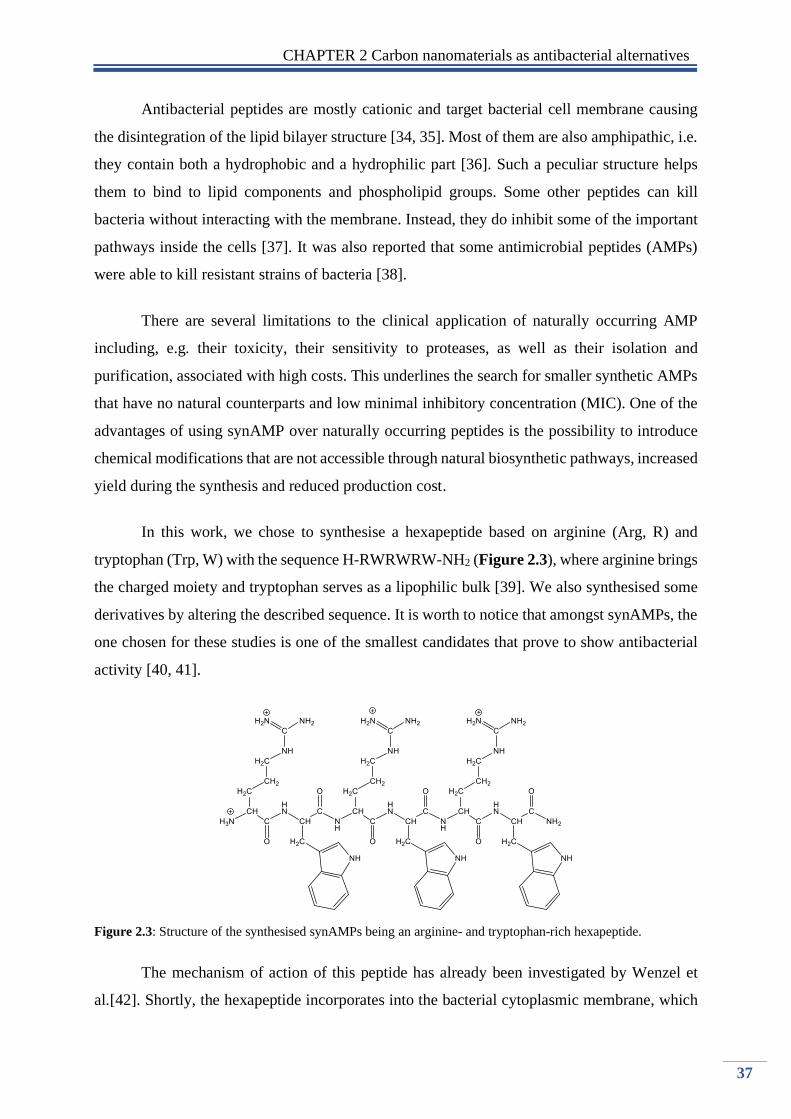
CHAPTER 2 Carbon nanomaterials as antibacterial alternatives
37
Antibacterial peptides are mostly cationic and target bacterial cell membrane causing
the disintegration of the lipid bilayer structure [34, 35]. Most of them are also amphipathic, i.e.
they contain both a hydrophobic and a hydrophilic part [36]. Such a peculiar structure helps
them to bind to lipid components and phospholipid groups. Some other peptides can kill
bacteria without interacting with the membrane. Instead, they do inhibit some of the important
pathways inside the cells [37]. It was also reported that some antimicrobial peptides (AMPs)
were able to kill resistant strains of bacteria [38].
There are several limitations to the clinical application of naturally occurring AMP
including, e.g. their toxicity, their sensitivity to proteases, as well as their isolation and
purification, associated with high costs. This underlines the search for smaller synthetic AMPs
that have no natural counterparts and low minimal inhibitory concentration (MIC). One of the
advantages of using synAMP over naturally occurring peptides is the possibility to introduce
chemical modifications that are not accessible through natural biosynthetic pathways, increased
yield during the synthesis and reduced production cost.
In this work, we chose to synthesise a hexapeptide based on arginine (Arg, R) and
tryptophan (Trp, W) with the sequence H-RWRWRW-NH2 (Figure 2.3), where arginine brings
the charged moiety and tryptophan serves as a lipophilic bulk [39]. We also synthesised some
derivatives by altering the described sequence. It is worth to notice that amongst synAMPs, the
one chosen for these studies is one of the smallest candidates that prove to show antibacterial
activity [40, 41].
Figure 2.3: Structure of the synthesised synAMPs being an arginine- and tryptophan-rich hexapeptide.
The mechanism of action of this peptide has already been investigated by Wenzel et
al.[42]. Shortly, the hexapeptide incorporates into the bacterial cytoplasmic membrane, which

CHAPTER 2 Carbon nanomaterials as antibacterial alternatives
38
contains negatively charged phospholipids. When attached to the cell membrane, it causes the
detachment of cytochrome C (a protein involved in the respiratory chain) and MurG (an enzyme
involved in the synthesis of the cell wall). The indifference of cytochrome C affects the
synthesis of adenosine triphosphate (ATP) and consequently, the cellular energy, whereas the
indifference of MurG affects the cell wall integrity.
The choice of this peptide was driven by its already recognised activity against some
methicillin-resistant S. aureus (MRSA) and vancomycin-intermediate-resistant S. aureus
(VISA) [43]. As it is also known to cause bacteria death through the disruption of a broad range
of cellular processes, such a peptide can be envisioned as a key structure to address the issue of
growing antimicrobial resistance.
Together with the multivalent character of nanoparticles which potentially can
significantly increase the affinity to appropriate target compared to the monovalent reference,
the conjugates might be a promising alternative to nowadays antibiotics by improving their
antibacterial activity and stability against protease degradation. The antibacterial activity of
peptides and carbon-peptides conjugates was investigated against two strains of pathogenic
bacteria: E.coli and S.aureus.
2.2. Synthesis of short synthetic antimicrobial peptides (synAMPs) and
their antibacterial activity
All the peptides were prepared by standard Fmoc/tBu solid-phase peptide synthesis
(SPPS). To increase the overall positive charge on the peptides and their organometallic
conjugates, they were synthesised as peptide amides. All the structures were purified by
reverse-phase high-pressure liquid chromatography (RP-HPLC), analytical HPLC established
their purity and the composition of the final product was confirmed by electron ionisation mass
spectrometry (ESI-MS) (Table 2.1). Additionally, their antibacterial activity was assessed by
determining their MIC values by the microdilution method (Table 2.1).
We decided to use the H-RWRWRW-NH2 as a model structure, that amongst short
cationic peptides showed the highest antibacterial activity [40, 41]. Another factor that
determined our choice was the ease of preparation and sequence modification without losing
the antibacterial activity. In general, a set of 14 differently modified peptides was prepared.

CHAPTER 2 Carbon nanomaterials as antibacterial alternatives
39
The modifications were made in order to improve the antibacterial activity of the
peptides (introduction of organometallic moiety on the N terminal end (2a-3b; 6a-7b) [44-46]
and introduce the necessary linkage for the CuAAC reactions between the nanoparticles
(introduction of propargyl group on the N terminal end (4a-4b), the addition of extra amino
acid with propargyl group on the C terminal end (5a-7b). Small cationic synAMPs owe their
antibacterial activity mainly to charge and lipophilic bulk. These two factors were taken into
consideration while designing the sequences to avoid eukaryotic toxicity.
Table 2.1: Summary of HPLC retention times (tR), ESI-MS data and MIC values for the peptides 1a-7b
Molecular mass MIC [µg/ml]
Compound Sequence tR
1
[min] Calc. Exp.1 E.coli
DSM
30083
S.aureus
(MRSA)
ATCC
43300
1a H-RWRWRW-NH2 6.28 1044.24 1044.79 32 16
1b H-WRWRWR-NH2 6.36 1044.24 1044.75 32 16
2a FcCO-WRWRW-NH2 7.4 1100.08 1100.53 32 16
2b FcCO-RWRWR-NH2 6.63 1070.06 1070.75 256 32
3a RcCO-WRWRW-NH2 7.43 1145.31 1146.04 32 8
3b RcCO-RWRWR-NH2 6.66 1115.28 1116.05 64 16
4a CHCCH2CH2C(O)-RWRWRW-NH2 6.61 1124.33 1124.8 32 16
4b CHCCH2CH2C(O)-WRWRWR-NH2 6.68 1124.33 1124.8 32 16
5a H-RWRWRWG(CH2CCH) -NH2 6.36 1139.34 1139.86 32 16
5b H-WRWRWRG(CH2CCH) -NH2 6.5 1139.34 1139.93 32 16
6a FcCO-WRWRWG(CH2CCH) -NH2 7.43 1195.18 1195.68 64 32
6b FcCO-RWRWRG(CH2CCH) -NH2 6.83 1165.16 1165.98 128 32
7a RcCO-WRWRWG(CH2CCH) -NH2 7.51 1240.41 1240.9 128 8
7b RcCO-RWRWRG(CH2CCH) -NH2 7.41 1210.39 1211.01 128 32
ref ciprofloxacin - - - 2< 4 1For more details see appendix 6.2.1.1.- 6.2.1.14.
All peptides were prepared in high purity and satisfying yield. The activity against
pathogenic microbes was as expected, higher for Gram-positive bacteria than Gram-negative.
In the case of peptides (1a-1b; 4a-5b) which have a balanced content of charged and bulky
lipophilic groups, there is no difference in the activity irrespective of the order of the amino
acids. Peptides with the structure McC(O)-WRWRW-NH2 showed higher activity than
McC(O)-RWRWR-NH2 where the one with ruthenocene showed an increase in antibacterial
activity over the one that contained ferrocene.

CHAPTER 2 Carbon nanomaterials as antibacterial alternatives
40
2.3. Synthesis and surface modifications of nanoparticles prior to the
formation of bioconjugates
Two different kinds of carbon particles were used as a scaffold to incorporate previously
prepared peptides (1a-7b). Commercially available nanodiamonds (NDs) and carbon dots
(CQDs). Both particles have hydroxyl, carboxy or amino groups on their surface, which enable
to create amid bond between the peptides and create azido groups, necessary for the “click”
reaction.
In this work, we synthesised amine rich carbon dots (CQDs-1), which surface was
modified in two ways (Figure 2.4). First, to enable the incorporation of peptide amides, amine
groups were reacted with ClCH2COONa under strong alkaline conditions resulting in carboxyl
capped CQDs (CQDs-2). The second modification includes grafting 2-azidoacetic acid onto
the surface in the presence of EDC/NHS (CQDs-3). In case of nanodiamonds, for the coupling
of peptide amides, we used commercially available NDs-COOH, and for the “click” reactions
ND-NH2 was directly modified with 4-azidobenzoic acid in the presence of N, N’-
dicyclohexylcarbodiimide (DCC) and a catalytic amount of 4-dimethyl aminopyridine (DMAP)
(ND-3) (Figure 2.4).
Figure 2.4: Schematic illustration of synthesis and surface functionalization used for carbon nanoparticles.
CQDs-1 were prepared by a hydrothermal method previously reported by Zhu et al.[47]
with small modifications. The reaction was conducted by carbonisation of the organic precursor
with a sealed Teflon lined autoclave chamber at elevated temperature under reduced pressure

CHAPTER 2 Carbon nanomaterials as antibacterial alternatives
41
for 5 h. The obtained product was left to cool down to be further purified by dialysis for 24 h,
resulting in a dark brown solution with the yield of the reaction- around 60%.
The morphology and structure of CQDs-1 were examined. Figure 2.5 shows
transmission electron microscopy (TEM) of as-prepared material confirming their spherical
shape with an average diameter of 4.5 ±0.2 nm. From the image, most of the observed particles
are of a crystalline character possessing well-resolved lattice fringes.
Figure 2.5: TEM image, magnified TEM, FFT images and size distribution histograms for CQDs-1, CQDs-2,
and CQDs-3.

CHAPTER 2 Carbon nanomaterials as antibacterial alternatives
42
With such a high carbon lattice structure content, D and G band were detected in the
Raman spectra of CQDs-1 (Figure 2.6A). G band at 1570 cm-1 related to in-plane vibration of
sp2 carbon, and the D band at 1350 cm-1 attributed to the presence of sp3 defects. The intensity
ratio of the D and G band (ID/IG) is 0.85±0.12. Also, a broad peak centred at around 2800 cm-1
corresponding to the 2D component of graphitic structures is observed. The XRD diffractogram
(Figure 2.6B) displays a broad diffraction peak centred at 25.5° corresponding to an interlayer
spacing of 0.35 nm, larger than the spacing between (100) planes in bulk graphite (0.23 nm)
due to the incorporation of functional groups along the edges of the CQDs.
(A) (B)
(C) (D)
Figure 2.6: Characterisation of CQDs-1: (A) Raman spectrum of CQDs-1; (B) XRD diffractogram of CQDs-1;
(C) UV-Vis absorption (solid lines) and fluorescence spectra (dash-dot, λex = 350 nm) of the CQDs-1; (D)
wavelength-dependent fluorescent emission properties of CQDs-1.
1000 2000 3000 4000
Inte
nsity /
a.u
.
Raman shift / cm-1
20 30 40 50 60 70 80
Inte
nsity / a
.u.
2 / degrees
0,35nm
200 300 400 500 600 700 800
CQDs-1
Ab
so
rba
nce
/ a
.u.
Wavelength / nm
340nm
237nm
Flu
ore
sce
nce
/ a
.u.
450nm
446nm
400 500 600 700
0,0
0,2
0,4
0,6
0,8
1,0
260
280
300
320
340
360
380
400
420
440
460
480
260→480nm
Flu
ore
sce
nce
/ a
.u.
Wavelength / nm
CQDs-1

CHAPTER 2 Carbon nanomaterials as antibacterial alternatives
43
The UV/Vis of CQDs-1 (Figure 2.6C) reveals an absorption maximum at ≈242 nm
attributed to π-π* transition of C=C and a band at 344 nm attributed to n-π* transition of C=O
and C=N bonds.[48, 49] Furthermore, the QY of as-prepared carbon dots was as high as 61%
(0,33) (using quinine sulphate as a reference; QY, 0.54 in 0.12 M H2SO4). Additionally, upon
increasing the excitation wavelength, the emission gradually shifts to the red region with an
increase in fluorescence intensity (Figure 2.6D). The phenomenon of excitation-dependent
emission is typical for these nanostructures.
To get more information about the chemical composition FTIR and XPS measurements
were performed. XPS data revealed the presence of oxygen, nitrogen, and carbon on the surface
of the particles. The deconvolution of the high-resolution spectrum of C1s confirms the
existence of three different types of carbon atoms graphitic or aliphatic (C=C and CC),
oxygenated, and nitrous (Figure 2.7A) on the surface. The high-resolution XPS spectrum of
N1s displays one peak at 399,7eV, which corresponds to C-NH (Figure 2.7B).
(A) (B)
Figure 2.7: Characterisation of CQDs-1: (A)XPS survey of CQDs-1 with inset of high resolution C1s
spectrum; (B) N1s spectrum of CQDs-1.
The FTIR spectrum of as-prepared dots exhibits a distinct band at 3432 cm-1 attributed
to stretching vibration of -OH. Another absorption band corresponding to stretching vibrations
are the one of NH (3262cm-1), C=CH (3018cm-1), CH (2923cm-1), C=O (1771cm-1 and
1645cm-1), C-C (1297cm-1). The existence of the amine functional groups is confirmed by the
presence of a band at around 1567cm-1 and 1396cm-1(Figure 2.8).
406 404 402 400 398 396 394
Inte
ns
ity
/ a
.u.
Binding energy / eV
N 1s
900 800 700 600 500 400 300 200
N 12,41%O 14,94%
C 72,62%
294 292 290 288 286 284 282 280 278
Binding energy / eV
C-C; C=C
C-N; C-O
C=O; C=N
Inte
ns
ity
/ a
.u.
Binding energy / eV
C 1s
N 1s
O 1s

CHAPTER 2 Carbon nanomaterials as antibacterial alternatives
44
Figure 2.8: Characterisation of CQDs: FTIR spectra of as-prepared CQDs-1 (black), CQDs-2 (blue), CQDs-3
(red).
Modifications that have been made in order to enable the incorporation of peptide
amides on the surface of as-prepared carbon dots were verified by FTIR and XPS. First one
includes the conversion of amine groups presented on the surface into carboxyl capped carbon
dots [50, 51]. Briefly, the amino groups reacted with ClCH2COONa under strongly alkaline
conditions. The suspension was further neutralised, resulting in CQDs-COOH (CQDs-2). As
observed in the FTIR spectra, the sharp peak at around 1650 cm-1 was enhanced, which
indicates that plenty of carboxyl groups were introduced after the treatment of CQDs-1. The
increase in the oxygen content for CQDs-2 compared to CQDs-1 is 26.87% and 14.94%
respectively, strongly indicating the successful introduction of additional carboxyl groups.
Additionally, we can observe an increase in negative zeta potential and hydrodynamic diameter
(Table 2.2). Moreover, the quantum yield decreased significantly which can be attributed to
the increase of electron withdrawing carboxyl groups (QY= 7% (0.038) using quinine sulphate
as a reference; QY, 0.54 in 0.12 M H2SO4).
In case of CQDs-3, the successful formation of azide groups on the surface of
nanoparticles was confirmed by the appearance of the band at 2110cm-1 corresponding to
vas(N3) (Figure 2.8) and further verified by XPS which revealed the presence of the peak
405.2eV (-N=N+=N-) and 401.6 eV (N=N+=N-) in a ratio of 1:2, as theoretically expected
4000 3500 3000 2500 2000 1500 1000 500
Tra
ns
mit
tan
ce
/ a
.u.
Wavenumber / cm-1
34
32
32
62 29
23
1771
2110
CQDs-1
CQDs-2
CQDs-316
61
16
28
15
67
1396
12
97
1068

CHAPTER 2 Carbon nanomaterials as antibacterial alternatives
45
(Figure 2.9B). In addition, we can observe the increase in the atomic percentage of the N1s
component as well as the increase in the potential on the surface from -9.9 for CQDs-1 to +7
for CQDs-3 (Table 2.2). Furthermore, we decided to quantify the free amino group on the
surface of CQDs before and after the modification with the technique of so-called Keiser test.
The Keiser test makes the use of the formation of Ruhemann’s Blue by the reaction of ninhydrin
with the primary amino groups. The successful implementation of this colorimetric assay on
the nanoparticles has been already reported [52]. We can see than the modification made on
CQDs-1 caused a decrease in the amount of free amino groups in both CQDs-2 and CQDs-3.
It may indicate the success of converting the surface from amino into carboxyl groups and
successful coupling of azido moieties. It is also giving us more information about possible
loading efficiency.
Table 2.2: Physicochemical properties of nanostructures
Particles (mV)1 Size (nm) Hydrodyna
mic size
(nm) 2
PDI C1s O1s N1s Surface
loading
mmol/g
Keiser test
CQDs-1 -9.9 ± 3.4 4.5±0.2 11 ± 0.1 0.22±0.11 72.62 14.94 12.43 0.144
CQDs-2 -27 ±2.1 5.75±0.78 17± 2 0.357±0.2 68.89 26.87 4.23 0.0445
CQDs-3 7.14±3 5.5 ± 0.3 12 ± 0.1 0.23 ± 0.11 68.8 13.9 17.3 0.055 1: zeta potential, PDI Polydispersity index; 2the hydrodynamic size was recorded at 37°C
The modification of nanodiamonds with azide moiety was based on the same principle
as CQDs with the use of different coupling reagents. The successful coupling was confirmed
by FTIR and XPS. (Figure 2.9A) shows the transmission FTIR spectra of commercially
available ND-NH2 (ND-2) and modified nanodiamonds (ND-3). The presence of the broadband
between 3000-3600 cm-1 corresponds to the vibrational mode of amino groups presented on the
surface and adsorbed water molecules. The peak at 1632 cm-1 can be assigned to a superposition
of N-H scissoring mode and OH deformation mode of adsorbed water. After the surface
modification, an intense peak at 2124cm-1 appears, which is characteristic for vas(N3) stretching
mode. Additionally, new bands appeared at 1600, 1376, 1286, and 1174 cm-1 that correspond
to the same observed bands present in an FTIR spectrum of neat 4-azidobenzoic acid (data not
shown). In addition to FTIR, XPS analysis was performed to characterise and confirm the
modification. Deconvolution of high resolution N1s spectrum (Figure 2.7B) reveals the
presence of azide function by the signals at 405.2eV and 401.6eV in a 1:2 ration, comparable
to the XPS spectra generated for CDots-3.

CHAPTER 2 Carbon nanomaterials as antibacterial alternatives
46
(A) (B)
Figure 2.9: (A) FTIR of ND-2, ND-3; (B) High resolution N1s XPS spectrum of CQDs-3 (up) and ND-3 (down).
Table2.2: Physical properties of nanodiamonds (ND-2; ND-3)
Particles (mV)1 Hydrodynamic
size (nm) 2 PDI
ND-2 18± 3 134±7 0.35±0.02
ND-3 24±3 183±5 0.34±0.10 1: zeta potential, PDI Polydispersity index;
2.4. Subsequent post functionalization of carbon nanostructures:
Nanoparticles- peptide bioconjugates
We decided to use two different approaches to prepare nanoparticle-peptide conjugates
(Figure 2.10). First, consists of a standard coupling reaction using carboxyl groups presented
on the surface of carbon nanomaterial and creation of amide bond between the peptides. As
prepared peptides comprised of 2 or 3 arginine residues and C-terminal was created as amid.
Which gives us multiple binding sites on the peptide chain. Therefore, to gain more control of
the reaction, we decided to introduce an alkyne in the sequence of the peptides which together
with an azide moiety presented on nanoparticles allow us to “click” the molecules together.
Cu(I) catalysed azide-alkyne cycloaddition (CuAAC- click reaction) is stereospecific forming
1,4-substituted 1,2,3-triazole and is relatively fast even at low temperature resulting in high
yield products.
4000 3500 3000 2500 2000 1500 1000 500
Tra
ns
mit
tan
ce
/ a
.u.
Wavenumber / a.u.
ND-2
ND-32124 1630 1277 11001600
410 408 406 404 402 400 398 396 394
ND-3
Binding energy / eV
-N=N=N
-N=N=N
-NH-C=O
N1s CQDs-3
Inte
ns
ity
/ a
.u.
-N=N=N
-N=N=N
-NH-C=O

CHAPTER 2 Carbon nanomaterials as antibacterial alternatives
47
Figure 2.10: Schematic illustration of surface functionalization used in this section.
The preparation of CQDs-peptides (1a-3b) is based on a two-step process. The first
step was the conversion of the amino coated surface into carboxyl capped CQDs-2. CQDs-2
has further reacted with peptide amide with the use of N-Ethyl-N′-(3-dimethyl
aminopropyl)carbodiimide hydrochloride (EDC•HCl) and N-hydroxysuccinimide (NHS) as
coupling reagents. The reaction was carried out in a buffer solution at physiological pH and
resulted in well dispersible carbon-conjugates. On the FTIR spectra (Figure 2.11A) we can see
the peak at 3450 cm-1 has broadened due to additional symmetric stretching vibration mode of
-NH. Additionally, a new band appears at 1530 cm-1 which corresponds to the symmetric
bending vibration of -NH commonly presented in amino acids. In the (Table 2.3), we can see
the changes in zeta potential and hydrodynamic diameter of as-prepared particles. Charge of
the modified carbon dots, changes towards more positive values in the case of all the
compounds. However, the changes are not as significant as expected. The hydrodynamic
diameter increases from 68±2.1 to be within the range of 137-157nm, which is probably the
result of the formation of aggregated structures. Surprisingly the calculated quantum yields do
not differ significantly from CQDs-2 reflecting that the modifications barely altered the
inherent emission features of CQDs-COOH.
For the preparation of ND-peptides, we decided to use commercially available ND-
COOH. For the coupling of the peptides, we chose N,N′-Diisopropylcarbodiimide (DIC) and

CHAPTER 2 Carbon nanomaterials as antibacterial alternatives
48
Hydroxybenzotriazole (HOBt). The FITR spectrum (Figure 2.11B) of as-prepared NDs
conjugates show similar features to the one of CQDs. The band at around 3450cm-1 is broadened
due to appearance of the stretching mode of -NH. Additionally, new bands appeared at 1534
cm-1, 1200cm-1, 1135 cm-1 which are attributed to stretching vibration and stretching rocking
mode of -NH. For the conjugates ND-1.2a and ND-1.3a we can observe an appearance of
additional bands at 837cm-1 and 802cm-1 which might be attributed to the presence of
metallocenoyl group on the surface of the particles. Similarly, to CQDs, the surface potential
of nanodiamonds changes towards less negative values but remaining negative. The
hydrodynamic diameter increases from 78±2.3 to ranging between 147-167nm.
(A) (B)
Figure 2.11: (A) FTIR spectra of CQDs-peptides (CQDs-2.1a, CQDs-2.2a, CQDs-2.3a); (B) FTIR spectra of ND-
peptides (ND-1.1a, ND-1.2a, ND-1.3a) bioconjugates.
Table 2.3: Physicochemical properties of carbon-conjugates
Particles (mV)1 Hydrodynamic
size (nm) PDI
Loading
µg/mg QY (%)
CQDs-2 -27 ±2 68±2.1 0.24±0.13 - 7
CQDs-2.1a 2±0.4 141±2.5 0.21±0.09 134 6.54
CQDs-2.1b 2±0.5 137±1.7 0.17±0.10 115 6.72
CQDs-2.2a -2±0.3 153±2.4 0.14±0.04 98 6.1
CQDs-2.2b 0.5±0.3 157±3.6 0.51±0.09 103 6.3
CQDs-2.3a -2±0.8 149±3.2 0.42±0.02 95 6.41
CQDs-2.3b -0.4±0.7 147±4.6 0.33±0.41 97 6.44
ND-1 -45±2 78±3 0.19±0.02 - -
ND-1.1a -25±2 154±2 0.14±0,02 284 -
ND-1.1b -17±3 147±3.5 0.16±0.03 292 -
ND-1.2a -19±3 161±4,1 0.17±0.02 266 -
ND-1.2b -15±3 167±3 0.17±0.02 268 -
ND-1.3a -20±2 159±2 0.18±0.02 275 -
ND-1.3b -15±3 160±2.7 0.18±0.02 267 - 1 : zeta potential, PDI Polydispersity index;
4000 3500 3000 2500 2000 1500 1000 500
Tra
ns
mit
tan
ce
/ a
.u.
Wavenumber / cm-1
CQDs-2
CQDs-2.2a
CQDs-2.1a
CQDs-2.3a
1530
4000 3500 3000 2500 2000 1500 1000 500
ND-1.2a
Tra
ns
mit
tan
ce
/ a
.u.
ND-1
ND-1.1a
ND-1.3a
Wavenumber / cm-1
1200
1135
1534 802
837

CHAPTER 2 Carbon nanomaterials as antibacterial alternatives
49
The formation of the click conjugates for both carbon structure was performed in the
same way using copper sulphate pentahydrate and ascorbic acid as catalysts for the reaction. In
both cases, the process was accomplished in two steps. The first step consists of the introduction
of the azide molecule on the surface of the nanoparticles, and the second step was achieved by
using a previously prepared “clickable” peptides. The successful modification can be easily
followed by the disappearance of the band at 2100 cm-1 for CQDs (Figure 2.12A) and
2123cm-1 for ND (Figure 2.12B). Additionally, for CQDs containing metallocene moieties, the
characteristic band at 3071cm-1 was observed. The band at 1200 cm-1 and 1130 cm-1 are
attributed to stretching rocking mode of -NH. For ND, the band assigned to stretching vibrations
of -NH appears at 1540cm-1.
(A) (B)
Figure 2.12: (A) FTIR spectra of CQDs-peptides (CQDs-3.4a, CQDs-3.5a, CQDs-3.6a, CQDs-3.7a); (B) FTIR
spectra of ND-peptides (ND-3.4a, ND-3.5a, ND-3.6a, ND-3.7a) bioconjugates.
Following the changes in the zeta potential of the particles (Table 2.4), we can assume
that the cationic peptide is incorporated on the surface, and its charge is not altered by additional
interactions. For CQDs, the potential changes form 7±3 up to 26±1 and for all the particles stay
in the same range. ND showed a higher charge on the surface, which is influenced by higher
loading of the cationic peptide on the surface. As expected, the hydrodynamic diameter
increases for all prepared nanoconjugates indicating the successful incorporation of peptides.
In the case of CQDs click particles, another factor indicating the success of the reaction is the
decrease in the quantum yield. The changes are quite significant, decreasing from 33% for
CQDs-3 to get as low as 9.8% after the modification. It is worth to notice that the peptides “b”
are the reverse sequence of peptides “a” showing the same FTIR spectra.
4000 3500 3000 2500 2000 1500 1000 500
Tra
ns
mit
tan
ce
/ a
.u.
Wavenumber / cm-1
2123
ND-3.4a
1540
ND-3.5a
ND-3.6a
ND-3.7a
ND-3
1314
4000 3500 3000 2500 2000 1500 1000 500
CQDs-3.7a
CQDs-3.6a
CQDs-3.5a
CQDs-3.4a
CQDs-3
Tra
ns
mit
tan
ce
/ a
.u.
Wavenumber / cm-1
1130
2110
12003071

CHAPTER 2 Carbon nanomaterials as antibacterial alternatives
50
Table 2.4: Physicochemical properties of carbon conjugates: “clicked” nanoparticles
Particles (mV)1 Hydrodynamic
size (nm) PDI
Loading
µg/mg QY (%)
CQDs-3 7±3 71±4 0.24±0.13 - 34
CQDs-3.4a 21±2 181±2 0.28±0.09 94 10
CQDs-3.4b 26±1 175±3 0.22±0.08 95 13
CQDs-3.5a 23±1 179±4 0.19±0.05 101 14
CQDs-3.5b 25±2 158±2 0.20±0.10 103 11
CQDs-3.6a 19±2 198±3 0.34±0.09 100 9.8
CQDs-3.6b 22±2 191±4 0.32±0.13 97 10
CQDs-3.7a 21±3 193±3 0.31±0.11 99 15
CQDs-3.7b 24±1 190±3 0.34±0.15 96 14.7
ND-2 18±3 134±7 0.35±0.02 - -
ND-3 24±3. 183±5 0.34±0.10 - -
ND-3.4a 34±3 213±4 0.36±0.02 243 -
ND-3.4b 33±3 234±6 0.36±0.02 239 -
ND-3.5a 33±3 219±2 0.34±0.04 257 -
ND-3.5b 29±3 229±3 0.39±0.10 256 -
ND-3.6a 28±3 261±5 0.41±0.09 261 -
ND-3.6b 29±3 267±7 0.40±0.07 273 -
ND-3.7a 31±2 253±6 0.39±0.15 269 -
ND-3.7b 30±2 249±8 0.4±0.11 258 - 1: zeta potential, PDI Polydispersity index;
2.5. Antibacterial activity of carbon-based conjugates
The antibacterial activity of all the compounds was assessed by determining their MIC
values by microdilution assay. MIC value is the lowest concentration of an antimicrobial that
will inhibit the visible growth of a microorganism after overnight incubation. In this work, one
Gram-negative (E.coli DSM 30038) and one Gram-positive (S.aureus ATCC 43300), the
bacterial strain was used.
The first group of compounds where the peptides were covalently linked showed no
antibacterial activity against studied bacterial strains (conjugates: 1a-3b) (Table 2.5). Peptides
used in this study owe their antibacterial properties to charge and lipophilic bulk. The
unprotected guanidine groups on the arginine side chain could react with the nanoparticles
during the coupling, resulting in the loss of the positive charge and increase lipophilicity. This
indicates the importance of preserving these groups during the reaction.

CHAPTER 2 Carbon nanomaterials as antibacterial alternatives
51
Table 2.5: Antibacterial properties of carbon nanomaterials (CQDs-1; CQDs-2; ND-1) and covalently modified
carbon-peptides conjugates (CQDs-2.1a- CQDs-2.3b; ND-1.1a- ND-1.3b).
n.a.: not active
To overcome this problem and protect the positive charge of the peptides, click
chemistry was developed to link AMPs to the nanostructures. Increased antibacterial activity
of the conjugates (Table 2.6) revealed the multivalent character of carbon nanostructures.
For the conjugates prepared by the “click” reaction, the activity is in general higher for
Gram-positive bacteria. However, for some compounds, the activity for Gram-negative E.coli
has been significantly increased. For carbon dots conjugates (CQDs-3.4a; CQDs-3.4b; CQDs-
3.5a; CQDs-3.5b) we can observe 5 fold increase of activity and 4-fold increase in case of
nanodiamonds (ND-3.4a; ND-3.4b; ND-3.5a; NDs-3.5b) with the MIC values ranging between
6.02µg mL-1- 6.59µg mL-1 (CQDs) and 7.65µg mL-1 – 8.22µg mL-1 (NDs) respectively. The
additional amino acid and the position of the propargyl group does not seem to alter the MIC
values. This contrast to the presence of the metallocene group (conjugates: 6a-7b) where the
activity in case of ND was slightly decreased, and carbon dots showed no activity in the studied
range.
Considering Gram-positive bacteria S.aureus (MRSA) 5-fold increase for carbon dots
and a 4-fold increase in the activity is also observed (conjugates: 6a-7b). In the case of peptides
containing ferrocene or ruthenocene, the activity increases 3-fold for CQDs being approx.
12µg mL-1 with an exception for compound CQDs-3.7a where the MIC value (6.34µg mL-1) is
close to the value of the free peptide (8µg mL-1). Nanodiamond conjugates showed a 2-fold
increase in case of ND-3.6b and ND-3.7b, no significant difference for ND-3.6a and decreased
Compound Sequence
MIC [µg/ml] MIC [µg/ml]
E.coli
DSM 30083
S.aureus (MRSA)
ATCC 43300
CQDs-1 - n.a. n.a.
CQDs-2 - n.a. n.a.
CQDs-2.1a H-RWRWRW-NH2 n.a. n.a.
CQDs-2.1b H-WRWRWR-NH2 n.a. n.a.
CQDs-2.2a FcCO-WRWRW-NH2 n.a. n.a.
CQDs-2.2b FcCO-RWRWR-NH2 n.a. n.a.
CQDs-2.3a RcCO-WRWRW-NH2 n.a. n.a.
CQDs-2.3b RcCO-RWRWR-NH2 n.a. n.a.
ND-1 - n.a. n.a.
ND-1.1a H-RWRWRW-NH2 n.a. n.a.
ND-1.1b H-WRWRWR-NH2 n.a. n.a.
ND-1.2a FcCO-WRWRW-NH2 n.a. n.a.
ND-1.2b FcCO-RWRWR-NH2 n.a. n.a.
ND-1.3a RcCO-WRWRW-NH2 n.a. n.a.
ND-1.3b RcCO-RWRWR-NH2 n.a. n.a.

CHAPTER 2 Carbon nanomaterials as antibacterial alternatives
52
activity for the compound ND-3.7a was observed. Lower activity compared to CQDs can be
attributed to the higher loading of ND, resulting in a higher amount of ferrocene/ ruthenocene
presented on the surface. Therefore, increasing the lipophilicity of the compounds.
Additionally, CuSO4 that was used as a catalyst during the click reaction (compounds
4a-7b) was tested, showing no inhibition of growth in case of both bacterial strains.
Table 2.6: Antibacterial properties of carbon nanostructures (CQDs-3, ND-3) and carbon nanoconjugates prepared
by Cu(I) catalysed azide-alkyne cycloaddition (CuAAC) (CQDs-3.4a-3.7b; ND-3.4a-ND-3.7b). MIC value for the
conjugates: [µg/ml] NP // [µg/ml] of the peptide conjugated on the surface
Indicated in orange: alkyne group necessary for the click reaction; Indicated in blue: conjugates that showed
increased antibacterial activity.
Compound Sequence
MIC [µg/ml]
Gram-negative Gram-positive
E.coli
DSM 30083
S.aureus (MRSA)
ATCC 43300
CQDs-3 - n.a. n.a.
4a CHCCH2CH2C(O)-RWRWRW-NH2
32 16
CQDs-3.4a 64 // 6.02 32 // 3.01
4b CHCCH2CH2C(O)-WRWRWR-NH2
32 16
CQDs-3.4b 64 // 6.08 32 // 3.04
5a H-RWRWRWG(CH2CCH) -NH2
32 16
CQDs-3.5a 64 // 6.46 32 // 3.23
5b H-WRWRWRG(CH2CCH) -NH2
32 16
CQDs-3.5b 64 / /6.59 32 // 3.3
6a FcCO-WRWRWG(CH2CCH) -NH2
64 32
CQDs-3.6a 512 // 51.2> 128 // 12.8
6b FcCO-RWRWRG(CH2CCH) -NH2
128 32
CQDs-3.6b 512 // 49.7> 128 // 12.4
7a RcCO-WRWRWG(CH2CCH) -NH2
128 8
CQDs-3.7a 512 // 50.07> 64 // 6.34
7b RcCO-RWRWRG(CH2CCH) -NH2
128 32
CQDs-3.7b 512 // 50.07> 128 // 12.7
ND-3 - n.a. n.a.
4a CHCCH2CH2C(O)-RWRWRW-NH2
32 16
ND-3.4a 32 // 7.78 16 // 3.89
4b CHCCH2CH2C(O)-WRWRWR-NH2
32 16
ND-3.4b 32 // 7.65 16 // 3.82
5a H-RWRWRWG(CH2CCH) -NH2
32 16
ND-3.5a 32 // 8.22 16 // 4.11
5b H-WRWRWRG(CH2CCH) -NH2
32 16
ND-3.5b 32 // 8.19 16 // 4.1
6a FcCO-WRWRWG(CH2CCH) -NH2
64 32
ND-3.6a 512 // 134> 128 // 33.4
6b FcCO-RWRWRG(CH2CCH) -NH2
128 32
ND-3.6b 512 // 140> 64 // 17.5
7a RcCO-WRWRWG(CH2CCH) -NH2
128 8
ND-3.7a 512 // 138> 64 // 17.2
7b RcCO-RWRWRG(CH2CCH) -NH2
128 32
ND-3.7b 512 // 132> 64 // 16.5

CHAPTER 2 Carbon nanomaterials as antibacterial alternatives
53
2.6. Conclusions
A set of 28 different carbon- peptide nanostructure conjugates were successfully
prepared, and their antibacterial activity was assessed by determining the MIC values against
Gram-negative E.coli DSM 30083 and S.aureus ATCC 43300. The first group of compounds
obtained by covalent linking showed no antibacterial activity against studied strains
(conjugates: 1a-3b) while the second group where click chemistry was applied (conjugates: 4a-
7b) showed a significant decrease in the minimal inhibitory concentrations for Gram-negative
and Gram-positive bacteria. Amongst them, conjugates 4a-5b showed the best MIC values for
the tested strains being 5-fold lower for peptide modified CQDs and 4-fold higher for peptide
modified ND when compared to the peptide in solution.
The lack of activity in some particle-conjugates in the tested range is associated with
the molecules loaded on the surface rather than particles themselves. The data collected in the
(Table 2.3) and (Table 2.5) indicate that charge is a significant factor in the antibacterial
activity. Even though the zeta potentials were decreased towards less negative charges
indicating successful incorporation of peptides, it did not increase as much as expected, like for
the “clicked” compounds suggesting that some if not all of the positive charge in the backbone
of the loaded peptides was blocked.
In addition, it was shown that the position of the propargyl group, as well as the reverse
order of amino acids in the sequence, does not influence the activity of tested compounds. This
contrast the results obtained for the conjugates where the organometallic moiety was present.
The presence of ferrocene or ruthenocene results in loss of activity for the Gram-negative
bacteria and only 2-3 fold higher for Gram-positive S.aureus with an exception for the
conjugate 7a where the MIC value did not improve. This might be explained by the increased
lipophilicity on the surface of nanoparticles due to the presence of ferrocene or ruthenocene.
Which indicated that the existence of organometallic moiety in case of as prepared conjugates
is not favourable and better MIC values are obtained for the “simple” peptides.

CHAPTER 2 Carbon nanomaterials as antibacterial alternatives
54
2.7. References
1. Cheng, G., et al., Antimicrobial Drugs in Fighting against Antimicrobial Resistance.
Frontiers in microbiology, 2016. 7: p. 470-470.
2. O'Neill, J. Antimicrobial Resistance: Tackling a crisis for the health and wealth of
nations. December 2014; Available from: https://amr-review.org/Publications.html.
3. Huh, A.J. and Y.J. Kwon, “Nanoantibiotics”: A new paradigm for treating infectious
diseases using nanomaterials in the antibiotics resistant era. Journal of Controlled
Release, 2011. 156(2): p. 128-145.
4. Weir, E., et al., The use of nanoparticles in anti-microbial materials and their
characterization. Analyst, 2008. 133(7): p. 835-845.
5. Allaker, R.P. and G. Ren, Potential impact of nanotechnology on the control of
infectious diseases. Transactions of The Royal Society of Tropical Medicine and
Hygiene, 2008. 102(1): p. 1-2.
6. Li, Q., et al., Antimicrobial nanomaterials for water disinfection and microbial
control: Potential applications and implications. Water Research, 2008. 42(18): p.
4591-4602.
7. Mühling, M., et al., An investigation into the effects of silver nanoparticles on
antibiotic resistance of naturally occurring bacteria in an estuarine sediment. Marine
Environmental Research, 2009. 68(5): p. 278-283.
8. Pal, S., Y.K. Tak, and J.M. Song, Does the Antibacterial Activity of Silver
Nanoparticles Depend on the Shape of the Nanoparticle? A Study of the Gram-
Negative Bacterium <em>Escherichia coli</em>. Applied and
Environmental Microbiology, 2007. 73(6): p. 1712.
9. Huang, Z., et al., Toxicological Effect of ZnO Nanoparticles Based on Bacteria.
Langmuir, 2008. 24(8): p. 4140-4144.
10. Kim, J.S., et al., Antimicrobial effects of silver nanoparticles. Nanomedicine:
Nanotechnology, Biology and Medicine, 2007. 3(1): p. 95-101.
11. Rabea, E.I., et al., Chitosan as Antimicrobial Agent: Applications and Mode of Action.
Biomacromolecules, 2003. 4(6): p. 1457-1465.
12. Zhang, L., et al., Development of Nanoparticles for Antimicrobial Drug Delivery.
Current Medicinal Chemistry, 2010. 17(6): p. 585-594.
13. Zhang, L., et al., Nanoparticles in Medicine: Therapeutic Applications and
Developments. Clinical Pharmacology & Therapeutics, 2008. 83(5): p. 761-769.
14. Santos-Magalhães, N.S. and V.C.F. Mosqueira, Nanotechnology applied to the
treatment of malaria. Advanced Drug Delivery Reviews, 2010. 62(4): p. 560-575.
15. Mengesha, A.E. and B.B.C. Youan, 8 - Nanodiamonds for drug delivery systems, in
Diamond-Based Materials for Biomedical Applications, R. Narayan, Editor. 2013,
Woodhead Publishing. p. 186-205.
16. Szunerits, S., A. Barras, and R. Boukherroub, Antibacterial Applications of
Nanodiamonds. International journal of environmental research and public health,
2016. 13(4): p. 413-413.
17. Mishra, V., et al., Carbon dots: emerging theranostic nanoarchitectures. Drug
Discovery Today, 2018. 23(6): p. 1219-1232.
18. Zheng, M., et al., Integrating Oxaliplatin with Highly Luminescent Carbon Dots: An
Unprecedented Theranostic Agent for Personalized Medicine. Advanced Materials,
2014. 26(21): p. 3554-3560.

CHAPTER 2 Carbon nanomaterials as antibacterial alternatives
55
19. Moore, L., et al., Biocompatibility Assessment of Detonation Nanodiamond in Non-
Human Primates and Rats Using Histological, Hematologic, and Urine Analysis. ACS
Nano, 2016. 10(8): p. 7385-7400.
20. Zhu, Y., et al., The biocompatibility of nanodiamonds and their application in drug
delivery systems. Theranostics, 2012. 2(3): p. 302.
21. Marcon, L., et al., Cellular and in vivo toxicity of functionalized nanodiamond in
Xenopus embryos. Journal of Materials Chemistry, 2010. 20(37): p. 8064-8069.
22. Liu, K.-K., et al., Biocompatible and detectable carboxylated nanodiamond on human
cell. Nanotechnology NANOTECHNOLOGY Nanotechnology, 2007. 1871: p.
325102-10.
23. Krueger, A. and D. Lang, Functionality is key: recent progress in the surface
modification of nanodiamond. Advanced Functional Materials, 2012. 22(5): p. 890-
906.
24. Krueger, A., New Carbon Materials: Biological Applications of Functionalized
Nanodiamond Materials. Chemistry – A European Journal, 2008. 14(5): p. 1382-1390.
25. Mochalin, V.N., et al., The properties and applications of nanodiamonds. Nature
Nanotechnology, 2012. 7(1): p. 11-23.
26. Yang, S.-T., et al., Carbon Dots for Optical Imaging in Vivo. Journal of the American
Chemical Society, 2009. 131(32): p. 11308-11309.
27. Wei, W., et al., Non-Enzymatic-Browning-Reaction: A Versatile Route for Production
of Nitrogen-Doped Carbon Dots with Tunable Multicolor Luminescent Display.
Scientific Reports, 2014. 4: p. 3564.
28. Weng, C.-I., et al., One-step synthesis of biofunctional carbon quantum dots for
bacterial labeling. Biosensors and Bioelectronics, 2015. 68: p. 1-6.
29. Wang, Q., et al., Hollow luminescent carbon dots for drug delivery. Carbon, 2013. 59:
p. 192-199.
30. Lai, C.-W., et al., Facile synthesis of highly emissive carbon dots from pyrolysis of
glycerol; gram scale production of carbon dots/mSiO2 for cell imaging and drug
release. Journal of Materials Chemistry, 2012. 22(29): p. 14403-14409.
31. Gogoi, N. and D. Chowdhury, Novel carbon dot coated alginate beads with superior
stability, swelling and pH responsive drug delivery. Journal of Materials Chemistry,
2014. 2: p. 4089.
32. Thakur, M., et al., Antibiotic Conjugated Fluorescent Carbon Dots as a Theranostic
Agent for Controlled Drug Release, Bioimaging, and Enhanced Antimicrobial
Activity. Journal of Drug Delivery, 2014. 2014: p. 9.
33. Sarkar, N., et al., Carbon quantum dot tailored calcium alginate hydrogel for pH
responsive controlled delivery of vancomycin. European Journal of Pharmaceutical
Sciences, 2017. 109: p. 359-371.
34. Shai, Y., Mode of action of membrane active antimicrobial peptides. Peptide Science,
2002. 66(4): p. 236-248.
35. Zhang, L.J., A. Rozek, and R. Hancock, Interaction of Cationic Antimicrobial
Peptides with Model Membranes. The Journal of biological chemistry, 2001. 276: p.
35714-22.
36. Jenssen, H., P. Hamill, and R.E.W. Hancock, Peptide Antimicrobial Agents. Clinical
Microbiology Reviews, 2006. 19(3): p. 491.
37. Brogden, K.A., Antimicrobial peptides: pore formers or metabolic inhibitors in
bacteria? Nature Reviews Microbiology, 2005. 3(3): p. 238-250.
38. Brumfitt, W., M.R.J. Salton, and J.M.T. Hamilton-Miller, Nisin, alone and combined
with peptidoglycan-modulating antibiotics: activity against methicillin-resistant

CHAPTER 2 Carbon nanomaterials as antibacterial alternatives
56
Staphylococcus aureus and vancomycin-resistant enterococci. Journal of
Antimicrobial Chemotherapy, 2002. 50(5): p. 731-734.
39. Strøm, M.B., et al., The Pharmacophore of Short Cationic Antibacterial Peptides.
Journal of Medicinal Chemistry, 2003. 46(9): p. 1567-1570.
40. Strøm, M.B., Ø. Rekdal, and J.S. Svendsen, Antimicrobial activity of short arginine-
and tryptophan-rich peptides. Journal of Peptide Science, 2002. 8(8): p. 431-437.
41. Liu, Z., et al., Length effects in antimicrobial peptides of the (RW)n series.
Antimicrobial agents and chemotherapy, 2007. 51(2): p. 597-603.
42. Wenzel, M., et al., Small cationic antimicrobial peptides delocalize peripheral
membrane proteins. Proceedings of the National Academy of Sciences of the United
States of America, 2014. 111(14): p. E1409-E1418.
43. Wenzel, M., et al., Towards Profiles of Resistance Development and Toxicity for the
Small Cationic Hexapeptide RWRWRW-NH2. Frontiers in cell and developmental
biology, 2016. 4: p. 86-86.
44. Chantson, J.T., et al., Solid-Phase Synthesis, Characterization, and Antibacterial
Activities of Metallocene–Peptide Bioconjugates. ChemMedChem, 2006. 1(11): p.
1268-1274.
45. Albada, H.B., et al., Modulating the activity of short arginine-tryptophan containing
antibacterial peptides with N-terminal metallocenoyl groups. Beilstein journal of
organic chemistry, 2012. 8: p. 1753-1764.
46. Albada, B. and N. Metzler-Nolte, Highly Potent Antibacterial Organometallic Peptide
Conjugates. Accounts of Chemical Research, 2017. 50(10): p. 2510-2518.
47. Zhu, S., et al., Highly Photoluminescent Carbon Dots for Multicolor Patterning,
Sensors, and Bioimaging. Angewandte Chemie International Edition, 2013. 52(14): p.
3953-3957.
48. Wang, S., et al., The dual roles of functional groups in the photoluminescence of
graphene quantum dots. Nanoscale, 2016. 8(14): p. 7449-7458.
49. Kim, T.H., et al., Yellow-Emitting Carbon Nanodots and Their Flexible and
Transparent Films for White LEDs. ACS App. Mater. Interfaces, 2016. 8: p. 33102–
33111.
50. Cui, X., et al., A fluorescent biosensor based on carbon dots-labeled
oligodeoxyribonucleotide and graphene oxide for mercury (II) detection. Biosensors
and Bioelectronics, 2015. 63: p. 506-512.
51. Zhu, L., et al., Fluorescence immunoassay based on carbon dots as labels for the
detection of human immunoglobulin G. Analytical Methods, 2014. 6(12): p. 4430-
4436.
52. Jarre, G., et al., Synthesis of nanodiamond derivatives carrying amino functions and
quantification by a modified Kaiser test. Beilstein journal of organic chemistry, 2014.
10: p. 2729-2737.

CHAPTER 3 On-demand-antimicrobial releasing platform triggered by near infrared light
57
CHAPTER 3
On-demand antimicrobial releasing platform triggered by near-
infrared light
The content below was done in collaboration work with Laura Chambre from Prof. Amitav Sanyal group (Boğaziçi
University, Istanbul, Turkey). Cryogels were fabricated by Laura Chambre. The author performed or coordinated
most of the experiments including the biological assays.
3.1. Introduction
Advances in controlled drug release systems and devices have addressed many of the
challenges encountered in traditional drug delivery [1-7]. Controlled drug release can be
triggered either by internal or external stimuli [8-11]. These systems may provide a long-term
solution to the main drawbacks of drug resistance development which is a widespread problem
encountered in many treatments, especially in bacterial infections therapy due to increasing
antibacterial resistance and severity of side effects [12, 13].
Stimuli-responsive systems are considered to be effective for “on-demand” drug
delivery under the influence of a particular stimulus, which can be endogenous or exogenous
in nature [14]. Endogenous systems are systems that result in drug release upon changes in pH,
temperature change, redox state etc. [8-11]. The limited control over drug release is the main
obstacle in designing and in the efficient use of such systems. Exogenous stimuli like light,
magnetic field, electric field and ultrasound have been successfully utilised helping to overcome
these limitations [15]. Among them, light is considered the most attractive stimulus due to ease
and spatiotemporal control [16-19]. Near-infrared (NIR) light has good tissue penetration due
to limited attenuation. It is also considered safe for cells and tissues [17, 20]. Therefore systems
that are NIR light-responsive can be utilised as a potential drug carrier for drug delivery in
clinical application [4]. In such systems, the release of a drug is achieved upon exposure to NIR
light where photothermally active material convert absorbed light radiation into heat.
Depending on the material, the local application of heat will result in either physical or chemical
change in the system, subsequently resulting in enhanced drug release [21]. Several
thermoresponsive nanomaterials have been successfully applied in on-demand release systems
such as gold nanorods, metallic nanoparticles, organic chromophores and carbon nanomaterials
notable graphene-based materials [1, 17, 22]. Graphene oxide (GO) and its derivatives, notably
reduced graphene oxide (rGO) have been used to fabricate drug delivery platforms either as

CHAPTER 3 On-demand-antimicrobial releasing platform triggered by near infrared light
58
discrete nanocarriers or have been incorporated into composite nanomaterial [23].
Modifications with nontoxic polymers enhance its biocompatibility and dispersibility.
Embedding GO into hydrogels would have in addition several advantages such as high
loading capacity, protection of an active molecule together with on-demand release possibilities
[21, 24, 25]. While hydrogels have been extensively explored, in recent years, the focus has
shifted towards utilisation of the macroporous member of hydrogel family- cryogel [26]. The
advantage of using cryogel over traditional hydrogel is its exceptional combination of highly
porous characteristic with sufficient osmotic stability, better drug release due to higher porosity
and swellability which allows for better transport of medium into their interiors [27].
We envisioned that graphene oxide (rGO) loaded cryogel patch, where the release of
antibacterial agent can be triggered photothermally, might be an effective on-demand delivery
platform with antibacterial properties for the ablation of skin related pathogens. The increased
temperature during NIR irradiation is used in this approach to promote the release of the loaded
drug either through enhanced diffusion out of the gel or by the disruption of the structure
(Figure 3.1). For that purpose, we utilise a photothermal active cryogels consisting of rGO
embedded in a gel composed of hydrophilic poly(ethylene glycol) (PEG) and hydrophobic
furfuryl groups. The furan groups served a dual-mode providing hydrophobic pockets and
covalent linkage sites for maleimide-modified antimicrobial peptide. We took advantage of
thermally controlled equilibrium of the Diels-Alder reaction, which at elevated temperature
induce the reverse reaction and the reformation of maleimide and furan moieties whereas lower
temperature favours the adduct formation.
Figure 3.1: Schematic illustration of different furan-based cryogels loaded non-covalently as well as covalently
via Diels-Alder reaction with an antimicrobial agent for the killing of bacteria triggered by near-infrared laser.

CHAPTER 3 On-demand-antimicrobial releasing platform triggered by near infrared light
59
3.2. Fabrication and photothermal properties of furan-modified
cryogels with and without embedded rGO
A series of maleimide reactive cryogels with and without embedded rGO (Figure 3.2B)
were synthesised using varying amounts of poly(ethylene glycol) methyl ethermethacrylate
(PEGMEMA) and furfuryl methacrylate (FuMA) monomers and an aqueous dispersion of rGO.
The fabrication of cryogels was carried out at -13°C under UV irradiation (365nm). 2,2-
dimethoxy-2-phenylacetophenone (DMPA) was used as a photoinitiator and poly(ethylene
glycol) dimethacrylate (PEGDMA) as a crosslinker (Figure 3.2B). As can be seen from Table
3.1), increasing the FuMA/PEDMEM ratio resulted in a decrease of pore size diameter;
likewise, the presence of rGO is causing the reduction of pore size. Additionally, the presence
of rGO changes the morphology of the cryogels (Figures 3.2B-C) significantly. The successful
incorporation of rGO is validated by the colour change of cryogel (Figure 3.2A).
(A)
(B)

CHAPTER 3 On-demand-antimicrobial releasing platform triggered by near infrared light
60
(C)
Figure 3.2: (A) Hydrogel samples prepared using a FuMA/PEDMEM= 20/80 with (left) and without (right)
embedded rGO; (B) SEM picture of furan-containing cryogels with (up) and without (down) embedded rGO (scale
bar: 100nm); (C) Schematic illustration of the fabrication of furan containing cryogels.
Table 3.1: Composition, pore size and swelling capacity of cryogels fabricated by photo-crosslinking
Cryogel
No embedded rGO FuMA/PEGMEMA Pore size / µm Swelling capacity
CG-0 0/100 36.4 ± 9.6 1123 ± 18
CG-10 10/90 26 ± 10.8 992 ± 30
CG-20 20/80 23.9 ± 5.1 934 ± 28
CG-30 30/70 16.2 ± 4.0 808 ± 33
CG-40 40/60 13.5 ± 2.7 759 ± 38
Cryogel
rGO embedded FuMA/PEGMEMA Pore size / µm Swelling capacity
CG-rGO-0 0/100 13.1 ± 1.6 468 ± 7
CG-rGO-10 10/90 10.3 ± 2.9 428 ± 5
CG-rGO-20 20/80 9.5 ± 2.6 317 ± 5
CG-rGO-30 30/70 7.7 ± 1.9 303 ± 7
CG-rGO-40 40/60 6.5 ± 1.6 278 ± 8

CHAPTER 3 On-demand-antimicrobial releasing platform triggered by near infrared light
61
The swelling capacity of the cryogels was investigated by recording the water uptake as
a function of time (Figure 3.3). In general, all cryogels showed a decrease in swelling capacity
with increased hydrophobic content (FuMA). Addition of rGO in the structure of as-prepared
cryogels decreases the overall water uptake.
Figure 3.3: Swelling behaviour of the different cryogels: Water uptake of different cryogels at 24 oC without
and with rGO (1 weight %) embedded into the cryogels.
As we can see from the UV-Vis absorption spectrum (Figure 3.4A), the presence of rGO causes
a strong absorption in the NIR region. This indicated the potential use of the cryogel for NIR
based photothermal heating. Therefore, the photothermal properties of cryogels containing rGO
were explored. As expected, the increasing amount of rGO embedded in the cryogel leads to an
increase in the temperature during NIR irradiation. Cryogels containing different amount of
rGO were irradiated with a continuous wave laser at 980 nm at 1Wcm-2, resulting in the
maximum surface temperature of 70±3 °C (Figure 3.4B).
(A) (B)
Figure 3.4: (A) UV/Vis absorption spectra of cryogel CG-40 without rGO and rGO embedded CG-40; (B)
Photothermal heating curves of cryogels with different amount of rGO when immersed in PBS (0.1 M, pH 7.4)
and exposed to laser light of 980 nm for 10 min at 1W cm-2.
0 2 4 6 8 10 12 14 16 18 20
0
200
400
600
800
1000
1200
Sw
ellin
g / %
Time / min
GC-0
GC-10
GC-20
GC-30
GC-40
GC-rGO-0
GC-rGO-10
GC-rGO-20
GC-rGO-30
GC-rGO-40
▪
0 3 6 9 12 15 18 21
0
20
40
60
80
100
mg rGO
0
0.2
0.4
0.6
0.8
1
T / °
C
Time / min
200 400 600 800 1000 1200
0,0
0,2
0,4
0,6
0,8
1,0
Ab
so
rpti
on
/ a
.u.
Wavelength / nm
rGO
Cryogel GC-40
rGO loaded cryogel (CG-rGO-40)

CHAPTER 3 On-demand-antimicrobial releasing platform triggered by near infrared light
62
3.3. Loading of cryogels with antimicrobial agents
Loading of antimicrobials was accomplished in two ways : a) by simple absorption of
the drug inside the gels via gel immersion into an aqueous solution of the antimicrobial agents;
b) by Diels-Alder reaction between the furan containing cryogel and a maleimide containing
antimicrobial (Figure 3.5A)
3.3.1. Loading of the antibacterial drug via absorption and subsequent release
The loading capacity of cryogel CG-rGO-40 for ampicillin was investigated first. The
amount of antibiotic-loaded onto the gel was calculated from the solution before and after the
loading using a calibration curve on HPLC. From Figure 3.5B, we can see that the
concentration of antibiotic used in the initial solution is proportional to the amount loaded inside
the cryogel. When 800 µg mL-1 of the antibiotic was used for loading, 114 µg mL-1 (14%) of
ampicillin was found to be loaded into the CG-rGO-40 gel.
To trigger the release of ampicillin (Figure 3.5C), a laser at a power density of
1 W cm-2 was used and resulted in a total release of 56 µg mL-1. In comparison, the passive
release of extensively washed cryogels was low. The minimal inhibitory concentration for
Gram-negative bacteria E.coli K12 was determined to be 10 µg ml-1. The amount of released
antimicrobial was higher than the minimum concentration inhibiting the growth of the bacteria
(Figure 3.5C). In addition, Figure 3.5D shows that already after 5minutes of irradiation, the
amount of released antibiotic is sufficient for complete bacterial ablation, while passively
released amount is below the MIC value for the tested bacteria, which shows the importance of
combining laser irradiation with rGO embedded cryogels in “on-demand” antibiotic release.
(A)

CHAPTER 3 On-demand-antimicrobial releasing platform triggered by near infrared light
63
(B) (C)
(D)
Figure 3.5: (A) Furan-maleimide chemistry; (B) Loading capacity of CG-rGO-40 (3 mg) for ampicillin using
different initial concentrations; (C) Release behaviour of ampicillin from CG-rGO-40 loaded with 114 µg mL-1 of
antibiotic under 10 min laser activation (1 W cm-2) and passively at 24oC; (D) Comparison of the action of free
ampicillin (2 µg mL-1, negative control), ampicillin (10 µg mL-1; positive control) and released ampicillin after 10
min upon activation and passively when incubated with E. coli (1108 CFU mL-1).
3.3.2. Loading of the antibacterial agent via Diels-Alder reaction and subsequent
release
Another way of incorporating biologically active molecule into the cryogels is the
possibility of furfuryl interaction with maleimide modified antimicrobial. In this section, we
evaluated loading efficiency using HPLC calibration curve or fluorescent microscopy of the
solution before and after loading. Fluorescent- maleimide dye was used as a model to validate
the loading mechanism. Cryogel CG-rGO-40 was immersed in a solution of the dye for 24h,
resulted in fluorescent cryogel with the loading efficiency of 40 ± 2% (Figure 3.6B). To
confirm that the dye is covalently bonded to the furan moiety, the same amount of the dye was
0 2 4 6 8 10 12
0
20
40
60
80
100
120
800 g mL-1
400 g mL-1
200 g mL-1
[am
pic
ilin
] /
g
mL
-1
Time / h
0 5 10 15 20 25 30
0
10
20
30
40
50
60
[am
pic
ilin
] /
g
mL
-1
time / min
active release
passive release
0,0
0,1
0,2
0,3
0,4
0,5a
mp
icil
lin
(1
0g
mL
-1)
OD
60
0
Activity of released ampicillin
am
pic
illi
n (
2g
mL
-1)
pa
ss
ive
(1
0m
in)
ac
tiv
e (
10
min
)

CHAPTER 3 On-demand-antimicrobial releasing platform triggered by near infrared light
64
loaded onto furan depleted cryogel (CG-rGO-0) for the same amount of time. Proving that, no
dye was left inside the gel as no fluorescent signal could be observed (Figure 3.6A).
The release mechanism is based on the temperature-dependent equilibrium between Diels-
Alder (DA) and reverse reaction(rDA), where at a lower temperature, the adduct formation is
favoured (Figure 3.6A). During the irradiation of the cryogel, the polymer network is exposed
to an increased temperature which affects the equilibrium rate between DA-rDA, resulting in
an increased release of maleimide-dye compared to release at 37 oC without irradiation.
(A)
(B) (C)
Figure 3.6: (A) Schematic representation of dye loading and microscopy images of CG-rGO-0(control) and CG-
rGO-40 (CG-Furan) after treatment with maleimide dye; (B) Capacity of furan free CG-rGO-0 (control) and
furfuryl-containing CG-rGO-40 cryogels for the integration N-(5-fluoresceinyl)maleimide via Diels-Alder
cycloaddition; (C) Release of fluorescein maleimide dye via retro Diels Alder from CG-rGO in % using a laser
(active) and at 37 oC (passive).
0 5 10 15 20 25
0
5
10
15
20
25
30
35
40
45
Inte
gra
tio
n o
f d
ye
/ %
Time / h
Furan containing CG-rGO
CG-rGO
0 10 20 30 40 50 60 70 80 90 100
0
1
2
3
4
5
6
7
Re
lea
se
d d
ye
/ %
Time / min
active release
passive release

CHAPTER 3 On-demand-antimicrobial releasing platform triggered by near infrared light
65
We decided to investigate the possibility of loading a short synthetic antimicrobial
peptide (AMP) onto the cryogel. The AMP chosen is a maleimide-modified cationic peptide
containing three arginine units serving as a positive charge donor and three tryptophan serving
as a bulky lipophilic part- necessary to obtain the optimum antibacterial activity (Figure 3.7A).
This small peptide targets the bacterial membrane and inhibits cellular respiration and cell wall
synthesis [28, 29] and was shown to be effective against Gram-positive bacteria including
methicillin-resistance S. aureus while being moderately active against Gram-negative bacteria.
MIC values obtained for the peptide and the peptide-maleimide conjugate remain unchanged,
indicating that the modification did not alter the antibacterial activity of AMP.
(A)

CHAPTER 3 On-demand-antimicrobial releasing platform triggered by near infrared light
66
(B) (C)
(D) (E)
Figure 3.7: (A) Synthesis of maleimide modified peptide via Thiol Michael addition reaction between a PEG bis-
maleimide and the cysteine end and Schematic representation of loading and release of maleimide containing
molecules based on Diels Alder based linkage and retro Diels Alder based cleavage at Temperature > 42 °C;
(B) Loading capacity of CG-rGO-40 cryogel with time from a stock solution of maleimide-modified peptide of
600 µg mL-1; (C) Release of maleimide containing peptide via retro Diels Alder from cryogel at 37 oC (passive)
and 70°C (active); (D) Photothermal heating curves of CG-40 via Kpaton/rGO and CG-rGO-40 when immersed
in PBS (0.1 M, pH 7.4) and exposed to laser light of 980 nm for 10 min at 1W cm-2; (E) Activity of maleimide-
modified peptide released from cryogel under different conditions against S. aureus ATCC4300.
The amount of peptide integrated into CG-rGO-40 was calculated to be 198 µg mL-1
(33%) (Figure 3.7B) after 20h incubation (HPLC). Unfortunately, the release from the cryogel
was <1% even after irradiation for 30 min at 70°C. These findings are comparable to those of
using N-(5-fluoresceinyl)maleimide, where only 6% of the loaded dye was released (0.6% at
37 oC without radiation) (Figure 3.6C). One possible explanation could be the strong π-π
stacking interaction between the peptide and the rGO inside the gel and/or electrostatic
interaction between positively charged peptide and negatively charged reduce graphene oxide.
To validate this theory, an rGO-free gel (CG-40), was loaded with the peptide/dye, and
photothermal release was achieved by using an rGO-covered Kapton patch on top of the cryogel
(Figure 3.7A). Such a construct does not influence significantly the max temperature of the
0 4 8 12 16 20
0
20
40
60
80
100
T / °
C
Time / min
CG-rGO-40
CG-40 via Kapton/rGO
0 4 8 12 16 20
0
40
80
120
160
200
[pe
pti
de
] /
g
mL
-1
Time / h
1 2 3 4100
101
102
103
104
105
106
107
108
109
1010
70
°C f
or
10 m
in
70
°C f
or
5 m
in
37
°C f
or
220
min
[s.a
ure
us
AT
CC
43
30
0]
/ c
fu m
L-1
no
tre
atm
en
t
0 50 100 150 200
0
50
100
150
200
37°C
[pe
pti
de
] /
g
mL
-1
Time / min
70°C

CHAPTER 3 On-demand-antimicrobial releasing platform triggered by near infrared light
67
solution reached during the irradiation (Figure 3.7D) but affects the release efficiency of the
loaded molecules. As shown in (Figure 3.7C) the peptide release was almost 100%
(194±6 µg mL-1) when heated to 70 °C for 240 min, while only 2.4 µg mL-1(1.4 %) was released
at 37 °C- as for the dye, using such a construct allows us to release 28% while heating and only
1,4% at 37 °C.
The activity of the released antibacterial peptide was tested on Gram-positive bacteria
S. aureus ATCC43300. The MIC value for the maleimide-modified peptide was determined by
microdilution method, and the obtained value was the same as for the peptide itself
(16µg mL-1). While the amount of the released AMP at 37°C was not efficient to inhibit the
growth of tested bacteria strain, the amount of the peptide-maleimide released upon heating of
the cryogel up to 70°C already after 10min of irradiation was enough to reach the MIC value
and ultimately decrease bacteria cell viability (Figure 3.7E).
Before applying this approach for real application (e.g. would healing platform), the cell
toxicity of the cryogel was determined using a HeLa cell line and the CCK-8 viability assay.
As shown in the (Figure 3.8), none of the prepared cryogels shows any cytotoxicity.
As expected for a PEG-based matrix [30].
Figure 3.8: HeLa cell viability of after 24h incubation with different cryogels with and without incorporated rGO.
3.4. Conclusions
Cryogels were fabricated using photo-polymerisation leading to embedded matrix
comprised of hydrophilic poly(ethylene glycol) and hydrophobic furfuryl groups. Furfuryl
groups were incorporated as control over the hydrophobicity as well as a handle for Diels-Alder
conjugation. Ampicillin was loaded inside the matrix via adsorption, while a maleimide-
containing anti-microbial peptide was conjugated via the Diels-Alder reaction.
0
20
40
60
80
100
120C
G-r
GO
-10
CG
-rG
O-0
CG
-rG
O-1
0
CG
-rG
O-1
0
CG
-rG
O-1
0
CG
-40
CG
-30
CG
-20
CG
-10
CG
-0
Ce
ll v
iab
ilit
y / %
co
ntr
ol

CHAPTER 3 On-demand-antimicrobial releasing platform triggered by near infrared light
68
While the system performed as expected for the release of conventional antibiotic like
ampicillin, the study reveals that integration of rGO inside the cryogels hindered the release of
AMPs due to the strong − interactions between the anti-microbial peptide and the rGO. To
overcome this problem, a bilayer device structure was employed. The furan containing
the rGO-depleted hydrogel was interfaced with NIR responsive Kapton patch modified with
rGO. In case of cryogel loaded via absorption of commercially available ampicillin- total
eradication of E.coli bacteria is achieved after 10 min. of NIR irradiation. Cryogels loaded via
Diels-Alder reaction with AMP inhibited the growth of bacteria by 75% after 10 min. Increasing
the irradiation time resulted in the higher release of a peptide, and after 30min, no bacteria could
be detected. In order to apply this approach in the clinical application, the cytotoxicity of the
cryogels was tested. Obtained results revealed no toxicity against HeLa cells. In addition to
cytotoxicity, the antibacterial activity of as-prepared cryogels was tested showing no activity
before antimicrobial loading.
3.5. References
1. Blum, A.P., et al., Stimuli-Responsive Nanomaterials for Biomedical Applications.
Journal of the American Chemical Society, 2015. 137(6): p. 2140-2154.
2. Deng, L., et al., Magnetothermally responsive star-block copolymeric micelles for
controlled drug delivery and enhanced thermo-chemotherapy. Nanoscale, 2015. 7(21):
p. 9655-9663.
3. Ge, J., et al., Drug Release from Electric-Field-Responsive Nanoparticles. ACS Nano,
2012. 6(1): p. 227-233.
4. Mura, S., J. Nicolas, and P. Couvreur, Stimuli-responsive nanocarriers for drug
delivery. Nature Materials, 2013. 12: p. 991.
5. Peer, D., et al., Nanocarriers as an emerging platform for cancer therapy. Nature
Nanotechnology, 2007. 2(12): p. 751-760.
6. Spizzirri, U.G., et al., Recent Advances in the Synthesis and Biomedical Applications
of Nanocomposite Hydrogels. Pharmaceutics, 2015. 7(4): p. 413-437.
7. Wolinsky, J.B., Y.L. Colson, and M.W. Grinstaff, Local drug delivery strategies for
cancer treatment: Gels, nanoparticles, polymeric films, rods, and wafers. Journal of
Controlled Release, 2012. 159(1): p. 14-26.
8. Fleige, E., M.A. Quadir, and R. Haag, Stimuli-responsive polymeric nanocarriers for
the controlled transport of active compounds: Concepts and applications. Advanced
Drug Delivery Reviews, 2012. 64(9): p. 866-884.
9. de la Rica, R., D. Aili, and M.M. Stevens, Enzyme-responsive nanoparticles for drug
release and diagnostics. Advanced Drug Delivery Reviews, 2012. 64(11): p. 967-978.
10. Ganta, S., et al., A review of stimuli-responsive nanocarriers for drug and gene
delivery. Journal of Controlled Release, 2008. 126(3): p. 187-204.
11. Rapoport, N., Physical stimuli-responsive polymeric micelles for anti-cancer drug
delivery. Progress in Polymer Science, 2007. 32: p. 962-990.

CHAPTER 3 On-demand-antimicrobial releasing platform triggered by near infrared light
69
12. Lobanovska, M. and G. Pilla, Penicillin's Discovery and Antibiotic Resistance:
Lessons for the Future? The Yale journal of biology and medicine, 2017. 90(1): p.
135-145.
13. Melander, R.J. and C. Melander, The Challenge of Overcoming Antibiotic Resistance:
An Adjuvant Approach? ACS Infectious Diseases, 2017. 3(8): p. 559-563.
14. Hang, C., et al., NIR and UV-responsive degradable hyaluronic acid nanogels for
CD44-targeted and remotely triggered intracellular doxorubicin delivery. Colloids
and Surfaces B: Biointerfaces, 2017. 158: p. 547-555.
15. Wang, Y. and D.S. Kohane, External triggering and triggered targeting strategies for
drug delivery. Nature Reviews Materials, 2017. 2(6): p. 17020.
16. Liu, G., W. Liu, and C.-M. Dong, UV- and NIR-responsive polymeric nanomedicines
for on-demand drug delivery. Polymer Chemistry, 2013. 4(12): p. 3431-3443.
17. Yang, G., et al., Near-infrared-light responsive nanoscale drug delivery systems for
cancer treatment. Coordination Chemistry Reviews, 2016. 320-321: p. 100-117.
18. Sahu, A., et al., Nanographene oxide as a switch for CW/pulsed NIR laser triggered
drug release from liposomes. Materials Science and Engineering: C, 2018. 82: p. 19-
24.
19. Rahoui, N., et al., Spatio-temporal control strategy of drug delivery systems based
nano structures. Journal of Controlled Release, 2017. 255: p. 176-201.
20. Li, A., et al., NIR-laser switched ICG/DOX loaded thermo-responsive polymeric
capsule for chemo-photothermal targeted therapy. European Polymer Journal, 2017.
92: p. 51-60.
21. Teodorescu, F., et al., Photothermally triggered on-demand insulin release from
reduced graphene oxide modified hydrogels. Journal of Controlled Release, 2017. 246:
p. 164-173.
22. Matteini, P., et al., Graphene as a photothermal switch for controlled drug release.
Nanoscale, 2014. 6(14): p. 7947-7953.
23. Liu, J., L. Cui, and D. Losic, Graphene and graphene oxide as new nanocarriers for
drug delivery applications. Acta Biomaterialia, 2013. 9(12): p. 9243-9257.
24. Robinson, J.T., et al., Ultrasmall Reduced Graphene Oxide with High Near-Infrared
Absorbance for Photothermal Therapy. Journal of the American Chemical Society,
2011. 133(17): p. 6825-6831.
25. Teodorescu, F., et al., Transdermal skin patch based on reduced graphene oxide: A
new approach for photothermal triggered permeation of ondansetron across porcine
skin. Journal of Controlled Release, 2017. 245: p. 137-146.
26. Aydin, D., et al., Hooked on Cryogels: A Carbamate Linker Based Depot for Slow
Drug Release. Bioconjugate Chemistry, 2017. 28(5): p. 1443-1451.
27. Memic, A., et al., Latest Advances in Cryogel Technology for Biomedical
Applications. Advanced Therapeutics, 2019. 2(4): p. 1800114.
28. Wenzel, M., et al., Toards Profiles and Resistance Development and Toxicity for the
small cationic hexapeptide RWRWRW-NH2. Frontiers in cell and Dev. Biol., 2016. 4:
p. 86.
29. Wenzel, M., et al., Small cationic antimicrobial paptides delocalized peripheral
memebrane proteins. Procc. Nat. Acad. Sci., 2014: p. E1408.
30. Chen, S.-L., et al., A PEG-Based Hydrogel for Effective Wound Care Management.
Cell transplantation, 2018. 27(2): p. 275-284.

CHAPTER 4 Functional carbon quantum dots as a medial countermeasure to human
coronavirus (HCoV)
70
CHAPTER 4
Functional carbon quantum dots as a medical countermeasure to
human coronavirus (HCoV)
Much of the content below served as a basis for a publication [1]. The author conceived and performed all
syntheses and performed or coordinated all experiments. Antiviral assays were performed in collaboration with
Dr.Karin Seron in the group of Dr. Jean Dubuisson (Institut Pasteur de Lille, U1019- UMR8204- CIIL- Center
for Infection and Immunity of Lille).
4.1. Introduction
The eradication of viral infections is an ongoing challenge in the medical field, not only
due to the problem of spreading but also owning to virus ability to escape therapy by genetic
mutations. The lack of targeted antiviral therapeutics, as well as the constant emergence of new
viruses, make the search for antiviral agents a challenging and extremely needed research task
[2]. As part of a global strategy to prevent epidemics, some severe emerging pathogens with
great epidemic potential have been identified by the World Health Organization (WHO) [3],
including, next to Ebola virus disease, the highly pathogenic human coronavirus (HCoV)
infections. While circulating HCoVs (HCoV-229E, HCoV-OC43, HCoV-NL63 and HKU1)
cause relatively mild common cold-like respiratory tract infections, severe acute respiratory
syndrome coronavirus (SARS-CoV) and Middle-East respiratory syndrome coronavirus
(MERS-CoV) lead to pneumonia, which requires hospitalisation and intensive care [4]. A total
of 2266 laboratory-confirmed cases of MERS-CoV, including 804 associated deaths, have been
declared to WHO until now, with a high case-fatality rate (35%) [5]. As the virus is circulating
in animals and humans, it may undergo further adaptation and cause a pandemic. Therefore,
therapeutic options are urgently needed.
The current treatments for MERS-CoV are extrapolated from SARS-CoV and H1N1
influenza outbreaks [6-8]. These include different combinations of small molecules with broad
antiviral activity (e.g. ribavirin, corticosteroids, interferons (IFN)), and monoclonal or
polyclonal antibody therapies [8, 9]. The membrane-anchored glycoprotein S was found lately
to be essential for the interaction between MERS-CoV and the host cell [9, 10], and the
development of MERS-CoV entry/fusion inhibitors targeting the S1 subunit is nowadays
considered as a viable antiviral strategy.

CHAPTER 4 Functional carbon quantum dots as a medial countermeasure to human
coronavirus (HCoV)
71
Recently, nanoscale materials have emerged as promising and efficient platforms to
modulate the viral infection cycle [11]. Given that the attachment of viruses to host cells is
favoured by multivalent interactions, the multivalent character of nanostructures and their high
surface-to-volume ratio, both allowing the attachment of several ligands, make them well
adapted to interfere with the viral attachment and to block the viral entry into cells.
We investigate here the potential of functional carbon quantum dots (CQDs) as
inhibitors of host cell infections by HCoV-229E coronavirus as the model virus (Figure 4.1).
CQDs with an average diameter below 10 nm and excellent water dispersibility are highly
attractive for nanomedical applications due to a lack of visible signs of toxicity in animals [12].
They can be synthesised quickly via several different, inexpensive and simple methods, and
their excellent optical properties offer in vivo tracking possibilities. It was recently
demonstrated that CQDs are suitable scaffolds to interfere with the entry of viruses into cells
[13-15]. As an example, boronic acid-modified CQDs were able to inhibit HIV-1 entry by
suppressing syncytium formation [14]. Additionally, CQDs functionalised with boronic acid
and amine moieties have been demonstrated to interfere with the entry of herpes simplex virus
type 1 [13]. Han and co-workers also reported the potential of CQDs as viral inhibitors by
activation of type I interferon responses [15].
The present study reveals that boronic acid functions can be responsible for the anti-
HCoV activity. CQDs derived from citric acid and ethylenediamine, further conjugated by
“click” chemistry with boronic acid functions (–B(OH)2), display an effective 50% inhibition
concentration EC50=52±8 µg mL-1. Likewise, CQDs derived from 4-aminophenylboronic acid
and phenylboronic acid without any further modification exhibit antiviral behaviour with an
effective EC50 lowered down to 5.2±0.7 µg mL-1. The underlying mechanism of action of these
CQDs proved to be their interaction with the HCoV-229E S protein. Surprisingly, an equally
large inhibition activity was observed at the viral replication step.

CHAPTER 4 Functional carbon quantum dots as a medial countermeasure to human
coronavirus (HCoV)
72
Figure 4.1: Influence of CQDs, prepared by hydrothermal carbonisation, on the binding of HCoV-229E virus to
cells: (a) inhibition of protein S receptor interaction, (b) inhibition of viral RNA genome replication.
4.2. First-generation of CQDs inhibitors of host cell infections by
HCoV-229E coronavirus: Boronic acid-modified CQDs
4.2.1. Formation, functionalization and characterisation
Boronic acid derivatives have been proposed as low-toxicity agents for inhibiting the
interaction of various viruses with cells [16, 17]. To test if such concepts can be extrapolated to
human coronavirus HCoV-229E infections, boronic acid functional groups were chemically
integrated onto CQDs formed through the hydrothermal carbonisation of ethylenediamine and
citric acid (CQDs-1, Figure 4.2A). Synthesis and characterisation of as-prepared CQDs have
already been discussed in details in CHAPTER 2.

CHAPTER 4 Functional carbon quantum dots as a medial countermeasure to human
coronavirus (HCoV)
73
(A)
(B) (C)
(D)
408 407 406 405 404 403 402 401 400 399 398 397
-NH-C=O
-N=N-
-NH2C-N
CQDs-5
CQDs-4
-NH-C=O
-N=N=N
Inte
ns
ity
/ a
.u.
Binding energy / eV
-N=N=N
CQDs-3
-NH2
-NH-C=O
-N=N-C-N
20 30 40 50 60
CQDs-5
CQDs-4
CQDs-3
Inte
ns
ity
/ a
.u.
2 /
CQDs-1
1000 1200 1400 1600 1800 2000
CQDs-5
CQDs-4
Inte
ns
ity
/ a
.u.
Raman shift / cm-1
CQDs-3

CHAPTER 4 Functional carbon quantum dots as a medial countermeasure to human
coronavirus (HCoV)
74
(E)

CHAPTER 4 Functional carbon quantum dots as a medial countermeasure to human
coronavirus (HCoV)
75
(F)
Figure 4.2: (A) Schematic representation of the synthesis of CQDs-1, -3, -4 and -5; (B) N1s high resolution XPS
spectra of CQDs-3, -4 and -5; (C) XRD diffractograms of CQDs-1, -3, -4, -5; (D) Raman spectra of CQDs-3; -
4, and -5; (E) TEM, magnified TEM, HR-TEM images and size distribution histograms of CQDs-1, -3, -4 and -
5; (F) Photographs of CQDs-1, -3, -4 and -5 suspensions (1 mg mL-1) after 1 month in water (W), PBS (0.01 M,
P) and Dulbecco’s Modified Eagle’s medium (M).
Table 4.1: Physico-chemical properties of CQDs-1, -3, -4 and -5
CQDs (mV)1 Size (nm) Hydrodynamic
size (nm)2
PDI3 C1s4
(at. %)
O1s4
(at. %)
N1s4
(at. %)
B1s4
(at. %)
CQDs-1 -9.9 ± 3.4 4.5 ± 0.2 11 ± 0.1 0.22 ± 0.11 72.6 12.5 14.9 -
CQDs-3 -7.9 ± 2.7 5.5 ± 0.3 12 ± 0.1 0.23 ± 0.11 68.8 13.9 17.3 -
CQDs-4 -15.9 ± 4.3 6.3 ± 0.4 12 ±0.25 0.15 ± 0.10 67.9 7.3 20.3 4.5
CQDs-5 -15.9 ± 1.3 6.5 ± 0.4 11 ±0.19 0.13 ± 0.10 68.5 13.6 17.9 - 1: zeta potential; 2The hydrodynamic size was recorded at 37 °C; 3PDI Polydispersity index; 4XPS was used to
determine the respective atomic percentages of the indicated elements.
The formation of CQDs-4 is based on a two-step chemical process. In a first step, azido-
functionalized CQDs-3 are prepared by coupling 2-azido acetic acid to CQDs-1. The N1s signal
of CQDs-3 shows signals at 405.2 (-N=N+=N-) and 401.6 eV (N=N+=N-) in a 1:2 ratio, as
theoretically expected (Figure 4.2B). The azide functions in CQDs-3 quantitatively react with
alkyne functions as indicated by the absence of the azide band at 405.2 eV in the relevant spectra
of CQDs-4 and CQDs-5 (Figure 4.2B). The band at 399.2 eV (-NH2) is most likely resulting
from partial hydrolysis of surface-linked 2-azido acetamide functions. CQDs-5 were
synthesised as a control to check whether the triazole function acts as a passive linker or not
[18, 19].
The morphologies of CQDs-4 and CQDs-5 are comparable to that of CQDs-1 with an
average diameter of 6.25± 0.17 nm (Figure 4.2E) and a diffraction peak centred at 25.3° for
CQDs-4 (Figure 4.3C), and an average diameter of 6.50± 0.40 nm and a diffraction peak
centred at 25.4° for CQDs-5. The Raman spectra of the CQDs-3, -4, -5 (Figure 4.2D) do not
show any significant difference compared to that of CQDs-1, displaying the characteristic G
and D bands with ID/IG =0.93±0.15 [20]. The colloidal stability of CQDs-1 and CQDs-3, -4, -
5 in water, phosphate buffer (PBS, 10 mM) and Dulbecco’s Modified Eagle’s medium (M) was

CHAPTER 4 Functional carbon quantum dots as a medial countermeasure to human
coronavirus (HCoV)
76
also examined. All the particles had good long-term colloidal stability as seen from the
photographs in Figure 4.2F.
The UV/Vis spectra of CQDs-3, -4, -5 reveal the same features as the one of CQDs-1
(Figure 4.3B), which are: an absorption maximum at ≈242 nm attributed to a π-π* transition of
C=C and a band at 344 nm attributed to n-π* transition of C=O and C=N bonds [21, 22].
However, CQDs exhibit different fluorescent quantum yields: 61% (CQDs-1), 34% (CQDs-3),
9.8 (CQDs-4) and 7.3 (CQDs-5) with respect to that of quinine sulphate as a reference (Figure
4.3C). The wavelength-dependent fluorescence emission properties of the CQDs are similar
(Figure 4.3A).
(A)

CHAPTER 4 Functional carbon quantum dots as a medial countermeasure to human
coronavirus (HCoV)
77
(B) (C)
Figure 4.3: (A) Wavelength-dependent fluorescence emission of CQDs-1, -3, -4 and -5; (B) UV-Vis absorption
spectra of 100 µg mL-1 dispersions of CQDs-1, -3, -4 and -5; (C) Fluorescence spectra of CQDs-1, -3, -4 and -5
upon excitation at 350 nm.
4.2.2. Cytotoxicity assay
The cell toxicity of CQDs-1, CQDs-4 and CQDs-5 were established on Huh-7 cell lines
after 8 h (time points corresponding to HCoV-229E infections) and 24 h incubation. The CQDs
toxicity was evaluated using cell viability assessment by the resazurin assay, based on the
conversion of a non-fluorescent dye to a fluorescent molecule by mitochondrial and
cytoplasmic enzymes. All CQDs are non-toxic to Huh-7 cells even at the highest concentration
(100 µg mL-1) investigated when incubated for 8 and 24 h (Figure 4.4A). Neither the presence
of boronic acid nor triazole units had a negative effect on cell toxicity.
The uptake mechanism proved to be the same for all of the nanostructures. Taking the
example of CQDs-4 (which later proves to have antiviral activity), Huh-7 cells were fixed after
1 h incubation at 4 °C and 37 °C. Then nuclei were stained with Hoechst 33342, a fluorescent
dye for labelling DNA in fluorescence microscopy (Figure 4.4B). The green fluorescence,
attributed to CQDs-4, is homogeneously distributed in the cytoplasm after 1 h when incubated
at 37 °C, which confirms the internalisation of CQDs-4 in the cells. The reduction of green
fluorescence, observed in the cytoplasm after 1 h incubation at 4 °C, suggests that the active
internalisation mechanism may be partially blocked and a small portion of CQDs was
internalised by passive penetration.
200 250 300 350 400 450 500 550 600
0,2
0,4
0,6
0,8
1,0
Ab
so
rpti
on
/ a
.u.
Wavelength / nm
CQDs-1
CQDs-3
CQDs-4
CQDs-5
350 400 450 500 550 600
0
200
400
600
800
1000CQDs-1
CQDs-3
CQDs-4
CQDs-5
Flu
ore
sc
en
ce
/ a
.u.
Wavelength / nm

CHAPTER 4 Functional carbon quantum dots as a medial countermeasure to human
coronavirus (HCoV)
78
The endocytosis of CQDs-4 was in addition quantitatively evaluated using flow
cytometry by treating Huh-7 cells with 100 µg mL-1 of CQDs-4 for 1 h at 4 °C and for 1 h, 3 h
and 6 h at 37 °C (Figure 4.4C). The excitation fluorescence of CQDs-4 at 488 nm allowed the
analysis of intracellular CQDs. A progressive shift in the cell population towards higher
fluorescence values was observed along with the increase of the incubation time, due to the
time-dependent cellular uptake, most likely through endocytosis. Lower fluorescence intensity
was observed upon incubation at 4 °C for 1 h, where an active uptake process is blocked. The
low percentage of green cells (0.8%) observed after 1 h at 4 °C suggests that only a very low
quantity of CQDs-4 penetrates via passive uptake.
(A)
(B)
0 1 10 25 50 75 100
0
20
40
60
80
100
120
Ce
ll v
iab
ilit
y / %
[CQDs] / g mL-1
CQDs-1 CQDs-4 CQDs-5
slightly toxic
severely toxic
toxic
24h
0 1 10 25 50 75 100
0
20
40
60
80
100
120
Ce
ll v
iab
ilit
y / %
[CQDs] / g mL-1
CQDs-1 CQDs-4 CQDs-5
severely toxic
slightly toxic
toxic
8h

CHAPTER 4 Functional carbon quantum dots as a medial countermeasure to human
coronavirus (HCoV)
79
(C)
Figure 4.4: Characterization of post-functionalized CQDs: (A) Viability of Huh-7 cells treated with the
different CQDs: Huh-7 cells were grown in 96-well plates (15×103 cells/well) with 100 µL of culture medium
containing increasing concentrations of CQDs for 8 h (left) and 24 h (right). The results, expressed as the
percentage of viability, are the mean value of two independent experiments with each treatment performed in
triplicate. Negative control: without CQDs; (B) Fluorescence microscopy of Huh-7 cells treated with 100 µg mL-
1 of CQDs-4 for 1 h at 4 °C (upper image) and 37 °C (lower image). The blue signal corresponds to the nuclei
stained with Hoechst 33342, while the green signal is attributed to CQDs-4. Scale bars = 50 µm. (C) Flow
cytometry analysis of cellular uptake of CQDs in Huh-7 cells treated with 100 µg mL-1 of CQDs-4 for 1 h at 4 °C
and 1 h, 3 h and 6 h at 37 °C.
4.2.3. Antiviral assay of first-generation CQDs
The antiviral activity of CQDs-1, CQDs-4 and CQDs-5 was evaluated on Huh-7 cells
monolayers, infected with HCoV-229E-Luc (Figure 4.5A). Addition of CQDs-1 1 h after the
infection and further incubation for 6 h at 37 °C shows no inhibition of the infection. This
contrasts to CQDs-4, where a concentration-dependent virus inactivation is observed with an
estimated EC50=52±8 µg mL-1 (Figure 4.5B). Addition of mannose to CQDs-4 results in a
complete loss of antiviral activity of the latter at low particle concentrations, with some antiviral
activity above 50 µg mL-1 CQDs. These data reveal two important findings. First, it highlights
that boronic acid functions, the mode of action of which is the selective and reversible formation
of tetravalent boronate complexes with cis-diols – and thus glycan units [23] – are interacting
with HCoV-229E. CQDs-4 are in this context speculated to be pseudolectins, targeting the S
protein of HCoV-229E via a lectin-carbohydrate binding mechanism, similar to that of the
oligomannose-specific lectin Griffithsin [24]. Thus, the addition of mannose blocking the

CHAPTER 4 Functional carbon quantum dots as a medial countermeasure to human
coronavirus (HCoV)
80
antiviral activity is in favour of this mechanistic assumption. The remaining little antiviral
activity of mannose-saturated CQDs-4 might be due to the presence of the triazole function
at the particles’ surface. Indeed, control particles CQDS-5, bearing no boronic acid function
but a triazole ring, display some antiviral activity, even though largely decreased when
compared to CQDs-4.
(A) (B)
Figure 4.5. Viral infection inhibition in the presence of CQDs: (A) Viral inhibition using CQDs at various
concentrations. Huh-7 cells (1.5×10⁴ cells/well) were inoculated with HCoV-229E-Luc for 1 h (in an atmosphere
with 5% CO2 at 37 °C) in the presence or absence of different CQDs in medium without FBS for 1 h. Afterwards,
the inoculum was removed and replaced with DMEM with FBS for 6 h. Cells were lysed, and luciferase activity
quantified. The results expressed as the percentage of infection normalised to the control without CQDs, which is
expressed as 100% infection. Data are means of two independent experiments with each treatment performed in
triplicate. (B) Determination of EC50 for CQDs-4 and CQDs-5, and the effect of viral inhibition using CQDs-4
after incubation with mannose (2:1) overnight at 4 °C.
4.3. Second- generation of CQDs inhibitors of host cell infections by
HCoV-229E coronavirus
4.3.1. Formation and characterisation of CQDs-6, -7, -8
To validate if boronic acid functions can be formed directly on CQDs, hydrothermal
carbonisation of phenylboronic acid and 4-aminophenylboronic acid were performed, resulting
in CQDs-6 and CQDs-7, respectively (Figure 4.6A). As a control, hydrothermal carbonisation
of aniline and poly(ethylene glycol) PEG600, both lacking boronic acid functions, was
conducted. Unfortunately, several attempts to prepare CQDs from aniline as a starting material
failed.
0 0,5 1 2 5 10 25 50 100
0
20
40
60
80
100
120
Re
lati
ve
in
fec
tio
n / %
[CQDs] / g mL-1
CQDs-1 CQDs-4 CQDs-5
0 20 40 60 80 100
0
50
100
150
CQDs-4 + mannose
CQDs-5
CQDs-4
Vir
al in
fec
tio
n / %
[CQDs] / g mL-1

CHAPTER 4 Functional carbon quantum dots as a medial countermeasure to human
coronavirus (HCoV)
81
(A)
(B) (C)
Figure 4.6: (A) Schematic representation of the hydrothermal carbonisation of the different organic precursors
employed for the synthesis of CQDs-6, -7, -8; (B) XRD diffractograms of CQDs-6, -7, -8; (C) Raman spectra of
CQDs-6, -7, -8; (D) TEM, magnified TEM and size distribution histograms of CQDs-6, -7, -8.
The TEM images of CQDs-6, -7 and -8 appear in Figure 4.7. CQDs-6 have an
average diameter of 9.2±0.3 nm, somehow larger than CQDs-7, which have a size distribution
centred on 7.6±0.2 nm (Table 4.2). The particles formed from PEG (CQDs-8) display a
spherical shape, with an average diameter of 8.0 ±0.2 nm. The XRD diffractograms (Figure
4.6B) display broad diffraction peaks centred at 21.3° for CQDs-6, 22.6° for CQDs-7, and
22.1° for CQDs-8, corresponding to an interlayer spacing of 0.42 nm (CQDs-6), 0.40 nm
(CQDs-7), and 0.39 nm (CQDs-8). Raman spectra of CQDs-6, -7 and -8 (Figure 4.6C)
remain similar to that of CQDs-1, displaying the characteristic G and D bands with ID/IG
=0.93±0.15 for all particles.
The UV/Vis absorption spectra of the CQDs are depicted in Figure 4.8B. The
absorption shoulders at 250-300 nm correspond to the typical absorption of an aromatic π
20 30 40 50 60
CQDs-7
CQDs-8
Inte
ns
ity
/ a
.u.
2 / o
CQDs-6
1000 1200 1400 1600 1800
CQDs-7
CQDs-8
Inte
ns
ity
/ a
.u.
Raman shift / cm-1
CQDs-6

CHAPTER 4 Functional carbon quantum dots as a medial countermeasure to human
coronavirus (HCoV)
82
system, in accordance with the literature data [25]. The CQDs exhibit different fluorescence
quantum yields (QY) of 0.03 (CQDs-6), 0.05 (CQDs-7), and 0.09 (CQDs-8) (Figure 4.8C).
The wavelength-dependent fluorescence emission properties of the CQDs are comparable
(Figure 4.8A). The zeta potential and hydrodynamic size of CQDs-6, -7 and -8 are summarised
in Table 4.2.
Figure 4.7: TEM, magnified TEM and size distribution histograms of CQDs-6, -7, -8.
The chemical composition of the particles was further assessed using X-ray
photoelectron spectroscopy and 11B boron NMR. The XPS survey spectra of the different CQDs
(Table 4.2) indicate the presence of C1s, O1s, N1s and B1s in agreement with the chemical

CHAPTER 4 Functional carbon quantum dots as a medial countermeasure to human
coronavirus (HCoV)
83
composition of the precursors. Deconvolution of the C1s XPS spectrum of CQDs-6 reveals
bands located at 284.3 eV (C=C, sp2); 285.1 eV (C-H, C-B) and a small contribution centred at
287.0 eV (C=O) (Figure 4.9C). The boron content is lower than that reported by Shen and Xi
[26] but comparable to that reported by Wang et al. [27]. This indicates that some of the
phenylboronic acid groups were carbonised under our experimental conditions. The low B
content might also indicate doping rather than the presence of boronic acid functions. CQDs-7
particles depict binding energy bands at 284.3 eV (C=C, sp2), 285.2 eV (C-H, C-B), 287.3 eV
(C=O) and a band at 290.3 eV due to O–C=O functions. In the case of CQDs-8, the C1s XPS
spectrum comprises three different carbon features: the graphitic C=C at 283.4 eV, 284.9eV
(C-H) and 286.4 eV (C-O, C-N). Analysis of the N1s high resolution spectrum of CQDs-7
reveals the presence of surface -NH2 groups (Figure 4.9B).
(A)

CHAPTER 4 Functional carbon quantum dots as a medial countermeasure to human
coronavirus (HCoV)
84
(B) (C)
Figure 4.8: (A) Wavelength-dependent fluorescence emission properties of CQDs-6, -7, -8; (B) UV-Vis
absorption spectra of 100 µg mL-1 suspensions of CQDs-6, -7, -8; (C) Fluorescence spectra of CQDs-6, -7, -8
upon excitation at 350 nm.
Table 4.2: Physico-chemical characteristics of CQDs-6, -7, and -8.
CQDs (mV)1 Size (nm) Hydrodynamic
size (nm)2 PDI3
C1s4
(at. %)
O1s4
(at. %)
N1s4
(at. %)
B1s4
(at. %)
CQDs-6 -20.0 ± 5.5 7.6 ± 0.2 13 ± 1.8 0.14±0.09 77.4 21.7 - 0.9
CQDs-7 -41.2 ± 1.0 9.2 ± 0.3 12 ± 0.2 0.11± 0.06 69.4 21.5 7.4 1.7
CQDs-8 -39.2 ±1.5 8.0 ± 0.2 13 ± 3.1 0.28± 0.34 60.8 39.2 - - 1: zeta potential; 2The hydrodynamic size was recorded at 37 °C; 3PDI: Polydispersity index; 4XPS was used to
determine the atomic percentage of the elements respectively.
The FTIR spectra (Figure 4.9A) of CQDs-6, -7, -8 exhibit a distinct band at 3465 cm-1
attributed to the stretching vibration of -OH groups and bands around 2874 and 2924 cm-1, due
to CH2 stretching. The sharp band at 1618 cm-1 is assigned to graphitic C=C, and the C-H
deformation mode is observed at 1460 cm-1. The C=O band at ~1780-1650cm-1 is also visible
in all cases. In the case of CQDs-7, the band at 1090 cm-1 might be due to C-B stretching modes.
This band is less defined in the case of CQDs-6, which might underline doping rather than the
presence of boronic acid functions. The FTIR spectrum of CQDs-8 displays the C-O-C bands
of the PEG units at 1043 cm-1.
CQDs-6, -7, -8 exhibited a negative zeta potential in water (pH 7.4) at room temperature
and showed excellent long-term stability even in a biological medium such as Dulbecco’s
Modified Eagle’s medium (Figure 4.9D).
The cytotoxicity of CQDs 5-7 (Figure 4.10A) is comparable to the CQDs discussed before
(Figure 4.4A), with CQDs-7 being slightly more toxic at concentrations >25 µg mL-1 after 24
200 250 300 350 400 450 500 550 600
0,0
0,1
0,2
0,3
0,4
0,5
0,6
0,7
Ab
so
rba
nc
e / a
.u.
Wavelength / nm
CQDs-8
CQDs-7
CQDs-6
350 400 450 500 550 600
0
200
400
600
800
1000
Ab
so
rba
nc
e / a
.u.
Wavelength / nm
CQDs-8
CQDs-7
CQDs-6

CHAPTER 4 Functional carbon quantum dots as a medial countermeasure to human
coronavirus (HCoV)
85
h incubation. This might be due to the presence of NH2 groups on these particles. The uptake
mechanism of these particles was comparable and is exemplified using CQDs-7 in Figure
4.10B. Due to the low intrinsic fluorescence of CQDs-7 particles, they were labelled with
fluorescein-NHS.
(A) (B)
(C) (D)
Figure 4.9: (A) FTIR spectra of CQDs-6, -7, -8; (B) N1s high resolution XPS spectrum of CQDs-7; (C) C1s high
resolution XPS spectrum of CQDs-6, -7, -8; (D) Photographs of CQDs CQDs-6, -7, -8 suspensions (1mg mL-1)
after 1 month in Dulbecco’s Modified Eagle’s medium.
4000 3500 3000 2500 2000 1500 1000 500
Inte
ns
ity
/ a
.u.
CQDs-8
CQDs-7
Wavelength / nm
CQDs-6
408 406 404 402 400 398 396
Inte
ns
ity
/ a
.u.
Binding energy / eV
N1sCQDs-7
294 292 290 288 286 284 282 280
Inte
ns
ity
/ a
.u.
CQDs-8
CQDs-7
Binding energy / eV
C1s
CQDs-6

CHAPTER 4 Functional carbon quantum dots as a medial countermeasure to human
coronavirus (HCoV)
86
(A)
(B)
Figure 4.10: Cell viability of CQDs-6, -7, -8: (A)Viability of Huh-7 cells grown in 96-well plates (15×103
cells/well) with 100 µL of culture medium containing an increasing concentration of CQDs-6, -7, -8 labelled for
8 and 24 h. The results, expressed as the percentage of viability, are the mean value of two independent experiments
with each treatment performed in triplicate. Negative control: without CQDs; (B) Fluorescence microscopy of
Huh-7 cells treated with 100 µg mL-1 of CQDs-6 labelled with fluorescein for 1 h at 4 °C (upper) and 37 °C
(lower). The blue signal corresponds to the nuclei stained with Hoechst 33342, while the green signal is attributed
to CQDs. Scale bars = 50 µm.
0 1 10 25 50 75 100
0
20
40
60
80
100
120
Ce
ll v
iab
ilit
y / %
[CQDs] / g mL-1
CQDs-6 CQDs-7 CQDs-8 8h
toxic
severly toxic
slightly toxic
0 1 10 25 50 75 100
0
20
40
60
80
100
120
Ce
ll v
iab
ilit
y / %
[CQDs] / g mL-1
CQDs-6 CQDs-7 CQDs-8 24h
severly toxic
toxic
slightly toxic

CHAPTER 4 Functional carbon quantum dots as a medial countermeasure to human
coronavirus (HCoV)
87
4.3.2. Antiviral assay of second-generation CQDs-6, -7, -8
Addition of CQDs-8 1 h after infection and further incubation for 6 h at 37 °C showed
no inhibition of infection (Figure 4.11A), indicating that these particles are not interfering with
HCoV-229E-Luc entry or replication. CQDs-6 and CQDs-7 display a concentration-dependent
virus inactivation. The dose-response curve (Figures 4.11B) reveals that the effective
concentration to achieve 50% inhibition (EC50) against HCoV-229E-Luc infection is 5.2±0.7
µg mL-1 for CQDs-7 and 11.6±1.1 µg mL-1 for CQDs-6. Surprisingly, the addition of mannose
did not result in a loss of the antiviral activity (Figure 4.11C), as observed previously for
CQDs-4.
(A) (B)
(C)
Figure 4.11: Viral infection inhibition in the presence of CQDs-6, -7, -8: (A) Viral inhibition using CQDs-6, -
7, -8 at various concentrations. Huh-7 cells (1.5×10⁴ cells/well) were inoculated with HCoV-229E-Luc for 1 h (in
an atmosphere with 5% CO2 at 37 °C) in the presence or absence of different CQDs in medium without FBS for 1
h. Afterwards, the inoculum was removed and replaced with DMEM with FBS for 6 h. Cells were lysed, and
luciferase activity quantified. The results expressed as the percentage of infection normalised to the control without
CQDs, which is expressed as 100% infection. Data are means of two independent experiments with each treatment
performed in triplicate; (B) Determination of EC50 for CQDs-6 and CQDs-7; (C) Viral inhibition using CQDs-6
and CQDs-7 after incubation with mannose (2:1) overnight at 4°C.
0 20 40 60 80 100
0
20
40
60
80
100
Vir
al in
fec
tio
n / %
[CQDs] / g mL-1
CQDs-6
CQDs-7
0 1 2 5 10 20 50 100
0
20
40
60
80
100
120
Re
lati
ve
in
fec
tio
n / %
[CQDs] / g mL-1
CQDs-6 CQDs-7 CQDs-8
0 20 40 60 80 100
0
20
40
60
80
100
120
Vir
al in
fec
tio
n / %
[CQDs] / g mL-1
CQDs-6 + mannose
CQDs-7 + mannose

CHAPTER 4 Functional carbon quantum dots as a medial countermeasure to human
coronavirus (HCoV)
88
Performing 11B NMR analysis of CQDs-6 and CQDs-7 (Figure 4.12) and comparing the
resulting spectra to those of the respective starting materials, 4-aminophenylboronic
acid(4APBA) and phenylboronic acid (PBA) (Figure 4.12), reveals large differences in the
chemical composition. 4-aminophenylboronic acid and phenylboronic acid both exhibit a
strong signal at around 29 ppm, in accordance with literature data for -B(OH)2 functions
[28, 29]. The small-signal at about 20 ppm arises most likely from residual B(OR)3 often used
in boronic acid synthesis [30]. The 11B NMR spectra of CQDs-6 and CQDs-7 display, on the
other hand, peaks at 13 ppm (CQDs-6) and a band at 10 ppm with a shoulder at 12 ppm for
CQDs-7. This means that, during the hydrothermal reaction, boron was incorporated through
doping rather than in the form of a surface function. Indeed, one-pot solvothermal synthesis
using aminophenylboronic acid as a precursor was reported by Wang et al. to result in N and B
co-doped CQDs. [31] They reported the presence of 0.7 at.% B measured by XPS, which is
comparable to the amount obtained in the present work (Table 4.2).
Figure 4.12: 11B NMR spectra of CQDs-6 and CQDs-7 prepared by hydrothermal method from phenylboronic
acid (PBA) and 4-aminophenylboronic acid (4-APBA) precursors respectively, and 11B NMR spectra of 4-
aminophenylboronic acid (4-APBA) and phenylboronic acid (PBA) starting materials.
60 50 40 30 20 10 0
CQDs-7
CQDs-6
Inte
ns
ity
/ a
.u.
4APBA
ppm
PBA

CHAPTER 4 Functional carbon quantum dots as a medial countermeasure to human
coronavirus (HCoV)
89
4.4. Mechanism of action
We further investigated the mechanism of action of CQDs-4 and CQDs-7 on viral
infection by performing a time-of-addition assay. CQDs (at 10 µg mL-1) were added at different
time points during infection, as represented in Figure 4.13A. As expected, a strong inhibition
of the infection was observed when CQDs were added 1 h after inoculation. Moreover, the
inhibition activity of CQDs was stronger when the nanoparticles were added during the entry
step, i.e. 30 min before and after inoculation and during inoculation. These results are in
agreement with our hypothesis of the interaction of CQDs with HCoV-229E S protein, or the
interaction of CQDs with entry factors. Surprisingly, a strong inhibitory activity of CQDs was
also observed when they were added 5.5 h after the entry step, that is at the replication step.
The inhibition is not significantly different for the entry step compared to the replication step.
It suggests that, in addition to its major effect on HCoV-229E entry, CQDs can also affect the
genomic replication of this virus. It could potentially be explained by an interaction between
the CQDs and a cell surface protein leading to signal transduction affecting viral replication, or
by interaction with cytosolic proteins as CQDs are internalised.
To determine if CQDs are interacting directly with viral particles, HCoV-229E-Luc was
incubated with CQDs at 10 µg mL-1 for 30 min at 37 °C before inoculation. The inoculum was
diluted ten times, leading to a final concentration of CQDs of 1 µg mL-1, and an infection assay
was performed. In parallel, Huh-7 cells were inoculated with HCoV-229E-Luc in the presence
of CQDs at 1 or 10 µg mL-1. The inoculum titers were kept constant in different conditions.
The results showed that the pre-incubation of the virus with CQDs at high concentration does
not impair HCoV-229E infection, meaning that CQDs are not interacting with the virus before
infection (Figure 4.13B). Taken together, our results are in favour of an interaction of the CQDs
with cellular factors, which could also explain that their antiviral effects occur at different steps
of the infection.

CHAPTER 4 Functional carbon quantum dots as a medial countermeasure to human
coronavirus (HCoV)
90
(A)
(B)
Wat
er
10 1
1(dilu
ted)
1
10
100
[CQDs] / µg mL-1
rela
tive lu
cif
era
se a
cti
vit
y
CQDs-4 CQDs-7
ns*
Figure 4.13: Time-of-addition assay of CQDs-4 and -7 during HCoV-229E infection. (A) CQDs at 10 µg mL-
1 were added at different time points during infection of Huh-7 cells with HCoV-229E-Luc as shown below the
graph. Cells were lysed, and luciferase activity quantified. Results are representative of 3 experiments performed
in triplicate. Error bars represent SD of three independent values; (B) Virus HCoV-229E-Luc was pre-incubated
with CQDs at 10 µg mL-1 for 30 min at 37 °C. The mixture was diluted 10 times in culture medium leading to a
final concentration of CQDs of 1 µg mL-1 and inoculated on Huh-7 cells for 1 h. In parallel, Huh-7 cells were
inoculated with HCoV-229E-Luc in the presence of CQDs at 1 and 10 µg mL-1 for 1 h. Cells were lysed 7 h post-
infection, and luciferase activity quantified. Results are means of 3 experiments performed in triplicate. Error bars
represent means of three independent values. Statistic evaluation (confidence interval of 95%), ns (p>0.99); *
(p<0.1);** (p<0.01).
Wat
er
Pre
incu
bIn
oc
Post
incu
b
Entr
y
Rep
licat
ion
Contin
ous
0.01
0.1
1
10
100
ns**
ns
CQDs-4 CQDs-7

CHAPTER 4 Functional carbon quantum dots as a medial countermeasure to human
coronavirus (HCoV)
91
4.5. Conclusions
The viral infection cycle produces important biological and structural changes in the
host cell, resulting in cell damage. The possibility to interfere with viral attachment to cells as
well as with viral replication to reduce viral infection and spreading is an appropriate antiviral
approach. We presented here the antiviral performance of seven different CQDs with their main
features summarised in Table 4.3. Three of these CQDs (CQDs-4, -6, -7) proved to interfere
significantly with HCoV-229E-Luc infection in a concentration-dependent manner. While
CQDs-6 showed the most modest antiviral activity, the estimated EC50 value decreased
considerably from CQDs-4 (boronic acid-modified quantum dots, derived from
ethylenediamine/citric acid as carbon precursors; EC50 = 52±8 µg mL-1) to CQDs-7 (5.2±0.7
µg mL-1). The presence of boronic acid functions proved to be essential for CQDs-4 to exhibit
antiviral activity, whereas CQDs-6 and CQDs-7 did not carry any substantial amount of
boronic acid functions, as revealed by 11B NMR and confirmed by mannose addition
experiments. These findings reveal the complex nature of the identification of viral inhibitors
for human coronaviruses such as HCoV-229E-Luc. Mechanistic studies suggest that the
particles are acting at the early stage of the virus infection, especially through the inhibition of
the virus entry which could itself be due to the inhibition of protein S-receptor interaction with
the host cell membrane. In addition, all of the three different particles interfere with the viral
replication step, something less common. These results are extremely encouraging to replace
currently used antiviral agents such as ribavirin, and IFN is known to have major side effects
such as confusion, short-term memory loss, deficits in executive functions, as well as
extrapyramidal effects. Further experimental confirmations are required to know if this
approach can be extrapolated to other coronaviruses (notably to the clinically relevant MERS-
CoV) and to validate the potential of these nanostructures as alternative anti-MERS therapeutics
or anti-MERS approaches able to confront this severe and life-threatening disease.
Additionally, the precise in vivo behaviour of such particles needs to be understood in the future,
so that the different possible inhibition mechanisms can be elucidated.

CHAPTER 4 Functional carbon quantum dots as a medial countermeasure to human
coronavirus (HCoV)
92
Table 4.3: Summary of the main features of CQDs-1 to -8.
CQDs Size (nm) charge functions antiviral EC50 / µg mL-1
CQDs-1 4.5 ± 0.2 -9.9 NH2, COO- -
CQDs-3 5.5 ± 0.3 -7.9 N3 -
CQDs-4 6.3 ± 0.4 -15.9 triazole, R-B(OH)2 ++ 52 ± 8
CQDs-5 6.5 ± 0.4 -15.9 triazole, OH + n.d.
CQDs-6 7.6± 0.2 -20.0 R-B(OH)2 +++ 11.6 ± 1.1
CQDs-7 9.2 ± 0.3 -41.2 R-B(OH)2, NH2 ++++ 5.2 ± 0.7
CQDs-8 8.0 ± 0.3 -39.2 PEG -
n.d. not determinable
4.6. References
1. Loczechin, A., et al., Functional Carbon Quantum Dots as Medical Countermeasures
to Human Coronavirus (HCoV). ACS Applied Materials & Interfaces, 2019.
2. Nii-Trebi, N.I., Emerging and Neglected Infectious Diseases: Insights, Advances, and
Challenges. BioMed Research International, 2017. 2017: p. 15.
3. http://www.who.int/csr/research-and-development/meeting-report-prioritization.pdf.
4. Al Hajjar, S., Z.A. Ziad A. Memish, and K. McIntosh, Middle East Respiratory
Syndrome Coronavirus (MERS-CoV): A Perpetual Challenge. Ann Saudi Med. , 2013.
33: p. 427–436.
5. http://www.emro.who.int/pandemic-epidemic-diseases/mers-cov/mers-situation-
update-november-2017.html.
6. Mo, Y. and D. Fisher, A review of treatment modalities of Middle Repsiratorey
Syndroms. . J. Antimicrob. Chemother., 2016. 71: p. 3340-3350.
7. Uyeki, T.M., et al., Development of Medical Countermeasures to Middle East
Respiratory Syndrome Coronavirus. Emerging Infect. Disease, 2016. 22: p. e1.
8. Zumla, A., et al., Coronavirus-drug discovery and therapeutic options. Nat. Rev.,
2016. 15: p. 327.
9. Du, L., et al., MERS-CoV spike protein: a key target for antivirals. Exp. Opin. Ther.
Targets, 2017. 21: p. 131-143.
10. Lu, L., et al., Structures-based discovery of Middle East repiratory syndrome
coronavirus fusion inhibitor. Nat. Commun., 2014. 5: p. 3067.
11. Szunerits, S., et al., Nanostructures for the Inhibition of Viral Infections. Molecules
2015. 20: p. 14051
12. Lim, S.Y., W. Shen, and Z. Gao, Carbon quantum dots and their applications. Chem.
Soc. Rev. , 2015. 44: p. 362.
13. Barras, A., et al., High Efficiency of Functional Carbon Nanodots as Entry Inhibitors
of Herpes Simplex Virus Type 1. ACS Appl. Mater. Interfaces 2016. 8: p. 9004.
14. Fahmi, M.Z., et al., Design of boronic acid-attributed carbon dots on inhibits HIV-1
entry. RSC Adv., 2016. 6: p. 92996-93002
15. Du, T., et al., Carbon dots as inhibitors of virus by activation of type I interferon response. Carbon, 2016. 110: p. 278-285.
16. P. C. Trippier, et al.
17. Khanal, M., et al., Boronic acid-modified lipid nanocapsules: a novel platform for the
highly efficient inhibition of hepatitis C viral entry. Nanoscale, 2015. 7: p. 1392.
18. Agalave, S.G., S.R. Maujan, and V.S. Pore, Click Chemistry: 1,2,3‐Triazoles as
Pharmacophores. Chem. Asian J., 2011. 6: p. 2696-2718.

CHAPTER 4 Functional carbon quantum dots as a medial countermeasure to human
coronavirus (HCoV)
93
19. Hilimire, T.A., et al., HIV-1 Frameshift RNA-Targeted Triazoles Inhibit Propagation
of Replication-Competent and Multi-Drug-Resistant HIV in Human Cells. ACS Chem
Biol., 2017. 12: p. 1674–1682.
20. Yaoping, H., et al., How do nitrogen-doped carbon dots generate from molecular
precursors? An investigation of the formation mechanism and a solution-based large-
scale synthesis. J. Mater. Chem. B, 2015. 3: p. 5608-5614
21. Wang, S., et al., The dual roles of functional groups in the photoluminescence of
graphene quantum dots. Nanoscale, 2016. 8(14): p. 7449-7458.
22. Kim, T.H., et al., Yellow-Emitting Carbon Nanodots and Their Flexible and
Transparent Films for White LEDs. ACS App. Mater. Interfaces, 2016. 8: p. 33102–
33111.
23. Yan, J., H. Fang, and B. Wang, Boronolectins and fluorescent boronolectins: an
examination of the detailed chemistry issues important for the design. Med. Res. Rev.,
2005. 25: p. 490-520.
24. O'Keefe, B.R., et al., Broad-Spectrum In Vitro Activity and In Vivo Efficacy of the
Antiviral Protein Griffithsin against Emerging Viruses of the Family Coronaviridae. J.
Virol., 2010. 84: p. 2511-2521.
25. Li, H., et al., Water-Soluble Fluorescent Carbon Quantum Dots and Photocatalyst
Design. Angew. Chem. Int. Ed., 2010. 49: p. 4430-4434.
26. Shen, P. and Y. Xia, Synthesis-Modification Integration: One-Step Fabrication of
Boronic Acid Functionalized Carbon Dots for Fluorescent Blood Sugar Sensing. Anal.
Chem., 2014. 86: p. 5323.
27. Wang, Y., et al., Multi-doped carbon dots with ratiometric pH sensing properties for
monitoring enzyme catalytic reactions. . Chem. Commun., 2016. 52: p. 9247-9250.
28. De Moor, J.E. and P. Van Der Kelen, Studies on trivalent boron compounds II. Dipole
moment measurements. J. Organomet. Chem. , 1967. 9: p. 23-29.
29. Beachell, H.C. and D.W. Beistel, Nuclear Magnetic Resonance Spectra of
Phenylboronic Acids. Inorg. Chem., 1964. 3: p. 1028.
30. Good, C.D. and D.M. Ritter, Alkenylboranes. II. Improved Preparative Methods and
New Observations on Methylvinylboranes. J. Am. Chem. Soc. , 1962. 84: p. 1162.
31. Wang, Y., et al., Multi-doped carbon dots with ratiometric pH sensing properties for
monitoring enzyme catalytic reactions. Chem. Commun., 2016. 52: p. 9247-9250.

CHAPTER 5 Conclusion and perspectives
94
CHAPTER 5
Conclusions and perspectives
Unprecedented antibacterial resistances, as well as new and more aggressive viruses,
are continuously emerging, with a global impact on public health and society at large. This has
made the search for antibacterial and antiviral drugs or therapeutics a very challenging research
task. In the case of viral infections, the general absence of availability of specific antiviral
therapeutics for a variety of viruses represents a considerable threat to the body’s immune
system as they hide inside cells, making it difficult for antibodies to reach them. In contrast to
bacterial infections, which are mostly treated using antibiotics, immunisation against viral
infections is not always possible. While vaccines against serious viral infections such as
measles, mumps, hepatitis A, and hepatitis B do exist, some viruses are capable of mutating
from one person to the next, making vaccination a difficult task as the viruses have already
changed their format by the time the corresponding vaccines become available. We considered
both antiviral and antibacterial aspects of this thesis.
Special attention was given to carbon nanomaterials, considered as promising
candidates for various biological applications because of their biocompatible nature and rich
surface chemistry. Among the possible candidates, diamond particles (NDs), were considered
as they represent an important class of highly stable nanomaterials limiting their decomposition
or transformation to materials with potential toxicity and decreased activity. Furthermore,
carbon quantum dots (CQDs), formed by green synthetic methods without the need of a metal
catalyst were investigated as they show good biocompatibility, small hydrodynamic size
(5-20 nm in diameter) and excellent hydrophilic character. These nanostructures were modified
with short cationic synthetic antimicrobial peptides (synAMP) that are rich in arginine (Arg)
and tryptophan (Trp). The peptides were coupled either through a condensation reaction or via
a copper(I)-catalyzed azide-alkyne cycloaddition (CuAAC) using an azido group originating
from a previous chemical modification at the nanoparticle surface. Among the 28 investigated
structures, conjugates 4a-5b showed the best MIC values for the tested bacteria strains (Gram-
negative E.coli DSM 30083 and S.aureus ATCC 43300) with a 5-fold lower MIC value for
peptide modified CQDs and 4-fold lower for peptide modified NDs, when compared to the free
peptides in solution, reaching MIC values of ca. 3 µg mL-1 and 4 µg mL-1 respectively.

CHAPTER 5 Conclusion and perspectives
95
The collected data indicated that charge is a significant factor in the antibacterial activity as
well as the presence of metallocenoyl group.
Additionally, rGO-embedded furan-modified cryogels conjugated with maleimide-
modified ampicillin and synAMP were formed, and the possibility of on-demand release of the
antibacterial agent to ablate bacteria was investigated. The cryogels were fabricated using
photo-polymerisation reaction using hydrophilic poly(ethylene glycol) and hydrophobic
furfuryl elements, where maleimide-containing anti-microbial peptide could be linked via the
Diels-Alder reaction. While the system performed as expected for the release of ampicillin, the
study reveals that integration of rGO inside the cryogels hindered the release of AMPs due to
the strong − interactions between the anti-microbial peptide and the rGO.
We also considered CQDs as antiviral agents. We have developed a first and second
generation of functional antiviral CQDs prepared by one-pot synthesis or by subsequent surface
post-modification. The structure showed a concentration-dependent virus inactivation with an
estimated EC50 of 52±8 µg mL-1 for the first generation, and 5.2±0.7 µg mL-1 for the second.
Our mechanistic studies revealed that the particles interact with the virus already at the early
stage of the viral infection, inhibiting the virus entry. This behaviour could be due to the
inhibition of the protein S-receptor interaction with the host cell membrane. In addition, the
existence of another interaction occurring during the replication step was discovered. As-
prepared CQDs could thus be a potential substitute for currently used antiviral agents. However,
further experimental confirmations are required to validate their potential as alternative
therapeutics as well as the possibility of this approach can to be extrapolated to other
coronaviruses, notably the clinically relevant MERS-CoV.
Overall, the scope of application of the different carbon-based nanostructures is
certainly broad. The third generation of functional antiviral CQDs is under development, using
MERS-CoV specific peptides as surface anchors. This will allow specific targeting of the
inhibition of MERS-CoV cell interactions. The concept will be also expanded to other
nanostructures (NDs, MoS2 nanosheets, etc.) to see if the concept is specific to CQDs or not.
Additionally, this will also allow answering questions concerning the importance of the size of
these nanostructures in the inhibition process.
In the case of antibacterial nanostructures, further work will be devoted to understanding
the mechanism of conjugates 4a-5b to ablate Gram-negative E.coli DSM 30083 and

CHAPTER 5 Conclusion and perspectives
96
Gram-positive S.aureus ATCC 43300. In addition, no in vivo studies have been performed in
this work, essential for establishing pharmacokinetic parameters of these nanostructures and
elucidating their real potential for antibacterial applications. This is also true for the proposed
antibacterial cryogels where testing on ex vivo infected skin will be one of the next steps. The
healing of diabetic foot ulcers is targeted with these gels where the effect of heat and local
antibacterial release might be of great advantage for treating such chronic wounds. The future
of this interdisciplinary research that brings organic chemistry, nanotechnology and biology
together is thus brighter than ever.

APPENDIX
97
APPENDIX
EXPERIMENTAL PART
6.1. Materials
Rink amid resin, Fmoc-Arg(Pbf)-OH, Fmoc-Trp(BOC)-OH, Fmoc-propargyl-Gly-OH,
Fmoc-Cys(Trt)-OH, triisopropyl silane (TIS), N,N-diisopropylethylamine (DiPEA),
2-(1H-benzotriazole-1-yl)-1,1,3,3-tetramethylammonium tetrafluoroborate (TBTU),
N,N′-diisopropylcarbodiimide (DIC), 1-hydroxy benzotriazole hydrate (HOBt), were purchased
from Iris Biotech or Novabiochem.
2,6-dichlorobenzoyl chloride, 1,2-dimethoxyethane, anhydrous aluminium chloride
(AlCl3), sodium sulfate (Na2SO4), potassium tert-butoxide (KOtBu), ascorbic acid, copper
sulfate pentahydrate (CuSO4•5 H2O), phenol, potassium cyanide (KCN), ninhydrin, N-
hydroxysuccinimide (NHS), 4-pentynoic acid, 4-dimethylaminopyridine (DMAP),
N-(3-dimethylaminopropyl)-N′-ethylcarbodiimide hydrochloride (EDC•HCl), quinine sulfate,
acetic acid, sodium acetate, propargyl alcohol, , 2-azidoacetic acid, ethylenediaminetetraacetic
acid (EDTA), sodium hydroxide (NaOH), hydrochloric acid (HCl), potassium bromide (KBr),
phenylboronic acid (PBA), 4-aminophenylboronic acid (4APBA), poly(ethylene glycol)
(PEG600, molecular weight 570-630 g.mol-1), citric acid, ethylenediamine, ampicillin, N,N′-
dicyclohexyl carbodiimide (DCC), sodium chloroacetate (ClCH2COONa), para-formaldehyde,
5/6-carboxyfluorescein succinimidyl ester (fluorescein-NHS), N-(5-fluoresceinyl)maleimide,
1,8-bismaleimidodiethyleneglycol (BM(PEG)2), Bis(cyclopentadienyl)-ruthenium(II),
dimethylformamide (DMF), dichloromethance (DCM), isopropanol (IPA), piperidine,
trifluoroacetic acid (TFA), formic acid (FA), n-hexane, diethyl ether (Et2O), acetonitrile
(MeCN), ethyl acetate (EtOAc), pyridine, sulfuric acid; deuterated solvents: dimethylsulfoxide-
d6 (DMSO), chloroform (CDCl3), water (D2O); Muller Hinton Broth (MHB), Muller Hinton
agar (MHA), brain heart infusion medium and agar (BHI), Luria-bertani medium and broth
(LB) were purchased from Sigma Aldrich, Thermofisher or Fisher Scientific.
4-azidobenzoic acid (TCI Europe), ferrocenecarboxylic acid (Alfa Aesar), Dulbecco’s
modified Eagle’s medium (DMEM, Gibco®), fetal bovine serum (FBS, Gibco®), penicillin-

APPENDIX
98
streptomycin (Gibco®), renilla lysis buffer (Promega, Madison, USA), renilla luciferase assay
system kit (Promega, Madison, USA), disposable PD10 columns G-25 PD-10 desalting
columns (GE Healthcare) and all the dialysis membranes were purchased from Spectrum
laboratories. Kapton® HN polyimide foils with a thickness of 125 µm were obtained from
DuPontTM. Graphene oxide (GO) was purchased from Graphenea, Spain. Amino- and carboxyl-
terminated nanodiamonds were purchased from International Technology Center (Raleigh, NC,
USA).
All the solvents used for solid-phase peptide synthesis were peptide grade solvents.
Ultrapure water (Milli-Q, 18 MWcm) was used for all the experiments. All the chemicals were
used as received unless otherwise noted.
6.2. Synthesis
6.2.1. General procedure for solid-phase peptide synthesis (SPPS)
Solid-phase peptide synthesis (SPPS) was performed manually using the Fmoc
protection strategy on a Rink amid resin. The resin was left to swell for 30 min in
dichloromethane (DCM) before use. Removal of Fmoc protection was achieved using 20%
(v/v) piperidine in dimethylformamide (DMF), deprotection was performed in duplicate in the
course of 5 min. For each coupling step, a mixture of Fmoc-aa-OH (4 eq); TBTU (4 eq); HOBt
(4 eq); DIPEA (8 eq) in DMF was used (metallocenes were attached using [Ru(Cp)(C5H4-
COOH)] or [Fe(Cp)(C5H4-COOH)] (2 eq); TBTU (2 eq); HOBt (2 eq); DIPEA (4 eq)). Each
coupling reaction was carried out 2 times for 60 min. In case of unsuccessful coupling, the
reagents were changed to Fmoc-aa-OH (4 eq); DIC (4 eq); HOBt (4 eq). After attaching the last
aa, the Fmoc group was removed, the resin was washed and dried under vacuum. Side-
protecting groups were removed along with the cleavage of the peptide from the resin in the
course of 3 h with a treatment of:
• TFA:water:TIS (95:2.5:2.5) for peptides 1a, 1b, 3a-5b, 7a, 7b.
• TFA:phenol:TIS (95:2.5:2.5) for ferrocene-containing peptides 2a, 2b, 6a, 6b.
• TFA:TIS:EDT (95:2.5:2.5) for peptide 8.
The cleavage cocktail was filtered, and the crude peptide was precipitated and washed
with ice-cold Et2O/hexane (v/v). Peptides were dissolved in a water/acetonitrile mixture and
lyophilised to be further purified by RP-HPLC.

APPENDIX
99
6.2.1.1. Synthesis of H-RWRWRW-NH2: 1a
The peptide was synthesised by solid-phase peptide synthesis as described in the general SPPS
procedure.
MS (ESI, m/z): 1044.79 (calc. 1044.24 for [M+H]+).
HPLC (tR, min): 6.28.
0 2 4 6 8 10 12 14
0,0
0,2
0,4
0,6
0,8
1,0
Re
lative
ab
so
rption
Retention time [min]
1044.79
0
1
2
3
4
4 x10 Intens.
200 400 600 800 1000 1200 m/z

APPENDIX
100
6.2.1.2. Synthesis of H-WRWRWR-NH2: 1b
The peptide was synthesised by solid-phase peptide synthesis as described in the general SPPS
procedure.
MS (ESI, m/z): 1044.75 (calc. 1044.24 for [M+H]+).
HPLC (tR, min): 6.36.
1044.75
0.0
0.2
0.4
0.6
0.8
4 x10 Intens.
200 400 600 800 1000 1200 m/z 0 2 4 6 8 10 12 14
0,0
0,2
0,4
0,6
0,8
1,0
Re
lative
ab
so
rption
Retention time [min]

APPENDIX
101
6.2.1.3. Synthesis of FcCO-WRWRW-NH2: 2a
The peptide was synthesised by solid-phase peptide synthesis as described in the general SPPS
procedure.
MS (ESI, m/z): 1100.53 (calc. 1100.08 for [M+H]+).
HPLC (tR, min): 7.4.
0 2 4 6 8 10 12 14
0,0
0,2
0,4
0,6
0,8
1,0
Re
lative
ab
so
rption
Retention time [min]
1100.53
0.0
0.2
0.4
0.6
0.8
1.0 5 x10
Intens.
200 400 600 800 1000 1200 m/z

APPENDIX
102
6.2.1.4. Synthesis of FcCO-RWRWR-NH2: 2b
The peptide was synthesised by solid-phase peptide synthesis as described in the general SPPS
procedure.
MS (ESI, m/z): 1070.75 (calc. 1070.06 for [M+H]+).
HPLC (tR, min): 6.63.
0 2 4 6 8 10 12 14
0,0
0,2
0,4
0,6
0,8
1,0
Re
lative
ab
so
rptio
n
Retention time [min]
1070.75
0
1
2
3
4 x10 Intens.
200 400 600 800 1000 1200 m/z

APPENDIX
103
6.2.1.5. Synthesis of RcCO-WRWRW-NH2: 3a
The peptide was synthesised by solid-phase peptide synthesis as described in the general SPPS
procedure.
MS (ESI, m/z): 1146.04 (calc. 1145.31 for [M+H]+).
HPLC (tR, min): 7.43.
1146.04
0
2
4
6
4 x10 Intens.
200 400 600 800 1000 1200 m/z 0 2 4 6 8 10 12 14
0,0
0,2
0,4
0,6
0,8
1,0
Re
lative
ab
so
rption
Retention time [min]

APPENDIX
104
6.2.1.6. Synthesis of RcCO-RWRWR-NH2: 3b
The peptide was synthesised by solid-phase peptide synthesis as described in the general SPPS
procedure.
MS(ESI, m/z): 1116.05 (calc. 1115.28 for [M+H]+).
HPLC (tR, min): 6.66.
1116.05
0
1
2
3
4
4 x10 Intens.
200 400 600 800 1000 1200 m/z 0 2 4 6 8 10 12 14
0,0
0,2
0,4
0,6
0,8
1,0
Re
lative
ab
so
rptio
n
Retention time [min]

APPENDIX
105
6.2.1.7. Synthesis of CHCCH2CH2C(O)-RWRWRW-NH2: 4a
The peptide was synthesised by solid-phase peptide synthesis as described in the general SPPS
procedure.
MS (ESI, m/z): 1124.80 (calc. 1124.33 for [M+H]+).
HPLC (tR, min): 6.61.
0 2 4 6 8 10 12 14
0,0
0,2
0,4
0,6
0,8
1,0
Re
lative
ab
so
rptio
n
Retention time [min]
1124.80
0
2
4
6
8 4 x10
Intens.
200 400 600 800 1000 1200 m/z

APPENDIX
106
6.2.1.8. Synthesis of CHCCH2CH2C(O)-WRWRWR-NH2: 4b
The peptide was synthesised by solid-phase peptide synthesis as described in the general SPPS
procedure.
MS (ESI, m/z): 1124.80 (calc. 1124.33 for [M+H]+).
HPLC (tR, min): 6.68.
0 2 4 6 8 10 12 14
0,0
0,2
0,4
0,6
0,8
1,0
Re
lative
ab
so
rptio
n
Retention time [min]
1124.80
0.0
0.2
0.4
0.6
0.8
5 x10 Intens.
200 400 600 800 1000 1200 m/z

APPENDIX
107
6.2.1.9. Synthesis of H-RWRWRWG(CH2CCH)-NH2: 5a
The peptide was synthesised by solid-phase peptide synthesis as described in general procedure
for SPPS.
MS (ESI, m/z): 1139.86 (calc. 1139.34 for [M+H]+).
HPLC (tR, min): 6.36.
0 2 4 6 8 10 12 14
0,0
0,2
0,4
0,6
0,8
1,0
Re
lative
ab
so
rption
Retention time [min]
1139.86
0.0
0.2
0.4
0.6
0.8
5 x10 Intens.
200 400 600 800 1000 1200 m/z

APPENDIX
108
6.2.1.10. Synthesis of H-WRWRWRG(CH2CCH)-NH2: 5b
The peptide was synthesised by solid-phase peptide synthesis as described in the general SPPS
procedure.
MS (ESI, m/z): 1139.93 (calc. 1139.34 for [M+H]+).
HPLC (tR, min): 6.5.
0 2 4 6 8 10 12 14
0,0
0,2
0,4
0,6
0,8
1,0
Re
lative
ab
so
rptio
n
Retention time [min]
1139.93
0.0
0.5
1.0
1.5
2.0
4 x10 Intens.
200 400 600 800 1000 1200 m/z

APPENDIX
109
6.2.1.11. Synthesis of FcCO-WRWRWG(CH2CCH)-NH2: 6a
The peptide was synthesised by solid-phase peptide synthesis as described in the general SPPS
procedure.
MS (ESI, m/z): 1195.68 (calc 1195.18 for [M+H]+).
HPLC (tR, min): 7.43min
0 2 4 6 8 10 12 14
0,0
0,2
0,4
0,6
0,8
1,0
Re
lative
ab
so
rption
Retention time [min]
1195.68
0
1
2
3
4 4 x10
Intens.
200 400 600 800 1000 1200 m/z

APPENDIX
110
6.2.1.12. Synthesis of FcCO-RWRWRG(CH2CCH)-NH2: 6b
The peptide was synthesised by solid-phase peptide synthesis as described in the general SPPS
procedure.
MS (ESI, m/z): 1165.98 (calc. 1165.16 for [M+H]+).
HPLC (tR, min): 6.83.
0 2 4 6 8 10 12 14
0,0
0,2
0,4
0,6
0,8
1,0
Re
lative
ab
so
rption
Retention time [min]
1165.9
8
0.0
0.5
1.0
1.5
2.0
2.5
4 x10 Intens.
20
0 40
0 60
0 80
0 100
0 120
0 m/
z

APPENDIX
111
6.2.1.13. Synthesis of RcCO-WRWRWG(CH2CCH)-NH2: 7a
The peptide was synthesised by solid-phase peptide synthesis as described in the general SPPS
procedure.
MS(ESI, m/z): 1240.9 (calc. 1240.41 for [M+H]+).
HPLC (tR, min): 7.51.
0 2 4 6 8 10 12 14
0,0
0,2
0,4
0,6
0,8
1,0
Re
lative
ab
so
rption
Retention time [min]
1240.9
0
0
2
4
6
4 x10
Intens.
20
0 40
0 60
0 80
0 100
0 120
0 m/
z

APPENDIX
112
6.2.1.14. Synthesis of RcCO-RWRWRG(CH2CCH)-NH2: 7b
The peptide was synthesised by solid-phase peptide synthesis as described in the general SPPS
procedure.
MS (ESI, m/z): 1211.01 (calc. 1210.39 for [M+H]+).
HPLC (tR, min): 7.41.
0 2 4 6 8 10 12 14
0,0
0,2
0,4
0,6
0,8
1,0
Re
lative
ab
so
rption
Retention time [min]
1211.0
1
0
2
4
6
4 x10 Intens.
20
0 40
0 60
0 80
0 100
0 120
0 m/
z

APPENDIX
113
6.2.1.15. Synthesis of H-WRWRWRC-NH2: 8
The peptide was synthesised by solid-phase peptide synthesis as described in the general SPPS
procedure.
MS (ESI, m/z): 1147.86 (calc. 1147.38 for [M+H]+).
HPLC (tR, min): 7.06.
1147.8
6
0
1
2
3
4
5
4 x10 Intens.
60
0 80
0 100
0 120
0 140
0 m/
z 0 2 4 6 8 10 12 14
0,0
0,2
0,4
0,6
0,8
1,0
Re
lative
ab
so
rption
Retention time [min]

APPENDIX
114
6.2.2. Organic compounds
6.2.2.1. Synthesis of H-WRWRWRC-maleimide
BM(PEG)2 (2.62 µmol, 3 eq) was dissolved in a minimum volume of DMF (200 µL).
Peptide 8 (0.871 µmol, 1 eq) was added to the solution. The reaction mixture was stirred
overnight at room temperature. The next day, the solution was precipitated in ice-cold diethyl
ether and centrifuged at 13,000 rpm for 5min, resulting in the desired maleimide-modified
peptide (yield 73%).
6.2.2.2. Ruthenocenecarboxylic acid
Ruthenocenecarboxylic acid was prepared by Friedel-Crafts acylation (1) followed by
hydrolysis of the ketone, resulting in the desired carboxylic acid (2). The synthesis was
performed according to the literature with minor modifications [1]. For the synthesis of
compound (1): ruthenocene (2.16 mmol) was suspended in freshly distilled dry DCM (20 mL),
and 2,6-dichlorobenzoyl chloride (2.16 mmol) was added. The mixture was cooled down in an
ice salt bath, and anhydrous AlCl3 (2.16 mmol) was added. Stirring was continued overnight at
room temperature. The reaction mixture was cooled down to 0 °C and water (3.5 mL) was
added. After separation of the layers, the organic phase was concentrated under reduced
pressure, and EtOAc was added. The solution was washed with brine, and the organic layer was
dried over Na2SO4. The product was purified using column chromatography (silica; eluent: n-
hexane/EtOAc 10:1, v/v) resulting in 2,6-dichlorobenzoylruthenocene (yellow solid: 389 mg,
0.96 mmol, 45% yield). 1H NMR (CDCl3): δ 7.32-7.14 (m, 3H, aromatic H), 4.89-4.8 (m, 2H,
Cp-R), 4.76-4.70 (m, 2H, Cp-R), 4.61-4.53 (s, 5H, C5H5). 13C NMR(CDCl3): δ 194.5, 138.1,
132.1, 130.2, 128.3, 84.5, 73.6, 72.5, 71.4.

APPENDIX
115
For the synthesis of (2), compound (1) (0.91 mmol) was dissolved in 1,2-
dimethoxyethane (8 mL). KOtBu (10 eq) and water were added, and the reaction mixture was
stirred under reflux until the next day. The solution was diluted with 10 mL of water and left to
cool down to room temperature. Et2O was added, and ruthenocenecarboxylate was extracted
using 1 N NaOH. Aqueous layers were combined, acidified with 1 N HCl, and the desired
carboxylic acid was extracted using Et2O. The organic phase was dried over Na2SO4 and
concentrated under reduced pressure, resulting in the final product as a yellow solid (230 mg;
0.82 mmol; 91% yield). 1H NMR (DMSO): δ 12.04 (s, 1H, COOH), 5.03 (t, 3JHH = 1.8 Hz, 2H,
Cp-R), 4.75 (t, 3JHH = 1.8 Hz, Cp-R, 2H), 4.65-4.59 (s, 5H, C5H5).
6.2.2.3. 4-[(1-oxo-4-pentyn-1-yl)amino]phenylboronic acid
4-[(1-oxo-4-pentyn-1-yl)amino]phenylboronic acid was synthesised according to the
literature with small modifications [2]. 4-pentynoic acid (1.5 mmol) was dissolved in water (5
mL), and the pH of the solution was adjusted to 4.8 using 1N NaOH. 4-aminophenylboronic
acid hydrochloride (1.8 mmol) was dissolved in water (5 mL), and the pH was adjusted to 4.8
with 1N NaOH. EDC hydrochloride (1.5 mmol) was added to the 4-aminophenylboronic acid
solution, and the mixture was stirred for 20 min at 0 °C. The solution of 4-pentynoic acid was
added dropwise, and the mixture was kept for another hour in ice then overnight at room
temperature. A white/grey precipitate appeared and was collected by filtration. The precipitate
was washed with ice-cold water and dried under vacuum, resulting in a grey powder (yield:
59%).
1H NMR (DMSO): δ 9.97 (s, 1H, O=CNH), 7.87 (s, 2H, B-OH), 7.73 (d, J = 8.1 Hz, 2H, Ar-
H), 7.55(d, J = 8.1Hz, 2H, Ar-H), 2.79 (t, J = 2.5 Hz, 1H, HCCR), 2.6-2.4 (m, 4H, CH2-CH2).
13C NMR (DMSO): δ 138.8, 117, 39.77, 35.22.

APPENDIX
116
6.2.3. Nanoparticles
6.2.3.1. Carbon dots: CQDs
6.2.3.1.1. CQDs-1: NH2-rich CQDs
Carbon dots were prepared following a method reported previously by Zhu et al. [3]
with some modifications. Citric acid (2.1014 g) was dissolved in Milli-Q water (20 mL), and
ethylenediamine was added (670 µL). The mixture was transferred into a Teflon-lined autoclave
(125 mL acid digestion vessel n° 4748, Parr, France) and heated at 250 °C for 5 h. The resulting
product was dialysed against Milli-Q water for 24 h (SpectraPor, pore size 1 kDa). To obtain
the powder, carbon dots were lyophilised and stored at 4 °C.
6.2.3.1.2. CQDs-2: CQDs-COOH
To introduce carboxyl groups at the surface of CQDs-1, a simple method was used
similar to the one reported to Zu et al. [4]. Briefly, 100 mg of CQDs-1 were dispersed in 50 mL
of Milli-Q water containing 2.5 g NaOH and 2.5 g ClCH2COONa. The mixture was sonicated
for several hours, left shaking overnight and neutralised with 1 N HCl to obtain CQDs-COOH.
The suspension was dialysed against Milli-Q water using a cellulose ester dialysis membrane
(Biotech CE N°131093, pore size 500-1000 Da) for 2 days to remove all ions.
6.2.3.1.3. CQDs-3: CQDs-N3
Azide-terminated carbon dots were prepared from CQDs-1 using carbodiimide
chemistry. To a solution of 2-azidoacetic acid (1 mg in 1 mL, 0.1 X PBS) were added equimolar
amounts of EDC•HCl and NHS. The solution was stirred for 20 min in order to activate the
carboxyl groups. To this solution, CQDs-1 (1 mg in mL, 0.1X PBS) were added in a 1:2 ratio
(v/v). The reaction was carried out for 5 h at room temperature, and the resulting suspension
was then dialysed against Milli-Q water using a cellulose ester dialysis membrane for 24 h
(Biotech CE N°131093, pore size 500-1000 Da) to remove all unreacted material.
6.2.3.1.4. CQDs-4: CQDs-BA
A dispersion of CQDs-3 (1 mg mL-1, 5 mL) was mixed with previously prepared 4-[(1-
oxo-4-pentyn-1-yl)amino]phenylboronic acid (2 mM), copper sulphate pentahydrate (200 µM)
and ascorbic acid (300 µM). The reaction mixture was stirred for 24 h at room temperature.

APPENDIX
117
EDTA was added to the mixture prior to dialysis (SpectraPor 1, pore size: 1000 Da) against
Milli-Q water for 48 h.
6.2.3.1.5. CQDs-5
A suspension of CQDs-3 (1 mg mL-1, 5 mL) was mixed with propargyl alcohol (2 mM),
copper sulphate pentahydrate (200 µM) and ascorbic acid (300 µM). The reaction mixture was
stirred for 24 h at room temperature. EDTA was added to the mixture prior to dialysis
(SpectraPor 1, pore size: 1000 Da) against Milli-Q water for 48 h.
6.2.3.1.6. CQDs-6
CQDs-6 particles were prepared according to the protocol described recently by our
group [5]. Phenylboronic acid (200 mg) was dissolved in water (20 mL), and the pH was
adjusted to 9.0 with NaOH (1 M). The solution was degassed with nitrogen gas for 1 h in order
to remove dissolved oxygen and heated in a Teflon-lined autoclave chamber (125 mL – acid
digestion vessel n°4748, Parr, France) for 8 h at 160 °C. After cooling to room temperature, the
suspension was dialysed against Milli-Q water for 24 h, water being changed every 6 h
(SpectraPor 1, pore size: 1000 Da). The formed functional CQDs-2 (200 µL) were dried and
then weighed with a Sartorius microbalance (TG 209 F3 Tarsus®, Netzsch) to estimate the
exact mass concentration. Particles were lyophilised and stored at 4 °C until use.
6.2.3.1.7. CQDs-7
CQDs-7 were prepared in a similar way to CQDs-6. In short, 4-aminophenylboronic
acid (200 mg) was dissolved in Milli-Q water (20 mL), and the pH of the solution was adjusted
to 9.0 with NaOH (1 M). The solution was degassed with nitrogen gas for 1 h in order to remove
dissolved oxygen and heated in a Teflon-lined autoclave chamber (125 mL – acid digestion
vessel n°4748, Parr, France) for 8 h at 160 °C. After cooling to room temperature, the solution
was centrifuged at 10,000 rpm for 30 min to remove large precipitates and dialysed against
water for 24 h with Milli-Q water, water being changed every 6 h (SpectraPor 1, pore size: 1000
Da). The formed functional CQDs-7 (200 µL) were dried and then weighed with a Sartorius
microbalance (TG 209 F3 Tarsus®, Netzsch) to estimate the exact mass concentration. The
suspension was further lyophilised and stored at 4 °C.

APPENDIX
118
6.2.3.1.8. CQDs-8
Particles were prepared in a similar manner to CQDs-6 and CQDs-7 by dissolving
PEG600 (200 mg) in water (20 mL) and adjusting the pH to 9.0 by using NaOH (0.5 M). The
solution was degassed with nitrogen gas for 1 h to remove dissolved oxygen and heated in a
Teflon-lined autoclave chamber (125 mL – acid digestion vessel n°4748, Parr, France) for 72 h
at 120 °C. After cooling to room temperature, the solution was dialysed against Milli-Q water
for 24 h, water being changed every 6 h (SpectraPor 1, pore size: 3500 Da).
6.2.3.1.9. Fluorescently labelled CQDs-7
CQDs-7 were dispersed in PBS buffer (pH 7.4) at the concentration of 2 mg mL-1.
Fluorescein-NHS was dissolved in DMF (10 mg mL-1). The CQDs-7 dispersion was cooled
down to 0 °C, and 10 µL of freshly prepared fluorescein was added. The reaction was stirred
on ice for another 3 h. After that, a Sephadex G-25 PD-10 desalting column was used to remove
the dye in excess. The formed fluorescent CQDs-7 were dried (200 µL) and then weighed with
a Sartorius microbalance (TG 209 F3 Tarsus®, Netzsch) to estimate the exact mass
concentration. Particles were stored as a dispersion.
6.2.3.1.10. CQDs-peptide conjugates: CQDs-2.1a; -2.1b; -2.2a; -2.2b; -2.3a; -2.3b
A suspension of CQDs-COOH was prepared in PBS buffer (pH 7.4) at the concentration
of 2 mg in 1 mL (10 mg). To this suspension, a two-fold excess of EDC and a 2.5-fold excess
of NHS (dissolved in the minimum amount of PBS) were added, and the mixture was stirred
for 20 min in order to activate the carboxyl groups. The peptide (1a-3b) (5 mg, 1 eq), dissolved
in the minimum amount of Milli-Q water or DMSO, was added and the reaction was stirred
overnight. 1 mL of the mixture was lyophilised to obtain powder and indirectly quantify the
free peptide by quantifying the unbounded one. The rest of the mixture was dialysed against
Milli-Q water for 24 h using a cellulose membrane (Spectra/Por 6 Standard RC Pre-wetted
Dialysis Tubing, MWCO 3.5 kDa). The sample collected before dialysis was weighed on a
microbalance (Mettler Toledo, XPR6UD5), dispersed in 1 mL Milli-Q water and dropped off
on a size exclusion PD10 disposable column (GE Healthcare) in order to separate the free
peptide from the particles. Some of the particles stayed at the top of the column, indicating the
formation of aggregates. Therefore, this method was not used for purification purposes. The

APPENDIX
119
separated peptide was lyophilised and quantified by HPLC using the corresponding calibration
curve.
6.2.3.1.11. CQDs-peptide conjugates: CQDs-3.4a; -3.4b; -3.5a; -3.6b; -3.7a; -3.7b
CQDs-N3 were dispersed in water at the concentration of 3 mg in 1 mL (9 mg). 5 eq of
copper sulphate pentahydrate,10 eq of ascorbic acid and ca. 3 mg (1 eq) of the peptide (4a-7b)
were added. The reaction mixture was stirred for 24 h. 1 mL of the reaction mixture was
collected in order to quantify the free peptide. To the rest of the solution, 1 mM solution of
EDTA was added, and the mixture was dialysed against Milli-Q water for 24 h using a cellulose
membrane (Spectra/Por 6 Standard RC Pre-wetted Dialysis Tubing, MWCO 3.5 kDa). The
sample collected before dialysis was weighed on a microbalance (Mettler Toledo, XPR6UD5),
dispersed in 1mL of Milli-Q water and dropped off on a size exclusion column (GE Healthcare,
disposable PD10 columns) in order to separate the free peptide from the particles. Some of the
particles stayed at the top of the column, indicating the formation of aggregates. Therefore, this
method was not used for purification purposes. The separated peptide was lyophilised and
quantified by HPLC using the adequate calibration curve.
6.2.3.2. Nanodiamonds: ND
6.2.3.2.1. ND-3: ND-N3
ND-NH2 were suspended in dry ACN at a concentration of 5 mg in 1 mL (10 mL).
Particles were sonicated for 30 min before the addition of 4-azidobenzoic acid (0.2 mmol), DCC
(0.22 mmol) and DMAP(0.066 mmol). The reaction mixture was stirred for 24 under a nitrogen
atmosphere. The particles were purified via several washing/centrifugation cycles at 12,500
rpm with ACN (3 times), ethanol (3 times), dried under vacuum and finally stored until further
use.
6.2.3.2.2. Nanodiamonds-peptide conjugates: ND-1.1a; -1.1b; -1.2a; -1.2b; -1.3a; -1.3b
ND-COOH (ND-1) were dispersed in acetonitrile at the concentration of 1 mg in 1 mL
(5 mg). The particles were sonicated for 30 min before addition of DIC (4 eq), HOBt (4 eq) and
the peptide (1a-3b, approx. 3mg, 1 eq) dispersed in the minimum amount of water. The reaction
was carried out for 12 h at room temperature. The particles were purified by repeated
washing/centrifugation cycles at 12,500 rpm with ACN (2 times) and water (until the

APPENDIX
120
absorbance at 214 nm = 0). The supernatants collected from the washing steps were lyophilised,
and the amount of the free peptide was quantified by HPLC (calibration curve).
6.2.3.2.3. Nanodiamonds- peptide conjugates: ND-3.4a; -3.4b; -3.5a; -3.5b; -3.6a; -3.6b;
-3.7a; -3.7b
ND-N3 (ND-3) were dispersed in Milli-Q water at the concentration of 2.5 mg in 1 mL
(5 mg). The particles were sonicated for 1 h before the addition of 5 eq of copper sulphate
pentahydrate, 10 eq of ascorbic acid and 3 mg (1 eq) of a peptide. The reaction mixture was
stirred for 24 h at room temperature, and the particles were purified by several
washing/centrifugation cycles at 12,500 rpm with water (until the absorbance at 214 nm = 0)
and EDTA (3 times). Supernatants collected from the washing steps in water were lyophilised,
and the amount of the free peptide was quantified by HPLC using a calibration curve.
6.2.3.3. Reduced graphene oxide: rGO
A GO aqueous suspension (150 mg, 3 mg mL-1) was prepared by sonication and
hydrazine hydrate (50 µL, 1.03 mmol) was added to this suspension. The mixture was heated
in an oil bath at 100 °C for 24 h over which reduced GO gradually precipitated out the
suspension. The product was isolated by filtration over a PVDF membrane with a 0.45 μm pore
size, washed copiously with water (5 × 20 mL) and methanol (5 × 20 mL), and dried in an oven.
6.2.4. Preparation of flexible patch photothermal heaters
Kapton foils (10 × 10 mm2) were cleaned with acetone in an ultrasonic water bath for
30 min, followed by isopropanol for 10 min and then dried under a nitrogen flow. The cleaned
Kapton foils were modified with rGO using a drop-casting procedure (100 μL) repeated three
times and dried at room temperature for several hours.
6.2.5. Fabrication of furfuryl-containing cryogels
A mixture of all monomers and crosslinkers (FuMA, PEGMEMA, PEGDMA, DMPA)
at the appropriate ratios were prepared in 1,4-dioxane. Gelation was undertaken at –13 °C under
UV irradiation exposure (365 nm). After 1 h, cryogels were thawed at room temperature,
washed with 1,4-dioxane and rinsed with water to remove unreacted monomers. The cryogels
were dried under vacuum to yield spongy materials.

APPENDIX
121
6.2.6. Synthesis of rGO embedded furfuryl containing cryogels
rGO was introduced into the reaction mixture, and the synthesis remains the same. Resulting
in a black crosslinked material after drying in vacuo
6.3. Swelling of cryogels
Swelling studies were conducted by sampling a circular piece of a dried hydrogel in a
vial containing distilled/deionised water at room temperature. After removing the hydrogel
from water and absorbing excess surface adhered water with a tissue paper, the mass increase
of the cryogel sample was recorded at periodic time points. The water uptake percentage was
obtained using the following equation:
Percentage of swelling (%) = (Wwet – Wdry)/Wwet ×100
where Wwet and Wdry are the weights of the hydrogels in their swollen and dried states,
respectively.
6.4. Drug loading
6.4.1. Non-covalent interaction with ampicillin
The cryogels (30 mg) were loaded with ampicillin (ampicillin, 1 mL, 800 µg mL-1)
under shaking for 12 h at 4 °C. The concentration of ampicillin loaded onto the gel was
determined using HPLC analysis.
6.4.2. Diels-Alder reaction
6.4.2.1. Conjugation of N-(5-fluoresceinyl)maleimide
A solution of N-(5-fluoresceinyl)maleimide in PBS (30 µg mL-1) was loaded into the
cryogels CG-40 (30 mg) and left to react for 24 h at room temperature. Any unbound dye was
washed with PBS. A solution of PBS was analysed by fluorescence to ensure total removal of
the dye.
APPENDIX
122
6.4.2.2. Conjugation of maleimide-functionalized antibacterial peptides
Maleimide-modified peptides (1 µL, 600 µg mL-1) were loaded into CG-40 (30 mg) for
24 h at room temperature. Any unbound peptide was washed with water. Washing solution was
analysed by UV-Vis to ensure the total removal of the free peptide.
6.5. High-Performance Liquid Chromatography for quantification
6.5.1. AMP loading and release
The concentration of AMP loaded onto CG-GrGO-40 was determined by an HPLC
system (Shimadzu, Tokyo, Japan) equipped with a 5 μm C4 QS Uptisphere® 300 Å, 250 mm ×
4.6 mm column (Interchim, Montluçon, France) heated to 40 °C. Mobile phase: A (water:0.1%
TFA); B (MeCN:0.1% TFA); flow rate of 1 mL min-1. The samples were injected at a volume
of 40 µL, and the detection wavelength was 214 nm. First, a calibration curve of a series of
AMP solutions of different concentrations was generated. The concentration of AMP remaining
in the supernatant solution used for loading was measured, allowing the determination of the
AMP concentration in CG-rGO. The concentration of an aliquot removed at a different time
interval during the photothermal and passive release was measured using the same parameters
as above to determine the amount and % of released AMP.
6.5.2. Quantification of peptides incorporated into the nanostructures of carbon
nanoparticles
The concentration of AMP loaded onto CQDs and NDs was determined by HPLC.
Calibration curves were recorded on HPLC system (Shimadzu, Tokyo, Japan) equipped with a
5 μm C18AQ Uptisphere® column (Interchim, Montluçon, France) heated to 40 °C at a flow
rate 1 mL min-1. As a mobile phase: 100% A (water: 0.1% FA); 100% B (MeCN: 0.1% FA)
was used.
6.6. Fluorescent plate reader for quantification of dye loading and
release
The concentration of N-(5-fluoresceinyl)maleimide loaded onto CG-rGO-40 was
determined using a plate reader (BioTek Instruments SAS, France). First, a calibration curve of
a series of dye solutions of different concentrations was generated. The concentrations of dye

APPENDIX
123
remaining in the supernatant used for loading and washing solutions were measured, allowing
the determination of the dye concentration in CG-GrGO. The concentration of an aliquot
removed at different time intervals during the photothermal treatment and passive release was
measured using the same parameters as above to determine the amount and % of released dye.
6.7. Purification and purity assessment of peptides: 1a-8
For purification purposes, semi-preparative HPLC was performed on a Knauer
instrument. A Macherey Nagel Nucleodur 100-5 C18 reverse phase column was used at a flow
rate of 5 mL min-1. The gradient used was changed as a function of the impurities of the crude
product. Chromatograms were recorded at λ = 214 nm and λ = 254 nm.
Purity was assessed by analytical HPLC, performed on Knauer instrument. Macherey
Nagel Nucleodur 100-5 Pyramid C18 reverse phase column was used at a flow rate of 1 mL
min-1. With the linear gradient of buffer B (95% in 7min) from 95% buffer A with a total run
time of 15min (Table 6.1). Chromatograms were recorded at λ = 214 nm and λ = 254 nm.
Table 6.1: Gradient for analytical HPLC (A = 100% Millipore-Q water, 0.1% TFA; B=100% acetonitrile, 0,1%
TFA).
15 min gradient
T [min] % A % B
0 95 5
2 95 5
9 5 95
11 5 95
13 95 5
15 95 5
6.8. Quantification of the amino groups by a modified Keiser test
The primary amino groups onto the surface of CQDs were quantified using a modified
photometric assay based on the Kaiser test reported by [65]. CQDs carrying amino groups (0.1-
0.5 mg of powder) were dispersed in 1 mL Milli-Q water. To this suspension, 1 mL of the buffer
solution (A) was added, followed by 15 min sonication. 1 mL of solution (C) and 1 mL of
solution (D) were added, and the dispersion was heated at 120 °C for 10 min; 1 mL of solution
(B) was added, and the mixture was heated for another 10 min. After cooling to room
temperature, 5 mL of the ethanol solution (E) were added. As a reference sample, benzylamine
was used at different concentrations in aqueous solution. The calibration curve is shown in
(Figure 6.1).

APPENDIX
124
A) Buffer pH 5.5: 36 g of sodium acetate were dissolved in 6.9 mL of acetic acid and completed
with Milli-Q water to a volume of 100 mL.
B) 5 % ninhydrin solution: 5 g of ninhydrin was dissolved in 100 mL of ethanol.
C) KCN solution: 2 mL of 0.03 M KCN solution was diluted to a final of the volume of 100
mL with pyridine.
D) Phenol solution: 80 g phenol were dissolved in 20 mL of ethanol.
E) 60 % ethanol solution.
Figure 6.1: UV-Vis calibration curve of benzylamine.
6.9. Quantum yield measurements
The quantum yields (Φ) of the CQDs were calculated by comparing their fluorescence
intensities (excitation at 350 nm) and absorbance values at 350 nm with those of quinine
sulphate using the following equation:
Φ=ΦREF × (I/IREF) × (AREF/A) × (η²/η²REF)
Where Φ is the quantum yield, A is the optical density, I is the measured integrated emission
fluorescence intensity, and η is the refractive index. Quinine sulfate (quantum yield of 0.54 at
350 nm) was chosen as standard and dissolved in 0.1 M H2SO4 (refractive index: 1.33), and the
CQDs were dissolved in water (refractive index: 1.33). Absorbance values of the CQDs
suspensions were kept under 0.05 at the excitation wavelength in order to minimise re-
absorption effects.
Table 6.2: Surface loading of different CQDs
(mmol/g) obtain by modified Keiser method
Sample
Sample
mass
(mg)
Absorbance
at 570nm
Surface loading
(mmol/g)
CQDs-1 0.1375 0.343 0.144
CQDs-3 0.056 0.068 0.0445
CQDs-2 0.099 0.116 0.055
0,00000 0,00001 0,00002 0,00003 0,00004 0,00005
0,0
0,1
0,2
0,3
0,4
0,5
0,6
0,7
0,8
Ab
so
rba
nce
/ a
.u.
Concentration / mol L-1
y=15837.6x+0.0285
R2=0.996

APPENDIX
125
6.10. Photothermal release studies
Release experiments were performed into 1 mL PBS buffer. The cryogels were
irradiated with laser in a continuous mode (Gbox model, Fournier Medical Solution) with an
output light at 980 nm (1W cm-2) for various time intervals (1-60 min). Thermal images were
captured by an Infrared Camera (Thermovision A40) and treated using ThermaCam Researcher
Pro 2.9 software. The amount of released antibiotic was evaluated by HPLC. The quantity of
N-(5-fluoresceinyl)maleimide released was evaluated by fluorescence spectroscopy (BioTek
Instruments SAS, France).
6.11. Biological assays
6.11.1. Cytotoxicity assay – Huh-7 cells
The Huh-7 hepatocarcinoma cell line was cultured and maintained in Dulbecco’s
Modified Eagle’s medium (DMEM, Gibco®) supplemented with 10% fetal bovine serum (FBS,
Gibco®) and 1% penicillin-streptomycin (Gibco®) in a humidified incubator at 37 °C and 5%
CO2. Cells were seeded at a density of 15×103 cells/well in a 96-well plate and grown for 24 h
before assay. The culture medium was replaced with a fresh medium that contains increasing
concentrations of CQDs for 2 h and 8 h from 1 to 100 µg mL-1. Then, the old medium was
aspirated, and the cells were washed with PBS. The cell viability was evaluated using resazurin
cell viability assay. Briefly, 100 mL of the resazurin solution (11 µg mL-1) in DMEM/10% FBS
were added to each well, and the plate was incubated for 4 h in the humidified incubator. The
fluorescence emission of each well was measured at 593 nm (20-nm bandwidth) with an
excitation at 554 nm (18-nm bandwidth) using a Cytation™ 5 Cell Imaging Multi-Mode Reader
(BioTek Instruments SAS, France). Each condition was replicated three times, and the mean
fluorescence value of non-exposed cells was taken as 100% cellular viability.
6.11.2. Cytotoxicity assay – HeLa cells
The HeLa cell line derived from the cervical carcinoma of a 31-year-old female
[ATCC® CCL-2™, ECACC, Sigma Aldrich, Saint-Quentin Fallavier, France] were cultured
and maintained in Dulbecco’s Modified Eagle’s medium (DMEM, Gibco®) supplemented with
10% fetal bovine serum (FBS, Gibco®) and 1% penicillin-streptomycin (Gibco®) in a
humidified incubator at 37 C and 5% CO2. Cells were grown for 24 h before the assay to be
treated with the cryogel for another 24 h. The cell viability was assessed by using CCK-8

APPENDIX
126
viability assay. Briefly, the cells were washed, CCK-8 was added, and cells were incubated for
an additional 2 h at 37°C. The absorbance values were then measured at 450 nm. The results
were expressed as a percentage compared to controls.
6.11.3. Uptake mechanism: fluorescently labelled CQDs-7
Cells were seeded at a density of 15×104 cells/well in a 24-well plate with sterile
coverslips at the bottom and grown for 24 h before assay. The culture medium was replaced
with a fresh medium that contained 100 µg mL-1 of CQDs. After 1 h incubation at 4 °C and 37
°C, the Huh-7 cells were washed with PBS (three times), fixed with 4% para-formaldehyde for
10 min at room temperature and then stained with 10 µg mL-1 Hoechst 33342 in PBS for 10
min at room temperature in the dark. After washing with PBS, the coverslips were mounted on
glass slides and recorded using a Cytation™ 5 Cell Imaging Multi-Mode Reader (BioTek
Instruments SAS, France) equipped with a 40× objective (Plan Fluorite WD 2.7 NA 0.6). The
fluorescence images were acquired with the same exposure using DAPI (377/447 nm) and GFP
(469/525 nm) excitation/emission filter sets. All the images were processed using Gen5 Image+
software.
For cellular uptake, cells were seeded at a density of 15×104 cells/well in a 6-well plate
and grown for 48 h before assay. The culture medium was replaced with a fresh medium that
contained 100 µg mL-1 of CQDs. After 1 h incubation at 4 °C and 1, 3, and 6 h incubation at 37
°C, the Huh-7 cells were washed with PBS (three times) and collected by trypsinisation. The
cells suspensions were resuspended in PBS/PFA 0.5% and analysed through a flow cytometer
(BD LSR Fortessa) with FITC channel. The data were collected (104 cells per sample) and
analysed using BD FACSDiva 8.0.1 software.
6.11.4. Antiviral assay: HCoV-229E-Luc
We used a modified HCoV-229E containing a renilla luciferase reporter gene HCoV-
229E-Luc. The viral stocks were produced in Huh-7 cells. Huh-7 cells were infected with a pre-
stock of HCoV-229E-Luc in flasks. After 5 days, the supernatants of flasks were collected. For
the infection assay, Huh-7 cells, 15,000/well seeded in 96-well plate, were inoculated with
HCoV-229E-Luc at a multiplicity of infection (MOI) of 1 for 1 h at 37 °C in DMEM without
serum, then the inoculum was removed and cells were incubated in complete culture medium
for 6 h at 37 °C. CQDs were added to cells during the 1-h infection. Cells were lysed in 20 µL

APPENDIX
127
of renilla lysis buffer (Promega, Madison, USA) and luciferase activity quantified using a
renilla luciferase assay system kit (Promega, Madison, USA) as recommended by the
manufacturer and a Tristar LB 941 luminometer (Berthold Technologies, Bad Wildbad,
Germany). To measure EC50, the dose-response experiment was performed with CQDs added
at different concentrations during the inoculation step and post-inoculation step. For time-of-
addition assays, CQDs were added at different time points at 10 µg mL-1. For all experiments,
water was used as a control because CQDs are suspended in water.
6.11.5. Time-of-addition assay
Huh-7 cells were infected with a pre-stock of HCoV-229E-Luc in flasks. After 5 days,
the supernatants of flasks were collected. For the infection assay, Huh-7 cells, 15,000/well
seeded in 96-well plate, were inoculated with HCoV-229E-Luc at a multiplicity of infection
(MOI) of 1 for 1 h at 37 °C in DMEM without serum, then the inoculum was removed and cells
were incubated in complete culture medium for 6 h at 37 °C. CQDs were added to cells at a
different time during the infection of Huh-7 with HCoV-229E-Luc. Cells were lysed in 20 µL
of renilla lysis buffer (Promega, Madison, USA) and luciferase activity quantified using a
renilla luciferase assay System kit (Promega, Madison, USA) as recommended by the
manufacturer and a Tristar LB 941 luminometer (Berthold Technologies, Bad Wildbad,
Germany). All the conditions were performed in triplicates.
6.11.6. Virus-nanoparticles interaction assay
Virus HCoV-229E-Luc was pre-incubated with CQDs at 10 µg mL-1 for 30 min at 37
°C. The mixture was diluted 10 times in culture medium leading to a final concentration of
CQDs of 1 µg mL-1 and inoculated on Huh-7 cells for 1 h. In parallel, Huh-7 cells were
inoculated with HCoV-229E-Luc in the presence of CQDs at 1 and 10 µg mL-1 for 1 h. Cells
were lysed 7 h post-infection, and luciferase activity quantified. All the experiments were
performed in triplicates.
6.11.7. Competitive assay with mannose
The suspensions of CQDs-4 and CQDs-7 were prepared at the concentration of 1mg
mL-1. pH was adjusted to 9 with 1N NaOH and excess of mannose (2:1) was added. The mixture
was incubated overnight at 4 °C to be further dialysed against water for 24 h
(SpectraPor 1, pore size: 1000 Da). As prepared CQDs were added to cells during the 1-h

APPENDIX
128
infection. Cells were lysed in 20 µL of renilla lysis buffer (Promega, Madison, USA) and
luciferase activity quantified using a renilla luciferase assay System kit (Promega, Madison,
USA) as recommended by the manufacturer and a Tristar LB 941 luminometer (Berthold
Technologies, Bad Wildbad, Germany). All the experiments were performed in triplicates.
6.11.8. Susceptibility testing – MIC values determination
E. coli DSM 30083 and S. aureus ATCC 43300 (MRSA) were used. 3-5 morphologically
similar colonies from a fresh agar plate were transferred into a sterile tube containing sterile
broth. The bacteria were incubated overnight at 37 °C. The next day OD600 of the overnight
culture was adjusted to reach OD600 0.1 and was left to continue growing until reaching the
mid-log phase. After preparing the inoculum at the concentration of 5×10⁵ CFU mL-1, the
bacterial suspension was used within 30 min to prevent changes in the cell number. Peptides
and peptide-carbon conjugates were tested. MIC values were established in a microtiter plate
assay according to CSLI guidelines [66]. Briefly, the peptide and the peptide conjugate were
dissolved in DMSO at the concentration of 10 mg mL-1. Serial dilution was prepared in MH
(Muller-Hinton) medium, inoculated with 5×10⁵ CFU mL-1 and incubated for 16 h at 37 °C.
Inoculated medium without the sample served as a growth control and uninoculated medium as
a sterility control. The lowest visible growth was reported as a MIC value. Visual counting of
the number of colonies upon overnight incubation at 37 °C allowed reading out the initial and
final concentrations of the bacteria strain in CFU mL-1. All the experiments were performed in
triplicates.

APPENDIX
129
6.12. Instrumentation
6.12.1. NMR Spectroscopy
1H NMR and 13C NMR spectra were recorded at room temperature on a Bruker DPX
200 (1H: 200 MHz, 13C: 50 MHz) spectrometer. Chemical shifts are given in ppm relative to
tetramethylsilane (TMS). Coupling constant J are reported in Hz. Multiplicities are noted as
singlet (s), doublet (d), triplet (t), multiplet (m).
11B NMR spectra were recorded on a Bruker Avance II 400MHz. All the spectra were
recorded in deuterated solvents.
6.12.2. Mass Spectrometry
ESI-MS spectra were measured on a Bruker Esquire 6000.
6.12.3. High-Performance Liquid Chromatography (HPLC)
Semi-preparative HPLC was performed on a Knauer instrument. A Macherey Nagel VP
Nucleodur 125/10 100-5 C18ec reverse phase column was used at a flow rate of 5 mL min-1.
As a mobile phase A (water:0.1% TFA); B (MeCN:0.1% TFA) was used.
Analytical HPLC was performed on a Knauer instrument. A Nagel EC 125/4 Nucleodur
C18 pyramid, 5µm reverse phase column was used at a flow rate of 1 mL min-1. As a mobile
phase A (water:0.1% TFA); (MeCN:0.1% TFA) was used. With the linear gradient of buffer B
(95% in 7min) from 95% buffer A with a total run time of 15min.
Calibration curves obtained for the loading and release studies were recorded on an
HPLC system (Shimadzu, Tokyo, Japan) equipped with a 5 μm C4 QS Uptisphere® 300 Å,
250 mm × 4.6 mm column (Interchim, Montluçon, France) heated to 40 °C at a flow rate 1 mL
min-1. As a mobile phase: A (water:0.1% TFA); B (MeCN:0.1% TFA) was used. With the linear
gradient of buffer B (80% in 15min) from 100% buffer A with a total run time of 23min.
Calibration curves obtained for peptide quantification on the surface of nanoparticles
were recorded on HPLC system (Shimadzu, Tokyo, Japan) equipped with a 5 μm C18AQ
Uptisphere® column (Interchim, Montluçon, France) heated to 40 °C at a flow rate 1ml/min. As
a mobile phase 100%A (water: 0.1%FA); 100B (MeCN: 0.1%FA) was used. With the linear
gradient of buffer B (80% in 15min) from 100% buffer A with a total run time of 23min.

APPENDIX
130
6.12.4. Fourier Transform Infrared (FTIR) Spectroscopy
FTIR spectra were recorded using a ThermoScientific FTIR instrument (Nicolet 8700)
in the 550 - 4000 cm-1 range at a spectral resolution of 6 cm-1. 1 mg of dried sample was mixed
with 200 mg of KBr powder in an agate mortar. The mixture was pressed into a pellet under 7
tons of load for 2-4 min, and the spectrum was recorded immediately. A total of 64
accumulative scans were collected. The signal from a pure KBr pellet was subtracted as the
background.
6.12.5. X-ray Photoelectron Spectroscopy (XPS)
XPS measurements were carried out using a ESCALAB 220 XL spectrometer from
Vacuum Generators featuring a monochromatic Al Kα X-ray source (1486.6 eV) and a
spherical energy analyser operated in the CAE (constant analyser energy) mode (CAE = 100
eV for survey spectra and CAE = 40 eV for high-resolution spectra), using the electromagnetic
lens mode. The angle between the incident X-rays and the analyser was 58°, and the detection
angle of the photoelectrons was 30°.
6.12.6. Raman Spectroscopy
Raman spectroscopy measurements were performed on a LabRam HR Micro-Raman
system (Horiba Jobin Yvon) using a 473-nm laser diode as an excitation source. Visible light
is focused by a 100× objective. The scattered light is collected by the same objective in
backscattering configuration, dispersed by an 1800 mm focal length monochromator and
detected by a CCD camera.
6.12.7. UV-Vis Spectroscopy
UV-Vis measurements were performed with a Perkin-Elmer Lambda 950 dual-beam
spectrophotometer (resolution of 1 nm) in a 1-cm quartz cuvette.
6.12.8. X-Ray Diffraction (XRD)
XRD patterns of the CQDs powders were recorded in the range of 10°-90° on a Bruker
AXS D8-advance X-ray diffractometer using the Cu Kα radiation ( = 1.54 Å) at 40 kV and 40
mA.

APPENDIX
131
6.12.9. Transmission Electron Microscopy (TEM)
TEM images were recorded on a JEOL JEM-2800 electron microscope operated at an
accelerating voltage of 200 kV.
6.12.10. Scanning Electron Microscopy (SEM)
Scanning electron microscope (SEM) images were obtained using an ESEM-
FEG/EDAX Philips XL-30 (Philips, Eindhoven, The Netherlands) instrument operated at an
accelerating voltage of 10 kV.
6.12.11. Emission fluorescence Spectroscopy
Emission fluorescence spectra were recorded between 340 and 800 nm using a Cary
Eclipse spectrometer (Agilent, France). Samples were excited at 350 nm, and different
wavelengths (excitation and emission slit: 10 nm, scan rate: 600 nm min-1).
6.12.12. Particle Size and Zeta Potential Measurements
The average hydrodynamic diameter, the polydispersity index (PDI), and the zeta-
potential were recorded on a Zetasizer® Nano ZS (Malvern Instruments S.A., Worcestershire,
UK). All the batches were diluted to 100 µg mL-1 in water and analysed in triplicate. The size
of suspended nanoparticles was measured at 25 °C or 37 °C in the 173° scattering geometry,
and zeta potentials were measured using the electrophoretic mode.
6.12.13. Photothermal Effect Measurements
All irradiations were performed in 12 or 6 well plates. The temperature changes were
captured by an infrared camera (ThermoVision A40) and treated using ThermaCam Researcher
Pro 2.9 software.

APPENDIX
132
6.13. References
1. Slootweg, J.C., et al., Efficient Reagent-Saving Method for the N-Terminal Labeling of
Bioactive Peptides with Organometallic Carboxylic Acids by Solid-Phase Synthesis.
Organometallics, 2016. 35(18): p. 3192-3196.
2. Kanayama, N. and H. Kitano, Interfacial Recognition of Sugars by Boronic Acid-
Carrying Self-Assembled Monolayer. Langmuir, 2000. 16(2): p. 577-583.
3. Zhu, S., et al., Highly Photoluminescent Carbon Dots for Multicolor Patterning,
Sensors, and Bioimaging. Angewandte Chemie International Edition, 2013. 52(14): p.
3953-3957.
4. Zhu, L., et al., Fluorescence immunoassay based on carbon dots as labels for the
detection of human immunoglobulin G. Analytical Methods, 2014. 6(12): p. 4430-
4436.
5. Barras, A., et al., High Efficiency of Functional Carbon Nanodots as Entry Inhibitors
of Herpes Simplex Virus Type 1. ACS Appl. Mater. Interfaces 2016. 8: p. 9004.
6. Approved Standard, M.A., Methods for Dilution Antimicrobial Susceptibility Tests for
Bacteria That Grow Aerobically;. vol 29, no. 2, 2009, 8th edition., 2009.

Scientific activity
133
SCIENTIFIC ACTIVITY
LIST OF PUBLICATIONS
• Loczechin A, Seron K, Barras A, Giovanelli E, Belouzard S, Chen YT, Metzler-Nolte
N, Boukherroub R, Debuisson J, Szunerits S; (2019) Functional Carbon Quantum Dots
as medical Countermeasures to Human Coronavirus (HCoV).
ACS Applied Materials & Interfaces (accepted)
DOI: 10.1021/acsami.9b15032
RESEARCH VISITS
• April 2017- October 2017; May 2018- August 2018; October 2018- April 2019- three
PhD mobilities at Ruhr University Bochum, Germany.
MEMBER GRANTS
• Pathogen and Graphene (September 2016- December 2019); funding from the European
Union's Horizon 2020 Research and Innovation Staff Exchange (RISE)
Marie Skłodowska-Curie Actions under grant agreement No 69083

RESUME
La résistance croissante aux antibiotiques et les limitations dans le développement de
nouveaux médicaments nécessitent la recherche de stratégies alternatives afin d’éradiquer les
infections bactériennes. Des problèmes semblables apparaissent dans le développement de
thérapeutiques antivirales, en raison de l’émergence constante de nouveaux virus et leur
capacité à contourner les thérapies par des mutations génétiques.
Ce travail de recherche examine la potentielle activité antibactérienne et/ou antivirale
de nanostructures à base de carbone telles que les nanoparticules de diamant et les points
quantiques carbonés (carbon quantum dots, CQDs), ainsi que l’oxyde de graphène réduit
(reduced graphene oxide, rGO) combiné à des cryogels. Les CQDs produits par synthèse
hydrothermale à partir de l’acide 4-aminophénylboronique comme précurseur carboné se sont
montrés efficace en tant qu’inhibiteurs de l’attachement du coronavirus humain HCoV-229E-
Luc aux cellules avec une EC50 de 5,2±0.7 µg mL-1. Les études mécanistiques suggèrent que
les CQDs agissent lors des tout premiers stades de l’infection virale ainsi que lors de l’étape de
réplication du virus. En parallèle, nous avons tiré parti du caractère multivalent des CQDs et
des nanodiamants pour les modifier en y fixant de courts peptides synthétiques antimicrobiens
(antimicrobial peptides, AMPs). Ces nanostructures ont été testées contre des bactéries
pathogènes à Gram positif Staphylococcus aureus et à Gram négatif Escherichia coli et ont
montré une activité antibactérienne plus élevée que celle des AMPs seuls. Dans le cas du rGO
combiné à des cryogels chargés en AMPs, l’éradication des bactéries a été réalisée efficacement
et à la demande en utilisant une irradiation infrarouge comme activateur externe permettant le
relargage des AMPs.
ABSTRACT
Increasing antibiotic resistance and limited development of new drugs necessitate the
search for alternative strategies to eradicate bacterial infections. Similar problems are faced in
the development of antiviral therapeutics, due to the constant emergence of new viruses and
their ability to escape therapy by genetic mutations.
This work investigates the potential antibacterial and/or antiviral activity of carbon-
based nanostructures such as diamond nanoparticles and carbon quantum dots (CQDs) as well
as reduced graphene oxide (rGO) in combination with cryogels. CQDs formed by hydrothermal
synthesis from 4-aminophenylboronic acid as the carbon precursor showed to be efficient in the
inhibition of the viral attachment of human coronavirus HCoV-229E-Luc to cells with an EC50
of 5.2±0.7 µg mL-1. Mechanistic studies suggest that the CQDs are acting at the early stage of
virus infection as well at the viral replication step. In parallel, we took advantage of the
multivalent character of CQDs as well as nanodiamonds and modified them with short synthetic
antimicrobial peptides (AMPs). Tests of these nanostructures against Gram-positive
Staphylococcus aureus and Gram-negative Escherichia coli pathogens showed increased
antibacterial activity when compared to AMPs alone. In the case of rGO combined with
cryogels loaded with AMPs, bacterial eradication was achieved efficiently and on-demand
using near-infrared light as external trigger to release AMPs.