Clinical Pharmacology and Therapeutic Drug Monitoring of ...
Upload
khangminh22Category
view
1download
0
1
THERAPEUTIC ALTERNATIVES FOR MULTIPLE DRUGRESISTANT BACTERIAL ISOLATES
A DissertationSubmitted to the Central Department of Microbiology
Tribhuvan University
In Partial Fulfillment of the Requirements for the Award of the Degreeof
Master of Science in Microbiology(Medical Microbiology)
By
Bhuwaneswor Prasad Kandel
Central Department of MicrobiologyTribhuvan University
Kirtipur, Kathmandu, Nepal2010

2
THERAPEUTIC ALTERNATIVES FOR MULTIPLE DRUGRESISTANT BACTERIAL ISOLATES
A DissertationSubmitted to the Central Department of Microbiology
Tribhuvan University
In Partial Fulfillment of the Requirements for the Award of the Degreeof
Master of Science in Microbiology(Medical Microbiology)
By
Bhuwaneswor Prasad Kandel
Central Department of MicrobiologyTribhuvan University
Kirtipur, Kathmandu, Nepal2010
Exam Roll No. : 54/2009Date of Submission : 6th Oct, 2010

3
RECOMMENDATION
This is to certify that Mr. Bhuwaneswor Prasad Kandel has completed this dissertation
work entitled “Therapeutic Alternatives for Multiple Drug Resistant Bacterial
Isolates” as a partial fulfillment of M.Sc. Degree in Medical Microbiology under our
supervision. To our knowledge, this is an original research work of him and has not been
submitted for any other degree.
Dr. Dwij Raj Bhatta Dr. Geeta Shakya, MD
M.Sc., Ph.D. (Microbiology), Director,
Associate Professor, National Public Health Laboratory,
Central Department of Microbiology, Department of Health Services,
Tribhuvan University, Teku, Kathmandu, Nepal.
Kirtipur, Kathmandu, Nepal.
Mr. Bishnu Prasad Upadhyay, M.Sc.
Microbiologist,
National Public Health Laboratory,
Department of health Services,
Teku, Kathmandu, Nepal.
Date: 6th Oct, 2010

4
CERTIFICATE OF APPROVAL
On the recommendation of Dr. Dwij Raj Bhatta, Dr. Geeta Shakya and Mr. Bishnu
Prasad Upadhyay, this dissertation work by Mr. Bhuwaneswor Prasad Kandel
entitled “Therapeutic Alternatives for Multiple Drug Resistant Bacterial Isolates”
has been approved for the examination and is submitted to the Tribhuvan University for
partial fulfillment of the requirement for M. Sc. Degree in Medical Microbiology.
Dr. Dwij Raj Bhatta, M. Sc., Ph. D.
Associate Professor and Head,
Central Department of Microbiology,
Tribhuvan University,
Kirtipur, Kathmandu, Nepal
Date: 6th Oct, 2010

5
BOARD OF EXAMINERS
Recommended by:Dr. Dwij Raj Bhatta, M.Sc, Ph. D. (Supervisor)
Dr. Geeta Shakya, MD (Supervisor)
Mr. Bishnu Prasad Upadhyay, M. Sc. (Supervisor)
Approved by:Dr. Dwij Raj Bhatta, M.Sc, Ph. D.
(Head of the Department)
Examined by:Prof. Dr. Bharat Mani Pokhrel, Ph. D.
(External examiner)
Mrs. Reshma Tuladhar, M. Sc.
(Internal examiner)
Date: 22nd Nov, 2010

6
ACKNOWLEDGEMENT
I express profound gratitude to respected supervisors Dr. Dwij Raj Bhatta, Head,
Central Department of Microbiology, Tribhuvan University, Dr. Geeta Shakya,
Director, National Public Health Laboratory, and Mr. Bishnu Prasad Upadhyay,
Microbiologist, National Public Health Laboratory, for their proficient guidance and
supervision during my research work.
I express earnest appreciation to the respected teachers, Dr. Shreekant Adhikari,
Dr. Anjana Singh, Dr. Prakash Ghimire, Mr. Binod Lekhak, Mrs. Reshma
Tuladhar, Ms. Shaila Basnyat, Dr. Megh Raj Banjara, Mr. Dev Raj Joshi and Mr.
Komal Raj Rijal from Central Department of Microbiology, Tribhuvan University, for
their constant encouragement. In addition, I am also thankful to all the visiting faculty
members and staffs of Central Department of Microbiology, Tribhuvan University, for
their constant encouragement and worthy academic support.
I am indebted to Ms. Srijana Devi Shrestha, Microbiologist, and other related staffs, of
National Public Health Laboratory, for their constant support and cooperation during the
research period. I am also grateful to Mr. Shyam Prakash Dumre for his boosting
advices and to Mr. Shishir Poudyal for his good cooperation during the research work.
Finally, I express deepest gratitude to my family members for inspiration and support in
every step. I am equally grateful to all my friends and to all the people who directly or
indirectly helped me in completing this work.
Mr. Bhuwaneswor Prasad KandelDate: 6th Oct, 2010

7
ABSTRACT
Worldwide emergence of multiple drug resistance among bacterial pathogens is agrowing health problem and demands for its proper monitoring and control in differentclinical settings. This study aimed to determine the prevalence of multidrug resistanceand ESBL production among various clinical bacterial isolates and to search for effectivetherapeutic alternatives. A total of 2141 various clinical specimens including urine,sputum, throat swabs, eye and ear specimens, pus and body fluids received atmicrobiology laboratory of National Public Health Laboratory, Nepal, from March, 2009to February, 2010, were processed and isolates were identified following standardmicrobiological procedures. Antibiotic susceptibility testing and ESBL detection wasdone by Disk Diffusion methods according to CLSI guidelines. Of the 2141 variousclinical specimens processed, 20.2% showed significant growth, 64.9% isolates wereMDR and most of them (78.1%) were urinary isolates. Significant MDR phenotype wasseen among major gram negative pathogens such as P. aeruginosa (92.3%), Klebsiellaspp. (82.4%), E. coli (75.5%), Acinetobacter spp. (71.4%) and C. freundii (66.7%), andgram positive E. faecalis (80.0%) and S. aureus (52.5%). Of the 142 MDR isolates testedfor ESBL production, 59.9% isolates produced ESBLs and E. coli (77.6%) remainedpredominant ESBL producer followed by K. oxytoca (75.0%). Among conventionalantibiotics, nitrofurantoin and chloramphenicol were effective against most MDRisolates. Clindamycin and vancomycin remained the drug of choice for MRSA isolates.Temocillin, meropenem, imipenem, and combination of cefepime, ceftazidime andcefotaxime with clavulanate were effective against most ESBL producers. Resistancerates for fluoroquinolones (75.4-97.6%) and aminoglycosides (44.4-85.2%) wererelatively higher. Fosfomycin was found to be the best drug against all MDR isolateswith lowest resistance (17.6%) followed by tigecycline (23.2%). ESBL production andincreased spectrum of drug resistance was statistically significant (p<0.05). To the best ofour knowledge, the study reports resistance patterns of MDR clinical bacterial isolates tofosfomycin, temocillin and tigecycline for the first time in Nepal. The higherpredominance of ESBL production and MDR phenotypes among common clinicalisolates mandates proper control measures and more potent drugs.
Key words: ESBL, MDR, fosfomycin, temocillin, tigecycline, Nepal
8
TABLE OF CONTENTS
Title Page i
Recommendation ii
Certificate of Approval iii
Board of Examiners iv
Acknowledgement v
Abstract vi
Table of contents vii
List of Abbreviations x
List of Tables xii
List of Figures xiii
List of Photographs xiv
List of Appendices xv
CHAPTER-I: Introduction 1-3
CHAPTER-II: Objectives 4
2.1 General objective 4
2.2 Specific objectives 4
CHAPTER-III: Literature Review 5-39
3.1 Antimicrobial Resistance 5
3.1.1 Emergence of antimicrobial resistance 5
3.1.2 Spread of antimicrobial resistance 6
3.1.3 General mechanisms of antimicrobial resistance 8
3.2 Resistance to Common Antibiotics 13
3.2.1 Resistance to β-lactam antibiotics 13

9
3.2.1.1 Classification of β-lactamases 15
3.2.1.2 Extended spectrum β-lactamases (ESBLs) 15
3.2.1.3 Types of ESBLs 16
3.2.1.4 Clinical implications of ESBLs 17
3.2.1.5 Screening for ESBLs 18
3.2.1.6 Phenotypic confirmation of ESBLs 20
3.2.1.7 Treatment options in infections by ESBL producers 23
3.2.1.8 Other β-lactamases 23
3.2.2 Resistance to aminoglycosides 24
3.2.3 Resistance to fluoroquinolones 25
3.2.4 Resistance to tetracyclines 26
3.2.5 Resistance to sulfonamides and trimethoprim 29
3.2.6 Resistance to other common antibiotics 30
3.3 Fosfomycin: An Old Antibiotic with New Scope 30
3.4 Multiple Drug Resistance 32
3.4.1 Multiple drug resistance: A global daunting challenge 35
3.4.2 Multiple drug resistance and ESBL production in Nepal 37
CHAPTER-IV: Materials and Methods 39-44
4.1 Materials, equipment and reagents 39
4.2 Methodology 39
4.2.1 Study site and study period 39
4.2.2 Study population 39
4.2.3 Number and types of specimen 39
4.2.4 Data collection 39
4.2.5 Specimen collection and transport 40
4.2.5.1 Urine specimens 40
4.2.5.2 Sputum and throat swab specimens 40
4.2.5.3 Eye and ear specimens 41
4.2.5.4 Pus specimens 41
10
4.2.5.5 Body fluid specimens 42
4.2.5.6 Semen 42
4.2.6 Gross and microscopic examination 42
4.2.7 Culture of specimens 43
4.2.7.1 Urine culture 43
4.2.7.2 Sputum culture 44
4.2.7.3 Pus, body fluids, CSF, eye and ear specimens,
and semen culture 44
4.2.8 Bacterial identification 44
4.2.9 Antimicrobial susceptibility testing 44
4.2.10 Preservation of MDR isolates 45
4.2.11 Screening for ESBL production 45
4.2.12 Confirmation of ESBL production 45
4.2.13 Data analysis 46
4.2.14 Quality control 46
4.2.15 Limitations of the study 47
CHAPTER-V: Results 50-72
CHAPTER-VI: Discussion and Conclusion 73-95
6.1 Discussion 73
6.2 Conclusion 95
CHAPTER-VII: Summary and Recommendations 96-100
7.1 Summary 96
7.2 Recommendations 100
CHAPTER-VIII: References 101-139
APPENDICES I-XVIII
11
LIST OF ABBREVIATIONS
AAC : Aminoglycoside acetyltransferase
ABC : ATP binding cassette
ABL : AmpC β-lactamase
ANT : Aminoglycoside nucleotidyltransferase
APH : Aminoglycoside phosphatase
ASM : American Society for Microbiology
ATCC : American type culture collection
BSAC : British Society for Antimicrobial Chemotherapy
CAT : Chloramphenicol acetyl transferase
CDC : Center for Disease Control and Prevention
CFU : Colony forming units
CLSI : Clinical and Laboratory Standards Institute
DHFR : Dihydrofolate reductase
DMT : Drug/metabolite transporters
EPI : Efflux pump inhibitor
ESBL : Extended spectrum β-lactamase
FDA : Food and Drug Administration
FNAC : Fine needle aspiration cytology
GISA : Glycopeptide-intermediate S. aureus
HCP : Healthcare professional
HPA : Health Protection Agency
ICU : Intensive care unit
LCR : Ligase chain reaction
LTCF : Long term care facility
MATE : The multidrug and toxic compound extrusion
MBL : Metallo-β-lactamase

12
MDR : Multidrug resistant
MFS : The major facilitator superfamily
MHA : Mueller Hintor agar PABL plasmid abls
MHB : Mueller Hinton Broth
MIC : Minimum inhibitory concentration
MOP : Multidrug/oligosaccharidyl-lipid/polysaccharide flippase
MRSA : Methicillin resistant S. aureus
MSU : Mid stream urine
NBUs : Non-replicating Bacteroides units
NCCLS : National Committee for Clinical Laboratory Standards
NNIS : National Nosocomial Infections Surveillance
NPHL : National Public Health Laboratory
PABA : Para-aminobenzoic acid
PBP : Penicillin binding protein
PCR : Polymerase chain reaction
PDR : Pandrug resistant
RND : The resistance nodulation division
SAT : Streptogramin Acetyl Transferase
SMART : Study for Monitoring Antimicrobial Resistance Trends
SMR : The small multidrug resistance
SPSS : Statistical package for social science
TUTH : Tribhuvan University Teaching Hospital
UTI : Urinary tract infections
VRE : Vancomycin resistant enterococci
VRSA : Vancomycin resistant S. aureus
WHO : World Health Organization
XDR : Extensively drug resistant

13
LIST OF TABLES
Table 3.2.1.7 ESBL screening breakpoints (BP)
Table 5.1.1 Clinical profiles of various specimens
Table 5.1.2 Microbiological profiles of various clinical specimens
Table 5.2.1 Antibiotic resistance among gram negative isolates
Table 5.2.2 Antibiotic resistance among gram positive isolates
Table 5.3.1 Distribution of MDR bacteria among various clinical specimens
Table 5.3.2 Microbiological profiles of MDR bacteria among various clinical specimens
Table 5.4.1 Distribution of ESBL producing MDR isolates among clinical specimens
Table 5.4.2 ESBL production patterns among various MDR bacterial genera
Table 5.5 Comparative efficacy of different ESBL screening agents
Table 5.6 Efficacy of different combination disks in ESBL confirmation
Table 5.7.1 ESBL production and penicillin resistance
Table 5.7.2 ESBL production and resistance to various antibiotics
Table 5.7.3 ESBL production and cephalosporin resistance
Table 5.7.4 ESBL production and resistance to carbapenems and combination of various
β-lactams with clavulanate
Table 5.7.5 ESBL production and fluoroquinolone resistance
Table 5.7.6 ESBL production and aminoglycoside resistance
Table 5.7.7 ESBL production and resistance to tetracyclines and fosfomycin
Table 5.8 Spectrum of drug resistance among MDR isolates and ESBL production

14
LIST OF FIGURES
Fig. 1 Bacterial efflux pumps
Fig. 2 Flow chart for organism isolation, identification and antibiotic susceptibilitytesting
Fig. 3 Flow chart for comparative ESBL screening by different screening agents andphenotypic confirmation of ESBL production by combined disk synergy test(CDST)
15
LIST OF PHOTOGRAPHS
Photograph 1. ESBL screening of MDR isolate showing ESBL screen positive by all
agents (ceftriaxone, ceftazidime, cefotaxime, cefpodoxime and
aztreonam)
Photograph 2. ESBL phenotypic confirmation of a MDR isolate by ceftazidime,
cefotaxime, cefpodoxime and cefepime each in alone and with
clavulanate showing ESBL positive by all these agents
Photograph 3. Fluoroquinolone susceptibility pattern of a bacterial isolate showing
resistance to nalidixic acid, ciprofloxacin, ofloxacin, gatifloxacin,
moxifloxacin and levofloxacin
Photograph 4. Susceptibility pattern of a bacterial isolate to amoxycillin, temocillin,
tetracycline, minocycline, tigecycline, cefpirome and fosfomycin
showing susceptibility to tigecycline and fosfomycin and resistance to
all other agents

16
LIST OF APPENDICES
APPENDIX-I Patient’s request form and microbiological procedures
APPENDIX-II A. Composition and preparation of different culture media
B. Biochemical Test Media
C. Staining and Test Reagents
APPENDIX-III A. List of Equipment, Materials and Supplies
APPENDIX-IV A. Gram-Staining
B. Biochemical Tests for Bacterial Identification
APPENDIX-VI A. Antimicrobial Susceptibility Testing by Disk Diffusion Method
APPENDIX-VII A. Inhibitor Potentiated Disk Diffusion (IPDD) Test/Combined
Disk Assay for phenotypic confirmation of ESBL production
APPENDIX-VIII Distinguishing biochemical reactions of common and pathogenic
Enterobacteriaceae
APPENDIX-IX Table of β-Lactamase Classification

17
CHAPTER-I
1. INTRODUCTION
The optimism of antimicrobial discovery has been tempered by the emergence of
bacterial strains resistant to various antimicrobial agents and the development of new
antibiotics at an unprecedented pace in recent years has been paralleled by the appearance
of resistance to these antibiotics. Thus, antibiotic resistance has become a growing health
problem of global magnitude (Bonomo and Rossolini, 2008; Levy, 1982).
Antimicrobial use and misuse in past decades has led to emergence and evolution of
antibiotic resistance among bacterial pathogens in both community and hospital settings.
The emergence of multidrug resistant (MDR) strains among common clinical microbes
frequently causes treatment failure, prolonged period of hospital stay along with
increased health service cost, morbidity and mortality. Therefore, we are in the midst of
an emerging crisis of antibiotic resistance throughout the world (Spellberg et al., 2008;
Stephen et al., 2004; Wilcox, 2004).
MDR bacteria arising from animal and environmental sources may pose greater clinical
risk to human health. Extensive and indiscriminant use of various antimicrobials in
clinical, veterinary and agricultural practices has been creating the major selective force
for emergence and global dissemination of resistant strains and resistance genes
(Goossens et al., 2005; Shea, 2003; Aryal, 2001). Misuse and overuse of antibiotics by
clinicians, their unnecessary dispensing by retailers, their use by patients in suboptimal
dose and duration, and use of leftover antibiotics by patients, all have contributed to
emergence and spread of MDR bacteria (Kardas et al., 2007; Wachter et al., 1999).
Residents of long-term care facilities (LTCF) are among the main reservoirs of most of
antibiotic-resistant bacteria. Severe and chronic illness, increased antimicrobial exposure,
altered physiological states, use of indwelling devices, surgery, etc. are reported risk
factors for infection with MDR organisms (Engel, 2009).
18
Among the MDR organisms, MRSA (Methicillin resistant Staphylococcus aureus),
VRSA (Vancomycin resistant S. aureus), VRE (vancomycin resistant Enterococci),
ESBL (extended spectrum β-lactamase)-producing gram-negative bacteria,
Pseudomonas, Acinetobacter, Klebsiella, Enterobacter species, and Escherichia coli
warrant special attention because of their limited therapeutic options (Reddy et al., 2009;
Siegel et al., 2007; Hasan et al., 2007). Extensively drug resistant (XDR) and pandrug
resistant (PDR) Acinetobacter baumannii, Pseudomonas aeruginosa and Klebsiella
pneumoniae are also emerging increasingly and frequently in hospitalized patients for
which no adequate therapeutic options exist (Souli et.al., 2008; Kuo et al., 2004).
Extended-spectrum β-lactamases (ESBLs) are plasmid-mediated bacterial enzymes that
confer resistance to the penicillins, first-, second-, and third-generation cephalosporins,
aztreonam, and related oxyimino-blactams (but not the cephamycins or carbapenems) by
hydrolyzing these antibiotics, and are inhibited by β-lactamase inhibitors such as
clavulanic acid, sulbactam and tazobactam (Livermore and Woodford, 2006). Most of
them also have the ability to hydrolyze fourth-generation cephalosporins e.g. cefepime
and cefpirome. Most common types include TEM, SHV, CTX-M and Toho- β-
lactamase, OXA class D types. PER, VEB, GES and IBC type β- lactamases are
uncommon and found only in specific geographic regions (Al-Jasser, 2006).
Predominant ESBL producers include E. coli and Klebsiella species, and they are also
found in Acinetobacter spp., Alcaligenes fecalis, Burkholderia cepacia, P. aeruginosa,
Salmonella enterica and Serratia marcescens (Bush, 2008; Al-Jasser, 2006; Paterson and
Bonomo, 2005; Bradford, 2001). All ESBL producing organisms should be considered
resistant to all penicillins (except temocillin), cephalosporins (except cefoxitin and
cefotetan) and aztreonam (Livermore and Woodford, 2004). In spite of their significant
activity against ESBLs in vitro, β-lactam-β-lactamase inhibitor combinations may not be
optimal therapeutic alternatives for serious infections by ESBL producers and their
clinical effectiveness is controversial. Moreover, ESBL-producing bacteria are also
resistant to fluoroquinolones, aminoglycosides, tetracyclines, chloramphenicol and co-

19
trimoxazole extremely limiting the antibiotic options in treating infections caused by
them (Sharma et al., 2010; Giske et al., 2008; Paterson and Bonomo, 2005).
The search for more potent agents appears to be otiose presently. Appropriate use of both
newer (e.g., tigecycline,), and many older antibiotics (e.g., fosfomycin, cotrimoxazole,
aminoglycosides, chloramphenicol, etc.) can be the valuable alternatives for the treatment
of difficult-to-treat infections (Flagas et al., 2009). Imipenem or meropenem alone or in
combination with aminoglycosides can be the good choice for severe infections by
ESBL-producing gram-negative bacteria (Rahal, 2008; Wright and Eiland, 2008).
Tigecycline can be an alternative in treating many complicated infections by
multiresistant pathogens, like Acinetobacter spp., ESBL producers, MRSA and
enterococci (Garau, 2008; Nathwani, 2005; Livermore, 2005). Fosfomycin can be a safe
and effective option in treating infections by MDR bacteria including ESBL-producers
(Flagas et al., 2009; Cueto et al., 2006).
Multiple drug resistant organisms render therapy more precarious and costly, and
sometimes unsuccessful. Individuals may succumb to MDR infections because all
available drugs have failed, especially in the developing world. The progressive increase
of ESBL-producing MDR pathogens has called for a re-evaluation of current antibiotic
therapy. Incorporation of feasible and sensitive ESBL detection methods has become
mandatory for rationalizing the use of third and fourth generation cephalosporins.
Monitoring MDR organisms in different healthcare settings is important to detect newly
emerging antimicrobial resistance profiles, to identify vulnerable patient populations, and
to assess the need for and effectiveness of interventions including antibiotic stewardship
programs. Hence, this study was carried out to determine prevalence and resistance
patterns of various clinical isolates, particularly focusing on multidrug resistance and
ESBL production with testing for broader antibiotic panels and searching for newer
therapeutic alternatives.

20
CHAPTER-II
2. OBJECTIVES
2.1 GENERAL OBJECTIVE
To determine the prevalence of multiple drug resistance among bacterial isolates from
various clinical specimens focusing on ESBL production and their susceptibilities to
broader antibiotic panels
2.2 SPECIFIC OBJECTIVES
To isolate and identify bacterial pathogens from various clinical specimens
To determine the prevalence of multiple drug resistance among bacterial isolates
To screen and confirm the MDR isolates for the possible presence of ESBLs
To determine the susceptibility of MDR isolates to broader antibiotic panels

21
CHAPTER-III
3. LITERATURE REVIEW
3.1. Antimicrobial Resistance
Antibiotic resistance is a usual and expected phenomenon in the environment when
potent and specific antimicrobial agents are used against diverse group of
microorganisms. Clinically antibiotic resistance is a relative concept and is indirectly
related to the microbiologic techniques often used to detect it, inoculum effect, intrinsic
susceptibility and tolerance of microbes to a particular antibiotic. The antimicrobial
resistance is recognized and categorized according to the determinations made by
different standard-setting bodies such as BSAC, CLSI, etc. (Murray et al., 2003).
Antimicrobial resistance is a common problem that complicates the treatment of both
community-acquired and nosocomial infections. It is the temporary or permanent ability
of an organism and its progeny to remain viable or multiply under environmental
conditions by opposing the inhibitory (bacteriostatic) or killing (bactericidal) effects of
antibiotics and such resistance usually arises from random mutations in existing genes or
from intact genes that already served a similar purpose. Antimicrobial resistance
increases morbidity, mortality, length of hospital stay, and health care costs. These
adverse outcomes may be the result of ineffectiveness of antibiotics by antimicrobial
resistance or a delay in therapy (Cosgrove, 2006; Wilcox, 2004; Stephen et al., 2004).
Antimicrobial resistance may be cross-resistance (resistance to a whole class of
antibiotics), co-resistance (presence of many resistance mechanisms in the same
organism) and co-selection (selection of multiple antibiotic resistance genes).
3.1.1. Emergence of antimicrobial resistance
Emergence of antibiotic resistance among bacterial pathogens has been threatening the
human health all the times (Spellberg et al., 2008). Emergence of antibiotic resistance in

22
bacteria is linked to the clinical or other use of antimicrobial agent against which the
resistance arises. Resistance is the result of selective pressure on the microbe due to its
prolonged exposure and interaction with an antibiotic in a sub-inhibitory concentration
whether in host, or in the environment. Resistance genes may originate from the
antibiotic-producing organism, e.g. aminoglycoside modifying enzymes. Resistant
bacteria may appear rapidly after antibiotic use, but disappear slowly, even in the absence
of the selecting antibiotic. Moreover, resistance to antibiotics that are not in clinical use is
often not tested and it remains unnoticed (Murray et al., 2003). Extensive use of
antibiotics in agricultural and veterinary fields (Shea, 2003; Aryal, 2001), excessive
outpatient antibiotic use (Goosens et al., 2005), unnecessary prescriptions of antibiotics
by physicians due to promotional gifts (Guldal and Semin, 2000), and suboptimal use of
antibiotics by patients (Kardas et al., 2007) all have contributed significantly to the
emergence of antibiotic resistance. The survival of the resistant strain after its emergence
is determined by the level of resistance expressed, antibiotic tolerance, linkage to other
resistance genes, site of primary colonization, and others. Severe illness,
immunocompromise, use of newer devices and procedures, increased introduction of
resistant organisms from community, increased antibiotic prophylaxis, ineffective
infection control and isolation practice, increased empirical polymicrobial antibiotic
therapy, and higher antibiotic use contribute to increased emergence of antibiotic
resistance (ASM, 2009; Denyer et al., 2007; Murray et al., 2003).
3.1.2. Spread of antimicrobial resistance
Dissemination of antibiotic resistance genes by horizontal transfer has led to the rapid
emergence of antibiotic resistance among clinical isolates, and occurs between different
bacterial species and genera easily and frequently in nature, even between bacteria that
normally reside in different sites. Selected resistance genes and their hosts spread and
propagate under continued antimicrobial selection to amplify and extend the problem to
other hosts and other geographic locations. Integrons are important horizontal gene
transfer systems of resistance genes in clinical isolates and integron-positive isolates were

23
more likely to be multiresistant than integron-negative isolates (Lin and Biyela, 2005;
Carattoli, 2001; Fluit and Schmitz, 1999; Martinez et al., 1998). Multiresistant bacteria
from environmental water and infected animals may be the potential reservoirs of
resistance genes that can spread to other members of the Enterobacteriaceae (Sidjabat et
al., 2007; Lin and Biyela, 2005). Self-transmissible and mobilizable plasmids,
conjugative transposons, non-replicating Bacteroides units (NBUs), transposons, gene
cassettes and integrons are different elements involved in resistance gene spread (Salyers
and Amabile-Cuevas, 1997). Host and clone specificity, plasmid and clone specificity,
virulence, interactions with commensal flora, duration of selection pressure, and variable
gene expression significantly determine the emergence and spread of resistant strains, e.g.
the staphylococcal β-lactamase gene is now almost universally present within
staphylococci, but not in enterococci (ASM, 2009; Murray et al., 2003; CDC, 2002).
Lack of appropriate infection control practices in hospitals and community, improper
hygienic practices of patient, their visitors, and the health care professionals (HCP) who
are transiently or persistently colonized with resistant bacteria, can transfer resistant
bacteria from patient to patient. Exposure of people to day care centers, long term care
facilities and nursing homes, and repeated movement to tertiary care centers and back,
also transmit resistant microorganisms (Adcock et al., 1998; Sherertz et al., 1996;
Reichler et al., 1992). Excessive and nonhuman use of antibiotics and use of counterfeit
drugs also contribute to emergence and spread of resistant microorganisms, e.g.
emergence of vancomycin resistant enterococci due to the excessive use of a
glycopeptide avoparcin as a growth promoter in food animals (Wegener et al., 1999).
Spread of resistant organisms may be through the food supply or due to population
mobility, e.g., salmonellae acquired from meat, or eggs. Population mobility is a main
factor in globalization of public health threats and risks, especially distribution of
antibiotic resistant microorganisms. These all factors indicate for the establishment of an
authority to provide proper guidelines for control and management of infection
(MacPherson et al., 2009; Siegel et al., 2007; CDC, 2006).

24
3.1.3. General mechanisms of antimicrobial resistance
A. Molecular mechanisms
After exposure to antibiotics, bacteria develop novel mechanisms to overcome their
effects. Single or multiple mechanisms together are involved in evolution and exchange
of resistant genes among bacterial pathogens. Usually point mutations occur in bacteria,
leading to changes in a receptor or binding sites on the antibiotic targets making the
antibiotics ineffective, e.g. rifampin resistance by mutation in rpoB gene coding RNA
polymerase, streptomycin resistance by ribosomal mutation, fluoroquinolone resistance
by mutation in topoisomerases, (Murray et al., 2003; Cloutier, 1995). Mutations may also
activate the expression of silent genes coding for resistant variants of the drug target,
result in the production of specific drug inactivating enzymes, or provide an alternative
biochemical pathway to avoid drug action, e.g. mutations in a cellular amidase gene
(ampD) in Enterobacter spp. result in buildup of a cell wall breakdown product and
increases the expression of ampC gene, downregulation of expression of the porin OMP2
in P. aeruginosa associated with imipenem resistance, etc. Most, but not all, resistance-
determining mutations and accessory elements engender some fitness cost, but those
costs are likely to be ameliorated by subsequent evolution (Anderson and Levin, 1999).
Bacteria can acquire the resistance genes in various ways either from antibiotic producers
or from other resistant bacteria living in the same ecological niche. Drug resistance,
especially multiple-drug resistance, in bacteria is often associated with integrons which
link antibiotic-resistant genes together to form large multiple loci of antimicrobial
resistance within the genome, e.g. acquired cfr gene, when linked with ermB gene and
coexpressed as ermB/cfr, confers resistance to almost all antibiotics whose target is the
large ribosomal subunit. Acquired resistance is usually distinguished after several months
or even after many years (Toh et al., 2007; Bass et al., 1999).
Some competitive bacteria can absorb naked DNA molecules from the surroundings
under suitable conditions and incorporate them into their chromosomes. The source of
25
DNA with resistance genes may be the dead and disrupted normal or genetically modified
bacteria (Heuer and Kornelia, 2007). Transformation does not account much for clinical
cases of resistance transfer. Bacteria may acquire resistance genes via bacteriophages by
the process of transduction which may be either specialized or generalized. The high
prevalence of β-lactamase production and methicillin resistance in staphylococci is
probably due to phage mediated transfer of non- conjugative plasmid having β-lactamase
gene (Stewart and Rosenblum, 1980). The major mechanism of transfer of drug
resistance in gram negative bacteria is conjugation and occurs more frequently by the
transfer of cojugative R-plasmids among specific but versatile hosts. Transferable drug
resistance in Enterobacteriaceae involves all antibiotics in common use. Production of
ESBLs, MBLs, carbapenemases and ABLs may also be mediated by conjugative
plasmids. Moreover, a single R-plasmid often contains many resistance genes and its
conjugative transfer can remarkably contribute to multidrug resistance Conjugation is
capable of mediating very broad host range gene transfers than transformation or
transduction (Nikaido, 2009; Salyers and Amabile-Cuevas, 1997). The further mutation
of acquired genes can lead to development of even broader spectrum of antimicrobial
resistance, e.g. over 100 mutational variants of the TEM β-lactamases in K. pneumoniae
(Woodford and Ellington, 2007, Jacoby and Medeiros, 1991).
Antibiotic resistance may also be transferred among bacteria by non-replicative genetic
elements, called transposons, e.g. Tn916-mediated tetracycline and minocycline
resistance through tet(M) resistance gene in Enterococcus faecalis and other various
hosts, Tn1545-mediated tetracycline, minocycline, erythromycin and kanamycin
resistance in Streptococcus pneumonia, transposon-mediated vancomycin resistance in
Enterococcus faecium strains, etc. Transposons may also encode genes for efflux of
antibiotics from the cell. Conjugative transposons can also mobilize coresident plasmids
or some small integrated elements called NBUs (nonreplicating Bacteroides units). Non-
conjugative transposons also transfer resistance most commonly by integrating
themselves into the transferable plasmids either transiently or permanently, e.g. Tn917

26
confers erythromycin resistance, Tn1546 confers vancomycin resistance, and Tn4001
confers gentamicin resistance (Salyers et al., 1995; Shaw et al., 1993).
B. Biochemical mechanisms
Many antibiotic modifying enzymes have been known including the β-lactamases,
aminoglycoside modifying enzymes (O-phosphotransferases, N-acetyl transfereases, and
nucleotidyl transferases), streptogramin acetyl transferases (SATs) and chloramphenicol
acetyl transferases (CATs). Most of these enzymes are acquired, and some of them are
intrinsic to certain species, though expressed at low levels under normal conditions, e.g.
chromosomal β-lactamases are intrinsic to almost all gram-negative bacteria (Rice et al.,
2000; Jacobs et al., 1995). Aminoglycoside modifying enzymes are sometimes intrinsic
to bacterial species as well, e.g. chromosomal acetyltransferases of Providencia stuartii
and Serratia marcescens Generally, antibiotic modifying enzymes confer high levels of
resistance. However, vancomycin does not have such enzymes against it (Murray et al.,
2003; Rather et al., 1993; Shaw et al., 1993).
Minor alterations of the highly specific binding sites on target molecule may have
pronounced effect on antibiotic binding. Expression of the novel penicillin binding
proteins (PBPs) alter the interaction of β-lactams with these proteins. Change in PBP2 or
PBP2a resulted in the emergence of MRSA. Interaction between erythromycin-ribosomal
methylase confers resistance to the macrolide-lincosamide-streptogramin B classes of
antibiotics. Most of these alterations are the result of mutations, but some resistance genes are
also found on plasmids, e.g. plasmid-mediated vancomycin resistance by the substitution of
lactate for D-alanine in peptidoglycan synthesis. Over-expression of the drug target InhA
leads to a low-level isoniazid (INH) resistance in Mycobacterium tuberculosis (Murray et
al., 2003). Mutations in the porin genes, resulting in their reduced expression or activity,
accounts for much of the observed decrease in membrane permeability. These mutations
occur frequently in gram-negative bacteria and resistance to β-lactam antibiotics,
aminoglycosides, chloramphenicol, and tetracycline may be partially attributed to the
decreased uptake (Bellis et al., 2000). Examples include β-lactam resistance in E. coli,
27
penicillin and tetracycline resistance in Neisseria gonorrhoeae, imipenem resistance in
P.aeruginosa, cefepime resistance in Enterobacter cloacae and cefoxitin or ceftazidime
resistance in K. pneumoniae (Livermore, 1992; Lee et al., 1991). Barriers to entry can
also exist in the cytoplasmic membrane, e.g. aminoglycosides are inactive against
anaerobes as their movement across the cytoplasmic membrane requires oxygen.
Some sulfonamide-resistant bacteria do not require para-aminobenzoic acid (PABA) for
folic acid synthesis, but utilize preformed folic acid which in turn is required for bacterial
nucleic acid synthesis. Many bacteria are intrinsically resistant to antimetabolites (co-
trimoxazole) and some are capable of transferring this genetic capacity to others via
plasmids. Moreover, bacteria acquire unaltered wild type genes for drug resistant
dihydropteroate synthetase and dihydrofolate reductase from another source expressing
both drug sensitive and resistant enzymes. Such type of resistance is usually shown by
Staphylococci, Streptococci, Neisseria and Enterobacteriaceae (Denyer et al., 2007;
Cloutier, 1995). Moreover, the prodrug antibiotic itself has no direct activity against the
bacteria and requires its activation by a bacterial enzyme, e.g. KatG (catalase-peroxidase)
for activation of isoniazide (INH), which produces a range of reactive metabolites
including reactive oxygen species and then reactive organic radicals, which then inhibit
multiple targets, including mycolic acid synthesis. Metronidazole is activated through
RdxA (nitroreductase) forming reactive species that damage the DNA. Thus, mutations in
these enzymes cause resistance to these drugs (Wei et al., 2005; Land and Johnson, 1999).
Formation of intact biofilms by persister cells confers resistance among S. aureus, S.
epidermidis, E. coli and P. aeruginosa which are genetically similar to susceptible cells
(Okajima et al., 2006; Keren et al., 2004; Drenkard and Ausubel, 2002; Kloos and Bannerman,
1994). Salycylate also mediates non-heretable resistance in some bacteria by binding to
MarR to release the suppression of the MarAB operon thus increasing transcription of the
efflux pump acrAB and membrane channel tolC leading to increased efflux of drug.
MarA also enhances the transcription of micF (an antisense RNA for ompF) which shuts
down the expression of ompF leading to reduced drug intake (Price et al., 2000).

28
C. Efflux pumps
Antibiotic efflux was first described in 1980 as a mechanism for tetracycline resistance in
enterobacteria. Various efflux pumps have become common ways for bacteria to resist
the action of numerous classes of antibiotics nowadays. Most of these pumps are located
in the cytoplasmic membrane and use protons as the motive force for efflux. An
increased efflux of antibiotic from the bacterium produces a reduction in drug
accumulation and an increment in the MIC. The most common antimicrobials expelled
by the efflux pumps are macrolides, tetracyclines and quinolones (Barker, 1999).
Phylogenetically, bacterial antibiotic efflux pumps belong to five superfamilies that are
classified in two mechanistically distinct types. The highly drug specific primary
transporters or ATP binding cassette (ABC) transporters extrude drugs from the cell with
ATP hydrolysis. They are mostly found in antibiotic-producing organisms and in
staphylococci and enteroccocci conferring resistance to macrolides and bacitracin.
Secondary transporters are energized by trans-membrane electrochemical gradients of
either H+ or Na+ ions (Webber and Piddock, 2003).
Secondary transporters include MFS, RND, SMR, MATE, etc. efflux pumps. Efflux
pumps of the major facilitator superfamily (MFS) are found in both gram positive and
gram negative bacteria; narrow spectrum, e.g. NorA of S. aureus, PmrA of S. pneumonia
and EmeA of E. faecalis, EmrB of E. coli and various Rv efflux pumps in M.
tuberculosis. The efflux pumps belonging to resistance nodulation division (RND)
superfamily are mainly found in gram-netative bacteria and have broad spectrum of
activity. They share genetic homology within and among different bacterial species and
function with auxiliary proteins present in the outer membrane (the channel-forming
OMF) and the periplasm, e.g. AcrAB-TolC of E. coli and MexAB-OprM of P.
aeruginosa, AdeABC of A. baumannii, etc. (Piddock, 2006).
Efflux pumps designated as the small multidrug resistance (SMR) subfamily of
drug/metabolite transporters (DMT) superfamily, only found in bacteria, are the smallest
drug efflux proteins known and function as homo-or hetero-oligomeric complexes. They

29
are involved in the efflux of lipophilic cationic drugs, e.g. Smr of S. aureus and EmrE of
E. coli. The MFS, RND and SMR families are proton antiporters. The efflux pumps
classified as multidrug and toxic compound extrusion (MATE) subfamily of the
multidrug/oligosaccharidyl-lipid/polysaccharide flippase (MOP) superfamily are either
Na+ or H+ antiporters. They are found in various bacteria like Vibrio parahaemolyticus
(NorM), V. cholerae (VcrM, VcmA), Haemophilus influenzae (HmrM), P. aeruginosa
(PmpM), Clostridium difficile (CdeA), and S. aureus (MepA) conferring resistance to
various antibiotics (Piddock, 2006; Webber and Piddock, 2003; Chung and Saier, 2001).
Efflux pump inhibitors (EPIs) such as valinomycin, dinitrophenol, peptidomimetics,
promethazine, nocardamine, antihypertensives (reserpine and verapamil), etc., inhibit
various efflux pumps potently, decrease the intrinsic bacterial resistance to antibiotics,
reverse acquired resistance due to efflux pump overexpression, reduce the bacterial
virulence in vivo (RND efflux pumps) and reduce the frequency of the emergence of
resistant mutant strains. EPIs target either driving force of MDR pumps or inhibit them
competitively or noncompetitively and can be used as adjunct therapy (Zechini and
Versace, 2009; Mahamoud et al., 2007; Lomovskaya et al., 2006, Li and Nikaido, 2004).
3.2. Resistance to Common Antibiotics
Antimicrobial chemotherapy has played a vital role in the treatment of human infectious
diseases in the 20th century. However, emergence of resistance against almost all
antibiotics available has challenged the clinicians and microbiologists to decide which
agents are appropriate for inclusion in routine and specialized susceptibility testing and
therapeutic implications according to the current resistance patterns.
3.2.1. Resistance to β-lactam antibiotics
Penicillins, cephalosporins, monobactams and carbapenems produce bactericidal effects
by inhibiting PBPs, transpeptidations, transglycosylations, and carboxypeptidations
during peptidoglycan synthesis. PBPs are serine peptidases that, like β-lactamases,

30
interact with β-lactams (structural analogues of the peptidyl-D-alaninyl-D-alanine termini
of peptidoglycan precursors) by catalytically disrupting the β-lactam bond, resulting in a
serine ester-linkage of the acylenzyme derivative. Some bacterial species are intrinsically
resistant to some β-lactams by virtue of decreased PBP affinity, e.g. cephalosporin
resistance in enterococci, nafcillin and oxacillin resistance in staphylococci, and
ticarcillin and carbenicilin resistance in Pseudomonas spp. PBP-mediated resistance may
also be due to overexpression of PBPs, e.g. methicillin resistance in staphylococci
(overexpressed PBP4), penicillin resistance in enterococci (overexpressed PBP5), etc.
Acquisition of foreign PBPs also accounts for β-lactam resistance, e.g. methicillin and all
β-lactam resistance in staphylococci due to acquired low-affinity PBP2a encoded by
mecA gene. Finally, PBP-mediated β-lactam resistance may result from mutations within
the pbp genes producing lower affinity PBPs, e.g. mutation in PBP5 of E. faecium raises
penicillin MIC very much (Murray et al., 2003).
Production of β-lactamases is the main mechanism of bacterial resistance to the β-lactam
class of antibiotics. Penicillinase was the first β-lactamase to be identified by Abraham
and Chain in 1940 from gram-negative E. coli. The β-lactamases are members of a
superfamily of active-site serine proteases and confer resistance to penicillins,
cephalosporins, cephamycins and carbapenems. Most β-lactamases are composed of α-
helices and β-pleated sheets, have structural similarities, and catalytically hydrolyse the
β-lactam (amide) bond to release penicilloyl or cephalosporyl moiety with 2-3000 times
higher hydolysis rates than PBPs (Knox et al., 1995; Ghuysen et al., 1991).
The β-lactamases can be chromosome, plasmid, or transposon encoded and produced in a
constitutive or inducible manner. Chromosomal β-lactamases, e.g., AmpC
cephalosporinases have some physiological role in peptidoglycan assembly or defend
against β-lactams produced by environmental bacteria and fungi. Resistance due to these
enzymes is non-transferable. The first plasmid-mediated β-lactamase TEM-1 was
reported in 1965 from an E. coli isolate belonging to a patient (Temoniera) in Athens,
Greece. The TEM-1 β-lactamase has spread worldwide among various bacterial species.

31
Other commoner plasmid-mediated β-lactamase is SHV-1 (sulfhydryl “variable” active
site). Plasmid-mediated β-lactamases are also called extended-spectrum β-lactamases
(ESBLs) and are inhibited by clavulanic acid. These can be transferred between various
species of Enterobacteriaceae. Most β-lactamases are secreted into the periplasmic space
in gram-negative bacteria or into the surrounding medium by their gram-positive
counterparts (Livermore, 1995).
Novel β-lactamases among gram-negative bacteria have evolved and the simultaneous
production of multiple types of β-lactamases encoded by interchangeable or spreadable
chromosomal and plasmid genes has challanged the therapy by β-lactams. Synergism
between β-lactamase action and other resistance mechanisms may lead to emergence of
multiple or total drug resistance (Thomson et al., 2000; Ahmad et al., 1999).
3.2.1.1 Classification of β-lactamases
Various classification schemes have been proposed by many researchers but the
molecular classification scheme by Ambler in 1980 based on similarities in nucleotide
and amino acid sequences, and whether they are plasmid- or chromosome-encoded and
functional classification scheme based on correlation of substrate and inhibitory
properties with molecular structure by Bush in 1989 which was later modified, are the
most frequently used classification schemes. The Bush-Jacoby-Medeiros scheme
integrates functional and molecular characteristics and puts 178 β-lactamases from
naturally occurring bacterial isolates into four groups based on substrate and inhibitor
profiles (Bush et al., 1995). Classification is summarised in the table in Appendix-IX.
3.2.1.2 Extended spectrum β-lactamases (ESBLs)
Microbial resistance through ESBLs was reported first in the early 1980s in Europe and
subsequently in the United States, soon after the introduction of third-generation cephalosporins
in clinical practice. Probably, the continuous, prolonged and rampant use of third generation
cephalosporions contributed to such emergence. There are now over 200 recognized ESBLs

32
in various gram-negative bacilli conferring resistance to penicillins, cephalosporins, a
monobactam, and even carbapenems (Wright and Eiland, 2008).
Extended-spectrum β-lactamases (ESBLs) are plasmid-mediated bacterial enzymes that
confer resistance to the penicillins, first-, second-, and third-generation cephalosporins,
aztreonam, and related oxyimino-blactams (but not the cephamycins or carbapenems) by
hydrolyzing these antibiotics, and are inhibited by β-lactamase inhibitors such as
clavulanic acid, sulbactam and tazobactam (Livermore and Woodford, 2006). Most of
them also have the ability to hydrolyze fourth-generation cephalosporins e.g. cefepime
and cefpirome. ESBLs are included in 2be and 2d group of the β-lactamases in Bush-
Jacoby-Medeiros classification system whereas ESBLs except OXA-type have been
grouped in class A in the classification scheme of Ambler (Bush et al., 1995).
Predominant ESBL producers include E. coli and Klebsiella species, and they are also
found in Acinetobacter spp., Alcaligenes fecalis, Burkholderia cepacia, P. aeruginosa,
Salmonella enterica and Serratia marcescens (Bush, 2008; Al-Jasser, 2006; Paterson and
Bonomo, 2005; Bradford, 2001).
3.2.1.3 Types of ESBLs
SHV (sulfahydril variable) type of ESBLs may be found in clinical isolates more
frequently than any other type of ESBLs. There are relatively few derivatives of SHV-1.
More than 50 SHV varieties are described worldwide. Most of them possess the ESBL
phenotype and a few are inhibitor-resistant. SHV-type of ESBLs has been detected in a
wide range of Enterobacteriaceae, P. aeruginosa and Acinetobacter spp. The first SHV-
type ESBL was reported in 1983 in Klebsiella ozaenae. The TEM type ESBls are the
mutational derivatives of TEM-1 and TEM-2 β-lactamases and are also called complex
mutants of TEM (CMT-1 to 4). They hydrolyze third-generation cephalosporins and are
resistant to inhibitor. They and are reported in enteric bacteria and in P. aeruginosa (Al-
Jasser, 2006). Cefotaximases (CTX-M) have become the most prevalent ESBLs
worldwide (Livermore et al., 2007). They have potent hydrolytic activity against
cefotaxime and hydrolyze cephalothin better than benzylpenicillin and cefotaxime over

33
ceftazidime. CTX-M-type β-lactamases also hydrolyze cefipime with high efficiency
(Tzouvelekis et al., 2000). They are better inhibited by tazobactam than sulbactam or
clavulanic acid. These are produced by plasmid-acquired β-lactamase genes normally
found in chromosome of Kluyvera spp. rather than by chromosomal mutation of TEM or
SHV genes. More than 40 varients of CTX type ESBLs have been reported and their
numbers are rapidly expanding. Most CTX-M-15 producers are resistant to multiple
antibiotics. Toho-1 and Toho-2 β-lactamases are structurally related to CTX-M types and
have similar hydrolytic activity against cefotaxime.
OXA types ESBLs are so named because of their greater hydrolytic activity (>50% ) for
cloxacillin and oxacillin than that for benzylpenicillin. They are found predominantly in
P. aeruginosa. Many OXA-type ESBLs have been derived from the original OXA-10 β-
lactamase (e.g., OXA-11, 14, 6 and 17). Majority of them confer resistance to
ceftazidime while OXA-17 confers resistance to cefotaxime and ceftriaxone rather than
ceftazidime. A novel OXA-18 is found to be inhibited by clavulanate. PER types ESBLs
share 25-27% homolgy with SHV and TEM type ESBLs, efficiently hydrolyse penicillins
and cephalosporins, and are inhibited by clavulanate. VEB-1 has greater structural
homology to PER-1 and PER-2 and confers high level resistance to ceftazidime,
cefotaxime and aztreonam. It is inhibited by clavulanic acid. Other VEB type enzymes
have also been detected in various geographic regions. Other rarely found ESBLs are
GES, IBC, BES, SFO and TLA. These are either plasmid-mediated or integron-
associated (Mavroidi et al., 2001; Bonnet et al., 2000).
3.2.1.4 Clinical implications of ESBLs
ESBL-producing organisms have been increasingly detected worldwide and their
prevalence varies geographically from country to country and from institution to
institution. Most ESBL-producing organisms are usually prevalent in tertiary care
centers. Higher prevalence of ESBL producers in clinical settings complicates therapy
and increases healthcare cost. All ESBL producers should be considered resistant to all
penicillins (except temocillin), cephalosporins (except cefamycins) and aztreonam

34
(Livermore and Woodford, 2004). Optimal therapy in serious infections due to ESBL-
producing organisms may not be achieved with β-lactam-β-lactamase inhibitor
combinations and their clinical effectiveness is controversial. ESBL-producing bacteria
are also resistant to other class of antibiotics such as fluoroquinolones, aminoglycosides,
tetracyclines, chloramphenicol and co-trimoxazole extremely limiting the antibiotic
options in treating infections caused by them (Sharma et al., 2010; Giske et al., 2008;
Paterson and Bonomo, 2005; Nathisuwan et al., 2001). ESBL producers contribute to the
selection and persistence of multidrug-resistant ESBL strains and plasmids in both
clinical and community settings (Canton et al., 2008; Morosini et al., 2006).
3.2.1.5 Screening for ESBLs
Clinical and Laboratory Standards Institute (CLSI) has developed disk diffusion and
broth microdilution screening tests for ESBL production with the use of five
cephalosporins, viz. cefpodoxime, ceftazidime, ceftriaxone, cefotaxime and aztreonam.
(Table 3.2.1.5)
Table 3.2.1.5 ESBL screening breakpoints (BP)
Antibiotic disks andtheir strengths (µg)
SusceptibleBP (≥mm)
ResistanceBP (≤mm)
ESBLscreeningBP (≤mm)
SusceptibleMICs
(≤µg/ml)
ResistantMICs
(≥µg/ml)Cefpodoxime (10) 27 17 17 8 8
Ceftazidime (30) 18 14 22 8 2
Aztreonam (30) 22 15 27 8 2
Cefotaxime (30) 23 14 27 8 2
Ceftriaxone (30) 21 13 25 8 2
Disk diffusion method: The CLSI has proposed disk diffusion methods for screening for
ESBL production in Klebsiella spp., E. coli and Proteus mirabilis by noting specific
inhibition zone diameters which indicate possible ESBL production (NCCLS, 2005).
Cefpodoxime (10µg), ceftazidime (30µg), cefotaxime (30µg), ceftriaxone (30µg), or
aztreonam (15µg) can be used for screening. The disks are so arranged that the distance
between them is approximately twice the radius of the inhibition zone produced by the

35
cephalosporins tested on its own. The results are interpreted using the size of zones of
inhibition given in table 3.2.1.5. K. pneumonia ATCC 700603 (ESBL producer) and E.
coli ATCC 25922 (ESBL nonproducer) are taken as controls.
Cefpodoxime and ceftazidime have been proposed as indicators of ESBL production as
compared to cefotaxime and ceftriaxone. An institution where only cefotaxime and
ceftriaxone are used in the routine sensitivity testing panel may have difficulty in
detecting ESBLs (Nathisuwan et al., 2001). The detection may also be affected by the
inoculum size. Not all ESBL producers are universally resistant to any one of extended
spectrum cephalosporins. They vary in their substrate specificity and may not
phenotypically express resistance to its own substrate. ESBLs can also be induced by
certain antibiotics, amino acids or body fluids and organisms possessing genes for
inducible β-lactamases show false susceptibility if tested in the uninduced state (Revathi
and Singh, 1997). For TEM and SHV type ESBLs, ceftazidime is a good detector while
for CTX-M type ESBLs, cefotaxime is more useful. All ESBLs show obvious resistance
to cefpodoxime. Therefore, use of either cefpodoxime or both cefotaxime and
ceftazidime resistance improves the sensitivity of ESBL detection (HPA, 2008).
ChromID ESBL: Bacterial strains are cultured in the chromogenic chromID ESBL agar
medium (bioMerieux, France) aerobically at 37ºC for 18 to 48 hours. Colonies of ESBL
producers develop species-specific colors (E. coli shows pink to burgundy coloration of
β-glucuronidase-producing colonies; Klebsiella spp., Enterobacter spp., Serratia spp.,
and Citrobacter spp. show green and/or blue coloration of β-glucosidase producing
colonies; and Proteus spp., Providencia spp., and Morganella spp. show orange to brown
coloration of deaminase-expressing colonies) on chromID ESBL agar. Non-ESBL
producers grow with colorless colonies or will not grow (Farber et al., 2008).
Screening by dilution antimicrobial susceptibility tests: The CLSI has proposed this
method for Klebsiella spp. and E. coli. Ceftazidime, cefotaxime, ceftriaxone, or
aztreonam can be used at a screening concentration of 1µg/ml. MIC of cephalosporins at
a range of ≥ 2µg/ml is suspicious of ESBL production.

36
3.2.1.6 Phenotypic confirmation of ESBLs
ESBLs confer resistance to oxyimino-β-lactams (e.g. ceftriaxone, cefotaxime,
ceftazidime, cefpodoxime and aztreonam) and are inhibited by β-lactamase inhibitors,
usually clavulanate and others sulbactam and tazobactam. Some phenotypic confirmatory
tests for the suspected ESBL producers include:
Cephalosporins/clavulanate combination disks: According to CLSI guidelines,
cefotaxime (30µg), cefpodoxime (30µg) and ceftazidime (30µg) disks with and without
clavulanate (10µg) or cefpodoxime (10µg) alone and cefpodoxime (10µg) plus
clavulanate (1µg) are placed on the already inoculated Mueller Hinton agar with standard
suspension of test organism and incubated for 18-24 hours. Regardless of the zone
diameters, a ≥5mm increase in a zone diameter for an antimicrobial agent tested in
combination with clavulanic acid versus its zone size when tested alone indicates
phenotypic confirmation of ESBL production. These methods, though useful, may not
detect those ESBLs that are poorly inhibited by β-lactamase inhibitors (Nathisuwan et al.,
2001). ESBLs are harder to detect in those Enterobacteriaceae with inducible AmpC
chromosomal enzymes (e.g. Enterobacter spp., C. freundii, M. morganii, Providencia
spp. and Serratia spp.). The AmpC enzymes may be induced by clavulanate and may
attack the cephalosporin, masking synergy arising from inhibition of the ESBL. Cefepime
or cefpirome is a more reliable detection agent for ESBLs in isolates simultaneously
producing AmpC-β-lactamase, as this drug is stable to AmpC β-lactamases, but labile to
ESBLs (Guleri et al., 2004; Livermore and Woodford, 2004). ESBL tests are not developed
for Acinetobacter spp., P. aeruginosa and S. maltophilia and should not be used for them.
Broth microdilution: It utilizes ceftazidime (0.25 to128µg/ml), ceftazidime plus
clavulanic acid (0.25-128/4 µg/ml), cefotaxime (0.25-64µg/ml), and ceffotaxime plus
clavulanic acid (0.25-64/4 µg/ml). Both of these antibiotic should be used. Phenotypic
confirmation is considered as ≥3-twofold-serial-decrease in MIC of either of
cephalosporin in the presence of clavulanic acid compared to its MIC when tested alone.

37
Double disk synergy/disk approximation test: This test incorporates the use of
cefotaxime (30µg), ceftazidime (30µg), or cefpodoxime (10µg) disks which are placed on
either side of co-amoxiclav (20+10µg) disk on an already inoculated Mueller Hinton
Agar (MHA) plate with the bacterial suspension adjusted to 0.5 McFarland turbidity
standard, at a center to center distance of 20-30mm. Plates are incubated at 35-37°C for
18-24 hours. Enhancement of zone of inhibition of either cephalosporin is indicative of
ESBL production. This method is not advocated for routine use as critical disk spacing
for various strains is of utmost imporance (Livermore and Woodford, 2004).
Alternatively, cefoxitin (inducer) disk is placed at a distance of 2.5cm from cephalosporin
disk. Production of inducible β-lactamase is indicated by flattening of the zone of
inhibition of the cephalosporin disk towards inducer disk by >1mm.
E-test for ESBLs: Two E-test combination strips, e.g. ceftazidime plus ceftazidime-
clavulanate and cefotaxime plus cefotaxime-clavulanate, having a cephalosporin gradient
at one end and a cephalosporin plus clavulanate gradient at the other, are employed to
perform the phenotypic confirmatory testing. These strips are applied on the inoculated
surface of the agar plate and incubated overnight. Any reduction of >3 log 2 (doubling)
dilution or >8-fold reduction in cephalosporin MICs in the presence of clavulanate, is
considered as positive. Strains with substrate specificities other than ceftazidime may not
be detected with the ceftazidime/clavulanic acid strip alone hence cefotaxime is also
used. The reported sensitivity of this method is 87-100% and specificity is 95-100%.
Disk replacement method: Three amoxycillin/clavulanate disks are applied to a MHA
plate inoculated with the test organism. After one hour at room temperature, these
antibiotic disks are removed and replaced on the same spot by disks containing
ceftazidime, cefotaxime and aztreonam. Control disks of these three antibiotics are
simultaneously placed at least 30mm from these locations. A zone increase of ≥5mm for
the disks which have replaced the amoxycillin/clavulanate disks compared to the control
disks gives positive test. This method is somewhat unreliable in ESBL detection.

38
Disk-on-disk test: In this test cefotaxime and ceftazidime disks are tested against test
organism both alone and in combination with co-amoxiclavulanic acid disk being placed
on top of the cephalosporin disk. The enhancement of inhibition zone by ≥10mm by
placing co-amoxyclav disks than their use alone indicates ESBL production.
The above tests distinguish AmpC β-lactamases from ESBLs.
Molecular and other instant methods of ESBL detection: Molecular methods assess
genetic variations, e.g. presence or absence of plasmids, restriction endonuclease profiles,
number and positions of repetitive elements, precise nucleotide sequence, mutations that
are associated with pattern variation to measure inter-strain relatedness. These methods
characterize ESBLs genotypicallly such as TEM, SHV, OXA, CTX-M, etc. and their
epidemiological patterns. Methodologies include DNA probes, various polymerase chain
reaction (PCRs), oligotyping, ligase chain reaction (LCR), nucleotide sequencing, etc.
The multiplex PCR assay detects ESBLs and PABLs with 100% sensitivity, identifies
them efficiently and reduces the time for their classification (Kim et al., 2009). Other
faster methods include:
Vitek ESBL Cards utilize cefotaxime and ceftazidime alone and in combination with
clavulanic acid.
BD Phoenix automated Microbiology System uses growth response to cefpodoxime,
ceftazidime, ceftriaxone, and cefotaxime with or without clavulanic acid.
Agar supplemented with clavulanate: 30μg disks of each ceftazidime, cefotaxime,
ceftriaxone and aztreonam are placed on 4μg/ml clavulanate-supplemented and on
clavulanate free MHA plates. A difference in β-lactam zone width of ≥10mm on the
two media is considered positive for ESBL production.
MicroScan Walkaway Panels: This is based on hydrolysis of fluorogenic substrates,
pH changes following substrate utilization, and rate of production of specific
metabolic byproducts after 2.5 hours incubation in the instrument.

39
3.2.1.7 Treatment options in infections by ESBL producers
Of all the available β-lactams, carbapenems such as meropenem, imipenem, doripenem
and etrapenem still remain effective (Mehrgan and Rahbar, 2008). But, community
outbreaks of ESBL producers will lead to increased carbapenem use and this may lead to
a much serious problems in treating infections by carbapenemase producers. However,
MBL-producing strains are less prevalent than those with other mechanisms of resistance
(Trevino et al., 2009). Cephamycins and latamoxef are often effective in the treatment of
such infections. Urinary tract infections may be treated safely with β-lactam/β-lactamase
inhibitor combination due to higher concentration of β-lactamase inhibitor in urine to
counteract the hydrolytic activity of ESBLs (Nordmann, 1998). Non β-lactam
antimicrobial agents (aminoglycosides, fluoroquinolones) may be beneficial; however,
co-resistance rates against these agents are frequent. Fosfomycin can be a safe and
effective alternative in treating infections by multiple drug bacteria including ESBL-
producers (Flagas et al., 2010; Flagas et al., 2009; Cueto et al., 2006). Tigecycline can
also be of newer and better option against various ESBL producers in treating various
types of infections (Livermore, 2005; Garau, 2008).
3.2.1.8 Other β-lactamases
AmpC β-lactamases (ABLs) cephalosporinases are species-specific chromosomally-
encoded β-lactamases, common but not ubiquitous in Enterobacteriaceae and
Pseudomonaceae, which have also become mobilized onto transmissible plasmids. They
mediate resistance to oxyiminocephalosporins (ceftazidime, cefotaxime, ceftriaxone,
etc.), cefamycins (cephalothin, cefazolin, cefoxitin), most penicillins, monobactams such
as aztreonam, and β-lactam/β-lactamase inhibitor combinations. The hydrolysis of
benzylpenicillin, cefepime, cefpirome and carbapenems are very low (Jacoby, 2009, Ding
et al., 2008). Types of ABLs include CMY, MIR, MOX, LAT, FOX, DHA, ACT, etc.
Pathgens harbouring plasmids for ABLs often carry resistance genes for other multiple
antibiotics and even for ESBLs thus indicating a significant therapeutic challange
(Shahid et al., 2009). The emergence of metallo-β-lactamases (MBLs) with activity

40
against carbapenems (e.g. the VIM, GIM, NDM, SIM, SPM and IMP families of
enzymes) has compromised the clinical utility of this class of antibiotics. Resistance to
carbapenems may also be induced due to increased production of either AmpC or ESBL,
coupled with a decrease in porin production or increased efflux MBLs can hydrolyse all
clinical β-lactams, with the exception of aztreonam. These enzymes are frequently
reported in P. aeruginosa, K. pneumoniae and A. baumannii. The appearance and rapid
spread in the USA, China, Israel and Europe of molecular class A carbapenem-hydrolysing
KPC-type β-lactamases is the most recent development in the epidemiology of carbapenem
resistance (Hawkey and Jones, 2009; Walsh, 2008).
3.2.2 Resistance to aminoglycosides
Most aminoglycosides are derived either from Micromonospora spp. (gentamicin,
sisomicin, and netilmicin) or from Streptomyces spp. (streptomycin, neomycin,
kanamycin, tobramycin, and paromomycin). Aminoglycosides contain amino sugars in
their structure and exert bactericidal activity against most aerobic bacteria. They inhibit
bacterial protein synthesis by binding irreversibly to the bacterial 30S ribosomal subunit,
thus blocking the translocation of mRNA during protein synthesis, thereby leading to cell
death. Aminoglycosides also cause misreading of the genetic code producing toxic
nonsense proteins. Bacterial uptake of these agents is facilitated by β-lactams and
vancomycin, providing synergistic action. All aminoglycosides exert intrinsic irreversible
ototoxic and reversible nephrotoxic effects.
Though resistance to aminoglycosides was reported in P. aeruginosa clinical isolates, it
is now too common in any clinical specimens and is virtually present in all areas of the
world. The aminoglycoside-resistant P. aeruginosa isolates carry multiple (i.e., two to
five) modifying enzymes and exhibit broad-spectrum aminoglycoside resistance as a
result. Aminoglycosides are inactivated by modifications that reduce the net positive
charges on these polycationic antibiotics (Wright, 1999; Davies and Wright, 1997). There
are three classes of these enzymes: four aminoglycoside acetyltransferases (AACs), seven
aminoglycoside phosphatases (APHs) and four aminoglycoside nucleotidyltransferases

41
(ANTs). Plasmid-encoded enzymes found in many aminoglycoside resistant gram-
nagative bacteria are derived form the chromosomal genes of organisms that produce
these antibiotics or microorganisms in the environment, especially the soil. Resistance to
all aminoglycosides is often associated with reduced aminoglycoside accumulation due to
reduced permeability and uptake, panaminoglycoside-resistant due to efflux involving
efflux systems of RND family such as MexXY, AmrAB-OprA, MexAB-OprM and
AcrAD-TolC, and a multidrug transporter of the SMR family, EmrEP.A. Resistance to all
aminoglycosides and loss of resistance in absence of drug may occur reversibly both in
vitro and in vivo, and is due to aminoglycoside induced efflux systems and enhanced
expression of genes associated with anaerobic respiration. Resistance may also be due to
altered ribosomal binding site (Poole, 2005; Karlowsky et al., 2003a; Li et al, 2003).
3.2.3 Resistance to fluoroquinolones
Quinolones are potent antibiotics biochemically related to nalidixic acid, which was
developed initially as a urinary antiseptic. Fluoroquinolones are new agents derived from
modifications of quinolones and contain a fluorine atom attached to the nucleus at
position 6. Norfloxacin, enoxacin, lomefloxacin, ciprofloxacin, ofloxacin, levofloxacin,
sparfloxacin, trovafloxacin, gatifloxacin, and moxifloxacin are currently available for
clinical use. Quinolones inhibit DNA gyrase (a type II DNA topoisomerase) and
fluoroquinolones also inhibit DNA topoisomerase IV leading to termination of
chromosomal replication killing the bacteria. The DNA gyrase A subunit in gram-
negative bacteria and topoisomerase IV in gram-positive bacteria are the main targets of
quinolones. Fluoroquinolones actively trap the topoisomerases as drug-enzyme-DNA
complexes in which double-stranded DNA breaks are held together. The enzymes are
unable to reseal the DNA so that chromosome becomes fragmented. The activity of
various quonolones is reduced by lower pH, urine, and presence of divalent cations like
Ca+ and Mg+ (Wolfson and Hooper, 1989).
Bacterial resistance to quinolones may occur either by mutations in the coding regions of
the gyrase subunits (gyrA and gyrB) and DNA topoisomerase IV (parC and parE),
42
mutations in regulatory genes governing bacterial outer membrane permeability to the
drug, and expression or overexpression of energy-dependent efflux pumps that can
actively remove drugs from the bacterial cell (Hooper, 2000). Mutations in gyrB and
parE leading to resistance are uncommon. Acumulation of mutations in several of these
genes increases the MIC in a stepwise manner (Drlica and Malik, 2003). There is also a
plasmid-encoded target DNA protection mechanism enabled by the qnr genes, among
some clinical strains of E. coli and K. pneumonia, with both genes being found on
plasmids carrying blaCTX-M and blaCMY that inactivate third-generation cephalosporins
(Cattoir and Nordmann, 2009; Lavilla et al., 2008). In addition, a widely distributed
plasmid-coded ciprofloxacin resistance gene encodes for a mutant aminoglycoside
acetylase, the AAC (6'')-Ib that acetylates the amino group of ciprofloxacin. Possibly, low
level plasmid-encoded fluoroquinolone resistance has provided a selective advantage for
bacteria exposed to fluoroquinolones for the easier selection of high-level resistance
mutations in gyrA, thus explaining the association of high-level quinolone resistance with
plasmid-encoded ESBL genes (Robicsek et al., 2006b; Wang et al., 2004).
3.2.4 Resistance to tetracyclines
First isolated in 1945, tetracyclines are broad-spetrum bacteriostatic antibiotics with the
hydronaphthacene nucleus containing four fused rings, e.g. tetracycline,
chlortetracycline, oxytetracycline, doxycycline and minocycline. Tetracyclines enter
bacterial cell by an active process and prevent protein synthesis by reversibly binding to
the 30S ribosomal subunits thus blocking the access of aminoacyl-tRNA to the RNA-
ribosome complex. Resistance to tetracyclines develops relatively slowly, but there is
cross resistance. Bacterial resistance to tetracyclines is due to active efflux of the drug
from the cell, an altered ribosomal target site, or production of modifying enzymes that
inactivate the drug (Spear et al., 1992). The tet(A-E), tet(G), tet(H), tet(K), tet(L) and
tet(X) genes have been identified in tetracycline-resistant E. coli strains, tet(B) gene
being the most prevalent gene. The tet(X) gene encodes an enzyme which modifies and
inactivates tetracyclines instead of efflux (Denyer et al., 2007; Wilkerson et al., 2004).

43
Tigecycline: A new glycylcycline antibiotic
Tigecycline is a bacteriostatic glycylcycline derived from minocycline and was licensed
for use in the United States in 2005 (O’Neill, 2008; Zhanel et al., 2004). Tigecycline
binds reversibly and 5-fold more strongly to the 30S subunit of the bacterial ribosome in
a different orientation than classical tetracyclines and blocks the binding of amino-acyl-
tRNA to the acceptor site on the mRNA-ribosome complex. This prevents the
incorporation of amino acids to the growing peptide chain, thereby inhibiting protein
synthesis (Nathwani, 2005; Bauer et al., 2004). Tigecycline evades the Tet(A-E) and
Tet(K) efflux pumps, and works on tet(M)-protected ribosomes. This enhanced binding,
probably, overcomes the ribosomal protection mechanisms mediated tet(M) gene. Like
tetracyclines, tigecycline also forms chelation complexes with divalent cations such as
Ca+, Mg+ and Fe++ resulting in food-drug and drug-drug interactions, thus influencing its
anti-microbial and pharmacokinetic properties (Garrison et al., 2005; Fluit et al., 2005).
Tigecycline is an FDA (Food and Drug Administration) approved drug and can be used
as an empiric monotherapy to treat a variety of both hospital and community acquired
serious bacterial infections, including complicated skin/skin-structure infections,
complicated intra-abdominal infections, community-acquired bacterial pneumonia
including cases with concomitant bacteremia., MDR A. baumannii meningitis in
combination with meropenem and netilmicin, deep soft tissue infections and infected
ulcers (Tutuncu et al., 2009; Frampton, 2005). Tigecycline in higher doses can also be
used successfully in treating UTI and urospesis caused by ESBL-producing MDR strains
of E. coli, K. pneumoniae and A. baumanii (Cunha, 2009; Krueger et al., 2007).
Tigecycline has proven to be beneficial in the treatment of serious infections in patients
with cancer (Chemaly et al., 2009). It is currently under review by regulatory agencies
worldwide for other indications and due to its proven activity against highly resistant
organisms, it should be reserved only for life-threatening situations.
Tigecycline is highly effective against MRSA, glycopeptide-intermediate S. aureus
(GISA), VRE, and penicillin resistant S. pneumoniae (Sorlozano et al., 2006;

44
Felmingham, 2005). Tigecycline shows high potency against gram-negative bacilli such
as MDR (including carbapenem-resistant) Acinetobacter spp., S. maltophilia and K.
pneumonia and other members of Enterobacteriaceae. Tigecycline is also active against
clinically relevant species of Enterobacteriaceae, including ESBL and/or MBL producing
strains, pan-resistant isolates, Legionella pneumophilia, Chlamydia, rapidly growing non-
tuberculosis bacteria and various anaerobes (Volles and Branan, 2008; Sorlozano et al.,
2006; Souli et al., 2006; Meagher et al., 2005; Ogtrop et al., 2000). Tigecycline is not
active against certain Proteus strains, including P. mirabilis, Pseudomonas, Morganella,
or Providencia (Greer, 2006). However, the activity of tigecycline is not universally
consistent, and may be affected if a more conservative breakpoint is adopted.
Tigecycline is not yet available in Nepal. It is currently available only for intravenous
(IV) use in other countries. The recommended dose of tigecycline is 50mg every 12 hours
given over 30-60 minutes after a 100mg loading dose for 5-14 days (Annonymous,
2005). Tigecycline has excellent tissue distribution profile and long half-life (~36 hours)
with good post antibiotic effect (Ogtrop et al., 2000). The major mode of its excretion is
biliary route with <15% unchanged tigecycline through in urine. The pharmacokinetics of
tigecycline are unaffected by food, age, gender, renal disease or hepatic disease
(Nathwani, 2005). Though it has good safety profiles in normal adults, its use during
tooth development may cause permanent discoloration of teeth (Rello, 2005).
The FDA has proposed susceptibility breakpoints of ≤0.25mg/L for streptococci,
≤0.5mg/L for staphylococci, ≤2mg/L for Enterobacteriaceae (inhibition zone diameter
≥19mm for susceptibility and ≤14mm for resistance) and ≤4mg/L for anaerobes. The
British Society of Antimicrobial Chemotherapy (BSAC) disk breakpoints are ≥24mm and
≤19mm for susceptibility and resistance, respectively (Hope et al., 2007). MICs of
≤2µg/ml and a breakpoint zone diameter of ≥16/≤12mm to define
susceptibility/resistance respectively assessed by CLSI methods (CLSI, 2006) have
consistent intermethod accuracy, instead of US FDA breakpoints (≥19/≤14mm,
respectively) for Enterobacteriaceae, giving physicians greater confidence in the

45
laboratory susceptibility test result (Jones et al., 2007). The potential development of
resistance to tigecycline during the course of therapy is of concern (Karageorgopoulos et
al., 2008). Recently tigecycline resistance has been reported in P. miribalis and P.
aeruginosa strains due to its extrusion by chromosomally-encoded multidrug efflux
pumps and Tet(X), a tetracycline-degrading mono-oxygenase rarely found in Bacteroides
spp. (Livermore, 2005; Yang et al., 2004). High resistance rates to tigecycline with
higher MICs in multiple clones of MDR A. baumannii have been reported perturbingly
(Capone et al., 2008; Navon-Venezia et al., 2007).
3.2.5 Resistance to sulfonamides and trimethoprim
Sulfonamides (sulfamethoxazole or SMX, sulfadiazine, sulfisoxozole, and
sulfamethizole) are chemical analogs to p-aminobenzoic acid (PABA) which
competitively inhibit the bacterial modification of p-aminobenzoic acid into
dihydrofolate, whereas trimethoprim (TMP) is a pyrimidine analog that inhibits bacterial
dihydrofolate reductase, a synergistic action. This sequential inhibition of folate
methabolism ultimately prevents the synthesis of bacterial DNA. Co-trimoxazole
(trimethoprim:sulfamethoxazole in 1:5 ratio) has proven to be very effective in the
treatment of many infections by many gram-positive cocci, including staphylococci and
streptococci, and most gram-negative bacilli, except P. aeruginosa. However, 10-50% of
S. pneumoniae strains are resistant in many parts of the world. Sulfonamides and
trimethoprim have irregular in vitro activity influenced by inoculums size and
composition of test media. The antibacterial activity of co-trimoxazole may be reduced in
patients receiving high doses of folinic acid. Resistance to trimethoprim can be caused by
overproduction of host dihydrofolate reductase (DHFR) and acquisition of the dfr gene
encoding resistant forms. DHFR of type I, II, or V of the 15 types is the most common
mechanism of trimethoprim resistance among the Enterobacteriaceae. For all species of
Enterobacteriaceae, co-trimoxazole resistance was more commonly observed in isolates
with a single-drug resistance phenotype (Karlowsky et al., 2003b).

46
3.2.6 Resistance to other common antibiotics
Chloramphenicol contains a nitrobenzene ring and is originally derived from
Streptomyces venezuelae. It is highly effective broad-spectrum antimicrobial agent and
inhibits protein synthesis by binding reversibly to the peptidyltransferase component of
the 50S ribosomal subunit and prevents the transpeptidation process of peptide chain
elongation. Resistance to chloramphenicol is mainly due to its inactivation by plasmid-
mediated chloramphenicol acetyltransferases (CATs) in both gram-negative and gram-
positive bacteria; however, decreased outer membrane permeability and active efflux
have been identified in gram-negative bacteria. Macrolide resistance is due to reduced
outer membrane permeability, target modification of 23S rRNA by adenine-N-
methyltransferase encoded by erm gene and increased efflux mediated by mef and msr
genes. Resistance to nitrofurantoin may be due to alteration in enzyme activity that
hydrolyzes the prodrug followed by efflux and reduced uptake. Vancomycin resistance is
mediated by five types of genes vanA to vanE. Phenotypic VanA resistance mediated by
a seven gene cluster in transposon Tn1546 is most common and confers high-level
vancomycin and teicoplanin resistance (Denyer et al., 2007).
3.3 Fosfomycin: An Old Antibiotic with New Scope
Fosfomycin is a phosphoenoyl pyruvate analogue (1,2-epoxypropylphosphonic acid)
originally isolated in 1969 from Streptomyces fradiae and other Streptomyces species but
now produced synthetically. Both oral (fosfomycin calcium and fosfomycin trometamol)
and intravenous (fosfomycin disodium) formulations are available. It exerts bactericidal
effect by inactivating the enzyme enolpyruvyl transferase, thereby irreversibly blocking
the peptidoglycan synthesis in gram-positive and gram-negative bacteria (Forsgen and
Walder, 1983). It also reduces bacterial adherence to uroepithelial cells. Fosfomycin can
have synergistic effects with β-lactams, aminoglycosides and fluoroquinolones (Tessier
and Quentin, 1997). Its action requires glucose-6-phosphate induced transport system
which enhances drug penetration into bacterial cells. The loss of this transport system

47
either in vitro by the presence of high concentrations of phosphate or glucose in the
growth medium or in vivo abolishes the action of fosfomycin (Takahata et al., 2010).
Fosfomycin has excellent tissue penetration property and it is excreted unchanged in the
urine. The drug can be used safely even in pregnancy if needed clearly. Fosfomycin can
be used in treating a diversity of infections such as bacteremias, and urinary, respiratory,
osteoarticular, and gynecological infections (Gobernado, 2003; Murray et al., 2003).
Fosfomycin is active against many bacterial strains, both in vitro and in clinical infections
(Greenwood et al., 1992; Barry, 1991). The susceptibility in terms of MIC (NCCLS, M7-
A3, 1993) and zone of inhibition in disk diffusion techniques for disk containing
fosfomycin 200μg and glucose-6- phosphate 50 μg (NCCLS, M2-A5, 1993) is interpreted
as: Susceptible (≥16mm or MIC≤64μg/mL), Intermediate (13-15mm or
MIC=128μg/mL) and Resistant (≤12mm or MIC≥256μg/mL).
Fosfomycin has excellent in vitro activity against MRSA, and various ESBL-producing
and other urinary isolates (Flagas et al., 2010; Maraki et al., 2009; Cueto et al., 2006;
Tharavichitkul et al., 2005). It has remarkable in vitro activity against vancomycin-
resistant enterococci (Superti et al., 2009). Fosfomycin can be an effective alternative in
treating infections by ESBL-producing MDR organisms (Paterson and Bonomo, 2005;
Giske et al., 2008; Sharma et al., 2010). The in vitro activity depends on the culture,
inoculums, medium and method of testing. Fosfomycin may also be suitable for
treatment of soft tissue infection (Frossard et al., 2000). Fosfomycin in combination with
other antibiotics such as antipseudomonal β-lactam, imipenem and aminoglycoside may
be a good alternative regimen for the treatment of MDR P. aeruginosa infections with a
low side effect profile (Flagas et al., 2009; Mirakhur et al., 2003). Fosfomycin has a
protective effect against nephrotoxicity of aminoglycoside by inhibiting aminoglycoside-
induced histamine release from mast-cell destruction. It also increases the oxygen levels
in the mitochondria and cyclic-AMP in the mast cells (Bedirdjian et al., 1978).
There is presumably little opportunity for the resistance due to lack of glucose-6-
phosphate induced transport system to develop in vivo, since the selection pressure is

48
minimal, but mutations in the constitutive α-glycerophosphate pathway that the drug also
uses may give rise to clinical resistance. These types of resistance involve chromosomal
genes, but plasmid-mediated resistance associated with enzymic inactivation of
fosfomycin has also been described (Arca et al., 1988; Llaneza et al., 1985). Moreover,
urinary E. coli isolates can acquire fosfomycin resistance from other fosfomycin resistant
and CTX-M-15-producing E.coli strains (Oteo et al., 2009). In multiple-dose use, rapid
chromosome- or plasmid-mediated bacterial resistance to fosfomycin may emerge
rapidly. Fosfomycin has no cross resistance with other classes of antibiotics as it differs
from other agents its chemical structure and site of action (Murray et al., 2003; Reeves,
1994). Novel amino acid substitutions in MurA or the loss of function of transporters
confer fosfomycin resistance in clinical isolates of E. coli (Takahata et al., 2010).
3.4 Multiple Drug Resistance
The definition of multidrug resistance depends on different clinical settings, distribution
pattern of pathogens, the drugs used to treat them, and their drug resistance patterns.
Most commonly multidrug resistance can be defined as resistance to at least two or more
different classes of antibiotics (Shorr, 2009; CDC, 2006; Huys et al., 2005; Ortega et al.,
2004). Multidrug-resistant (MDR) bacteria were detected in the late 1950s and early
1960s. Enteric gram-negative bacilli, such as Escherichia coli, Shigella, and Salmonella,
were the first MDR bacteria identified (Levy, 2002). Thenafter, other multidrug resistant
bacteria have been emerging continuously challenging the antibiotic chemotherapy.
Recently, the term extreme drug resistance (XDR) has been suggested to designate gram-
negative bacilli that are resistant to all authorized antimicrobial agents except tigecycline
and the polymyxins, and pan-drug resistance (PDR) to those bacteria that are also
resistant to the latter two drugs (Paterson and Doi, 2007).
In most instances, infections by MDR organisms have clinical manifestations that are
similar to infections caused by susceptible pathogens. However, options for treating
patients with these infections are often extremely limited, e.g. until recently, only
vancomycin provides effective therapy for potentially life-threatening MRSA infections

49
and during the 1990’s there were virtually no antimicrobial agents to treat infections
caused by VRE. Moreover, infections due to MDR organisms increase the health cost
significantly (Wilcox, 2004; Stephen et al., 2004). Although antimicrobials are now
available for treatment of MRSA and VRE infections, resistance to each new agent has
already emerged in clinical isolates. Resistance to multiple, commonly prescribed
antimicrobials among multiple drug resistant gram-negative bacteria raises concerns
about available therapeutic options to infections by them (O’Fallon et al., 2009).
Therapeutic options are limited for ESBL-producing isolates of gram-negative bacilli,
strains of A. baumannii resistant to all antimicrobial agents except imipenem and
intrinsically resistant Stenotrophomonas spp. (Clarke et al., 2003; Simor et al., 2002).
These limitations may influence antibiotic usage patterns in ways that suppress normal
flora and create a favorable environment for development of colonization when exposed
to potential MDR pathogens, i.e. selective advantage. Increased lengths of hospital stay,
costs, and mortality also have been associated with MDR organisms. To reduce the
selection pressure for resistance by extensive use of broad spectrum antibiotics, hospital
patients can be treated with more narrow-spectrum and target-specific antibiotics after
proper antibiotic susceptibility testing of the etiological isolate (Gorgani et al., 2009).
Multidrug resistance in bacteria is mainly due to the expulsion of more than one class of
drug by multidrug efflux pumps that recognize a broad range of structural and chemically
different substrates. However, transfer of the genes coding for these pumps across
organisms is uncommon because of the inherent genetic complexity of these pumps.
Efflux pumps responsible for multiple drug resistance include highly drug specific ABC
transporters and major secondary transporters, viz. MFS, RND, SMR, DMT and MATE
transporters that are involved in multiple drug resistance (Poole, 2002).
The chromosomally encoded RND pumps in gram-negative bacteria can be
overexpressed easily and some of them can easily pump out most of the antibiotics
currently in use. Sometimes combinations of different types of efflux pumps can have
synergy between outer membrane impermeability and chromosomally-encoded multidrug

50
efflux pumps of the RND-MFP-OMF type (Li et al., 2000; Germ et al., 1999) resulting in
higher level of intrinsic multidrug resistance (Nikaido, 2009; Lee et al., 2000). Four
RND-MFP-OMF type MDR efflux systems have been described in P. aeruginosa,
including MexAB-OprM, MexCD-OprJ, MexEF-OprN and MexXY-OprM. Homologues
of these pumps play an equally important role inintrinsic and acquired resistance to
antimicrobials in a number of gram-negative organisms (Poole, 2001).
Fig. 1 Bacterial efflux pumps. The figure shows diagrammatically the five structural
classes of antibiotic transporters. Abbreviations: OM: outer membrane, P: periplasm,
CM: cytoplasmic membrane, MFP: membrane fusion protein, MACs: macrolides, TETs:
tetracyclines, FQs: fluorquinolones, CHL: chloramphenicol, CD: cationic drugs, AMGs:
aminoglycosides, BLAs: β-lactams (Zechini and Versace, 2009).
Limited numbers of bacterial ABC pumps confer MDR phenotype, e.g. LmrA from
Lactococcus lactis and others described in gram-positive and gram-negative bacteria,
such as E. faecalis and V. cholerae, as well as in Mycoplasma pathogens. MacB in E. coli
confers resistance to macrolides when overexpressed. DrrAB, an ABC efflux pump in M.
tuberculosis, confers resistance to tetracycline, ethambutol, macrolides, aminoglycosides
and chloramphenicol (Piddock, 2006; Chung and Saier, 2001).

51
The antibiotic susceptibility of bacterial cells is also affected by their physiological states
such as occurrence of “persister” cells which is a strategy whereby bacteria naturally
generate mixtures of phenotypically different populations, such that one of them can be
advantageous to a changing environmental demand limiting the efficacy of antibiotic
therapy (Nikaido, 2009). Many bacteria such as S. aureus and P. aeruginosa become
resistant to various antibiotics by biofilm formation (Rao et al., 2008).
3.4.1 Multiple drug resistance: A global daunting challange
Multidrug resistance among bacteria is a growing problem in the world. The relentless
threat posed by microbial drug resistance has achieved the dimension of a global
pandemic, with a relevant impact in terms of morbidity, mortality and health-care
associated costs (Cosgrove, 2006). The major resistance challenges are encountered
among Enterobacteriaceae, P.aeruginosa and A. baumannii, which are among the most
important causes of nosocomial infections and some Enterobacteriaceae are also
important causes of community-acquired infections. Among these gram negative
bacteria, clustering of multiple resistance determinants to various classes of antimicrobial
agents is a common finding which results in complex MDR phenotypes. MDR and VRE,
MRSA, MDR M. tuberculosis and MDR gram-negative bacteria are responsible for this
emerging crisis (Giske et al., 2008; Werner et al., 2008; WHO, 2002). Carbapenemase-
producing members of Enterobacteriaceae have now been identified in hospitals in at
least 20 states in the United States, as well as in other parts of the world, including South
America, Israel, China, and, less commonly, Europe (Nordmann et al., 2009). The
problem is worsened by the dearth of new agents active on multidrug-resistant gram-
negatives in the pipeline (Rossolini et al., 2007). The emergence of extensively drug
resistant (XDR), or even increasingly resistant to vitrually all active antimicrobial agents
or pandrug-resistant (PDR), i.e. resistant to all available antibiotics, A. baumannii, P.
aeruginosa and Enterobacteriaceae in Europe has been reported (Souli et al., 2008). S.
maltophilia and N. gonorrhoeae also require special attention (Engel, 2009). The
growing threat posed by increasing prevalence of ESBLs and carbapenemases among

52
gram-negative bacteria has resulted in infections that can be extremely difficult to treat,
leading to increased morbidity and mortality (Coque et al., 2008). Carbapenems may also
be losing their effectiveness (Paterson and Bonomo, 2005).
According to a study conducted in Spain, of the 3112 blood isolates of S. aureus, 24.5%
were oxacillin resistant and among them 68.1% were MDR (Oteo et al., 2004). A similar
study in India showed methicillin resistance in 34.78% of S. aureus isolates, of which
37.5% were found to be resistant to all commonly used antibiotics (Saikia et al., 2009).
Analysis of data from the National Nosocomial Infections Surveillance System from
1986 to 2003 demonstrated a tenfold increase in resistance among Klebsiella spp, twofold
increase in resistance among E. coli, threefold increase in multidrug resistance among P.
aeruginosa, and 20% increase in carbapenem-resistant Acinetobacter spp. The Study for
Monitoring Antimicrobial Resistance Trends (SMART) demonstrated increased detection
of ESBL-producing bacteria from 2003 to 2004. In 2004, the percentages of ESBL-
producing E coli, Klebsiella spp, and Enterobacter spp. were 10%, 17%, and 22%,
respectively (Shorr, 2009; Rossi et al., 2006; Gaynes and Edwards, 2005; NNIS, 2004).
Increase in ESBL production and carbapenem resistance in K. pneumoniae has been
reported form Pakistan (Khan et al., 2010). All 500 clinical isolates from hospital
inpatients and outpatients in Iran were resistant to more than 3 antibiotic classes and a
total of 53.8% of them expressed ESBL production. E. coli and K. pneumoniae were
most common in outpatients, and inpatients samples respectively (Mansouri and Abbasi,
2010). Most of urinary isolates isolates in India were resistant to 4 or more number of
antibiotics with 42.0% of isolates producing ESBL (Akram et al., 2007). A study in
Nigeria showed that of the 300 K. pneumoniae, 186 (62.0%) isolates produced ESBLs.
ESBL producing K. pneumoniae were most frequently isolated from blood 76 (40.0%)
followed by urine 66 (30.5%) and sputum 44 (23.6%) (Romanus et al., 2009). In
Singapore, of the 1846 clinical E.coli and Klebsiella spp. isolates, 28.2% isolates
produced ESBL (Chlebicki and Oh, 2004). These studies show the higher prevalence and
emergence of multidrug resistance globally among common clinical pathogens.

53
3.4.2 Multiple drug resistance and ESBL production in Nepal
Nepal is frequently facing a number of different problems and challenges pertaining to
antimicrobial resistance. Indiscriminate use of antibiotics by clinicians or pharmacists
without the proper clinical and microbiological diagnosis remain the major caouse of
emergence of multidrug resistance. No measures are designed to monitor content and
quality of antibiotics in various commercial formulations and control the non-medical use
of antibiotics also. Large scale researches for antimicrobial resistance surveillance are
also deficient. Routine detection of ESBLs is not being practiced.
Besides these causes, the real scenario of emerging antibiotic resistance in the country
has been revealed by various studies. In a study of antibiotic resistance pattern of S.
aureus, carried out in Manipal Teaching Hospital, of the 117 S. aureus isolates tested,
15.4% were found to be MRSA, 14 (77.8%) of which were resistant to all agents tested
(Subedi and Brahmadathan, 2005). In a study on urinary isolates carried out at National
Public Health Laboratory (NPHL), 45.0% of various isolates were MDR among which E.
coli (51.3%) predominated (Shrestha et al., 2007). Of the 121 S. enterica serovars Typhi
and Paratyphi A, 7 S. Typhi isolates were MDR and and all of them had integron-
associated multidrug resistance (Tamang et al., 2007). A study to assess the load and the
antibiotic resistance pattern of bacterial isolates of healthcare liquid waste generated in 10
central hospitals in Nepal showed that more than 50.0% bacteria were multidrug resistant
(Sharma, 2004). A study at a tertiary hospital showed that about 56.0% isolates were
multi-drug resistant which was most common among E. faecalis followed by P.
aeruginosa (Basnet et al., 2009).
ESBL-producing Enterobacteriaceae form a significant percentage (29.6%) of isolates
both from the hospital and the community in Nepal. There was increased resistance to
other antibiotics in the E. coli and K. pneumonia isolates that had ESBL and/or AmpC
activity. Nearly one-third of these infections are becoming untreatable with the available
antibiotics in a country that has limited healthcare resources. Fluoroquinolone resistance
exceeds 75%, probably reflecting antibiotic selection pressure as some fluoroquinolones
54
are available over the counter in Nepal (Hammer et al., 2007). Many researches have
recorded the production of ESBL by various clinical isolates. In a study conducted at
Tribhuvan University Teaching Hospital (TUTH), 47.6% of the isolates from the sputum
and 60.4% of urinary isolates were MDR strains among which 24.3% and 16.0% of the
isolates from sputum and urine respectively were ESBL producers (Pokhrel et al., 2006).
Another study of 541 blood isolates of S. enterica in TUTH showed that 5.0% isolates
were MDR strains with 3 isolates of Salmonella Paratyphi A demonstrating ESBL
activity (Pokhrel et al., 2006). ESBLs of SHV type among environmental Salmonella
Enteritidis isolates has also been detected in Nepal (Bhatta et al., 2007). In a similar study
at TUTH, 68.3% of the urinary and 71.4% of the sputum isolates showed multidrug
resistance among which 12 urinary and 3 sputum isolates produced ESBL (Bomjan,
2005). Similarly, among various clinical isolates in Kathmandu Model Hospital, 41.1%
of the clinical isolates were found to be MDR with E. coli (46.1%) being the most
predominant MDR strain and 100% the MDR E. coli demonstrated ESBL production
(Baral, 2008). In a study among 203 various clinical isolates at Patan Hospital, ESBLs
were detected in 29.6% and AmpC β -lactamase in 11.8%; 5.4% isolates had both ESBL
and AmpC (Hammer et al., 2007).
Controlling antibiotic resistant bacteria and subsequent infections more efficiently
necessiates the prudent and responsible use of antibiotics. It is mandatory to prevent the
needless use of antibiotics and to improve the timely prescription of appropriate
antibiotics to a patient so as to prevent the spread of infection by resistant organisms.

55
CHAPTER-IV
4. MATERIALS AND METHODS
4.1 Materials, Equipment and Reagents
The materials, equipment and reagents used in different stages of this study are listed in
Appendix III
4.2 Methodology
4.2.1 Study site and study period
The study was conducted prospectively at the microbiology laboratory of National Public
Health Laboratory (NPHL), Teku, Kathmandu, from March, 2009 to February, 2010.
4.2.2 Study population
The study included patients of all age groups and both sexes visiting NPHL, from whom
the samples were sent for routine culture and antibiotic susceptibility testing.
4.2.3 Number and types of specimen
A total of 2141 different specimens including urine, pus, sputum, body fluids, ear and eye
specimens, and throat swabs that were sent for routine culture and antibiotic
susceptibility testing were processed during the study period. Specimens obtained in a
clean, leak proof container without visible contamination and proper label with sufficient
patient information were processed. Otherwise, a second proper specimen was requested.
4.2.4 Data collection
The demographic parameters, brief clinical history, prior antibiotic use, if yes duration
and type, were recorded using a standard questionnaire as shown in Appendix I.

56
4.2.5 Specimen collection and transport
4.2.5.1 Urine specimens
The older children, non-menstruating women, and men patients were asked to collect 10-
20 ml of clean-catch first morning mid stream urine (MSU) in a sterile, dry, wide necked,
leakproof container, instructing them not to stop and restart the urination for a midstream
urine collection but preferably move the container into the path of the continuous stream
of voiding urine. Catheterized specimens or supra-pubic aspirates were collected with the
assistance of a physician from infants and patients who are unable to provide clean-catch
specimens because of urologic or neurologic problems including impaired consciousness.
The container was then properly labelled and immediately delivered to the laboratory
with the request form for further processing. In case of any delay, the specimen was
refrigerated at 4-6°C, and when a delay of more than 2 hours in delivery was anticipated,
boric acid (1.8% w/v) was added as preservative to the urine.
4.2.5.2 Sputum and throat swab specimens
The sputum sample was collected in a sterile, leakproof, disposable container. The patient
was asked to gargle the mouth with sterile water (not with mouthwash) prior to sample
collection and to collect specimen (4-6ml sputum) resulting from deep cough, not the
saliva or post-nasal discharge, preferably in the morning soon after he wakes up. The
patient was also instructed not to soil the outside of the container with the sputum. The
container was then properly labelled and immediately delivered to the laboratory. For
collecting throat swab, first the patient’s head was leaned in a backward position as far as
possible and the swab was inserted through the mouth with the aid of tongue depressor.
The swab was rubbed over the inflamed tonsillar area and posterior pharynx, selecting
inflamed or membranous areas. The swab was rotated over all of the affected areas and
withdrawn avoiding the touch to tongue or other areas of the mouth. It was then replaced
in Stuart’s Transport Medium (STM), labeled and transported to the laboratory promptly.
In case of delayed transport, specimen was refrigerated.

57
4.2.5.3 Eye and ear specimens
For conjunctival swab, first the excess debris from the outside of the eye was cleaned
with normal saline and guaze pad, wiping from inner to outer canthus. Then, one or two
drops of topical anesthetic were instilled and a moistened swab with sterile physiologic
saline was taken and carefully rubbed over lower conjunctiva to collect epithelial cells
without touching other surfaces. The swab was held parallelly to the eye rather than
pointed directly to it to avoid irritation. One swab per eye was used. For
conjunctival/corneal scrapings, specimens were collected by ophthalmologist using a
wire culture loop to get epithelial cells. The conjunctival swab or wire culture loop was
then transferred into STM, properly labeled and transported to the laboratory. Vitreous
taps (0.1-0.3ml) were collected by ophthalmologist by needle aspiration method.
For external ear specimens, the excess debris was cleaned from the patient’s ear by using
normal saline and guaze pads. The sterile swab was inserted into the ear canal and rotated
gently against the walls of the canal avoiding damage to the eardrum. The swab was
drawn out without touching the other surfaces to prevent contamination, transferred into
STM, properly labeled and immediately transported to the laboratory.
4.2.5.4 Pus specimens
Pus specimens were usually obtained from wounds or abscesses that were clinically
infected or deteriorating or stubborn. In case of closed wounds and aspiration, surface
disinfection was done with 2.0% cholorohexidine followed by an iodine solution,
whereas for open wound, the wound was first debrided and rinsed thoroughly with sterile
normal saline prior to collection of pus specimen. Pus specimen was collected aseptically
in such a way that it contained the deepest portion of the lesion or exudate rather than
superficial debris. Mostly, aspirates (~ 5ml pus) or biopsy samples were preferred as far
as possible, and in other cases swabbing was done with sterile cotton wool swab by
gently rolling over the surface of the wound approximately five times, particularly
focusing the obviously purulent, inflamed or necrotic tissue. The pus swabs thus

58
collected were transported to the laboratory in Stuart’s Transport Medium, properly
labelled with demographic information, type of specimen and anatomic site. For fine
needle aspirarion cytology (FNAC) and other aspirated specimens, the syringe was
properly capped, labelled and immediately dispathced to the laboratory.
4.2.5.5 Body fluid specimens (pleural, peritoneal, cerebrospinal and synovial fluids)
Body fluid specimens were obtained with the help of trained physicians by aseptic
percutaneous aspiration. The puncture site was first cleaned and disinfected with alcohol
followed by iodine solution. Then, about 1-5ml of the fluid was drawn with syringe and
transported to laboratory, properly labelled, in a sterile tube or vial for further processing.
Cerebrospinal fluid (CSF) specimens were aseptically collected with the help of
physicians either by lumbar or ventricular puncture. The puncture site was disinfected
with the help of antiseptic solution and alcohol prior to collection then 1-2ml of CSF was
allowed to drain slowly into dry, sterile, leakproof tubes. The tubes were properly
labelled, and dispatched to laboratory as soon as possible. In case of delay in processing,
they were kept at room temperature rather than refrigerated.
4.2.5.6 Semen
The patient was given a sterile, clean, dry, leakproof container and requested to collect
the semen at home by masturbation, not by coitus interruptus, following 3-7 days of
sexual abstinence. The patient was also instructed to label the specimen properly with
collection time, period of abstinence and dispatch to laboratory as soon as possible at
around body temperature by wrapping it in plastic bag and carrying it in his clothing.
4.2.6 Gross and microscopic examination
The urine specimens were grossly observed for color, turbidity, presence of blood and
crystals, deposits, and reported accordingly. The sputum specimens were observed for
presence of only saliva or real sputum. If it was found only to be watery, it was reported

59
as ‘unsuitable for microbiological examination’ and another proper specimen was
requested. Body fluid specimens also were observed for presence of blood, turbidity,
quantity, any deposits or suspended particles, visible contamination, and proper labeling.
Other specimens such as pus, swabs, scrappings, etc. were observed whether they were
sent in proper transporatation media or not. Microscopy of smears of sputum and throat
swab, pus, body fluids, various swabs and scrapings was also done following appropriate
staining techniques for the presumptive diagnosis of the infection.
4.2.7 Culture of Specimens
4.2.7.1 Urine culture
The urine specimens were cultured onto the MacConkey agar (MA) and Blood agar (BA)
plates by the semi-quantitative culture technique using a standard calibrated loop that
delivered 0.01ml of urine. A loopful of urine was taken by immersing the calibrated loop
vertically just below the surface of throughly mixed uncentrifuged/unspun urine
specimen and then streaked onto the plate accordingly. The loop was touched to the
center of the plate from which the inoculum was spread in a line across the diameter of
the plate. Then without flaming or re-entering urine, the loop was drawn across the entire
plate, crossing the first inoculum streak numerous times to produce isolated colonies. The
plates were then incubated aerobically at 37°C overnight. Colony count was performed so
as to calculate the number of colony forming units (CFU) per ml of urine and the
bacterial count was reported as:
≤104 CFU/ml organisms: not significant bacteriuria
104-105 CFU/ml organisms: doubtful (suggested for repeated specimen
collection)
≥105 CFU/mL of urine: significant bacteriuria
In case of significant growth of two bacteria, both were identified and tested for antibiotic
susceptibility. However, growth of ≥3 pathogens was reported as mixed bacterial growth
and the patient was asked to recollect the specimen and deliver it to laboratory properly.

60
4.2.7.2 Sputum culture
The culture of sputum specimens was carried out in BA, Chocolate agar (CA) and MA
plates. For sputum, a 5µg Optochin disk and a 10U Bacitracin disk were added in CA
plate to screen out S. pneumoniae and H. influenzae respectively, whereas for throat
swab, 0.05U bacitracin disk was added to the plate to screen S. pyogenes. The CA and
BA plates were incubated overnight at 37°C in 5-10% CO2 environment whereas the MA
plate was incubated at 37°C aerobically.
4.2.7.3 Pus, body fluids, CSF, eye and ear specimens, and semen culture
These samples were priomarily inoculated into BA, CA and MA plates for isolation and
Mueller Hinton Broth (MHB) tubes for enrichment if the specimen contained very low
number of organisms. The BA and CA plates were incubated in a 5-10% CO2 enriched
atmosphere at 37°C whereas MA plates and MHB tubes were incubated aerobically at
37°C overnight. In case of no growth from primary inoculation, the MHB enriched
culture was used to re-inoculate the plates.
4.2.8 Bacterial identificaion
The identification of the bacterial isolates was performed by following standard
diagnostic procedures. Gram-staining, bacitracin-optochin sensitivity test, bile solubility
test, various biochemical tests, etc. were performed to identify gram positive bacteria
whereas gram negative bacteria were identified by techniques based on morphological,
staining and biochemical properties according to Bergey's Manual of Systematic
Bacteriology. Various conventional biochemical tests used are described in Appendix V.
4.2.9 Antimicrobial susceptibility testing
Antimicrobial susceptibility test of the different clinical isolates was performed by
following Kirby-Bauer disk diffusion method for the commonly isolated pathogens using
Mueller Hinton agar (MHA) whereas for the less frequently isolated or fastidious bacteria

61
5.0% blood containing MHA or CA were used as described by CLSI. MDR bacteria were
screened by testing with routinely used common antibiotics. Isolates showing
intermediate susceptibility were interpreted as resistant ones. The MDR isolates were also
screened for ESBL production by using all CLSI recommended screening agents and
suspected MDR isolates for ESBL production were subsequently subjected to antibiotic
susceptibility testing to broader panels of antibiotics and phenotypic confirmation of
ESBL production. Any aberrant result obtained during the experiment was confirmed by
repeating the test twice and processing accordingly. The detailed account of test
procedures is given in Appendix VI.
4.2.10 Preservation of the MDR isolates
The suspected MDR isolates for ESBL production after performing the primary
antimicrobial susceptibility testing and ESBL screening were preserved in pure culture in
Tryptic Soya Broth containing 20.0% Glycerol and kept at -70°C until subsequent tests
for ESBL detection and susceptibility to broader antibiotic panels were performed.
4.2.11 Screening for ESBL production
All MDR isolates were subjected to screening for ESBL production using all CLSI
recommended screening agents, viz. ceftriaxone (30µg), ceftazidime (30µg), cefotaxime
(30µg), cefpodoxime (10µg), and aztreonam (30µg). The MDR isolates showing reduced
susceptibility to one or all of the above with zone of inhibiton diameter for cefpodoxime
<17mm, ceftazidime <22mm, aztreonam <27mm, cefotaxime <27mm, and ceftriaxone
<25mm were considered as the possible ESBL producers.
4.2.12 Confirmation of ESBL production
The suspected ESBL producers were subjected to Combined Disk (CD) test for
phenotypic confirmation of ESBL production using MASTDISCSTM ID Extended
Spectrum Beta-Lactamase (ESβL) Detection Discs (D52C) and MASTDISCSTM ID
Cefepime ESβL ID Disc Set (D63C). The former kit consisted of ceftazidime (30µg) and
62
ceftazidime (30µg) plus clavulanic acid (10µg), cefotaxime (30µg) and cefotaxime
(30µg) plus clavulanic acid (10µg), and cefpodoxime (30µg) and cefpodoxime (30µg)
plus clavulanic acid (10µg). The later consisted of cefepime (30µg) and cefepime (30µg)
plus clavulanic acid (10µg). The zone of inhibition for the ceftazidime, cefotaxime,
cefpodoxime and cefepime disks was compared with that of disks containing their
respective combinations with clavulanic acid and an increase in zone diameter by ≥5mm
in the presence of clavulanic acid for any one or all of the sets was concluded as
confirmed ESBL producers. The detailed working protocol is described in appendix VII.
K. pneumonia ATCC 700603 and E. coli ATCC 25922 were used as positive (ESBL
producer) and negative (ESBL non-producer) controls, respectively.
4.2.13 Data analysis
All the results obtained were entered in to the worksheet of statistical package for social
science (SPSS) software (Version 17.0) and analyzed. Chi-square test was used to
determine significant association between dependent variables like resistance to
fluoroquinolones, aminoglycosides, etc. to independent variable like ESBL production.
Sensitivity and positive predictive value of the screening and confirmatory methods for
ESBL detection were also determined.
4.2.14 Quality Control
A. Monitoring and regular evaluation of laboratory euipment, reagents and media
Laboratory equipment like incubator, refrigerator, autoclave and hot air oven were
regularly monitored for their efficiency. The temperature of the incubator and refrigerator
was monitored twice a day. Reagents and media were regularly monitored for their
manufacture and expiry date and proper storage conditions. After preparation, they were
properly labelled with prepartion date and self-life. The quality of media prepared was
checked by subjecting one plate of each batch for sterility and performance testing.

63
B. Purity Plate
The purity plate was used to ensure that the inoculation used for the biochemical tests
was pure culture and also to check maintenance of aseptic conditions. Thus, while
performing biochemical tests, the same inoculum was subcultured in respective medium,
incubated and checked for the appearance of pure growth of organisms.
C. Quality control during antimicrobial susceptibility testing
MHA and the antibiotic disks were checked for their lot number, manufacture and expiry
date, and proper storage. For the standardization of Kirby-Bauer test and for performance
testing of antibiotics and MHA, control strains of Escherichia coli ATCC 25922 and
Staphylococcus aureus ATCC 25923 were tested primarily. The thickness of MHA was
maintained at 4mm and the pH at 7.2-7.4. Similarly antibiotic disks containing the correct
amount of the antibiotic as required by the test were used.
4.2.15 Limitations of the study
Inability to include large number of various clinical specimens from different territories,
no determination of MICs and resistance mechanisms of antibiotics tested except ESBL
detection, and no genetic analysis of the isolates remained the limitations of the study.

64
LABORATORY PROCEDURES
A. Organism isolation, identification and antimicrobialsusceptibility testing
Specimens(Urine, pus and body fluids, sputum and throat swab, eye and ear specimens)
Culture at 37°C for 24 hrs in respective medium
Growth No growth
Identification of organisms
Antimicrobial susceptibility testing of organism (conventional antibiotics) andESBL screening by all CLSI recommended agents
Non-MDR MDR isolates (resistant to ≥2 different classes ofOr ESBL screen antibiotics and ESBL screen positive by at leastnegative MDR isolates one or all agents)
Preservation
a. Phenotypic confirmation for ESBL productionb. Testing for non-conventional antibioticsc. Testing for fluoroquinolone panelsd. Testing for aminoglycoside panels
Fig. 2 Flow chart for organism isolation, identification and antibiotic susceptibilitytesting

65
B. Detection of ESBL production
1. Screening for ESBL production
MDR Isolates
ESBL screening by disk diffusion tests on MHA using CRO (30µg), CAZ(30µg), CPD (10µg), ATM (30µg) and CTX (30µg)
Zone of inhibition ≤25mm for CRO, ≤22mm for CAZ, ≤17mm for CPD,≤27mm for ATM and ≤27mm for CTX
Possible ESBL producers
2. Phenotypic Confirmation of ESBL Production
ESBL screen positive MDR isolates
Disk diffusion test for CPD (30µg), CTX (30µg), CAZ (30µg) and CPM(30µg) alone and each in combination with 10µg clavulanate
Regardless of the zone diameters, a ≥5mm increase in a zone diameter forany one or all of the cephalosporin agents tested in combination with
clavulanic acid versus their zone sizes when tested alone
ESBL production confirmed phenotypically
Fig. 3 Flow chart for comparative ESBL screening by different screening agents and
phenotypic confirmation of ESBL production by combined disk synergy test (CDST).

66
CHAPTER-V
5. RESULTS
5.1 Clinical and Microbiological Profiles of Various Clinical Specimens
5.1.1 Clinical profiles of various specimens
Of the total 2141 clinical specimens processed (1710 urinary specimens, 136 pus and
body fluids, 170 sputa and throat swabs, and 125 eye and ear specimens of which 53.4%
of them were from males and 46.6% were from females), 20.2% specimens grew
significantly with maximum isolates (61.2%) from urine. Maximum growth rate was
observed in eye and ear specimens (51.2%) followed by pus and body fluids (40.4%),
sputum and throat swab (28.8%) and least in urine specimens (15.5%). Among urinary
isolates, greater growth rate was observed in females (18.1%) than males (13.1%) and
similar was the case among other specimens too. (Table 5.1.1)
Table 5.1.1 Clinical profiles of various specimens
Specimens
Female Male
Total No.of
specimens
Totalgrowth
positivity(No. and
%)
No. ofspecimens
Growthpositivity(No. and
%)
No. ofspecimens
Growthpositivity(No. and
%)
Urine 815 148 (18.1) 895 117 (13.1) 1710 265 (15.5)
Pus andbodyfluids
42 22 (52.4) 94 33 (35.1) 136 55 (40.4)
Sputumand throat
swabs75 24 (32.0) 95 25 (26.3) 170 49 (28.8)
Eye andear
specimens66 37 (56.1) 59 27 (45.8) 125 64 (51.2)
Total 998 231 (23.1) 1143 202 (17.7) 2141 433 (20.2)

67
5.1.2 Microbiological profiles of various clinical specimens
Of the 433 various bacterial isolates, gram negative bacteria predominated with 292
(67.4%) isolates among which E. coli (51.7%) was the most predominant organism
followed by K. pneumoniae with (14.7%). On the other hand, S. aureus and S. pyogenes
predominated among gram positive bacteria with 41.8% and 28.9% isolates, respectively.
Most of the gram positive bacteria (31.9%) were isolated from eye and ear specimens
while most of the gram negative bacteria (83.2%) were isolated from urinary specimens
with (56.4%) isolates from females. S. aureus was the dominant species among pus and
body fluids and eye and ear specimens with 79.4% and 48.9% isolates, respectively.
Moreover, S. pyogenes was the most frequently isolated gram positive bacteria among
sputum and throat swab specimens with 65.0% isolates and E. faecalis among urinary
specimens with 45.4% isolates. The most predominant isolate among urinary gram
negative bacteria was E. coli (60.9%) followed by K. pneumoniae (11.9%) among which
60.8% E. coli and 62.1% K. pneumoniae isolates were isolated from females and males,
respectively. (Table 5.1.2)

68
Table 5.1.2 Microbiological profiles of various clinical specimens
Organisms
No. of isolates from different specimensTotalNo. ofisolates
Urine Pus andbodyfluids
Sputum andthroat swabs
Eye and earspecimensFemale Male
Gram negative bacteria
Acinetobacter spp. 14 9 3 0 2 28Alkaligenes spp. 1 0 0 0 0 1C. freundii 4 3 0 0 2 9Edwardsiella spp. 1 1 0 0 0 2Enterobacter spp. 2 1 0 0 0 3E. coli 90 58 3 0 0 151H. influenzae 0 0 0 1 2 3H. parainfluenzae 0 0 0 1 0 1K. oxytoca 7 5 2 0 0 14K. pneumoniae 11 18 6 5 3 43Moraxella spp. 3 0 1 0 8 12M. morganii 1 1 0 0 0 2Neisseria spp. 0 0 0 0 1 1P. mirabilis 2 1 0 0 0 3P. vulgaris 0 1 0 0 0 1Providencia spp. 0 3 1 0 0 4P. aeruginosa 1 5 4 2 1 13Serratia spp. 0 0 1 0 0 1
Total 137 106 21 9 19 292Gram positive bacteria
CONS 0 2 3 0 0 5E. faecalis 8 2 0 0 0 10
Hemolyticstreptococci
0 2 0 0 0 2
S. pneumoniae 0 0 0 4 15 19S. pyogenes 0 0 2 26 5 33S. aureus 3 5 27 2 22 59
Viridansstreptococci
0 0 2 8 3 13
Total 11 11 34 40 45 141

69
5.2 Antibiotic Resistance Patterns of Clinical Isolates
5.2.1 Antibiotic resistance among gram negative isolates
Of the 14 primarily tested antibiotics to screen MDR gram negative bacteria from various
clinical specimens, amikacin was found to be the drug of choice with only 28.0%
resistance. Ceftazidime, nitrofurantoin and chloramphenicol also had relatively lower
resistance rates of 28.6%, 29.3% and 30.1%, respectively. Amoxycillin had the highest
resistance rate of 89.0% followed by azithromycin with 82.6% and nalidixic acid with
80.5% resistance. Resistance rates to other antibiotics tested were also relatively higher.
(Table 5.2.1)
Table 5.2.1 Antibiotic resistance among gram negative isolates
S.N.Antibiotics used and their
potenciesPercentage of resistant isolates
Total No. ofisolates tested
1. Tetracycline (30 µg) 60.6 292
2. Amoxycillin (30 µg) 89.0 292
3. Chloramphenicol (30 µg) 30.1 292
4. Co-trimoxazole (25 µg) 62.2 291
5. Nitrofurantoin (300 µg) 29.3 246
6. Nalidixic acid (30 µg) 80.5 256
7. Ciprofloxacin (5 µg) 66.1 292
8. Ofloxacin (5 µg) 62.1 288
9. Norfloxacin (10µg) 62.9 248
10. Ceftriaxone (30 µg) 43.5 292
11. Azithromycin (15 µg) 82.6 167
12. Gentamicin (10 µg) 44.7 282
13. Amikacin (30 µg) 28.0 282
14. Ceftazidime (30 µg) 28.6 21

70
5.2.2 Antibiotic resistance among gram positive isolatesOf the 18 primarily tested antibiotics against various gram positive bacteria,
nitrofurantoin and norfloxacin were found to be the best drugs with least resistance of
only 9.5% for each. Chloramphenicol, tetracycline, ceftriaxone, nalidixic acid and
ceftazidime had lower resistance rates of 10.6%, 12.8%, 14.2%, 14.3% and 14.7%,
respectively. Lower resistance patterns were also seen with erythromycin, ofloxacin and
penicillin G. Most (47.5%) isolates were resistant to ciprofloxacin followed by
gentamicin (44.4%) and co-trimoxazole (41.8%). Fewer (21.3%) isolates were resistant to
erythromycin than azithromycin (28.7%). Of the 59 isolates tested, 22.0% isolates were
found to be oxacillin resistant of which 76.9% were resistant to methicillin. (Table 5.2.2)
Table 5.2.2 Antibiotic resistance among gram positive isolates
S.N.Antibiotics used and their
potencyPercentage of resistant
isolatesTotal No. of isolates
tested
1. Tetracycline (30 µg) 12.8 141
2. Amoxycillin (30 µg) 39.0 141
3. Chloramphenicol (30 µg) 10.6 141
4. Co-trimoxazole (25 µg) 41.8 141
5. Nitrofurantoin (300 µg) 9.5 21
6. Nalidixic acid (30 µg) 14.3 21
7. Ciprofloxacin (5 µg) 47.5 141
8. Ofloxacin (5 µg) 23.4 141
9. Norfloxacin (10µg) 9.5 21
10. Ceftriaxone (30 µg) 14.2 141
11. Azithromycin (15 µg) 28.7 136
12 Gentamicin (10 µg) 44.4 18
13. Amikacin (30 µg) 35.3 17
14. Oxacillin (1 µg) 22.0 59
15. Erythromycin (15 µg) 21.3 141
16. Penicillin G (10 U) 25.9 135
17. Methicillin (5 µg) 76.9 13
18. Ceftazidime (30 µg) 14.7 34

71
5.3 Distribution and Microbiological Profiles of MDR Isolates amongVarious Clinical Specimens
5.3.1 Distribution of MDR bacteria among various clinical specimens
Of the total 433 isolates from various clinical specimens, 64.9% of them were MDR
isolates. Maximum multiple drug resistance was observed among urinary isolates
(78.1%) and least multiple drug resistance was seen among the eye and ear specimens
(32.8%). Predominance of MDR isolates among sputum and throat swab (51.0%), and
pus and body fluid specimens (50.9%) was almost same. Of the total 281 MDR isolates,
most of them (73.7%) were urinary isolates followed by 9.9% pus and body fluid isolates,
8.9% sputum and throat swab isolates and 7.5% eye and ear specimen isolates.
Distribution of MDR bacteria among males and females was found statistically
insignificant (p>0.05). (Table 5.3.1)
Table 5.3.1 Distribution of MDR bacteria among various clinical specimens
SpecimensNo. of
specimens
Growthpositivity
(%)
No. of MDR isolates (%) among thegenders
Males Females Total
Urine 1710 15.5 38.1 40.0 78.1
Pus and body fluids 136 40.4 30.9 20.0 50.9Sputum and throat
swabs170 28.8 20.4 30.6 51.0
Eye and earspecimens
125 51.2 12.5 20.3 32.8
Total 2141 20.2 31.4 33.5 64.9
5.3.2 Microbiological profiles of MDR bacteria among various clinicalspecimensOf the total 281 MDR isolates, 77.9% were gram negative bacteria and most of them
(87.7%) were urinary isolates. Least (2.7%) MDR gram negative isolates were isolated
from sputa and throat swabs. Among MDR gram positive bacteria, 30.6% were isolated
from sputa and throat swabs, 24.2% from urinary specimens and 22.6% from both pus

72
and body fluids, and eye and ear specimens. Among MDR gram negative bacteria, E. coli
predominated with 52.0% isolates followed by K. pneumoniae (15.5%) and
Acinetobacter spp. (9.1%) isolates.
Similarly, of the 62 MDR gram positive isolates, S. aureus constituted 50.0% of the
isolates followed by S. pyogenes (17.4%). All the isolates of Edwardsiella spp., M.
morganii, P. vulgaris and Serratia spp. were MDR. Among gram negative bacteria,
higher MDR rate was seen in K. oxytoca (92.8%), P. aeruginosa (92.3%), K. pneumoniae
(79.1%), E. coli (75.5%), Providencia spp. (75.0%) and Acinetobacter spp. (71.4%).
Multidrug resistance among gram positive bacteria was more frequent among isolates of
E. faecalis (80.0%) and S. aureus (52.5%). Multidrug resistance among viridans
streptococci was also considerable (46.1%). (Table 5.3.2)

73
Table 5.3.2 Microbiological profiles of MDR bacteria among various clinical
specimens
Organisms (total No. ofisolates)
No. of MDR bacteria among variousspecimens Total
MDRIsolates
(%)UrinePus and
bodyfluids
Sputumand throat
swabs
Eye andear
specimens
Gram negative bacteria
Acinetobacter spp. (28) 18 2 0 0 20(71.4)Alkaligenes spp. (1) 0 0 0 0 0(0.0%)C. freundii (9) 6 0 0 0 6(66.7%)Edwardsiella spp. (2) 2 0 0 0 2(100.0)Enterobacter spp. (3) 2 0 0 0 2(66.7)E. coli (151) 112 2 0 0 114(75.5)H. influenza (3) 0 0 0 0 0(0.0)H. parainfluenzae (1) 0 0 0 0 0(0.0)K. oxytoca (14) 11 2 0 0 13(92.8)K. pneumoniae (43) 25 2 5 2 34(79.1)Moraxella spp. (12) 3 0 0 4 7(58.3)M. morganii (2) 2 0 0 0 2(100.0)Neisseria spp. (1) 0 0 0 0 0(0.0)P. mirabilis (3) 2 0 0 0 2(66.7)P. vulgaris (1) 1 0 0 0 1(100.0)Providencia spp. (4) 2 1 0 0 3(75.0)P. aeruginosa (13) 6 4 1 1 12(92.3)Serratia spp. (1) 0 1 0 0 1(100.0)
Total (292) 192 14 6 7 219 (75.0)Gram positive bacteria
CONS (5) 1 0 0 0 1(20.0)E. faecalis (10) 8 0 0 0 8(80.0)Hemolytic streptococci (2) 1 0 0 0 1(50.0)S. pneumonia (19) 0 0 2 2 4(21.0)S. pyogenes (33) 0 0 11 0 11(33.3)S. aureus (59) 5 13 1 12 31(52.5)Viridans streptococci (13) 0 1 5 0 6(46.1)
Total (141) 15 14 19 14 62 (44.0)

74
5.4 Distribution of MDR Isolates Among Various Clinical Specimensand Their ESBL Production Profiles
5.4.1 Distribution of ESBL producing MDR isolates among clinical specimens
Among 281 MDR isolates from various clinical specimens, 142 isolates suspected of
ESBL production were subjected to the ESBL detection test and 59.9% of them were
found to produce ESBL. Maximum number of urinary isolates (68.4%) produced ESBLs.
ESBL production was also observed among 50.0% of tested isolates from sputum and
throat swab and 20.8% isolates from pus and body fluids whereas no isolates from eye
and ear specimens were found to produce ESBL possibly in screening test and were not
subjected to phenotypic confirmatory test for ESBL. (Table 5.5.1)
Table 5.4.1 Distribution of ESBL producing MDR isolates among clinical specimens
SpecimensNo. ofMDRstrains
No. ofisolates tested
No. ofESBL
producers(%)
No. of ESBLNon-
producers(%)
Urine 207 114 68.4 31.6
Pus and Body Fluids 28 24 20.8 79.2
Sputum and Throat swab 25 4 50.0 50.0
Eye and Ear specimens 21 0 00.0 0.0
TOTAL 281 142 59.9 40.1
5.4.2 ESBL production patterns among various MDR bacterial genera
Among 85 ESBL positive isolates, E. coli isolates constituted to most of the ESBL
producers (77.6%) followed by K. oxytoca (7.0%), K. pneumoniae (7.0%), C. freundii
(3.5%), Acinetobacter spp. (3.5%) and P. mirabilis (1.2%). Moreover, 77.6% E. coli,
75.0% K. oxytoca, 66.7% K. pneumoniae, 50.0% C. freundii and 16.7% Acinetobacter
spp. tested ESBL positive. One isolate of P. mirabilis tested also produced ESBL. No
ESBL production was detected in Providencia spp., P. aeruginosa and S. aureus. Most of

75
the ESBL producers (91.8%) were from urinary specimens among which E. coli was
most predominant organism with 80.8% isolates. Also, 5.9% ESBL producers were from
pus and body fluids and 2.3% were from sputum and throat swabs. (Table 5.4.2)
5.4.2 ESBL production patterns among various MDR bacterial genera
Organisms (No. ofMDR isolates tested)
ESBLproduction
No. of isolates in different clinical specimens
Pus andbody fluids
Sputum Urine Total
Acinetobacter spp.(18)
Positive 1 0 2 3
Negative 9 0 6 15
C. freundii (6)Positive 0 0 3 3
Negative 1 0 2 3
E. coli (85)Positive 3 0 63 66
Negative 0 0 19 19
K. oxytoca (8)Positive 1 0 5 6
Negative 0 0 2 2
K. pneumoniae (9)Positive 0 2 4 6
Negative 0 1 2 3
P. mirabilis (1)Positive 0 0 1 1
Negative 0 0 0 0
Providencia spp. (1)Positive 0 0 0 0
Negative 0 0 1 1
P. aeruginosa (7)Positive 0 0 0 0
Negative 3 0 4 7
S. aureus (7)Positive 0 0 0 0
Negative 6 1 0 7
Total (142)Positive 5 2 78 85
Negative 19 2 36 57

76
5.5 Comparative Efficacy of Different ESBL Screening Agents
Of the 281 MDR bacterial isolates subjected to ESBL screening test by using all five
screening agents recommended by CLSI to compare their relative efficacy, altogether 142
isolates were ESBL screen positive by one or all agents. Cefpodoxime and ceftriaxone
had the highest sensitivities of 96.5% but cefpodoxime had lowest PPV of 67.2% and
ceftriaxone had lower PPV of 68.3%. Aztreonam and cefotaxime both had equal
sensitivity of 94.1% and PPV of 67.8%. Though ceftazidime had lowest sensitivity of
only 83.5%, it had the highest PPV of 79.8%. The screening results obtained with
ceftazidime versus cefotaxime was found statistically insignificant (p>0.05). (Table 5.5)
Table 5.5 Comparative efficacy of different ESBL screening agents
Screeningagents for
ESBLproduction
Screeningcriteria withinhibitionzone size(≤mm)
No. of isolatesin screening
results
No. ofESBL
confirmedisolates
Sensitivity(%)
Positivepredictive
value(PPV)(%)
Aztreonam(30µg)
27Positive 118 80
94.1 67.8Negative 24 5
Cefotaxime(30µg)
27Positive 118 80
94.1 67.8Negative 24 5
Cefpodoxime(10µg)
17Positive 122 82
96.5 67.2Negative 20 3
Ceftazidime(30µg)
22Positive 89 71
83.5 79.8Negative 53 14
Ceftriaxone(30µg)
25Positive 120 82
96.5 68.3Negative 22 3

77
5.6 Efficacy of Different Combined Disks in ESBL Confirmation
Among 142 MDR isolates tested for confirmation of ESBL production by using four
types of combination disks, maximum ESBL producers 97.6% were detected by
Cefepime-clavulanate combination disks. Ceftazidime-clavulanate and Cefotaxime-
clavulanate combination disks both detected only 95.3% ESBL producers. Ceftazidime
and cefotaxime disks each in combination with clavulanate showed 10 and 1 more ESBL
producers respectively than in respective screening tests. Cefpodoxime-clavulanate
combination disks detected least number of ESBL producers with only 91.8% positive
isolates and it detected 4 less ESBL producers than in respective screening test.
Table 5.6 Efficacy of different combination disks in ESBL confirmation
Combineddisks and
theirpotencies
Criterion forconfirmation
No. ofisolatestested
No. ofESBL
confirmedisolates
(%)
No. ofESBL
negativeisolates
Total No.of ESBL
confirmedisolates
(%)
TotalNo. ofESBL
negativeisolates
CPD 10 µgand CPD10 µg plusCV 1 µg
Increase insize of
inhibitionzone by ≥5
mm forcombinationdisk than the
respectivedisk alone forat least one or
all agents
142 78 (91.8) 7
85 (59.9) 57
CAZ 30 µgand CAZ30 µg plusCV 10 µg
142 81 (95.3) 4
CTX 30 µgand CTX30 µg plusCV 10 µg
142 81 (95.3) 4
CPM 30 µgand CPM30 µg plusCV 10 µg
142 83 (97.6) 2
78
5.7 ESBL Production and Resistance Patterns of MDR Isolates to
Various Antibiotics
5.7.1 ESBL production and penicillin resistance
Almost all isolates tested were highly resistant to amoxycillin (97.9%) and ticarcillin
(90.1%). Among 85 ESBL positive isolates, maximum resistance (97.6%) was observed
against amoxycillin followed by ticarcillin (94.1%). On the flip side, 98.2% ESBL
negative isolates were also resistant to amoxycillin and 84.2% were resistant to ticarcillin.
All S. aureus isolates tested were resistant to amoxycillin, ticarcillin, penicillin G,
oxacillin and methicillin.
Altogether, only 27.5% of all MDR isolates were resistant to temocillin. Regarding to
ESBL production, 24.7% ESBL positive and 31.6% ESBL negative isolates were
resistant to temocillin. Among ESBL positive isolates, 66.7% Acinetobacter spp., 50.0%
K. pneumoniae, and 33.3% of each of C. freundii and K. oxytoca were resistant to
temocillin. Among ESBL negative isolates, all isolates of C. treundii, 85.7% of P.
aeruginosa and 66.7% of K. pneumoniae showed resistance to temocillin. No isolate of
ESBL negative K. oxytoca and fewer (13.3%) ESBL negative Acinetobacter spp. were
resistant to temocillin. Activity of temocillin against most MDR organisms, particularly
against MDR Acinetobacter spp. isolates was of note. (Table 5.7.1)

79
Table 5.7.1 ESBL production and penicillin resistance
Organisms (No. ofMDR isolates tested)
ESBL production(No. of organisms)
Antibiotics and No. of resistantisolates
A TC OX M PG TEM
Acinetobacter spp.(18)
Positive (3) 3 1 - - - 2
Negative (15) 15 15 - - - 2
C. freundii (6)Positive (3) 3 3 - - - 1
Negative (3) 3 2 - - - 3
E. coli (85)Positive (66) 65 64 - - - 13
Negative (19) 18 16 - - - 4
K. oxytoca (8)Positive (6) 6 6 - - - 2
Negative (2) 2 1 - - - 0
K. pneumoniae (9)Positive (6) 5 5 - - - 3
Negative (3) 3 2 - - - 2
P. mirabilis (1)Positive (1) 1 1 - - - 0
Negative (0) 0 0 - - - 0
Providencia spp. (1)Positive (0) 0 0 - - - 0
Negative (1) 1 1 - - - 1
P. aeruginosa (7)Positive (0) 0 0 - - - 0
Negative (7) 7 4 - - - 6
S. aureus (7)Positive (0) 0 0 - - - -
Negative (7) 7 7 7 7 7 -
Total (142)Positive (85) 83 80 - - - 21
Negative (57) 56 48 7 7 7 18
5.7.2 ESBL production and resistance to various antibiotics
Nitrofurantoin was the drug of choice for all MDR isolates with least resistance (29.6%)
followed by chloramphenicol (39.4%). Highest resistance was seen with azithromycin
(93.7%) followed by co-trimoxazole (77.5%). Among ESBL positive isolates, highest
resistance (95.3%) was observed with azithromycin followed by co-trimoxazole (82.3%)
and lesser resistance was seen with chloramphenicol (40.0%), and least with
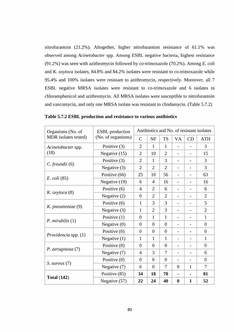
80
nitrofurantoin (21.2%). Altogether, higher nitrofurantion resistance of 61.1% was
observed among Acinetobacter spp. Among ESBL negative bacteria, highest resistance
(91.2%) was seen with azithromycin followed by co-trimoxazole (70.2%). Among E. coli
and K. oxytoca isolates, 84.8% and 84.2% isolates were resistant to co-trimoxazole while
95.4% and 100% isolates were resistant to azithromycin, respectively. Moreover, all 7
ESBL negative MRSA isolates were resistant to co-trimoxazole and 6 isolates to
chloramphenicol and azithromycin. All MRSA isolates were susceptible to nitrofurantoin
and vancomycin, and only one MRSA isolate was resistant to clindamycin. (Table 5.7.2)
Table 5.7.2 ESBL production and resistance to various antibiotics
Organisms (No. ofMDR isolates tested)
ESBL production(No. of organisms)
Antibiotics and No. of resistant isolates
C NF TS VA CD ATH
Acinetobacter spp.(18)
Positive (3) 2 1 1 - - 3
Negative (15) 2 10 2 - - 15
C. freundii (6)Positive (3) 2 1 3 - - 3
Negative (3) 2 2 2 - - 3
E. coli (85)Positive (66) 25 10 56 - - 63
Negative (19) 6 4 16 - - 16
K. oxytoca (8)Positive (6) 4 2 6 - - 6
Negative (2) 0 2 2 - - 2
K. pneumoniae (9)Positive (6) 1 3 3 - - 5
Negative (3) 1 2 3 - - 2
P. mirabilis (1)Positive (1) 0 1 1 - - 1
Negative (0) 0 0 0 - - 0
Providencia spp. (1)Positive (0) 0 0 0 - - 0
Negative (1) 1 1 1 - - 1
P. aeruginosa (7)Positive (0) 0 0 0 - - 0
Negative (7) 4 3 7 - - 6
S. aureus (7)Positive (0) 0 0 0 - - 0
Negative (7) 6 0 7 0 1 7
Total (142)Positive (85) 34 18 70 - - 81
Negative (57) 22 24 40 0 1 52

81
5.7.3 ESBL production and cephalosporin resistance
Altogether, maximum resistance (93.0%) was observed with cephalothin and least
resistance (62.7%) was observed with ceftazidime. Among ESBL positive bacteria,
lowest resistance (43.5%) was observed with cefoxitin while maximum resistance
(98.8%) was seen with aztreonam followed by cefpodoxime (96.5%) and ceftriaxone
(96.5%). Fourth generation cephalosporins also showed higher resistance rates of 92.9%
for cefpirome and 87.0% for cefepime. Almost all cephalosporins tested showed
increased resistance among ESBL producing organisms.
Of the 57 ESBL negative isolates tested against different cephalosporins, ceftazidime
showed least resistance (31.6%) while maximum numbers of isolates (89.5%) were
resistant to cephalothin. Cefepime and cefpirome also showed higher resistance rates of
57.9% and 59.6%, respectively. All 7 isolates of P. aeruginosa and 80.0% of ESBL
negative Acinetobacter spp. were susceptible to ceftazidime. Moreover, cefepime and
cefpirome showed lesser resistance of 21.0% and 26.3%, respectively, among ESBL
negative E. coli isolates and of 28.7% for both among P. aeruginosa isolates. All the
MRSA isolates were resistant to all cephalosporins tested. (Table 5.7.3)

82
Table 5.7.3 ESBL production and cephalosporin resistance
Organisms (No. ofMDR isolatestested)
ESBLproduction (No.of organisms)
Antibiotics and No. of resistant isolates
KF
FO
X
AT
M
CR
O
CP
D
CA
Z
CT
X
CP
M
CF
P
Acinetobacter spp.(18)
Positive (3) 3 2 3 2 2 2 2 2 3Negative (15) 15 15 15 15 15 3 15 15 15
C. freundii (6)Positive (3) 3 3 3 3 3 2 3 3 3Negative (3) 3 3 3 3 3 2 3 2 2
E. coli (85)Positive (66) 63 26 55 64 64 55 63 57 60Negative (19) 13 5 5 4 4 3 4 4 5
K. oxytoca (8)Positive (6) 6 4 6 6 6 6 6 6 6Negative (2) 2 0 1 0 0 0 0 0 0
K. pneumoniae (9)Positive (6) 5 2 6 6 6 5 5 5 6Negative (3) 3 3 2 2 3 2 2 2 2
P. mirabilis (1)Positive (1) 1 0 1 1 1 1 1 1 1Negative (0) 0 0 0 0 0 0 0 0 0
Providencia spp.(1)
Positive (0) 0 0 0 0 0 0 0 0 0Negative (1) 1 1 1 1 1 1 1 1 1
P. aeruginosa (7)Positive (0) 0 0 0 0 0 0 0 0 0Negative (7) 7 7 4 6 7 0 6 2 2
S. aureus (7)Positive (0) 0 0 0 0 0 0 0 0 0Negative (7) 7 7 7 7 7 7 7 7 7
Total (142)Positive (85) 81 37 78 82 82 71 80 74 79Negative (57) 51 41 38 38 40 18 38 33 34
5.7.4 ESBL production and resistance to carbapenems and combination of
various β-lactams with clavulanate
Meropenem and imipenem with no and 2.3% resistance, respectively, were found to be
the drug of choice among ESBL positive isolates. Imipenem resistance was remarkably
high among ESBL negative Acinetobacter spp. (86.7%) and MRSA (71.4%), and all
MRSA isolates were resistant to meropenem. Carbapenem resistance was also observed
among C. freundii, E. coli, K. pneumoniae, Providencia spp. and P. aeruginosa. Higher
degree of carbapenem resistance, 40.3% for imipenem and 19.2% for meropenem, was
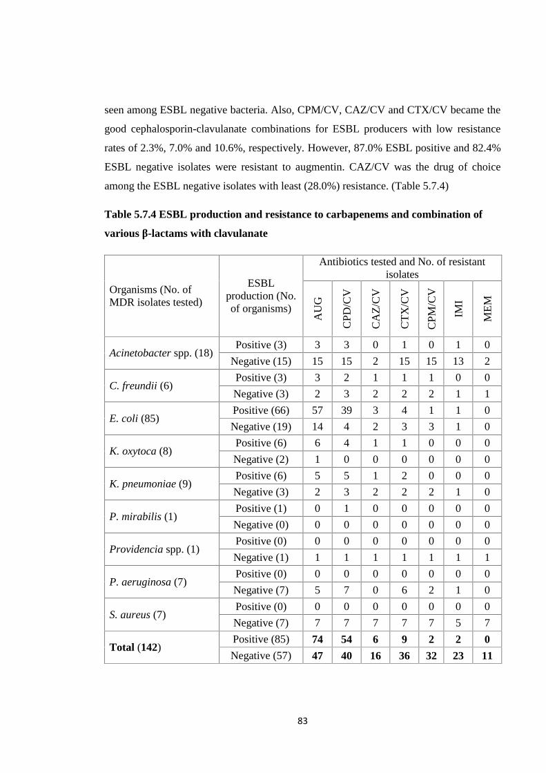
83
seen among ESBL negative bacteria. Also, CPM/CV, CAZ/CV and CTX/CV became the
good cephalosporin-clavulanate combinations for ESBL producers with low resistance
rates of 2.3%, 7.0% and 10.6%, respectively. However, 87.0% ESBL positive and 82.4%
ESBL negative isolates were resistant to augmentin. CAZ/CV was the drug of choice
among the ESBL negative isolates with least (28.0%) resistance. (Table 5.7.4)
Table 5.7.4 ESBL production and resistance to carbapenems and combination of
various β-lactams with clavulanate
Organisms (No. ofMDR isolates tested)
ESBLproduction (No.of organisms)
Antibiotics tested and No. of resistantisolates
AU
G
CP
D/C
V
CA
Z/C
V
CT
X/C
V
CP
M/C
V
IMI
ME
M
Acinetobacter spp. (18)Positive (3) 3 3 0 1 0 1 0
Negative (15) 15 15 2 15 15 13 2
C. freundii (6)Positive (3) 3 2 1 1 1 0 0
Negative (3) 2 3 2 2 2 1 1
E. coli (85)Positive (66) 57 39 3 4 1 1 0
Negative (19) 14 4 2 3 3 1 0
K. oxytoca (8)Positive (6) 6 4 1 1 0 0 0
Negative (2) 1 0 0 0 0 0 0
K. pneumoniae (9)Positive (6) 5 5 1 2 0 0 0
Negative (3) 2 3 2 2 2 1 0
P. mirabilis (1)Positive (1) 0 1 0 0 0 0 0
Negative (0) 0 0 0 0 0 0 0
Providencia spp. (1)Positive (0) 0 0 0 0 0 0 0
Negative (1) 1 1 1 1 1 1 1
P. aeruginosa (7)Positive (0) 0 0 0 0 0 0 0
Negative (7) 5 7 0 6 2 1 0
S. aureus (7)Positive (0) 0 0 0 0 0 0 0
Negative (7) 7 7 7 7 7 5 7
Total (142)Positive (85) 74 54 6 9 2 2 0
Negative (57) 47 40 16 36 32 23 11

84
5.7.5 ESBL production and fluoroquinolone resistance
Of the 7 different fluoroquinolones tested, all of them showed very high degree of
resistance among both ESBL positive and negative isolates. The maximum resistance of
97.6% was seen with each of nalidixic acid, ciprofloxacin and moxifloxacin among
ESBL positive isolates and of 94.0% with naldixic acid and moxifloxacin among ESBL
negative isolates. Norfloxacin comparatively showed lower resistance of 85.9% and
75.4% than other quinolones among ESBL positive and negative isolates, respectively.
Newer quinolones, viz. gatifloxacin (88.7%) and moxifloxacin (96.5%) showed
remarkably higher degree of resistance. ESBL production and fluoroquinolone resistance
was statistically significant (p<0.05). (Table 5.7.5)
Table 5.7.5 ESBL production and fluoroquinolone resistance
Organisms (No. ofMDR isolates tested)
ESBLproduction
(No. oforganisms)
Antibiotics tested and No. of resistant isolates
NA CIP OFX GAT MOX LEV NX
Acinetobacter spp.(18)
Positive (3) 1 2 1 1 3 1 3Negative
(15)15 15 15 15 15 15 13
C. freundii (6)Positive (3) 3 3 3 3 3 1 1Negative (3) 2 2 2 2 2 2 1
E. coli (85)Positive (66) 66 65 64 64 64 63 58
Negative(19)
17 13 13 13 16 13 12
K. oxytoca (8)Positive (6) 6 6 6 6 6 6 6Negative (2) 2 2 2 2 2 2 2
K. pneumoniae (9)Positive (6) 6 6 6 6 6 6 5Negative (3) 3 2 2 2 3 2 2
P. mirabilis (1)Positive (1) 1 1 1 1 1 1 0Negative (0) 0 0 0 0 0 0 0
Providencia spp. (1)Positive (0) 0 0 0 0 0 0 0Negative (1) 1 1 1 1 1 1 1
P. aeruginosa (7)Positive (0) 0 0 0 0 0 0 0Negative (7) 7 3 3 3 7 5 5
S. aureus (7)Positive (0) 0 0 0 0 0 0 0Negative (7) 7 7 7 7 7 7 7
Total (142)Positive (85) 83 83 81 81 83 78 73
Negative(57)
54 45 45 45 54 47 43

85
5.7.6 ESBL production and aminoglycoside resistance
Of the six aminoglycosides tested, fewer MDR isolates (44.4%) were resistant to
amikacin while maximum resistance (85.2%) was seen with kanamycin followed by
tobramycin (77.5%). Resistance of 35.3% and 57.9% was observed with amikacin among
both ESBL positive and negative isolates, respectively. Aminoglycoside resistance was
more predominant among ESBL negative Acinetobacter spp. Amikacin (14.3%) and
tobramycin (42.8%) showed lower resistance against P. aeruginosa isolates. All the
MRSA isolates were resistant to all aminoglycosides except one isolate was susceptible
to amikacin. (Table 5.7.6)
Table 5.7.6 ESBL production and aminoglycoside resistance
Organisms (No. ofMDR isolatestested)
ESBLproduction (No.of organisms)
Antibiotics tested and No. of resistant isolates
AK GM TN NET NE K
Acinetobacter spp.(18)
Positive (3) 0 1 2 1 1 2Negative (15) 15 15 15 14 15 15
C. freundii (6)Positive (3) 2 3 3 2 2 3Negative (3) 2 2 2 2 2 2
E. coli (85)Positive (66) 26 39 55 39 33 56Negative (19) 6 6 10 6 9 14
K. oxytoca (8)Positive (6) 0 4 4 3 3 5Negative (2) 1 0 1 1 1 2
K. pneumoniae (9)Positive (6) 1 1 4 1 3 5Negative (3) 1 2 2 2 2 2
P. mirabilis (1)Positive (1) 1 1 1 1 1 1Negative (0) 0 0 0 0 0 0
Providencia spp.(1)
Positive (0) 0 0 0 0 0 0Negative (1) 1 1 1 1 0 1
P. aeruginosa (7)Positive (0) 0 0 0 0 0 0Negative (7) 1 4 3 5 4 6
S. aureus (7)Positive (0) 0 0 0 0 0 0Negative (7) 6 7 7 7 7 7
Total (142)Positive (85) 30 49 69 47 43 72Negative (57) 33 37 41 38 40 49

86
5.7.7 ESBL production and resistance to tetracyclines and fosfomycin
For the 142 MDR isolates tested, fosfomycin was found to be the best drug of choice
with lowest resistance of 17.6% followed by tigecycline with lower resistance rate of
23.2%. Highest resistance was observed with tetracycline (85.2%) followed by
doxycycline and minocycline with equal resistance rate of 82.4%.
Fosfomycin and tigecycline were the drugs of choice among ESBL producing bacteria
with least resistance rates of 4.7% and 14.1%, respectively. However, resistance rates to
both of these antibiotics among ESBL non-producing isolates were 4.7% and 36.8%,
respectively. Of the 28 isolates, 55.5% of all Acinetobacter spp. were resistant to
tigecycline and all ESBL negative Acinetobacter spp. were resistant to fosfomycin.
71.4% of P. aeruginosa were also resistant to tigecycline while only 28.6% of them were
resistant to fosfomycin. Moreover, all MRSA isolates were susceptible to fosfomycin
while only 14.3% MRSA were resistant to tigecycline. ESBL production and resistance
to tigecycline and fosfomycin was statistically insignificant (p>0.05). (Table 5.7.7)

87
Table 5.7.7 ESBL production and resistance to tetracyclines and fosfomycin
Organisms (No. ofMDR isolates tested)
ESBLproduction (No.of organisms)
Antibiotics tested and No. of resistantisolates
T DO MN TGC FOT
Acinetobacter spp.(18)
Positive (3) 3 2 2 0 0
Negative (15) 15 14 13 10 15
C. freundii (6)Positive (3) 3 3 3 1 0
Negative (3) 2 2 1 1 1
E. coli (85)Positive (66) 54 54 53 8 3
Negative (19) 17 16 16 2 1
K. oxytoca (8)Positive (6) 6 6 6 2 0
Negative (2) 1 1 1 0 0
K. pneumoniae (9)Positive (6) 5 3 5 0 1
Negative (3) 1 1 2 2 2
P. mirabilis (1)Positive (1) 1 1 1 1 0
Negative (0) 0 0 0 0 0
Providencia spp. (1)Positive (0) 0 0 0 0 0
Negative (1) 1 1 1 0 0
P. aeruginosa (7)Positive (0) 0 0 0 0 0
Negative (7) 6 7 7 5 2
S. aureus (7)Positive (0) 0 0 0 0 0
Negative (7) 6 6 6 1 0
Total (142)Positive (85) 72 69 70 12 4
Negative (57) 49 48 47 21 21
5.8 ESBL Production and Spectrum of Drug Resistance
Of the 142 MDR isolates tested, 62.7% isolates showed resistance to 21-30 antibiotics.
Greater number of the MDR isolates that produced ESBL showed increased resistance
towards antibiotics with majority of them (81.2%) resistant to 21-30 drugs and all of
them were gram negative bacteria. Among ESBL positives, 2.3% isolates were further
resistant to 31-40 drugs tested. Among 57 ESBL negative isolates, 35.1% isolates, which

88
were all gram negative bacteria, were resistant to 21-30 antibiotics. All 7 ESBL negative
MRSA isolates were resistant to greater than 40 antibiotics. ESBL production and
increased drug resistance was statistically significant (p<0.05). On the other hand, 77.5%
MDR isolates were resistant to 5-7 classes of antibiotics of which 61.8% were ESBL
positive and all of them were gram negative bacteria. Moreover, 5.9% of ESBL positive
and 12.3% of ESBL negative isolates were further resistant to >7 classes of antibiotics
and all of them were also gram negative bacteria. All 7 ESBL negative MRSA isolates
were also resistant to 5-7 classes of antibiotics. ESBL production and drug resistance to
more classes of antibiotics was statistically significant (p<0.05).
Table 5.8 Spectrum of drug resistance among MDR isolates and ESBL production
Organismtype
ESBLproduction
Spectrum of drug resistance (No. of bacteria)Total≤10
Drugs11 – 20Drugs
21 – 30Drugs
31- 40Drugs
>40Drugs
Grampositive
Positive 0 0 0 0 0 0
Negative 0 0 0 0 7 7
Gramnegative
Positive 0 14 69 2 0 85
Negative 5 18 20 7 0 50
Total 5 32 89 9 7 142
Organismtype
ESBLproduction
Spectrum of resistance to different antibiotic classes(No. of Bacteria)
2-4 classes 5-7 classes >7 classes Total
Grampositive
Positive 0 0 0 0
Negative 0 7 0 7
Gramnegative
Positive 12 68 5 85
Negative 8 35 7 50
Total 20 110 12 142

89
CHAPTER-VI
6. DISCUSSION AND CONCLUSION
6.1 Discussion
A total of 2141 clinical specimens (1710 urinary specimens, 136 pus and body fluid
specimens, 170 sputa and throat swabs, and 125 eye and ear specimens) were received
and processed of which 53.4% were from males. Similar distribution of various
specimens was seen among clinical specimens in other studies also (Baral, 2008;
Shrestha et al., 2007). Therefore, the overall result of the study may be affected by urine
itself. The higher proportion of urinary specimens showed the greater prevalence of the
urinary diseases than others. Among the total specimens, 20.2% specimens showed
significant growth and maximum percentage (61.2%) of total isolates was contributed by
the urinary isolates. A growth rate of 22.3% among different clinical specimens was seen
in a similar study (Baral, 2008).
Least growth rate (15.5%) was observed among urine specimens that resembled with low
growth rate of 16.3% among urinary specimens observed in India (Ramesh et al., 2008)
and with such low growth rates in other similar studies in Nepal by various researchers
(Basnet et al., 2009; Baral, 2008; Kattel et al., 2008; Shrestha et al., 2007; Bomjan, 2005;
Dhakal, 1999). The low growth rate among urinary specimens may probably be due to
request for urine culture in other urogenital problems rather than overt urinary tract
infection, non-gonococcal urethritis or other clinical conditions that mimic UTI, higher
health consciousness among patients, prior antibiotic consumption and presence of
fastidious pathogens. In contrary to the normal patterns, we received more urine
specimens (52.3%) from males than from females (47.7%). However, the growth rate was
higher among females (18.1%) than males (13.1%). Similar results were obtained in the
previous studies (Basnet et al., 2009; Baral, 2008; Shrestha et al., 2007; Bomjan, 2005).
It might be attributed to easy access of uropathogens to female urinary tract due to shorter
urethra in closer proximity to anus. Moreover, higher growth rate of 43.3% among

90
sputum specimens was also seen in other studies (Pokhrel et al., 2006). Growth rate
among eye and ear specimens (51.2%) and pus and body fluids (48.9%) was higher
which may be apparently attributed to poor hygienic practices among persons. The low
growth rate (28.8%) of sputum and throat swab may be due to continuing antibiotic
therapy, or viral and fungal infection or infection by organisms like Mycoplasma,
Chlamydiae, Legionella, etc. which are not routinely cultured (Read and Finch, 1998).
Growth patterns among various clinical specimens may differ in different territories
depending on the prevalence of the particular disease, hygiene, antibiotic therapy and
other disease phenomena.
Of the 433 bacterial isolates, gram negative bacteria predominated with 67.4% isolates
and such predominance was also observed in other similar studies (Basnet et al., 2009;
Baral, 2008; Bomjan, 2005; Shrestha et al., 2007; Blomberg et al., 2005). Among gram
negative isolates, E. coli was the most predominant organism with 51.7% isolates
followed by K. pneumoniae with 14.7% isolates. These results resembled with that of the
studies done by various other workers (Baral, 2008; Shrestha, 2007; Pokhrel et al., 2006;
Kumari et al., 2005; Sharma, 2004; Tuladhar, 1999). Most E. coli isolates were from
urinary specimens as they have special virulent properties to cause UTI, thus being the
major uropathogens throughout the world. E. coli can bind to the glycol-conjugate
receptor (Gal α 1→4 Gal) of the uroepithelial cells of human urinary tract so it can
initiate infection itself (Johnson, 1991). E. coli is the most predominant organism to
colonize the urethral meatus (Schaeffer and Chmiel, 1983) and perineum (Levy, 2001)
before ascending to the bladder.
Maximum numbers of gram positive bacteria were isolated from eye and ear specimens
(31.9%). S. aureus and S. pyogenes predominated among gram positive bacteria with
41.8% and 28.9% isolates. S. aureus was the dominant species among pus and body
fluids (79.4%) and eye and ear specimens (48.9%). Higher isolation rates of S. aureus
were also seen in the similar studies done by various other workers, i.e. 25.3% among
wound isolates (Banjara, 2002), 70.97% isolates in pus samples (Dhungel, 2001), 49.40%
in ear discharges (Aryal, 2001) and 57.66% isolates in pus samples (Tuladhar, 1999). S.
91
aureus is responsible for about 70% of all site specific soft tissue infections in humans
(Collier et al., 1998). Because of their rigid body, resistance to dry conditions and high
salt concentrations, S. aureus is well suited for skin and soft tissue infections. Other
major virulent factors like exotoxins, leucocidin, exfoliatins, haemolysins, enzymes
(coagulase, phosphatase, dioxyribonuclease, hyaluronidase, staphylokinases, lipases and
proteases) are responsible for occurrence of higher prevalence of S. aureus mediated skin
and soft tissue infections. S. pyogenes and E. faecalis were the most frequently isolated
gram positive bacteria among sputum and throat swab specimens and urinary specimens
with 65.0% and 45.4% isolates, respectively. Significant isolation of enterococci from
urine specimens has also been reported from India (Ramesh et al., 2008). High frequency
of E. coli isolation may be due to the predominance of urinary specimens and of S.
aureus and S. pyogenes may be due to their common predominance among pus, swabs
and respiratory specimens. Predominance of E. coli (60.9%) followed by K. pneumoniae
(11.9%) among urinary isolates resembled with the findings of other similar studies by
various researchers (Basnet et al., 2009; Banjara, 2002).
Of the 14 primarily tested antibiotics against various gram negative bacteria, amikacin
was the best drug with only 28.0% resistance. Ceftazidime, nitrofurantoin and
chloramphenicol also had relatively lower resistance rates of 28.6%, 29.3% and 30.1%,
respectively. Higher amikacin and ceftazidime susceptibility was also reported among
urinary isolates (Das et al., 2006). Amoxycillin had the highest resistance rate of 89.0%
followed by azithromycin (82.6%) and nalidixic acid (80.5%). This finding resembled the
results of other similar works (Puri, 2006; Pokhrel, 2006; Bomjan, 2005; Paneru, 2002;
Oteo et al., 2001; Dhakal, 1999). Other antibiotics tested also showed relatively higher
resistance rates. The lower resistance against nitrofurantoin and chloramphenicol may be
due to their rarer use in recent days resulting in reduced selective pressure on bacteria.
Higher amoxycillin resistance is due to its extensive and empirical use in minor
infections and also in clinical conditions due to viral etiologies. Production of various β-
lactamases and ESBLs under the extensive selection pressure by excessive β-lactam use
also has contributed to penicillin and cephalosporin resistance (Goossens et al., 2005).

92
Of the 18 primarily tested antibiotics against various gram positive bacteria,
nitrofurantoin and norfloxacin were found to be the drugs with least resistance of only
9.5%. Resistance to most of the other antibiotics among gram positive bacteria was
relatively lower as most of the gram positive bacteria except MRSA, VRSA and VRE are
still susceptible to most antibiotics worldwide. Maximum resistance was seen with
ciprofloxacin (47.5%) followed by gentamicin (44.4%) and co-trimoxazole (41.8%)
which were the drugs used most frequently used in the country. Erythromycin showed
lower resistance (21.3%) than azithromycin (28.7%). Of the 59 isolates of S. aureus
tested, 16.9% of them were MRSA. A study in a Malaysian teaching hospital showed
44.1% prevalence of MRSA (Ghaznavi-Rad et al., 2010). In Nepal, 15.4-39.6%
predominance of MRSA isolates among various clinical S. aureus isolates has been
recorded (Sanjana et al., 2010; Kumari et al., 2008; Subedi and Brahmadathan, 2005).
Resistance to methicillin is mediated by the chromosomal mecA gene which is the part of
a mobile genetic element found in many MRSA strains called Staphylococcal Cassette
Chromosome (SCCmec). This gene encodes for an altered PBP2a that has lower binding
affinity for β-lactam antibiotics (Timothy, 2004).
Of the 433 isolates, 64.9% isolates were MDR with maximum multiple drug resistance
(78.1%) among urinary isolates with MDR isolates and least multiple drug resistance
(32.8%) among the eye and ear specimens. Predominance of MDR isolates among
sputum and throat swab, and pus and body fluid specimens was almost same with 51.0%
and 50.9% MDR isolates, respectively. Similar results with 47.6-83.0% MDR isolates
among urinary isolates and about 60.0% MDR isolates among sputum specimens has
been reported nationally and internationally (Ullah et al., 2009; Pokhrel et al., 2006;
Bomjan, 2005). Higher prevalence of MDR isolates from pus specimens has been
similarly reported (Banjara, 2002). The multi-drug resistance has been found as a major
problem in the management of uropathogens (Akram et al., 2007; Hasan et al., 2007). Of
the total MDR isolates, most of them (73.7%) were urinary isolates. That was because of
predominance of urinary specimens among total specimens processed and higher
emergence of MDR bacteria among urinary isolates than other specimens due to common
93
infections and higher antibiotic exposure. Prevalence of MDR organisms varies globally
and by different anatomical sites as it depends on the local antibiotic using policies,
prevalence of particular type of infection and hence the pathogens too. Moreover, the
higher transferable multidrug-resistance may be due to plasmids with several resistance
genes which are transferred from one bacterium to another (Ram et al., 2000).
Of the total 281 MDR isolates from different clinical specimens, 77.9% were gram
negative bacteria and most of them (87.7%) were urinary isolates. Most of the gram
positive MDR bacteria (30.6%) were isolated from sputum and throat swab specimens.
Of the 219 MDR gram negative bacteria, E. coli constituted 52.0% isolates. The higher
levels of drug resistance among E. coli, Acinetobacter spp., K. pneumoniae and other
bacterial species to β-lactams, fluoroquinolones, aminoglycosides, co-trimoxazole and
some other antibiotics might be due to production of various β-lactamases (ESBL, AmpC
and MBL), mutations in gyrA and parC genes, production of aminoglycoside modifying
enzymes and presence of many different multidrug efflux systems (Picao et al., 2008;
Walsh et al., 2005; Sulavik et al., 2001; Ozeki et al., 1997; Livermore, 1995). As most of
the antibiotics are commonly used in the country since several years, increasing selective
pressure might have contributed to the emergence of resistance.
In our study, 59.9% ESBL screen positive MDR isolates produced ESBL. Of the 85
ESBL positive isolates, E. coli with 77.6% isolates constituted to most of the ESBL
producers followed by K. oxytoca with 75.0% isolates. Moreover, 66.7% K. pneumoniae,
50.0% C. freundii and 16.7% Acinetobacter spp. also tested ESBL positive. ESBL
production was also observed among 50.0% isolates from sputum and throat swab and
20.8% isolates from pus and body fluid. Similar results were obtained among various
clinical isolates in other studies in Nepal (Baral, 2008; Bomjan, 2005). Similarly in other
study, 8%, 12.5%, 12.5%, 25%, and 5% isolates of K. pneumoniae, E. coli, C. freundii, A.
calcoaceticus and P. aeruginosa, respectively produced ESBLs (Sharma, 2004). In
Pakistan, 56.9%% ESBL production among 116 urinary E. coli isolates was also
observed (Ullah et al., 2009). In our study, ESBL production was most predominant

94
(68.4%) among urinary isolates which resembled the findings of other similar studies
(Melzer and Patersen, 2007; Jamal et al., 2005). Moreover, in India 100% K. oxytoca,
79.0% E. coli and 69.4% K. pneumoniae produced ESBLs. Higher prevalence of ESBL
producing E. coli isolates in China (55.0%) and Thailand (50.8%) have been reported
(Hawser et al., 2009). Prevalence ESBLs varies in different countries as ESBL
prevalence of 67%, 42%, and 43% has been reported in E .coli from Iran, India and
Bangladesh respectively (Mehrgan and Rahbar, 2008; Taneja et al., 2008; Rahman et al.,
2004). Substantial geographical difference in the occurrence of ESBLs among and within
countries, in territories, and among hospitals has been observed (Babini and Livermore,
2000). ESBL production is more common in E. coli and Klebsiella spp. than in other
members of Enterobacteriaceae. They are sometimes difficult to detect as clavulanate
induces production of chromosomal AmpC enzymes that hydrolyze indicator
cephalosporin, thus masking any synergy. Among C. freundii, Enterobacter spp.,
Morganella spp., Providencia spp. and Serratia spp., mutational hyperproduction of
AmpC enzymes is responsible for most of the cephalosporin resistance. Derepressed
mutants of AmpC inducible species copiously produce AmpC enzyme without induction
and become resistant to almost all penicillins and cephalosporins (Freeman et al., 2009;
Livermore et al., 2001). However, plasmid-mediated AmpC β-lactamases in lower
proportions have also been reported among different bacterial strains (Li et al., 2008;
Moland et al., 2006). Variation in ESBL production rates has also been observed among
various bacterial genera. These findings bespeak emergence of MDR and ESBL
producing organisms worldwide limiting the therapeutic options. This finding suggests
for the proper and routine detection of ESBLs before the use of β-lactams in therapy as
well as evaluation of other old and new potent therapeutic agents.
All 281 MDR isolates were subjected to ESBL screening test by using all five screening
agents recommended by CLSI and 142 isolates were found to be screen positive by one
or all screening agents. Among them, cefpodoxime and ceftriaxone had the highest
sensitivities of 96.5% each but cefpodoxime had lowest PPV (67.2%) and ceftriaxone had
lower PPV of 68.3%. Aztreonam and cefotaxime both had equal sensitivity of 94.1% and

95
PPV of 67.8%. Ceftazidime had lowest sensitivity of only 83.5%, but had the highest
PPV of 79.8%. Higher sensitivity of cefotaxime in ESBL screening was reported from
Israel with its convenient use and improved negative predictive value for most of the
genera in areas where Klebsiella spp. are not the dominant ESBL producers (Navon-
Venezia et al., 2003). In a similar study to investigate the performance of ESBL
screening methods for Enterobacteriaceae in South-East England, many isolates found
resistant only to cefpodoxime at the source sites proved not to have ESBLs or AmpC and,
hence, screening with cefotaxime and ceftazidime allowed better specificity for
identification of mechanism-based resistance, as did the automated systems (Hope et al.,
2007). However, the screening results with ceftazidime versus cefotaxime was found
statistically insignificant (p>0.05).
Among 142 MDR isolates tested for confirmation of ESBL production by using four
types of combination disks, maximum ESBL producers (97.6%) were detected by
cefepime-clavulanate combination disks. Higher sensitivity (98.0%) of cefepime-
clavulanate in ESBL Etests followed by 83.0% with cefotaxime-clavulanate, and 74.0%
with ceftazidime-clavulanate strips among Enterobacteriaceae has also been reported in
similar work (Sturenburg et al., 2004). According to that study, reliable ESBL detection
among Enterobacter spp. could only be achieved by the cefepime–clavulanate strip with
100% sensitivity. A limitation of using the cefepime-clavulanate combination strip was
less than optimal specificity with K1 phenotypes of K. oxytoca: 66.7% cases were false-
positive by Etest strips containing cefepime–clavulanate. In a similar study using E-test
strips, the cefotaxime ESBL strip detected ESBL activity in 96.5% of the 87 ESBL
producing isolates tested while ceftazidime Etest strips detected it only in 54 % of the
study isolates (Dashti et al., 2006). Moreover, cefotaxime-clavulanate showed 91.0%
overall sensitivity and 100% sensitivity among Enterobacteriaceae in detecting ESBLs.
Ceftazidime and cefotaxime disks each in combination with clavulanate showed 10 and 1
more ESBL producers respectively than in respective screening tests. Combined use of
cefotaxime-clavulanate and ceftazidime-clavulanate showed 100% sensitivity in
detecting ESBLs among all various clinical isolates and cefpodoxime-clavulanate did not

96
add any benefit (Navon-Venezia et al., 2003). The use of the combination of both
cefotaxime and ceftazidime Etest ESBL strips detected ESBLs in 86 of the 87 test
isolates (98.8%). Cefpodoxime-clavulanate combination disks detected least number of
ESBL producers with only 91.8% positive isolates. This may be due to presence of some
cefpodoxime hydrolyzing β-lactamases in some strains that were not sensitive to
clavulanic acid, which could have reduced the sensitivity of the test. Moreover, changes
in major outer membrane proteins change in porin number and raised production of
chromosomal AmpC enzymes might have contributed to the lower sensitivity of this
agent (Oliver et al., 2002). Higher false-positive ESBL comfirmation may also be due to
presence of pampC genes with or without other β-lactamase genes and false-negative
results may be due to presence of both ESBL and pampC genes (Robberts et al., 2009).
The differences in sensitivities of various agents in detecting ESBLs might be due to
differences in dominant enzymes in various geographical areas. Moreover, the particular
enzymes harbored by the tested organisms might have influenced the test performance.
Among 85 ESBL positive isolates, maximum resistance (97.6%) was observed against
amoxycillin followed by ticarcillin (94.1%). On the flip side, 98.2% ESBL negative
isolates were also resistant to amoxycillin and 84.2% were resistant to ticarcillin. Almost
all isolates tested were resistant to amoxycillin and ticarcillin. Temocillin showed
remarkably less resistance of 27.5%. Only 24.7% of ESBL positive and 31.6% of ESBL
negative isolates were resistant to temocillin. The higher susceptibilities of MDR
pathogens, particularly the MDR Acinetobacter spp. isolates to temocillin was worthy to
note. We adopted the temocillin susceptibility breakpoints according to the BSAC
guidelines as diameter of inhibition zone of ≥20mm for susceptibility and ≤19mm for
resistance for systemic Enterobacteriaceae isolates, and for urinary Enterobacteriaceae
isolates, ≥12mm and ≤11mm zone diameters were regarded as susceptible and resistant
(Andrews et al., 2007). All S. aureus isolates tested were resistant to amoxycillin,
ticarcillin, penicillin G, oxacillin and methicillin. Higher ampicillin resistance among S.
aureus (81%) and E. coli (92.26%) from various clinical specimens was reported in
Nepal (Datta, 2004) while around 90.0% amoxycillin resistance was observed among

97
enteric bacteria in Iran (Mansouri and Abbasi, 2010). Higher amoxycillin resistance is
obvious as it remains the most frequently sold antibiotic in Nepal (Kafle et al., 2007).
Among ESBL positive isolates, all of Acinetobacter spp., 83.3% of K. pneumoniae and
66.7% of C. freundii showed resistance to temocillin. Among ESBL negative isolates, all
isolates of C. freundii, 85.7% of P. aeruginosa and 66.7% of K. pneumoniae showed
resistance to temocillin. No isolate of ESBL negative K. oxytoca and fewer (13.3%)
ESBL negative Acinetobacter spp. isolates were resistant to temocillin.
Temocillin is β-lactamase resistant penicillin. It is stable against hydrolysis by most β-
lactamases, including extended-spectrum β-lactamases (ESBLs) and AmpC-type β-
lactamases with studies reporting MICs at which 90% of bacteria are inhibited (MIC90s)
between 16 and 32 µg/ml (Denyer et al., 2007; Glupczynski et al., 2007; Livermore et al.,
2006). In a study on KPC-β-lactamase producing K. pneumoniae and E. coli isolates that
were resistant to carbapenem (ertapenem), the MICs ranged between 16 µg/ml and 64
µg/ml (MIC90=32µg/ml) for K. pneumoniae. The E. coli clinical isolates had MICs
between 8 and 16µg/ml. An inoculum effect was not observed at 105 CFU, whereas a
mild inoculum effect averaging within a twofold MIC difference was seen with K.
pneumoniae when 106 CFU was inoculated (Adams-Haduch et al., 2009). The common
dosage is 2g intravenously every 12 hours or in severe disease as a single loading dose of
2g is given intravenously followed by a 4g infusion over 24 hours (Jongh et al., 2008).
Temocillin is thus drawing attention as a potential alternative to carbapenems in treatment
of infections caused by the Enterobactericeae producing these broad-spectrum β-
lactamases. It is not active against gram positive bacteria, anaerobes, Acinetobacter
species, Pseudomonas aeruginosa or bacteria with altered penicillin-binding proteins. Its
primary use is against Enterobacteriaceae, and in particular against strains producing
extended spectrum β-lactamase or AmpC β-lactamase (Livermore and Tulkens, 2009;
Livermore et al., 2006). Temocillin showed comparable efficacy with other IV infusions
in treating cystic fibrosis patients (Kent et al., 2008).

98
Altogether, nitrofurantoin was the drug of choice with least resistance of 29.6% followed
by chloramphenicol (39.4%). Highest resistance was seen with azithromycin (93.7%)
followed by co-trimoxazole (77.5%). Similar high co-trimoxazole resistance has also
been reported by other workers (Mansouri and Abbasi, 2010; Ullah et al., 2009; Akram et
al., 2007; Tankhiwale et al., 2004). Higher azithromycin resistance in our country can be
justified by its excessive empirical use in many enteric and respiratory infections due to
its dose convenience. Higher nitrofurantion resistance (61.1%) was observed among
Acinetobacter spp. reducing its efficacy in UTIs by such bacteria.
All MRSA isolates were ESBL negative and resistant to co-trimoxazole and 85.7% of
them were resistant to chloramphenicol and azithromycin. All MRSA isolates were
susceptible to nitrofurantoin and vancomycin which was of good remark and only one
MRSA isolate was resistant to clindamycin. These findings were in accordance with the
results in other similar works (Baral, 2008; Dhungel, 2001; Tuladhar, 1999). Moreover,
of the 18 Acinetobacter spp. tested, only 3 isolates were resistant to co-trimoxazole and 4
isolates were resistant to chloramphenicol which was also of good note and suggests for
the inclusion of these drugs in therapeutic regime for treating Acinetobacter infections.
However, high resistance of co-trimoxazole among Acinetobacter spp. was also observed
in a study in Italy (Capone et al., 2008). Though older and commoner, these antibiotics
can still be used effectively for treating severe infections caused by them. This higher
susceptibility may be attributed to the less common infections by such bacteria and thus
their lesser previous exposure to these drugs sufficient to develop resistance. However,
higher co-trimoxazole resistance was observed among other commoner pathogens the
infections by which were empirically treated by this agent in previous years.
Among ESBL positive bacteria, lowest resistance (43.5%) was observed with cefoxitin
while maximum resistance (98.8%) among them was seen with aztreonam followed by
cefpodoxime and ceftriaxone (96.5%). Lower cefoxitin resistance may imply the lower
rate of production of AmpC enzymes (Robberts et al., 2009). Fourth generation
cephalosporins also showed higher resistance rates of 92.9% for cefpirome and 87.0% for

99
cefepime. In a study in Indian setting at tertiary care referral hospital by among 516
bacterial isolates, 54.2% (n=177) gram positive isolates, 66% (n=209)
Enterobacteriaceae, 70.7% (n=130) P. aeruginosa and other nonfermenters were resistant
to cefpirome (Chaudhary, 2003). In the 1980s and 1990s, reports from North America,
Europe, and Japan have revealed an uniformly higher efficacy of cefpirome as compared
to the other β-lactams including cephalosporins (Jones et al., 1991). At that time,
cefpirome showed better activity than other third generation cephalosporins against both
inducible β-lactamase producing and not producing Enterobacteriaceae species (Reeves
et al., 1993). This scenario may be imperceptibly changing in the light of some reports
from Thailand with 13.4% resistance to cefpirome in all gram negative isolates tested and
from Japan with 41.7% resistance among 14,216 P. aeruginosa isolates but only 15.7%
resistance among them to ceftazidime (Nitsuma et al., 2001; Biedenbach et al., 1999).
However, no P. aeruginosa isolate in our study was resistant to ceftazidime and it
remained the good antipseudomonal cephalosporin (Denyer et al., 2007). Resistance to
cefotaxime, ceftriaxone and ceftazidime among Citrobacter spp. has been reported within
range of 70.4-82.2% (Patil and Lakshmi, 2000).
The high frequency of resistance against cefpirome in our study is not surprising as
cefpirome belongs to the cephalosporin group and can become a target of potent and
constantly evolving β-lactamases. Moreover, higher cefepime resistance among clinical
isolates of ESBL producing E. coli and K. pneumoniae has been reported in India (Arya
et al., 2007) and in Iran (Feizabadi et al., 2006). Almost all MDR Acinetobacter spp.
isolates were resistant to cefepime, ceftazidime and aztreonam in Italy (Capone et al.,
2008). This higher cephalosporin resistance among these organisms might be due to
hyperproduction of AmpC β-lactamases rather than ESBLs. The over-the-counter sale of
such medicines and use of counterfeit medicines has now become common without any
benefits for the patients (Aldhous, 2005). Furthermore, any suboptimal dose and/or
duration of therapy would support selection and dissemination of cefepime resistant
ESBL-producing K. pneumoniae and E. coli. There would be some delay in prescribing
antibiotics matching the in-vitro susceptibility for an ICU isolate.

100
Almost all cephalosporins tested showed increased resistance among ESBL producing
organisms. As cefalosporins are susceptible to various types of ESBLs by definition,
higher cephalosporin resistance rates among ESBL producers is obvious. Higher
cephalosporin resistance among ESBL producing bacteria have been reported in various
studies (Khadri and Alzohairy, 2009). In a similar study carried out in Nigeria on 100
MDR isolates of Enterobacteriaceae, only 54.8% of the Klebsiella species isolated were
sensitive to ceftazidime, 48.4% to ceftriaxone and 30.7% to cefotaxime (Okesola and
Makanjuola, 2009). Higher ceftazidime resistance among enteric bacteria has been
reported from Iran (Mansouri and Abbasi, 2010). With E. coli however, the susceptibility
pattern to the third-generation cephalosporins was better (65.6% were sensitive to
ceftazidime, 62.5% to ceftriaxone and 71.9% to cefotaxime). In Proteus species, the
susceptibility pattern was generally poor to the three classes of antibiotics (50.0% were
susceptible to ceftazidime and ceftriaxone, 0.0% to cefotaxime, 33.3% to ciprofloxacin,
50.0% to gentamycin and 0.0% to amoxycillin/clavulanate).
Among 57 ESBL negative isolates, ceftazidime showed least resistance of 31.6% while
maximum numbers of isolates (89.5%) were resistant to cephalothin. Cefepime and
cefpirome also showed higher resistance rates of 57.9% and 59.6%, respectively. This
may be due to production of inhibitor-resistant β-lactamases. All (100%) isolates of P.
aeruginosa and 80% of ESBL negative Acinetobacter spp. were susceptible to
ceftazidime. Higher ceftazidime susceptibility among P. aeruginosa isolates has also
been previously mentioned (Denyer et al., 2007). Moreover, cefepime and cefpirome
showed lesser resistance of 21.0% and 26.3%, respectively, among ESBL negative E. coli
isolates and of 28.7% for both among P. aeruginosa isolates showing their possible
therapeutic efficacy against these organisms. All the MRSA isolates were resistant to all
cephalosporins tested. Adequate data on fourth-generation cephalosporin use in Nepal
was not available as their launch was quite recent, third-generation cephalosporins and
penicillins still have a significant market share in Nepal (Kafle et al., 2007).

101
Meropenem and imipenem with no and 2.3% resistance, respectively, were found to be
the drug of choice among ESBL positive isolates. Higher degree of carbapenem
resistance, 40.3% for imipenem and 19.2% for meropenem, was seen among ESBL
negative bacteria. Imipenem was found to be the most effective drug against gram
negative isolates in the similar studies (Baral, 2008; Puri, 2006; Oteo et al., 2001).
Meropenem is more stable than imipenem to the enzyme dehydropeptidase produced by
kidney and can be administered without cialistin and hence can be a better therapeutic
option (Jones et al., 2007). Imipenem resistance was remarkably high among ESBL
negative Acinetobacter spp. (86.7%) and MRSA (71.4%), and all MRSA isolates were
resistant to meropenem. Carbapenem resistance was also observed among C. freundii, E.
coli, K. pneumoniae, Providencia spp. and P. aeruginosa. In India, 16% imipenem
resistance among 50 clinical isolates of P. aeruginosa has been reported (Hemalatha et
al., 2005). MBL-mediated imipenem resistance in P. aeruginosa is of concern in the
therapy of critically ill patients. Resistance to carbapenems is due to reduced permeability
due to porin channel downregulation (porin loss), upregulated efflux of the drug and most
importantly the production of plasmid borne AmpC like β-lactamase, oxacillinases,
Klebsiella pneumoniae carbapenemases (KPCs 1-4) and metallo-β-lactamases. Moreover,
co-existance of blaAmpC and blaCTX-M genes in bacteria with decreased outer membrane
permeability may lead to carbapenem resistance (Shahid et al., 2009; Wang et al., 2009).
Higher resistance rates of carbapenem resistance among Acinetobacter spp. has also been
seen in Israel and Italy (Capone et al., 2008; Navon-Venezia et al., 2007). The clinical
utility of carbapenems is under threat due to emergence of acquired carbapenemases, i.e
MBLs among clinically important pathogens, such as Pseudomonas spp., Acinetobacter
spp., and members of Enterobacteriaceae family, which are also capable of integron-
mediated transfer. Therefore, early detection of MBL-producing organism is
recommended (Picao et al., 2008; Franklin et al., 2006).
The CPM/CV, CAZ/CV and CTX/CV became the good cephalosporin-clavulanate
combinations for ESBL producers with respective low resistance rates of 2.3%, 7.0% and
10.6%. Because there were no published breakpoints for such combinations, we used the

102
susceptibility criteria for them if the inhibitor combination gave the inhibition zone
greater than their ESBL screening criteria. For cefepime, inhibition zone >18mm was
interpreted as susceptible. Augmentin showed higher resistance of 87.0% and 82.4%
among both ESBL positive and negative isolates. This might be due to excessive use of
amoxycillin in past and recent years and also due to production of inhibitor resistant β-
lactamases by the isolates. CAZ/CV was the drug of choice among the ESBL negative
isolates with least (28.0%) resistance. Good stability of drugs against various β-
lactamases in presence of clavulanate can be attributed to these findings. As relatively
higher concentration of β-lactamase inhibitor in urine is achieved, safe and successful
treatment of UTIs by β-lactam/β-lactamase inhibitors has also been described
(Nordmann, 1998). In a study on 54 CTX-M β-lactamase producing E. coli isolates from
respiratory, pus and urinary specimens, 96.2% isolates were susceptible to
ceftriaxone/sulbactam combination which was equal to imipenem susceptibility; however
the susceptibility to ticarcillin/clavulanate and piperacillin/tazobactam combinations was
unworthy (Shahid et al., 2007). Similarly, in a study among various clinical isolates from
ventilator-associated pneumonia in Turkey, cefoperazone-sulbactam showed highest
susceptibility (89.0%) against MDR Acinetobacter spp. (Erdem et al., 2008). These
combination agents may be of worthy clinical use in this era of emerging ESBL threat.
All of the 7 different fluoroquinolones tested showed very high degree of resistance
among both ESBL positive and negative isolates. The maximum resistance of 97.6% was
seen with nalidixic acid, ciprofloxacin and moxifloxacin among ESBL positive isolates
and of 94.0% with naldixic acid and moxifloxacin among ESBL negative isolates.
Norfloxacin comparatively showed lower resistance of 85.9% and 75.4% than other
quinolones among ESBL positive and negative isolates, respectively. Newer quinolones,
viz. gatifloxacin and moxifloxacin, showed remarkably higher degree of resistance.
Disappointing results of ciprofloxacin resistance among urinary isolates have been
reported in eastern part of Nepal (Kumari et al., 2005), in Turkey (Erdem et al., 2008)
and in Iran (Mansouri and Abbasi, 2010). Increasing trend in fluoroquinolone resistance
due to their increased use in various clinical cases has also been reported (Neuhauser et

103
al., 2003; Shankar et al., 2007). Fluoroquinolones still share a greater market sale
percentage in Nepal which has been creating a constant selective pressure on
microorganisms (Kafle et al., 2007). Greater resistance among ESBL producing E .coli
isolates to nalidixic acid (63.6%) and ciprofloxacin (54.6%) was seen in a study in
Palestine (Astal et al., 2004; Astal, 2005). Fluoroquinolone resistance and MDR
phenotype is associated with ESBL production. In a study among 34 ESBL producing E.
coli isolates in Canada, 79.4% isolates were resistant to ciprofloxacin and higher
percentage of them were also resistant to gentamicin, doxycycline and co-trimoxazole.
CTX-M-14 ESBLs were associated with fluoroquinolone resistance only while CTX-M-
15 genotypes were associated with multiple drug resistance (Lagace-Wiens et al., 2007).
Higher resistance to ciprofloxacin and levofloxacin was also observed in Italy (Capone et
al., 2008). The higher level of fluoroquinolone resistance observed in the study might be
due to involvement of several resistance mechanisms such as mutation of the
topoisomerase targets, mutational activation of efflux systems, protection of the
topoisomerase target by Qnr proteins and inactivation of the drug by the AAC(6’)-Ib-cr
variant of the common AAC(6’)-Ib aminoglycoside acetyl-transferase (Robicsek et al.,
2006b). Quinolone resistance mostly originates from chromosomal mutations but
plasmid-mediated quinolone resistance with the involvement of qnrA, qnrB, or qnrS
genes has also been reported in several parts of the world (Oktem et al., 2008). These
findings question the recent clinical utility of fluoroquinolones and mandates for
avoidance of their empirical and monotherapeutic use.
Fewer isolates (44.4%) were resistant to amikacin while maximum resistance (85.2%)
was seen with kanamycin followed by tobramycin (77.5%). Lesser resistance of 35.3%
and 57.9% was observed with amikacin among both ESBL positive and negative isolates,
respectively. Aminoglycoside resistance was more predominant among ESBL negative
Acinetobacter spp. The presence of integrons containing aminoglycoside-modifying
enzymes is associated with resistance to gentamicin and tobramycin but not with
resistance to amikacin among Acinetobacter isolates (Lin et al., 2010). Amikacin (14.3%)
and tobramycin (42.8%) showed lower resistance against P. aeruginosa isolates. All the

104
MRSA isolates were resistant to all aminoglycosides tested except that one isolate was
susceptible to amikacin. Higher amikacin susceptibility and gentamicin resistance among
ESBL producing enteric bacteria has been reported in UAE (Al-Zarouni et al., 2008) and
higher gentamicin resistance among enteric bacteria in Iran (Mansouri and Abbasi, 2010).
Higher amikacin and gentamicin resistance have also been observed in Italy (Capone et
al., 2008). In similar study, gentamicin and tobramycin typically demonstrated poor in
vitro activity against ESBL-producing Klebsiella species, E. coli and Proteus species
(Spanu et al., 2002). In Pakistan, urinary E. coli isolates showed 57% kanamycin
resistance and 52% gentamicin resistance (Ullah et al., 2009). Such resistant isolates pose
serious problems to the physicians as therapeutic options are limited. ESBLs are plasmid-
mediated and multidrug resistance is a characteristic feature of strains producing ESBLs.
Our study confirms this observation in Klebsiella and Proteus species, as these isolates
were resistant to different classes of antibiotics including aminoglycosides. Amikacin still
retains its clinical utility and tobramycin has good efficacy in treating specific P.
aeruginosa infections. The higher aminoglycoside resistance observed might be due to
reduced uptake, decreased permeability, altered ribosomal binding site and most
importantly the production of aminoglycoside modifying enzymes, viz. AACs, ANTs and
(Mingeot-Leclercq et al., 1999; Shaw et al., 1993). The much resistance against
kanamycin may be due to excessive production of ANT (4’) (4”) I, APH (2”)/AAC (6’),
APH (3’) and AAC (3’) III, and to tobramycin may be due to excessive production of
ANT(4’)(4”)I, APH(2”)/AAC(6’) (Livermore et al., 2001).
Among tetracyclines, the highest resistance rate was observed with tetracycline (85.2%)
followed by doxycycline and minocycline with equal resistance rate of 82.4%. Higher
tetracycline resistance among enteric bacteria has also been reported from Iran (Mansouri
and Abbasi, 2010). In contrary to our finding, higher susceptibility (92%) of doxycycline
among 25 A. baumannii strains but higher MICs for 35 P. aeruginosa was seen in other
study (Timurkaynak et al., 2006). This higher resistance observed in our study might be
due to consequence of excessive and imprudent use of these antibiotics in the past years
and the involvement of various resistance mechanisms such as various efflux pumps,

105
alteration of ribosomal target site that prevents binding of the drug, or production of
modifying enzymes that inactivate the drug (Spear et al., 1992). The tet(A-E), tet(G),
tet(H), tet(K), tet(L) and tet(X) genes have been identified in tetracycline-resistant
organisms. The tet(X) gene encodes an enzyme which modifies and inactivates
tetracyclines instead of efflux (Denyer et al., 2007; Wilkerson et al., 2004). Cross
resistance has also been reported.
For 142 MDR isolates tested, fosfomycin was the best drug with lowest resistance
(17.6%). Similar higher susceptibility rate to fosfomycin among both gram positive and
gram negative bacteria was reported in Japan (Wachino et al., 2010) and Greece (Maraki
et al., 2009). Though we observed such slight resistance, evidence for the in vivo
development of fosfomycin resistance is lacking. Fosfomycin has been extensively used
for more than 20 years in Japan and Europe for treatment of UTI, but resistance rate as
low as <2% among urinary E. coli isolates has been reported (Shimizu et al., 2000).
Fosfomycin was also found considerably active against both ESBL positive and negative
isolates of E. coli and K. pneumoniae and similar results have also been reported by
different workers (Flagas et al., 2010; Maraki et al., 2009; Cueto et al., 2006;
Tharavichitkul et al., 2005). As ESBL-producing bacteria are also resistant to other
various antibiotics, fosfomycin can be an effective alternative in treating infections by
such MDR organisms (Paterson and Bonomo, 2005; Giske et al., 2008; Sharma et al.,
2010). Fosfomycin can also be a good option for treatment of soft tissue infection
(Frossard et al., 2000). However, all ESBL negative Acinetobacter spp. isolates of our
study were resistant to fosfomycin. Similar higher fosfomycin resistance of 96.5%
(82/85) among Acinetobacter baumannii has been reported in other studies (Flagas et al.,
2009; Maraki et al., 2009). Though, the drug was never used previously in Nepal and this
is the test for the first time, observance of resistance among these isolates was of note and
makes question on its use in treating infections by such organisms. The observed
resistance rate could be confirmed by MIC tests, genetic analyses and large scale studies.

106
Only 28.6% of the P. aeruginosa isolates were resistant to fosfomycin. Similarly, ≤10%
and 10-50% resistance rates of P. aeruginosa among 7/19 and 4/19 relevant studies,
respectively have been identified (Flagas et al., 2009). Fosfomycin in combination with
other antibiotics such as antipseudomonal β-lactam, imipenem and aminoglycoside has shown a
very good efficacy in the treatment of MDR P. aeruginosa infections with a low side effect
profile and can be a good alternative regimen for such infections (Mirakhur et al., 2003;
Flagas et al., 2009). Moreover, all seven MRSA isolates in our study were susceptible to
fosfomycin and similar good in vitro activity of fosfomycin against MRSA and other
staphylococci alone or in combination with other antibiotics was observed in the similar
studies (Flagas et al., 2009; Maraki et al., 2009; Flagas et al., 2010). Fosfomycin also
showed remarkable in vitro activity against more than 98% of vancomycin-resistant
enterococci (Superti et al., 2009). Therefore, fosfomycin is a very good therapeutic
alternative in various clinical conditions by a vast array of MDR bacteria. It has good
pharmacological properties and better safety profiles in both oral and parenteral
administration (Woodruff et al., 1977). There is presumably little chance of in vivo
fosfomycin resistance due to lack of glu-6-phosphate induced transport system since the
selection pressure is minimal.
Plasmid genes encoding thiol transferase enzymes FosA and FosB confer low level
fosfomycin resistance. The resistance may be due to mutation of chromosomally encoded
α-glycerolphosphate transport (GlpT) transport system, involvement of transferable
plasmid-mediated genes fosA, fosB and fosC which efficaciously inactivate fosfomycin
through glutathione S-transferase activity which add glutathione to fosfomycin and a
nontransferable ATP dependent novel mechanism comparable to mechanisms by fosC,
and fomA and fomB of fosfomycin producing Pseudomonas syringae and Streptomyces
wedmorensis (Wachino et al., 2010; Shimizu et al., 2000; Arca et al., 1997).
The next drug of choice was found to be the tigecycline with resistance rate of (23.2%).
Except Acinetobacter spp. and P. aeruginosa, which contributed this higher resistance
rate, all the tigecycline susceptibility rates for other organisms tested were in accordance
107
with the studies by some other workers (Brown and Traczewski, 2007; Hope et al.,
2006). In clinical studies to evaluate the clinical efficacy of tigecycline on MDR
Enterobacteriaceae, 69.7% of the 33 reported patients treated with tigecycline achieved
resolution of an infection caused by a carbapenem-resistant or ESBL-producing or MDR
Enterobacteriaceae (Kelesidis et al., 2008). Tigecycline showed excellent activity against
a collection of difficult-to-treat and various β-lactamase producing pathogens (Sorlozano
et al., 2006; Souli et al., 2006; Bouchillon et al., 2005). Tigecycline showed higher
efficacy over classical tetracyclines because it binds more strongly to the 30S subunit of
the bacterial ribosome in a different orientation than classical tetracyclines to halt protein
synthesis (Nathwani, 2005; Bauer et al., 2004). Tigecycline also evades the Tet(A-E) and
Tet(K) efflux pumps, and works on tet(M)-protected ribosomes (Garrison et al., 2005;
Fluit et al., 2005). This enhanced binding, probably, overcomes the ribosomal protection
mechanisms mediated tet(M) gene.
Of the 28 isolates, 55.5% of all Acinetobacter spp. were resistant to tigecycline. Similar
high level of tigecycline resistance (66%) among 82 clinical isolates and 62% resistance
among 58 clinical isolates of MDR Acinetobacter spp. was observed in Israel and India
(Navon-Venezia et al., 2007; Behera et al., 2009). Recent data on the activity of
tetracyclines against A. baumannii are conflicting. In large surveys from the UK, the
USA, Germany and Italy, the frequency of A. baumannii isolates resistant to tigecycline,
minocycline and doxycycline varied in the ranges 4% to 6%, 0% to 18% and 6% to 44%,
respectively, in association with high overall rates of resistance to tetracycline
(Mezzatesta et al., 2008; Seifert et al., 2006; Henwood et al., 2002). Moreover, 3.8%,
16.3% and 86.3% of the the 80 presumed MDR A. baumannii isolates have been reported
resistant to minocycline, doxycycline and tetracycline, respectively (Capone et al., 2008).
Significant tetracycline and minocycline resistance with the involvement of tet(M), tet(K)
and tet(A) genes and high tigecycline susceptibility among MRSA, MDR E. coli, K.
pneumoniae, Acinetobacter spp., Enterobacter spp. and P. aeruginosa has been reported
from Italy (Patersen et al., 2010). In our study, of the 15 ESBL negative Acinetobacter
spp. isolates, 13 were imipenem resistant isolates, and similarly 10 (66.7%) such isolates

108
were also resistant to tigecycline which coincided with the results of study in Israel
(Navon-Venezia et al., 2007). However, this study reported only 37% minocycline
resistance and our results substantially differed with those findings for minocycline as we
observed higher minocycline resistance of 83.3% among Acinetobacter spp. and 82.4%
among all isolates tested. This indicated its least usefulness. Minocycline has limited
clinical implications due to the unavailability of its intravenous formulation.
Though resistance mechanisms for tetracyclines were not determined in this study and
data regarding to the minocycline susceptibility was not available in Nepal, we assumed
the involvement of common tetracycline resistance mechanisms, making the drug
clinically ineffective. The high resistance to tigecycline observed in our study differs
substantially from previous reports in which Acinetobacter spp. isolates were almost
uniformly susceptible to tigecycline, even in studies that tested MDR Acinetobacter
isolates with high imipenem resistance rates (Souli et al., 2006; Pachon-Ibanez et al.,
2004). We used the FDA breakpoints for susceptibility criteria in disk diffusion
techniques and the different susceptibility rates in other works may be referring to the
less strict breakpoints. Recently, non-susceptibility to tigecycline has also been reported
in a single clone of MDR A. baumannii in the Chicago area (Lolans et al., 2006). The
higher degree of tigecycline resistance among Acinetobacter spp. might be, at least
partly, due to the involvement of increased levels an efflux pump AcrAB (Peleg et al.,
2007). Resistance to tigecycline have also been reported in Enterobacter cloacae due to
treatment with ciprofloxacin, a substrate of AcrAB in E. cloacae, which possibly selected
for cross-resistance to tigecycline as a result of RamA-mediated AcrAB upregulation
(Hornsey et al., 2010)a. Moreover, adoption of FDA breakpoints for disk diffusion tests
might have reported the overresistance among these isolates as advocated by the study of
(Jones et al., 2007) who have suggested the correct disk diffusion breakpoints of ≥16mm
for susceptible and ≤12mm for resistant. Furthermore, susceptibility breakpoint of
≥13mm for Acinetobacter spp. with 99.0% sensitivity and 100% specificity has been
suggested (Thamlikitkul et al., 2007). This report is the first description of a high in vitro
tigecycline resistance among multiple clones of MDR A. baumannii in Nepal.

109
Tigecycline resistance, particularly among MDR Acinetobacter spp. isolates that had not
previously been exposed to the drug is worrysome and limits the antibiotic options
available to clinicians to combat these bacteria. When treating patients with severe
infections caused by MDR Acinetobacter spp., in vitro susceptibility to tigecycline
should be evaluated. Also 71.4% of P. aeruginosa and one isolate of P. mirabilis were
resistant to tigecycline. Nons-susceptibility of tigecycline among P. aeruginosa and P.
mirabilis has also been described in other studies (Greer, 2006). The higher tigecycline
resistance among P. aeruginosa might be due to their intrinsic resistance to tigecycline
mediated by various efflux pumps such as MexAB-OprM, MexCD-OprJ, MexEF-OprN
and MexXY-OprM and tigecycline also induces transcription of mexZ, mexX and mexY
genes. Overexpression of MexAB-OprM efflux pump is also responsible for higher
tigecycline and tetracycline resistance among P. aeruginosa (Dean et al., 2003).
Therefore, a combination of tigecycline and the efflux pump inhibitor to augment its
efficacy may present an attractive therapeutic option (Rajendrana et al., 2010).
Up-regulation of endogenous SdeXY-HasF-mediated efflux is associated with tigecycline
resistance in S. marcescens along with MIC rises for tetracycline, ciprofloxacin and
cefpirome (Hornsey et al., 2010)b. In addition to various other severe infections by MDR
bacteria, off-level use of tigecycline can also treat UTIs as a single agent that covers a
polymicrobial infection due to VRE and MDR Acinetobacter spp. and avoids nephrotoxic
antimicrobials such as colistin and amikacin, and 22.0% of tigecycline is excreted
unchanged in the urine (Bantar, 2007; Anonymous, 2007). Only 1 of 7 MRSA isolates in
our study was resistant to tigecycline. Higher susceptibilities of >98.0% to tigecycline
among MRSA has also been reported (Verkade et al., 2010; Souli et al., 2006).
Fosfomycin and tigecycline were the drug of choice among ESBL producing bacteria
with least resistance rate of 4.7% and 14.1%, however, resistance rate to both of these
antibiotics among ESBL non-producing isolates was same and somewhat higher (36.8%),
respectively. The association between such resistance was statistically insignificant
(p>0.05). Despite the resistance among certain MDR isolates, fosfomycin and tigecycline
were found the best drugs to treat infections by highly resistant MDR bacteria.

110
Increasing spectrum of multidrug resistance was seen with ESBL production, particularly
among gram negative isolates, which was statistically significant (p<0.05). Most of the
ESBL producers (81.2%) were found to be resistant to 21-30 different drugs tested and
some of them (2.3%) were further resistant to 31-40 drugs tested further increasing the
spectrum of antibiotic resistance. In a similar study carried out in Iran, 92.8% ESBL
producing isolates were resistant to nine antibiotics tested (Mansouri and Abbasi, 2010).
More than one-third (35.1%) of ESBL negative isolates were also resistant to 21-30
antibiotics. Considerable resistance rate observed among ESBL negative isolates
indicates involvement of other mechanisms of drug resistance or masking of ESBL
detection. Moreover, comparison among smaller sample size might have given the larger
resistance percentage. All MRSA isolates were ESBL negative and resistant to more than
40 antibiotics. On the other hand, 77.5% MDR isolates were resistant to 5-7 classes of
antibiotics of which 61.8% were ESBL positive and all of them were gram negative
bacteria. ESBL production and drug resistance to more classes of antibiotics was found
statistically significant (p<0.05). All the MRSA isolates were also resistant to 5-7 classes
of antibiotics. Similar significant multidrug resistance against commonly used antibiotics
such as fluoroquinolones, aminoglycosides, tetracyclines, chloramphenicol and co-
trimoxazole has also been observed among ESBL-producing bacteria (Sharma et al.,
2010; Tsering et al., 2009; Giske et al., 2008; Paterson and Bonomo, 2005). Multidrug
resistance among MRSA is usual. Though data on resistance to such larger number of
antibiotics is scanty to the best of our knowledge, higher resistance against commonly
used antibiotics among MRSA has been reported (Sanjana et al., 2010; Kumari et al.,
2008). This is probably due to the indiscriminate and empirical use of the drugs due to
relatively cheaper price and easy availability over-the-counter in Nepal. This higher
proportion of resistance among bacterial pathogens extremely limits the therapeutic
options in treating infections caused by them, directs toward the prudent use of antibiotics
and sophisticated testing methods, and urges for finding more potent options.
111
6.2 Conclusion
Emergence of multiple drug resistance among common bacterial patohgens limits the
therapeutic options in the management of several bacterial infections. Higher percentage
of multiple drug resistance (64.9%) prevailed among the common clinical isolates in this
study. E. coli, S. aureus, Klebsiella spp., Acinetobacter spp. and P. aeruginosa were the
most frequently isolated bacterial etiologies among various infectious clinical conditions
with high level of multiple drug resistance. Predominance of multiple drug resistance was
higher among urinary isolates than others. Higher rate of ESBL production (59.9%) and
increased multiple drug resistance among the tested MDR bacterial isolates was
statistically significant (p<0.05). Use of both cefotaxime and ceftazidime in screening
ESBL producers and use of cefepime and cefepime plus clavulanate in phenotypic ESBL
confirmation was found to be most effective. Temocillin showed greater efficacy against
most MDR isolates including ESBL producers than other penicillins. Chloramphenicol
and nitrofurantoin still remained useful against various bacterial pathogens. Other
conventional first line drugs, newer fluroquinolones, aminoglycosides and fourth
generation cephalosporins showed high level of resistance. Vancomycin and clindamycin
had best activity against MRSA isolates. Carbapenems, fosfomycin, tigecycline and
combination of third and fourth generation cephalosporins with clavulanate remained
highly active against most MDR isolates. The higher predominance of ESBL production
and MDR phenotypes among common clinical isolates mandates for immediate control
measures and search for more potent drugs.

112
CHAPTER-VII
7. SUMMARY AND RECOMMENDATIONS
7.1 Summary
1. Of the 2141 specimens (1710 urine, 136 pus and body fluids, 170 sputum and
throat swab, and 125 eye and ear specimens) processed, 20.2% specimens showed
significant growth.
2. Slightly higher rate of infection was observed among females (23.1%) than males
(17.7%). However, the association between growth rate and gender was found
statistically insignificant (p>0.05). Among urinary isolates, statistically significant
greater growth rate was observed in females (18.1%) than in males (13.1%).
3. Among 433 various clinical isolates, gram negative bacteria predominated with
67.4% isolates and most of them (83.2%) were from urine. Altogether 25 different
bacterial species were isolated (18 gram negative and 7 gram positive) from 4
different types of clinical specimens. E. coli (51.7%) predominated among them
followed by K. pneumoniae (14.7%) and Acinetobacter spp. (9.6%). S. aureus
(41.8%) predominated among gram positive bacteria and most of them were from
pus and body fluids.
4. Amikacin was found as the drug of choice among gram negative isolates with low
(28.0%) resistance rate followed by ceftazidime, nitrofurantoin and
chloramphenicol with relatively lower resistance rates of 28.6%, 29.3% and
30.1%, respectively.
5. Nitrofurantoin and norfloxacin were found to be the best drugs among gram
positive isolates with least resistance of only 9.5% followed by chloramphenicol,
tetracycline, ceftriaxone, nalidixic acid and ceftazidime with lower resistance
rates of 10.6%, 12.8%, 14.2%, 14.3% and 14.7%, respectively. Erythromycin,
ofloxacin and penicillin G also showed relatively lower resistance patterns.
6. Of the 59 S. aureus isolates, 16.9% of them were MRSA.

113
7. Of the 433 various clinical isolates, 281 (64.9%) isolates were MDR. Maximum
multiple drug resistance was observed among urinary isolates with 78.1% MDR
isolates followed by about 51.0% among sputum and throat swab, and pus and
body fluid specimens, and 32.8% among eye and ear specimens.
8. Of the total 281 MDR isolates from different clinical specimens, 77.9% were
gram negative bacteria and most of them (87.7%) were urinary isolates. Multidrug
resistance was more predominant among gram negative bacteria (75.0%) than
gram positive bacteria (44.0%). Most of the gram positive MDR bacteria (30.6%)
were isolated from sputum and throat swab specimens. E. coli (52.0%) and S.
aureus isolates were the most predominant MDR isolates.
9. Significant multidrug resistance was seen among major gram negative pathogens
such as E. coli (75.5%), Acinetobacter spp. (71.4%), C. freundii (66.7%),
Klebsiella spp. (82.4%) and P. aeruginosa (92.3%). Multidrug resistance was also
more pronounced among E. faecalis (80.0%) and S. aureus (52.5%).
10. Of the 281 MDR isolates, 142 isolates were ESBL screen positive of which
59.9% produced ESBLs.
11. Of the 85 ESBL positive isolates, E. coli with 77.6% isolates remained
predominant ESBL producers followed by K. oxytoca (75.0%). Other enteric
bacteria also produced ESBLs in varying degrees. No ESBL production was
detected among isolates of Providencia spp., P. aeruginosa and S. aureus.
12. Most of the ESBL producers (91.8%) were from urinary specimens among which
E. coli was most predominant organism. Also, 5.9% ESBL producers were from
pus and body fluids and 2.3% were from sputum and throat swabs.
13. In comparative ESBL screening, cefpodoxime and ceftriaxone had the highest
sensitivities of 96.5% but with lower respective PPV of 67.2% and 68.3%.
Aztreonam and cefotaxime both had equal sensitivity of 94.1% and PPV of
67.8%. Ceftazidime had lowest sensitivity (83.5%) and highest PPV (79.8%). The
screening result obtained with ceftazidime versus cefotaxime was found
statistically insignificant (p>0.05).
114
14. Four types of combination disks were utilized to confirm ESBL production. Of 85
ESBL positive isolates, maximum ESBL producers (97.6%) were detected by
Cefepime-clavulanate combination disks while least number of ESBL producers
(91.8%) was detected by cefpodoxime-clavulanate combination.
15. All penicillins tested showed high level of resistance except temocillin which
showed 72.5% susceptibility among all isolates. Good activity of temocillin
against MDR Acinetobacter spp. was of note.
16. Altogether, nitrofurantoin was the drug of choice with least resistance of 29.6%
followed by chloramphenicol (39.4%) against MDR isolates tested. Other
commoner drugs showed higher level of resistance. Clindamycin and vancomycin
remained the drug of choice for MRSA isolates.
17. Cefoxitin showed least in vitro resistance (43.5%) among ESBL producers while
ceftazidime showed least in vitro resistance (31.6%) among ESBL non-producers.
Other third and fourth-generation cephalosporins showed high level of in vitro
resistance among ESBL producers. All isolates of P. aeruginosa and 80% ESBL
negative Acinetobacter spp. isolates were susceptible to ceftazidime. All the
MRSA isolates were resistant to all cephalosporins tested.
18. Meropenem and imipenem with no and 2.3% resistance, respectively, were found
to be the drug of choice among ESBL positive isolates. Higher degree of
carbapenem resistance, 40.3% for imipenem and 19.2% for meropenem, was seen
among ESBL negative bacteria. Imipenem resistance was remarkably high among
ESBL negative Acinetobacter spp. (86.7%) and MRSA (71.4%), and all MRSA
isolates were resistant to meropenem.
19. Carbapenem resistance was also observed among C. freundii, E. coli, K.
pneumoniae, Providencia spp. and P. aeruginosa. Also, CPM/CV, CAZ/CV and
CTX/CV became the good cephalosporin-clavulanate combinations for ESBL
producers with low resistance rates of 2.3%, 7.0% and 10.6%, respectively.
CAZ/CV was the drug of choice among the ESBL negative isolates with least
(28.0%) resistance.

115
20. Resistance rates for all newer and fluoroquinolones ranged from 75.4-97.6%.
Fewer MDR isolates (44.4%) were resistant to amikacin. Aminoglycoside
resistance was more predominant among Acinetobacter spp. Almost all MRSA
isolates were resistant to all aminoglycosides tested. ESBL production and
fluoroquonolone resistance was statistically significant (p<0.05).
21. Fosfomycin was found to be the best drug with lowest resistance (17.6%)
followed by tigecycline with lower resistance rate of (23.2%). However, 55.5% of
all Acinetobacter spp. and all ESBL negative Acinetobacter spp. were resistant to
tigecycline and fosfomycin, respectively. Moreover, 71.4% of P. aeruginosa were
also resistant to tigecycline while only 28.6% of them were resistant to
fosfomycin. Moreover, all MRSA isolates were susceptible to fosfomycin while
only 14.3% MRSA were resistant to tigecycline.
22. Greater number of the MDR isolates, i.e. 81.2% that produced ESBL showed
increased resistance towards antibiotics with majority of them resistant to 21-30
drugs. Also, 2.35% ESBL positive ones were further resistant to 31- 40 drugs
tested. Moreover, 35.1% ESBL negative isolates were resistant to 21-30
antibiotics. All MRSA isolates were resistant to ≥40 antibiotics. Moreover, 77.5%
MDR isolates were resistant to 5-7 classes of antibiotics. ESBL production and
increased spectrum of drug resistance was statistically significant (p<0.05).

116
7.2 Recommendations
1. Prevalence of MDR organisms among various clinical specimens should be
determined in large scale using most frequently used antibiotics on regular basis
to know the exact scenario of drug resistance.
2. Organism specific use of antibiotics after proper in vitro susceptibility test
should be encouraged for treatment rather than the empirical use of broad
spectrum antibiotics.
3. Use of two screening agents, viz. cefotaxime and ceftazidime should be used in
routine ESBL screening to detect most ESBLs rather than using single agent.
4. Inclusion of routine ESBL detection tests in all clinical laboratories should be
encouraged.
5. ESBL producers should be reported resistant to all penicillis (except temocillin),
cephalosporins (except cephamycins) and aztreonam regardless of their in vitro
susceptibilities determined by disk diffusion and MIC determination methods.
6. Molecular characterization of ESBLs should be done in large scale to determine
the exact prevalence of dominant type of ESBLs in different territories, their
source and transfer phenomena.
7. Isolates that are ESBL non-producers but still resistant to cephalosporins and
carbapenems should be subjected to tests for detection of ABLs and MBLs.
8. Efficacy of commonly used, non-conventional, older and newer antibiotics in
different clinical settings should be regularly evaluated.
9. Dissemination of such types of findings to health care professionals and
educating patients to avoid self-medication, overuse and underuse of antibiotics
should be emphasized. Any problems regarding antimicrobial chemotherapy
should be immediately addressed.
10. Establishment of authority to devise guidelines for antibiotic policy with
antibiotic stewardship programme is mandatory.
11. Fosfomycin, tigecycline and temocillin should be included in therapeutic regime
in treating difficult-to-treat infections by MDR organisms.

117
CHAPTER-VIII
8. REFERENCES
Adams-Haduch JM, Potoski BA, Sidjabat HE, Paterson DL and Doi Y (2009) Activity of
temocillin against KPC-producing Klebsiella pneumoniae and Escherichia coli.
Antimicrobial Agents and Chemotherapy 53(6): 2700-801.
Adcock PM, Pastor P, Medley F, Patterson JE and Murphy TV (1998) Methicillin
resistant Staphylococcus aureus in two child care centers. Journal of Infectious
Disease 178:577-80.
Ahmad M, Urban C, Mariano N, Bradford PA, Calcagni E, Projan SE, Bush K and Rahal
JJ (1999) Clinical characteristics and molecular epidemiology associated with
imipenem-resistant Klebsiella pneumoniae. Clinical Infectious Diseases 29:352-5.
Akram M, Shahid M and Khan AU (2007) Etiology and antibiotic resistance patterns of
community-acquired urinary tract infections in JNMC Hospital Aligarh, India.
Annals of Clinical Microbiology and Antimicrobials 6:4.
Aldhous P (2005) Counterfeit pharmaceuticals: murder by medicine. Nature 434:132-6.
Al-Jasser AM (2006) Extended spectrum β-lactamases (ESBLs): A global problem.
Kuwait Medical Journal 38(3):171-85.
Al-Zarouni M, Senok A, Rashid F, Al-Jesmi SM and Panigrahi D (2008) Prevalence and
antimicrobial susceptibility pattern of extended-spectrum-β-lactamase-producing
Enterobacteriaceae in the United Arab Emirates. Medical Principles and Practice
17:32-6.
Anderson DI and Levin BR (1999) The biological cost of antibiotic resistance. Current
Opinion in Microbiology 2:489-93.

118
Andrews JM, Jevons G, Walker R, Ashby J and Fraise AP (2007) Temocillin
susceptibility by BSAC methodology. Journal of Antimicrobial Chemotherapy
60(1):85-7.
Anonymous (2007) Tigacil (package insert). Philadelphia, PA: Wyeth Pharmaceuticals
Inc.
Arca P, Reguera G and Hardisson C (1997) Plasmid-encoded fosfomycin resistance in
bacteria isolated from the urinary tract in a multicentre survey Journal of
Antimicrobial Chemotherapy 40:393-9.
Arya SC, Agarwal N, Solanki BS and Agarwal S (2007) Use of cefepime for the
treatment of infections caused by extended spectrum β-lactamase-producing
Klebsiella pneumoniae and Escherichia coli. Singapore Medical Journal
48(6):600-1.
Aryal C (2001) Bacteriology of ear discharges. M.Sc. Dissertation submitted to the
Central Department of Microbiology, Tribhuvan University, Kathmandu, Nepal.
Aryal S (2001) Antibiotic Resistance: A concern to veterinary and human medicine.
Nepal Agricultural Research Journal 5:66-70.
Astal ZE (2005) Increasing ciprofloxacin resistance among prevalent urinary tract
bacterial isolates in the Gaza Strip. Singapore Medical Journal 46(9):457.
Astal ZE, Sharif FA, Abdallah SA and Fahd MI (2004) Extended spectrum β-lactamases
in Eschericia coli isolated from community-acquired urinary tract infections in
the Gaza Strip, Palestine. Annals of Saudi Medicine 24(1):55-7.
Babini GS and Livermore DM (2000) Antimicrobial resistance amogst Klebsiella spp.
Collected from intensive care units in southern and western Europe in 1997-1998.
Journal of Antimicrobial Chemotherapy 45:183-9.

119
Banjara MR (2002) Study of air, water and wound infection in different wards of TU
Teaching Hospital. M.Sc. Dissertation submitted to the Central Department of
Microbiology, Tribhuvan University, Kathmandu, Nepal.
Bantar C (2007) Tigecycline: Searching the Rationale to the Enforced Off-Level Usage.
Pharmacotherapy 27(9):e58-9.
Baral P (2008) Multidrug resistance among various clinical bacterial isolates and
production of different types of β-lactamases with subsequent transfer mechanism
by plasmid DNA analysis. M.Sc. Dissertation submitted to the Central
Department of Microbiology, Tribhuvan University, Kathmandu, Nepal.
Barker KF (1999) Antibiotic resistance: a current perspective. Journal of Clinical
Pharmacology 48:109-24.
Barry AL and Fuchs PC (1991) In vitro susceptibility testing procedures for fosfomycin
tromethamine. Antimicrobial Agents and Chemotherapy 35:1235-8.
Basnet BB, Thakur D, Acharya K, Karmacharya N, Dahal RK, Upreti HC and Rijal BP
(2009) Multidrug resistance patterns of urinary isolates in a tertiary care hospital
of Nepal. Journal of Nepal Association for Medical Laboratory Sciences
10(1):47-52.
Bass L, Liebert CA, Lee MD, Summers AO, White DG, Thayer SG and Maurer JJ (1999)
Incidence and characterization of integrons, genetic elements mediating multiple-
drug resistance, in avian Escherichia coli. J Antimicrobial Agents and
Chemotherapy 43(12):2925-9.
Bauer G, Berens C, Projan SJ and Hillen W (2004) Comparison of tetracycline and
tigecycline binding to ribosomes mapped by dimethylsulphate and drug-directed
Fe2+ cleavage of 16S rRNA. Journal of Antimicrobial Chemotherapy 53:592-9.

120
Bedirdjian JP, Morin JP, Fouchet B and Fillastre JP (1978) Effect of fosfomycin on
respiration by rat kidney mitochondria. Drugs Under Experimental and Clinical
Research 4:57-62.
Behera B, Das A, Mathur P, Kapil A, Gadepalli R and Dhawan B (2009) Tigecycline
susceptibility report from an Indian tertiary care hospital. Indian Journal of
Medical Research 129:446-50.
Bellis RJD and Zdanawicz M (2000) Bacteria battle back: Addressing antibiotic
resistance. Boston, MA: Massachusetts College of Pharmacy and Health
Sciences. http://www.tufts.edu/med/ apua/Educ/CME/BBB.pdf.
Bhatta DR, Bangtrakulnonth A, Tishyadhigama P, Saroj SD, Bandekar JR, Hendriksen
RS and Kapadnis BP (2007) Serotyping, PCR, phage-typing and antibiotic
sensitivity testing of Salmonella serovars isolated from urban drinking water
supply systems of Nepal. Letters in Applied Microbiology 44:588-94.
Bidenbach DJ, Johnson DM and Jones RN (1999) In vitro evaluation of cefepime and
other broad spectreum β-lactams in eight medical centres in Thailand. Diagnostic
Microbiology and Infectious Disease 35:325-31.
Blomberg B, Jureen R, Manji KP, Tamim BS, Mwakagile DSM, Urassa WK, Fataki M,
Msangi V, Tellevik MG, Maselle SY and Langeland N (2005) High rate of fatal
cases of pediatric septicemia caused by gram-negative bacteria with extended-
spectrum β-lactamases in Dares Salaam, Tanzania. Journal of Clinical
Microbiology 43(2):745-9.
Bomjan R (2005) Prevalence of multidrug resistant strains with reference to Extended-
spectrum β-lactamase producing strains among the bacterial pathogens isolated
from different clinical samples at Tribhuvan University Teaching Hospital. M.Sc.
Dissertation submitted to the Central Department of Microbiology, Tribhuvan
University, Kathmandu, Nepal.

121
Bonomo R and Rossolini GM (2008) Importance of antibiotic resistance and antibiotic
and resistance mechanisms. Expert Review of Anti-infective Therapy 6:549-50.
Bouchillon SK, Hoban DJ, Johnson BM, Johnson JL, Hsiung A, Dowzicky MJ and
Tigecycline Evaluation and Surveillance Trial (TEST) Group (2005) In vitro
activity of tigecycline against 3989 gram-negative and gram-positive clinical
isolates from the United States: Tigecycline Evaluation and Surveillance Trial
(TEST Program; 2004). Diagnostic Microbiology and Infectious Disease
52(3):173-9.
Bradford PA (2001) Extended spectrum β-lactamases in the 21st century:
characterization, epidemiology and the detection of this important resistance
threat. Clinical Microbiology Reviews 14:933-51.
Brown SD and Traczewski MM (2007) Comparative in vitro antimicrobial activity of
tigecycline, a new glycylcycline compound, in freshly prepared medium and
quality control. Journal of Clinical Microbiology 45(7):2173-9.
Bush K (2008) Extended-spectrum β-lactamases in North America, 1987–2006. Clinical
Microbiology and Infection 14 (S1):134-43.
Bush K, Jacoby GA and Medeiros AA (1995) A functional classification scheme for β-
lactamases and its correlation with molecular structure. Antimicrobial Agents and
Chemotherapy 39:1211-33.
Canton R, Novais A, Valverde A, Machado E, Peixe L, Baquero F and Coque TM (2008)
Prevalence and spread of extended-spectrum β-lactamase-producing
Enterobacteriaceae in Europe. Clinical Microbiology and Infection 14(S1):144-
53.
Capone A, D’Arezzo S, Visca P and Petrosillo N on behalf of the Romano G (2008) In
vitro activity of tigecycline against multidrug-resistant Acinetobacter baumannii.
Journal of Antimicrobial Chemotherapy 62:422-5.

122
Carattoli A (2001) Importance of integrons in the diffusion of resistance. Veterinary
Research 32:243-59.
Cattoir V and Nordmann P (2009) Plasmid-mediated quinolone resistance in gram-
negative bacterial species: an update. Currrnt Medical Chemistry 16:1028-46.
Center for Disease Control and Prevention (2006) Introduction and overview,
antibiotic/antimicrobial resistance content source: National Center for
Preparedness, Detection, and Control of Infectious Diseases/Division of
Healthcare Quality Promotion.
Chaudhary A (2003) In vitro activity of cefpirome: A new fourth generation
cephalosporin. Indian Journal of Medical Microbiology 21(1):52-5.
Chemaly RF, Hanmod SS, Jiang Y, Rathod DB, Mulanovich V, Adachi JA, Rolston KV,
Raad II and Hachem RY (2009) Tigecycline use in cancer patients with serious
infections: a report on 110 cases from a single institution. Medicine (Baltimore)
88(4):211-20.
Chlebicki MP and Oh HML (2004) Extended-spectrum β-lactamases in clinical isolates
of E.coli and Klebsiella spp. in a Singapore hospital: clinical spectrum. Annals of
the Academy of Medicine, Singapore 33:302-6.
Chung YJ, Saier MH Jr (2001) SMR-type multidrug resistance pumps. Current Opinion
in Drug Discovery and Development 4:237-45.
Clarke NM, Patterson J and Lynch IJ (2003) Antimicrobial resistance among gram-
negative organisms in the intensive care unit. Current Opinion in Critical Care
9(5):413-23.
Clinical and Laoratory Standards Institute/NCCLS (2006) Performance standards for
antimicrobial disk susceptibility tests. Approved standard M2-A9, 9th edition.
Clinical and Laboratory Standards institute, Wayne, PA.

123
Cloutier MJ (1995) Antibiotics: Mechanisms of action and the acquisition of resistance-
when magic bullets lose their magic. American Journal of Pharmaceutical
Education 59:67-72.
Collier L, Balows A and Sussman M (1998) Topley &Wilson’s Microbiology and
Microbial Infections. 9th edition. Arnold Publication, Great Britian Pg: 319-22.
Coque TM, Baquero F and Canton R (2008) Increasing prevalence of ESBL-producing
Enterobacteriaceae in Europe. European Surveillance 13(47): pii=19044.
Cosgrove SE (2006) The relationship between antimicrobial resistance and patient
outcomes: mortality, length of hospital stay and health care costs. Clinical
Infectious Disease 42(S2):S82-9.
Cueto M, Lopez L, Hernandez JR, Morillo C and Pascual A (2006) In vitro activity of
fosfomycin against extended-spectrum-β-lactamase- producing Escherichia coli
and Klebsiella pneumoniae: Comparison of susceptibility testing procedures.
Antimicrobial Agents and Chemotherapy 50(1):368-70.
Cunha BA (2009) Pharmacokinetic considerations regarding tigecycline for multidrug-
resistant (MDR) Klebsiella pneumoniae or MDR Acinetobacter baumannii
urosepsis. Journal of Clinical Microbiology 47(5):1613.
Das RN, Chandrashekhar TS, Joshi HS, Gurung M, Shrestha N and Shivananda PG
(2006) Frequency and susceptibility profile of pathogens causing urinary tract
infections at a tertiary care hospital in western Nepal. Singapore Medical Journal
47(4):281-5.
Dashti AA, West P, Paton R and Amyes SGB (2006) Characterization of extended-
spectrum b-lactamase (ESBL)-producing Kuwait and UK strains identified by the
Vitek system, and subsequent comparison of the Vitek system with other
commercial ESBL-testing systems using these strains. Journal of Medical
Microbiology 55:417-21.

124
Datta SK (2004) In vitro activity of cefazolin and ampicillin. Kathmandu University
Medical Journal 2(1):47-8.
Davies J and Wright GD (1997) Bacterial resistance to aminoglycoside antibiotics.
Trends in Microbiology 5:234-40.
Dean CR, Visalli MA, Projan SJ, Sum PE and Bradford PA (2003) Efflux-mediated
resistance to tigecycline (GAR-936) in Pseudomonas aeruginosa PA01.
Antimicrobial Agents and Chemotherapy 47(3):972-8.
Denyer SP, Hodges NA and Gorman SP (2007) Hugo and Russell’s Pharmaceutical
Microbiology. 7th edition. Blackwell Publishing Company, UK.
Dhakal BK (1999) A prospective study of urinary tract infections based on culture and
direct microscopy of urine along with the antibiotic sensitivity test of urinary
pathogens. M.Sc. dissertation submitted to the Central Department of
Microbiology, Tribhuvan University, Kathmandu, Nepal.
Dhungel R (2001) Prevalence of common bacterial pathogens in different clinical
samples submitted at Tribhuvan University Teaching Hospital and their antibiotic
sensitivity test profiles. M.Sc. Dissertation submitted to the Central Department of
Microbiology, Tribhuvan University, Kathmandu, Nepal.
Ding H, Yang Y, Lu Q, Wang Y, Chen Y, Deng L, Wang A, Deng Q, Zhang H and
Wang C, Liu L, Xu X, Wang L and Shen X (2008) The prevalence of plasmid-
mediated AmpC β-lactamases among clinical isolates of Escherichia coli and
Klebsiella pneumoniae from five children’s hospitals in China. European Journal
of Clinical Microbiology and Infectious Disease 27:915-21.
Drenkard E and Ausubel FM (2002) Pseudomonas biofilm formation and antibiotic
resistance are linked to phenotypic variation. Nature 416(6882):740-3.

125
Drlica K and Malik M (2003) Fluoroquinolones: action and resistance. Current Topics in
Medicinal Chemistry 3:249-82.
Engel LS (2009) Multidrug-Resistant Gram-Negative Bacteria: Trends, Risk Factors, and
Treatments. Emergency Medicine 41(11):18-27.
Erdem I, Ozgultekin A, Inan AS, Dincer E, Turan G, Ceran N, Engin DO, Akcay SS,
Akgun N and Goktas P (2008) Incedence, etiology and antibiotic resistance
patterns of gram-negative microorganisms isolated from patients with ventilator-
associated pneumonia in medical-surgical intensive care unit of a teaching
hospital in Istanbul, Turkey (2004-2006). Japanese Journal of Infectious Disease
61:339-42.
Farber J, Moder KA, Layer F, Tammer I, Konig W and Konig B (2008) Extended-
spectrum β-lactamase detection with different panels for automated susceptibility
testing and with a chromogenic medium. Journal of Clinical Microbiology
46(11):3721-7.
Feizabadi MM, Etemadi G, Yadegarinia D, Rahmati M, Shabanpoor S and Bokaei S
(2006) Antibiotic-resistance patterns and frequency of extended-spectrum β-
lactamase-producing isolates of Klebsiella pneumoniae in Tehran. Medical
Science Monitor 12:BR362-5.
Felmingham D (2005) Tigecycline: The first glycylcycline to undergo clinical
development: an overview of in vitro activity compared to tetracycline. Journal of
Chemotherapy 17:5-11.
Flagas ME, Kastoris A, Kapaskelis AM and Karageorgopoulos DE (2010) Fosfomycin
for the treatment of multidrug-resistant, including extended-spectrum β-lactamase
producing, Enterobacteriaceae infections: a systematic review. Lancet Infectious
Diseases 10:43-50.

126
Flagas ME, Roussos N, Gkegkes ID, Rafailidis PI and Karageorgopoulos DE (2009)
Fosfomycin for the treatment of infections caused by gram positive cocci with
advanced antimicrobial drug resistance: a review of microbiological, animal and
clinical studies. Expert Opinion on Investigational Drugs 18(7):921-44.
Fluit AC and Schmitz FJ (1999) Class 1 integrons, gene cassettes, mobility, and
epidemiology. European Journal of Clinical Microbiology and Infectious Disease
8:761-70.
Fluit AC, Florijn A, Verhoef J and Milatovic D (2005) Presence of tetracycline resistance
determinantsand susceptibility to tigecycline and minocycline. Antimicrobial
Agents and Chemotherapy 49:1636-8.
Forsgen A and Walder M (1983) Antimicrobial activity of fosfomycin in vitro. Journal of
Antimicrobial Chemotherapy 11:467-71.
Frampton JE and Curran MP (2005) Tigecycline. Drugs 65:2023-35.
Franklin C, Liolios L, and Peleg AY (2006) Phenotypic detection of carbapenem-
susceptible metallo-β-lactamase-producing gram-negative bacilli in clinical
laboratory. Journal of Clinical Microbiology 44(9):3139-44.
Freeman JT, Sexton DJ and Anderson DJ (2009) Emergence of extended-spectrum β-
lactamase–producing Escherichia coli in community hospitals throughout North
Carolina: A harbinger of a wider problem in the United States? Clinical Infectious
Diseases 49e:30-2.
Frossard M, Joukhadar C, Erovic BM, Dittrich P, Mrass PE, Houte MV, Burgmann H,
Georgopoulos A and Muller M (2000) Distribution and antimicrobial activity of
fosfomycin in the interstitial fluid of human soft tissues. Journal of Antimicrobial
Chemotherapy 44:2728-32.

127
Garau J (2008) Other antimicrobials of interest in the era of extended-spectrum β-
lactamases: fosfomycin, nitrofurantoin and tigecycline. Clinical Microbiology and
Infection 14 (S1):98-102.
Garrison MW, Neumiller JJ and Setter SM (2005) Tigecycline: an investigational
glycylcycline antimicrobial with activity against resistant gram-positive
organisms. Clinical Therapeutics 27(1):12-22.
Gaynes R and Edwards JR (2005) National Nosocomial Infections Surveillance System:
Overview of nosocomial infections caused by gram-negative bacilli. Clinical
Infectious Diseases 41(6):848-54.
Germ M, Yoshihara E, Yoneyama H and Nakae T (1999) Interplay between the efflux-
pump and the outer membrane permeability barrier in fluorescent dye
accumulation in Pseudomonas aeruginosa. Biochemical and Biophysical
Research Communications 261:452-5.
Ghaznavi-Rad E, Shamsudin MN, Sekawi Z, Khoon LY, Aziz MN, Hamat RA, Othman
N, Chong PP, Belkum A, Ghasemzadeh-Moghaddam H and Neela V (2010)
Predominance and emergence of clones of hospital-acquired methicillin-resistant
Staphylococcus aureus in Malaysia. Journal of Clinical Microbiology 48(3):867-
72.
Ghuysen JM (1991) Serine β-lactamases and Penicillin Binding Proteins. Annual Review
of Microbiology 45:37-67.
Giske CG, Monnet DL, Cars O, Carmeli Y (2008) Clinical and economic impact of
common multidrug-resistant gram-negative bacilli. Antimicrobial Agents and
Chemotherapy 52:813-21.
Glupczynski Y, Huang TD, Berhin C, Claeys G, Delmee M, Ide L, Ieven G, Pierard D,
Rodriguez-Villalobos H, Struelens M and Vaneldere J (2007) In vitro activity of
temocillin against prevalent extended-spectrum β-lactamases producing

128
Enterobacteriaceae from Belgian intensive care units. European Journal of
Clinical Microbiology and Infectious Disease 26:777-83.
Gobernado M (2003) Fosfomicina. Revista Espanola de Quiminotherapia 16:15-40.
Goossens H, Ferech M, Stichele RV, Elseviers M, and the ESAC Project Group (2005)
Outpatient antibiotic use in Europe and association with resistance: a cross-
national database study. Lancet 365:579-87.
Gorgani N, Ahlbrand S, Patterson A and Pourmand N (2009) Detection of point
mutations associated with antibiotic resistance in Pseudomonas aeruginosa.
International Journal of Antimicrobial Agents 34(5):414-8.
Greenwood D, Edwards R, Brown J and Ridout P (1992) The comparative activity of
fosfomycin trometamol against organisms isolated from infected urines. Infection
20(S4):S302-4.
Greer ND (2006) Tigecycline (Tygacil): the first in the glycylcycline class of antibiotics.
Proceedings (Baylor University Medical Center) 19(2):155-61.
Guldal D and Semin S (2000) The influences of drug companies' advertising programs on
physicians. International Journal of Health Services 30(3):585-95.
Guleri A, Alcock SR, Corcoran GD, Samavedam S, Robinson J and Wilson J (2004) A
pilot study for developing a clinically relevant protocol for ESBL detection in a
tertiary care hospital. SCIEH weekly report 38(50):4-5.
Hammer DA, Dangol S, Anderson TP, Wong JSJ, Werno AM and Murdoch DR (2007)
High prevalence of extended-spectrum β-lactamase producing Enterobacteriaceae
in Nepal. International Journal of Antimicrobial Agents 30:471-2.
Hasan AS, Nair D, Kaur J, Baweja G, Deb M and Aggrwal P (2007) Resistance patterns
of urinary isolates in a tertiary Indian hospital. Journal of Ayurb Medical College
Abbottabad 19(1):39-41.
129
Hawkey PM and Jones AM (2009) The changing epidemiology of resistance. Journal of
Antimicrobial Chemotherapy 64(S1):i3-i10.
Hawser SP, Bouchillon SK, Hoban DJ, Badal RE, Hsueh P and Paterson DL (2009)
Emergence of high levels of extended-spectrum β-lactamase-producing gram-
negative bacilli in the Asia-Pacific region: Data from the Study for Monitoring
Antimicrobial Resistance Trends (SMART) Program, 2007. Antimicrobial Agents
and Chemotherapy 53(8):3280-4.
Health Protection Agency (2008) Laboratory detection and reporting of bacteria extended
spectrum β-lactamases. National Standard Method QSOP 51(2.2)
Hemalatha V, Sekar U and Kamat V (2005) Detection of metallo-β-lactamase producing
Pseudomonas areuginosa in hospitalized patients. Indian Journal of Medical
Research 122:148-52.
Henwood CJ, Gatward T, Warner M, James D, Stockdale MW, Spence RP, Towner KJ,
Livermore DM and Woodford N (2002) Antibiotic resistance among clinical
isolates of Acinetobacter in the UK, and in vitro evaluation of tigecycline (GAR-
936). Journal of Antimicrobial Chemotherapy 49:479-87.
Heuer H and Smalla K (2007) Horizontal gene transfer between bacteria. Environmental
Biosafety Research 6(1-2):3-13.
Hooper DC (2000) Mechanisms of action and resistance of older and newer
fluoroquinolones. Clinical Infectious Diseases 31(S2):S24-8.
Hope R, Parsons T, Mushtaq S, James D and Livermore DM (2007) Determination of
disk breakpoints and evaluation of Etests for tigecycline susceptibility testing by
the BSAC method. Journal of Antimicrobial Chemotherapy 60:770-4.

130
Hope R, Potz NAC, Warner M, Fagan EJ, Arnold E and Livermore DM (2007) Efficacy
of practised screening methods for detection of cephalosporin-resistant
Enterobacteriaceae. Journal of Antimicrobial Chemotherapy 59:110-3.
Hope R, Warner M, Potz NAC, Fagan EJ, James D and Livermore DM (2006) Activity
of tigecycline against ESBL-producing and AmpC-hyperproducing
Enterobacteriaceae from South-East England. Journal of Antimicrobial
Chemotherapy 58(6):1312-4.
Hornsey M, Ellington MJ, Doumith M, Hudson S, Livermore DM and Woodford N
(2010)b Tigecycline resistance in Serratia marcescens associated with up-
regulation of the SdeXY-HasF efflux system also active against ciprofloxacin and
cefpirome. Journal of Antimicrobial Chemotherapy 65(3):479-82.
Hornsey M, Ellington MJ, Doumith M, Scott G, Livermore DM and Woodford N (2010)a
Emergence of AcrAB-mediated tigecycline resistance in a clinical isolate of
Enterobacter cloacae during ciprofloxacin treatment. International Journal of
Antimicrobial Agents 35(5):478-81.
Huys G, Cnockaert M, Vaneechoutte M, Woodford N, Nemec A, Dijkshoorn L and
Swings J (2005) Distribution of tetracycline resistance genes in genotypically
related and unrelated multiresistant Acinetobacter baumannii strains from
different European hospitals. Research in Microbiology 156:348-55.
Jacobs C, Joris B, Jamin M, Klarsov K, Beeumen JV, Lecreuix DM, Heijenoort JV, Park
JT, Normark S and Frere JM (1995) AmpD, essential for both β-lactamase
regulation and cell wall recycling, is a novel cytosolic N- acetylmuramyl-L-
alanine amidase. Molecular Microbiology 15:553-9.
Jacoby GA (2009) AmpC β-Lactamases. Clinical Microbiological Reviews 22:161-182.
Jacoby GA and Medeiros AA (1991) More extended-spectrum β-lactamases.
Antimicrobial Agents and Chemotherapy 35:1697-704.

131
Jamal W, Rotimi VO, Khodakhast F, Saleem R, Pazhoor AAI and Hasim G (2005)
Prevalence of ESBLs in Enterobacteriaceae, Pseudomonas and Stenotrophomonas
as determined by the VITEK 2 and E test systems in a Kuwait teaching hospital.
Medical Principles and Practice 14(5):325-31.
Jones RN, Ferraro MJ, Reller LB, Schreckenberger PC, Swenson JM and Sader HS
(2007) Multicenter studies of tigecycline disk diffusion susceptibility results for
Acinetobacter spp. Journal of Clinical Microbiology 45(1):227-30.
Jones RN, Pfaller MA, Allen SD, Gerlach EH, Fuchs PC and Aldridge KE (1991)
Antimicrobial activity of cefpirome an update compared to five third-generation
cephalosporins against nearly 6000 recent clinical isolates from five medical
centers. Diagnostic Microbiology and Infectious Disease 14(4):361-4.
Jongh RD, Hens R, Basma V, Mouton JW, Tulkens PM and Carryn S (2008) Continuous
versus intermittent infusion of temocillin, directed spectrum penicillin for
intensive care patients with nosocomial pneumonia: stability, compatibility,
population pharmacokinetic studies and breakpoint selection. Journal of
Antimicrobial Chemotherapy 61(2):382-8.
Kafle KK, Karkee SB and Rajbhandari V (2007) Report on consumption of antibiotics
and other medicines. Pharmaceutical Horizon of Nepal (PHON), Kathmandu,
Nepal.
Karageorgopoulos DE, Kelesidis T, Kelesidis I and Falagas ME (2008) Tigecycline for
the treatment of multidrug-resistant (including carbapenem-resistant)
Acinetobacter infections: a review of the scientific evidence. Journal of
Antimicrobial Chemotherapy 62:45-55.
Kardas P, Pechere JC, Hughes DA and Cornaglia G (2007) A global survey of antibiotic
leftovers in the outpatient setting. International Journal of Antimicrobial Agents
30(6):530-6.

132
Karlowsky JA, Draghi DC, Jones ME, Thornsberry C, Friedland IR and Sahm DF
(2003)a Surveillance for antimicrobial susceptibility among clinical isolates of
Pseudomonas aeruginosa and Acinetobacter baumannii from hospitalized
patients in the United States, 1998 to 2001. Antimicrobial Agents and
Chemotherapy 47:1681-8.
Karlowsky JA, Jones ME, Thornsberry C, Friedland IR and SahmDF (2003)b Trends in
antimicrobial susceptibilities among Enterobacteriaceae isolated from hospitalized
patients in the United States from 1998 to 2001. Antimicrobial Agents and
Chemotherapy 47(5):1672-80.
Kattel HP, Acharya J, Mishra SK, Rijal BP and Pokhrel BM (2008) Bacteriology of
urinary tract infection among patients attending Tribhuvan University Teaching
Hospital, Kathmandu, Nepal. Journal of Nepal Association for medical
Laboratory Sciences 9(1):25-9.
Kelesidis T, Karageorgopoulos DE, Kelesidis I and Falagas ME (2008) Tigecycline for
the treatment of multidrug-resistant Enterobacteriaceae: a systematic review of
the evidence from microbiological and clinical studies. Journal of Antimicrobial
Chemotherapy, Advance Access Publicatio:1-10.
Kent L, Bradley JM, France M, Doring G, Carryn S, Bradbury I, Rendall J, Jones A and
Elborn JS (2008) Temocillin in cystic fibrosis: a retrospective pilot study. Journal
of Cystic Fibrosis 7(6):551-4.
Keren I, Shah D, Spoering A, Kaldalu N and Lewis K (2004) Specialized persister cells
and the mechanism of multidrug tolerance in Escherichia coli. Journal of
Bacteriology 186(24):8172-80.
Khadri H and Alzohairy M (2009) High prevalence of multi-drug-resistance (MDR) and
extended spectrum β-lactamases (ESBL) producing bacteria among community-

133
acquired urinary tract infections (CAUTI). Journal of Bacteriology Research
1(9):105-10.
Khan E, Ejaz M, Shakoor S, Inayat R, Zafar A, Jabeen K and Hasan R (2010) Increased
isolation of ESBL producing Klebsiella pneumoniae with emergence of
carbapenem resistant isolates in Pakistan: Report from a tertiary care hospital.
Journal of Pakistan Medical Association 60:186-90.
Kim J, Jeon S, Rhie H, Lee B, Park P, Lee H, Lee J and Kim S (2009) Rapid detection of
extended spectrum β-lactamase (ESBL) for Enterobacteriaceae by use of a
multiplex PCR-based method. Infection and Chemotherapy 41(3):181-4.
Kloos WE and Bannerman TL (1994) Update on clinical significance of coagulase-
negative staphylococci. Clinical Microbiology 7:117-40.
Knox JR (1995) Extended spectrum and inhibitor resistant TEM-type β-lactamases:
mutations, specificity, and three-dimensional structure. Antimicrobial Agents and
Chemmotherapy 39:2593-601.
Krueger WA, Kempf VA, Peiffer M, Nagele U, Unertl KE and Schroeder TH (2007)
Treatment of recurrent urosepsis caused by ESBL-producing Escherichia coli
with tigecycline. Journal of Clinical Microbiology 46:817-20.
Kumari N, Ghimire G, Magar JK, Mohapatra TM and Rai A (2005) Antibiogram pattern
of isolates from UTI cases in eastern part of Nepal. Nepal Medical College
Journal 7(2):116-8.
Kumari N, Mohapatra TM and Singh YI (2008) Prevalence of Methicillin-resistant
Staphylococcus aureus (MRSA) in a tertiary-care hospital in eastern Nepal.
Journal of Nepal Medical Association 47(170):53-6.

134
Kuo LC, Teng LJ, Yu CJ, Ho SW and Hsueh PR (2004) Dissemination of a clone of
unusual phenotype of pandrug-resistant Acinetobacter baumannii at a university
hospital in Taiwan. Journal of Clinical Microbiology 42(4):1759-63.
Lagace-Wiens PRS, Nichol KA, Nicolle LE, DeCorby MR, McCracken M, Alfa MJ,
Mulvey MR and Zhanel GG (2007) ESBL genotypes in fluoroquinolone-resistant
and fluoroquinolone-susceptible ESBL-producing E. coli urinary isolates in
Manitoba. Canadian Journal of Infectious Disease and Medical Microbiology
18(2):133-7.
Land KM and Johnson PJ (1999) Molecular basis of metronidazole resistance in
pathogenic bacteria and protozoa. Drug Resistance Updates 2:289-94.
Lavilla S, Gonzalez-Lopez JJ, Sabate M, Gracia-Fernandez A, Larossa MN, Bartolome
RM, Carattoli A and Prats G (2008) Prevalence of qnr genes among extended-
spectrum β-lactamase-producing enterobacterial isolates in Barcelona, Spain.
Journal of Antimicrobial Chemotherapy 61(2):291-5.
Lee A, Mao W, Warren MS, Mistry A, Hoshino K, Okumura R, Ishida H, and
Lomovskya O (2000) Interplay between efflux pump may provide either additive
or multiplicative effects on drug resistance. Journal of Bacteriology 182:3142-50.
Lee EH, Nicolas Mh, Kitzis MD, Pialoux G, Collatz E and Gutmann L (1991)
Association of two resistance mechanism in a clinical isolate of Enterobacter
cloacae with high level resistance to imipenem. Antimicrobial Agents and
Chemotherapy 35:1093-8.
Levy SB (1982) Microbial resistance to antibiotics: an evolving and persistent problem.
Lancet 2(8289):83-8.
Levy SB (2001) Antibiotics resistance: Consequences of inaction. Clinical Infectious
Diseases 33(S3):S124-9.

135
Levy SB (2002) Factors impacting on the problem of antibiotic resistance. Journal of
Antimicrobial Chemotherapy 49:25-30.
Li XZ and Nikaido H (2004) Efflux-mediated drug resistance in bacteria. Drugs
64(2):159-204.
Li XZ, Poole K and Nikaido H (2003) Contributions of MexAB-OprM and an EmrE
homologue to intrinsic resistance of Pseudomonas aeruginosa to
aminoglycosides and dyes. Antimicrobial Agents and Chemotherapy 47:27-33.
Li XZ, Zhang L and Poole K (2000) Interplay between the MexABOprM multidrug
efflux system and the outer membrane barrier in the multiple antibiotic resistance
of Pseudomonas aeruginosa. Journal of Antimicrobial Chemotherapy 45:433-6.
Li Y, Li Q, Du Y, Jiang X, Tang J, Wang J, Li G, and Jiang Y (2008) Prevalence of
plasmid-mediated AmpC-β-lactamases in a Chinese university hospital from 2003
to 2005: First report of CMY-2-Type AmpC-β-lactamase resistance in China.
Journal of Clinical Microbiology 46 (4):1317-21.
Lin J and Biyela PT (2005) Convergent acquisition of antibiotic resistance determinants
amongst the Enterobacteriaceae isolates of the Mhlathuze River, KwaZulu-Natal
(RSA). Water SA 31(2):257-60.
Lin YC, Hsia KC, Chen YC, Sheng WH, Chang SC, Liao MH and Li SY (2010) Genetic
basis of multidrug resistance in Acinetobacter clinical isolates in Taiwan.
Antimicrobial Agents and Chemotherapy 54(5):2078-84.
Livermore DM (1992) Interplay of impermeability and chromosomal β-lactamase activity
in imipenem resistant Pseudomonas aeruginosa. Antimicrobial Agents and
Chemotherapy 36:2046-8.
Livermore DM (1995) β-lactamases in laboratory and clinical resistance. Clinical
Microbiological Reviews 8:557-84.

136
Livermore DM (2005) Tigecycline: what is it and where should it be used? Journal of
Antimicrobial Chemotherapy 56:11-4.
Livermore DM and Tulkens PM (2009) Temocillin reviewed. Journal of Antimicrobial
Chemotherapy 63(2):243-5.
Livermore DM and Woodford N (2004) Guidance to diagnostic laboratories: Laboratory
detection and reporting of bacteria with extended-spectrum β-lactamases. Health
Protection Agency (HPA):1-13.
Livermore DM and Woodford N (2006) The β-lactamase threat in Enterobacteriaceae,
Pseudomonas and Acinetobacter. Trends in Microbiology 14:413-20.
Livermore DM, Canton R, Gniadkowski M, Nordmann P, Rossolini GM, Arlet G, Ayala
J, Coque TM, Kern-Zdanowicz I, Luzzaro F, Poirel L and Woodford N (2007)
CTX-M: changing the face of ESBLs in Europe. Journal of Antimicrobial
Chemotherapy 59:165-74.
Livermore DM, Hope R, Fagan EJ, Warner M, Woodford N and Potz N (2006) Activity
of temocillin against prevalent ESBL- and AmpC-producing Enterobacteriaceae
from south-east England. Journal of Antimicrobial Chemotherapy 57(5):1012-4.
Livermore DM, Winstanley TG and Shannon KP (2001) Interpretative reading:
recognizing the unusual and inferring resistance mechanisms from resistance
phenotypes. Journal of Antimicrobial Chemotherapy 48(S1):87-102.
Llaneza J, Villar CJ, Salas JA, Suarez JE, Mendoza MC and Hardisson C (1985)
Plasmid-mediated fosfomycin resistance is due to enzymic modification of the
antibiotic. Anrimicrobial Agents and Chemotherapy 28:163-4.
Lolans K, Rice TW, Munoz-Price S and Quinn JP (2006) Multicity outbreak of
carbapenem-resistant Acinetobacter baumannii isolates producing the
carbapenemase OXA-40. Anrimicrobial Agents and Chemotherapy 50:2941-5.

137
Lomovskaya O and Bostian KA (2006) Practical applications and feasibility of efflux
pump inhibitors in the clinic-A vision for applied use. Biochemical Pharmacology
71:910-8.
MacPherson DW, Gushulak BD, Baine WD, Bala S, Gubbins PO, Holtom P and
Newnham MS (2009) Population mobility, globalization and antimicrobial drug
resistance. Emeging Infectious Diseases 15(11):1127-31.
Mahamoud A, Chevalier J, Libert-Franco S, Kern WV and Pages JM (2007) Antibiotic
efflux pumps in gram negative bacteria: the inhibitory response strategy. Journal
of Antimicrobial Chemotherapy 59:1223-9.
Mansouri S and Abbasi S (2010) Prevalence of multiple drug resistant clinical isolates of
extended-spectrum β-lactamase producing Enterobacteriaceae in Southeast Iran.
Iranian Journal of Medical Sciences 35(2):101-8.
Maraki S, Samonis G, Rafailidis PI, Vouloumanou EK, Mavromanolakis E and Falagas
ME (2009) Susceptibility of urinary tract bacteria to fosfomycin. Antimicrobial
Agents and Chemotherapy 53(10):4508-10.
Mavroidi A, Tzelepi E, Tsakris A, Miriagou V, Sofianou D and Tzouvelekis LS (2001)
An integron-associated β-lactamase (IBC-2) from Pseudomonas aeruginosa is a
variant of the extended-spectrum β-lactamase IBC-1. Journal of Antimicrobial
Agents and Chemotherapy 48:627-30.
Meagher AK, Ambrose PG, Grasela TH and Ellis-Grosse EJ (2005) Pharmacokinetic/
pharmacodynamic profile for tigecycline: A new glycylcycline antimicrobial
agent. Diagnostic Microbiology and Infectious Disease 52:165-71.
Mehrgan H and Rahbar M (2008) Prevalence of extended-spectrum β-lactamase-
producing Escherichia coli in a tertiary care hospital in Tehran, Iran. International
Journal of Antimicrobial Agents 31(2):147-51.

138
Melzer M and Patersen I (2007) Mortality following bacteraemic infection caused by
extended spectrum β-lactamase (ESBL) producing E. coli compared to non-ESBL
producing E. coli. Journal of Infection 55(3):254-9.
Mezzatesta ML, Trovato G, Gona F, Nicolos VM, Nicolos D, Carattoli A, Fadda G,
Nicoletti G and Stefani S (2008) In vitro activity of tigecycline and comparators
against carbapenem-susceptible and resistant Acinetobacter baumannii clinical
isolates in Italy. Annals of Clinical Microbiology and Antimicrobials 7:4.
Mingeot-Leclercq MP, Glupczynski Y and Tulkens PM (1999) Aminoglycosides:
Activity and resistance. Antimicrobial Agents and Chemotherapy 43:727-37.
Mirakhur A, Gallagher MJ, Ledson MJ, Hart CA and Walshaw MJ (2003) Fosfomycin
therapy for multiresistant Pseudomonas aeruginosa in cystic fibrosis. Journal of
Cystic Fibrosis 2:19-24.
Moland ES, Hanson ND, Black JA, Hossain A, Song W and Thomson KS (2006)
Prevalence of newer β-lactamases in gram-negative clinical isolates collected in
the United States from 2001 to 2002. Journal Of Clinical Microbiology 44
(9):3318-24.
Morosini MI, Garcia-Castillo M, Coque TM, Valverde A, Novais A, Loza E, Baquero F
and Canton R (2006) Antibiotic co-resistance in extended-spectrum-β-lactamase-
producing Enterobacteriaceae and in vitro activity of tigecycline. Antimicrobial
Agents and Chemotherapy 50(8):2695-9.
Nathisuwan S, Burgess DS and Lewis JS II (2001) ESBLs: Epidemiology, Detection and
Treatment. Pharmacotherapy 21(8):920-8.
Nathwani D (2005) Tigecycline: Clinical evidence and formulary positioning.
International Journal of Antimicrobial Agents 25(3):185-92.

139
National Committee for Clinical Laboratory Standards (2005) Performance standards for
antimicrobial susceptibility testing; 15th informational supplement (M100–S15).
National Committee for Clinical Laboratory Standards, Wayne, PA.
National Committee for Clinical Laboratory Standards (NCCLS) (1993) Methods for
Dilution: Antimicrobial Susceptibility Tests for Bacteria that Grow Aerobically.
Third Edition. Approved Standard NCCLS Document M7-A3 13(25), Villanova,
PA.
National Committee for Clinical Laboratory Standards (NCCLS) (1993) performance
standard for antimicrobial disk susceptibility tests. Fifth edition. Approved
standard NCCLS document M2-A5 13(24), Villanova, PA.
National Nosocomial Infections Surveillance (NNIS) System Report (2004) Data
summary from January 1992 through June 2004. American Journal of Infection
Control 32(8):470-85.
Navon-Venezia S, Hammer-Munz O, Schwartz D, Turner D, Kuzmenko B and Carmeli
Y (2003) Occurrence and phenotypic characteristics of extended-spectrum β-
lactamases among members of the family Enterobacteriaceae at the Tel-Aviv
Medical Center (Israel) and evaluation of diagnostic tests. Journal of Clinical
Microbiology 41(1):155-8.
Navon-Venezia S, Leavitt A and Carmeli Y (2007) High tigecycline resistance in
multidrug-resistant Acinetobacter baumannii. Journal of Antimicrobial
Chemotherapy 59:772-4.
Neuhauser MM, Weinstein RA, Rydman R, Danziger LH, Karam G and Quinn JP (2003)
Antibiotic resistance among gram-negative bacilli in us intensive care units:
Implications for fluoroquinolone use. Journal of the American Medical
Association 289(7):885-8.

140
Niitsuma K, Saitoh M, Kojmabara M, Kashiwabara N, Aoki T, Tomizawa M, Maeda J
and Kosenda T (2001) Antimicrobial susceptibility of Pseudomonas aeruginosa
in Fukushima Prefecture. Japanese Journal of Antibiotics 54:79-87.
Nikaido H (2009) Multidrug resistance in bacteria. Annual Review of Biochemisty
78:119-46.
Nordmann P (1998) Trends in β-lactam resistance among Enterobacteriaceae. Clinical
Infectious Diseases 27(S1):100-6.
Nordmann P, Cuzon G and Naas T (2009) The real threat of Klebsiella pneumoniae
carbapenemase-producing bacteria. Lancet Infectious Diseases 9:228-36.
O’Neill AJ (2008) New antibacterial agents for treating infections caused by multi-drug
resistant Gram-negative bacteria. Expert Opinion on Investigational Drugs
17(3):297-302.
O'Fallon E, Schreiber R, Kandel R, and D'Agata EM (2009) Multidrug-resistant gram-
negative bacteria at a long-term care facility: assessment of residents, healthcare
workers, and inanimate surfaces. Infection Control and Hospital Epidemiology
30(12):1172-9.
Ogtrop MLV, Andes D and Stamstad TJ (2000) In vivo pharmacodynamic activities of
two glycylcylines (GAR-936 and WAY-152,288) against various gram-positive
and gram-negative bacteria. Antimicrobial Agents and Chemotherapy 44:943-9.
Okesola AO and Makanjuola O (2009) Resistance to third-generation cephalosporins and
other antibiotics by Enterobacteriaceae in western Nigeria. American Journal of
Infectious Diseases 5 (1):17-20.
Oktem IMA, Gulay Z, Bicmen M, Gur D and The HITIT Project Study Group (2008)
qnrA prevalence in extended-spectrum β-lactamase-positive Enterobacteriaceae
isolates from Turkey. Japanese Journal of Infectious Disease 61 (1):13-7.

141
Oliver A, Weigel LM, Rasheed JK, McGowan JE, Raney P and Tenover FC (2002)
Mechanisms of decreased susceptibility to cefpodoxime in Escherichia coli.
Antimicrobial agents and chemotherapy 46(12):3829-36.
Ortega B, Groeneveld AB and Schultsz C (2004) Endemic multidrug-resistant
Pseudomonas aeruginosa in critically ill patients. Infection Control and Hospital
Epidemiology 25:825-31.
Oteo J, Baquero B, Vindel A and Campos J (2004) Antibiotic resistance in 3113 blood
isolates of Staphylococcus aureus in 40 Spanish hospitals participating in the
European Antimicrobial Resistance Surveillance System (2000–2002). Journal of
Antimicrobial Chemotherapy 53(6):1033-8.
Oteo J, Campos J, Baquero F and Spanish members of the European Antimicrobial
Surveillance System (2001) Escherichia coli in 27 Spanish hospitals participating
in the European Antimicrobial Surveillance System. Journal of Antimicrobial
Chemotherapy 42:16-8.
Oteo J, Orden B, Bautista V, Cuevas O, Arroyo M, Martínez-Ruiz R, Perez-Vazquez M,
Alcaraz M, Garcia-Cobos S and Campos J (2009) CTX-M-15-producing urinary
Escherichia coli O25b-ST131-phylogroup B2 has acquired resistance to
fosfomycin Journal of Antimicrobial Chemotherapy 64(4):712-7.
Ozeki S, Deguchi T, Yasuda M, Nakano M, Kawamura T, Nishino Y and Kawada Y
(1997) Development of a rapid assay for detecting gyrA mutations in Escherichia
coli and determination of incidence of gyrA mutations in clinical strains isolated
from patients with complicated urinary tract infections. Journal of Clinical
Microbiology 35:2315-9.
Pachon-Ibanez ME, Jimenez-Mejıas ME and Pichardo C, Llanos AC and Pachon J
(2004) Activity of tigecycline (GAR-936) against Acinetobacter baumannii
142
strains, including those resistant to imipenem. Antimicrobial Agents and
Chemotherapy 48:4479-81.
Paneru TP (2005) Antibiotic sensitivity profile of E. coli, Klebsiella and Pseudomonas of
patients visiting TUTH Kathmandu. M.Sc. dissertation submitted to the Central
Department of Microbiology, Tribhuvan University, Kathmandu, Nepal.
Patersen PJ, Tuckman M and Jones CH (2010) In vitro activity of tigecycline against
patient isolates collefted during phase 3 clinical trials for hospital acquired
pneumoniae. Infectious Disease Reports 2: e1, Wyeth Research, NY, USA.
Paterson DL and Bonomo RA (2005) Extended-spectrum β-lactamase: a Clinical update.
Clinical Microbiology Reviews 18(4):657-86.
Paterson DL and Doi Y (2007) A step closer to extreme drug resistance (XDR) in gram-
negative bacilli. Clinical Infectious Diseases 45:1179-81.
Patil MA and Lakshmi V (2000) Antibiotic resistance among Citrobacter spp. from
different clinical specimens. Indian Journal of Medical Microbiology 28:25-9.
Peleg AY, Potoski BA, Rea R, Adams J, Sethi J, Capitano B, Husain S, Kwak EJ, Bhat
SV and Paterson DL (2007) Acinetobacter baumannii bloodstream infection
while receiving tigecycline: a cautionary report. Journal of Antimicrobial
Chemotherapy 59:128-31.
Picao RC, Andrade SS, Nicoletti AG, Campana EH, Moraes GC, Mendes RE, Gales AC
(2008) Metallo-β-lactamase detection: Comparative evaluation of double disk
synergy versus combined disk tests for IMP-, GIM-, SIM-, SPM-, or VIM-
producing isolates. Journal of Clinical Microbiology 46 (6):2028-37.
Piddock LV (2006) Clinically relevant chromosomally encoded multidrug resistance
efflux pumps in bacteria. Clinical Microbiology Reviews 19:382-402.

143
Pokhrel BM, Koirala J, Mishra SK, Dahal RK, Khadka P and Tuladhar NR (2006)
Multidrug resistance and extended spectrum β-lactamase producing strains
causing lower respiratory tract and urinary tract infection. Journal of Institute of
Medicine 8(3):30-4.
Poole K (2001) Multidrug efflux pumps and antimicrobial resistance in Pseudomonas
aeruginosa and related organisms. Journal of Molecular Microbiology and
Biotechnology 3(2):255-64.
Poole K (2002) Outer membranes and efflux: the path to multidrug resistance in gram-
negative bacteria. Current Pharmaceutical Biotechnology 3:77-98.
Poole K (2005) Aminoglycoside resistance in Pseudomonas aeruginosa. Antimicrobial
Agents and Chemotherapy 49(2):479-87.
Price CTD, Lee IR and Gustafson JE (2000) The effect of salicylate on bacteria. The
International Journal of Biochemistry and Cell Biology 32(10):1029-43.
Puri N (2006) Study on the incidence of urinary tract infection among the diabetic
patients and the prevalence of multidrug resistant strains among the bacterial
pathogenic isolates. M.Sc. Dissertation submitted to the Central Department of
Microbiology, Tribhuvan University, Kathmandu, Nepal.
Rahal JJ (2008) The role of carbapenems in initial therapy for serious gram negative
infections. Critical Care 12(S4):S5.
Rahman MM, Haq JA, Hossain MA, Sultana R, Islam F and Islam AH (2004) Prevalence
of extended-spectrum β-lactamase-producing Escherichia coli and Klebsiella
pneumonie in an urban hospital in Dhaka, Bangladesh. International Journal of
Antimicrobial Agents 24(5):508-10.

144
Rajendrana R, Quinna RF, Murraya C, McCullochb E, Williamsb C and Ramagea G
(2010) Efflux pumps may play a role in tigecycline resistance in Burkholderia
species. Journal of Antimicrobial Chemotherapy 36(2):151-4.
Ram S, Gupta R and Gaheer M (2000) Emerging antibiotic resistance among the
uropathogens. Indian Journal of Medical Sciences 54(9):388-94.
Ramesh N, Sumathi CS, Balasubramanian V, Palaniappan KR and Kannan VR (2008)
Urinary tract infection and antimicrobial susceptibility pattern of extended
spectrum of β-lactamase producing clinical isolates. Advances in Biological
Research 2(5-6):78-82.
Rao RS, Karthika RU, Singh SP, Shashikala P, Kanungo R, Jayachandran S and
Prashanth K (2008) Correlation between biofilm production and multiple drug
resistance in imipenem resistant clinical isolates of Acinetobacter baumannii.
Indian Journal of Medical Microbiology 26(4):333-7.
Rather PN, Orosz E, Shaw KJ, Hare R and Miller G (1993) Characterization and
transcriptional regulation of the 2’-N-acetyltransferase gene from Providencia
stuartii. Journal of Bacteriology 175:6492-8.
Reddy P, Chadaga S, and Noskin GA (2009) Antibiotic considerations in the treatment of
multidrug-resistant (MDR) pathogens: A case-based review. Journal of Hospital
Medicine 4(6):E8-15
Reeves DS (1994) Fosfomycin trometamol. Journal of Antimicrobial Chemotherapy
34:853-8.
Reeves DS, Bywater MJ and Holt HA (1993) The activity of cefpirome and ten other
antibacterial agents against 2858 clinical isolates collected from 20 centres.
Journal of Antimicrobial Chemotherapy 31:345-62.

145
Reichler MR, Allphin AA, Breiman RF, Schreiber JR, Arnold JE, Mcdougal LK,
Racklan RR, Noxerbaum D, May D, Walton RO and Jacobs MR (1992) The
spread of multiply resistant Streptococcus pneumoniae at a day care center in
Ohio. Journal of Infectious Disease 166:1346-53.
Rello J (2005) Pharmacokinetics, pharmacodynamics, safety and tolerability of
tigecycline. Chemotherapy 17:12-22.
Revathi G and Singh S (1997) Detection of expanded spectrum cephalosporin resistance
due to inducible lactamases in hospital isolates. Indian Journal of Medical
Microbiology 15(3):113-5.
Rice LB, Carias LL, Hujer A M, Bonafede M, Hutton R and Bonomo RA (2000) High
level expression of chromosomally encoded SHV-1 β-lactamase and outer
membrane protein change confer resistance to ceftazidime and piperacillin-
tazobactam in a clinical isolate of Klebsiella pneumonia. Antimicrobial Agents
and Chemotherapy 44:362-4.
Robberts FJL, Kohner PC and Patel R (2009) Unreliable extended-spectrum β-lactamase
detection in the presence of plasmid-mediated AmpC in Escherichia coli clinical
isolates. Journal of Clinical Microbiology 47(2):358-61.
Robicsek A, Jacoby GA and Hooper DC (2006a) The worldwide emergence of plasmid-
mediated quinolone resistance. Lancet Infectious Diseases 6:629-40.
Robicsek A, Strahilevitz J, Jacoby GA, Macielag M, Abbanat D, Park CH, Bush K and
Hooper DC (2006b) Fluoroquinolone-modifying enzyme: a new adaptation of a
common aminoglycoside acetyltransferase. Nature Medicine 12:83-8.
Romanus II, Egwu OA, Ngozi AT, Chidiebube NA and Chika EP (2009) Extended
Spectrum β-Lactamase (ESBL) Mediated Resistance to Antibiotics Among
Klebsiella pneumoniae in Enugu Metropolis. Macedonian Journal of Medical
Sciences 2(3):196-9.

146
Rossi F, Baquero F, Hsueh PR, Paterson DL, Bochicchio GV, Snyder TA,
Satishchandran V, McCarroll K, DiNubile MJ and Chow JW (2006) In vitro
susceptibilities of aerobic and facultatively anaerobic Gram-negative bacilli
isolated from patients with intra-abdominal infections worldwide: 2004 results
from SMART (Study for Monitoring Antimicrobial Resistance Trends). Journal
of Antimicrobial Chemotherapy 58(1):205-10.
Rossolini GM, Mantengoli E, Docquier JD, Musmanno RA and Coratza G (2007)
Epidemiology of infections caused by multiresistant Gram-negatives: ESBLs,
MBLs, panresistant strains. New Microbiologica 30:332-9.
Saikia L, Nath R, Choudhury B and Sarkar M (2009) Prevalence and antimicrobial
susceptibility pattern of methicillin-resistant Staphylococcus aureus in Assam.
Indian Journal of Critical Care Medicine 13:156-8.
Salyers AA and Amabile-Cuevas CF (1997) Why are antibiotic resistance genes so
resistant to elimination? Antimicrobial Agents and Chemotherapy 41(11):2321-5.
Salyers AA, Shoemaker NB, Li LY and Stevens AM (1995) Conjugative transposons: an
unusual and diverse set of integrated gene transfer elements. Microbiological
Reviews 59:579-90.
Sanjana RK, Shah R, Chaudhary N and Singh YI (2010) Prevalence and antimicrobial
susceptibility pattern of methicillin-resistant Staphylococcus aureus (MRSA) in
CMS-teaching hospital: a preliminary report. Journal of College of Medical
Sciences-Nepal 6(1):1-6.
Schaeffer AJ and Chmiel J (1983) Urethral meatal colonization in the pathogenesis of
catheter associated bacteriuria. Journal of Urology 130:1096-9.
Seifert H, Stefanik D and Wisplinghoff H (2006) Comparative in vitro activities of
tigecycline and 11 other antimicrobial agents against 215 epidemiologically

147
defined multidrug-resistant Acinetobacter baumannii isolates. Journal of
Antimicrobial Chemotherapy 58:1099-100.
Shahid M, Singhai M, Malik A, Shukla I, Khan HM, Shujatullah F and Tahira F (2007)
In vitro efficacy of ceftriaxone/sulbactam against Escherichia coli isolates
producing CTX-M-15 extended-spectrum β-lactamase. Journal of Antimicrobial
Chemotherapy 60(1):187-8.
Shahid M, Sobia F, Singh A, Khan H, Hawkey PM, Huq A and Khardori N (2009)
AmpC β-lactamases and bacterial resistance: an updated mini review. Reviews in
Medical Microbiology 20(3):41-55.
Shankar PR, Upadhyay DK, Mishra P, Subish P, Dubey AK and Saha AC (2007)
Fluoroquinolone utilization among inpatients in a teaching hospital in western
Nepal. Journal of Pakistan Medical Association 57:78-82.
Sharma J (2004) Prospective study on Microbiology of Lower Respiratory tract
Infections and antibiotic sensitivity profile with special interest in multidrug
resistant and extended spectrum β-lactamase producing strain. M.Sc. Dissertation
submitted to the Central Department of Microbiology, Tribhuvan University,
Kathmandu, Nepal.
Sharma J, Ray P, Sharma M (2010) Plasmid profile of ESBL producing Gram-negative
bacteria and correlation with susceptibility to β-lactam drugs. Indian Journal of
Pathology and Microbioloty 53(1):83-6.
Shaw KJ, Rather PN, Hare RS, et al. (1993) Molecular genetics of aminoglycoside
resistance genes and familial relationships of the aminoglycoside-modifying
enzymes. Microbiological Reviews 57:138-63.
Shea KM (2003) Antibiotic resistance: What is the impact of agricultural uses of
antibiotics on children’s health? Pediatrics 112(1):253-8.

148
Sherertz RJ, Reagan DR, Hampton KD, Robertson KL, Streed SA, Hoen HM, Thomas R
and Gwaltney JM (1996) A cloud adult: the Staphylococcus aureus-virus
interaction revisited. Annals of Internal Medicine 124:539-47.
Shimizu M, Shigenobu F, Miyakozawa I, Nakamura A, Nakazawa K, Suzuki M,
Mizukoshi S, O’Hara K and Sawai T (2000) Novel fosfomycin resistance of
Pseudomonas aeruginosa clinical isolates recovered in Japan in 1996.
Antimicrobial Agents and Chemotherapy 44(7):2007-8.
Shorr AF (2009) Review of studies of the impact of gram-negative bacterial resistance on
outcomes in the intensive care unit. Critical Care Medicine 37(4):1463-9.
Shrestha P, Malla S, Basnyat S, Dumre S, Upadhaye B and Lamichhane S (2007)
Antimicrobial resistance patterns of bacteria causing urinary tract infections.
Journal of Nepal Health Research Council 5(2):49-54.
Sidjabat HE, Hanson ND, Smith-Moland E, Bell JM, Gibson JS, Filippich LJ and Trott
DJ (2007) Identification of plasmid-mediated extended spectrum and AmpC β-
lactamases in Enterobacter spp. isolated from dogs. Journal of Medical
Microbiology 56:426-34.
Siegel JD, Rhinehart E, Jackson M and Chiarello L (2007) Management of multidrug-
resistant organisms in healthcare settings. American Journal of Infection Control
35(10S2):S165-93.
Simor AE, Lee M, Vearncombe M, Jones-Paul L, Barry C, Gomez M, Fish JS, Cartotto
RC, Palmer R and Louie M (2002) An outbreak due to multiresistant
Acinetobacter baumannii in a burn unit: risk factors for acquisition and
management. Infection Control and Hospital Epidemiology 23(5):261-7.
Read RC and Finch RG (1998) Bacterial infections of the respiratory tract: Topley and
Wilson’s Principles of Bacteriology, Virology and Immunity, 8th edition, Volume
III Bacterial Diseases. BC Decker Inc., Philadelphia, USA. Pg: 319-345.

149
Sorlozano A, Gutierrez J, Salmeron A, Lunac JD, Martınez-Checa F, Roman J and
Piedrola G (2006) Activity of tigecycline against clinical isolates of
Staphylococcus aureus and extended-spectrum β-lactamase-producing
Escherichia coli in Granada, Spain. International Journal of Antimicrobial Agents
28:532-6.
Souli M, Kontopidou FV, Koratzanis E, Antoniadou A, Giannitsioti E, Evangelopoulou
P, Kannavaki S and Giamarellou H (2006) In vitro activity of tigecycline against
multiple-drug-resistant, including pan-resistant, gram-negative and gram-positive
clinical isolates from Greek hospitals. Antimicrobial agents and Chemotherapy
50(9):3166-9.
Souli M, Galani I and Giamarellou H (2008) Emergence of extensively drug-resistant and
pandrug-resistant gram-negative bacilli in Europe. European Surveillance
13(47):pii=19045
Spanu T, Luzzano F, Perilli M, Amicosante Q, Toniolo A, Fadda G and Italian ESBLs
study group (2002) Occurrence of extended spectrum β-lactamases in members of
family Enterobacteriaceae in Italy: Implication of resistance to β-lactams and
other antimicrobial agents. Antimicrobial Agents and Chemotherapy 46:196-02.
Spear BS, Shoemaker NB and Salyers AA (1992) Bacterial resistance to tetracycline:
mechanisms, transfer, and clinical significance. Clinical Microbiology Reveiws
5:387-99.
Spellberg B, Guidos R, Gilbert D, Bradley J, Boucher HW, Scheld WM, Bartlett JG, and
Edwards J (2008) The epidemic of antibiotic-resistant infections: a call to action
for the medical community from the Infectious Diseases Society of America.
Clinical Infectious Diseases 46:155-64.

150
Stephen JW, Cynthia JK, Kari MG, Joyce EG, Helen MV and Rajiv S (2004) Direct costs
of multidrug-resistant Acinetobacter baumannii in the burn unit of a public
teaching hospital. American Journal of Infection Control 32 (6):342-4.
Stewart GC and Rosenblum ED (1980) Transduction of methicillin resistance in
Staphylococcus aureus: Recipient effectiveness and β-lactamase production
Antimicrobial Agents and Chemotherapy 18(3):424-32.
Sturenburg E, Sobottka I, Noor D, Laufs R and Mack D (2004) Evaluation of a new
cefepime–clavulanate ESBL Etest to detect extended-spectrum b-lactamases in an
Enterobacteriaceae strain collection. Journal of Antimicrobial Chemotherapy
54:134-8.
Subedi S and Brahmadathan KN (2005) Antimicrobial susceptibility patterns of clinical
isolates of Staphylococcus aureus in Nepal. Clinical Microbiology and Infection
11(3):235-7.
Sulavik MC, Houseweart C, Cramer C, Jiwani N, Murgolo N and Greene J (2001)
Antibiotic susceptibility profiles of Escherichia coli strains lacking multidrug
efflux pump genes. Antimicrobial Agents and Chemotherapy 45:1126-36.
Superti S, Dias CAG and Azevedo PA (2009) In vitro fosfomycin activity in
vancomycin-resistant Enterococcus faecalis. Brazilian Journal of Infectious
Diseases 13(2):123-4.
Takahata S, Ida T, Hiraishi T, Sakakibara S, maebashi K, Terada S, Muratani T,
Matsumoto T, Nakahama C and Tomono K (2010) Molecular mechanisms of
fosfomycin resistance in clinical isolates of E. coli. International Journal of
Antimicrobial Agents 35(4):333-7.
Tamang MD, Oh JY, Seol SY, Kang HY, Lee JC, Lee YC, Cho DT and, Kim J (2007)
Emergence of multidrug-resistant Salmonella enterica serovar Typhi associated

151
with a class 1 integron carrying the dfrA7 gene cassette in Nepal. International
Journal of Antimicrobial Agents 30(4):330-5.
Taneja N, Rao P, Arora J and Dogra A (2008) Occurrence of ESBL and AmpC β-
lactamases and susceptibility to newer antimicrobial agents in complicated UTI.
Indian Journal of Medical Research 127(1):85-8.
Tankhiwale SS, Jalgaonkar SV, Ahamad S and Hassani U (2004) Evaluation of extended
spectrum β-lactamase in urinary isolates. Indian Journal of Medical Research
120(6):553-6.
Tessier F and Quentin C (1997) In vitro activity of fosfomycin combined with
ceftazidime, imipenem, amikacin and ciprofloxacin against Pseudomonas
aeruginosa. European Journal of Clinical Microbiology and Infectious Diseases
16:159-62.
Thamlikitkul V and Trakulsomboon S (2006) In vitro Activity of tigecycline against
Burkholderia pseudomallei and Burkholderia thailandensis. Antimicrobial Agents
and Chemotherapy 50(4):1555-7.
Thamlikitkul V, Tiengrim S and Tribuddharat C (2007) Comment on: High tigecycline
resistance in multidrug-resistant Acinetobacter baumannii. Journal of
Antimicrobial Chemotherapy 60(1):177-8.
Tharavichitkul P, Khantawa B, Bousoung V and Boonchoo M (2005) Activity of
fosfomycin against extended-spectrum-β-lactamase-producing Klebsiella
pneumoniae and Escherichia coli in Maharaj Nakorn Chiang Mai Hospital.
Journal of Infectious Diseases and Antimicrobial Agents 22:121-6.
Thomson KS and Moland ES (2000) The new β-lactamases of gram-negative bacteria at
the dawn of new millenium. Microbes and Infection 2:1225-35.

152
Timothy F (2004) The staphylococcus aureus “superbug”. Journal of Clinical
Ivestigation 114:1693-6.
Timurkaynak F, Can F, Azap OK, Demirbelek M, Arslan H and Karaman SO (2006) In
vitro activities of non-traditional antimicrobials alone or in combination against
multidrug-resistant strains of Pseudomonas aeruginosa and Acinetobacter
baumanii isolated from intensive care units. International Journal of
Antimicrobial Agents 27(3):224-8.
Toh SM, Xiong L, Arias CA, Villegas MV, Lolans K, Quinn J and Mankin AS (2007)
Acquisition of a natural resistance gene renders a clinical strain of methicillin-
resistant Staphylococcus aureus resistant to the synthetic antibiotic linezolid.
Molecular Microbiology 64(6):1506-14.
Trevino M, Moldes L, Martinez-Lamas L, Varon C and Regueiro BJ (2009)
Carbapenem-resistant Enterobacter cloacae and the emergence of metallo-β-
lactamase-producing strains in a third-level hospital (Santiago de Compostela,
NW Spain). European Journal of Clinical Microbiology and Infectious Diseases
28(10):1253-8.
Tsering DC, Das S, Adhikari L, Pal R and Singh TSK (2009) Extended spectrum β-
lactamase detection in gram-negative bacilli of nosocomial origin. Journal of
Global Infectious Disease 1(2):87-92.
Tuladhar P (1999) A prospective study on bacteriology of wound infection at TUTH.
M.Sc. dissertation submitted to the Central Department of Microbiology,
Tribhuvan University, Kathmandu, Nepal.
Tutuncu EE, Kuscu F, Gurbuz Y, Ozturk B, Haykir A and Sencan I (2009) Tigecycline
use in two cases with multidrug-resistant Acinetobacter baumannii meningitis.
International Journal of Infectious Diseases (epub).

153
Tzouvelekis LS, Tzelepi E, Tassios PT and Legakis NJ (2000) CTX-M-type-β-
lactamases: an emerging group of extended spectrum enzymes. International
Journal of Antimicrobial Agents 14:137-42.
Ullah F, Mallik SA and Ahmad J (2009) Antibiotic susceptibility pattern and ESBL
prevalence in nosocomial E.coli from urinary tract infection in Pakistan. African
Journal of Biotechnology 8(16):3921-6.
Verkade EJM, Verhulst CJMM, Huijsdens XW and Kluytmans JAJW (2010) In vitro
activity of tigecycline against methicillin-resistant Staphylococcus aureus,
including livestock-associated strains. European Journal of Clinical Microbiology and
Infectious Diseases 29:503-7.
Volles DF and Branan TN (2008) Antibiotics in the intensive care unit: focus on agents
for resistant pathogens. Emergency Medicine Clinics of North America
26(3):813-34.
Wachino J, Yamane K, Suzuki S, Kimura K and Arakawa Y (2010) Prevalence of
fosfomycin resistance among CTX-M-producing Escherichia coli clinical isolates
in Japan and identification of novel plasmid-mediated fosfomycin-modifying
enzymes. Antimicrobial Agents and Chemotherapy 54(7):3061-4.
Wachter DA, Joshi MP and Rimal B (1999) Antibiotic dispensing by drug retailers in
Kathmandu, Nepal. Tropical Medicine and International Health 4(11):782-8.
Walsh TR (2008) Clinically significant carbapenemases: an update. Current Opinion in
Infectious Diseases 21:67-71.
Walsh TR, Toleman MA, Poirel L and Nordman P (2005) Metallo-β-Lactamases: the
Quiet before the Storm? Clinical Microbiology Reviews 18(2):306-25.
Wang M, Sahm DF, Jacoby GA and Hooper DC (2004) Emerging plasmid-mediated
quinolone resistance associated with the qnr gene in Klebsiella pneumoniae

154
clinical isolates in the United States. Antimicrobial Agents and Chemotherapy
48:1295-9.
Wang XD, Cai JC, Zhou HW, Zhanf R and Chen GX (2009) Reduced susceptibility to
carbapenems in Klebsiella pneumoniae clinical isolates associated with plasmid-
mediated β-lactamase production and OmpK36 porin deficiency. Journal of
Medical Microbiology 58:1196-202.
Webber MA and Piddock LJV (2003) The importance of efflux pumps in bacterial
antibiotic resistance. Journal of Antimicrobial Chemotherapy 51(1):9-11.
Wegener HC, Aarestrup FM, Jensen LB, Hammerum AM and Bager F (1999) Use of
antimicrobial growth promoters in food animals and Enterococcus faecium
resistance to therapeutic antimicrobial drugs in Europe. Emerging Infectious
Diseases 5:329-35.
Wei CJ, Lei B, Musser JM and Tu SC (2005) Isoniazid activation defects in recombinant
Mycobacterium tuberculosis catalase-peroxidase (KatG) mutants evident in InhA
inhibitor production. Antimicrobial Agents and Chemotherapy 47(2):670-5.
Werner G, Coque TM, Hammerum AM, Hope R, Hryniewicz W, Johnson A, Klare I,
Kristinsson KG, Leclercq R, Lester CH, Lillie M, Novais C, Olsson-Liljequist0 B,
Peixe LV, Sadowy E, Simonsen GS, Top J, Vuopio-Varkila J, Willems RJ, Witte
W and Woodford N (2008) Emergence and spread of vancomycin resistance
among enterococci in Europe. European Surveillance 13(47):pii=19046.
Wilcox M (2004) Healthcare-associated infection: morbidity, mortality and costs.
Hospital Medicine 65(2):88-91.
Wilkerson C, Samadpour M, Kirk N and Roberts MC (2004) Antibiotic resistance and
distribution of tetracycline resistance genes in Escherichia coli O157:H7 isolates
from humans and bovines. Antimicrobial Agents and Chemotherapy 48(3):1066-
7.

155
Wolfson JS and Hooper DC (1989) Fluoroquinolone antimicrobial agents. Clinical
Microbiology Reviews 2:378-424.
Woodford N and Ellington MJ (2007) The emergence of antibiotic resistance by
mutation. Clinical Microbiology and Infection 13(1):5-18.
Woodruff HB, Mata JM, Hernandez S, Mochales S, Rodriguez A, Stapley EO, Wallick
H, Miller AK and Hendlin D (1977) Fosfomycin: laboratory studies.
Chemotherapy 23(S1):1-22.
World Health Organization (2002) Antimicrobial resistance. Fact sheet 194.
World Health Organization (2002) Use of antimicrobials outside human medicine and
resultant antimicrobial resistance in humans. Fact sheet 268.
Wright BM and Eiland EH (2008) Current perspectives on extended-spectrum β-
lactamase-producing gram-negative bacilli. Journal of Pharmacy Practice
21(5):338-45.
Wright GD (1999) Aminoglycoside-modifying enzymes. Current Opinion in
Microbiology 2:499-503.
Yang W, Moore IF, Koteva KP, Bareich DC, Hughes DW and Wright GD (2004) TetX is
a flavin-dependent monooxygenase conferring resistance to tetracycline
antibiotics. Journal of Biological Chemistry 279:52346-52.
Zechini B and Versace I (2009) Inhibitors of multidrug resistant efflux systems in
bacteria. Recent Patents on Anti-Infective Drug Discovery 4(1):37-50.
Zhanel GG, Homenuik K and Nichol K (2004) The glycylcyclines: A comparative review
with the tetracyclines. Drugs 64:63-88.

156
APPENDIX-I
PATIENT’S REQUEST FORMName: … … … … … … … … … … … … … … Lab No: … … …Age / Gender: … … … … … … … …… … … … … Date: … … … …Address: … … … … … … … … … … … … …Brief Clinical History:Culture requested for: … … … … …Patient under antibiotic treatment: a). Yes … … b). No … …If Yes, Antibiotic(s) taken: 1) … … … … … … … … 2) … … … …… … … …
Duration of treatment: … … … …… … … …… … … …
MICROBIOLOGICAL PROCEDURES
Day 1
Date: … … … … …Specimen type: … … … … … … Time of specimen collection: … … … … … …Method of specimen collection: … … … … … … … …Time of specimen receipt at the laboratory: … … … …Condition of specimen: … … … … … … … …Culture on: 1) … … … … 2) … … … … … 3) … … … … …Incubation: 1) Aerobic 2) Anaerobic 3) MicroaerophilicIncubation temperature: … … … … … … … …Incubation time: … … … … … … …
Day 2
Date: … … … … …
Observation for colony characteristics, Gram-staining reaction preliminary tests on primary culture
plates:
Specimen
ID
Isolate
ID
Colony characteristics Gram’s reaction
Med
ium
Shap
e
Size
Col
or
Tex
ture
Mar
gin
Ele
vatio
n
Opa
city
Con
sist
ency
Catalase: … … … … … … … … … Oxidase: … … … … … … … … …
Coagulase: … … … … … … … … … Others: … … … … … … … … …
Provisional Identification of Organism: … … … … … … … … … … …

157
Inoculation on different biochemical test media 1) … … … … … 2) … … … … … 3) … …
… … … 4) … … … … …

158
Day 3
Date: … … … … …
A. Observation of Biochemical Tests
Sample ID Isolate ID
SIM
MR
VP
TSI
Citr
ate
Lys
ine
Ure
ase
Orn
ithin
e
Oth
ers
Identifiedorganism
Note: SIM, Sulfide Indole Motility test; MR, Methyl Red; VP, Voges Proskauer; TSI, Triple Sugar Ironagar test; Lysine and Ornithine, decarboxylase tests.
B. Antibiotic susceptibility Test: Kirby-Bauer Method
Date: … … … … …
Disks Manufacturer: … … … … … Manufacture Date: ... … … ... Expiry Date: … … …
Medium used/Manufacturer: … … … … … … Medium Manufacture Date: … … … … Medium
Expiry Date: … … … … … … Medium preparation Date: … … … …
Note: S = Susceptible, I = Intermediate, R = Resistant
Day 4
Date: … … … …
C. Screening of the Multi-drug resistant bacteria for ESBL Production
IsolateID
Organisms Diameter of Zone of Inhibition (mm) of antibiotic disksused for screening and their disk contents
Interpretation
CTX
30µg
CRO
30µg
CAZ
30µg
CPD
10µg
ATM
30µg
IsolateID
Organisms
Antibiotic disks used and Disk contentsRemarks
(MDR, R to≥2 antibiotic
classes)
A10µg
T30µg
TS25µg
C30µg
NF300µg
NA30µg
CIP5
µg
GM10µg
… … …

159
Day 5
Date: … … … … …
D. ESBL Confirmatory Test
Isol
ate
ID
Org
anis
ms
Diameter of zone of inhibition (mm) of antibiotic disks alone and incombination with clavulanate and their disk contents used for ESBL phenotypicconfirmation
Inte
rpre
tatio
n
CT
X30
µg
CT
X30
µg
+ C
V10
µg
CA
Z30
µg
CA
Z30
µg
+ C
V10
µg
CPD
30µ
g
CPD
30µ
g +
CV
10µ
g
CPM
30µ
g
CPM
30µ
g
+ C
V10
µg
Day 6
Date: … … … … …
Disks Manufacturer: … … … … … Manufacture Date: ... … … ... Expiry Date: … … …
Medium used/Manufacturer: … … … … … … Medium Manufacture Date: … … … … Medium
Expiry Date: … … … … … … Medium Preparation Date: … … … …
E. Testing for susceptibility of ESBL-Positive and ESBL-Negative Isolates towards widespectrum of conventional and newer antibiotics
IsolateID
Organisms
Antibiotic disks used and Disk contents
Remarks
TG
C 1
5 µ
g
DO
30
µg
MN
30
µg
FOT
200
µg
TE
M 3
0µg
CFP
30 µ
g
GA
T 5
µg
TN
10
µg
… … …

160
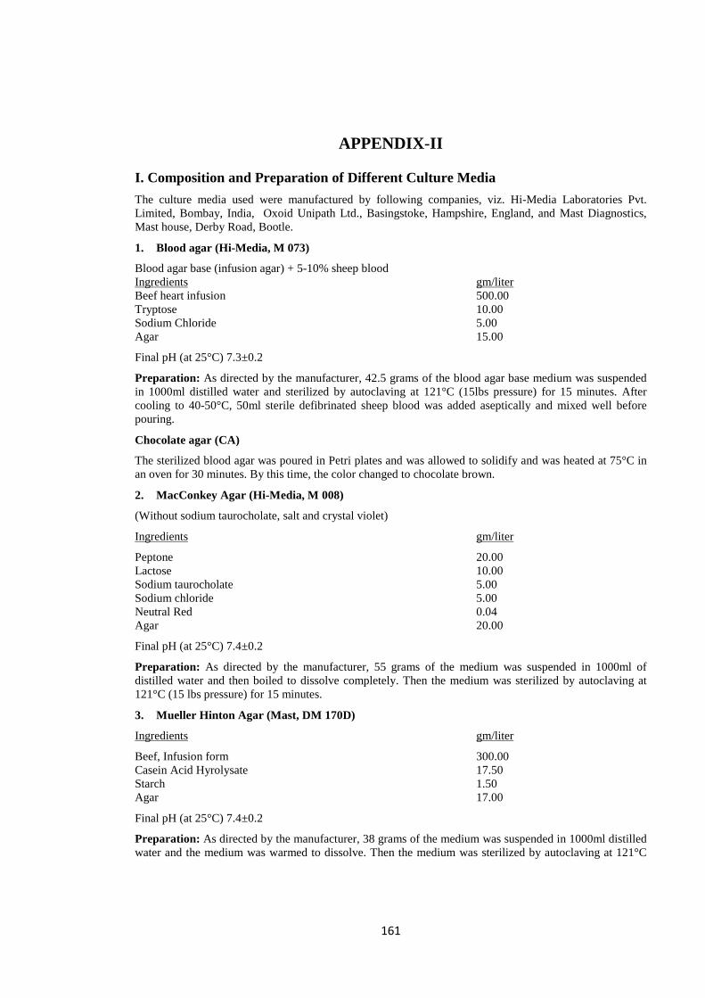
161
APPENDIX-II
I. Composition and Preparation of Different Culture Media
The culture media used were manufactured by following companies, viz. Hi-Media Laboratories Pvt.Limited, Bombay, India, Oxoid Unipath Ltd., Basingstoke, Hampshire, England, and Mast Diagnostics,Mast house, Derby Road, Bootle.
1. Blood agar (Hi-Media, M 073)
Blood agar base (infusion agar) + 5-10% sheep bloodIngredients gm/literBeef heart infusion 500.00Tryptose 10.00Sodium Chloride 5.00Agar 15.00
Final pH (at 25°C) 7.3±0.2
Preparation: As directed by the manufacturer, 42.5 grams of the blood agar base medium was suspendedin 1000ml distilled water and sterilized by autoclaving at 121°C (15lbs pressure) for 15 minutes. Aftercooling to 40-50°C, 50ml sterile defibrinated sheep blood was added aseptically and mixed well beforepouring.
Chocolate agar (CA)
The sterilized blood agar was poured in Petri plates and was allowed to solidify and was heated at 75°C inan oven for 30 minutes. By this time, the color changed to chocolate brown.
2. MacConkey Agar (Hi-Media, M 008)
(Without sodium taurocholate, salt and crystal violet)
Ingredients gm/liter
Peptone 20.00Lactose 10.00Sodium taurocholate 5.00Sodium chloride 5.00Neutral Red 0.04Agar 20.00
Final pH (at 25°C) 7.4±0.2
Preparation: As directed by the manufacturer, 55 grams of the medium was suspended in 1000ml ofdistilled water and then boiled to dissolve completely. Then the medium was sterilized by autoclaving at121°C (15 lbs pressure) for 15 minutes.
3. Mueller Hinton Agar (Mast, DM 170D)
Ingredients gm/liter
Beef, Infusion form 300.00Casein Acid Hyrolysate 17.50Starch 1.50Agar 17.00
Final pH (at 25°C) 7.4±0.2
Preparation: As directed by the manufacturer, 38 grams of the medium was suspended in 1000ml distilledwater and the medium was warmed to dissolve. Then the medium was sterilized by autoclaving at 121°C

162
(15 lbs pressure) for 15 minutes. The sterilized medium was then poured into sterile Petri plates andallowed to cool.
4. Nutrient Agar (Hi-Media, M 001)
Ingredients gm/litre
Peptone 10.00Sodium Chloride 5.00Beef Extract 10.00Yeast Extract 1.50Agar 12.00
Final pH (at 25°C) 7.4±0.2
Preparation: As directed by the manufacturer, 37 grams of the medium was suspended in 1000ml ofdistilled water and then boiled to dissolve completely. The medium was sterilized by autoclaving at 121°C(15 lbs pressure) for 15 minutes, and poured into sterile Petri plates.
5. Nutrient Broth (Hi-Media, M 002)
Ingredients gm/litre
Peptone 5.00Sodium Chloride 5.00Beef Extract 1.50Yeast Extract 1.50
Final pH (at 25°C) 7.4±0.2
Preparation: As directed by the manufacturer, 13 grams of the medium was dissolved in 1000ml distilledwater Then the medium was dispensed in test tube in amount of 3ml in each and and autoclaved at 121°C(15 lbs pressure) for 15 minutes. The sterilized medium was then cooled to room temperature.
6. Mueller Hinton Broth (Oxoid, CM 0405)
Ingredients gm/litre
Beef 300.00Casein Hydroxylate 17.50Starch 1.50Calcium 0.003665Magnesium 6.29
Final pH (at 25°C) 7.3±0.1
Preparation: As directed by the manufacturer, 21 gram of the media was added to 1000ml of distilledwater, mixed well to dissolve, dispensed in screw-capped bottles in amount of 3ml in each and sterilized byautoclaving at 121°C (15 lbs pressure) for 15 minutes. the sterilized medium was then cooled to roomtemperature.
7. Mannitol Salt Agar (Hi-Media, M 118)
Ingredients gm/litre
Proteose peptone 10.00Beef etract 1.00NaCl 75.00D-Mannitol 10.00Phenol Red 0.025Agar 15.00

163
Final pH (at 25°C) 7.4±0.2
Preparation: As directed by the manufacturer, 111.02 grams of the medium was dissolved in 1000mldistilled water, boiled to dissolve completely and sterilized by autoclaving at 121°C (15 lbs pressure) for 15minutes. The sterile medium was then poured into sterile petriplates and allowed to cool.
8. Tryptone Soy broth+ 20% Glycerol
Ingredients gm/litre
Pancreatic Digest of Casein 15.00Enzymatic Digest of Soybean Meal 5.00Sodium Chloride 5.00Glycerol 200ml
Preparation: As directed by the manufacturer, 40 gram of the medium was suspended in 1000ml ofdistilled water containing 200ml glycerol and mixed thoroughly. It was boiled completely and autoclaved at121°C for 15 minutes.
II. Biochemical Test Media
1. MR-VP Medium (Hi-Media, M 070I)
Ingredients gm/litre
Buffered Peptone 7.00Dextrose 5.00Dipotassium Phosphate 5.00
Final pH (at 25°C) 6.9±0.2
Preparation: As directed by the manufacturer, 17 grams of medium was dissolved in 1000ml distilledwater, heated to dissolve completely, dispensed in the amount of 3 ml in each test tube and autoclaved at121°C (15 lbs pressure) for 15 minutes. The tubes were then allowed to cool.
2. Hugh and Leifson’s Medium (Hi-Media, M 826)
Ingredients gm/litre
Tryptone 2.00Sodium Chloride 5.00Dipotassium Phosphate 0.30Bromothymol Blue 0.08Agar 2.00
Final pH (at 25°C) 6.8±0.2
Preparation: As directed by the manufacturer, 19.40 grams of the medium was dissolved in 1000mldistilled water and then heated to boiling to dissolve completely. The medium was then sterilized byautoclaving for 15 minutes at 15 lbs pressure (121°C). To 1000ml sterile medium 100ml of sterile Dextrosewas added aseptically, mixed thoroughly and dispensed in 5ml quantities into sterile culture tubes.
3. Sulphide Indole Motility (SIM) medium (Hi-Media, M 181)
Ingredients gm/litre
Beef Extract 3.00Peptone 30.00Peptonized Iron 0.20Sodium Thiosulphate 0.025Agar 3.00

164
Final pH (at 25°C) 7.3±0.2
Preparation: As directed by the manufacturer, 36.23 grams of the medium was suspended in 1000mldistilled water and dissolved completely. It was dispensed into test tubes to a depth of about 3 inches andautoclaved for 15 minutes at 15 lbs pressure (121°C).
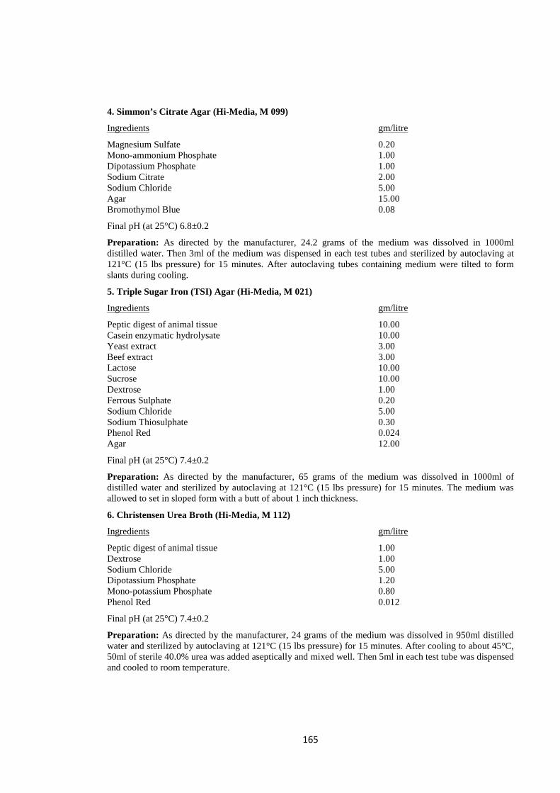
165
4. Simmon’s Citrate Agar (Hi-Media, M 099)
Ingredients gm/litre
Magnesium Sulfate 0.20Mono-ammonium Phosphate 1.00Dipotassium Phosphate 1.00Sodium Citrate 2.00Sodium Chloride 5.00Agar 15.00Bromothymol Blue 0.08
Final pH (at 25°C) 6.8±0.2
Preparation: As directed by the manufacturer, 24.2 grams of the medium was dissolved in 1000mldistilled water. Then 3ml of the medium was dispensed in each test tubes and sterilized by autoclaving at121°C (15 lbs pressure) for 15 minutes. After autoclaving tubes containing medium were tilted to formslants during cooling.
5. Triple Sugar Iron (TSI) Agar (Hi-Media, M 021)
Ingredients gm/litre
Peptic digest of animal tissue 10.00Casein enzymatic hydrolysate 10.00Yeast extract 3.00Beef extract 3.00Lactose 10.00Sucrose 10.00Dextrose 1.00Ferrous Sulphate 0.20Sodium Chloride 5.00Sodium Thiosulphate 0.30Phenol Red 0.024Agar 12.00
Final pH (at 25°C) 7.4±0.2
Preparation: As directed by the manufacturer, 65 grams of the medium was dissolved in 1000ml ofdistilled water and sterilized by autoclaving at 121°C (15 lbs pressure) for 15 minutes. The medium wasallowed to set in sloped form with a butt of about 1 inch thickness.
6. Christensen Urea Broth (Hi-Media, M 112)
Ingredients gm/litre
Peptic digest of animal tissue 1.00Dextrose 1.00Sodium Chloride 5.00Dipotassium Phosphate 1.20Mono-potassium Phosphate 0.80Phenol Red 0.012
Final pH (at 25°C) 7.4±0.2
Preparation: As directed by the manufacturer, 24 grams of the medium was dissolved in 950ml distilledwater and sterilized by autoclaving at 121°C (15 lbs pressure) for 15 minutes. After cooling to about 45°C,50ml of sterile 40.0% urea was added aseptically and mixed well. Then 5ml in each test tube was dispensedand cooled to room temperature.

166
7. Decarboxylase test medium (Hi-Media, M 912)
Ingredients gm/litrePeptic digest of animal tissue 5.00Yeast extract 3.00Dextrose 1.00Bromo-cresol purple 0.02Final pH (at 25°C) 6.8±0.2
Preparation: As directed by the manufacturer, 9 grams of the medium was dissolved in 1000ml of thedistilled water. The medium was then divided into four equal parts. One part was dispensed in test tubeswithout adding amino acid, and to the remaining three parts three amino acids (L-Lysine, L-Arginine andL-Ornithine) were added separately to a final concentration of 0.5% and dispensed in test tubes. The tubeswere then sterilized by autoclaving at 121°C (15 lbs pressure) for 15 minutes.
III. Staining and Test Reagents
1. Preparation of gram staining reagents
(a) Hucker’s crystal violet stain
In a clean piece of paper, 40gm of crystal violet (90.0-95.0% dye content) was weighed, dissolved in 400mlof ethanol (95.0%), filtered and stored a clean brown bottle at room temperature. For the preparation ofworking solution of crystal violet, 40ml of stock solution was added to 160ml of filtered ammoniumoxalate solution (1.0%).
(b) Lugol’s iodine
To prepare stock solution of Lugol’s iodine, 25gm of iodine crystals and 50gm of potassium iodide crystalswere dissolved in 500ml of distilled water in a brown glass bottle. For the preparation of working solution,60 ml of Lugol’s iodine stock solution was mixed with 220ml of distilled water and 60 ml of 5.0% sodiumbicarbonate solution.
(c) Acetone-alcohol decolorizer (1:1)
To 50ml of ethanol (95.0%), 50ml of acetone was mixed well and transferred into a clean bottle. Thepreparation was labeled with date and stored at room temperature.
(d) Safranin (Counterstain)
The stock solution was prepared by dissolving 5gm of safranine in 200ml of 95.0% ethanol and theworking solution was prepared by mixing 20ml of stock solution with 180ml of distilled water.
3. Normal saline
Preparation: In 100ml of distilled water, 0.85gm of sodium chloride was dissolved well in a leak-proofbottle. The bottle was then labeled and stored at room temperature.
4. Test reagents
A. For catalase testTo make 100ml Catalase Reagent (3% H2O2)Hydrogen peroxide 3.00mlDistilled Water 97.00ml
Preparation: To 97ml of distilled water, 3ml of hydrogen peroxide was added and mixed well.
B. For oxidase test
To make 100ml Oxidase Reagent (impregnated in Whatman’s No. 1 filter paper)Tetramethyl p-phenylene diamine dihydrochloride (TPDD) 1.00gm

167
Distilled Water 100.00ml
Preparation: In 100ml of distilled water, 1gm of TPDD was dissolved. To that solution strips ofWhatman’s No. 1 filter paper were soaked and drained for about 30 seconds. Then those strips were freeze-dried and stored in a dark bottle tightly sealed with a screw cap.
C. Kovac’s reagent for indole test
To prepare 40ml reagentIsoamyl alcohol 30.00 ml4-dimethyl aminobenzaldehyde 2.00gmConc. hydrochloric acid 10.00ml
Preparation: In 30ml of isoamylalcohol, 2gm of 4-dimethyl aminobenzaldehyde was dissolved in a cleanbrown bottle. Then, 10ml of conc. HCl was added to that and mixed well.
D. Methyl red solution for methyl red test
To make 50ml solutionMethyl red 0.05gmEthyl alcohol (absolute) 28.00mlDistilled Water 22.00ml
Preparation: To 28ml ethanol, 0.05gm of methyl red was dissolved in a clean brown bottle. Then 22mldistilled water was added to that bottle and mixed well.
E. Barritt's reagent for Voges-Proskauer testVoges-Proskauer reagent ATo make 100mlα-Napthol 5.00gmEthyl alcohol (absolute) 100.00ml
Preparation: To 25ml distilled water, 5gm of α-napthol was dissolved in a clean brown bottle. Then thefinal volume was made 100ml by adding distilled water.Voges-Proskauer reagent BTo make 100mlPotassium hydroxide 40.00gmDistilled Water 100.00ml
Preparation: To 25ml distilled water, 40gm of KOH was dissolved and transferred into a clean brownbottle. Then the final volume was made 100ml by adding distilled water.
The three parts of reagent A (5.0% α-napthol) was mixed with one part of reagent B (40.0% KOH) to getthe working solution of the Barritt’s reagent.

168
APPENDIX-IIII. List of Equipment, Materials and Supplies
A. General microbiology laboratory equipment
(a). Autoclave (Stermite, Japan) (b). Incubator (Sakura, Japan) (c). Hot air oven (Memmert, Germany;and GallenKamp) (d). Microscope (Olympus, Japan) (e). Refrigerator, 4-8°C (Sanyo, Japan); -20°C(Videocon, India); -75°C (Sanyo, Japan) (f). Weighing machine (Ohaus Corporation, USA) (g). WaterBath (Boekel Scientific, USA) (h). Gas burner (i). Glasswares (j). Inoculating loops and wires (k).Sterile cotton swabs (l). Sterile Petri plates (m). McFarland 0.5 turbidity standards (n). Vortex mixer(o). Ruler (p). Forceps
B. Microbiological media
(a). Blood Agar (b). Chocolate Agar (c). MacConkey Agar (d). Mueller Hinton Agar (e). Mannitol SaltAgar (f). Mueller Hinton Broth (g). Hugh and Leifson Medium (h). Sulfide Indole Motility medium(i). MRVP broth (j). Triple Sugar Iron Agar (k). Urea broth (l). Simmon’s Citrate Agar (m). NutrientAgar (n). Tryptone Soy Broth
C. Chemicals and reagents
Catalase reagent (3% H2O2), Oxidase reagent (1% Tetramethyl p-phenylene diaminedihydrochloride), Kovac’s reagent, Barritt’s reagent (40% KOH, 5% α-naphthol in a ratio of 1:3),Barium Chloride, Conc. H2SO4, , Glycerol, Gram’s reagent, etc.
D. Antibiotic disks
The following antibiotic disks were used for the antibiotic susceptibility tests which were fromMast Group Ltd., Mast House, Derby Road, Bootle, Merseyside, L201EA, UK.
Amikacin (AK/30µg), Amoxycillin (A/20µg), Augmentin (AUG/30µg), Azithromycin(ATH/15µg), Erythromycin (E/15µg), Aztreonam (ATM/30µg), Bacitracin (BA/10U), Bacitracin(BA/0.04IU), Cefotaxime (CTX/30µg), Ceftazidime (CAZ/30µg), Cefpodoxime (CPD/10µg/30µg),Ceftriaxone (CRO/30µg), Cotrimoxazole (TS/25µg), Cephalothin (KF/30µg), Cefoxitin (FOX/30µg),Nalidixic acid (NA/30µg), Ciprofloxacin (CIP/5µg), Ofloxacin (OFX/5µg), Levofloxacin (LEV/5µg),Moxifloxacin (MFX/1µg), Gatifloxacin (GAT/5µg), Tobramycin (TN/10µg), Neomycin (NE/30µg),Gentamicin (GM/10µg), Netilmicin (NET/30µg), Kanamycin (K/30µg), Chloramphenicol (C/30µg),Cefepime (CPM/30µg), Cefpirome (CFP/30µg), Clindamycin (CD/2µg), Imipenem (IMI/10µg),Meropenem (MEM/10µg), Norfloxacin (NOR/10µg), Nitrofurantoin (NI/300µg), Oxacillin (OX/1µg),Penicillin G (PG/10U), Tetracycline (T/30µg), Doxycycline (DXT/30µg), Minocycline (MN/30µg),Tigecycline (TGC/15µg), Ticarcillin (TC/75µg), Fosfomycin (FOT/200µg), and Vancomycin (VA/30µg).
Methicillin (M/5µg) disks used were from Hi-Media Laboratories Pvt. Ltd., India.
E. Disks for ESBL detection
(a). MASTDISCSTM ID Extended Spectrum Beta-Lactamase (ESβL) Detection Discs (D52C):Cefotaxime (CTX/30µg), cefpodoxime (CPD/30µg) and ceftazidime (CAZ/30µg) alone and each incombination with clavulanic acid (CV/10µg)
(b). MASTDISCSTM ID Cefepime ESβL ID Disc Set (D63C): Cefepime (CPM/30µg) alone and itscombination with clavulanic acid (CV/10µg)

169
APPENDIX-IVA. Gram-Staining
The method was first devised by Sir Hans Christian Gram during the late 19th century.This method can beused effectively to divide all bacterial species into two major groups: Gram-positive (those that take up andretain the basic dye crystal violet even on washing) and Gram-negative (those that allow the crystal violetdye to wash out easily with the decolorizer of alcohol or acetone). The following steps were performed forGram-staining:
1. A thin oval smear of the material to be examined was prepared on a clean grease free glass slide andair dried. The material on the slide was then heat fixed and allowed to cool before staining.
2. The slide was flooded with crystal violet stain and allowed to remain without drying for 30-45seconds. Then the slide was rinsed with tap water to remove the excess stain.
3. Then the slide was flooded with Gram’s iodine solution and allowed to remain on the surfacewithout drying for twice as long as the crystal violet was in contact with the slide surface. Again theslide was rinsed with tap water to remove excess iodine.
4. The slide was flooded with acetone/alcohol decolorizer for 10 seconds and rinsed immediately withsufficient tap water until no further color flowed from the slide with the decolorizer. Thicker smearrequired more aggressive decolorizing.
5. The slide was flooded with counter stain (safranin) for 30 seconds and washed off with tap water.
6. The slide was blotted between two clean sheets of bibulous paper and examined microscopicallyunder oil immersion at 1000X.
Quality control
The laboratory staining procedure and reagents were tested prior to use of new lots of each staining anddecolorizing agents and at least weekly thereafter, using Escherichia coli ATCC 25922 (Gram negative)and Staphylococcus aureus ATCC 25923 (Gram positive).
B. Biochemical Tests for Bacterial Identification
Catalase test
Microorganisms produce hydrogen peroxide (H2O2) during aerobic respiration, which is lethally toxic tothe cell itself. Catalase enzyme breaks down hydrogen peroxide into water and oxygen. The enzymecatalase is present in most cytochrome containing aerobic and facultative anaerobic bacteria, with majorexception being Streptococcus species. About 2-3 drops of 3.0% H2O2 was put on the surface of a cleanglass slide and a small amount of a culture from Nutrient Agar plate was applied with sterile wooden orglass stick. Presence of effervescence indicated the positive test. A false positive reaction was prevented byavoiding the use of the culture medium with catalase (e.g., Blood Agar) or an iron loop.
Oxidase test
This test is performed for the detection of cytochrome oxidase in bacteria which catalyzes the transport ofelectrons between electron donors. In the presence of redox dye, Tetramethyl-p-phenylene diaminedihydrochloride (TPDD), the cytochrome oxidase oxidizes it into a deep purple colored end product,Indophenol, which is detected in this test. A small aliquot of colony of the test organism was rubbed on thefilter paper soaked in oxidase reagent with the help of sterile glass rod. The positive test was indicated bythe appearance of blue-purple color within 10 seconds.
Oxidation-fermentation test
This test is done to determine the oxidative or fermentative metabolism of carbohydrate resulting inproduction of various organic acids as end products. Some bacteria are capable of metabolizing

170
carbohydrates (as exhibited by acid production) only under aerobic conditions, while others produce acidboth aerobically and anaerobically. Most medically important bacteria are facultative anaerobes. The testorganism was stabbed into the bottom of two sets of tubes with Hugh and Leifson's media, containingbromothymol blue as the pH indicator. The inoculated medium in one of the tubes was covered with a10mm deep layer of sterile paraffin oil. The tubes were then incubated at 37°C for 24 hours. Afterincubation the tubes were examined for carbohydrate utilization as shown by acid production. Fermentativeorganisms utilize the carbohydrate in both the open and sealed tubes as shown by a change in colour of themedium from green to yellow. However, oxidative organisms are able to use the carbohydrate only in theopen tube.
Indole production test
This test detects the ability of the organism to produce an enzyme: ‘tryptophanase’ which oxidizestryptophan to form indolic metabolites: indole, skatole (methyl indole) and indoleacetic acid. Indole, ifpresent, combines with the aldehyde present in the reagent to produce red color in the alcohol layer. Thecolor reaction is based on the presence of the pyrrole structure present in indole. A smooth bacterial colonywas stabbed on Sulfide Indole Motility (SIM) medium by a sterile inoculating wire and the inoculatedmedia was incubated at 37°C for 24 hours. After 24 hours incubation, 0.5ml of Kovac's reagent was added.Appearance of red color on the top of media indicated the indole positive reaction.
Motility test
Motile organisms migrate from the stabline and diffuse into the medium causing turbidity whereas non-motile bacteria show the growth only along the stabline, and the surrounding medium remains colorless andclear. This test was macroscopically observed in semisolid Sulfide Indole Motility (SIM) medium.
Methyl red test
This test is performed to test the ability of an organism to produce sufficient acid from the fermentation ofglucose. The degree of acidity is denoted by intensity of color change of methyl red indicator over a pHrange of 4.4-6.0. A pure colony of the test organism was inoculated into 2ml of MRVP medium and wasincubated at 37°C for 24 hours. After incubation, about 5 drops of methyl red reagent was added and mixedwell. The positive test was indicated by the development of bright red color, indicating acidity.
Voges-Proskauer (VP) test
This test detects the production of acetyl methyl carbinol (a neutral end product) or its reduction product 2,3-butanidiol during fermentation of carbohydrates. A pure colony of the test organism was inoculated into2ml of MRVP medium and was incubated at 37°C for 24 hours. After incubation, about 5 drops of Barritt'sreagent was added and shaken well for maximum aeration and kept for 15 minutes, positive test is indicatedby the development of pink red colour.
Citrate utilization test
This test detects whether an organism can utilize citrate as a sole source of carbon for metabolism withresulting alkalinity. Organisms capable of utilizing citrate as sole carbon source also utilize the ammoniumsalts present in the medium as sole nitrogen source. Production of ammonia imparts alkalinity, thuschanging the color of indicator Bromothymol blue from green (pH 6.0-7.6) to blue (pH ≥7.6). A loopful oftest organism was streaked on the slant area of Simmon's Citrate Agar medium and incubated at 37°C for24 hours. Growth of organism and change of color of media from green to blue indicated a positive test.
Triple sugar iron (TSI) agar test
The TSI agar is used to determine the ability of an organism to utilize specific carbohydrate incorporated inthe medium (glucose, sucrose and lactose in concentrations of 0.1%, 1.0% and 1.0% respectively), with orwithout the production of gas (indicated by cracks in the media as well as an air gap at the bottom of thetube) along with determination of possible hydrogen sulfide production (detected by production of blackcolor in the medium). The test organism was stabbed on the butt and streaked on the surface of TSI slantand incubated at 37°C for 24 hours. Acid production limited only to the butt region of the tube is indicative

171
of glucose utilization, while acid production in slant and butt indicated sucrose or lactose fermentation.Phenol red is the pH indicator which gives yellow colour at acidic pH, and red colour at alkaline condition.

172
Urease test
This test demonstrates the ability of bacteria to produce urease which decomposes urea, releasing ammoniaand carbon dioxide. Ammonia thus produced imparts alkalinity and changes the color of phenol redindicator incorporated in the medium. The test organism was inoculated in a medium containing urea andthe indicator phenol red and incubated at 37°C overnight. Change in color of the medium to deep pink redindicated positive reaction, i.e. urease production.
Amino acid decarboxylase test
This test identifies the ability of some bacteria to decarboxylate an amino acid to the corresponding aminewith the liberation of carbon dioxide. Lower pH induces the production of these decarboxylases and pHrises to the neutrality or above as a result of their action. Lysine, ornithine and arginine are the threeprincipal amino acids used in this test which are converted to cadaverine, putrescine and citrullinerespectively by the specific enzymes. A pure colony of test organism was inoculated lightly through theparaffin layer of each broth medium containing lysine, ornithine and arginine separately with a straightwire. The tubes were incubated at 37°C and read daily for 4 days. The development of violet color in themedium indicated decarboxylation.
DNase (Deoxyribonuclease) test
This test is used to identify Staphylococcus aureus which produces deoxyribonuclease (DNase) enzyme.The deoxyribonuclease enzyme hydrolyses the DNA. The test organism was cultured on a mediumcontaining DNA. After overnight incubation, the colonies were tested for DNase production by floodingthe plate with a weak hydrochloric acid solution as acid precipitates unhydrolysed DNA. Clear areasdeveloped around the DNase producing colonies due to DNA hydrolysis.
Coagulase tests
Coagulase tests differentiate species within the genus Staphylococcus: S.aureus (usually positive) from S.epidermidis (negative). A positive coagulase test is usually the final diagnostic criterion for theidentification of S. aureus. Free coagulase and bound coagulase are the two types of coagulase possessedby this organism most strains of S.aureus possess both free and bound coagulase.
Slide coagulase test
Slide test detects bound coagulase (Clumping Factor) which is bound to the bacterial cell wall andreacts directly with fibrinogen resulting in alteration of fibrinogen so that it precipitates on thestaphylococcal cell, causing cell clumping when a bacterial suspension is mixed with plasma. For slidecoagulase test, a drop of physiological saline was placed on three places of a slide, and then a colonyof the test organism was emulsified in two of the drops to make thick suspensions. Later a drop ofplasma was added to one of the suspensions and mixed gently. Then a clumping was observed within10 seconds for the positive coagulase test. No plasma was added in second suspension. This was usedfor the differentiation of any granular appearance of the organism from true coagulase clumping. Thethird drop of saline was used for a known strain of coagulase positive staphylococci as a control.
Tube coagulase test
This test detects free coagulase. Plasma contains coagulase reacting factor (CRF) which activates freecoagulase. The activated coagulase converts prothrombin to thrombin which in turn convertsfibrinogen into fibrin which is detected as a firm gel (clot) in the tube test. Tube test is performed incase of negative or doubtful results in slide test. The plasma was first diluted 1 in 10 in physiologicalsaline. Four small tubes were taken, one for test organism, other for positive control, another fornegative control, and next to observe self clotting of plasma. Then 0.5ml of the diluted plasma waspipetted into each tube and 0.5ml of test organism, 0.5ml of positive control (Staphylococcus aureusculture), and 0.5ml negative control (S. epidermidis culture) was added to three respective tubes. Tothe fourth tube, 0.5ml sterile broth was added. After mixing gently, all tubes were incubated at 37°C on

173
a waterbath for 6 hours and observed for gel formation in every 30 minutes. Any clot formationindicated the positive coagulase test.

174
Optochin susceptibility test
This test is used to differentiate pneumococci from other streptococci. Optochin (ethyl hydrocuprienehydrochloride) lyses pneumococci (positive test); whereas the viridans group of streptococci are resistant(negative test). It can be performed conveniently with a paper disc containing optochin 5µg. Thepneumococci susceptible to optochin show a zone of inhibition of >14mm, while other alpha hemolyticstreptococci grow to the edge of the disk. A paper disk containing 5µg of optochin was placed in theprimary inoculum of a chocolate agar plate streaked with the pure culture of suspected pneumococci andthe plate was incubated at 37°C in candle jar for overnight. After incubation the plate was observed forzone of inhibition around the disk.
Bacitracin susceptibility test
This test is used to screen H. influenzae from the primary agar plate. H. influenzae is resistant to the 10 UnitBacitracin disc but the viridans streptococci, Neisseria spp., diptheroid bacilli and staphylococci aresusceptible and get supressed. A paper disk containing 10U of bacitracin was placed in the primaryinoculum of a chocolate agar plate streaked with the material from the specimen and the plate wasincubated at 37°C in candle jar for overnight. After incubation, the plate was observed for the colonies ofH. influenzae growing near the bacitracin disk.
Bile solubility test
This test differentiates pneumococci and viridans streptococci. Bile lyses the pneumococcal cells byattacking the peptidoglycan, whereas the viridans streptococci are unaffected. Solution of 10% sodiumdeoxycholate prepared in distilled water was autoclaved and stored at room temperature. Colonies ofsuspected pneumococci were inoculated in two test tubes containing 5ml nutrient broth from the blood orchocolate agar and was incubated at 37°C for overnight. Then 0.5ml of bile salt solution was added in onetube while another was taken as control. Incubation was continued for 15-30 minutes. In the positive test,turbidity of broth was cleared as compared to the control tube.
Tests for X and V factor requirements
This test is carried out to differentiate H. influenzae from H. parainfluenzae. H.influenzae requires both X-factor (haemin or various iron containing compounds such as protoporphyrin IX) and the V-factor (NAD orNADP) as the accessory growth factors. H. parainfluenzae requires only the V-factor for growth. Disksimpregnated with X factor alone, V factor alone and X and V factor together were placed on Mueller-Hinton agar which had been inoculated with a light suspension of the organism. After overnight incubationat 37°C in candle jar, the plate was examined for growth around each disk. The growth only around the XVdisc was indicative of the species being H. influenzae.

175
APPENDIX-VI
1. Antimicrobial Susceptibility Testing by Disk Diffusion Method
A. Principle
A standardized inoculum of bacteria is swabbed onto the surface of MHA plate. Filter paper disksimpregnated with antimicrobial agents are placed flat on the agar surface. After overnight incubation, thediameter of the zone of inhibition is measure around each disk. By referring to the tables in the CLSI diskdiffusion standard, a qualitative report of susceptible, intermediate or resistant is obtained.
B. Procedures
1. Preparation of 0.5 McFarland Standard turbidity solution: To 0.5ml of 0.048M BaCl2 (1.17% w/vBaCl2.2H2O), 99.5ml of 0.18M H2SO4 (1%v/v) was added with constant stirring. The solution was thendistributed into test tubes, well screw-capped, wrapped with almunium foil and stored in dark till use.
2. The agar plates and antibiotic disk cartridges were brought to room temperature before use. Agar plateswere made to contain no excess moisture on their surface by placing them in the 35°C ambient airincubator with lids ajar for evaporation.
3. Inoculum preparation: By touching 2-3 morphologically similar colonies not more than 24 hours oldwith sterile loop on nonselective agar plate, MHB or NB was inoculated and incubated at 37°C untilturbidity matched with that of the 0.5 McFarland Standard turbidity solution.
4. Inoculation of agar plates: Within 15 minutes of adjusting turbidity, a sterile cotton swab was dipped intothe inoculums and rotated against the wall of the tube above the liquid to remove excess inoculum. Theentire surface of the Mueller Hinton agar plate was swabbed by rotating plates approximately 60° betweensuccessive swab strokes to ensure even distribution. Any touching to sides of the plates and aerosolformation was avoided. Finally, the swab was run around the edge of the agar to remove any excessmoisture. The plates were left for about 10-15 minutes to let the agar surface dry before the application ofthe disks.
5. Appropriate antibiotic disks were placed evenly (not closer than 24mm from center to center) on theinoculated agar surface either with dispenser or manually with sterile forceps and a gentle pressure wasapplied on each disk with sterile forceps to ensure complete contact of disk with agar. Not more than sixdisks were placed on a 90mm Petri plate. Within 30 minutes of disk application, the plates were incubatedin inverted position at 35°C in an ambient air incubator for 16-18 hours.
6. Reading, interpretation and reporting: Only the plates with lawn of confluent or nearly confluent growthwere read. The diameter of zones of inhibition was measured in nearest whole millimeter with a measuringscale by observing the plate against nonreflecting surface. In case of sulfonamides, trimethoprim and co-trimoxazole, light growth (<20.0% of lawn growth) was disregarded and zone of inhibition was measuredby using the edge of the more obvious margin of the zone. In case of development of discrete colonieswithin the zone of inhibition (which may represent mixed culture or resistant variants), eachmorphologically different single colonies were subcultured, re-identified and retested for antibioticsusceptibility. If they were still present, the colony-free inner zone was measured. Any aberrant resultswere confirmed by repeating the test twice and processing accordingly. Criteria specified by the CLSI (formost antibiotics), BSAC (for temocillin and moxifloxacin) and FDA (for tigecycline) were usedunambiguously to interpret the zone of inhibition and the categorical results were reported as eithersusceptible (S), intermediate (I), or resistant (R).
Quality control
a. QC strains: i). Escherichia coli ATCC 25922 ii). Staphylococcus aureus ATCC 25923
b. Monitoring Accuracy: QC strains were tested daily and weekly by following routine procedures andresults were recorded. Also the lot number and expiry date of the antibiotic disks and MHA were noted.

176
The obtained results were compared to expected results (CLSI QC tables) and in case of any aberrantresult, the procedure was continued with corrective action.
APPENDIX-VIIA. Inhibitor Potentiated Disk Diffusion (IPDD) Test/Combined Disk Assay forphenotypic confirmation of ESBL production
ESBL production was confirmed among the suspected bacterial strains according to the CLSI (Clinical andLaboratory Standard Institute) guidelines for phenotypic confirmatory testing. According to theseguidelines, when confirming ESBL production among the suspects using Combined Disk assay, an increasein zone size of ≥5 mm from either of the combination disk, i.e. clavulanate containing disk indicates thepresence of ESBL in the test organism.
i). The suspected organism was inoculated onto Mueller Hinton broth and incubated at 35°C untilthe turbidity matched with 0.5 McFarland standard turbidity solution.
ii). The test organism was carpet-cultured on a MHA plate using a sterile cotton swab.
iii). Using sterile forceps, the ESBL detection disks were applied onto the inoculated mediumensuring that they are evenly spaced and in proper contact with the agar surface.
iv). The plates were incubated at 35-37°C for 18-24 hours and the results were interpretedaccordingly.
InterpretationZones of inhibition for cefotaxime, cefpodoxime, ceftazidime and cefepime were compared to that of theirrespective combinations with clavulanic acid. An increase in zone diameter by ≥5mm in the presence ofclavulanic acid for any or all of the disks indicated the presence of ESBL in the test organism.
Quality control
Quality control was maintained by performing the test with at least one organism to demonstrate positivereaction and at least one other organism to demonstrate negative reaction. For positive controls,Escherichia coli NCTC 13351 and Klebsiella pneumonia ATCC 700603 were tested while for negativecontrol, Escherichia coli ATCC 25922 was tested.
Other QC measures related to antibiotic susceptibility test were also strictly followed.

177
APPENDIX-VIII
Distinguishing biochemical reactions of common and pathogenic Enterobacteriaceae
a lac, inos, fermentation of lactose, inositol; mot, motility; gas, gas from glucose; ind, indole production;VP, Voges-Proskauer; cit, Citrate utilization (Simmons'); PDA, phenylalanine deaminase; ure, urease; lys,lysine decarboxylase; H2S, H2S produced in TSI agar; ONPG, metabolism of o-nitrophenyl-β-D-galactopyranoside.b Some strains of Serratia marcescens may produce a red pigmentc Yersinia species are motile at 22°C.{Key: +, ≥85% of strains positive; -, ≥ 85% of strains negative; 16-84% of strains are positive after 24-48hour at 36°C}(Source: Collee et al., 1996)
Organisms
Tests/ Substratesa
lac mot gas ind VP cit PDA ure lys H2S inos ONPG
Escherichia coli + + + + - - - - + - - +
Shigella groups A, B, C - - - ± - - - - - - - -
Sh. sonnei - - - - - - - - - - - +
Salmonella (mostserotypes)
- + + - - + - - + + ± -
Salmonella typhi - + - - - - - - + + - -
Salmonella paratyphi A - + + - - - - - - - - -
Citrobacter freundii ± + + - - + - ± - ± - +
C. koseri ± + + + - + - ± - - - +
Klebsiella pneumoniae + - ++ - + + - + + - + +
K. oxytoca + - ++ + + + - + + - + +
Enterobacter aerogens + + ++ - + + - - + - + +
Ent. cloacae + + + - + + - ± - - - +
Hafnia alvei - + + - + - - - + - - +
Serratia marcescensb - + ± - + + - - + - ± +
Proteus mirabilis - + + - ± ± + ++ - + - -
P. vulgaris - + + + - - + ++ - + - -
Morganella morganii - + + + - - + ++ - ± - -
Providencia rettgeri - + - + - + + ++ - - + -
Prov. stuartii - + - + - + + ± - - + -
Prov. alcalifaciens - + + + - + + - - - - -
Yersinia enterocoliticac - - - ± - - - ± - - ± +
Y. pestis - - - - - - - - - - - ±
Y. pseudotuberculosis - - - - - - - + - - - ±
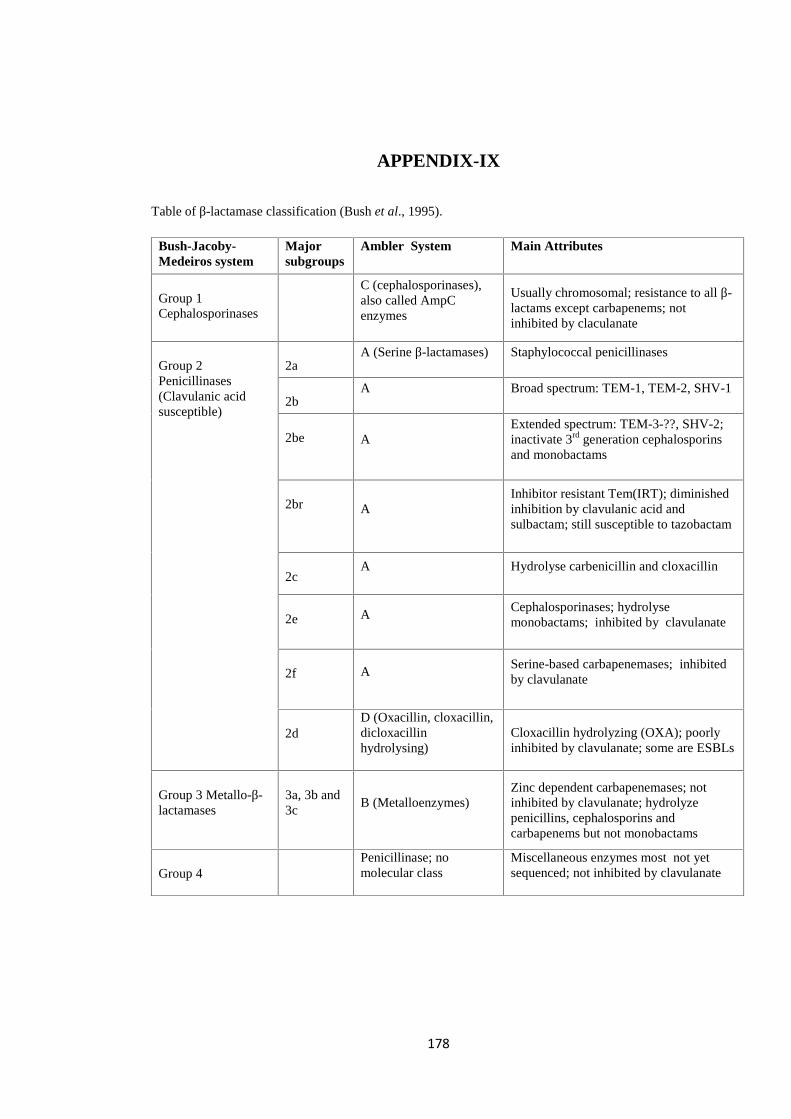
178
APPENDIX-IX
Table of β-lactamase classification (Bush et al., 1995).
Bush-Jacoby-Medeiros system
Majorsubgroups
Ambler System Main Attributes
Group 1Cephalosporinases
C (cephalosporinases),also called AmpCenzymes
Usually chromosomal; resistance to all β-lactams except carbapenems; notinhibited by claculanate
Group 2Penicillinases(Clavulanic acidsusceptible)
2aA (Serine β-lactamases) Staphylococcal penicillinases
2bA Broad spectrum: TEM-1, TEM-2, SHV-1
2be AExtended spectrum: TEM-3-??, SHV-2;inactivate 3rd generation cephalosporinsand monobactams
2br AInhibitor resistant Tem(IRT); diminishedinhibition by clavulanic acid andsulbactam; still susceptible to tazobactam
2cA Hydrolyse carbenicillin and cloxacillin
2e ACephalosporinases; hydrolysemonobactams; inhibited by clavulanate
2f ASerine-based carbapenemases; inhibitedby clavulanate
2dD (Oxacillin, cloxacillin,dicloxacillinhydrolysing)
Cloxacillin hydrolyzing (OXA); poorlyinhibited by clavulanate; some are ESBLs
Group 3 Metallo-β-lactamases
3a, 3b and3c
B (Metalloenzymes)Zinc dependent carbapenemases; notinhibited by clavulanate; hydrolyzepenicillins, cephalosporins andcarbapenems but not monobactams
Group 4Penicillinase; nomolecular class
Miscellaneous enzymes most not yetsequenced; not inhibited by clavulanate
Copyright © 2022 FDOKUMEN