Detect the Antibacterial and Antitumor of synthesized Silver ...
9
1 Volume 7 / Issue 2 / 30 / http://dx.doi.org/10.21931/RB/2022.07.02.30 Detect the Antibacterial and Antitumor of synthesized Silver Nanoparticles Using Microbacterium sp Thuraya Mehbas Dewan and Rashid Rahim Hateet* Abstract: Metal nanoparticles are widely utilized in biotechnology and biomedicine for various applications, including medication delivery, imaging, and bacterial growth control. Silver nanoparticles (AgNPs) were synthesized by bacteria, fungi, algae, and plants. The Study aimed to synthesize nanomaterial with a cost-effective, environmentally friendly, and the uses of AgNPs as antibacterial (against pathogenic bacteria) and anticancer (on MCF7 cell line). In this Study, bacteria were collected from different soil samples. Isolated, purified by selective media, identification genotypically by 16rRNA sequencing analysis, then compared with NCBI, GenBank as Microbacterium sp. Biosynthesis of silver nanoparticles using Microbacterium for extracellular synthesis by reducing silver ions to silver nanoparticles. The color change to brown and reddish-brown was the first indication of the AgNPs formation; physical characterization using UV-Visible spectroscopy showed a wavelength in 489 nm, while X-ray diffraction (XRD) revealed that the silver nanoparticles were crystalline; transmission electron microscope (TEM) image showed that AgNPs spherical in shape with an average diameter of around 50 nm, in SEM (Scanning electron microscope) AgNPs formed with a diameter of 41-44 nm, spherical and uniform, while Energy-dispersive X-ray show very high silver peaks. Bioactivity of AgNPs by antimicrobial on pathogenic bacteria, which collected from Al- Sadr hospital in Misan (identified by using VITEK). This experiment showed that the inhibition zone was rung from (6- 38mm) on pathogenic bacteria; it was tremendous compared with antibiotics (Gentamycin in this project ranged from(7-28.5mm). Antitumor activity of extracellular biosynthesized AgNPs was determined using the MTT test against breast cancer cells (MCF7 cell line), which showed very high results. AgNPs inhibition breast cancer cell line by about 81% at 100ug/ml, indicating that the rate is outstanding. Finally, different biomedical approaches can benefit from AgNPs as antibacterial agents and anticancer agents with many results. Key words: Silver Nanoparticles, biosynthesis, antibacterial, and antitumor. ARTICLE / INVESTIGACIÓN Introduction Recently, the immunological compatibility of humans has greatly enhanced the emergence of microbial diseases. As a result, many novel antibiotics and therapeutic phar- maceutical substances with a wide range of applications have been made available on the market to protect humans from various diseases 1 , but they can potentially affect the environment, especially in developing nations 25 . Metals containing nanoparticles have the potential to be used in the control of several types of infection, but little is known about their antibacterial capabilities18. Due to a growing desire to generate environmentally friendly products, the synthesis of nanoparticles has become a hot topic at the junction of nanotechnology and biotechnology 2,26,27 . Using bacteria, fungus, algae, actinomycetes, plants, and other organisms, biogenic synthesis of metal nanoparticles has been demonstrated 3 , Actinobacteria such as Streptomyces sp. 4 , Nocardia sp. 5 , and Rhodococcus sp. 6 , have been re- ported to synthesize and characterize silver and gold nano- particles. Metals such as zinc, silver, titanium, and copper have antibacterial properties that have been recognized for decades, allowing them to be used in various current me- dical applications to manage microbial infection disorders 33 . According to one idea, free metal ion toxicity arising from nanoparticle surfaces may play an essential role in infec- tion prevention 34 . The catalytic, electrical, and optical cha- racteristics of AgNPs are well-known 7,8 . A new generation of dressings comprising antimicrobial compounds like silver was created to prevent or minimize infection 9 . Hybrids of silver nanoparticles with amphiphilic hyperbranched ma- cromolecules have recently been proven to have efficient antibacterial surface coating 10 . Because of their widespread use, silver nanoparticles are in high demand. Silver nano- particles have attracted much attention among noble metal nanomaterials because of their appealing physicochemical feature 11 . Individual silver nanoparticles are great candida- tes for molecular labeling because of their surface Plasmon resonance and large effective scattering cross section 12,13 . Even Antifungal candidates could be AgNPs 19 . Silver ions have a well-known bactericidal action on microorganisms; however, the bactericidal process is only partially unders- tood. It's been proposed that ionic silver interacts signifi- cantly with the thiol groups of essential enzymes, rendering them inactive 29 . Experiments show that when bacteria are exposed to silver ions, their DNA loses its replication capacity. Other research has found indications of structural alterations in cell membranes and the creation of tiny electron-dense gra- nules from silver and sulfur 30,31 . Chemotherapy is one of the Department of Biology, College of Science, University of Misan, Maysan, Iraq. Corresponding author: [email protected] DOI. 10.21931/RB/2022.07.02.30 Citation: Dewan T, Hateet R . Detect the Antibacterial and Antitumor of synthesized Silver Nanoparticles Using Microbacterium sp. Revis Bionatura 2022;7(2) 30. http://dx.doi.org/10.21931/RB/2022.07.02.30 Received: 28 December 2021 / Accepted: 23 February 2022 / Published: 15 May 2022 http://www.revistabionatura.com Publisher’s Note: Bionatura stays neutral with regard to jurisdictional claims in published maps and institutional affiliations. Copyright: © 2022 by the authors. Submitted for possible open access publication under the terms and conditions of the Creative Commons Attribution (CC BY) license (https://creativecommons.org/licenses/by/4.0/).
-
Upload
khangminh22 -
Category
Documents
-
view
1 -
download
0
Transcript of Detect the Antibacterial and Antitumor of synthesized Silver ...

1
Volume 7 / Issue 2 / 30 / http://dx.doi.org/10.21931/RB/2022.07.02.30
Detect the Antibacterial and Antitumor of synthesized Silver Nanoparticles Using Microbacterium spThuraya Mehbas Dewan and Rashid Rahim Hateet*
Abstract: Metal nanoparticles are widely utilized in biotechnology and biomedicine for various applications, including medication delivery, imaging, and bacterial growth control. Silver nanoparticles (AgNPs) were synthesized by bacteria, fungi, algae, and plants. The Study aimed to synthesize nanomaterial with a cost-effective, environmentally friendly, and the uses of AgNPs as antibacterial (against pathogenic bacteria) and anticancer (on MCF7 cell line). In this Study, bacteria were collected from different soil samples. Isolated, purified by selective media, identification genotypically by 16rRNA sequencing analysis, then compared with NCBI, GenBank as Microbacterium sp. Biosynthesis of silver nanoparticles using Microbacterium for extracellular synthesis by reducing silver ions to silver nanoparticles. The color change to brown and reddish-brown was the first indication of the AgNPs formation; physical characterization using UV-Visible spectroscopy showed a wavelength in 489 nm, while X-ray diffraction (XRD) revealed that the silver nanoparticles were crystalline; transmission electron microscope (TEM) image showed that AgNPs spherical in shape with an average diameter of around 50 nm, in SEM (Scanning electron microscope) AgNPs formed with a diameter of 41-44 nm, spherical and uniform, while Energy-dispersive X-ray show very high silver peaks. Bioactivity of AgNPs by antimicrobial on pathogenic bacteria, which collected from Al- Sadr hospital in Misan (identified by using VITEK). This experiment showed that the inhibition zone was rung from (6- 38mm) on pathogenic bacteria; it was tremendous compared with antibiotics (Gentamycin in this project ranged from(7-28.5mm). Antitumor activity of extracellular biosynthesized AgNPs was determined using the MTT test against breast cancer cells (MCF7 cell line), which showed very high results. AgNPs inhibition breast cancer cell line by about 81% at 100ug/ml, indicating that the rate is outstanding. Finally, different biomedical approaches can benefit from AgNPs as antibacterial agents and anticancer agents with many results.
Key words: Silver Nanoparticles, biosynthesis, antibacterial, and antitumor.
ARTICLE / INVESTIGACIÓN
IntroductionRecently, the immunological compatibility of humans
has greatly enhanced the emergence of microbial diseases. As a result, many novel antibiotics and therapeutic phar-maceutical substances with a wide range of applications have been made available on the market to protect humans from various diseases1, but they can potentially affect the environment, especially in developing nations25. Metals containing nanoparticles have the potential to be used in the control of several types of infection, but little is known about their antibacterial capabilities18. Due to a growing desire to generate environmentally friendly products, the synthesis of nanoparticles has become a hot topic at the junction of nanotechnology and biotechnology2,26,27. Using bacteria, fungus, algae, actinomycetes, plants, and other organisms, biogenic synthesis of metal nanoparticles has been demonstrated3, Actinobacteria such as Streptomyces sp.4, Nocardia sp.5, and Rhodococcus sp.6, have been re-ported to synthesize and characterize silver and gold nano-particles. Metals such as zinc, silver, titanium, and copper have antibacterial properties that have been recognized for decades, allowing them to be used in various current me-dical applications to manage microbial infection disorders33. According to one idea, free metal ion toxicity arising from nanoparticle surfaces may play an essential role in infec-
tion prevention34. The catalytic, electrical, and optical cha-racteristics of AgNPs are well-known7,8. A new generation of dressings comprising antimicrobial compounds like silver was created to prevent or minimize infection9. Hybrids of silver nanoparticles with amphiphilic hyperbranched ma-cromolecules have recently been proven to have efficient antibacterial surface coating10. Because of their widespread use, silver nanoparticles are in high demand. Silver nano-particles have attracted much attention among noble metal nanomaterials because of their appealing physicochemical feature11. Individual silver nanoparticles are great candida-tes for molecular labeling because of their surface Plasmon resonance and large effective scattering cross section12,13. Even Antifungal candidates could be AgNPs19. Silver ions have a well-known bactericidal action on microorganisms; however, the bactericidal process is only partially unders-tood. It's been proposed that ionic silver interacts signifi-cantly with the thiol groups of essential enzymes, rendering them inactive29.
Experiments show that when bacteria are exposed to silver ions, their DNA loses its replication capacity. Other research has found indications of structural alterations in cell membranes and the creation of tiny electron-dense gra-nules from silver and sulfur30,31. Chemotherapy is one of the
Department of Biology, College of Science, University of Misan, Maysan, Iraq.Corresponding author: [email protected]
DOI. 10.21931/RB/2022.07.02.30
Citation: Dewan T, Hateet R . Detect the Antibacterial and Antitumor of synthesized Silver Nanoparticles Using Microbacterium sp. Revis Bionatura 2022;7(2) 30. http://dx.doi.org/10.21931/RB/2022.07.02.30Received: 28 December 2021 / Accepted: 23 February 2022 / Published: 15 May 2022
http://www.revistabionatura.com
Publisher’s Note: Bionatura stays neutral with regard to jurisdictional claims in published maps and institutional affiliations.Copyright: © 2022 by the authors. Submitted for possible open access publication under the terms and conditions of the Creative Commons Attribution (CC BY) license (https://creativecommons.org/licenses/by/4.0/).

2
most common treatments for cancer, and a large number of antitumor compounds are found in nature as a whole or as derivatives, mostly formed and produced by microorganis-ms, particularly Actinomycetes, which produce a large num-ber of natural products with various biological and bioactive properties, as well as antitumor properties, they work by in-ducing apoptosis through one of the suitable mechanisms. Topoisomerase, I or II inhibition, causes such DNA cleava-ge. Inhibition of essential enzymes affects signal transduc-tion, such as proteases, mitochondrial permeabilization, ce-llular metabolism, and tumor-induced angiogenesis in some situations32. The Study's goal was to isolate and identify Microbacterium from the soil. Biosynthesis of silver nano-particles, which were characterized using physicochemical methods such as UV-spectroscopy, XRD, SEM, TEM, EDX, and Study of the antibacterial activity of the AgNPs synthe-sized biologically against pathogenic bacteria; the last goal was evaluation the activity of AgNPs that synthesized in the lab as antitumor on MCF7 cell line (breast cancer cells).
Materials and methods
Soil Samples CollectingTn sterile polythene bags, soil samples were collected
from sugar cane fields and gardens in Misan at 11cm below the surface. The samples were named with numbers. Clo-sely tightened, and were taken to the laboratory14.
Isolation of ActinomycetesThe soil samples were dried in the oven at 60oC for
three hours to reduce the number of bacteria other than Ac-tinomycetes in the soil. Actinomycetes form spores and then grow in the media. Serial dilution was done for each sample. The isolation media SCN Agar (Starch-casein-nitrite agar)28 contained 1 ml of Cycloheximide (100ùg/ml) as antifungal agents; the samples were incubated for 5 days 30oC. The isolation bacteria were Isolated in pure culture on transfer medium YEG agar (Yeast Extract Glucose agar)28, with one colony on each plate.
16S rRNA Gene Sequencing of Isolated BacteriaSeveral methods for determining DNA sequence have
been used using a universal primer (Macrogen/Korea), as shown in table1.
Genomic DNA was extracted from isolates using DNA Kit (Presto' Mini g DNA Bacteria Kit, Geneaid, Taiwan), PCR reaction mixture with a final volume of 20µl consisting of 2µl for each 27F and1492R primers (10 picomoles), 9µl De-io-nized water, and 7µl of the DNA of the isolate, were added into the Maxime TM PCR Premix i-Taq (Intronbio/Korea), then amplified by polymerase chain reaction (PCR) techni-que under the following conditions: An initial denaturation at 94°C for 1 min, followed by 30 cycles of denaturation at 94°C for 1 min. Annealing at 58°C for 30 sec. and 72°C for
1 min, with a final extension at 72C° for 7 min. The PCR product that amplified was then Electrophoresis on Agaro-se Gels34. The isolation strain was identified genotypically by 16S rRNA sequencing analysis and then compared with (NCBI) GenBank14.
Biosynthesis of AgNPs LaboratoryColonies transfer into a conical flask containing 200 ml
of MGYP (Malt extract glucose yeast extract peptone bro-th)7 at PH 7.0 and put in a shaker incubator (rpm 150) at room temperature for 7 days. After that colony developed on the medium, filtrated through Whatman filter paper No.1 (Sigma/USA). The supernatant was added to 2mM of Ag-NO3(V/V) and incubated in an orbital shaker (rpm 150) for 7days at room temperature; after that, the color changed into dark color (reddish-brown dark) indicating the formation of AgNPs in the culture solution7.
Characterization of Biosynthesis AgNPsThe AgNPs were Characterized physically by using:
UV-Visible spectroscopyUV-Vis analyzed silver nanoparticles that were biosyn-
thesized laboratory. Spectroscopy (Elettrofor/Italy) to deter-mine the absorption spectrum, The sample of bio- AgNPs collected in a quartz cuvette (1cm path length) contains 2ml of the solution to fill past the instrument light path. At room temperature, the untreated supernatant was set as referen-ce control while treated supernatants were used to monitor their UV-Visible absorbance Spectra between 300-800 nm wave length36.
X-ray diffraction (XRD) analysisX-ray diffraction (Broker/Germany) is one of the most
widely used techniques for characterizing NPs. XRD usua-lly provides information regarding crystal structure, phase nature, lattice dimensions, and crystal sizes20.
Transmission Electron Microscopy (TEM) examinationThe formation type (shape) and size of the generated
silver nanoparticles were determined by Transmission Elec-tron Microscopy examination (Broker/Germany), according to magnification TEM micrographs37.
Scanning Electron Microscopy (SEM)SEM (Buker/Germany) was used to examine the Ag-
NPs in the sample. Thin films of the sample were made on carbon-coated copper grids by dropping an amount of the filtrate on the grid and blotting away the excess solution using blotting paper, then allowing the films to dry overnight at room temperature under sterilized conditions. The silver nanoparticles were imaged using a scanning electron mi-croscope equipped with EDX attachment38,39.
Thuraya Mehbas Dewan and Rashid Rahim HateetVolume 7 / Issue 2 / 30 • http://www.revistabionatura.com
Table 1. This is a table of the sequence of Universal primer kite36 used in this experiment.

3
Antibacterial ActivityThe antibacterial activity of synthesized AgNPs was
tested using the disc diffusion method15 against some hu-man pathogens from both gram-negative and gram-positive bacteria collected from Al-Sadr hospital in Misan/Iraq, as shown in table 2.
USA) and incubating the cells for 2 hours at 37°C. Following removal of the MTT solution, the crystals in the wells were solubilized by adding 100ul of DMSO (Dimethyl Sulfoxide) and incubating at 37°C for15minutes with shaking42. The absorbency was measured using a microplate reader at 620 nm (test wavelength), and the assay was done three time16,17.
ResultsAfter incubation period the bacteria was grown on cultu-
re media, then purified on transfer media. The isolates were identified genotipically14.
Identification of the Isolated Strains genotypicallyAfter DNA extraction from isolated bacteria, the DNA
must be amplified by polymerase chain reaction (PCR) te-chnique, then the nucleotide sequences of the 16S rRNA gene were compared to the nucleotide sequences of refe-rence strains retrieved from the GenBank database. One of the isolated was a new strain, so it was registered in my name on GenBank the other one was already registered on GenBank; the first isolation (the new one) was:
1. Microbacterium paraoxydans strain shahooda, 16S rRNA gene, partial sequence100% identical, Sequence ID: MZ701742.1, Length: 1388bp. The second isolation:
2. Microbacterum lacticum, strainSTM54,16S rRNA sequence gave 949 base pair, an NCBI, BLAST search revealed that the sequence was 100% identical to the se-quence of Microbacterium lacticum. strain STM54 16S rRNA gene, partial sequence, Sequence ID: KY393059.1, Length: 949bp.
Producing NanoparticlesThis Study was focused on the extracellular synthesis
by supernatant to form AgNPs using Microbacterium sp, af-ter incubation period the color change from white to reddish brown was the first indicating of silver nanoparticles forma-tion7, as shown in figure 1.
Detect the Antibacterial and Antitumor of synthesized Silver Nanoparticles Using Microbacterium sp
Table 2. This is a table of pathogenic bacteria that were collected from Al-Sadr hospital in Misan and used in our experiment.
Using sterile cotton swabs, each strain was swabbed uniformly into the individual Muller Hinton agar plates. 30ul of synthesized AgNPs were placed onto a plate using a ste-rile micropipette. It was then applied to a sterile paper disc (0.6 mm) and left to dry. After putting the AgNPs disc on the plates then incubation for 24 hours at 37°C, inhibitory zones appeared around the filter paper disc, showing the bioacti-vity of produced AgNPs40. The clear zone diameters were measured and compared to Gentamycin (30ul).
Antitumor activityMcf7 (breast cancer cells) were obtained from the IRAQ
Biotech Cell Bank Unit in Basrah and cultured in RPMI 1640 (Gibco/USA) with 10% Fetal bovine serum (Sigma/USA), 100 units/mL penicillin, and 100 g/mL streptomycin. Cells were passaged twice a week with Trypsin-EDTA, reseeded at 50% confluence, and incubated at 37°C with 5% CO241. The cytotoxicity test (measured by MTT assay) performed the MTT cell viability assay on 96 well plates to detect the cytotoxic effect. The mcf7 cell line was planted at 1 ×10⁴ cells per well. Cells were treated with the tested substan-ce at a final concentration of 1000ug/ml after 24 hours or when a confluent monolayer was attained. After 72 hours of treatment, cell viability was determined by removing the medium, adding 28 liters of a 2mg/ml MTT solution (Gibco/
Figure 1. This is a figure of (a) supernatant before synthe-sis. (b) supernatant after the formation of AgNPs.
4
Characterization of Ag Nanoparticles
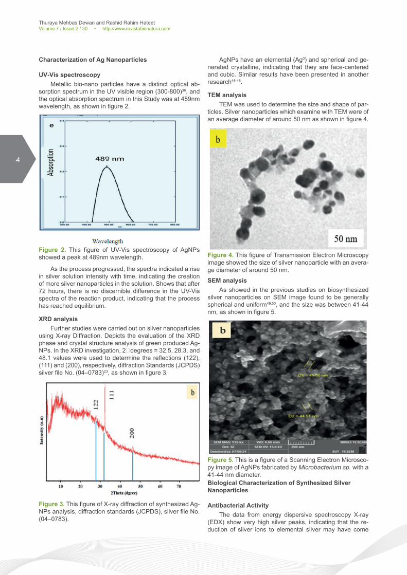
UV-Vis spectroscopyMetallic bio-nano particles have a distinct optical ab-
sorption spectrum in the UV visible region (300-800)36, and the optical absorption spectrum in this Study was at 489nm wavelength, as shown in figure 2.
AgNPs have an elemental (Ag0) and spherical and ge-nerated crystalline, indicating that they are face-centered and cubic. Similar results have been presented in another research46-48.
TEM analysisTEM was used to determine the size and shape of par-
ticles. Silver nanoparticles which examine with TEM were of an average diameter of around 50 nm as shown in figure 4.
Figure 2. This figure of UV-Vis spectroscopy of AgNPs showed a peak at 489nm wavelength.
As the process progressed, the spectra indicated a rise in silver solution intensity with time, indicating the creation of more silver nanoparticles in the solution. Shows that after 72 hours, there is no discernible difference in the UV-Vis spectra of the reaction product, indicating that the process has reached equilibrium.
XRD analysisFurther studies were carried out on silver nanoparticles
using X-ray Diffraction. Depicts the evaluation of the XRD phase and crystal structure analysis of green produced Ag-NPs. In the XRD investigation, 2𝜃degrees = 32.5, 28.3, and48.1 values were used to determine the reflections (122), (111) and (200), respectively, diffraction Standards (JCPDS) silver file No. (04–0783)23, as shown in figure 3.
Figure 3. This figure of X-ray diffraction of synthesized Ag-NPs analysis, diffraction standards (JCPDS), silver file No. (04–0783).
Figure 4. This figure of Transmission Electron Microscopy image showed the size of silver nanoparticle with an avera-ge diameter of around 50 nm.SEM analysis
As showed in the previous studies on biosynthesized silver nanoparticles on SEM image found to be generally spherical and uniform49,50, and the size was between 41-44 nm, as shown in figure 5.
Figure 5. This is a figure of a Scanning Electron Microsco-py image of AgNPs fabricated by Microbacterium sp. with a 41-44 nm diameter.Biological Characterization of Synthesized Silver Nanoparticles
Antibacterial Activity The data from energy dispersive spectroscopy X-ray
(EDX) show very high silver peaks, indicating that the re-duction of silver ions to elemental silver may have come
Thuraya Mehbas Dewan and Rashid Rahim HateetVolume 7 / Issue 2 / 30 • http://www.revistabionatura.com

5
from molecules connected to the AgNPs' surface. The sil-ver's dense peak was a clear indicator of the formation of silver nanoparticles from silver ions20. As shown in figure 6.
of MCF7 were able to form formazan product and remai-ned as alive cells (viability, after 72hr of incubation period. There was significant change in cancer cell that inhibition with synthesized silver nanoparticles at a different con-centration (10,30,60,80,100ug/ml) of AgNPs, by about (9.263%,65.252%,81.565%, 80.447%,81.732%), as shown in) figure 9.
The median inhibitory dose (IC50) value of 24.93ug/ml. This means that biosynthesis AgNPs have excellent anti-tumor activity on breast cancer cells. These results agreed with those described by (24).
DiscussionCompared to other bio-reductants, the manufacture of
metallic nanoparticles using microorganisms is more fruitful in terms of ease51. Phytochemicals are well known for con-verting Ag1+ to Ag0 and capping these nanoparticles, making them highly stable52. In this Study, Microbacterium was used after being isolated from soil to green synthesis of AgNPs, and This indicates that this isolated bacteria had the enzy-me that reduced Ag+ to Ag0 by Nitrate reductase, then the aggregation of silver atoms was formed AgNPs. Then was tested of the synthesized AgNPs were for bactericidal and cytotoxic activity. Spectroscopy in the ultraviolet and visible ranges was used to determine metallic nanoparticles' for-mation and exploit their optical properties. Plasmon bands were recorded at various points during the bioreduction pro-cess. The AgNPs were prepared principally by the emer-gence of a reddish-brown color after 72h. The reaction was finished and validated by the Plasmon absorption peak, which reached a constant value. The SPR (surface Plasmon resonance) band of the spherical AgNPs at 489 nm is visible
Figure 6. This figure of EDX analysis showed a strong peak of silver at 3kev.
Antibacterial testing revealed that the Nanoparticle is effective against bacterial pathogens21,22. Both Gram-nega-tive and Gram-positive bacteria alike (identified by using-VITEK-2)23 and compared with Gentamycin antibiotic, as shown in figure 7 and figure 8.
Microbacterium's silver nanoparticles displayed the most significant inhibition zones (38mm) against S.aureas bacteria; it was enormous compared with antibiotics (Gen-tamycin) in this project; the lowest inhibition zone was 7 mm on pseudomonas aeruginosa bacteria, as shown in table 3.
Antitumor activity of AgNPs biosynthesized laboratorySilver nanoparticles play important role as antitu-
mor24. At low concentrations demonstrated very great ac-tivity on MCF-7 cell line, AgNPs inhibition breast cancer cells by about 81.732 % at 100ug/ml, and only18.268 %
Figure 7. This figure of the Antibacterial activity of synthe-sized AgNPs against ten pathogenic bacteria.
Figure 8. This figure of the Antibacterial activity of antibiotic (Gentamycin) against the same pathogenic bacteria.
Detect the Antibacterial and Antitumor of synthesized Silver Nanoparticles Using Microbacterium sp

6
in the spectra, showing that the synthesis of AgNPs in the reaction mixture is consistent with the AgNPs synthesized before53. After one month under ambient circumstances, the stability of the nanoparticles was assessed throughout time (30 days), and no change in absorption peak value was de-tected, indicating that the nanoparticles are highly stable.
The particle size and shape of the produced AgNPs were studied using TEM and SEM. The synthesized AgNPs were monodispersed, spherical in shape, and ranged in size from 41 to 44 nanometers. The interaction of hydrogen
bonds and electrostatic interactions between the bioorganic capping molecules linked to the AgNPs resulted in silver na-noparticles. Even within the aggregates, the nanoparticles were not in direct contact, indicating that the nanoparticles had been stabilized by a capping agent and crystalline in nature45, and well dispersed, primarily spherical, which is agreed with other reports44,46.
Correlated to the (111), (200), and (220), and indica-ting that AgNPs have been prepared. Several Bragg reflec-tions with 20 values of 32.5O, 28.3O, and 48.1O are achieved
Table 3. This table of Inhibi-tion zone on ten pathogenic bacteria by synthesized Ag-NPs and Gentamicin.
Figure 9. This figure of viability percent on the mcf7 cell line in different concentrations of synthesized AgNPs (10, 30, 60, 80, 100) ug/ml.
Thuraya Mehbas Dewan and Rashid Rahim HateetVolume 7 / Issue 2 / 30 • http://www.revistabionatura.com

7
and related with the diverse set of (111), (200), and (220), which can be recorded as the band for face-centered cubic (JCPDS file no. 89-3722). As a result, XRD indicates that the samples are pure AgNPs that are highly crystalline. The silver particle size histograms revealed that the particles va-ried in size. Planes appear in the selected area Electron Di-ffraction Pattern (EDX), indicating that the produced AgNPs are crystalline. The dots are aligned with a face-centered cubic structure. The AgNPs are crystalline, according to the EDX pattern. The elemental detection of AgNPs was also done with the EDX. The presence of a prominent peak of silver at 3 KeV can be seen in the EDX pattern; the pro-duced AgNPs have outstanding antibacterial effectiveness. When clinical bacterial pathogens are exposed to AgNPs, the membrane permeability is altered, resulting in cellular leakage, restricting cell growth and replication. Some bac-terial macromolecules can be affected by AgNPs, resulting in disintegration and cell death54. Compared to chemically synthesized AgNPs, green produced AgNPs are more bio-compatible and have a stronger antibacterial impact55. The AgNPs first bind the cell membrane at numerous points be-fore quickly penetrating it, causing structural changes and, as a result, perforations that allow compounds from intrace-llular storage to flow out56. When AgNPs reach the interior, silver ions are released, resulting in the production of reacti-ve oxygen species (ROS), which can affect membrane pro-teins, causing the electron transport chain to be disrupted57.
The MTT assay findings show that AgNPs have high cytotoxicity against MCF7. The MTT assay was carried out in triplicate. AgNPs have higher toxicity at lower concen-trations as they were incubated for 24 hours. At doses of 10,30,60,80, and 100ug/ml of AgNPs at a different concen-tration, respectively, toxicity if there were (9.163%,65.252%,81.565%,80.447%,81.732%) found after 72 hours of incu-bation respectively this indicates that as the concentration of AgNPs rises so does its toxicity. The results showed that AgNPs strongly suppressed MCF-7 cell proliferation. For AgNPs-treated MCF7, IC50 values were calculated over a wide concentration range and incubation duration. Several studies have previously concentrated on using cell culture methods to perform AgNPs cytotoxicity tests58,59. The cyto-toxicity of any natural or synthetic substance on an establi-shed cell line must be determined before moving to in vivo experiments60. When tested against breast cancer cells, our biogenic AgNPs have high cytotoxicity.
ConclusionsThe soil was shown to be a rich supply of various
bacteria in the current study, and even a novel strain was discovered (M. paraoxydans strain shahooda). As a result of this research, it can be concluded that it has been em-ployed effectively for AgNPs extracellular synthesis using soil microbes. The presence of elemental silver and its crys-talline structure and size were confirmed by using UV-Vis. Spectroscopy, EDX, SEM, TEM, XRD. Both Gram-negative and Gram-positive pathogenic bacteria are susceptible to AgNPs. The anticancer effects of extracellular AgNPs on MCF7 (breast cancer cells) yielded the best results, with AgNPs significantly suppressing MCF7 cell multiplication. Finally, compared to commercially available antimicrobial drugs, the current Study provides an environmentally friend-ly and cost-effective approach for synthesizing powerful an-tibacterial silver nanoparticles (biologically) against patho-
genic bacteria and the capacity of silver nanoparticles as antitumors on breast cancer cells line.
Authors contributionsConceptualization, TMD and RRH; methodology, TMD;
software, TMD; validation, TMD and RRH; formal analysis, TMD; investigation, TMD; resources, TMD; data curation, TMD; writing—original draft preparation, TMD; writing re-view and editing, TMD; visualization, TMD; supervision, RRH; project administration, RRH. All authors have read and agreed to the published version of the manuscript.
FundingThis research received no external funding.
Institutional Review BoardAl- Sadr hospital in Misan had been informed about the
aims of the Study before collecting samples and declared their agreement to give samples (175; 1/6/2021). The Study follows the rules of scientific research at Misan University, Iraq.
Informed ConsentInformed consent was obtained from all subjects invol-
ved in the Study. The patient's consent was oral.
AcknowledgementsWe like to thank the head of the Biology Department,
Assist. Prof. Dr. Maytham Abdul Kadhim Dragh for his help, and I would like to thank M. Sc. Shaima R. Banoon, Biology department, college of Science, Misan University.
Conflicts of interestThe authors declare no conflict of interest.
Bibliographic references1. Al-Dhabi NA, Mohammed Ghilan AK, Arasu MV. Characteriza-
tion of silver nanomaterials derived from marine Streptomyces sp. al-dhabi-87 and its in vitro application against multidrug re-sistant and extended-spectrum beta-lactamase clinical patho-gens. Nanomaterials. 2018 May;8(5):279.
2. Patra JK, Das G, Fraceto LF, Campos EV, Rodriguez-Torres MD, Acosta-Torres LS, Diaz-Torres LA, Grillo R, Swamy MK, Sharma S, Habtemariam S. Nano based drug delivery sys-tems: recent developments and future prospects. Journal of nanobiotechnology. 2018 Dec;16(1):1-33.
3. Golinska P, Wypij M, Ingle AP, Gupta I, Dahm H, Rai M. Bio-genic synthesis of metal nanoparticles from actinomycetes: biomedical applications and cytotoxicity. Applied microbiology and biotechnology. 2014 Oct;98(19):8083-97.
4. Karthik L, Kumar G, Kirthi AV, Rahuman AA, Bhaskara Rao KV. Streptomyces sp. LK3 mediated synthesis of silver nanoparti-cles and its biomedical application. Bioprocess and biosyste-ms engineering. 2014 Feb;37(2):261-7
5. Subbaiya R, Selvam MM, Sundar K. Biological synthesis of silver nanorods from Nocardia mediterranei-5016 and its anti-tumor activity against non-small cell lung carcinoma cell line. Int. J. PharmTech Res. 2015;8(2):974-9.
6. Ahmad A, Senapati S, Khan MI, Kumar R, Ramani R, Srinivas V, Sastry M. Intracellular synthesis of gold nanoparticles by a novel alkalotolerant actinomycete, Rhodococcus species. Nanotechnology. 2003 Jun 6;14(7):824.
7. Dayma PB, Mangrola AV, Suriyaraj SP, Dudhagara P, Patel RK. Synthesis of bio-silver nanoparticles using desert isolat-ed Streptomyces intermedius and its antimicrobial activity. J. Pharm. Chem. Biol. Sci. 2019;7:94-101.
Detect the Antibacterial and Antitumor of synthesized Silver Nanoparticles Using Microbacterium sp

8
8. Singh D, Rathod V, Ninganagouda S, Hiremath J, Singh AK, Mathew J. Optimization and characterization of silver nanopar-ticle by endophytic fungi Penicillium sp. isolated from Curcuma longa (turmeric) and application studies against MDR E. coli and S. aureus. Bioinorganic chemistry and applications. 2014 Oct;2014.
9. Durán N, Marcato PD, De Souza GI, Alves OL, Esposito E. Antibacterial effect of silver nanoparticles produced by fungal process on textile fabrics and their effluent treatment. Journal of biomedical nanotechnology. 2007 Jun 1;3(2):203-8.
10. Sondi I, Salopek-Sondi B. Silver nanoparticles as antimicrobial agent: a case study on E. coli as a model for Gram-negative bacteria. Journal of colloid and interface science. 2004 Jul 1;275(1):177-82.
11. Martínez Espinosa JC, Carrera Cerritos R, Ramírez Morales MA, Sánchez Guerrero KP, Silva Contreras RA, Macías JH. Characterization of silver nanoparticles obtained by a green route and their evaluation in the bacterium of pseudomonas aeruginosa. Crystals. 2020 May;10(5):395.
12. Aymonier C, Schlotterbeck U, Antonietti L, Zacharias P, Thomann R, Tiller JC, Mecking S. Hybrids of silver nanopar-ticles with amphiphilic hyperbranched macromolecules ex-hibiting antimicrobial properties. Chemical Communications. 2002(24):3018-9.
13. Lee SH, Jun BH. Silver nanoparticles: synthesis and applica-tion for nanomedicine. International journal of molecular sci-ences. 2019 Jan;20(4):865.
14. Sapkota A, Thapa A, Budhathoki A, Sainju M, Shrestha P, Ary-al S. Isolation, characterization, and screening of antimicro-bial-producing actinomycetes from soil samples. International journal of microbiology. 2020 Mar 26;2020.
15. Loo YY, Rukayadi Y, Nor-Khaizura MA, Kuan CH, Chieng BW, Nishibuchi M, Radu S. In vitro antimicrobial activity of green synthesized silver nanoparticles against selected gram-nega-tive foodborne pathogens. Frontiers in microbiology. 2018 Jul 16;9:1555.
16. Abd-Elnaby HM, Abo-Elala GM, Abdel-Raouf UM, Hamed MM. Antibacterial and anticancer activity of extracellular syn-thesized silver nanoparticles from marine Streptomyces rochei MHM13. The Egyptian Journal of Aquatic Research. 2016 Sep 1;42(3):301-12.
17. El𝜃Sersy NA, Abdelwahab AE, Abouelkhiir SS, Abou𝜃Zeid DM, Sabry SA. Antibacterial and Anticancer activity of ε𝜃po-ly𝜃L𝜃lysine (ε𝜃PL) produced by a marine Bacillus subtilis sp. Journal of basic microbiology. 2012 Oct;52(5):513-22.
18. Aldujaili NH, Banoon SR. Antibacterial characterization of tita-nium nanoparticles nano synthesized by Streptococcus ther-mophilus. Periodico Tche Quimica (Online). 2020;17(34):311-20.
19. Hassan BA, Lawi ZKK, Banoon SR. Detecting the activity of silver nanoparticles, Pseudomonas fluorescens and Bacillus circulans on inhibition of Aspergillus niger growth isolated from moldy orange fruits. Periodico Tche Quimica. 2020;17(35):678-690.
20. Kasithevar M, Saravanan M, Prakash P, Kumar H, Ovais M, Barabadi H, Shinwari ZK. Green synthesis of silver nanoparti-cles using Alysicarpus monilifer leaf extract and its antibacte-rial activity against MRSA and CoNS isolates in HIV patients. Journal of Interdisciplinary Nanomedicine. 2017 Jun;2(2):131-41.
21. Devadass BJ, Paulraj MG, Ignacimuthu S, Theoder PA, Dhabi NA. Antimicrobial activity of soil actinomycetes isolated from Western Ghats in Tamil Nadu, India. J Bacteriol Mycol Open Access. 2016;3(2):224-32.
22. Bruna T, Maldonado-Bravo F, Jara P, Caro N. Silver nanopar-ticles and their antibacterial applications. International Journal of Molecular Sciences. 2021 Jan;22(13):7202.
23. Malarkodi C, Rajeshkumar S, Vanaja M, Paulkumar K, Gnana-jobitha G, Annadurai G. Eco-friendly synthesis and charac-terization of gold nanoparticles using Klebsiella pneumoniae. Journal of Nanostructure in Chemistry. 2013 Dec;3(1):1-7.
24. Gomes HI, Martins CS, Prior JA. Silver nanoparticles as carri-ers of anticancer drugs for efficient target treatment of cancer cells. Nanomaterials. 2021 Apr;11(4):964.
25. Banoon S, Ali Z, Salih T. Antibiotic resistance profile of local thermophilic Bacillus licheniformis isolated from Maysan prov-ince soil. Comunicata Scientiae. 2020 Jul 13;11:e3291.
26. Banoon SR, Ghasemian A. The characters of graphene ox-ide nanoparticles and doxorubicin against HCT-116 colorectal cancer cells in vitro. Journal of Gastrointestinal Cancer. 2021 Mar 19:1-5.
27. Al-Abboodi A., Alsaady HAM, Banoon SR, Al-Saady M. Conju-gation strategies on functionalized iron oxide nanoparticles as a malaria vaccine delivery system. Revista Bionatura. 2021; 6 (3): 2009-2015.
28. Bizuye A, Moges F, Andualem B. Isolation and screening of antibiotic producing actinomycetes from soils in Gondar town, North West Ethiopia. Asian Pacific journal of tropical disease. 2013 Oct 1;3(5):375-81.
29. Gupta A, Silver S. Molecular genetics: silver as a biocide: will resistance become a problem?. Nature biotechnology. 1998 Oct;16(10):888.
30. Feng QL, Wu J, Chen GQ, Cui FZ, Kim TN, Kim JO. A mech-anistic study of the antibacterial effect of silver ions on Esche-richia coli and Staphylococcus aureus. Journal of biomedical materials research. 2000 Dec 15;52(4):662-8.
31. Ahmad SA, Das SS, Khatoon A, Ansari MT, Afzal M, Hasnain MS, Nayak AK. Bactericidal activity of silver nanoparticles: A mechanistic review. Materials Science for Energy Technolo-gies. 2020 Jan 1;3:756-69.
32. Olano C, Méndez C, Salas JA. Antitumor compounds from ac-tinomycetes: from gene clusters to new derivatives by combi-natorial biosynthesis. Natural product reports. 2009;26(5):628-60.
33. Zhang E, Zhao X, Hu J, Wang R, Fu S, Qin G. Antibacterial metals and alloys for potential biomedical implants. Bioactive materials. 2021 Aug 1;6(8):2569-612.
34. Kim JS, Kuk E, Yu KN, Kim JH, Park SJ, Lee HJ, Kim SH, Park YK, Park YH, Hwang CY, Kim YK. Antimicrobial effects of silver nanoparticles. Nanomedicine: Nanotechnology, biology and medicine. 2007 Mar 1;3(1):95-101.
35. Miyoshi T, Iwatsuki T, Naganuma T. Phylogenetic charac-terization of 16S rRNA gene clones from deep-groundwater microorganisms that pass through 0.2-micrometer-pore-size filters. Applied and environmental microbiology. 2005 Feb;71(2):1084-8.
36. Chelius MK, Triplett EW. The Diversity of Archaea and Bac-teria in Association with the Roots of Zea mays L. Microbial ecology. 2001 Apr 1:252-63.
37. Alsamhary KI. Eco-friendly synthesis of silver nanoparticles by Bacillus subtilis and their antibacterial activity. Saudi Journal of Biological Sciences. 2020 Aug 1;27(8):2185-91.
38. Fissan H, Ristig S, Kaminski H, Asbach C, Epple M. Compari-son of different characterization methods for nanoparticle dis-persions before and after aerosolization. Analytical Methods. 2014;6(18):7324-34.
39. Zhang XF, Liu ZG, Shen W, Gurunathan S. Silver nanoparti-cles: synthesis, characterization, properties, applications, and therapeutic approaches. International journal of molecular sci-ences. 2016 Sep;17(9):1534.
40. Gould JC, Bowie JH. The determination of bacterial sensitivity to antibiotics. Edinburgh medical journal. 1952 Apr;59(4):178.
41. Rageh MM, El-Gebaly RH, Afifi MM. Antitumor activity of silver nanoparticles in Ehrlich carcinoma-bearing mice. Naunyn-Schmiedeberg's archives of pharmacology. 2018 Dec;391(12):1421-30.
42. Al-Shammari AM, Al-Esmaeel WN, Al Ali AA, Hassan AA, Ahmed AA. Enhancement of Oncolytic Activity of Newcastle Disease virus Through Combination with Retinoic Acid Against Digestive System Malignancies. InMOLECULAR THERAPY 2019 Apr 22 (Vol. 27, No. 4, pp. 126-127). 50 HAMPSHIRE ST, FLOOR 5, CAMBRIDGE, MA 02139 USA: CELL PRESS.
Thuraya Mehbas Dewan and Rashid Rahim HateetVolume 7 / Issue 2 / 30 • http://www.revistabionatura.com

9
43. Nguyen VT, Vu VT, Nguyen TA, Tran VK, Nguyen-Tri P. Antibac-terial activity of TiO2-and ZnO-decorated with silver nanoparti-cles. Journal of Composites Science. 2019 Jun;3(2):61.
44. Osorio-Echavarría J, Osorio-Echavarría J, Ossa-Orozco CP, Gómez-Vanegas NA. Synthesis of silver nanoparticles using white-rot fungus Anamorphous Bjerkandera sp. R1: Influence of silver nitrate concentration and fungus growth time. Scientif-ic Reports. 2021 Feb 15;11(1):1-4.
45. Yousefzadi Nobakht A, Shin S. Anisotropic control of thermal transport in graphene/Si heterostructures. Journal of Applied Physics. 2016 Dec 14;120(22):225111.
46. Fouad H, Hongjie L, Yanmei D, Baoting Y, El-Shakh A, Ab-bas G, Jianchu M. Synthesis and characterization of silver nanoparticles using Bacillus amyloliquefaciens and Bacillus subtilis to control filarial vector Culex pipiens pallens and its antimicrobial activity. Artificial Cells, Nanomedicine, and Bio-technology. 2017 Oct 3;45(7):1369-78.
47. Manikandan R, Beulaja M, Thiagarajan R, Palanisamy S, Goutham G, Koodalingam A, Prabhu NM, Kannapiran E, Basu MJ, Arulvasu C, Arumugam M. Biosynthesis of silver nanopar-ticles using aqueous extract of Phyllanthus acidus L. fruits and characterization of its anti-inflammatory effect against H2O2 exposed rat peritoneal macrophages. Process Biochemistry. 2017 Apr 1;55:172-81.
48. Mahmoud WM, Abdelmoneim TS, Elazzazy AM. The impact of silver nanoparticles produced by Bacillus pumilus as anti-microbial and nematicide. Frontiers in microbiology. 2016 Nov 10;7:1746.
49. Singh T, Jyoti K, Patnaik A, Singh A, Chauhan R, Chandel SS. Biosynthesis, characterization and antibacterial activity of sil-ver nanoparticles using an endophytic fungal supernatant of Raphanus sativus. Journal of Genetic Engineering and Bio-technology. 2017 Jun 1;15(1):31-9.
50. Lallawmawma H, Sathishkumar G, Sarathbabu S, Ghatak S, Sivaramakrishnan S, Gurusubramanian G, Kumar NS. Syn-thesis of silver and gold nanoparticles using Jasminum ner-vosum leaf extract and its larvicidal activity against filarial and arboviral vector Culex quinquefasciatus Say (Diptera: Culici-dae). Environmental Science and Pollution Research. 2015 Nov;22(22):17753-68.
51. Mukherjee S, Chowdhury D, Kotcherlakota R, Patra S. Poten-tial theranostics application of bio-synthesized silver nanopar-ticles (4-in-1 system). Theranostics. 2014;4(3):316.
52. Arokiyaraj S, Vincent S, Saravanan M, Lee Y, Oh YK, Kim KH. Green synthesis of silver nanoparticles using Rheum palma-tum root extract and their antibacterial activity against Staphy-lococcus aureus and Pseudomonas aeruginosa. Artificial cells, nanomedicine, and biotechnology. 2017 Feb 17;45(2):372-9.
53. Wani IA, Khatoon S, Ganguly A, Ahmed J, Ahmad T. Structural characterization and antimicrobial properties of silver nanopar-ticles prepared by inverse microemulsion method. Colloids and Surfaces B: Biointerfaces. 2013 Jan 1;101:243-50.
54. Singh K, Panghal M, Kadyan S, Chaudhary U, Yadav JP. Green silver nanoparticles of Phyllanthus amarus: as an an-tibacterial agent against multi drug resistant clinical isolates of Pseudomonas aeruginosa. Journal of nanobiotechnology. 2014 Oct;12(1):1-9.
55. Mukherjee S, Chowdhury D, Kotcherlakota R, Patra S. Poten-tial theranostics application of bio-synthesized silver nanopar-ticles (4-in-1 system). Theranostics. 2014;4(3):316.
56. Dizaj SM, Lotfipour F, Barzegar-Jalali M, Zarrintan MH, Adib-kia K. Antimicrobial activity of the metals and metal oxide nanoparticles. Materials Science and Engineering: C. 2014 Nov 1;44:278-84.
57. Ahmed S, Ahmad M, Swami BL, Ikram S. A review on plants extract mediated synthesis of silver nanoparticles for antimi-crobial applications: a green expertise. Journal of advanced research. 2016 Jan 1;7(1):17-28.
58. Gaiser BK, Hirn S, Kermanizadeh A, Kanase N, Fytianos K, Wenk A, Haberl N, Brunelli A, Kreyling WG, Stone V. Effects of silver nanoparticles on the liver and hepatocytes in vitro. Toxicological sciences. 2013 Feb 1;131(2):537-47.
59. Albers CE, Hofstetter W, Siebenrock KA, Landmann R, Klenke FM. In vitro cytotoxicity of silver nanoparticles on osteoblasts and osteoclasts at antibacterial concentrations. Nanotoxicolo-gy. 2013 Feb 1;7(1):30-6.
60. Gopinath P, Gogoi SK, Chattopadhyay A, Ghosh SS. Implica-tions of silver nanoparticle induced cell apoptosis for in vitro gene therapy. Nanotechnology. 2008 Jan 29;19(7):075104.
Detect the Antibacterial and Antitumor of synthesized Silver Nanoparticles Using Microbacterium sp