A scaffolded approach to unearth potential antibacterial ...
Upload
khangminh22Category
view
0download
0
Evaluation of anthelmintic,
antiamoebic and antibacterial activity
in traditional South African medicinal
plants
by
Lyndy Joy McGaw
Submitted in fulfilment of the requirements for the degree of
Doctor of Philosophy
in
School of Botany and Zoology
University of Natal, Pietermaritzburg
June 2001

DECLARATION
The experimental work described in this thesis was conducted in the Research
Centre for Plant Growth and Development, School of Botany and Zoology,
University of Natal Pietermaritzburg, from May 1998 to May 2001, under the
supervision of Professor J. van Staden and Doctor A. K. Jager.
These studies are the result of my own investigations, except where the work of
others is acknowledged, and have not been submitted in any other form to another
University.
Lyndy Joy McGaw
I declare the above statement to be true.
Profes or J. van Staden
(SUPERVISOR)
b~'Docto ~tager
(CO-SUPERVISOR)
June 2001

11
ACKNOWLEDGEMENTS
I would like to express special thanks to Professor J. van Staden for supervising this
research, and for his invaluable advice and encouragement during the time we have
worked together. The able co-supervision extended by Or A. K. Jager, along with her
essential support and indispensable advice, is greatly appreciated.
As a member of my research committee, Or W. Burnett offered sound advice and I
am sincerely grateful for her interest and encouragement.
The technical, academic and research staff are gratefully acknowledged for their
time and expertise which was always generously given. I would like to thank Or E.
Elgorashi and Professor D. Mulholland (University of Natal, Durban) for their
assistance in identifying the compounds described in this thesis. I am also grateful
to Or S. Beck (ICFR, Pietermaritzburg) for her help with the statistical analysis. I
would like to thank Ms Driekie Fourie (ARC-Grain Crops Institute, Potchefstroom) for
the generous donation of nematode cultures, and Mr S. Suparsad (Medical
Research Council, Durban) for providing valued practical knowledge as well as
amoebal cultures and culture medium.
The National Research Foundation is gratefully acknowledged for their generous
financial support during my studies.
I am extremely grateful to all my friends, especially Cathy and Sascha, and my
parents and sister, for their endless encouragement and unstinting belief in me
through many difficult times. Special thanks go to Luke and Leila for their love and
dedicated support.

111
ABSTRACT
Traditional medicine in southern Africa draws upon a vast selection of plants to treat
gastrointestinal disorders such as diarrhoea and intestinal parasites. The evaluation
of these plants for biological activity is necessary, both to substantiate the use of
these plants by healers, and also a~~J?9~~L~!e leC!Q fQ~~d~or !ter:b9L
~.
After a survey of the existing ethnobotanicalliterature, plants used to treat stomach
ailments such as diarrhoea, dysentery or intestinal worm infestations were selected
and submitted to bioassays according to their traditional uses. Extracts of the
chosen plants were made using the solvents hexane, ethanol and water, to ensure
the extraction of compounds with a wide range of polarity. In total, 138 extracts were
tested for antibacterial activity, 72 for anthelmintic activity, and 42 for antiamoebic
activity. Antibacterial activity was evaluated using the disc-diffusion assay, and
Minimal Inhibitory Concentration (MIC) values were determined using a microdilution
assay. The extracts were tested against the Gram-positive bacteria Bacillus subtilis
and Staphylococcus aureus, and the Gram-negative bacteria Escherichia coli and
Klebsiella pneumoniae. Ethanolic extracts showed the greatest activity and Gram
positive bacteria were the most susceptible microorganisms.
The free-living nematode Caenorhabditis elegans, which is morphologically similar
to parasitic nematodes, was used in two different assays to evaluate anthelmintic
activity. A microdilution technique was employed to investigate antiamoebic activity
.against the enteropathogenic Entamoeba histolytica, the causal organism of
amoebic dysentery. These assays were suitable for the screening of a large number
of extracts at one time. Several plants exhibited significant activity against these test
organisms.
Many species of plants belonging to the family Combretaceae are used in southern
African traditional medicine against a variety of ailments, including abdominal
complaints, bilharzia and diarrhoea. Extracts of powdered leaf material of 24 species

IV
belonging to the Combretaceae were prepared using the solvents ethyl acetate,
acetone, methanol and water. These extracts were screened for anthelmintic
activity. Significant activity was exhibited by C. apiculatum, C. hereroense and C.
mossambicense. The most anthelmintic activity was shown by acetone extracts,
followed by ethyl acetate, water and then methanol extracts.
The aromatic rhizomes of Acarus calamus L. are used extensively in traditional
medicine worldwide. They reportedly relieve stomach cramps and dysentery, and
are used as anthelmintics. Rhizome extracts of A. calamus growing in KwaZulu
Natal, South Africa, exhibited anthelmintic and antibacterial activity in the initial
general screening. Using bioassay-guided fractionation, the phenylpropanoid ~
asarone was isolated from the rhizome. This compound possessed both
anthelmintic and antibacterial activity. It has previously been isolated from A.
calamus, and a related species, A. gramineus. Different varieties of A. calamus
exhibit different levels of ~-asarone,with the diploid variety containing none of the
compound. Mammalian toxicity and carcinogenicity of asarones has been
demonstrated by other researchers, supporting the discouragement of the medicinal
use of Acarus calamus by traditional healers in South Africa.
Schotia brachypetala was another plant to show good antibacterial activity in the
initial screening. The roots and bark of S. brachypetala are used in South African
traditional medicine as a remedy for dysentery and diarrhoea. The lack of
pharmacological and chemical data on this plant prompted a further investigation
into its antibacterial activity. The differences in activity of ethanol and water extracts
with respect to plant part, season and geographical position were analysed. No
extreme fluctuations in activity were noted. Two other Schotia species, S. afra and
S. capitata, were included in the study, and both displayed good antibacterial
activity. The storage of the plant, either as dried, ground plant material at room
temperature, or as an extract residue at -15°C, had little effect on the antibacterial
activity. Preparing the extracts from fresh or dry material also did not notably affect
the activity. In general, the ethanolic extracts were more active than the aqueous
extracts. The chemical profiles on TLC chromatograms were compared and found to
be very similar in the case of ethanol extracts prepared in different months of the
year, and from different trees. The extracts of the three species, and of the leaves
stored under various conditions, as well as extracts prepared from fresh or dry
material, also showed similar TLC fingerprints. However, various plant parts of S.
brachypetala showed distinctly different chemical compositions.
The leaves of S. brachypetala showed slightly higher antibacterial activity than the
roots. Fractionation of the ethanol extract of the dried leaves using liquid-liquid
partitioning and chromatographic techniques yielded 9,12,15-octadecatrienoic
(linolenic) acid and methyl-5, 11,14,17-eicosatetraenoate. These fatty acids
displayed antibacterial activity against the Gram-positive bacteria Bacillus subtilis
and Staphylococcus aureus, and activity to a lesser extent against the Gram
negative Escherichia coli and Klebsiella pneumoniae. Linolenic acid is known to
have antibacterial activity.
The screening of plants for biological activity yielded valuable preliminary
information about the plants used by traditional healers to treat gastrointestinal
illnesses. The isolation of biologically active compounds from two highly active
plants was achieved.
v

VI
PAPERS PREPARED FROM THIS THESIS
McGaw, L. J., Jager, A. K. and van Staden, J. 2000. Antibacterial, anthelmintic and
antiamoebic activity in South African medicinal plants. Journal of
Ethnopharmacology 72: 247-263
McGaw, L. J., Rabe, T., Sparg, S. G., Jager, A. K., Eloff, J. N. and van Staden, J.
2000. An investigation on the biological activity of Combretum species. Journal of
Ethnopharmacology 75: 45-50
McGaw, L. J., Jager, A. K. and van Staden, J. 2001. Isolation of ~-asarone, an
antibacterial and anthelmintic compound, from Acorus calamus in South Africa.
South African Journal of Botany (In Press)
Taylor, J. L. S., Rabe, T., McGaw, L. J., Jager, A. K. and van Staden, J. 2001.
Towards the scientific validation of traditional medicinal plants. Plant Growth
Regulation 34: 23-37
McGaw, L. J., Jager, A. K. and van Staden, J. 2001. Variation in antibacterial activity
of Scholia species. South African Journal of Botany (Submitted)
McGaw, L. J., Jager, A. K. and van Staden, J. 2001. Isolation of antibacterial fatty
acids from Scholia brachypetala. (In preparation)

vu
CONFERENCE CONTRIBUTIONS FROM THIS THESIS
1999
25th Annual Congress of SAAB (South African Association of Botanists),
Umtata (South Africa):
Paper: McGaw, L. J., Jager, A. K. and van Staden, J. Screening of traditional South
African medicinal plants for anthelmintic, antiamoebic and antibacterial
activity.
Indigenous Plant Use Forum (IPUF), Richards Bay (South Africa):
Poster: McGaw, L. J., Jager, A. K. and van Staden, J. Anthelmintic and antibacterial
activity in South African muthi plants.
5th Joint Meeting of the American Society of Pharmacognosy (ASP),
Association Francaise pour l'Enseignement et la Recherche en
Pharmacognosie (AFERP), Gesellschaft fUr Arzneipflanzenforschung (GA) and
the Phytochemical Society of Europe (PSE): 2 000 Years of Natural Products
Research - Past, Present and Future, Amsterdam (The Netherlands):
Poster: McGaw, L. J., Jager, A. K. and van Staden, J. Biological activity of South
African medicinal plants against nematodes, amoebae and bacteria.
2000
26th Annual Congress of SAAB, Potchefstroom (South Africa):
Paper: McGaw, L. J., Jager, A. K. and van Staden, J. Antibacterial, anthelmintic and
antiamoebic effects of South African medicinal plants.
5th Annual Symposium of CIPUR (Centre for Indigenous Plant Use Research),
Pietermaritzburg (South Africa):
Paper: McGaw, L. J., Jager, A. K. and van Staden, J. Antibacterial and anthelmintic

V111
compounds from Acorus calamus in South Africa.
2001
27th Annual Congress of 8MB, Johannesburg (South Africa):
Paper: McGaw, L. J., Jager, A. K. and van Staden, J. Isolation of bioactive
compounds from Acarus calamus.
World Conference on Medicinal and Aromatic Plants: possibilities and
limitations of medicinal and aromatic plant production towards the 21 st
century, Budapest (Hungary):
Paper: McGaw, L. J., Jager, A. K. and van Staden, J. Antibacterial fatty acids from
Schotia brachypetala.

IX
TABLE OF CONTENTS
Declaration ; i
Acknowledgements .ii
Abstract iii
Papers prepared from this thesis vi
Conference contributions {rom this thesis vii
Table of contents .ix
List of Figures and Plates xvi
List of Tables xix
List of Abbreviations xxi
CHAPTER 1 LITERATURE REVIEW,"~,) .
y\ \,.,\ - 1.1 Introduction 1
),-1.2 Plants as source~ of ~seful drugs 1
1.3 Selection of plants for ,biological activity screening 6
*"1.4 Plants as sources of antimicrobial drugs 8
X- 1f1'1.5 Traditional medicine in South Africa 10
1.6 Gastrointestinal disorders 13
1.7 The effect of intestinal parasites in humans 15
1.8 Aims and objectives 16
CHAPTER 2 PLANT COLLECTION AND EXTRACTION
2.1 Introduction 17
2.2 Materials and Methods 18
2.2.1 Plant collection ; 18
2.2.2 Plant extract preparation 19
2.3 Results 20
2.4 Discussion 30
2.5 Conclusion 30

x
CHAPTER 3 ANTHELMINTIC SCREENING
3.1 Introduction 31
3.1.1 Helminthiasis 31
3.1.2 Control of helminth infection 33
3.1.3 Anthelmintic assays 34
3.2 Materials and Methods 36
3.2.1 Culture of Caenorhabditis elegans 36
3.2.2 Mortality assay 37
3.2.3 Reproductive ability assay 37
3.3 Results 38
3.3.1 Standardization of results 38
3.3.2 Anthelmintic activity of plant extracts 38
3.4 Discussion 44
3.4.1 Comparison of anthelmintic assays .44
3.4.2 Anthelmintic activity of plant extracts .44
3.5 Conclusion 45
CHAPTER 4 ANTIAMOEBIC SCREENING
4.1 Introduction 47
4.1.1 Amoebiasis 47
4.1.2 Control of amoebiasis .48
4.1.3 Antiamoebic assays 50
4.2 Materials and Methods 51
4.2.1 Antiamoebic assay ; 51
4.3 Results 52
4.3.1 Standardization of assay 52
4.3.2 Antiamoebic activity of plant extracts 53
4.4 Discussion 55
4.4.1 Antiamoebic activity of plant extracts 55
4.5 Conclusion 55

Xl
CHAPTER 5 ANTIBACTERIAL SCREENING
5.1 Introduction ··· ··········· ················ ······· .56
5.1.1 Infectious diarrhoea and dysentery 56
5.1.2 Control of enteropathogens causing infectious diarrhoea and
dysentery 57
5.1.3 Antibacterial assays 58
5.1.3.1 Diffusion methods 59
5.1.3.2 Dilution methods 60
5.1.3.3 Bioautographic methods 60
5.2 Materials and Methods 61
5.2.1 Quantification of bacteria 61
5.2.2 Disc-diffusion assay 62
5.2.3 Microdilution assay 62
5.3 Results 64
5.3.1 Quantification of bacteria 64
5.3.2 Antibacterial activity of plant extracts 64
5.4 Discussion 68
5.4.1 Antibacterial activity of plant extracts 68
5.5 Conclusion 70
CHAPTER 6 ANTHELMINTIC ACTIVITY IN THE FAMILY
COMBRETACEAE
6.1 Introduction 71
6.1.1 The family Combretaceae 71
6.1.2 Traditional medicinal usage 71
6.1.3 Biological activity and chemical constituents 72
6.2 MaterialS and Methods 73
6.2.1 Plant extract preparation 73
6.2.2 Screening of plant extracts for anthelmintic activity 73
6.3 Results 74
6.3.1 Anthelmintic activity of Combretaceae extracts 74
6.4 Discussion 78

XlI
6.4.1 Anthelmintic activity of Combretaceae extracts 78
6.5 Conclusion ····· ···············78
CHAPTER 7 ISOLATION AND IDENTIFICATION OF l3ASARONE FROM ACORUS CALAMUS
7.1 Introduction 79
7.1.1 Acarus calamus 79
7.1.1.1 Description and traditional medicinal uses 79
7.1.1.2 Biological activity .' 80
7.1.1.3 Chemical constituents 81
7.2 Materials and Methods 82
7.2.1 Plant extraction 82
7.2.2 Antibacterial assays 83
7.2.3 Anthelmintic assay 83
7.2.4 Thin Layer Chromatography (TLC) 83
7.2.5 Bioassay-guided fractionation for isolation of active compound 84
7.2.5.1 Vacuum Liquid Chromatography 84
7.2.5.2 Preparative Thin Layer Chromatography 84
7.2.3 Identification of purified active compound 85
7.3 Results 85
7.3.1 Plant extraction 85
7.3.2 Antibacterial and anthelmintic activity in different plant parts 85
7.3.3 Bioassay-guided fractionation for isolation of active compound 86
7.3.4 Identification of purified active compound 87
7.4 Discussion 91
7.4.1 Isolation of active compound 91
7.4.2 Identification of isolated compound 92
7.5 Conclusion 92
CHAPTER 8 VARIATION IN ANTIBACTERIAL ACTIVITY OF
SCHOTIA BRACHYPETALA
8.1 Introduction 93

X111
8.1.1 Schotia brachypetala 93
8.1.2 Variation in biological activity 94
8.1.3 Thin Layer Chromatography 95
8.2 Materials and Methods 95
8.2.1 Collection of plant material 95
8.2.2 Plant extract preparation 96
8.2.3 Antibacterial activity screening 96
8.2.4 TLC fingerprinting 97
8.3 Results 97
8.3.1 Plant part variation 97
8.3.2 Locational variation 98
8.3.3 Seasonal variation 98
8.3.4 Species variation 99
8.3.5 Variation after extraction of fresh or dry material 99
8.3.6 Effect of storage of plant material and extracts 100
8.4 Discussion 111
8.4.1 Plant part variation 111
8.4.2 Locational variation 112
8.4.3 Seasonal variation 113
8.4.4 Species variation 113
8.4.5 Variation after extraction of fresh or dry material. 113
8.4.6 Effect of storage of plant material and extracts 114
8.4.7 TLC fingerprinting 115
8.5 Conclusion 116
CHAPTER 9 ISOLATION AND IDENTIFICATION OF
ANTIBACTERIAL FATTY ACIDS FROM SCHOTlA
BRACHYPETALA
9.1 Introduction 117
9.1.1 Scholia brachypetala: biological activity and chemical constituents 117
9.2 Materials and Methods 118
9.2.1 Bulk extraction 118

XIV
9.2.2 Bioassay-guided fractionation for isolation of active compounds 118
9.2.2.1 Liquid-liquid partitioning 119
9.2.2.2 Vacuum Liquid Chromatography 119
9.2.2.3 Chlorophyll extraction 120
9.2.2.4 Gravity-assisted Column Chromatography 120
9.2.2.5 Preparative Thin Layer Chromatography 120
9.2.3 Identification of purified active compound 121
9.3 Results 121
9.3.1 Bulk extraction 121
9.3.2 Bioassay-guided fractionation for isolation of active compounds 121
9.3.2.1 Liquid-liquid partitioning 122
9.3.2.2 Vacuum Liquid Chromatography 122
9.3.2.3 Chlorophyll extraction 122
9.3.2.4 Gravity-assisted Column Chromatography 122
9.3.2.5 Preparative Thin Layer Chromatography 123
9.3.3 Identification of purified active compound 123
9.4 Discussion 125
9.4.1 Isolation and identification of active compounds 125
9.4.2 Fatty acids as constituents of plants 125
9.4.3 Compound A: 9,12, 15-octadecatrienoic acid 126
9.4.4 Compound B: methyl-5,11,14,17-eicosatetraenoate 128
9.4.5 Fatty acids as antimicrobial agents 128
9.4.6 Structure-function relationships of fatty acids 130
9.4.7 Mechanism of antimicrobial action offatty acids 132
9.5 Conclusion 139
CHAPTER 10 GENERAL CONCLUSIONS
10.1 Introduction 141
10.2 Screening of plants for biological activity 142
10.3 Variation in activity and chemical composition of plant extracts 143
10.4 Isolation and identification of active compounds in plants 144
10.5 Conclusion 144
xv
REFERENCES 146
APPENDIX 1 174

XVI
LIST OF FIGURES AND PLATES
CHAPTER 3
Figure 3.1. Inhibition of nematodes by levamisole (2 h mortality assay) 39
Figure 3.2. Inhibition of nematodes by levamisole (7 day reproductive ability
assay) 39
CHAPTER 4
Figure 4.1. Amoebicidal activity of metronidazole 52
CHAPTER 7
Figure 7.1. Structure of ~-asarone 87
Plate 7.1. Acarus calamus (A) uprooted from a shallow pond in the Botanic
Gardens, UNP and (B) rhizome. The length of the plant from rhizome to leaf tip was
72 cm, and the length of the rhizome portion displayed was 17 cm. TLC fingerprints
of ethanol extracts of A. calamus leaf, rhizome and root viewed (C) under visible
light, (0) under UV254 and (E) under UV366. The solvent system used was
hexane:ethyl acetate (2: 1). Marked with an arrow is the compound isolated (~-
asarone) 89
Plate 7.2. TLC of Vacuum Liquid Chromatography (VLC) fractions in the bioassay
guided fractionation of A. calamus ethanolic rhizome extract. Viewed (A) under
UV254, (B) under UV366 and (C) after staining with anisaldehyde. The bioautography
plate (0) shows white areas of no bacterial growth, indicating the presence of
antibacterial compounds. The active compound of interest (~-asarone) is marked
with an arrow 90
CHAPTER 8
Plate 8.1. A flowering Schotia brachypetala tree in Umfolozi Nature Reserve,

XVll
KwaZulu-Natal (A), a close-up view of the flowers, leaves and seed pods with seeds
(8), and photographs of a tree from which bark had recently been harvested (C, D
and E) in Pietermaritzburg 101
Plate 8.2. TLC separation of the ethanol extracts (A) of different plant parts of S.
brachypetala, viewed (1) under visible light, (2) after staining with anisaldehyde, (3)
under UV254nm and (4) under UV366nm' The solvent system used was toluene:ethyl
acetate (4: 1).
[If =leaf, st =stem, bk =bark, rt =root, rb =root bark, fI =flower, sp =seeds plus pods]
TLC separation of the ethanol extracts (8) of S. brachypetala (Sb), S. afra (Sa) and
S. capitata (Sc) viewed (1) under visible light, (2) after staining with anisaldehyde,
(3) under UV254 nm and (4) under UV366 nm 102
Plate 8.3. TLC separation of the ethanol extracts of leaves of genetically different
trees of S. brachypetala, viewed (1) under visible light, (2) after staining with
anisaldehyde, (3) under UV254nm and (4) under UV366nm. The solvent system used
was toluene:ethyl acetate (4:1).
[Source of plant material: a - 9 = different trees growing in the Botanic Gardens, UNP; h =
Silverglen Nature Reserve; i =Umfolozi Nature Reserve; j =Scottsville, Pietermaritzburg; k
=Hilton, Pietermaritzburg] 103
Plate 8.4. TLC separation of the ethanol extracts of S. brachypetala leaf material
collected during different months, viewed (1) under visible light, (2) after staining
with anisaldehyde, (3) under UV254 nm and (4) under UV366 nm' The solvent system
used was toluene:ethyl acetate (4:1).
[Collection month: a =January, b =February, c =March, d =April, e =May, f =June, 9 =July, h =August, i =September, j =October, k =November, I =December]. 104
Plate 8.5. TLC separation (A) of the ethanol extracts of S. brachypetala leaf extracts
prepared from fresh (F) and dry (D) material, viewed (1) under visible light, (2) after
staining with anisaldehyde, (3) under UV254 nm and (4) under UV366 nm'
TLC separation of (8) the ethanol extracts of S. brachypetala leaf material after

xviii
being stored under various conditions, viewed (1) under visible light, (2) after
staining with anisaldehyde, (3) under UV254nm and (4) under UV366nm' The solvent
system used was toluene:ethyl acetate (4:1).
[a =extract screened immediately after collection, drying and extraction of material; b =extract stored in freezer at -15°C for 18 months; c = dried, intact plant material stored in
dark cupboard at room temperature before being ground, extracted and screened]. 105
CHAPTER 9
Plate 9.1. TLC separation (A) of the VLC fractions of the hexane fraction from the
liquid-liquid partitioning step, viewed (1) after staining with anisaldehyde, (3) under
UV254nm and (4) under UV366nm' The bioautography plate (2) shows white areas
where bacterial growth was inhibited. The arrows indicate the two fatty acids,
compounds a (9,12, 15-octadecatrienoic acid) and b (methyl-5,11,14,17
eicosatetraenoate) isolated from S. brachypetala. The solvent system used was
hexane:ethyl acetate (2: 1).
[H = hexane extract from the liquid-liquid partitioning step before being submitted to VLC]
TLC separation (8) of the different plant parts of S. brachypetala viewed (1) after
staining withanisaldehyde. The bioautography plate (2) shows the position of the
two subsequently isolated fatty acids, indicated by the arrows. It is clear that these
compounds are present in the leaves, bark and roots; the roots possess additional
antibacterial compounds, as do the flowers. The solvent system used was
hexane:ethyl acetate (2: 1).
[If = leaf, st = stem, bk = bark, rt = root, rb = root bark, fl = flower, sp = seeds plus
pods] 124

XIX
LIST OF TABLES
CHAPTER 2
Table 2.1. South African medicinal plants investigated for anthelmintic, antiamoebic
and antibacterial activity 20
CHAPTER 3
Table 3.1. Inhibition of nematodes by plant extracts (2 hand 7 day anthelmintic
assays) 40
CHAPTER 4
Table 4.1. Antiamoebic activity in South African medicinal plants 54
CHAPTER 5
Table 5.1. Correlation of bacterial numbers and optical density readings of overnight
cultures 64
Table 5.2. Determination of the antibacterial activity of South African medicinal
plants with the disc-diffusion and microdilution assays (MIC recorded in mg ml-1) ...65
CHAPTER 6
Table 6.1. Results of the anthelmintic screening of Combretaceae leaf extracts.....75
CHAPTER 7
Table 7.1. Comparison of MIC values obtained using the microplate method
(ELOFF, 1998) for Acarus calamus ethanol extracts, isolated ~-asarone and the
antibiotic neomycin against various bacteria 88
CHAPTER 8
Table 8.1. Antibacterial activity of different plant parts of Schotia brachypetala
(voucher number McGaw58NU). Treatments in each column denoted by the same

xx
letters are not significantly different at the 5 % level, with a standard error (SE) for
the treatment means =1.618 · 106
Table 8.2. Antibacterial activity of leaves from different trees (Schotia brachypetaJa).
Treatments in each column denoted by the same letters are not significantly
different (p < 0.05), with S.E. (treatment means) = 1.3043 107
Table 8.3. Antibacterial activity of Schotia brachypetaJa leaves (voucher number
McGaw85NU) collected monthly from the same tree. Treatments in each column
denoted by the same letters are not significantly different at the 5 % level, with SE
(treatment means) = 1.593 108
Table 8.4. Antibacterial activity of leaves of different Schotia species. Treatments in
the same column denoted by the same letters are not significantly different (p <
0.05), with SE (treatment means) =1.457 109
Table 8.5. Antibacterial activity of Schotia brachypetaJa leaf extracts prepared from
fresh and dry plant material. Treatments in the same column denoted by the same
letters are not significantly different (p < 0.05), with SE (treatment means) =
0.614 109
Table 8.6. Antibacterial activity of Schotia brachypetaJa leaf extracts immediately
after being prepared, after being stored in the freezer for 18 months, and extract
made from leaf material stored in the dark for 18 months. Treatments in the same
column denoted by the same letters are not significantly different (p < 0.05), with
SE (treatment means) = 0.844 11 0
CHAPTER 9
Table 9.1. MIC values (mg ml-1) offatty acids 123
LIST OF ABBREVIATIONS
ABFA. albumin bound fatty acids
ANOVA. analysis of variance
AS anisaldehyde/sulphuric acid spray reagent
ATP adenosine triphosphate
EIMS Electron Impact Mass Spectrometry
FFA. .free fatty acids
HPLC High Performance Liquid Chromatography
INT. p-iodonitrotetrazolium violet .-/
LPS lipopolysaccharide
M molar
MH Mueller-Hinton /
MIC Minimallnhibitory Concentration ./
MRC Medical Research Council
MRSA. methicillin-resistant Staphylococcus aureus
MS Mass Spectrometry
NEFA. non-esterified fatty acids
NG nematode growth
NMR. Nuclear Magnetic Resonance
OD Optical Density
Rf•..•..•...................mobility relative to front J
SE standard error J
TLC .Thin Layer Chromatography J
TZC 2,3,5-triphenyltetrazolium chloride
UNP University of Natal Pietermaritzburg
UV ultraviolet /
VFA. volatile fatty acid
VLC Vacuum Liquid Chromatography
WHO World Health Organization-j
XXI

1
CHAPTER 1
LITERATURE REVIEW
1.1 INTRODUCTION
The use of medicinal plants in seeking relief from illness can be traced back over
five millennia to documents produced by the early civilizations in China, India and
the Near East, but it indisputably stretches even earlier into the history of mankind
(HAMBURGER and HOSTETTMAN, 1991). The major role played by plants as
sources of pharmacologically active substances is beyond dispute (PRINCIPE,
1989). Some have questioned the future need for plants as sources of these'.J
substances, but new and important plant-based drugs are still being introduced, with. "
considerable economic significance attached (PRINCIPE, 1989).
No accurate data are available to assess the value and extent of the use of plants or
active constituents derived from them in worldwide health care (FARNSWORTH et
al., 1985). The World Health Organization (WHO) has estimated that 80 % of the
inhabitants of the world rely chiefly on traditional medicines for their primary health
care needs, and it may be presumed that a major part of traditional healing involves
the use of plant extracts or their active principles (FARNSWORTH et al., 1985).
1.2 PLANTS AS SOURCES OF USEFUL DRUGS
Ecological awareness and an increased demand for alternative therapies after the
emphasis on synthetic pharmaceutical chemistry from 1945, among other reasons,
has resulted in a resurgence of interest in drugs of plant origin (HAMBURGER and
HOSTETTMAN, 1991). Major pharmaceutical companies are focussing renewed
attention on higher plants as a source for lead structures. The economic value of

2
both current plant-based pharmaceuticals, and those as yet undiscovered is
considerable (PRINCIPE, 1989). Since the number of plant species that are likely to
become extinct by the year 2000 is approximately 50 000 (PRINCIPE, 1989), the
analysis of plants for active compounds assumes great importance. The number of
marketable prescription pharmaceuticals that will be lost can be estimated to be
about 25, notwithstanding the other lost benefits, such as insight into biological
mechanisms or new over-the-counter drugs (PRINCIPE, 1989).
In industrialized countries, about 25 % of all prescription drugs contain active
principles that are still extracted from higher plants (FARNSWORTH, 1984;
FARNSWORTH et al., 1985). FARNSWORTH et al. (1985) reported that at least
119 compounds derived from 90 plant species can be considered as important
drugs currently in use in one or more countries, with 77 % of these being derived
from plants used in traditional medicine. Close to half of the best selling
pharmaceuticals in 1991 were either natural products or their derivatives (O'NEILL
and LEWIS, 1993), which provides further evidence for the importance of natural
products. PHILLlPSON (2001) reported that six of the top twenty prescriptions
dispensed in 1996 were natural products, and the clinical use of drugs such as
artemisinin, etoposide and taxol has once again focussed attention on plants as
sources of novel drug entities. Taxol is an anticancer taxane diterpenoid derived
from the Pacific or western yew tree, Taxus brevifolia (BALANDRIN et al., 1993).
Etoposide is a relatively new semisynthetic antineoplastic agent based on
podophyllotoxin, a constituent of the mayapple, Podophyllum peltatum (BALANDRIN
et al., 1993). Artemisinin, from Artemisia annua, is a sesquiterpene lactone with an
unusual endoperoxide group that is essential for its antimalarial activity (KLAYMAN,
1993). FARNSWORTH (1984) noted that less than 10 well-established plant-derived
drugs are produced commercially by synthesis, even though laboratory syntheses
have been described for most of them. Although the percentage of plant-based
drugs varies from country to country, for example 15-20 % in Japan and 35-40 % in
Germany in the 1980's according to PRINCIPE (1989), 25 % is a good estimate of
the average.
.,~ ....

3
The potential of higher plants as sources for new drugs is still mostly unexplored
(HOSTETTMAN et al., 1996). Approximately 5 to 15 % of the estimated 250 000
existing species of higher plants have been systematically surveyed for the presence
of biologically active compounds (BALANDRIN et al., 1993). Even plants that are
considered to have been investigated have often been screened for only a single
type, or at best a few types, of biological activity (BALANDRIN et al., 1993). Plants
may contain hundreds or thousands of metabolites, so phytochemical analysis of a
given plant will reveal only a narrow spectrum of its constituents (HAMBURGER and
HOSTETTMAN, 1991; HOSTETTMAN et al., 1996). A multidisciplinary collaboration
of botanists, pharmacognosists, chemists, pharmacologists, toxicologists and others
such as microbiologists is optimal for the investigation of medicinal agents from
higher plants, leading to isolation of the pure constituents (VLlETINCK, 1987;
HOSTETTMAN et al., 1996). Botanical knowledge is essential for the conservation
of medicinal plants, particularly with the present problems of over-harvesting and
loss of natural habitats caused by over-population, resulting in loss of potentially
useful plants (HEDBERG, 1993).
A significant development in natural product research is the introduction of high
throughput assays for biological activity, used in drug discovery by industry but also
on a smaller scale in academic research (HOUGHTON, 1999). This lends the
advantage of allowing the investigation of a large number of samples in a shorter
time. The amount of sample required is usually small. The process enables the
investigation of a large num~er of plants and also fractions from individual plant
extracts so that the active chemicals can be determined (HOUGHTON, 1999). A
major drawback of using bioactivity screening to find novel compounds is the
possibility that known compounds may be responsible for the activity detected in the
plant under study (HOUGHTON, 1999).
Bioassay-guided fractionation of crude plant extracts has led to the isolation of
numerous compounds with interesting biological activities (HOSTETTMAN et al.,
1996). The availability of suitable bioassays to detect the biological activity of
interest is essential. To facilitate bioassay-guided fractionation, these assays must
4
have the capacity for high sample throughput in order to cope with the numbers of
extracts and fractions generated by the process (HOSTETTMAN et al., 1996). The
test system should be simple, rapid, inexpensive and reproducible (HOSTETTMAN
et aI., 1996). The bioassays have to be sensitive enough to detect activity in active
principles present at low concentrations in the crude extracts so as to avoid false
negative results; at the same time the occurrence of false positives should be
reduced to a minimum (RASOANAIVO and RATSIMAMANGA-URVERG, 1993;
HOSTETTMAN et al., 1996). O'NEILL and LEWIS (1993) stated that the presence in
higher plant extracts of polyphenols, saponins, certain pigments or fatty acids may
result in false positives, particularly in the case of enzyme-based assays.
In a bioassay-guided fractionation, failure to isolate active constituents from an
active extract is sometimes a problem. This may occur if the active compound is
labile under certain conditions, if there is a synergistic effect with other constituents,
or if the fractionation procedure is inadequate to isolate the bioactive constituents
(RASOANAIVO and RATSIMAMANGA-URVERG, 1993). Synergism between
different components would be lost upon separation, and chemical breakdown of the
active compound may occur after removal of compounds present in the total extract,
which may prevent its hydrolysis, oxidation or some other cause of decomposition
(HOUGHTON, 1999).
Plants synthesize very original or highly complex natural products, useful directly as
medicinal agents, and also as new models or lead substances for medicinal
chemists and pharmacologists to synthesize large quantities of new compounds
(ANTON et al., 1987). Any biological molecule is a natural product, but the term is
usually reserved for secondary metabolites, which are small molecules produced by
an organism that are not absolutely necessary for the survival of the organism,
unlike the more prevalent macromolecules such as proteins, nucleic acids and
polysaccharides that are necessary for the more fundamental processes of life
(CANNELL, 1998). Secondary metabolites are a very broad group of metabolites.
For instance, they may be products of overflow metabolism as a result of nutrient
limitation, or shunt metabolites produced as defence mechanisms {CANNELL,

5
1998). If a secondary metabolite has no adverse effect on the organism, it may be
conserved for a relatively long period, during which time it may come to confer a
selective advantage (CANNELL, 1998). This concept is supported by the fact that
secondary metabolites are often unique to a particular species or group of
organisms, and while many act as antifeedants, attractants or antibiotic agents,
many have no apparent biological role (CANNELL, 1998).
Phytochemical research linked with a pharmacological study can be the key to the
discovery of new biologically active compounds. Plants have a remarkable ability to
produce complicated molecules with very specific stereochemistry and it would be
difficult for synthetic chemists to conceive or synthesize many of the structures
found in plants (HOUGHTON, 1999). These new structures may have novel modes
of action, possess better activity, or have fewer detrimental side-effects than
presently-used compounds (HOUGHTON, 1999). HOUGHTON (1999) concluded
that it is reasonable to assume that a large number of phytochemicals remain to be
isolated. Another reason for investigating plants for new chemicals is that they may
provide insights into biosynthetic pathways to known molecules (HOUGHTON,
1999).
VLlETINCK (1987) contended that the potential of traditional drugs for the discovery
of substances with valuable pharmacological activities is considerable. Several
factors are involved in deciding the ability of such compounds to play a role in the
development of new drugs, as templates for synthesis or semi-synthesis, and as
biochemical tools (VLlETINCK, 1987). CRAGG et al. (1997) warned that the
renewed interest of the past 15 years in the investigation of natural materials may be
waning in favour of new approaches to drug discovery, such as combinatorial
chemistry and computer-based molecular modelling designs. Biochemical
manipulation in tissue culture to produce novel compounds is an interesting
technique as it offers prospects for the discovery of new compounds, since cultures
sometimes produce substances different from those found naturally (HOUGHTON,
1999). PRINCIPE (1989) pointed out the argument made by some that those plants
with the highest probability of containing novel useful bioactive compounds have

6
already been investigated, and that this ratio cannot be expected to remain constant.
PRINCIPE (1989) contended that it is more likely for the ratio to increase in favour of
discovering more plants with interesting properties, as geographical areas that have
yet to be explored for new plants are often the ones producing the highest
percentage of pharmacologically active plants. There is still a solid base for the
claims made for the importance of natural products in drug discovery and
development. Also, there is the significance of scientifically validating the choice and
use of plants by traditional healers.
1.3 SELECTION OF PLANTS FOR BIOLOGICAL ACTIVITY SCREENING
The cost of making a new drug is estimated to be from 50 to 100 million dollars, and
approximately 10 years is needed to develop it (FARNSWORTH, 1984;
LEWINGTON, 1990). It is impossible to screen each plant for biological activity in a
world with limited financial resources; a reliable collection strategy of useful
medicinal plants is required. It is important for each researcher, who can only deal
with a relatively small number of species at a time, to select the most suitable plants
for biological screening and possible isolation of novel compounds. There are three
main approaches to select plants for biological investigation: the
ethnopharmacological, chemotaxonomical and random selection approaches.
Ethnopharmacology has been defined as the study of the use of local materials for
medicinal purposes by ethnic groups, usually designated as those indigenous to a
geographical area (HOUGHTON, 1999). Using the 'ethnopharmacological
approach', many bioactive constituents have been discovered through scientific
investigations of traditional medicinal plants (FARNSWORTH, 1984). The
'chemotaxonomical approach' relies upon correlations between plant taxonomy and
the occurrence of specific chemical constituents (RASOANAIVO and
RATSIMAMANGA-URVERG, 1993). In the 'random selection approach', a variety of
plants (and plant parts) are submitted to routine extraction and bioassay without
preconceived selection on the basis of ethnobotanical knowledge or

7
chemotaxonomical data (RASOANAIVO and RATSIMAMANGA-URVERG, 1993).
KHAFAGI and DEWEDAR (2000) described five different approaches of selecting
plants for pharmacological screening. Along with the above-mentioned more
classical techniques, these researchers included 'phytochemical targeting', which
involves the collection of all members of a plant family known to be rich in bioactive
compounds, and a method based on 'specific plant parts', such as seeds. Another
interesting approach augmenting the variety is the 'ecological approach', which
concentrates on species growing where competition may necessitate the
mobilisation of chemical defence systems (HOUGHTON, 1999). HOUGHTON
(1999) emphasized the need to explore the diversity within one species owing to the
existence of chemical races, but cautioned against ignoring chronological factors, as
the identity and quantity of secondary metabolites vary throughout the life of the
plant. HOUGHTON (1999) described intraspecific variation as the variation in a
species which occurs owing to its genetics, growth conditions, location, stage in its
life cycle or the part of it which is being investigated.
Tropical ecologists have proposed that tropical plants should produce a wider range
of defensive chemicals because herbivores and pathogens are active throughout the
year, so the selective pressures are higher (NASH, 2001). However, in temperate
regions, the extremes of seasonality and the ferocity of herbivore attack, combined
with the necessity to survive frost and drying winds, could be argued to increase the
requirement for good chemical defences and production of interesting natural
products (NASH, 2001). GENTRY (1993) anticipated that most pharmacologically
active compounds would come from tropical forests because an estimated half to
two-thirds of the world's plant species grow there.
The ethnobotanical approach to drug discovery has proven successful on many
occasions (COX, 1994). Results obtained by KHAFAGI and DEWEDAR (2000)
indicated that plant sampling based on the ethnobotanical approach produced a
greater number of plants showing antimicrobial activity, compared to plants collected
using the random method. FOURIE et al. (1992) screened for pharmacological

8
activity in about 300 plants used in South African traditional medicine. It was found
that at least 31 % of plants showed marked activity, whereas only 21 % could be
considered to be inactive and 48 % were moderately active (FOURIE et al., 1992).
FOURIE et al. (1992) suggested that the majority of the plants used in folk medicine
in southern Africa possess some pharmacological activity, making folk medicine an
excellent starting point for effective research. Of some concern was the finding that
9 % of the plants reputed to have medicinal properties investigated by FOURIE et al.
(1992) showed definite toxic effects. FARNSWORTH (1984) maintained that virtually
all the currently useful drugs derived from plants were discovered through scientific
investigation of traditional medicinal usage and claims of human efficacy. However,
the random selection approach may be valuable for identifying the presence of
bioactive compounds from plants with unknown medicinal uses (KHAFAGI and
DEWEDAR, 2000).
COX (1994) noted that not all Western diseases are equally likely to be diagnosed
by traditional medical practitioners. Diseases such as gastrointestinal maladies,
inflammation, skin infections and some viral diseases are probably fairly easily
detected by healers, whereas cancer and cardiovascular illnesses, among others,
are unlikely to be as easily recognized (COX, 1994). Possibly then, the likelihood of
obtaining effective plant remedies from indigenous knowledge systems against the
recognizable ailments is relatively high, and these remedies may provide sound
leads for the development of new pharmaceutical preparations. Therefore, while the
strength of the ethnobotanical method of plant selection appears to be its great
potential for discovering potent new compounds, it may be limited in the type of
drugs it is likely to provide (COX, 1994).
1.4 PLANTS AS SOURCES OF ANTIMICROBIAL DRUGS
Natural products have played, and continue to play, an invaluable role in the drug
discovery process, particularly in the area of infectious diseases (CRAGG et al.,
1997). Many phytochemical laboratories are actively involved in the search for

9
antimicrobial agents (HAMBURGER and HOSTETTMAN, 1991). Although no plant
derived compound has been discovered which can compete with clinically used
antibiotics, the search for compounds exhibiting a spectrum of activity
complementary to existing drugs and novel lead structures remains an important
task (HAMBURGER and HOSTETTMAN, 1991).
The crisis of new and reemerging infectious diseases for which no effective
therapies are available, and the development of resistance of many pathogens to
currently used drugs is widely recognized as being of serious and immediate
concern (CRAGG et aI., 1997). There is an urgent need to identify novel, active
chemotypes as leads for effective drug development, and nature is an incomparable
source of such lead discoveries, as demonstrated by the discovery of the "wonder"
antibiotics of the 1940s and 1950s (CRAGG et al., 1997). Higher plants and marine
sources, as well as microbial sources apart from the Actinomycetes, which have
been well investigated and have already produced many novel microbial
metabolites, have great potential for the origination of new chemicals (CRAGG et al.,
1997). HOSTETTMAN et al. (1995) emphasized the importance of field
observations, stating that if a bush or tree shows no signs of being attacked by pests
and has neither pieces eaten out of the leaves nor discolourations owing to the
presence of some foreign organism, then there is a good chance that some
metabolites are present which act as insecticides or antimicrobial agents.
Western market demands heavily influence pharmaceutical companies, which are
based primarily in Western cultures and economies (SHELDON et al., 1997). The
largest market for pharmaceuticals comprise Western patients, the needs of whom
therefore guide pharmaceutical research. As a result, far more research emphasis is
placed on treatments for afflictions such as cancer, heart disease and chronic
stress, than on health problems of developing countries, such as basic infections,
dysentery and parasitic diseases (SHELDON et al., 1997).The purchasing power of
those affected is simply not sufficient to justify or encourage a company's
investment. Research emphasis in developing countries should then perhaps be
focussed on evaluating traditional medicinal preparations used to treat illnesses

10
important in these countries for subsequent incorporation into the national health
care system.
1.5 TRADITIONAL MEDICINE IN SOUTH AFRICA
The African continent is fortunate to contain an extraordinaryQiyersity of plant
species, and aligned with this are a large number of traditional healers who exploit
this resource (MARSTON et al., 1993). In South Africa, up to 80 % of Zulu patients
seen by medical practitioners also consult traditional healers (GUMEDE, 1990), and
many people in rural areas rely completely on traditional healers owing to the lack of
Western medical facilities. FARNSWORTH (1984) estimated that two-thirds of the
people in developing countries rely on plants as sources of drugs.
Medicinal plants are an important part of South African culture. Well over 30000
species of higher plants may be found in southern Africa (VAN WYK et al., 1997).
With the remarkable plant and cultural diversity in South Africa, it is unsurprising that
approximately 3 000 species of plants are used as medicines, and of these, about
350 species are the most commonly used and traded medicinal plants (VAN WYK et
al., 1997).
There are an estimated 200 000 indigenous traditional healers in South Africa and .
about two-thirds of South Africans consult these healers, usually in addition to using
modern biomedical services (VAN WYK et al., 1997). The South African Medical
Association has estimated that in the cities South Africa has one doctor for every
700 people, but in rural areas, it has one doctor for every 10 000 people (SUNDAY
TIMES BUSINESS TIMES, 2001). With the acute shortages of Western medical
doctors and health clinics in rural areas, people in these districts often have to rely
solely on traditional healers for treatment. Indigenous healers therefore have an
indispensable role to play in the Primary Health Care System of South Africa. It is
essential for these traditional healers to be registered and regulated so they may be
given official recognition and support. They must also be held accountable for
11
malpractice and fraud, as are Western medical doctors.
Most of the health problems in Africa are attributable to unsafe drinking water, lack
of personal hygiene and malnutrition (ABEBE, 1987). Only by educating the
community about ways to prevent the outbreak and spread of diseases can we
overcome these and many other health problems (ABEBE, 1987). The herbalists
and traditional medical practitioners are in a good position to fulfil this function, as
they have the acceptance of the public to change the existing health outlook of the
rural communities (ABEBE, 1987). More scientific work on traditional remedies
needs to be performed to prove the efficacy of plant remedies, so they may be used
at least at the primary health care level. Also of concern is the possible toxicity,
manifested as possible short term or long term effects, of medicinal plants. Scientific
investigation into the standardization of natural remedies, as well as toxicity testing
and evaluation of potential side effects is therefore necessary.
Another problem is the conservation of medicinal plant resources. In the past, plants
were protected from over-harvesting as traditional healers collected and stored their
medicinal plants in accordance with traditions and taboos 01AN WYK et al., 1997).
Modern times have seen the emergence of urbanized healers who may be less
rigorously trained than their forebears, and who mainly purchase their plants from
gatherers who would be otherwise unemployed, or street markets and stores,
providing an economic incentive for the destructive harvesting of vulnerable
medicinal plants (VAN WYK et al., 1997). The exponential population growth in
South Africa has resulted in more people demanding medicinal plants, with the
consequence of increased collection from wild plant populations. Some sangomas
and farmers are cultivating rare and valuable plants used for medicinal purposes,
thus reducing the pressure on wild populations (VAN WYK et al., 1997).
Traditional healers in South Africa are known commonly as "inyanga" and
"sangoma" (Zulu), "ixwele" and "amaquira" (Xhosa), "nqaka" (Sotho), "bossiedokter"
and "kruiedokter" in the Western and Northern Cape (VAN WYK et al., 1997). The
designations "inyanga" and "sangoma" previously referred exclusively to herbalist

12
and diviner respectively, but in modern times the distinction has become blurred
(VAN WYK et al., 1997). Supporting the herbalists and diviners, who are believed to
be spiritually empowered, are the traditional birth attendants, prophets, spiritual
healers, spirit mediums, intuitives and dreamers (VAN WYK et al., 1997). In rural
areas, most of the elderly members of the community have a knowledge of herbal
remedies (VAN WYK et al., 1997).
Although held by traditions of the past, indigenous medicine systems are adaptive
and changing (VAN WYK et al., 1997). This is illustrated by the incorporation into the
materia medica of introduced medicinal herbs such as liquorice root'(Glycyrrhiza
glabra) and calamus root (Acarus calamus) (VAN WYK et al., 1997). SIMON and
LAMLA (1991) described the incorporation of Western pharmaceutical products
such as penicillin into the traditional dispensaries of Xhosa healers. Also significant
is the interest in modern Primary Health Care training programmes shown by
traditional healers' associations (VAN WYK et al., 1997). This ability to adapt
suggests that with the necessary official support and recognition, traditional
medicine will survive in the future, and not be overwhelmed by modern science
(VAN WYK et al., 1997). For several years, the World Health Organization (WHO)
has been promoting the resolution adopted in 1977 by the Thirtieth World Health
Assembly urging interested governments to give adequate importance to the
utilization of their traditional systems of medicine, with appropriate regulations as
suited to their national health systems (AKERELE, 1984). According to AKERELE
(1984), the WHO will continue to promote the development, teaching, and
application of analytical methods that can be used to evaluate the safety and
efficacy of various elements of traditional medicine.
Indigenous plants make an extremely important contribution to the Primary Health
Care systems in South Africa (VAN WYK et al., 1997). However, there is a lack of
detailed documentation on the use of medicinal plants in this country. Some recent
publications have set out to alleviate this problem by recording plant use by South
African healers, including HUTCHINGS et al. (1996), VAN WYK et al. (1997) and
VAN WYK et al. (2000). The esteemed work by WATT and BREYER-BRANDWIJK

13
(1962) is also essential to any literature search on the topic of southern African
medicinal plant usage. The wealth of indigenous knowledge of the medicinal value
of plants is the result of centuries of experimentation - essentially what amounts to
many lifetimes of empirical testing (MALONE, 1983). The urgency of committing to
written record the knowledge of plants used medicinally and otherwise, passed down
the generations by fragile oral tradition cannot be underestimated. VAN WYK et al.
(1997) reported that the informal oral-tradition medical systems of the Khoi-San
people, the Nguni and the Sotho-speaking people have not yet been systematized.
VAN WYK et al. (1997) believe that with formal documentation, scientific research
and official support, the beneficial components of Africa's indigenous systems of
medicine will someday claim their rightful position among the accepted and
recognized healing traditions of the world.
1.6 GASTROINTESTINAL DISORDERS
Diarrhoea is an increase in the fluidity and frequency of stools and is one of the
most common disorders of man. Fortunately in many cases the disorder is short
lived or responds satisfactorily to a number of well-tried remedies (LEWIS and
ELVIN-LEWIS, 1977). There are numerous causes of diarrhoea: acute diarrhoea
results from bacterial and viral enteritis, food and toxin poisoning, chemical
poisoning, and gastrointestinal allergy; chronic diarrhoea may be caused by chronic
intestinal infections among other things (LEWIS and ELVIN-LEWIS, 1977).
Diarrhoea and dysentery epidemics are mostly common where living conditions are
crowded and unhygienic (OTSHUDI et al., 2000). Dysentery is a microbial infection
of the gastrointestinal tract, and symptoms include fever, vomiting, abdominal pain,
and diarrhoea (OTSHUDI et al., 2000). The onset of the disease usually occurs
within 2-3 days after infection and lasts for up to several weeks. Dehydration occurs
rapidly, especially in children, and can cause death if treatment is not given
(OTSHUDI et al., 2000).
Many intestinal parasites may cause diarrhoea. It is common in trichuriasis (caused

14
by the nematode Trichuris trichiura) and giardiasis (caused by the amoeba Giardia
lamblia) , especially when infestation is heavy (TAYLOR et al., 1995). The cause of
diarrhoea may be treated by the administration of an antibiotic or antiamoebic drug,
or by replacing abnormal losses of water, electrolytes, protein and blood.
Symptomatic relief may be obtained by reducing the number of bowel actions with
drugs, or harmful substances in the bowel may be neutralized (LEWIS and ELVIN
LEWIS, 1977).
Diseases such as diarrhoea and dysentery are the main causes of the high mortality
rate in developing countries (OTSHUDI et al., 2000). In these countries, the World
Health Organization has estimated that over five million children under the age of
five die each year from severe diarrhoeal diseases (WHO, 1996). Socioeconomic
status plays an important role in the incidence of diarrhoeal disease, with poor rural
children experiencing a greater number of diarrhoeal episodes than more affluent
urban children (BERN et al., 1992). A recent outbreak of cholera (causal organism
Vibrio cholerae) greatly impacted on rural areas of KwaZulu-Natal, South Africa,
while urban areas were largely unaffected. In this epidemic, 192 people died and 90
888 cases were reported from mid-August 2000 to 1 May 2001. The mortality rate
was decreased significantly by the setting up of a number of rehydration centres
where patients not within easy reach of a health clinic could obtain treatment.
Most rural people have limited access to formal and adequate health care, and the
assistance of traditional medical practitioners is sought (OTSHUDI et al., 2000).
Encouragingly, BERN et al. (1992) reported that over the last 20 years, there have
been many studies documenting a decline in diarrhoeal mortality. This is probably
owing to diarrhoeal disease control programmes in many countries, as well as
general improvements in standards of living, improved nutrition and better
immunization. However, despite the indication of a decline in diarrhoeal mortality in
many developing countries, there are also some small-scale studies that give clear
evidence of continuing high mortality in certain areas (BERN et al., 1992). Overall,
the estimated global mortality has declined substantially, although the estimated
global morbidity from diarrhoea has remained constant over the last ten years

15
(BERN et al., 1992). To maintain the reduction in mortality, and to have an impact on
morbidity statistics, BERN et al. (1992) advised the intensification of diarrhoeal
disease control efforts, including promotion of the correct management of acute
watery diarrhoea and dysentery, and adequate access to treatment.
1.7 THE EFFECT OF INTESTINAL PARASITES IN HUMANS
Parasite infestations, aside from dysentery and acute or chronic diarrhoea, are
rarely associated with mortality, but cause significant impaired physical and mental
development. Intensity of infestation affects morbidity (TAYLOR et al., 1995). In
medical terms, morbidity is defined as being of the nature of or indicative of disease.
Academically less able students in Jamaica were found by NOKES and BUNDY
(1992) to have greater prevalence and intensity of infection with the nematode
Trichuris trichiura. The effect of worm load on children's nutritional status is also
important. Research on children suffering from mild to moderate undernutrition has
shown that treatment of helminth infestation improved height for age and weight for
height indices (SIMEON and GRANTHAM-MCGREGOR, 1990). Improvement in
physical fitness has been noted as a result of the successful treatment of ascariasis
and trichuriasis, caused by the nematodes Ascaris lumbricoides and Trichuris
trichiura respectively (STEPHENSON et al., 1990).
A significant improvement in spatial memory demonstrated by children in the Congo
was noted by BOIVIN et al. (1993) after successful treatment for infestation with
serious types of chronic intestinal parasites. In KwaZulu-Natal, KVALSVIG (1986)
reported that spontaneous energetic activity increased in children treated for
schistosomiasis relative to controls. Parasite infection has also been shown to be
associated with poor performance on an attention task (KVALSVIG et al., 1991a). In
addition, many individuals host more than one parasite infestation simultaneously
(KVALSVIG et al., 1991a). In the 1920s, white children in the former eastern
Transvaal were treated for parasitic diseases by the Medical Officer of Health
attached to the Transvaal Education Department on the grounds that these diseases

16
affected their schoolwork, but many decades later prevalence rates among black
children remain high and for the most part the diseases remain untreated
(KVALSVIG et al., 1991b).
Lack of clean water and adequate sanitation are major contributors to infestation
and reinfestation (TAYLOR et al., 1995). TAYLOR et al. (1995) investigated whether
targeted chemotherapy can reduce parasite prevalence in rural black preschool
children in southern KwaZulu-Natal. They reported unacceptably high rates of
reinfestation with A. lumbricoides, T. trichiura and Giardia lamblia 12 weeks after
initial treatment. Reinfestation is a problem because in the southern KwaZulu-Natal
area where the study was conducted, most people do not have clean water or
adequate sewerage facilities. TAYLOR et al. (1995) suggested that, while ongoing
repeated courses of chemotherapy may help, provision of pure water and
acceptable sanitation facilities is vital in ensuring the long-term health of
communities. In addition, TAYLOR et al. (1995) maintained that regular treatment
with a suitable anti-parasitic agent may have a major impact on morbidity reduction,
as the effects of repeated intervention may change both parasite transmission
characteristics and the pattern of infestation.
1.8 AIMS AND OBJECTIVES
Intestinal parasites are an important problem in South Africa, particularly in rural
communities. ~stounding number and variety of plants are used by traditional-----.•~,__,,_, __ , .,..._, .~__".,.,,,,~.~ _,_•• ,...••,~~ .,.,_•••,-~ .. .., _""., ""..".~.,,_.~ ,._.---.... ,..'''-,..,.~", _ ••, _ ,~ "'_" .•'''.,•.~",_..." _..;;.-:>'o,..,~'-.
~~c:il~[~JQJ!~~! __~_tQ!!!.~<?h~.~Ir.D.~Q.t~.:. The discovery of new anthelmintic, antiamoebic
or antibacterial compounds from traditional plant remedies would assist in the
development of new preparations to combat intestinal parasites. It would also help in
validating the work of traditional healers.
The aim of this study was to evaluate South African medicinal plants fo~
anthelmintic, antiamoebic and antibacterial activity using bioassay screening, and to
identify active compounds in plants showing high activity in the initial testing.. .

17
CHAPTER 2
PLANT COLLECTION AND EXTRACTION
2.1 INTRODUCTION
In this project, the ethnopharmacological approach was chosen because
gastrointestinal ailments are readily recognized and treated by traditional healers,
who use a large number of plants for this purpose. This approach is a proven
method of plant selection for active biological compound screening and isolation
(CHAPTER 1), and, importantly, it is also a means of validating the work of
traditional South African healers.
Various solvents have been used to extract plant metabolites (ELOFF, 1998b).
Many scientists employ Soxhlet extraction of dried plant material using solvents with
increasing polarity, but this technique cannot be used if the plants contain
thermolabile compounds (ELOFF, 1998b). It is desirable to extract a large variety of
compounds from the plant to improve the chances of extracting the biologically
active components if a specific class of chemical is not targeted (ELOFF, 1998b).
The use of a range of extracting solvents differing in polarity would be suitable for a
screening procedure, ensuring the extraction of both hydrophilic and lipophilic
compounds.
SHIMIZU (1998) warned that simple maceration of fresh biological material with
water or aqueous buffers fails to extract water-soluble compounds because they are
mostly stored in protected states. The mechanism of such protection is varied:
binding to the membranes, compartmentalization, or protection by lipophilic material, .
(SHIMIZU, 1998). This problem is usually not encountered in the extraction of
lipophilic compounds because organic solvents break up the compartmental
structures (SHIMIZU, 1998). Methods which interrupt the compartmentalization

18
include sonication, freezing-thawing, freeze-drying, heating, and enzyme digestion
(SHIMIZU, 1998).
Fresh or dried plant material may be used as a source for secondary plant
components (ELOFF, 1998b). Most scientists have tended to use dried material
because there are fewer problems associated with the large scale extraction of dried
rather than fresh plant material (ELOFF, 1998b). The time delay between collecting
and processing the plant material makes it difficult to work with fresh material
because differences in water content may affect solubility (ELOFF, 1998b). Also, the
secondary metabolites should be relatively stable if they are to be used as
antimicrobial agents, and most plants are used in the dried form (or as an aqueous
extract) by traditional healers (ELOFF, 1998b).
SILVA et al. (1998) recommended drying plant material at room temperature or in an
oven away from direct sunlight, because ultraviolet radiation may produce chemical
reactions giving rise to compound artifacts. The plant material should be well
ventilated to avoid fungal infestation and elevated temperatures by fermentation
(SILVA et al., 1998). Grinding the plant material assists the penetration of the
solvent to the cellular structure of the plant tissues, thereby helping to dissolve the
secondary metabolites and increase the yields of extraction (SILVA et al., 1998).
Generally, it has been found that the smaller the particle size of the plant material
the more efficient the extraction (SILVA et al., 1998).
2.2 MATERIALS AND METHODS
2.2.1 Plant collection
A list of plants used by Zulu, Xhosa and Sotho traditional healers for treatment of
intestinal worms, diarrhoea and dysentery was compiled from work done by various
researchers (WAIT and BREYER-BRANDWIJK, 1962; HUTCHINGS et al., 1996;
VAN WYK et al., 1997). From this list, plants were collected, predominantly from the
19
KwaZulu-Natal region of South Africa. Herbarium voucher specimens for each plant
were deposited at the Herbarium of the University of Natal, Pietermaritzburg. The
traditional usage and voucher specimen numbers of the plants collected are
recorded in Table 2.1. The plant material for use in the screening procedures was
dried in a 50°C oven for 3 days, or until dry, and stored at room temperature until
extraction.
2.2.2 Plant extract preparation
Dried plant material was ground to a powder using a Wiley mill. Two separate
samples of 1 g were extracted with 10 ml water and ethanol respectively. Four g of
plant material was extracted with 40 ml hexane. Extraction was performed by
sonication for 30 min in a Julabo ultrasound bath. The plant extracts were filtered
through Whatman No. 1 filter paper into pill vials. The filtrates were evaporated in
front of a fan and the residues stored at -15°C.

20
2.3 RESULTS
Table 2.1. South African medicinal plants investigated for anthelmintic, antiamoebic
and antibacterial activity
Family and botanical name
(voucher specimen1)
ALLlACEAE (L1L1ACEAE)
Tulbaghia violacea Harv.
(McGawSONU)
ANACARDIACEAE
Harpephyllum caffrum Bernh. ex Krauss
(McGaw38NU)
Sclerocarya birrea (A. Rich.) Hochst.
(McGaw44NU)
Plant pare assayed
TB
BK
BK
Traditional uses and administration
Pounded tuber decoctions used as
enemas for stomach ailments
(HULME, 1954). Tubers used as
anthelmintics (WATT and BREYER
BRANDWIJK, 1962)
Bark decoctions used as emetics
(PUJOL, 1990)
Bark decoctions taken as enemas
for diarrhoea (GERSTNER, 1938;
PUJOL, 1990) and dysentery
(WATT and BREYER-BRANDWIJK,
1962)
APIACEAE (UMBELLlFERAE)
Heteromorpha trifoliata (Wend!.) Eck!. & Zeyh. LF
(Zschocke2N U)
Infusions given as enemas for
abdominal disorders (WATT and
BREYER-BRANDWIJK, 1962;
BRYANT, 1966). Leaf decoctions
given for intestinal worms in
children and roots used for
dysentery (WATT and BREYER
BRANDWIJK,1962)
Pimpinella caffra (Eck!. & Zeyh.) D. Dietr.
(McGaw67NU)
WH Unspecified parts used against
intestinal worms (WATT and
BREYER-BRANDWIJK,1962)

Table 2.1 continued...
Family and botanical name
(voucher specimen1)
Plant pare assayed
21
Traditional uses and administration
APOCYNACEAE
Acokanthera oblongifolia (Hochst.) Codd
(McGaw49NU)
Rauvolfia caffra Sond.
(McGaw42NU)
LF
LF
Unspecified parts used as an
anthelmintic (HUTCHINGS et al..
1996)
Latex used as an emetic for
abdominal complaints (WATT and
BREYER-BRANDWIJK. 1962)
ARACEAE
Acorus calamus L. RH Rhizomes used as carminatives.
(McGaw47NU) stomachics. for dysentery (WATT
and BREYER-BRANDWIJK. 1962)
and diarrhoea (VAN WYK et al.,
1997)
ARALlACEAE
Cussonia spicata Thunb. LF Emetics made from the decorticated
(McGaw56NU) base of the fruit, stem or root taken
for nausea (WATT and BREYER-
BRANDWIJK,1962)
ASCLEPIADACEAE
Asclepias fruticosa L. LF Leaf infusions administered for
(McGaw36NU) diarrhoea and stomach pain in
children (HULME. 1954; WATT and
BREYER-BRANDWIJK, 1962).
Root decoctions used for stomach
ailments (MABOGO, 1990)
ASPHODELACEAE (L1L1ACEAE)
Aloe arborescens Mill. LF Leaf infusions used for stomach
(McGaw48NU) ache (HUTCHINGS and JOHNSON,
1986)
Table 2.1 continued...
Family and botanical name Plant pare assayed
(voucher specimen1)
Aloe marlothii Berger LF
(McGaw62NU)
Bulbine latifolia (L. f.) Roem. &Schult. TB
(McGaw73NU)
22
Traditional uses and administration
Leaf and root decoctions
administered orally or as enemas
against roundworms; shoot
decoctions widely used for stomach
ailments (WAn and BREYER
BRANDWIJK, 1962)
Tuber decoctions used for
dysentery and diarrhoea (PUJOL,
1990)
ASTERACEAE
Artemisia afra Jacq. ex Willd.
(McGaw30NU)
Bidens pilosa L.
(McGaw74NU)
Brachylaena discolor DC.
(McGaw84NU)
Tarchonanthus camphoratus L.
(McGaw55NU)
BIGNONIACEAE
Kigelia africana (Lam.) Benth.
LF
LF
LF
LFfTW
LF
Enemas made from ground plants
administered for constipation and
intestinal worms (ROBERTS, 1990).
Plants widely used in southern
Africa as anthelmintics and emetics
(HUTCHINGS et al., 1996)
Leaf or root infusions taken or
administered as enemas for
stomach complaints (BRYANT,
1966). Flowers used for diarrhoea
(HUTCHINGS et al., 1996)
Infusions of pounded leaves taken
as purgatives against intestinal
parasites (BRYANT, 1966;
MABOGO, 1990)
Leaf infusions used for abdominal
pain (HUTCHINGS et al., 1996)
Leaves, stems and twigs used for

Table 2.1 continued...
Family and botanical name
(voucher specimen1)
(McGaw57NU)
Tecomaria capensis (Thunb.) Spach
(McGaw52NU)
Plant parf assayed
BK
23
Traditional uses and administration
dysentery; roots used for
constipation and tapeworm; fruit
and ground bark decoctions taken
or administered as enemas to
children with stomach ailments
(HUTCHINGS et al., 1996)
Dried powdered bark infusions used
for diarrhoea, dysentery and
stomach pains (ROBERTS, 1990)
CAESALPINACEAE
Senna didymobotrya (Fresn.) Irwin + Barneby LF
(McGa\Al64NU)
Senna spp. are used
pharmaceutically in laxative
preparations (HUTCHINGS et al.,
1996)
CELASTRACEAE
Cassine transvaalensis (Burtt Davy) Codd
(McGaw70NU)
Catha edulis (Vahl) Forssk. Ex End!.
(McGa\Al63NU)
BK
RT
Bark infusions taken or
administered as emetics or enemas
for stomach ache (GERSTNER,
1939). Decoctions of powdered bark
taken for diarrhoea and intestinal
cramps (PUJOL, 1990) and as an
anthelmintic (MABOGO, 1990).
Roots used for diarrhoea
(HUTCHINGS et al., 1996)
Roots used for stomach ailments
(HUTCHINGS et al., 1996)
COMBRETACEAE
Combretum apiculatum Sond. subsp. apiculatum LF
(McGaw78NU)
Leaf decoctions used as steam
baths or administered as enemas
for abdominal disorders (WAIT and

Table 2.1 continued...
Family and botanical name
(voucher specimen1)
CORNACEAE
Curtisia dentata (Burm. f.) CA Srn.
(McGaw53NU)
DRACAENACEAE (L1L1ACEAE)
Sansevieria hyacinthoides (L.) Druce
(McGaw51 NU)
EBENACEAE
Euclea divinorum Hiern
(McGaw60NU)
EUPHORBIACEAE
Clutia pulchella L.
(McGaw61NU)
Croton sylvaticus Hochst.
(Zschocke1 NU)
Ricinus communis L.
(McGaw28NU)
Plant pare assayed
BK
LF
BK
LF
BK
LF
24
Traditional uses and administration
BREYER-BRANDWIJK, 1962)
Bark used for stomach ailments
including diarrhoea (PUJOL, 1990)
Used for intestinal worms (WATT
and BREYER-BRANDWIJK, 1962),
stomach disorders and diarrhoea
(PUJOL, 1990)
Bark and roots used as
anthelmintics, tonics and purgatives
(KOKWARO, 1976)
Leaf infusions taken for stomach
ache (BRYANT, 1966), diarrhoea
and dysentery (HUTCHINGS et al.,
1996)
Bark used for abdominal disorders
(BRYANT, 1966)
Leaf infusions administered orally or
as enemas for stomach ache
(GERSTNER, 1939). Roots chewed
as anthelmintics, root decoctions
taken for abdominal complaints,
stems and leaves used for stomach
ache and diarrhoea (KOKWARO,
1976)

Table 2.1 continued...
Family and botanical name
(voucher specimen1)
Spirostachys africana Sond.
(McGaw45NU)
Plant pare assayed
LF
25
Traditional uses and administration
Roots or stems used for diarrhoea,
dysentery and stomach pains
(MABOGO,1990)
FABACEAE
Albizia adianthifolia (Schumach.) W.F. Wight LF
(McGaw34NU)
Unspecified parts used for stomach
ailments (GERSTNER, 1939).
Leaves and roots used for stomach
ache and dysentery (MABOGO,
1990)
Erythrophleum lasianthum Corbishley
(Zschocke5NU)
Schotia brachypeta/a Sond.
(McGaw58NU)
GUNNERACEAE
Gunnera perpensa L.
(McGaw39NU)
ICACINACEAE
Apodytes dimidiata E. Mey. ex Arn.
(McGaw32NU)
IRIDACEAE
Crocosmia paniculata (Klatt) Goldbl.
(McGaw83NU)
BK
LF
RH/RT
LF
CM
Bark taken internally for abdominal
pains (WATT and BREYER
BRANDWIJK, 1962) and as an
anthelmintic (PALMER and
PITMAN,1972)
Roots used for dysentery and
diarrhoea (BRYANT, 1966)
Root decoctions used for stomach
ailments (WATT and BREYER
BRANDWIJK,1962)
Decoctions of root bark
administered as enemas for
intestinal parasites (BRYANT, 1966)
Corm decoctions taken for
dysentery and diarrhoea, followed
by enemas of the same decoctions
(GERSTNER, 1941; WATT and
Table 2.1 continued...
Family and botanical name
(voucher specimen1)
Plant parf assayed
26
Traditional uses and administration
BREYER-BRANDWIJK, 1962)
LAMIACEAE
Leonotis leonurus (L.) R. Br. LF Infusions of leaves and stems taken
(McGaw35NU) for dysentery (GERSTNER, 1941;
WATT and BREYER-BRANDWIJK,
1962). Leaves and flowers used for
tapeworm (HUTCHINGS et al.,
1996)
Tetradenia riparia (Hochst.) Codd LF Leaves used for diarrhoea (WATT
(McGaw31 NU) and BREYER-BRANDWIJK, 1962)
LAURACEAE
Cinnamomum camphora (L.) J. Presl. LF Used as an antibacterial and to treat
(McGaw65NU) diarrhoea (WATT and BREYER-
BRANDWIJK, 1962)
Ocotea bullata (Burch.) Baill. BK Infusion of the powdered bark used
(Jager16NU) for stomach trouble (PUJOL, 1990)
and infantile diarrhoea (VAN WYK
et al., 1997)
LOGANIACEAE
BUddleja salviifolia (L.) Lam. LF Root decoctions used to treat
(McGaw43NU) stomach upsets and diarrhoea
(ROBERTS, 1990)
Strychnos spinosa Lam. LF Unspecified parts used for
(McGaw79NU) dysentery (WATT and BREYER-
BRANDWIJK,1962)
MELlACEAE
Ekebergia capensis Sparrm. LF Roots used for dysentery (POOLEY,(McGaw69NU) 1993); leaf infusion used as a

Table 2.1 continued...
Family and botanical name
(voucher specimen1)
Melia azedarach L.
(McGaw77NU)
MYRSINACEAE
Maesa lanceolata Forssk.
(McGaw75NU)
Plant parf assayed
LF
LF
27
Traditional uses and administration
purgative parasiticide (HUTCHINGS
et al., 1996)
Cold water leaf infusions taken for
abdominal pains and as
anthelmintics (HUTCHINGS et al.,
1996)
Powdered whole fruit or seeds used
as anthelmintics (WATT and
BREYER-BRANDWIJK,1962)
OLEACEAE
Olea europaea L. LF Immature fruits used as astringents
(McGaw59NU) against diarrhoea (IWU, 1993)
PEDALlACEAE
Ceratotheca triloba (Bernh.) Hook. f. LF Leaf infusions administered for
(McGaw81 NU) diarrhoea and gastrointestinal
cramps (WATT and BREYER-
BRANDWIJK, 1962; ROBERTS,
1990)
PERIPLOCACEAE
Mondia whitei (Hook. f.) Skeels LF Roots chewed for stomach ache
(McGaw82NU) (GERSTNER, 1941; BRYANT,
1966)
PITTOSPORACEAE
Pittosporum viridiflorum Sims BK Bark decoctions administered as
(McGaw37NU) emetics or enemas for stomach
troubles (WATT and BREYER-
BRANDWIJK, 1962). Roasted bark
used for dysentery (HUTCHINGS

Table 2.1 continued...
Family and botanical name
(voucher specimen1)
POlYGONACEAE
Rumex lanceo/atus
(McGaw29NU)
RHAMNACEAE
Ziziphus mucronata Willd.
(McGawSONU)
RUBIACEAE
Canthium inerme (L. f.) Kuntze
(McGaw68NU)
Psychotria capensis (Eck!.) Vatke
(McGaw72NU)
Plant parf assayed
RT
IF
IF
RT
28
Traditional uses and administration
et al., 1996)
Cold root infusions used for
tapeworm (WAn and BREYER
BRANDWIJK, 1962)
Root infusions used for dysentery
(HUTCHINGS et al., 1996)
leaves used to cure stomach
complaints (BRYANT, 1966); leaf
infusions in milk taken for dysentery
and diarrhoea (HUTCHINGS et al.,
1996)
Root infusions taken as emetics
(WAn and BREYER-BRANDWIJK,
1962). Used for gastric complaints
(POOlEY, 1993)
RUTACEAE
Clausena anisata (Willd.) Hook. f. ex. Benth. RT
(McGaw66NU)
Zanthoxylum capense (Thunb.) Harv. IF
(McGaw41 NU)
Roots used as tapeworm remedy
(BRYANT, 1966); leaf infusions
taken as parasiticides and
purgatives (HUTCHINGS et al.,
1996)
leaves are an ingredient in
infusions used as purgative
parasiticides and for stomach
complaints (HUTCHINGS et al.,
1996)
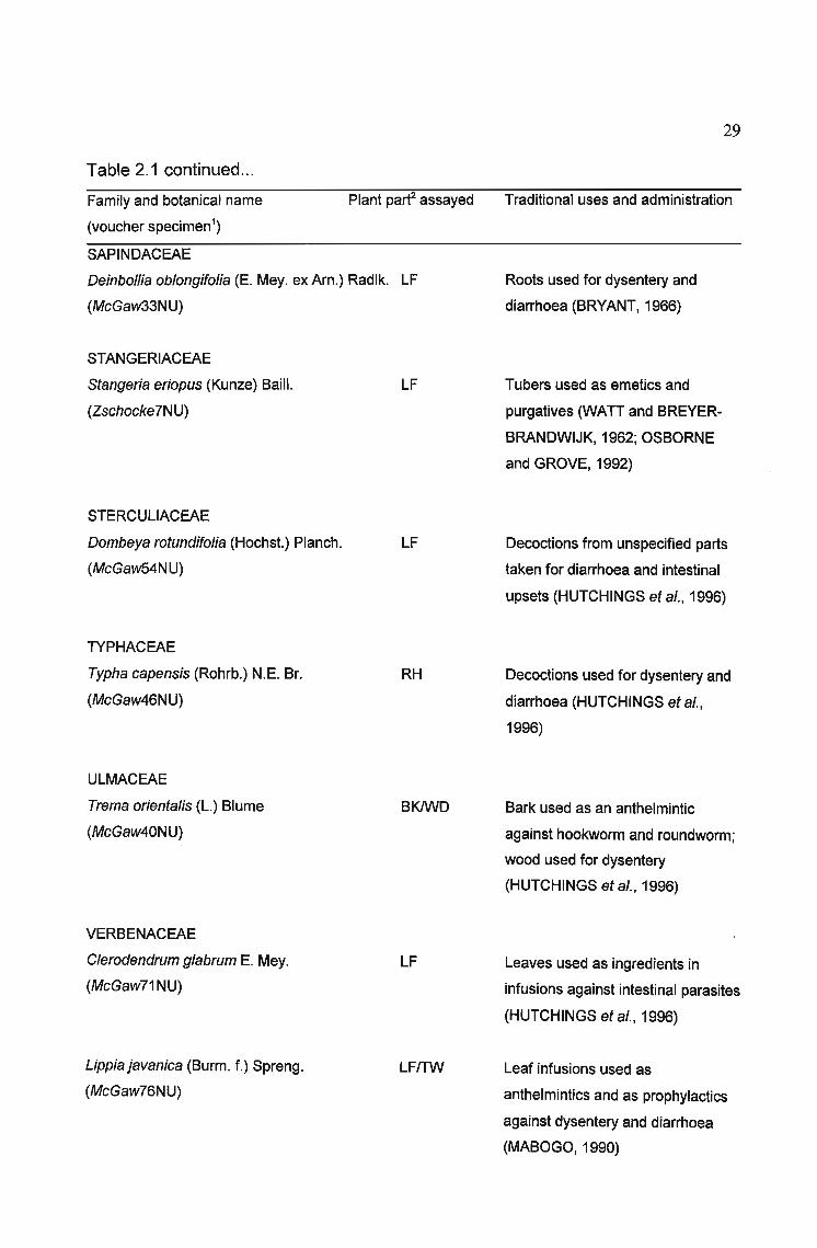
Table 2.1 continued...
Family and botanical name
(voucher specimen1)
Plant parf assayed
29
Traditional uses and administration
SAPINDACEAE
DeinbolJia oblongifolia (E. Mey. ex Am.) Radlk. LF
(McGaw33NU)
Roots used for dysentery and
diarrhoea (BRYANT, 1966)
STANGERIACEAE
Stangeria eriopus (Kunze) Bail!.
(Zschocke7NU)
STERCULlACEAE
Dombeya rotundifolia (Hochst.) Planch.
(McGaw54NU)
TYPHACEAE
Typha capensis (Rohrb.) N.E. Br.
(McGaw46NU)
ULMACEAE
Trema orientalis (L.) Blume
(McGaw40NU)
VERBENACEAE
Clerodendrum glabrum E. Mey.
(McG~w71NU)
Lippia javanica (Burm. f.) Spreng.
(McGaw76NU)
LF
LF
RH
BKlWD
LF
LFITW
Tubers used as emetics and
purgatives 0NATI and BREYER
BRANDWIJK, 1962; OSBORNE
and GROVE, 1992)
Decoctions from unspecified parts
taken for diarrhoea and intestinal
upsets (HUTCHINGS et al., 1996)
Decoctions used for dysentery and
diarrhoea (HUTCHINGS et al.,
1996)
Bark used as an anthelmintic
against hookworm and roundworm;
wood used for dysentery
(HUTCHINGS et al., 1996)
Leaves used as ingredients in
infusions against intestinal parasites
(HUTCHINGS et al., 1996)
Leaf infusions used as
anthelmintics and as prophylactics
against dysentery and diarrhoea
(MABOGO, 1990)

30
1Voucher specimen: NU =Natal University Herbarium, Pietermaritzburg
2Plant part: BK =bark, BL =bulb, CM =corm, FR =fruit, LF =leaf, LX =latex, RH =rhizome, RT =root, RT BK =root bark, ST =stem, TB =tuber, TW =twig, WD =wood, WH =whole plant
2.4 DISCUSSION
Hexane, ethanol and water were chosen as extracting solvents because of their
wide range in polarity. This selection of solvents would be expected to extract both
water-soluble and lipophilic compounds, allowing a greater possibility of discovering
biologically active compounds in the subsequent screening procedure. It was
decided to use dried plant material to eliminate the possibility of the water content of
different plants influencing the amount of resiC!ue resulting from the extraction. The
dried plant material was ground to a powder to allow the extracting solvent improved
access to plant cell contents. The extracts were stored in a freezer at -15 QC to
prevent decomposition of natural products before the screening procedure was
undertaken.
2.5 CONCLUSION
/ The selection of plants for screening inbioassays is important to maximise the
potential for discovering plants with high biological activity. Selecting plants based
on ethnopharmacological leads serves a dual purpose in both substantiating the
claims of traditional healers,_ and revealing pharmacologically interesting plants for
further investigation. This may involve isolat~ng the active components using
\ bioassay-guided fractionation, or producing galenical preparations for the herbal\
\\ medicine market.
"'------

31
CHAPTER 3
ANTHELMINTIC SCREENING
3.1 INTRODUCTION
3.1.1 Helminthiasis
Helminthiasis is a disease characterized by the presence of parasitic worms in the
body. Gastrointestinal helminthiases are estimated to be the most common of all
parasitic infections (BUNDY and COOPER, 1989). There are several types of worms
found inhabiting the intestines of humans and their domesticated animals, of which
the most important are nematodes or roundworms. Nematodes are widely
distributed and are found in almost every type of environment (LEE, 1965). There is
much morphological uniformity throughout nematode species. Differences in
behaviour and physiology have been instrumental in the success of the group
(CROLL, 1970). Most nematodes are free-living, but many species are parasitic on
insects, fish, birds, fungi, plants or animals including man. It has been estimated that
about 16 000 nematode species have been described and that at least 40 000
species exist (ANDERSON, 1992). About 33 % of all the nematode genera which
have been described occur as parasites of vertebrates (ANDERSON, 1992). The
economic importance of parasitic nematodes has focussed attention on these,
rather than on free-living species.
Most of the worms that affect man live unobtrusively in the intestine, doing little to
impair the health of their host, with symptoms and signs usually appearing only in
cases of heavy infection (LEWIS and ELVIN-LEWIS, 1977). The World Health
Organization (WHO) estimates that one billion people are infected with Ascaris
lumbricoides and 500 million with Trichuris trichiura (RASOANAIVO and
RATSIMAMANGA-URVERG, 1993). EVANS et al. (1~87) reported prevalences in

32
KwaZulu-Natal children of 64.5 % for A. lumbricoides and 61.1 % for T. trichiura.
Tissue-living nematodes are also an important health problem, with 100 million
people being infected with filarial worms such as Wucheraria bancrofti and
Onchocerca volvulus (RASOANAIVO and RATSIMAMANGA-URVERG, 1993). The
prevalence of helminthiasis is particularly intense in children in countries with poor
sanitation, a low standard of education, and little health education (ENWEREM et
al., 2001). The extent of debility and ill health caused by worms is related to the
worm load, type and duration of the infection, with treatment being directed primarily
at reduction of worm load (ENWEREM et al., 2001). Although the mortality rate
associated with parasite infestations is negligible, morbidity such as impaired
physical and mental development is significant (TAYLOR et al., 1995). The major
impact of parasitic infection is the impairment of nutritional status in the patient
(CROMPTON, 1991). A commonly reported consequence of parasitic infection is
lethargy, connected not only with a lack of energy for physical activity, but also for
mental effort (CONNOLLYand KVALSVIG, 1993). FINCHAM et al. (1998) stated
that the "vast mass" of helminth infection is contributing to impairment of nutrition,
growth, learning and physical performance of South African children. Since the
population has multiplied exponentially, and the consequent deterioration in
environmental conditions has enhanced infection by intestinal parasites, it is likely
that clinical cases are being seen increasingly frequently at most hospitals and
clinics (FINCHAM et al., 1998). In addition to the direct impact of nematodes on
human health, helminthiasis is a major factor adversely influencing animal breeding
(ASUZU and NJOKU, 1996).
The nematode life cycle includes an egg, four larval stages, each separated by a
moult, and the adult stage (CROLL, 1970). In the parasitic nematodes most studied,
the adults are usually parasitic, and the larval stages free-living or parasitic in an
intermediate host (CROLL, 1970). One stage has usually been developed which
completes the transition from free-living to parasitic - the infective stage (CROLL,
1970). The free-living infective stage in animal parasitic nematodes is often a long
lived ensheathed, resistant, non-feeding stage, bridging the gap between the free
living and the parasitic habits (CROLL, 1970). Roundworms are soil-transmitted,

33
each species spending an obligatory period of development on the soil, free from its
host (CROMPTON, 1991).
3.1.2 Control of helminth infection
A range of control measures for soil-transmitted helminthiases is available based on
chemotherapy, sanitation and health education (CROMPTON, 1991). NJOKU et al.
(1996) proposed that the most practical way to control helminth infection is by
chemotherapy using broad spectrum anthelmintics. There are several anthelmintics
available for treating helminthiasis, but a major setback to their continued usage is
the development of resistance (ASUZU and NJOKU, 1996). In addition, some
currently used synthetic anthelmintics are expensive or not readily available (ASUZU
and NJOKU, 1996). There is thus an urgent need for new, inexpensive drugs which
will be able to act for longer periods before resistance sets in. Much effort is being
made at the discovery of new anthelmintics of plant origin (NJOKU et aI., 1996).
An anthelmintic drug must have a wide margin of safety between its toxicity to the
worm and its toxic side effect on the host. lEWIS and ElVIN-lEWIS (1977) stated
that anthelmintic properties of many naturally occurring products have been known
since the beginning of civilization and are still widely used today. To be effective they
should be orally active, produce results in a single dose, and be cheap.
Chemotherapeutic agents are administered widely today to rid the host of helminth
parasites, acting either by reducing intestinal flora, which may serve as food for
certain worms, or by direct toxic action on the worms (lEWIS and ElVIN-lEWIS,
1977). Often the use of these drugs is followed by a purge (COlE, 1971, cited by
lEWIS and ElVIN-lEWIS, 1977). Therapeutic agents may only kill certain stages of
the life cycle, so if the adult stage is killed but the eggs persist, a purge is necessary
to cleanse the intestine of eggs to prevent the recurrence of infection.
Generally-used anthelmintics include the benzimidazoles such as albendazole and
thiabendazole, and the non-benzimidazoles such as levamisole, pyrantel and
avermectin (SIMPKIN and COlES, 1981). levamisole continues to be a major

34
broad spectrum anthelmintic for the treatment of gastrointestinal nematodes in man
and animals despite the occurrence of some side effects (AMERY and
BRUYNSEELS, 1992). Levamisole and pyrantel act by disrupting neuromuscular
transmission in nematodes (ATCHISON et al., 1992). Levamisole can penetrate the
cuticle of nematodes and then act on the cholinergic receptors at the neuromuscular
junctions to paralyse the worms in a contractile state, and once rendered immobile,
the worms are excreted (FRAYHA et al., 1997).
Anthelmintic properties of microbial secondary metabolites, such as avermectins
and milbemycins, have been discovered in members of the genus Streptomyces
(MISHRA et al., 1987). The avermectins (which include ivermectin) and milbemycins
are structurally related and belong to the family of macrolide antibiotics with activity
against a wide range of nematodes (MISHRA et al., 1987). The avermectins and
milbemycins have a similar mode of action in that they are able to stimulate the
conductance of chloride ions across muscle membranes (HABER et al., 1991).
Benzimidazoles, which include thiabendazole, mebendazole and albendazole,
appear to act by binding to tubulin and disrupting the assembly of microtubules, and
by altering transmembrane proton discharge (GROVE, 1996). Paraherquamide is a
nematocidal indole alkaloid first isolated from Penicillium paraherquei (SCHAEFFER
et aI., 1992). Ivermectin-resistant and thiabendazole-resistant nematodes remain
sensitive to paraherquamide treatment, suggesting a novel mode of action for the
alkaloid (SCHAEFFER et al., 1992). The study of SCHAEFFER et al. (1992)
supported the hypothesis that the biological activity is mediated via a ligand-receptor
interaction. The action of many, if not all anthelmintics is due to their ability to reduce
larval motility (ENWEREM et al., 2001).
3.1.3 Anthelmintic assays
NEAL in 1980 (cited by RASOANAIVO and RATSIMAMANGA-URVERG, 1993)
stated that about 50 new compounds need to be evaluated every week for 4 years
to stand a good statistical chance of success in finding a new parasiticidal
compound (1/10000). To achieve this goal, a rapid and relevant first stage screen

35
using 15 mg or less of compound or plant extracts would be easier than any animal
model. Animal tests require relatively large quantities of test chemicals and plenty of
space for the animals, which can themselves be expensive (SIMPKIN and COlES,
1981). ASUZU and NJOKU (1996) indicated that medicinal plants may be the most
important sources of new anthelmintics. Many vascular plants are in common use in
domestic medicine in tropical areas where they are readily available and inexpensive
(lEWIS and ElVIN-lEWIS, 1977).
A number of in vitro test systems have been reported, using various nematodes as
test organisms. These systems have been briefly reviewed by SIMPKIN and COlES
(1981). In 1958, TINER used eggs and larvae oftrichostrongyles from rabbits but
the rate of screening was relatively low. TINER (1958) also reported that cultured
Caenorhabditis briggsae did not respond reliably to the anthelmintic phenothiazine.
Cooperia punctata responded to anthelmintics in vitro, but long pre-test incubation
procedures with several media changes were required, and it is not certain whether
the method is sufficiently selective for use as a random screen (lElAND et al.,
1975). COlES and MCNEllLlE (1977) used eggs of Nematodirus spathigerto
detect benzimidazole-like drugs and cultured adult Nippostrongylus brasiliensis to
detect other anthelmintics, but a single step test would be more convenient.
PLATZER et al. (1977) evaluated nine benzimidazoles against the free-living
nematode Caenorhabditis elegans and suggested that it could be used in a test for
anthelmintics.
Caenorhabditis elegans is a self-reproducing hermaphrodite, each animal producing
both sperm and eggs (BRENNER, 1974). Occasionally, cultures are found with a
few males, which may be mated with the hermaphrodites. The adults are about 1
mm in length and the life cycle for worms grown on E. coli is 3.5 days at 20°C
(BRENNER, 1974). C. elegans has been adopted as a convenient model to study
important problems in the developmental genetics and biology of metazoans
(BRENNER, 1974). WHARTON (1986) predicted that knowledge of nematode
biology as a whole will increasingly be based on this species. The essentially
complete DNA sequence of C. elegans has been determined, and this knowledge

36
will be a significant aid to understanding and controlling nematode parasites
(BLAXTER, 1998). Manipulations of this sequence will be important in studies of
gene function, as genes involved in development are often homologous or shared by
the worm and other multicellular organisms (PENNISI, 1998).
SIMPKIN and COlES (1981) recommended screening anthelmintic compounds
using free-living nematodes, as the culture and screen are quite simple and do not
require sophisticated equipment. After successfully using Caenorhabditis elegans for
in vitro screening for potential anthelmintics and for screening some standard
anthelmintics, SIMPKIN and COlES (1981) suggested the use of C. elegans in high
through-put in vitro assays for anthelmintics, as this test system has a remarkable
degree of selectivity, and is very cheap and easy to operate.
3.2 MATERIALS AND METHODS
3.2.1 Culture of Caenorhabditis e/egans
Caenorhabditis elegans var. Bristol (N2) nematodes were cultured on nematode
growth (NG) agar (APPENDIX 1) seeded with E. coli as a food source for the
nematodes, according to the method of BRENNER (1974). Subculturing was
performed on a laminar flow bench to prevent contamination of the nematode
cultures. An aliquot (50 ,ul) of E. coli culture grown overnight at 35 QC was pipetted
onto each NG agar plate and spread with a glass spreader. Five ml of sterile M9
buffer (APPENDIX 1) was added to a previous nematode culture plate to wash the
.nematodes off the plate. Portions of this nematode suspension (500 ,ul) were
pipetted onto each fresh NG agar plate seeded with E. coli and incubated in the dark
at 20 Q C.
The extracts of plants used by traditional healers for treating intestinal worm
infestations were tested at concentrations of 1 and 2 mg ml-1. Ethanol and water
.extracts were resuspended in their extracting solvents, while hexane extracts were
37
resuspended in acetone or dimethylsulfoxide (DMSO).
3.2.2 Mortality assay
Two types of anthelmintic assay were carried out. The first was a simple bioassay
described by RASOANAIVO and RATSIMAMANGA-URVERG (1993) where
nematode mortality after addition of nematocidal compound or plant extract was
assessed. In this assay, 500-1000 nematodes (7 to 10-day old cultures) in M9 buffer
(APPENDIX 1) were incubated with plant extract for 2 h at 25°C in the dark. The
assay was first standardized using the anthelmintic drug levamisole in place of plant
extract. A standard concentration of 5 ,ug ml-1 levamisole was used as a control in all
subsequent experiments. A second control, or blank, consisted of nematodes
incubated with no plant extract or levamisole. Using a dissecting microscope, the
percentage of live worms was estimated, and their movement noted and compared
to the controls.
3.2.3 Reproductive ability assay
The second anthelmintic assay to determine the effect of plant extracts on
reproductive ability of nematodes was performed following the protocol of SIMPKIN
and COlES (1981). Nematodes for the test were washed from 4 to 8-day old
cultures with M9 buffer and held in M9 supplemented with 1 ,ug ml-1 chlorhexidine
digluconate for 1 hour. This treatment does not harm the worms and helps to avoid
contaminating bacteria overgrowing the test medium. The test medium was made by
adding 10 ml of 3 to 5-day old E. coli (grown at 20°C) to 100 ml M9 buffer along with
5 mg ampicillin and 10 000 U nystatin. The ampicillin inhibits the E. coli so that
multiplication is reduced and the bacteria are less likely to metabolize the added
compounds. After 2 h at room temperature, 2 ml of the test medium was placed in
each well of a sterile repli-dish (Sterilin) and 10 ,ul of extract or levamisole solution
was added to each well. A drop of the worm suspension containing about 20
nematodes was added to the wells after a few minutes so the worms did not drop
into a region of high concentration of extract or levamisole. In each repli-dish were

38
negative control wells containing test medium, nematodes and water, ethanol,
DMSO or acetone as well as blank wells containing test medium only. In the positive
control,5,ug ml-1 levamisole was added in place of plant extract. Two wells were
allocated to each concentration of plant extract or levamisole. The plates were
incubated in the dark at 20°C for 7 days. After incubation the number and
movement of nematodes in each well was assessed compared to the controls using
a dissecting microscope.
In both assays, the number and movement of nematodes as compared to the
control (nematodes incubated with no plant extract or levamisole) was noted and
scored.
3.3 RESULTS
3.3.1 Standardization of results
Standard curves (Figures 3.1 and 3.2) were prepared using levamisole as the
standard anthelmintic drug for each of the assays. Using regression analysis, the
50% inhibitory concentration (ICso) values for levamisole were calculated to be 4.674
and 6.901 ,ug ml-1 for the 2 hand 7 day assays respectively. SIMPKIN and COlES
(1981) reported the minimum detectable dose of levamisole on C. eJegans as 0.5,ug
ml-1; no literature available reports ICso values.
3.3.2 Anthelmintic activity of plant extracts
Inhibition of C. elegans by plant extracts in both anthelmintic assays is recorded in
Table 3.1.

100
80
e0 60E
.l:J
.r-e
40~0
20
0
1
39
10 100
Log concentration, Jlg mr1
Figure 3.1. Inhibition of nematodes by levamisole (2 h mortality assay)
. 100 • •80
e0 60;;:s.r-e 40~0
20
0
1 10 100 1000 104
Log concentration, J.Lg mr1
Figure 3.2. Inhibition of nematodes by levamisole (7 day reproductive ability assay)

40
Table 3.1. Inhibition of nematodes by plant extracts (2 hand 7 day anthelmintic
assays)
Plant name Plant part1 Extracf Concentration Activitf
(mg ml-1)
2h 7 day
Acokanthera ob/ongifoJia LF H 21
E 21
W 21
Acorus calamus RH/RT H 2 + ++
1 + +
E 2 ++ +++
1 + ++
W 2 -+ +
1 -+ +
A/bizia adianthifoJia LF H 2 +
1E 2 + +
1 -+
W 2 + +++
1 + +++
A/oe mar/othii LF H 2 -+
1 -+
E 2 ++ ++
1 + ++
W 2 + ++
1 -+
Apodytes dimidiata LF H 21
E 2 + ++
1 + +
W 2 -+
1 -+Artemisia afra LF H 2
1E 2 + ++
1 + ++
W 2 ++ +++
1 + ++Brachy/aena disc%r LF H 2 + +
1 -+
E 2 + ++
1 ++
W 2 -+ ++

41
Table 3.1 continued...Plant name Plant part' Extracf Concentration Activity"
(mg rnl")
2h 7 day
1Clausena anisata LF H 2
1E 2 + ++
1 + ++
W 2 + +++
1 + +
Clerodendrum glabrum LF H 21
E 2 -+ ++
1W 2 +
1Ekebergia capensis LF H 2 +
1E 2 -+ +
1W 2 -+
1 -+Erythrophleum lasianthum LF H 2
1E 2 -+ ++
1 +
W 2 -+ ++
1 +Euclea divinorum BK H 2
1E 2 +
1 -+
W 2 -+ ++
1 -+ ++Heteromorpha trifoliata LF H 2
1E 2 -+
1W 2 +
1Kigelia africana LF H 2 -+ -+
1 -+E 2
1W 2 -+ ++
1 +Leonotis leonurus LF H 2 -+

42
Table 3.1 continued...Plant name Plant part1 Extrace Concentration Activity3
(mg ml-1)
2h 7 day
1E 2 -+ ++
1 ++
W 2 ++
1 ++
Lippia javanica LFITW H 2 -+
1E 2 -+ +
1W 2 +++
1 +++
Maesa lanceolala LF H 2 -+
1 -+
E 2 +
1 -+
W 21
Melia azedarach LF H 2 -+
1E 2
1W 2 -+ +++
1 ++Pimpinella caffra WH H 2
1E 2
1W 2 ++
1 +Ricinus communis LF H 2 -+ ++
1 -+ +E 2 ++
1 +W 2 + ++++
1 +++Sansevieria hyacinthoides LF H 2
1E 2
1W 2 +++
1 ++Trema orientalis BKlWD H 2 -+ ++
1 -+ +

43
Table 3.1 continued...Plant name Plant part1 Extract2 Concentration Activitf
(mg ml-1)2 h 7 day
E 2 ~ ~
1 -+ ++W 2 +++
1 +++Tulbaghia violacea TB H 2
1E 2 + +++
1 -+ ++W 2 -+ ++++
1 +++Zanthoxylum capense LF H 2
1E 2 +
1 +W 2
1
1Plant part: BK =bark, LF =leaf, RH =rhizome, RT =root, TB =tuber, TW =twig, WO =wood, WH
= whole plant
2Extract: H =hexane, E =ethanol, W =water
3Activity:
same number of nematodes alive as control (nematodes incubated without
levamisole or plant extract), worms move vigorously
-+ about 80% of nematodes compared to control, worms move slightly slower
+ about half number of nematodes compared to control, worms move slowly
++ less than 20% of nematodes compared to control, worms move slowly
+++ less than 10% of nematodes compared to control, worms move very slowly
++++ no movement of worms
The number of extracts tested for anthelmintic activity was 72, derived from 24
genera (18 families). Several crude extracts used by traditional healers for treating
intestinal worms displayed activity against C. elegans.

3.4 DISCUSSION
. 3.4.1 Comparison of anthelmintic assays
The results of the two anthelmintic assays correlated well, with the 7 day incubation
assay appearing to be more sensitive than the shorter assay. A larger number of
extracts revealed anthelmintic activity in the 7 day assay. This may indicate that
some extracts exerted an effect on the ability of the nematodes to reproduce rather
than having a direct killing effect, which was monitored in the 2 h assay.
3.4.2 Anthelmintic activity of plant extracts
.The anthelmintic test systems using C. e/egans are cheap and easy to use. A large
number of extracts or compounds may be tested at one time, leaving few requiring
screening in infected animals. Many extracts exhibited activity at a concentration of 2
mg ml-1 but a more selective effect was noted at 1 mg ml-1. In general, the water
extracts were more active than ethanol extracts, and very little anthelmintic activity
was shown by the hexane extracts.
Acokanthera oblongifolia was the only plant displaying no anthelmintic activity.
Anthelmintic activity has been reported in ethanol extracts of root bark of Albizia
anthelmintica, and the active principle was identified as musennin (TSCHESCHE
and FORSTMAN, 1957). However, Albizia adianthifolia did not display much
anthelmintic activity.
Extracts of Acorus calamus exhibited a high level of anthelmintic activity. There are
possibly carcinogenic and toxic compounds in Acorus calamus, with toxic effects
owing to the presence of ~-asarone, a phenylpropanoid (LANDER and SCHREIER,
1990; BRUNETON, 1995). This compound may cause liver and duodenal cancer
(BRUNETON, 1995). The anthelmintic activity of Acorus calamus may possibly be
attributed to the presence of the toxic ~-asarone. Aloe marlothii displayed some
activity against the nematodes. Other species of Aloe are known to have a laxative

45
action, for example Aloe ferox (VAN WYK et al., 1997). The main purgative principle
of A. ferox is the anthrone C-glucoside aloin, or barbaloin (VAN WYK et al., 1997).
This compound has been shown to occur in A. marlothii (L1NDSEY et al., 2001). A.
marlothii also contains aloesin, aloeresin A and aloeresin 0 (L1NDSEY et al., 2001).
Other plants with marked activity against C. elegans included Artemisia afra,
Brachylaena discolor, Clausena anisata, Ricinus communis, Trema orientalis and
Tulbaghia violacea. Many chemical constituents have been isolated from different
parts of Clausena anisata (HUTCHINGS et al., 1996). The volatile oil of the leaves
yields the toxic estragole (OKUNADE and OLAIFA, 1987). The alkaloids imperatorin
and xanthoxyletin, isolated from the root or root bark, have molluscicidal activity
(HUTCHINGS et al., 1996). Ricinus communis, the castor oil plant, contains the fatty
acid ricinoleic acid, which stimulates intestinal peristalsis (VAN WYK et al., 1997).
This purgative action assists in clearing the body of intestinal parasites, but the
direct effect of the plant extract on the nematodes remains unexplained. The bark
and wood of Trema orientalis contain tannins (WATT and BREYER-BRANDWIJK,
1962). Many chemical constituents of Tulbaghia violacea have been isolated,
including several sulphur compounds and steroidal saponins (BURTON, 1990) but it
is not known whether these compounds are responsible for the biological activity of
the plant.
3.5 CONCLUSION
The anthelmintic assays in this study analysed the direct killing effect as well as the
effect on the reproductive ability of plant extracts on free-living nematodes. Owing to
the similarity in morphology of free-living and parasitic nematodes, it is probable that
extracts displaying anthelmintic activity against free-living nematodes will inhibit
parasitic nematodes. Many of the extracts tested revealed anthelmintic activity,
supporting their use by traditional healers for the treatment of helminth infestations.
~_~~e_?~~~~~bl_LndJ.9~_no~~~~~}orth~i!"J~~rgaJJ.Y~~~ p~Y~ml~
~_~ theE~n~_of in~~s!inalJ~.~~sites;.As a result of this, a lack of a direct killing
46
effect on the nematodes does not necessarily mean that the pla!JJsJM~J~! n~ use in~ ---_...--,-"",--...,..-.-......_---_.....~~__._._ \_~ .....__,..,_....... ~._, ..,_~_~' '----k '_'''''''''-",,--- _______treating nematode i[lfestatiQn§.c,.fQmQ!n9!iQJ:1~._QtpJpnt§.,~reJ:!su_~!!y'~~mpl?yedby
~",._<,.....o.-,,""-o_
healers, some plants possibly having dir~.a..':',-th.el~i_,!!ic activity, and othersw~...--._------"'~---~._----""""'-..-...--...-~ .......' - '-" .-."." ,. ~-...._--_.-----... --_.~ -
purgative c.tiQQtQJ2l~ar the body_Qf p'ar~sites.---- - - - ..... .-~' .~-.
The w~s._p-[eaclnatuJe__o(wQ!'-'11Jnfestations in rural parts of southern Africa, where~.---- -.--._-----_•• ,. -__.h,· • • _-~~ ~ • .~.~_ .... ~.~ ..._-.~__• ..........__ •• _.__• ~.. • __._'....__ ..
sanitation is poor or nonexistent, and wher~Lb~altb cli.ojcs__are_s-pa(SJ~,jDdicates an~""'''''''''''''~,",------_ .._'•• - ...._-=-,._,....-~.._~_._.,.",.~-.-.---~._.~.-- .. ;'''-''.....-._----.~~~.~ ......._-_._.."._., . ., _._-----
important role for the treatment~ administered by traqiiionaLheaJers... It would be~_... ·..,_··._._·'c·,· ....._ .- ... .;<;,.."._:..:....~......-,-.., ••~~-._.... - -~-. -,' .•. '.-- .• 1
worthwhile to investigate further the plants used by local healers for efficacy and
toxicity in an effort to maximise the beneficial influence on primary healthcare in
these areas.
Prevention of parasite infestation is a priority, especially with the unacceptably high
reinfestation rates in children after treatment (TAYLOR et al., 1995). Improvement in
the socio-economic status of poor communities, and the provision of clean water
and adequate sewerage facilities are high priorities in South Africa.

47
CHAPTER 4
ANTIAMOEBIC SCREENING
4.1 INTRODUCTION
4.1.1 Amoebiasis
Amoebiasis is a major disease caused by infections with the protozoan Entamoeba
histolytica, which exists in the large intestine as either mobile trophozoites or as
cysts. The protozoa usually live and multiply in the lumen of the large intestine, living
in symbiosis with the bacterial flora as commensal organisms. The host, who may
exhibit no symptoms of the disease, will pass infective cysts in the faeces (KEENE
et aI., 1986). If the cysts are transferred to another host as a result of poor hygiene,
they withstand the acidic conditions of the stomach and release trophozoites in the
small intestine (KEENE et al., 1986). Mild cases of amoebiasis cause little
disturbance to the patient, but a heavy infection results in amoebic dysentery
(KEENE et al., 1986). Invasive trophozoites cause local ulceration in the large
intestine resulting in dysentery, possibly leading to perforation and fatal peritonitis; if
trophozoites move to other organs such as the liver, brain or lung, they form
abscesses, and such widespread infections may be fatal (PHILLlPSON et al., 1987).
Amoebiasis occurs throughout the world, and is more closely related to sanitation
and socio-economic status than to climate, although the occurrence of the disease
is most severe in tropical and subtropical regions (DI STASI, 1995).
In 1986, WARREN (cited by PHILLlPSON et al., 1987) reported thatfor 1977-1978
in Asia, Latin America and Africa there were an estimated 400 million infections
annually with 30 000 deaths and 1.5 million cases of the disease. WRIGHT et al.
(1988) reported that an estimated 12 % of the world's population harbour
Entamoeba histolytica.
48
4.1.2 Control of amoebiasis
Since the turn of the century, the isoquinoline alkaloid emetine from Cephaelis
ipecacuanha (ipecac) has been used successfully to treat amoebic dysentery, and in
1959 its analogue dihydroemetine (which is less toxic on the heart muscle) was
introduced (LEWIS and ELVIN-LEWIS, 1977; PHILLlPSON et al., 1995). The severe
side effects of emetine include abdominal cramps, dizziness, fainting, vomiting,
neuromuscular and cardiovascular effects, and pain or even necrosis at the site of
injection (KEENE et al., 1987). Although emetine has been superseded by newer
drugs with fewer side effects, it is still considered to be one of the most potent
amoebicidal agents and is used in the initial treatment of severe cases of acute
amoebic dysentery (KEENE et al., 1987). The synthetic 2,3-dihydroemetine has a
similar activity but is less toxic than ~metine owing to faster rates of metabolism and
excretion (KEENE et al., 1987). Chloroquine and antibiotics such as paromomycin
also have direct amoebicidal action; tetracycline and erythromycin indirectly affect
survival of amoebae by modifying the intestinal flora necessary for the existence of
the organisms (SEATON, 1971, cited by LEWIS and ELVIN-LEWIS, 1977).
PHILLlPSON et al. (1987) described a series of pharmaceuticals available for the
treatment of amoebiasis, including emetine, chloroquine, diloxamide furoate,
paramomycin, 8-hydroxyquinolines and metronidazole. Metronidazole is currently
the preferred drug, but it has disadvantages in that it is not well tolerated by some
patients, causing side effects such as severe nausea (PHILLlPSON et al., 1987;
WRIGHT et al., 1988). Metronidazole is metabolized only in the cytosol of the
Entamoeba trophozoite stage (FRAYHA et al., 1997). The toxic reduction products
exert their killing effects on the parasite cell by reacting and damaging intracellular
macromolecules, including DNA (FRAYHA et al., 1997). The enzyme
oxidoreductase, which is responsible for the reduction of metronidazole in the
anaerobic parasites, does not have a counterpart in the aerobic mammalian cell,
thus accounting for the low toxicity of the drug to the host (FRAYHA et al., 1997).
Metronidazole has been reported to have mutagenic effects in bacteria, and to
cause tumours in experimental animals (PHILLlPSON et al., 1987; WRIGHT et al.,

49
1988). Hence, it appears that no ideal drug exists for the treatment of amoebiasis
(PHILLlPSON and O'NEILL, 1987). Additionally, the possibility of the future
development of resistant strains, as has been demonstrated by other protozoa,
cannot be excluded (WRIGHT et al., 1987).
WRIGHT et al. (1988) discovered highly antiamoebic quassinoids from Brucea
javanica fruits and Simarouba amara stems, which are both used as Chinese
traditional remedies against dysentery. A number of naturally occurring alkaloids
have amoebicidal activity, including alstonine from Alstonia scholaris and A.
macrophylla, conessine from Holarrhena antidysenterica, as well as tubulosine,
cryptopleurine and berberine (KEENE et al., 1986). The major modes of action of
antiprotozoal drugs include intercalation with DNA (e.g. alstonine and berberine),
inhibition of protein synthesis (e.g. emetine and quassinoids), alkylation and oxidant
stress (quinones) (PHILLlPSON and WRIGHT, 1991).
A number of natural products which have anticancer activities are also active against
protozoa (PHILLlPSON et al., 1995). In addition to in vitro amoebicidal assays,
PHILLlPSON et al. (1995) advised the use of a mammalian cell line in vitro for
comparison with antiprotozoal activity to establish that compounds are not acting
solely as cytotoxic agents. There is a considerable lack of knowledge concerning the
effects of total extracts which may act by more than one mechanism and in which
different compounds may potentiate or antagonize the activity of antiprotozoal
components (PHILLlPSON et al., 1995).
In traditional medicine, a range of plant species is used for the treatment of
amoebiasis. Worldwide, species from about 139 plant genera have been reported to
be used for the treatment of amoebiasis (FARNSWORTH, 1985, pers. comm. to
PHILLlPSON et al., 1987). Useful leads for new antiamoebic drugs may be obtained
by the systematic investigation of plants used in the traditional treatment of
amoebiasis (PHILLlPSON et al., 1987; PHILLlPSON and WRIGHT, 1991). It is vital
that the efficacy and safety of traditional medicines be validated and their active
constituents identified so that reliable quality controls may be established

50
(PHILLlPSON and O'NEILL, 1987). PHILLlPSON et al. (1995) anticipated that
research into antiprotozoal natural products from higher plants will lead to novel
chemotherapeutic agents and to the selection and standardization of plants used in
traditional medicine.
4.1.3 Antiamoebic assays
In vitro test methods have relatively recently been developed with Entamoeba
histolytica (DIAMOND et al., 1978; NEAL, 1983). KEENE et al. (1986) demonstrated
that such an in vitro test is effective in assessing the activity of plant extracts. A rapid
and sensitive procedure for assessing in vitro activity of plant extracts against E.
histolytica using microdilution techniques in microtiter wells has been developed by
WRIGHT et al. (1988). In this test, metronidazole and emetine were shown to be
highly active against E. histolytica, while some antibacterial and antifungal drugs
were not active. PHILLlPSON et al. (1987) stated that sensitive in vitro tests for
assessing activity against E. histolytica are applicable to the bioassay-guided
fractionation of plant extracts.
Antiamoebic activity in vitro has previously been assessed utilizing techniques such
as observing clonal growth of amoebae in semisolid medium to determine viability
(GILLlN et al., 1982). Another method incorporated using a haemocytometer to
count amoebae grown in flat-sided tissue culture tubes (KEENE et al., 1986).
WRIGHT et al. (1988) judged these techniques to be time-consuming, producing
variable results, and needful of relatively large quantities of test compounds and
materials. CEDENO and KROGSTAD in 1983 described a micromethod based on
measuring the incorporation of [3H] thymidine into E. histolytica grown in microtiter
plates. The results with this method correlated well with quantitative parasite counts.
A similar test has been developed by WRIGHT et al. (1988), but assessment of
amoebae present in the microtiter plate wells is made by staining with eosin. The
growth of amoebae may alternatively be visualized as turbidity in the wells, and
numbers of amoebae may be estimated using a haemocytometer (WRIGHT, pers.
comm., 1999). This simple micromethod does not require the use of radiolabeled
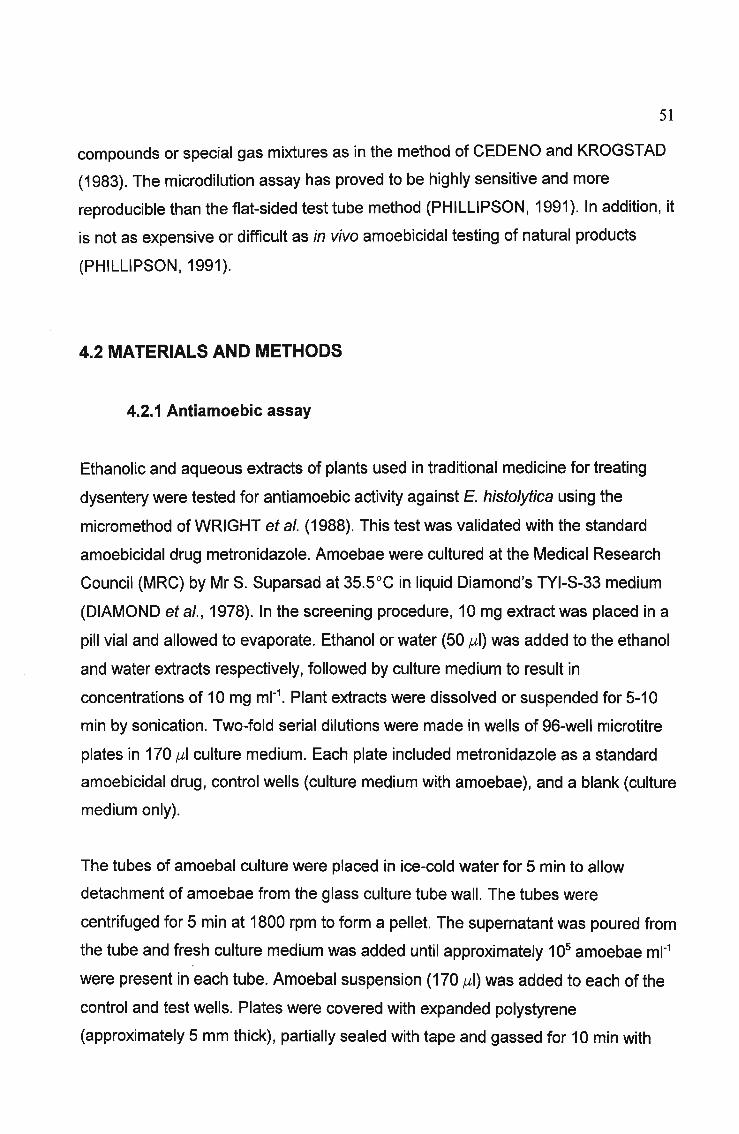
51
compounds or special gas mixtures as in the method of CEDENO and KROGSTAD
(1983). The microdilution assay has proved to be highly sensitive and more
reproducible than the flat-sided test tube method (PHILLlPSON, 1991). In addition, it
is not as expensive or difficult as in vivo amoebicidal testing of natural products
(PHILLlPSON, 1991).
4.2 MATERIALS AND METHODS
4.2.1 Antiamoebic assay
Ethanolic and aqueous extracts of plants used in traditional medicine for treating
dysentery were tested for antiamoebic activity against E. histolytica using the
micromethod of WRIGHT et al. (1988). This test was validated with the standard
amoebicidal drug metronidazole. Amoebae were cultured at the Medical Research
Council (MRC) by Mr S. Suparsad at 35.5°C in liquid Diamond's TYI-S-33 medium
(DIAMOND et al., 1978). In the screening procedure, 10 mg extract was placed in a
pill vial and allowed to evaporate. Ethanol or water (50 ,ul) was added to the ethanol
and water extracts respectively, followed by culture medium to result in
concentrations of 10 mg ml-1. Plant extracts were dissolved or suspended for 5-10
min by sonication. Two-fold serial dilutions were made in wells of 96-well microtitre
plates in 170 ,ul culture medium. Each plate included metronidazole as a standard
amoebicidal drug, control wells (culture medium with amoebae), and a blank (culture
medium only).
The tubes of amoebal culture were placed in ice-cold water for 5 min to allow
detachment of amoebae from the glass culture tube wall. The tubes were
centrifuged for 5 min at 1800 rpm to form a pellet. The supernatant was poured from
the tube and fresh culture medium was added until approximately 105 amoebae ml-1
were present in each tube. Amoebal suspension (170 ,ul) was added to each of the
control and test wells. Plates were covered with expanded polystyrene
(approximately 5 mm thick), partially sealed with tape and gassed for 10 min with
52
nitrogen before being sealed and incubated at 35.5°C for 72 hours.
After incubation, the growth of amoebae was checked with a microscope. Numbers
of amoebae in each well were estimated using a haemocytometer and compared to
the metronidazole standard and control wells.
4.3 RESULTS
4.3.1 Standardization of assay
A standard curve (Figure 4.1) was prepared for the amoebicidal compound
metronidazole. The ICso value of 0.202,ug ml-1 (calculated using regression analysis)
was close to the values of 0.22 and 0.32,ug ml-1 obtained in previously published
experiments (KEENE et al., 1986; WRIGHT et al., 1988).
100
100.10 1.00
Log concentration, Ilg mr1
80
c0 60:;:;:s.-~
.E 40~0
20
0
0.01
Figure 4.1. Amoebicidal activity of metronidazole

53
4.3.2 Antiamoebic activity of plant extracts
The activity of plant extracts against Entamoeba histolytica is shown in Table 4.1.
Forty-two extracts from 21 genera (18 families) were assayed. Plants exhibiting no
activity in both the ethanol and water extracts are not displayed in Table 4.1, and
were as follows: Bulbine latifolia, Canthium inerme, Clutia pulchella, Ekebergia
capensis,Heteromorpha trifoliata, Kigelia africana, Leonotis leonurus, Lippia
javanica, Senna didymobotrya, Spirostachys africana, Tecomaria capensis and
Trema orientalis. WRIGHT et al. (1988) found that the extracts of many plant
species were inactive, attributing this to the selectivity of the antiamoebic test. These
researchers also reported that most activity was found in methanol extracts, with
water extracts of the same plants displaying less activity. This is reflected to an
extent in the present study with the plants Acorus calamus and Sclerocarya birrea,
where the ethanol extracts were more active than the water extracts. In total, 8
ethanol extracts showed antiamoebic activity while 5 water extracts were active.

Table 4.1. Antiamoebic activity in South African medicinal plants
Botanical name Plant part1 Extract ICS02 (mg ml-1)
Acorus calamus RH E 0.3125
W 2.5
Albizia adianthifolia LF E >5.0
W >5.0
Bidens pilosa LF E
W >5.0
Deinbollia oblongifolia RT E >5.0
W >5.0
Piftosporum viridiflorum BK E >5.0
W
Schotia brachypetala RT E >5.0
W
Sclerocarya birrea BK E 1.25
W >5.0
Typha capensis RH E 5.0
W
Ziziphus mucronata LF E >5.0
W
Metronidazole 0.202 x 10⁻³
1Plant part: BK = bark, LF =leaf, RH =rhizome, RT =root, TB =tuber, TW =twig, WO =wood
2_ = no activity (no effect on amoebae)
54

55
4.4 DISCUSSION
4.4.1 Antiamoebic activity of plant extracts
The high activity of Acorus calamus may be attributed to the presence of the toxic
phenylpropanoid ~-asarone (LANDER and SCHREIER, 1990). Other active plants
included Albizia adianthifolia, Deinbollia oblongifolia and Sclerocarya birrea. Several
compounds have been isolated from the bark and roots o~ Albizia adianthifolia
(HUTCHINGS et al., 1996) but little chemical work has beeninitiat~d on the leaves.. ~
Little chemical investigation has been carried out on Deinbollia oblongifolia. The
bafk of Sclerocarya birrea contains tannins and traces of alkaloids (WATT and
BREYER-BRANDWIJK, 1962). GALVEZ et al. (1991) and GALVEZ et al. (1993)
reported that procyanidin from the bark inhibits peristaltic reflexes in guinea-pig
colon and shows antidiarrhoeal activity on isolated guinea-pig ileum and against
various induced diarrhoeas in mice.
4.5 CONCLUSION
Ti~9j!io~~L~~_~~~r~ in So~tb..Africa LIs~.~JJJ!lJ1beJ.Qtplants.to.treat.dys.eJlieric;\
dil?.e.g.§§, bl.:lt.iU$.J~I~arly not possible for the healers to ascertain whether the illness...."'''''','''......- .,'_ ..__~. " __"'~" _"'_'~"" '_""~~__"~"""_'_~~<''''_~''''''''''''_'",,,",'-__.~...• ',_~. _r"'~_""'"'_'_" .•~._- ,.,~_., __ "_' '~~"_"_"'"'~'_'-"_"" "_-"" '_"'"'.~ ~'''''''••_.~,_. ~.~~ .,..~ --
~~!~_~!~...of ~.~~~~~~.9~Q~gteJjaL-f:tgtllQg~n~. Consequently, lack of
antiprotozoal activity by the plant extracts tested in this study does not rule out the
possibility that the plant preparation may be efficacious against dysentery. Although
amoebic dysentery is not a commonly encountered affliction in South Africa, its
occurrence in poverty-stricken rural areas warrants the investigation of plants used
by indigenous healers. The discovery of novel antiprotozoal compounds with
possibly new modes of action from p~ts w9u1dJ~rovide-l!:np~ for..tt:1~
d~",§Lo.pment0f-Aew.antip[Q.tQzQ~LQr!J.9~_~ff~~~iv~_.§g~1!ll?lI.~.§j.§)tantpathogens.--~_..--_. _._----..-..
56
CHAPTERS
ANTIBACTERIAL SCREENING
5.1 INTRODUCTION
5.1.1 Infectious diarrhoea and dysentery
Diarrhoeal disease ranges from a mild and inconvenient illness to a major cause of
malnutrition, particularly among children in developing countries. In some cases
diarrhoea protects the body from harmful substances in the intestine by hastening
their removal. The most common causes of diarrhoea are viral infection, food
poisoning and parasite infestation, but diarrhoea also occurs in other illnesses, as a
side effect of some drugs for example.
Diarrhoeal diseases are a leading cause of morbidity and mortality in developing
countries (MECKES et al., 1997). Gastroenteritis is an inflammation of the stomach
and intestines, characterized by abdominal distress, nausea, vomiting and diarrhoea
(LEWIS and ELVIN-LEWIS, 1977). The cause of the disease may be viral, but
outbreaks produced by bacteria are either intoxications resulting from ingesting food
contaminated with toxic bacterial products or infections resulting from the ingestion
of viable bacteria (LEWIS and ELVIN-LEWIS, 1977). Among the latter are the
enteropathogenic strains of Escherichia coli, Vibrio parahaemolyticus, V. cholerae,
Salmonella species (particularly S. typhimurium) and Shigella species, which cause
bacillary dysentery (LEWIS and ELVIN-LEWIS, 1977). TORRES et al. (1995) cited
by MECKES et al. (1997) stated that most cases of bloody diarrhoea are owing to
bacterial enteropathogens such as Shigella spp. (the most virUlent), Salmonella spp.
and Vibrio parahaemolyticus, whereas E. coli and V. cholera generally cause watery
diarrhoea. Shigella spp. cause severe dysentery in children (MECKES et al., 1997).
All of these bacteria are Gram-negative species.
57
Diarrhoea and dysentery epidemics are common where living conditions are
crowded and hygiene is poor (OTSHUDI et al., 2000). Dysentery, a microbial
infection of the gastrointestinal tract, has symptoms of fever, vomiting, abdominal
pain and diarrhoea which often contains blood and pus (OTSHUDI et al., 2000). The
onset of the disease usually occurs within 2-3 days after infection and lasts for up to
several weeks. Dehydration occurs rapidly, especially in children and may cause
death if treatment is not given (OTSHUDI et al., 2000). As mentioned in CHAPTER
1, parasitic diseases such as dysentery and diarrhoea are the main causes of the
high mortality rate in developing countries, where over five million children under the
age of five die annually from severe diarrhoeal diseases (WHO, 1996).
Food poisoning may be caused by Clostridium perfringens and its toxins, Bacillus
cereus, species of Proteus, Klebsiella, Providencia (Paracolon), Citrobacter,
Pseudomonas, Enterobacter, and Actinomyces (LEWIS and ELVIN-LEWIS, 1977).
Overgrowth of organisms such as Staphylococcus aureus, Candida albicans,
Streptococcus faecalis, Pseudomonas aeruginosa and Proteus mirabilis may occur
when there is suppression of gut flora owing to antibiotic therapy, r~sulting in
diarrhoea among other symptoms (LEWIS and ELVIN-LEWIS, 1977).
5.1.2 Control of enteropathogens causing infectious diarrhoea and
dysentery
A mechanism to counteract infectious diarrhoea is by inhibiting the growth of the
enteropathogen causing the disease (MECKES et al., 1997). This is usually done by
prescribing antibiotic therapy. However, MECKES et al. (1997) warned that
enteropathogens have a high level of resistance to commonly used antibiotics and
resistance to new drugs emerges very fast. Bacteria have become resistant to
antimicrobial agents as a result of chromosomal changes or the exchange of genetic
material via plasmids and transposons (NEU, 1992). Fuelling this crisis is the
extensive and often inappropriate use of antibiotics in the community and hospitals,
leading to increased selective pressure on microorganisms (NEU, 1992). The
widespread use of antibiotics in animal feed may explain the high antibiotic

58
resistance of Salmonella species (NEU, 1992). Strategies employed to reduce the
emergence of resistant bacteria in animals should include the prohibition of the use
for growth promotion in animals of antimicrobials approved for human therapy
(WHO, 1996). Streptococcus species and staphylococci, organisms that cause
respiratory and cutaneous infections, as well as members of the Enterobacteriaceae
and Pseudomonas families, organisms that cause diarrhoea, urinary infection and
sepsis, are now resistant to virtually all of the older antibiotics (NEU, 1992). NEU
(1992) recommended the adoption of mechanisms such as antibiotic control
programs, better hygiene, and synthesis of agents with improved antimicrobial
activity in order to limit bacterial resistance. The need for new antibiotics will
continue because bacteria have a remarkable ability to overcome each new agent
synthesized (NEU, 1992). The search for new drugs with activity against
enteropathogens is a priority. The antimicrobial compounds from plants may inhibit
bacteria by a different mechanism than the presently used antibiotics and may have
clinical value in treatment of resistant microbial strains (ELOFF, 1999a).
5.1.3 Antibacterial assays
Antimicrobial activity of medicinal plants has been evaluated previously using
various methods, which are classified into three groups, Le. the disc-diffusion,
dilution and bioautographic assays. According to RIOS et al. (1988) many factors
can influence the results, inclUding the extraction method, inocula volume, culture
medium composition, pH and incubation temperature. In addition, there is no
standardized method for expressing the results of antimicrobial screening. The
diffusion and dilution methods are also used to determine the degree of
susceptibility of a test microorganism to a given antimicrobial agent (susceptibility\
tests), while bioautographic methods are mainly applied to bioassay screening and
bioassay-guided fractionation (RASOANAIVO and RATSIMAMANGA-URVERG,
1993).
The diffusion method enables several plant extracts to be screened against various
micoorganisms at the same time, and is therefore suitable for preliminary bioassay

59
scanning (RASOANAIVO and RATSIMAMANGA-URVERG, 1993). The dilution
methods are used to determine more precisely the antibacterial activity of extracts,
giving the MIC values of test extracts for a given microorganism (RASOANAIVO and
RATSIMAMANGA-URVERG, 1993). Bioautographic methods allow a rapid
identification of bioactive constituents of plant extracts (RASOANAIVO and
RATSIMAMANGA-URVERG, 1993) and are valuable in the bioassay-guided
fractionation of active extracts.
5.1.3.1 Diffusion methods
Diffusion methods make use of discs, holes or cylinders containing samples to be
tested, which are brought into contact with an inoculated medium. After incubation,
the diameter of the clear zone around the reservoir (inhibition diameter) is
measured. In the disc method, or disc-diffusion assay, a concentration of different
plant extracts is applied to filter paper discs, which are placed onto the surface of
inoculated agar medium in Petri dishes. Discs impregnated with plant extracts
exhibiting antibacterial activity will be surrounded with a clear zone of inhibition after
incubation. Inhibition diameters higher than 8 mm are generally considered as
positive results (RASOANAIVO and RATSIMAMANGA-URVERG, 1993). In the hole
plate assay method, holes are punched in inoculated agar in a Petri dish, plant
extract or antibiotic is placed in the holes, and the zone of inhibited growth of the test
organism is measured (RIOS et al., 1988). The cylinder method is similar to the hole
plate assay. Antibiotic or plant extract is applied to stainless steel or porcelain
cylinders, placed on inoculated agar, and the resulting zones of inhibition measured.
The inhibition diameters produced by the plant extracts in the diffusion assays may
be compared to those obtained by antibiotic controls. Diffusion assays are useful in
the preliminary screening for antimicrobial activity of plant extracts. It is not possible
to relate MIC values with inhibition diameters as there is no relationship between the
two (RIOS et al., 1988).

60
5.1.3.2 Dilution methods
Dilution assays are those which require a homogeneous dispersion of the sample in
water (RIOS et al., 1988). They are mainly used to determine the Minimum Inhibitory
Concentration (MIC) values of an extract or pure compound. These values are taken
as the lowest concentration of the extract that completely inhibits bacterial growth
after incubation for 24 h.
In the liquid dilution method, turbidity is taken as an indication of bacterial growth, so
where the sample is inactive against the microorganism tested, the liquid will appear
turbid (RIOS et al., 1988). The grade of inhibition is related to the turbidity of the
medium, and may be measured by spectrophotometry (RIOS et al., 1988). With the
agar dilution method, a fixed amount of an antibiotic- or extract-containing mixture is
mixed with nutrient agar and allowed to set (RIOS et al., 1988). The advantages of
this method are its simplicity and speed, and the possibility of using it in the
antimicrobial study of water-soluble or insoluble samples such as essential oils
(RIOS et al., 1988). Up to six microorganisms can be seeded in each Petri dish, and
antimicrobial activity is indicated when the bacteria do not grow (RIOS et al., 1988).
ELOFF (1998a) developed a microdilution technique using 9B-well microtitre plates.
A two-fold serial dilution of the extract, pure compound or antibiotic is prepared in
the wells of the microplate, and bacterial culture added. After incubation, p
iodonitrotetrazolium violet is added, and in the wells where bacterial growth occurs,
a deep red colour develops. Wells containing antibacterial compounds remain clear.
This scaled-down technique requires only a small amount of test compound, and is
quick, sensitive and reproducible. It is useful for screening plants for antimicrobial
activity, for the bioassay-guided fractionation of extracts, and for the MIC
determination of extracts or pure compounds (ELOFF, 1998a).
5.1.3.3 Bioautographic methods
Bioautography is an important detection method for new or unidentified antimicrobial

61
compounds (RIOS et al., 1988). In the agar-overlay bioautographic assay, a plant
extract is separated into individual compounds on a TLC plate. A molten agar
overlay containing a suspension of bacteria and a tetrazolium salt such as 2,3,5
triphenyltetrazolium chloride (TZC) is applied to the TLC plate. The separated
compounds on the TLC plate diffuse into the agar and, if they possess antibacterial
activity, will inhibit the growth of bacteria. Bacteria reduce the tetrazolium salt
through dehydrogenase activity to the corresponding intensely coloured formazan
(HAMBURGER and CORDELL, 1987; SLUSARENKO et al., 1989), so where
bacterial growth is inhibited, a colourless zone appears against a dark pink
background when TZC is used as an indicator. In the direct bioautography assay, a
suspension of microorganisms in liquid medium is sprayed on a developed TLC
plate and incubated overnight. A solution of tetrazolium salt is then sprayed on the
plate and incubated to detect the areas of bacterial growth inhibition. With this
method, there are no problems associated with differential diffusion of compounds
through the agar (HAMBURGER and CORDELL, 1987). An advantage of
bioautography is that it allows the localization of activity, even in complex mixtures
(HAMBURGER and CORDELL, 1987).
5.2 MATERIALS AND METHODS
5.2.1 Quantification of bacteria
The test organisms in the investigations of antibacterial activity, Bacillus subtilis,
Escherichia coli, Klebsiella pneumoniae and Staphylococcus aureus, were obtained
from the bacterial collection of the Department of Microbiology, University of Natal,
Pietermaritzburg. The number of cells in Mueller-Hinton (MH) broth cultures of each
bacterial species was estimated using a serial dilution technique (LECH and
BRENT, 1987). Ten-fold serial dilutions of overnight MH broth cultures were
prepared, and 100 ,ul of each dilution were spread onto MH agar plates using a
glass spreader. The plates were incubated overnight at 37°C and colonies were
counted using an Anderman Colony Counter. Following the assumption that each

62
living bacterial cell will grow into a separate colony on the plate, the number of cells
present per ml of the original overnight cultures was calculated. The Optical Density
(OD) at 600 nm (00600) for each dilution was determined using a Varian Cary 50
Spectrophotometer, and used to indicate numbers of bacterial cells in cultures for
the antibacterial screening and MIC determination.
5.2.2 Disc-diffusion assay
The disc-diffusion assay (RASOANAIVO and RATSIMAMANGA-URVERG, 1993)
was used in the antibacterial screening procedure. Plants used for treating
diarrhoea, dysentery and unspecified stomach ailments were tested for antibacterial
activity. Residues of plant extracts were resuspended in their extracting solvents to a
concentration of 100 mg ml-1• MH agar base plates were prepared using sterile 90
mm Petri dishes. MH agar at 48°C was inoculated with a MH broth culture (106- 108
bacteria ml-1) of each bacterial species and poured over the base plates to form a
homogenous layer. Filter paper discs (Whatman No. 3, 6 mm diameter) were
sterilized by autoclaving. One mg of plant residue (10,u1 of 100 mg ml-1 suspension)
was applied to each filter paper disc and allowed to air-dry. The dry discs were
placed on the seeded MH agar plates; each extract was tested in quadruplicate, with
4 discs of one extract per plate. Each plate also contained a reference antibiotic,
neomycin (500,ug ml-1), as a positive control. Air-dried solvent (hexane, ethanol and
water) saturated discs were used as negative controls. The plates were incubated at
37°C overnight, after which the zones of inhibition, if present, around each disc were
measured. The ratio of the inhibition zone (mm) produced by the plant extract and
the inhibition zone around the neomycin reference (mm) was used to express
antibacterial activity (VLlETINCK et al., 1995).
5.2.3 Microdilution assay
The microplate method of ELOFF (1998a) was used with slight modifications to
determine the Minimal Inhibitory Concentration (MIC) values for plant extracts with
antibacterial activity. Residues of plant extracts active in the disc-diffusion assay

63
were made up to 50 mg ml-1 with the extracting solvent in the case of ethanol and
water. Hexane extracts were resuspended in acetone to a concentration of 50 mg
ml-1. The plant extracts (100 ,ul) were serially diluted 50 % with water in 96-well
microtitre plates. Overnight MH broth cultures (grown at 37°C) of the test bacteria
were diluted 1:100 (overnight bacterial culture:MH broth), and 100 ,ulofthe resulting
culture were added to each well. Neomycin was used as the reference antibiotic and
controls with no extract and solvent only, or MH broth only were used. The covered
microplates were incubated overnight at 37°C. As an indicator of bacterial growth,
40 ,ul of p-iodonitrotetrazolium violet (INT) dissolved in water were added to the
microplate wells and incubated at 37°C for 30 min. MIC values were recorded as the
lowest concentration of extract that completely inhibited bacterial growth, i.e. a clear
well. The colourless tetrazolium salt acts as an electron acceptor and is reduced to a
red-coloured formazan product by biologically active organisms (ELOFF, 1998a).
Where bacterial growth is inhibited, the solution in the well will remain clear after
incubation with 1Nl.
In an effort to determine the effect of culture age on MIC determination, ELOFF
(1998a) showed that S. aureus culture age from 1 to 6 h, and storing cultures up to
10 days in a cold room, had little or no effect on the MIC values. This contrasts with
larger volume serial dilution assays where the number of cells inoculated has an
effect on the MIC (HEWITT and VINCENT, 1989; cited by ELOFF, 1998a). ELOFF
(1998a) suggested that the 50 % inoculum size used in the microdilution assay
compared to the approximate 50 times lower inoculum used in the standard tube
MIC method (ELOFF, 1998b) may explain the difference.

64
5.3 RESULTS
5.3.1 Quantification of bacteria
The 00600 of overnight cultures was compared with the estimated number of
bacteria in the overnight cultures of each bacterial species (Table 5.1). This supplied
an indication of the inoculum of bacteria used in the antibacterial assays, as the
Optical Density of each culture was confirmed before each assay to ensure that
differences in inoculum size did not become a factor in the MIC determination.
Table 5.1. Correlation of bacterial numbers and optical density readings of overnight
cultures
Bacteria 00600 Number of bacteria
B subtilis 0.71 1.1 x 10⁸
E. coli 1.13 3.5 x 10⁹
K. pneumoniae 1.28 6.4 x 10⁹
S. aureus 1.63 5.7 x 10⁸
5.3.2 Antibacterial activity of plant extracts
The antibacterial activity of crude extracts showing positive results (hexane, ethanol
and water) is presented in Table 5.2. In total, 138 extracts from 45 genera (31
families) were tested. Plants exhibiting no antibacterial activity were Albizia
adianthifolia, Aloe arborescens, Aloe marlothii, Buddleja salviifolia, Bulbine latifolia,
Canthium inerme, Ceratotheca triloba, Cinnamomum camphora, Clutia pulchella,
Croton sylvaticus, Deinbollia oblongifolia, Kigelia africana, Lippia javanica, Melia
8zedarach, Mondia whitei, Ocotea bullata, Olea europaea, Ricinus communis,
Stangeria eriopus, Strychnos spinosa, Tarchonanthus camphoratus, Tetradenia
riparia, Tulbaghia violacea and Zanthoxylum capense.

65
Table 5.2. Determination of the antibacterial activity of South African medicinal
plants with the disc-diffusion and microdilution assays (MIC recorded in mg ml-1)
Botanical name Plant part1 Extracf Bacteria tested3
B.s. E.c. K.p. S.a.
Dif4 MIC5 Dif MIC Dif MIC Dif MIC
Acorus calamus RH H 0.29 0.39 0 0 0
E 0.33 0.78 0 0 0
W 0 0 0 0
Artemisia afra LF H 0 0 0 0
E 0.2 0.39 0 0 0.33 0.39
W 0 0 0 0
Asclepias fruticosa LF H 0 0.22 6.25 0 0
E 0 0 0 0
W 0 0 0 0
Cassine transvaalensis BK H 0 0 0 0
E 0.23 0.195 0 0 0.58 0.098
W 0.18 0.78 0 0 0.33 0.195
Catha edulis RT H 0 0 0 0
E 0.21 0.012 0 0 0.50 0.012
W 0 0 0 0
Combretum apiculatum LF H 0 0 0 0
E 0.17 0.049 0 0 0.29 0.049
W 0.15 0.39 0 0 0.43 0.39
Curtisia dentata BK H 0 0 0 0
E 0.28 0.78 0 0 0
W 0.13 3.13 0 0 0
Cussonia spicata LF H 0.14 12.5 0.29 6.25 0 0.14 12.5
E 0.29 3.13 0.29 3.13 0.125 12.5 0
W 0 0 0 0Dombeya rotundifolia LF H 0 0 0 0
E 0.40 1.56 0 0 0.33 1.56
W 0.40 1.56 0 0 0.36 1.56Erythrophleum lasianthum LF H 0 0 0 0
E 0.17 0.78 0 0 0
W 0 0 0 0Gunnera perpensa RT/RH H 0 0 0 0
E 0 0 0 0.23 3.13

66
Table 5.2 continued...
Botanical name Plant part1 Extracf Bacteria tested3
B.s. E.c. K.p. S.a.
Dif4 MIC5 Dif MIC Dif MIC Dif MIC
W 0 0 0 0.23 0.78
Harpephyllum caffrum BK H 0.33 0.78 0 0 0
E 0.50 0.098 0.17 1.56 0.17 3.13 0.17 3.13
W 0.25 1.56 0 0 0
Heteromorpha trifoliata LF H 0 0 0 0
E 0.25 0.78 0 0 0
W 0 0 0 0
Pittosporum viridiflorum BK H 0.17 3.13 0 0 0
E 0.50 1.56 0 0 0.29 1.56
W 0 0 0 0
Psychotria capensis RT H 0 0 0 0.43 12.5
E 0 0 0 0.71 0.39
W 0 0 0 0.43 3.13
Rauvolfia caffra LF H 0 0 0 0.13 0.78
E 0 0 0 0.13 1.56
W 0 0 0 0
Sansevieria hyacinthoides LF H 0 0 0 0
E 0 0 0 0
W 0.42 6.25 0 0 0
Schotia brachypetala LF H 0 0 0 0
E 0.29 0.024 0 0 0.29 0.195
W 0.25 1.56 0 0 0.14 6.25Sclerocarya birrea BK H 0 0 0 0
E 0.33 0.012 0 0 0.29 0.049
W 0.33 0.78 0 0 0.43 0.78Spirostachys africana RT/ST H 0 0 0 0
E 0 0 0 0.36 3.13
W 0 0 0 0
Tecomaria capensis BK H 0 0 0 0
E 0 0 0 0.17 3.13
W 0 0 0 0Typha capensis RH H 0 0 0 0
E 0.17 3.13 0 0 0

67
Table 5.2 continued...
Botanical name Plant part1 Extracf Bacteria tested3
B.s.
Dif4E.c.
MIC5 Dif
K.p.
MIC Dif
S.a.
MIC Dif MIC
WOO 0 0
Neomycin 2.0 x 10⁻⁴ 1.3 X 10⁻² 7.8 X 10⁻⁴ 3.9 X 10⁻⁴
1Plant part: BK = bark, LF = leaf, RH = rhizome, RT = root, ST = stem, TB = tuber, WH =whole plant
2Extract: H = hexane, E = ethanol, W = water
3Bacteria: B.s. = Bacillus subtilis, E.c. = Escherichia coli, K.p. = Klebsiella pneumoniae, S.a. =
Staphylococcus aureus
4Dif: Results obtained in the disc-diffusion assays. Antibacterial activity is expressed as the ratio of
the inhibition diameter around the extract to the inhibition zone around the reference neomycin
antibiotic. The symbol 0 indicates no activity, Le. no inhibition zone around the extract discs
5MIC: Results obtained in the dilution assays. Antibacterial activity is expressed as the Minimum
Inhibitory Concentration (MIC) in mg ml-1. The symbol - is used where the MIC was not determined
Gram-positive bacteria (B. subtilis and S. aureus) were significantly more
susceptible to the extracts than the Gram-negative bacteria (E. coli and K.
pneumoniae). This is to be expected as Gram-negative bacteria are generally more
resistant to antibiotic agents compared with Gram-positive bacteria (VLlETINCK et
al., 1995). Only four extracts showed activity against E. coli, while two extracts were
active against K. pneumoniae. Concerning the Gram-positive bacteria, 27 extracts
were active against B. subtilis and 24 against S. aureus. The ethanolic extracts
displayed the most antibacterial activity, with 20 active extracts. Ten aqueous
extracts showed activity while only seven hexane extracts were active.

68
5.4 DISCUSSION
5.4.1 Antibacterial activity of plant extracts
The activity of Acorus calamus may be attributed to the presence of the toxic
compound J3-asarone in this plant (LANDER and SCHREIER, 1990). Cassine
transvaalensis bark contains tannins, which are sometimes used for their
antidiarrhoeal properties (BRUNETON, 1995). No antibacterial compounds have
been reported from Catha edulis. MARTINI and ELOFF (1998) isolated several
unidentified antibacterial compounds from Combretum erythrophyllum.
BREYTENBACH and MALAN (1989) isolated three antibacterial compounds from
Combretum zeyheri. These researchers ascribed the ethnopharmacological use of
this plant against diarrhoea to its antibacterial properties, particularly towards Gram
positive species. Antimicrobial components have been found in several other
Combretum species (ALEXANDER et al., 1992; ELOFF, 1999b). Harpephyllum
caffrum is reported to contain phenolic compounds which may be responsible for its
biological activity (EL SHERBEINY and EL ANSARI, 1976).
Antimicrobial activity has been detected in stembark aqueous extracts of Kigelia
africana (synonym K. pinnata) (AKUNYILI et al., 1991). This activity has been
partially attributed to the presence of iridoids present in the bark (AKUNYILI et al.,
1991). Ethanol and ethyl acetate extracts of the fruit and bark of K. africana
displayed antibacterial activity against Gram-positive and Gram-negative extracts,
but aqueous extracts had no activity (GRACE et al., 2001). In the present study no
activity was found in leaf extracts of the same species. Tetradenia riparia leaves
have been reported to inhibit several mycobacteria (VAN PUYVELDE et al., 1986). A
diterpene diol from the same species exhibited significant antimicrobial activity
against several bacteria and fungi (VAN PUYVELDE et ai, 1986). No activity was
displayed by leaf extracts of T. riparia in this investigation.
The chemical compounds of Schotia species have not been well investigated.
Polyhydroxystilbenes have been isolated from the heartwood of S. brachypetala

69
(DREWES, 1971; DREWES and FLETCHER, 1974). Many stilbenes have antibiotic
properties but the biological activity of the Schotia stilbenes is not known (VAN WYK
et al., 1997). Astringent tannins are present in the bark (BRUNETON, 1995), and
activity may be partly attributed to these compounds. Inpreliminary studies, the
antidiarrhoeal effects of Sclerocarya birrea bark have been linked to procyanidins
(GALVEZ et al., 1993). Extracts from stem bark have shown antibacterial activity
against several species of bacteria (HUSSEIN and DEENI, 1991).
Polyphenols including tannins, which are known to have antimicrobial activity, are
soluble in water, and these may explain the activity of some of the water extracts
(IEVEN et al., 1979). Tannins, which have the ability to precipitate proteins from
solution (a property known as astringency), are commonly found in a large array of
higher plant species, and may accumulate in large amounts in almost any plant part
(SCALBERT, 1991). Tannins may deter herbivores from predation, and they may
also increase resistance against microbial pathogens (SCALBERT, 1991).
Increasing attention is being paid to the use of tannins as antimicrobial agents, as
the toxicity of tannins towards microorganisms is well documented (SCALBERT,
1991). The astringent character of tannins may induce complexation with enzymes
or substrates, and tannins may also directly affect the metabolism of
microorganisms, in addition, possibly acting on membranes (SCALBERT, 1991).
NISHIZAWA (1990) isolated from Nuphar variegatum gallotannins and elagitannins
with antibacterial activity against Staphylococcus aureus and Proteus vulgare.
Amongst the plants tested, Cassine transvaalensis, Catha edulis, Combretum
apiculatum, Harpephyllum caffrum, Heteromorpha trifoliata, Schotia brachypetala
and Sclerocarya birrea showed the best activities with ~/IICs < 200,ug ml-1 and could
provide useful leads for the discovery of antibacterial compounds. The only species
displaying activity against Gram-negative bacteria were Cussonia spicata,
Harpephyllum caffrum and Asclepias fruticosa. Emphasis should be placed on the
discovery of new antimicrobials with activity against Gram-negative bacteria, as
many species from this group are pathogenic, and there is a serious lack of
antibiotics effective against them.

70
5.5 CONCLUSION
The evaluation of plants used by traditional healers to treat diarrhoeal disease is a
necessity in South Africa. Diarrhoea is a common affliction, especially in settlements
in rural areas that depend upon rivers as a source of water. Members of poor rural
communities may not have ready access to expensive antibiotics to treat stomach
ailments, particularly if health clinics are far away. Traditional healers are relied upon
to provide treatment, and the efficacy of the plant remedies given to dispel
pathogens causing diarrhoea and bacterial dysentery necessitates investigation.
Scientific exploration of these plants may lead to the isolation of novel compounds
with different mechanisms of action to those presently in use. With the compelling
issue of emerging drug-resistant bacterial pathogens, Western medicine is
searching for new and effective antimicrobial substances to combat this world-wide
threat.
In this study, a number of plants exhibited promising activity against a variety of
bacteria. These in vitro experiments are a step towards validating the use of these
plants by indigenous healers in South Africa. Most extracts showed activity against
Gram-positive bacteria, which are implicated in causing various stomach ailments.
However, Gram-negative enteropathogenic bacteria pose a greater threat by
cau$ing more serious, life-threatening diarrhoeal and dysenteric diseases,
warranting further research into the discovery of plant extracts active against these
bacteria.

71
CHAPTER 6
ANTHELMINTIC ACTIVITY IN THE FAMILY
COMBRETACEAE
6.1 INTRODUCTION
6.1.1 The family Combretaceae
The Combretaceae consists of 18 genera. The largest genus is Combretum, with
about 370 species, and Tenninalia is the second largest, with about 200 species
(LAWRENCE, 1951). The other genera are much smaller, including Calopyxes and
Buchenavia with 22 species each, Quisqualis with 16, Anogeissis with 14,
Conocarpis with 12 and Pteleopsis with 10 species (ROGERS and VEROTTA,
1996). Species from the genus Combretum and to a lesser extent Terminalia are
most widely used for medicinal purposes. These genera are widespread in parts of
Africa where they are often the most abundant species (ROGERS and VEROTTA,
1996). They are easily characterised by the wing-shaped appendages of the fruits,
and may be trees, shrubs or climbers (ROGERS and VEROTTA, 1996).
6.1.2 Traditional medicinal usage
Indigenous healers throughout southern Africa employ species of the Combretaceae
for many medicinal purposes. These include the treatment of abdominal pains,
backache, bilharzia, chest coughs, colds, conjunctivitis, diarrhoea, dysmenorrhea,
earache, fever, headache, hookworm, infertility in women, leprosy, pneumonia,
scorpion and snake bite, swelling caused by mumps, syphilis, toothache and general
weakness (WATT and BREYER-BRANDWIJK, 1962; KOKWARO, 1976; GELFAND
et al., 1985; HUTCHINGS et al., 1996; VAN WYK et al., 1997). The leaves and bark

72
of Combretum species are predominantly used. Fruits do not feature in medicine
owing to their reported toxicity to humans (ROGERS and VEROTTA, 1996).
6.1.3 Biological activity and chemical constituents
Several investigations into the antimicrobial activity of members of the
Combretaceae have been undertaken in recent years. BREYTENBACH and MALAN
(1989) found that most antibacterial activity resided in the leaves, twigs and fruits
with the latter being the most active. For reasons of availability, further investigation
was done on leaf material. These researchers isolated three unidentified
antibacterial compounds from leaves of Combretum zeyheri. ALEXANDER et al.
(1992) reported six species of Combretum with antimicrobial activity. In 1998,
MARTINI and ELOFF demonstrated the presence of at least 14 different unidentified
bacterial inhibitors, of widely differing polarity, in leaves of C. erythrophyllum. ELOFF
(1999b) examined other members of the Combretaceae, namely 27 species of
Combretum, Terminalia, Pteleopsis and Quisqualis. Acetone leaf extracts of all the
species inhibited the growth of the test bacteria to varying degrees. BABA-MOUSSA
et al. (1999) investigated seven species of the West African Combretaceae for
antifungal activity, reporting that all species displayed a degree of activity. These.
researchers suggested that the tannins and saponins, detected in the phytochemical
screening of the plant extracts, may be responsible for the antifungal activity.
The chemistry of Combretum species has been studied by a number of researchers.
Substituted phenanthrenes, dihydrophenanthrenes and bibenzyls have been
isolated from C. apiculatum (LETCHER and NHAMO, 1971), C. molle (LETCHER et
al., 1972) and C. hereroense (LETCHER and NHAMO, 1973). CARR and ROGERS
(1987) demonstrated the presence of triterpenoids and f1avonoids in leaf extracts of
several Combretum species. Several stilbenes and dihydrostilbenes (the
combretastatins) with cytotoxic activity, as well as acidic triterpenoids and their
glycosides with molluscicidal, antifungal, antimicrobial and anti-inflammatory activity
have also been isolated from species of Combretum (PETTIT et al., 1988;
ROGERS, 1989a; ROGERS, 1989b).

73
The widespread use of Combretum species in indigenous medicine for many
different ailments, as well as the significant antimicrobial and other activity exhibited
by several species, justifies the further investigation on other possible biological
activities, such as anthelmintic activity, of this group of plants.
6.2 MATERIALS AND METHODS
6.2.1 Plant extract preparation
Leaf material of various Combretaceae species, predominantly that of Combretum,
was supplied by Professor J. N. Eloff of the Department of Pharmacology, University
of Pretoria, p.a. Box 2034, Pretoria 0001, South Africa. This material was collected
in late summer (February) from plants growing in the Lowveld National Botanical
Garden in Nelspruit. A voucher specimen from each tree is present in the herbarium
of this Botanical Garden. The collection numbers and year in which each specimen
was collected are shown in Table 6.1.
Extracts of powdered leaf material were prepared using the solvents ethyl acetate,
acetone, methanol and water. For each species, separate samples of 0.5 g were
extracted in 5 ml of each solvent, sonicated for 30 min and filtered through
Whatman No. 1 filter paper. The plant material was re-extracted twice more, and the
filtrate was air-dried at room temperature.
6.2.2 Screening of plant extracts for anthelmintic activity
Anthelmintic activity of the ethyl acetate, acetone, methanol and water extracts was
determined against the free-living nematode Caenorhabditis elegans var. Bristol
(N2) as described in CHAPTER 3 (Section 3.2.2), with slight modifications. Plant
extracts at concentrations of 1 and 0.5 mg ml-1 plant extract were tested in the 2 h
mortality assay. Nematodes from 6 to 7-day old cultures were incubated with 0.5
and 1 mg ml-1 plant extract for 2 h at 25cC in the dark. The standard nematocidal

74
drug levamisole was used as a positive control in all experiments. A second control
consisted of nematodes incubated with no plant extract or levamisole. Solvent
blanks were included. Using a dissecting microscope, the percentage of living
worms was estimated, and their movement recorded and compared to the controls.
6.3 RESULTS
6.3.1 Anthelmintic activity of Combretaceae extracts
The results of the anthelmintic screening of Combretaceae leaf extracts are
presented in Table 6.1. Several extracts out of 88 water, acetone, methanol and
ethyl acetate extracts had an effect on the mortality and movement of nematodes. At
0.5 mg ml-1, one water, one ethyl acetate and nine acetone extracts were active,
while at 1 mg ml-1, one water, five ethyl acetate and 14 acetone extracts exhibited a
nematocidal effect. The only methanol extract to display activity was that of
Combretum apiculatum, which was active at both concentrations tested. C.
apiculatum extracts had by far the most detrimental effect on the nematodes, with
water, acetone, methanol and ethyl acetate extracts possessing varying degrees of
anthelmintic activity. C. hereroense and C. mossambicense showed activity in both
the acetone and ethyl acetate extracts. The only species apart from Combretum to
show activity was Quisqualis Iittorea, the ethyl acetate extract of which was mildly
nematocidal.

Table 6.1. Results of the anthelmintic screening of Combretaceae leaf extracts
Species and reference number1 Concentration of Anthelmintic activity using extraction solvents:
plant extract (mg ml-1) water acetone ethyl acetate methanol
C. apicuJatum Sond. sUbsp. 0.5 1 4 4 4
apicuJatum 36/86 1 1 5 7 5
C. bracteosum (Hochst.) 0.5 0 0 0 0
Brandis ex Engl. 127/88 1 0 0 0 0
C. ceJastroides Welw ex Laws 0.5 0 0 0 0
subsp. ceJastroides 49/91 1 0 0 0 0
C. coJlinum Fresen. subsp. suJuense 0.5 0 1 0 0
(Engl. & Diels) Okafor 134/85 1 0 1 0 0
C. edwardsii Exell 0.5 0 0 0 0
55/83 1 0 1 0 0
C. erythrophylJum (Burch.) Sond. 0.5 0 0 0 0
274/85 1 0 1 1 0
C. hereroense Schinz 0.5 0 0 0 0
82170 1 0 2 1 0
C. imberbe Wawra 0.5 0 0 0 0
260fi6 1 0 1 0 0
C. kraussii Hochst. 0.5 0 1 0 0
89fiO 1 0 2 0 0
C. microphylJum Klotzsch 0.5 0 1 0 0
151fi2 1 0 1 0 0
C. mkuzense Carr & Retief 0.5 0 1 0 0
-.)VI
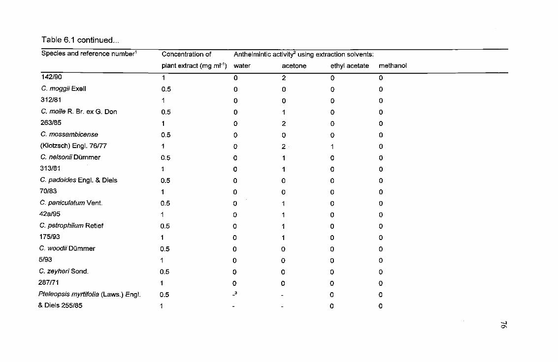
Table 6.1 continued...
Species and reference number1 Concentration of Anthelmintic activitf using extraction solvents:
plant extract (mg ml·1) water acetone ethyl acetate methanol
142/90 1 0 2 0 0
C. moggii Exell 0.5 0 0 0 0
312/81 1 0 0 0 0
C. molle R. Br. ex G. Don 0.5 0 1 0 0
263/85 1 0 2 0 0
C. mossambicense 0.5 0 0 0 0
(Klotzsch) Engl. 76/77 1 0 2 1 0
C. nelsonii DQmmer 0.5 0 1 0 0
313/81 1 0 1 0 0
C. padoides Engl. & Diels 0.5 0 0 0 0
70/83 1 0 0 0 0
C. paniculatum Vent. 0.5 0 1 0 0
42a/95 1 0 1 0 0
C. petrophilum Retief 0.5 0 1 0 0
175/93 1 0 1 0 0
C. woodii DQmmer 0.5 0 0 0 0
5/93 1 0 0 0 0
C. zeyheri Sond. 0.5 0 0 0 0
287171 1 0 0 0 0
pteleopsis myrtifolia (Laws.) Engl. 0.5 -3 - 0 0
& Diels 255/85 1 - - 0 0
-.J0'1

Table 6.1 continued...
Species and reference number1 Concentration of Anthelmintic activitl using extraction solvents:
plant extract (mg ml-1) water acetone ethyl acetate methanol
Quisqualis /ittorea (Engl.) Exell.
41a195
Terminalia brachystemma Welw.
ex Hiern. 167/96
Terminalia sericea Burch. ex DC.
159/85
0.5
1
0.5
1
0.5
1
1
2
oooo
oooooo
1Reference number: first part indicates collection number and second part the year in which collected.
2Anthelmintic activity scoring system: 0 =same as blank, 1 =80-90% nematodes alive, 2 =70-80% alive, 3 =60-70% alive, 4 =50-60% alive, 5 =40-50%alive, 6 =30-40% alive, 7 =20-30% alive, 8 =10-20% alive, 9 =0-10% alive.
3_: not tested
.....:J
.....:J

78
6.4 DISCUSSION
6.4.1 Anthelmintic activity of Combretaceae extracts
Several species exhibited significant activity in the anthelmintic mortality assay. Of
particular interest was Combretum apiculatum. The ethyl acetate, acetone and
methanol extracts of this plant displayed excellent activity and some activity was
present in the water extract, implying a wide range in polarity of active compounds.
C. mossambicense and C. hereroense also showed good activity. C.
mossambicense has also been shown to possess good antibacterial activity
(ELOFF, 1999b). Acetone extracts were the most active with respect to anthelmintic
activity followed by ethyl acetate and water, and lastly methanol.
6.5 CONCLUSION
The results obtained provide impetus for the further investigation of highly active
Combretum species to discover the compounds responsible for their biological
activity. Similar compounds are likely to be present in several species, providing
support for chemotaxonomic studies (ALEXANDER et al., 1992; ROGERS and
VEROTTA, 1996). More investigations into the biological activity of compounds
already isolated from active species are justified. In addition, the results support the
use of many Combretum species by traditional healers for a wide range of common
ailments in southern Africa.

79
CHAPTER 7
ISOLATION AND IDENTIFICATION OF ~-ASARONE FROM
ACORUS CALAMUS
7.1 INTRODUCTION
7.1.1 Acarus calamus
7.1.1.1 Description and traditional medicinal uses
Acarus calamus, of the family Araceae, is a reed-like, semi-aquatic, perennial plant
with a stout aromatic rhizome. The inconspicuous flowers are compactly arranged
on a long, fleshy axis, surrounded by a large leaf-like spathe. All parts of the plant
have a pungent, not unattractive smell. It is indigenous to South East Asia, but is
now Widely distributed in Europe, eastern North America and parts of Africa (VAN
WYK et aI., 1997). It has been cultivated in South Africa for many years and has
become a popular component of Zulu medicine. The plant is used extensively in
traditional medicine worldwide. Photographs of the plant growing in the Botanic
Gardens (UNP), and a portion of the rhizome, are shown in Plate 7.1.
Rhizomes have been used as carminatives, stomachics and to treat dysentery in
southern Africa, Europe and Asia (WATT and BREYER-BRANDWIJK, 1962). They
are used as tonics, stimulants and aphrodisiacs, and to treat rheumatism, toothache
and respiratory ailments (HUTCHINGS et al., 1996). In the Cape, the use of the
rhizome as a carminative and diarrhoea remedy has been recorded (WATT and
BREYER-BRANDWIJK, 1962). In India, the powdered rootstock and rhizome are
used as an antispasmodic, anthelmintic and insecticide (WATT and BREYER
BRANDWIJK, 1962). The fragrant oils obtained from the rhizome are also used as
flavouring in alcoholic beverages and as fragrant essences in perfumes and sacred
80
oils (MOTLEY, 1994).
7.1.1.2 Biological activity
Much work has been done on investigating the biological activity of Acorus calamus,
particularly in India and Europe. Extracts from the plant show larvicidal activity
(CHAVAN et al., 1976, cited by HUTCHINGS et al., 1996). The antigonadal activity
of ~-asarone is potentially useful in insect control (SAXENA et al., 1977). The
ethanolic extract has shown anti-secretagogue, anti-ulcer and cytoprotective
properties in rats (RAFATULLAH et al., 1994), supporting the use of calamus for the
treatment of gastropathy in traditional medicine. Rhizomes and roots have sedative,
hypotensive, analgesic and hallucinogenic effects (AGARWAL et al., 1956;
SHARMA and DANDIYA, 1969). The active hallucinogenic principles are presumed
to be a-asarone (chemically similar to mescaline, a psychoactive alkaloid) and the
eis-isomer ~-asarone (chemically similar to myristicin and kava alkaloids) (LEWIS
and ELVIN-LEWIS, 1977).
Fungicidal effects of A. calamus have been reported (ALANKARARAO and
RAJENDRA PRASAD, 1981; VASHI and PATEL, 1987; SAXENA et al., 1990;
JATISATIENR and JATISATIENR, 1999). An anti-mycotic principle was identified as
~-asarone, with a-asarone displaying a similar level of activity (OHMOTO and
SUNG, 1982). The essential oil and alcoholic extracts of the rhizomes have
antibacterial activity against many Gram-positive and Gram-negative bacteria (KAR
and JAIN, 1971; ALANKARARAO and RAJENDRA PRASAD, 1981; VASHI and
PATEL, 1987). The compounds responsible for the antibacterial activity have not
been well investigated.
The dried rhizome gave positive results as an antiamoebic agent when tested on
Paramecium caudatum (CHOPRA et al., 1957). Alcoholic extracts of the rhizomes
were active against the human nematode Ascaris lumbricoides (KALEYSA RAJ,
1974) and the rhizome oil was active against the larva of the root knot nematode
Meloidogyne incognita (SINGH et al., 1991). Both forms of asarone showed a dual

81
effect on the larvae of the nematode Toxocara canis (SUGIMOTO et a/., 1995). The
first was a fast acting temporary paralytic effect, and the other was a slowly
emerging killing effect. Anthelmintic and pesticidal activity of a related species,
Acarus gramineus, has been reported to be associated with the phenylpropanoids
a- and ~-asarone (PERRETT and WHITFIELD, 1995) . 80th isomers of asarone,
particularly the ~ form, are therefore associated with a wide range of biological
activity.
Extracts of Acarus calamus rhizomes are active in the antibacterial, anthelmintic and
antiamoebic tests (CHAPTERS 3, 4 and 5). 8ioassay-guided fractionation for the
isolation of the active compound(s) was attempted using the antibacterial agar
overlay technique (SLUSARENKO et a/., 1989) and an anthelmintic assay
(RASOANAIVO and RATSIMAMANGA-URVERG, 1993).
7.1.1.3 Chemical constituents
Essential oils are found in the leaves, rhizomes and roots, tannins occur in the
rhizome and roots, and ascorbic acid is found in the leaves and rhizomes
(PAMAKSTYTE-JUKNEVICIENE, 1971, cited by HUTCHINGS et a/., 1996). Owing
to its wide usage as a crude drug, much research has been undertaken on the
constituents of the oil. The major chemical constituents of the essential oils are
phenylpropanes, monoterpenes and thermolabile sesquiterpenoids (ROST and
80S, 1979, cited by MOTLEY, 1994). Methyleugenol, cis-methylisoeugenol, ~
asarone, geranylacetate, ~-farnesene, shyobunone, epishyobunone and
isoshyobunone are the most abundant chemical compounds (ROST and 80S,
1979, cited by MOTLEY, 1994). Other chemical components include a-asarone, y
asarone, calamenene, asaronaldehyde, acorenon~, calamenone, n-heptanic acid,
calamendiol, many sesquiterpenes, and trace amounts of other compounds
(MAZZA, 1985). The proportion of each chemical constituent of the oil, particularly in
the case of ~-asarone, varies between the varieties of A. calamus, corresponding to
the ploidy (ROST and 80S, 1979, cited by SCHMIDT and STRELOKE, 1994). The
diploid caryotype grows in North America and in parts of Asia (Siberia), and the

82
rhizomes contain little or no ~-asarone. In the triploid caryotype, present in central
Europe and Kashmir, the ~-asarone content of the rhizomes varies from 9 - 13 %. In
the tetraploid caryotype, found in India, east Asia and Japan, the essential oil of the
rhizomes is 70 - 96 % ~-asarone. The ploidy of African plants has not been
investigated.
~-Asarone is toxic (ABEL, 1987; LANDER and SCHREIER, 1990), and there is
evidence that it may induce duodenal and liver cancer in rats (HABERMAN, 1971,
cited by GOGGELMAN and SCHIMMER, 1983). As a result, many countries have
discontinued the use of Acarus in digestive medicines. Calamus has been banned
by the FDA as a food additive and within the last few years many herbal shops have
stopped recommending or dispensing it. Although calamus and its products are
banned in the United States, they are authorized in Europe, where the highest
acceptable concentration in foods and beverages is 0.1 mg kg-1 and in alcoholic
beverages,1 mg kg-1 (LANDER and SCHREIER, 1990).
7.2 MATERIALS AND METHODS
7.2.1 Plant extraction
Acarus calamus L. plants were collected from a shallow (approximately 0.3 m deep)
pond at Silverglen Nursery in Durban, South Africa in October 1999. A voucher
specimen (McGaw47NU) was deposited at the Herbarium of the University of Natal,
Pietermaritzburg. The leaves, roots and rhizomes were separated and dried at
50°C. The dried, powdered plant parts (4.33 g, 13.27 g and 17.56 g for the leaves,
roots and rhizomes respectively) were extracted separately with 100 ml ethanol.
Extraction was performed by sonication for 30 min in a Julabo ultrasound bath,
followed by overnight maceration. The extracts were filtered through Whatman No. 1
filter paper and the procedure was repeated twice with the same plant material. The
filtrates were air-dried and stored at -15°C.

83
7.2.2 Antibacterial assays
The antibacterial activity of fractions resulting from each purification stage was
tested using the agar-overlay bioautographic assay (SLUSARENKO et al., 1989) as
described in CHAPTER 5 (Section 5.1.3.3). The inhibition of bacterial growth by
compounds separated on the TLC plate was visible as white spots against a deep
red background. The test organism was the Gram-positive Staphylococcus aureus
(ATCC 12600).
The Minimal Inhibitory Concentration (MIC) values of the pure compound were
determined against the Gram-positive Bacillus subtilis (ATCC 6051) and
Staphylococcus aureus (ATCC 12600) and the Gram-negative Escherichia coli
(ATCC 11775) and Klebsiella pneumoniae (ATCC 13883). The microplate method of
ELOFF (1998a) as described in CHAPTER 5 (Section 5.2.3) was used. The pure
compound was dissolved in ethanol and serially diluted twofold with water in
microtiter plate wells. The antibiotic neomycin and extract-free solutions were
included as standard and blank controls. MIC values were recorded as the lowest
concentration resulting in complete inhibition of bacterial growth.
7.2.3 Anthelmintic assay
The anthelmintic activity of extracts, fractions and the pure compound was
determined against Caenorhabditis elegans var. Bristol (N2) nematodes using the
mortality assay described in CHAPTER 3 (Section 3.2.2). Solvent blanks, and
levamisole as a standard anthelmintic drug, were included. The percentage and
movement of live nematodes after incubation with the plant extracts was recorded
and compared to that of the controls.
7.2.4 Thin Layer Chromatography (TLC)
Thin Layer Chromatography (TLC) analysis was used to compare the chemical
composition of the crude ethanol extracts of Acorus leaves, roots and rhizomes. A

84
spot (0.5 mg) of each extract was loaded on a silica gel TLC plate (Merck Silica gel
60 F254' 0.25 mm thick) and developed using the solvent system hexane:ethyl
acetate (2:1). The TLC plate was visualized under ultraviolet light at 254 nm and 366
nm, under visible light, and after staining with anisaldehyde/sulphuric acid (AS)
spray reagent (465 ml ethanol, 5 ml glacial acetic acid, 13 ml concentrated sulphuric
acid and 13 ml para-anisaldehyde mixed in order). The TLC plate strip was heated
for 10 min at 100°C to allow development of the stain.
7.2.5 Bioassay-guided fractionation for isolation of active compound
All solvents were re-distilled using a rotary evaporator.
7.2.5.1 Vacuum Liquid Chromatography
Bioassay-guided fractionation was performed using the rhizome extract. The
ethanolic rhizome extract (1.14 g) was separated by Vacuum Liquid
Chromatography (VLC) over silica gel (Merck 230-400 mesh) using a hexane:ethyl
acetate gradient solvent system of increasing polarity. A column 13 cm in length and
with an internal diameter of 2 cm was packed with 20 g silica. The gradient solvent
system was increased by 5 % increments from 100 % hexane to 60 % hexane in
ethyl acetate, and subsequently by 10 % increments to 100 % ethyl acetate. At each
concentration, 100 ml solvent was flushed through the column. Bioautography
established that the most active fraction (270 mg) was eluted with 5 % and 10 %
ethyl acetate in hexane.
7.2.5.2 Preparative Thin Layer Chromatography
The active fraction was further separated by preparative TLC on three silica gel
plates (Merck Silica gel 60 F254, 0.25 mm thick) using toluene:ethyl acetate (93:7).
The active fraction (identified as a dark purple band under UV254) was scraped off
the TLC plates and eluted from the silica with ethanol. The active compound in
ethanol was filtered through Millipore filters (0.45 ,um and 0.22 ,um) to remove the
85
silica. To ensure complete removal of the silica particles, the filtrate was passed
through a Sephadex LH-20 column prepared in a Pasteur pipette. The Sephadex
was loaded to a height of 30 mm, and the pipette had an internal diameter of 5 mm.
The purity of the isolated compound, a yellow oil, was confirmed by TLC using
various solvent systems.
7.2.3 Identification of purified active compound
High Resolution Mass Spectrometry (MS) using a VG70-SEQ spectrometer was
performed. Nuclear Magnetic Resonance Spectroscopy CH NMR and 13C NMR) was
carried out to confirm the structure of the compound. A Varian Unity Inova 500 MHz
spectrometer was used.
7.3 RESULTS
7.3.1 Plant extraction
The extraction from the dried, powdered plant material yielded residues of 0.5546 g
(12.81 %) for the leaves, 0.2976 g (2.24 %) for the roots and 1.1357 g (6.47 %) for
the rhizomes.
7.3.2 Antibacterial and anthelmintic activity in different plant parts
The leaf, root and rhizome ethanol extracts of Acorus calamus exhibited
antibacterial and anthelmintic activity. The rhizome extract possessed a lower
Minimal Inhibitory Concentration (MIC) than the leaf and root extracts (Table 7.1),
against Bacillus subtilis and Staphylococcus aureus. Therefore, the rhizome extract
was selected to perform bioassay-guided fractionation for isolation of active
compound(s). Additionally, TLC and bioautography displayed fewer compounds,
other than the target active compound, in the rhizome extract than in the leaf and
root extracts. Photographs of the TLC fingerprints of the leaf, rhizome and root

86
extracts are presented in Plate 7.1. The compound of interest, which was shown
using bioautography to have antibacterial activity, is marked with an arrow.
7.3.3 Bioassay-guided fractionation for isolation of active compound
VLC on the rhizome ethanol extract resulted in a good separation of compounds.
Photographs of the TLC plates prepared from the VLC fractions, viewed under
ultraviolet light and after staining with AS spray reagent, are shown in Plate 7.2. Also
represented in Plate 7.2 is the bioautography plate of the VLC fractions. As
previously mentioned, the white areas indicate the presence of antibacterial
compounds, as the lack of bacterial growth cannot convert the indicator tetrazolium
salt to a red product. Marked with an arrow is the compound of interest, which eluted
in the second and third fractions. It is apparent that several other antibacterial
compounds, indicated by further white areas on the plate, are present in the
remaining VLC fractions. However, when the solvent was evaporated from these
fractions, there was insufficient residue to continue separations leading to
identification of these compounds. There was limited source plant material, so the
focus of the investigation remained on the compound of interest. The third fraction
was selected to continue the isolation as TLC indicated the presence of fewer
contaminating compounds than in the second fraction.
The activity-directed fractionation, ending with preparative TLC, of 1.14 g of rhizome
extract yielded 15.6 mg of oily yellow p-asarone. This represented 1.4 % of the
extract and 0.09 % of the rhizome. This pure compound possessed antibacterial and
anthelmintic activity. The structure of J3-asarone is presented in Figure 7.1. The MIC
values for the isolated J3-asarone against the test bacteria are presented in Table
7.1.
The anthelmintic activity of the crude rhizome extract and isolated J3-asarone was
very similar, both resulting in the death of 60-70 % of nematodes after incubation.
No other fractions resulting from the fractionation of the rhizome extract exhibited
anthelmintic activity.

87
1
63'
35
4
Figure 7.1. Structure of ~-asarone
7.3.4 Identification of purified active compound
The compound identified as ~-asarone was a yellow oil, C12H1603 [Mr 208.11016.
ElMS m/z (reI. int.): 208 [Mr (100),193 (47),177 (3),165 (27),162 (12),150 (6),
134 (3), 103 (4), 91 (11),77 (7), 69 (3),51 (2). The 1H NMR spectra 'in CDCI3were 0
1.84 (3H, dd, J = 7.3 Hz & 1.8 Hz, -CH=CH-C.!::6), 3.81, 3.83 and 3.89 (each 3H, s,
three OCH3), 5.76 (1 H, dq, J = 12 Hz &6.8 Hz, H-2'), 6.49 (1 H, dq, J = 11.4 Hz &
1.83 Hz, H-1'), 6.53 (1 H, s, H-3) and 6.84 (1 H, s, H-6). The 13C NMR spectra were
recorded in CDCI3, 0 151.5 s (C-4), 148.5 s (C-2), 142.3 s (C-1), 125.8 d (C-1'),
124.7 d (C-2'), 118.0 s (C-5), 114.1 d (C-6), 56.6 q, 56.4 q, 56.0 q (OCH3), 14.6 q (C
3').
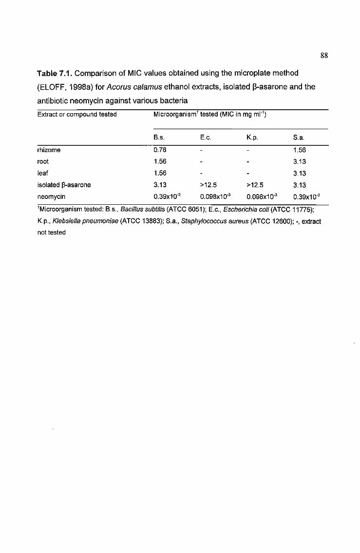
88
Table 7.1. Comparison of MIC values obtained using the microplate method
(ELOFF, 1998a) for Acarus calamus ethanol extracts, isolated ~-asarone and the
antibiotic neomycin against various bacteria
Extract or compound tested Microorganism1 tested (MIC in mg ml-1)
8.s. E.c. K.p. 5.a.
rhizome 0.78 1.56
root 1.56 3.13
leaf 1.56 3.13
isolated ~-asarone 3.13 >12.5 >12.5 3.13
neomycin 0.39x10⁻³ 0.098x10⁻³ 0.098x10⁻³ 0.39x10⁻³
1Microorganism tested: 8.s., Bacillus subtilis (ATCC 6051); E.c., Escherichia coli (ATCC 11775);
~.p., Klebsiella pneumoniae (ATCC 13883); 5.a., Staphylococcus aureus (ATCC 12600); -, extract
not tested

89
,\
Plate 7.1. Acorus calamus (~) uprooted from a shallow pond in the Botanic
Gardens, UNP and (B) rhizome. The length of the plant from rhizome to leaf tip was
72 cm, and the length of the rhizome portion displayed was 17 cm. TLC fingerprints
of ethanol extracts of A. calamus leaf, rhizome and root viewed (C) under visible
light, (D) under UV254 and (E) under UV366. The solvent system used was
hexane:ethyl acetate (2: 1). Marked with an arrow is the compound isolated (~
asarone).

-~ -- --- --
90
Plate 7.2. TLC of Vacuum Liquid Chromatography (VLC) fractions in the bioassay
guided fractionation of A. calamus ethanolic rhizome extract. Viewed (A) under
UV254 , (B) under UV366 and (C) after staining with anisaldehyde. The bioautography
plate (0) shows white areas of no bacterial growth, indicating the presence of
antibacterial compounds. The active compound of interest (~-asarone) is marked
with an arrow.
91
7.4 DISCUSSION
7.4.1 Isolation of an active compound
The isolated active compound ~-asarone has been isolated previously by others
from Acarus calamus growing in Europe and Asia (BAXTER et al., 1960). In this
study, the antibacterial and anthelmintic activity of ~-asaronewas demonstrated.
The anthelmintic activity of the isolated ~-asarone was very similar to that of the
crude rhizome extract. The crude extract and isolated compound were both tested at
the same concentration (1 mg ml-1). It was expected that the isolated ~-asarone
would display higher anthelmintic activity than the original extract as it would be
present in a higher concentration. However, this was not the case, and a possible
explanation may be the instability of ~-asaroneafter isolation, as some breakdown
of the compound was visible after TLC analysis. No other anthelmintic compounds
were apparent in the VLC fractions.
The antibacterial activity of the isolated ~-asarone was lower than that of the crude
extract. This may again be partly attributed to the lability of the isolated compound.
Also, several other compounds with antibacterial activity in the VLC fractions of the
crude extract were visualized in the bioautography assay. They made up such a
small part of the overall composition that their identification was not attempted. ~
Asarone appeared to be the major component of the crude extract (from TLC
analysis, and with its yield of 1.4 % of the extract). However, if it were the major
antibacterial compound, it would be expected to have a much lower MIC value. This
was not the case, so other minor compounds may well have significantly higher
antibacterial activity, contributing to a greater proportion of the overall activity even
though they are not present in such high quantities as ~-asarone. Unfortunately, it is
not possible to quantify antibacterial actiVity from a bioautography assay, where the
inhibition spot is by nature a positive/negative response, and does not reveal detail
about the degree of activity of a compound.

92
7.4.2 Identification of isolated compound
The mass spectrometry, 1H and 13C NMR data were consistent with those previously
reported for ~-asarone (PATRA and MITRA, 1981; MAZZA, 1985; NIGAM et al.,
1990; OPREAN et al., 1998). Asarone has been synthesized by two different
methods (SHARMA and DANDIYA, 1969).
7.5 CONCLUSION
The evidence presented indicates that if the Acorus calamus tested in this study is
representative of that found in southern Africa, then the South African Acorus is
likely to be triploid or tetraploid, as these caryotypes of A. calamus are known to
contain ~-asarone (ROST and 80S, 1979, cited by SCHMIDT and STRELOKE,
1994). Also, its origins possibly lie in Asia or Europe. Of some concern is the
evidence implicating ~-asarone in causing duodenal and liver cancer in test animals.
Owing to the possible carcinogenic and toxic effects, the use of Acorus in digestive
medicines has been discontinued in most countries. In South Africa, Acorus calamus
is still widely used by Zulu traditional healers to treat a variety of ailments, including
gastrointestinal disorders. The content of ~-asaronesubstantiates the use of this
plant as an anthelmintic and antibacterial remedy, but owing to the toxicity of the
active compound, it is recommended that the use of the plant is restricted to external
application for antibacterial use only, or discontinued altogether. A. calamus
contains several additional compounds with antibacterial activity. The use of
varieties of A. calamus which do not contain ~-asarone, but which still have
antibacterial activity, would be preferable.

93
CHAPTERS
VARIATION IN ANTIBACTERIAL ACTIVITY OF
SCHOTIA BRACHYPETALA
8.1 INTRODUCTION
8.1.1 Schotia brachypetala
Schotia brachypeta/a Sond. (Caesalpinaceae) is an attractive medium-sized to large
deciduous tree, endemic to the north-eastern parts of southern Africa (COATES
PALGRAVE, 1977). In South Africa, the roots are used for dysentery and diarrhoea
(WATT and BREYER-BRANDWIJK, 1962; BRYANT, 1966). In Zimbabwe, roots
are used to treat diarrhoea (GELFAND et al., 1985). Decoctions of the bark or roots
are used medicinally, alone or as an ingredient of a mixture (COATES PALGRAVE,
1977). An apparently emetic decoction of the bark is taken for heartburn and after
an excess of beer (WATT and BREYER-BRANDWIJK, 1962). A photograph taken
in July 2000 of a flowering S. brachypeta/a tree in Umfolozi Nature Reserve,
KwaZulu-Natal is shown in Plate 8.1. Also shown in Plate 8.1 are photographs of a
tree in Pietermaritzburg after a local labourer had harvested bark for his personal
medicinal use.
There are four species of Schotia in southern Africa, namely S. afra (L.) Thunb var.
afra, S. brachypeta/a Sond., S. capitata Bolle and S. latifolia Jacq. (POOLEY, 1993).
The medicinal use of the bark of S. afra (WATT and BREYER-BRANDWIJK, 1962)
and S. capitata (HUTCHINGS et al., 1996) in traditional medicine has been
recorded, but no details are supplied. Schotia latifolia is used by the Xhosa to treat
dysentery, diarrhoea and asthma (SIMON and LAMLA, 1991). Little is known about
the chemical constituents and biological activity of Schotia species.

94
A study of Schotia brachypetala was initiated after this plant displayed good
antibacterial activity in a broad screening of South African medicinal plants
(CHAPTER 5; MCGAW et al., 2000a). The lack of chemical data and
pharmacological knowledge concerning this plant was a further factor in the decision
to focus investigation on S. brachypetala. Possible variations in antibacterial activity
with respect to season, plant part and geographical location were investigated. The
effect on antibacterial activity of storing the dry leaf material for an extended period
at room temperature before screening, as well as storing a frozen, prepared plant
extract for the same period, was studied. Extraction of fresh and dry material was
carried out to see if this had an influence on the degree of activity in the leaves.
Other available Schotia species were included in this investigation. TLC fingerprints
of all the plant extracts, which gave information on the chemical constituents of each
plant, were compared.
8.1.2 Variation in biological activity
The variation of activity in a species which occurs owing to its genetics, growth
conditions, location, stage in its life cycle or the part of it which is being investigated
is known as intraspecific variation (HOUGHTON, 1999). Biological activities are
related to chemical compounds, particularly secondary metabolites (RASOANAIVO
and RATSIMAMANGA-URVERG, 1993). Some bioactive compounds are species
specific, and others may be found in several or many species of a genus, in several
related genera, or even families. These active constituents may be present in small
quantities in plant species from which they were first discovered. Other species in
related genera may be superior sources of these compounds, or may lead to new
bioactive derivatives. Chemotaxonomic relationships, therefore, are important in the
possible discovery of similar compounds of interest in related species
(RASOANAIVO and RATSIMAMANGA-URVERG, 1993; HOUGHTON, 1999).
Many plants concentrate certain secondary metabolites in specific organs (O'NEILL
and LEWIS, 1993), so variation in bioactivity is often encountered between different
parts of the same plant. Different parts of the plant may produce chemical

95
constituents quantitatively or qualitatively different from those in another part
(HOUGHTON, 1999). The season and geographical location may also affect the
presence and quantity of active compounds in higher plants (ELOFF, 1999a).
Consequently, bioassays of extracts made from other parts of the plant, or of plant
parts collected at a different time or from a different specimen, may yield novel
active compounds.
8.1.3 Thin Layer Chromatography
Thin layer chromatography (TLC) is a rapid and effective means of obtaining a
characteristic analytical fingerprint of a plant extract (WAGNER and BLADT, 1996).
This technique clearly illustrates differences in chemical composition of plant
extracts. Chemical data obtained from TLC are important additions to morphological
features in taxonomic studies, as they reinforce relationships of plants at a different
level. The chemical evidence may also assist in identifying the plants under study, if
reference TLC chromatograms for known plants and plant parts are available.
8.2 MATERIALS AND METHODS
8.2.1 Collection of plant material
The plant parts, namely leaves, bark, root bark, roots, seed pods with seeds, flowers
and young stems, were harvested individually from a small S. brachypetala tree
(about 3 years old) growing in Hilton, Pietermaritzburg. Leaves were collected at
about the same time of year from trees in different geographical locations in
KwaZulu-Natal, i.e. Umfolozi Nature Reserve, Silverglen Nature Reserve near
Durban, Scottsville (Pietermaritzburg), and the Botanic Garden of the University of
Natal Pietermaritzburg (UNP). From a tree approximately 12 years old found in the
Botanic Gardens (UNP), leaves were harvested in the middle of the month every
month for a year. Leaf collections of S. afra (from Val-Lea Vista Nursery,
Pietermaritzburg) and S. capitata (growing in the Botanic Gardens, UNP) were
96
made. Voucher specimens for each plant were deposited in the Herbarium of the
University of Natal, Pietermaritzburg. The voucher numbers are recorded in Tables
8.1 to 8.4.
8.2.2 Plant extract preparation
The plant material to be assayed was dried at 50°C, and ground to a powder in a
Wiley mill. Separate samples of 2 g were extracted with 20 ml ethanol and water
respectively, while a further 8 g were extracted with 80 ml hexane. Extraction was
performed by sonication for 30 min in a Julabo ultrasound bath, followed by filtration
through Whatman No. 1 filter paper.
To determine whether drying the leaves affected the antibacterial activity or
chemical profile on TLC, 10 g of freshly picked leaves were extracted with 100 ml
hexane, and separate samples of 5 g each were extracted with 50 ml ethanol and 50
ml water respectively. Extraction was performed by blending the leaf material
together with the extracting solvent in a Waring blender at high speed for 5 min. As
before, the extracts were filtered through Whatman No. 1 filter paper. The filtrates
were air-dried and stored at -15°C after being assayed for antibacterial activity. Also
investigated was the effect on activity of storing intact plant material in the dark at
room temperature for 18 months prior to grinding and extract preparation, and of
storing the plant extracts at -15°C for 18 months.
8.2.3 Antibacterial activity screening
The Minimal Inhibitory Concentration (MIC) values of the plant extracts were
determined against the Gram-positive bacteria Bacillus subti/is and Staphylococcus
aureus, and the Gram-negative bacteria Escherichia coli and Klebsiella
pneumoniae. The test organisms were obtained from the bacterial collection of the
Department of Microbiology, UNP. The microplate method of ELOFF (1998a) in 96
well microtiter plates as previously described (CHAPTER 5, SECTION 5.2.3) was
used to determine the Minimal Inhibitory Concentration (MIC) values. The MIC

97
values were used to compare the antibacterial activity of the ethanol and water
extracts. As before, the MIC values were recorded as the lowest concentration
resulting in complete inhibition_9tbacterial growth. The MIC determination
experiments were repeated a further two times for each extract. The results were
analysed by two-way analysis of variance (ANOVA), using least significant
differences (LSD) to compare differences between the means of MIC values, at a
5 % level of significance (p < 0.05).
8.2.4 TLC fingerprinting
To determine the composition of the extracts, 0.5 mg of each of the ethanol extracts
were loaded in a 1 cm band, with bands approximately 3 mm apart, on TLC plates
(Merck Silica gel 60 F254)' The plates were developed using the solvent system
toluene:ethyl acetate (4: 1). The solvents were redistilled using a rotary evaporator.
After evaporation of the solvent from the TLC plates, the separated components
were visualized under visible and ultraviolet light at 254 and 366 nm. The TLC plates
were then sprayed with AS spray reagent and heated at 110°C for 5-10 min to allow
the development of colours reSUlting from the presence of various compounds.
8.3 RESULTS
The Minimal Inhibitory Concentration (MIC) values of ethanol and water extracts
against the test bacteria are give'n in Tables 8.1 to 8.6. The MIC values reported are
the means of three experiments. The TLC chromatograms of the ethanolic extracts
are represented in Plates 8.2 to 8.4.
8.3.1 Plant part variation
With regard to the plant part variation (Table 8.1), there were no significant
differences (p < 0.05) in antibacterial activity of the ethanol extracts against all four
test bacteria. In contrast, there were significant differences (p < 0.05) for the water
98
extracts against the Gram-positive B. subtilis and S.aut(jus. The antibacterial
activity of the water extracts ranged from 0.71 mg ml-1 to more than 12.5 mg ml-1.
Only the leaves exhibited a small degree of activity against the Gram-negative
bacteria.
The TLC chromatogram (Plate 8.2) comparing the ethanolic extracts of the different
plant parts of the same tree indicated some differences in chemical composition.
The leaf, seed plus pod, and stem ethanol extracts showed similar profiles, with
more of the red fluorescence (UV366 nm) typical of chlorophyll visible in the stem
fingerprint than in that of the seed plus pod, and even more in that of the leaf
extract. There were very few compounds present in the fingerprints of the root bark
and flower extracts. The bark and root extracts produced similar chemical profiles ·on
TLC. The profile of the leaf extract showed many compounds in common with the
bark and root, but with the addition of chlorophyll compounds.
8.3.2 Locational variation
The results of the investigation on the antibacterial activity of genetically different
leaf material are recorded in Table 8.2. There were significant differences (p < 0.05)
in activity of ethanol extracts made from leaves of different trees against the Gram
negative bacteria E. coli and K. pneumoniae, but none against the Gram-positive
bacteria. The water extracts showed significant differences (p < 0.05) in activity
against all of the bacteria. The TLC chromatograms of the ethanol extracts (Plate
8.3) were again remarkably similar. The only noteworthy difference was that the
extract prepared from a tree growing in the Umfolozi Game Reserve (KwaZulu
Natal) exhibited a bright apple-green compound seen under visible light at Rf = 0.25.
This compound was not apparent in any of the other ethanol extracts.
8.3.3 Seasonal variation
Considering the monthly analysis of antibacterial activity (Table 8.3), there were
significant differences (p < 0.05) in the activity of the water extracts against all four
99
test bacteria, and in the activity of ethanol extracts against the Gram-negative
bacteria E. coli and K. pneumoniae. However, the range of activity was small, so
good activity was constantly shown against the Gram-positive bacteria, and fair
activity was present against the Gram-negative species most of the time. The
antibacterial activity of ethanol extracts against the Gram-positive B. subtilis and S.
aureus was not significantly different (p < 0.05) for all the months of the year. The
TLC chromatogram (Plate 8.4) showed a similar range of compounds in ethanol
extracts prepared from plant material collected in different months of the year. There
were no striking differences in composition of the extract.
8.3.4 Species variation
The only significant differences (p < 0.05) in activity between the three Schotia
species (Table 8.4) were apparent when the water extracts were tested against B.
subtilis and E. coli. The TLC fingerprints of the ethanol extracts of the three species
were similar (Plate 8.2). The fingerprint of S. afra showed two extra compounds not
seen in the fingerprints of S. brachypetala and S. capitata. These compounds were
apparent in visible light and under ultraviolet light at 366 nm, at Rf = 0.27 and Rf
=0.22.
8.3.5 Variation after extraction of fresh or dry material
There were no significant differences (p < 0.05) in activity between the fresh and dry
material extracts when the ethanol extracts were tested against B. subtilis and when
the water extracts were tested against S. aureus (Table 8.5). There were significant
differences (p < 0.05) in all other cases. The antibacterial activity against all four test
bacteria was stronger in the ethanol extracts of fresh material than in the ethanol
extracts of dry material. The reverse was true in the case of the water extracts,
where the dry leaf extracts were more antibacterial than those aqueous extracts
prepared from fresh material.
The TLC profiles of the fresh and dry extracts shown in Plate 8.5 were similar except

100
for 3 additional compounds, seen under visible and UV366nm, which were present in
the dried material extract at R, = 0.37, 0.32 and 0.27.
8.3.6 Effect of storage of plant material and extracts
Regarding the investigation into the effects of storage of dried leaf material and
extracts of Schotia brachypetala (Table 8.6), there were significant differences (p <
0.05) in activity only when the water extracts were tested against E. coli and S.
aureus. The TLC profiles (Plate 8.5) were similar with the only difference occurring
where the dried material stored for 18 months before being ground and extracted
lacked two compounds at Rf =0.32 and 0.27. The chromatograms of the extract
screened immediately after preparation, and the extract stored in the freezer for 18
months after preparation were remarkably similar.

101
,\
Plate 8.1. A flowering Schotia~brachypetala tree in Umfolozi Nature Reserve,
KwaZulu-Natal (A), a close-up view of the flowers, leaves and seed pods with seeds
(B) and photographs of a tree from which bark had recently been harvested (C,D
and E) in Pietermaritzburg.

r
102
:;
"
Plate 8.2. TLC separation of the ethanol extracts (A) of different plant parts of S.
brachypetala, viewed (1) under visible light, (2) after staining with anisaldehyde, (3)
under UV254nm and (4) under WV366nm' The solvent system used was toluene:ethyl
acetate (4:1).
[If =leaf, st =stem, bk =bark, rt =root, rb =root bark, fl =flower, sp =seeds plus pods]
TLC separation of the ethanol extracts (8) of S. brachypetala (Sb), S. afra (Sa) and
S. capitata (Sc) viewed (1) under visible light, (2) after staining with anisaldehyde,
(3) under UV254 nm and (4) under UV366 nm'
®
®
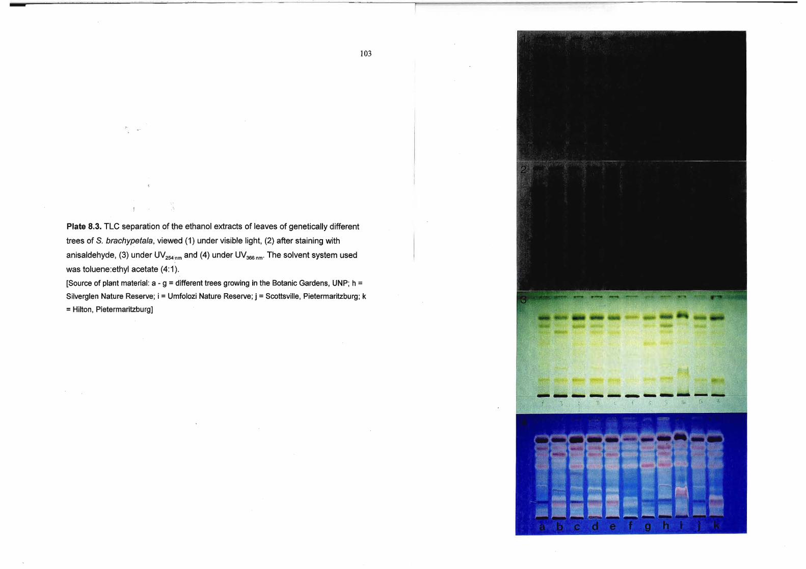
103
Plate 8.3. TLC separation of the ethanol extracts of leaves of genetically different
trees of S. brachypetaJa, viewed (1) under visible light, (2) after staining with
anisaldehyde, (3) under UV254;nm and (4) under UV366nm- The solvent system used
was toluene:ethyl acetate (4: 1).
[Source of plant material: a - 9 =different trees growing in the Botanic Gardens, UNP; h =Silverglen Nature Reserve; i =Umfolozi Nature Reserve; j =Scottsville, Pietermaritzburg; k
=Hilton, Pietermaritzburg]

r
104
Plate 8.4. TLC separation of the ethanol extracts of S. brachypetala leaf material
collected during different months, viewed (1) under visible light, (2) after staining
with anisaldehyde, (3) under l)V254nm and (4) under UV366nm' The solvent system
used was toluene:ethyl acetate. (4:1).
[Collection month: a =January, b =February, c =March, d =April, e =May, f =June, g =July, h =August, i =September, j =October, k =November, I =December]

105
I. \
Plate 8.5. TLC separation (A) of the ethanol extracts of S. brachypetala leaf extracts
prepared from fresh (F) and dry (D) material, viewed (1) under visible light, (2) after
staining with anisaldehyde, (3) under UV254 nm and (4) under UV366 nm'
TLC separation of (B) the eth?nol extracts of S. brachypetala leaf material after
being stored under various conditions, viewed (1) under visible light, (2) after
staining with anisaldehyde, (3) under UV254nm and (4) under UV366nm• The solvent
system used was toluene:ethyl acetate (4: 1).
[a =extract screened immediately after collection, drying and extraction of material; b =extract stored in freezer at -15°C for 18 months; c =dried, intact plant material stored in
dark cupboard at room temperature before being ground, extracted and screened]
@
@
f",'1
14
'I
'i"]" ', "",.',J
'I' ,

106
Table 8.1. Antibacterial activity of different plant parts of Schotia brachypetala
(voucher number McGaw58NU). Treatments in each column denoted by the same
letters are not significantly different at the 5 % level, with a standard error (SE) for
the treatment means = 1.618
Plant part1 Extract and bacteria2tested (MIC values in mg ml-1)
Ethanol Water
8.s. E.c. K.p. S.a. 8.s. E.c. K.p. S.a.
0.458 3.138 3.138 1.308 12.58 - 5.21 b
0.278 3.138 4.698 1.438 6.25c - 6.25b
0.398 3.138 3.138 2.608 8.33bc - 4.69b
ST
8K
R8
RT 0.328 3.138 4.698 1.568 10.428b- 12.58
FL 0.658 3.138 4.1r 3.138 1.30d- 0.78c
SP 0.168 1.308 2.088 0.088 0.78d- 0.71 c
LF 0.168 1.568 3.138 1.308 1.04d 4.178 12.58 5.21 b
1Plant part: 8K, bark; FL, flowers; LF, leaves; R8, root bark; RT, roots; SP, seeds and pod; ST,
young stems
28acteria: 8.s., Bacillus subtilis; E.c., Escherichia coli; K.p., Klebsiella pneumoniae; S.a.,
Staphylococcus aureus; -, MIC >12.5 mg ml-1
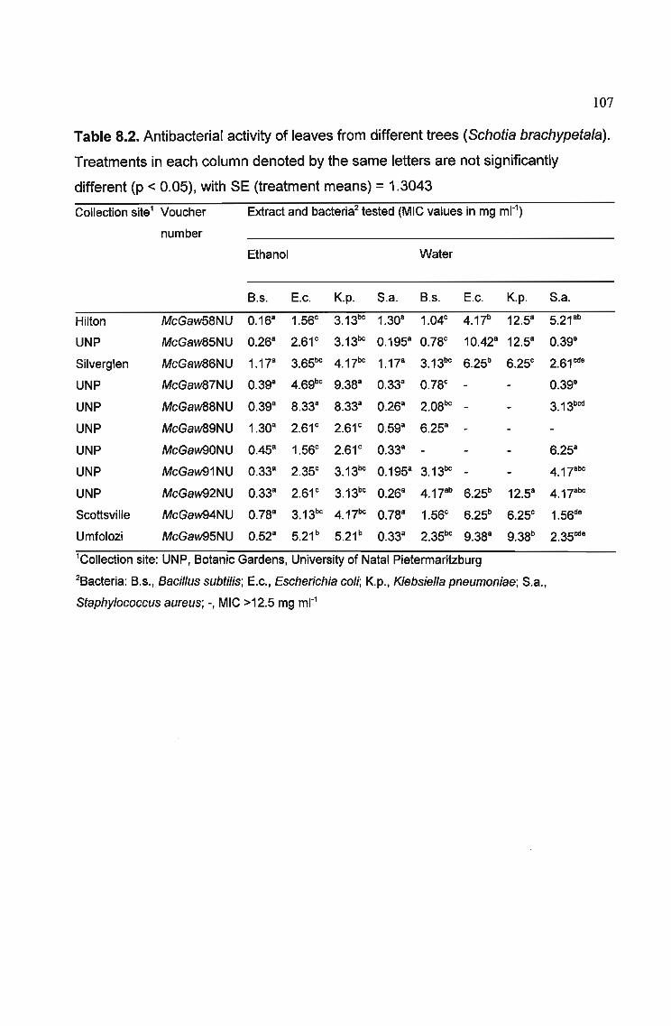
107
Table 8.2. Antibacterial activity of leaves from different trees (Schotia brachypetala).
Treatments in each column denoted by the same letters are not significantly
different (p < 0.05), with SE (treatment means) = 1.3043
Collection site1 Voucher Extract and bacteria2tested (MIC values in mg ml-1)
number
Ethanol Water
8.s. E.c. K.p. S.a. 8.s. E.c. K.p. S.a.
Hilton McGaw58NU 0.16a 1.56c 3.13bc 1.30a 1.04c 4.17b 12.5a 5.21 ab
UNP McGaw85NU 0.26a 2.61 c 3.13bc 0.195a 0.78c 10.42a 12.5a 0.398
Silverglen McGaw86NU 1.17a 3.65bc 4.17bc 1.17a 3.13bc 6.25b 6.25c 2.61 cd8
UNP McGaw87NU 0.39a 4.69bc 9.38a 0.33a 0.78c - 0.398
UNP McGaw88NU 0.39a 8.33a 8.33a 0.26a 2.08bC - 3.13bcd
UNP McGaw89NU 1.30a 2.61 c 2.61 c 0.59a 6.25a -UNP McGaw90NU 0.45a 1.56c 2.61 c 0.33a - 6.25a
UNP McGaw91NU 0.33a 2.35c 3.13bc 0.195a 3.13bc - 4.17abc
UNP McGaw92NU 0.33a 2.61 c 3.13bc 0.26a 4.17ab 6.25b 12.5a 4.17abc
Scottsville McGaw94NU 0.78a 3.13bc 4.17bc 0.78a 1.56c 6.25b 6.25c 1.56d8
Umfolozi McGaw95NU 0.52a 5.21 b 5.21 b 0.33a 2.35bc 9.38a 9.38b 2.35cd8
1Collection site: UNP, 80tanic Gardens, University of Natal Pietermaritzburg
28acteria: 8.s., Bacillus subtilis; E.c., Escherichia coli; K.p., Klebsiella pneumoniae; S.a.,
Staphylococcus aureus; ., MIC >12.5 mg ml-1

108
Table 8.3. Antibacterial activity of Schotia brachypetala leaves (voucher number
McGaw85NU) collected monthly from the same tree. Treatments in each column
denoted by the same letters are not significantly different at the 5 % level, with SE
(treatment means) =1.593
Collection time Extract and bacteria1 tested (MIC in mg ml-1)
Ethanol Water
B.s. E.c. K.p. S.a. B.s. E.c. K.p. S.a.
January 0.32a 0.41 c 0.12c 0.195a 0.52b 6.25bc 6.25c 0.19b
February 0.195a 2.61 bc 3.65b 0.13a 1.30b 3.13c 8.33bc 0.45b
March 0.15a 4.17ab 2.61 bc 0.11 a 0.78b 5.21 bc 7.29bc 0.65b
April 0.16a 4.69ab 5.21 ab 0.29a 0.39b 8.33b 10.42ab 0.58b
May 0.26a 7.29a 7.29a 0.39a 0.65b 7.29b 12.5a 0.91 b
June 0.52a 3.65b 4.17ab 0.65a 3.12ab 8.33b 10.42ab 4.30a
July 0.13a 3.13bc 4.17ab 0.42a 4.69a 12.5a 12.5a 4.69a
August 0.52a 4.1rb 5.21 ab 0.59a 1.82ab 12.5a 12.5a 2.35ab
September 0.33a 5.21 ab 7.29a 1.17a 3.12ab 10.42ab 12.5a 4.43a
October 0.268 3.65b 5.21 8b 0.55a 0.78b 5.21 bc 12.5a 1.10b
November 0.168 2.61 bc 2.61 bc 0.55a 0.65b 12.5a 12.5a O.84b
December 0.26a 4.69ab 4.69ab 0.29a 0.78b 6.25bc 7.29bc 0.91 b
1Bacteria: B.s., Bacillus subtilis; E.c., Escherichia coli; K.p., Klebsiella pneumoniae; S.a.,
Staphylococcus aureus

109
Table 8.4. Antibacterial activity of leaves of different Schotia species. Treatments in
the same column denoted by the same letters are not significantly different (p <
0.05), with SE (treatment means) =1.457
Collection site Voucher Extract and bacteria1tested (MIC in mg ml-1)
number
Ethanol Water
8.s. E.c. K.p. S.a. 8.s. E.c. K.p. S.a.
S. afra McGaw96NU 0.39& 2.61& 2.0S& 0.195& 6.25& 12.5& 12.5& 0.91&
S. brachypetala McGaw5SNU 0.26& 2.61 & 3.13& 0.195& 0.7Sb 10.42&b 12.5& 0.39&
S. capitata McGaw93NU 0.39& 2.61& 3.65& 0.195& 1.04b S.33b 12.5& 0.7S&
18acteria: 8.s., Bacillus subtilis; E.c., Escherichia coli; K.p., Klebsiella pneumoniae; S.a.,
Staphylococcus aureus
Table 8.5. Antibacterial activity of Schotia brachypetala leaf extracts prepared from
fresh and dry plant material. Treatments in the same column denoted by the same
letters are not significantly different (p < 0.05), with SE (treatment means) = 0.614
Plant material Extract and bacteria1tested (MIC in mg ml-1)
Ethanol Water
8.s. E.c. K.p. S.a. 8.s. E.c. K.p. S.a.
Fresh 0.32& 1.56b 3.13b 0.26b 1.56& 12.5& 12.5& 0.46&
Dry 1.04& 4.17& 12.5& 5.21& 0.26b 2.61b 3.13b 0.195&
18acteria: 8.s., Bacillus subtilis; E.c., Escherichia coli; K.p., Klebsiella pneumoniae; S.a.,
Staphylococcus aureus

110
Table 8.6. Antibacterial activity of Schotia brachypetala leaf extracts immediately
after being prepared, after being stored in the freezer for 18 months, and extract
made from leaf material stored in the dark for 18 months. Treatments in the same
column denoted by the same letters are not significantly different (p < 0.05), with SE
(treatment means) = 0.844
Plant material1 Extract and bacteria2tested (MIC recorded in mg ml-1)
Ethanol Water
8.s. E.c. K.p. 5.a. 8.s. E.c. K.p. 5.a.
1 0.26a 2.61 a 3.13a 0.20a 0.78a 10.42a 12.58 0.39b
2 0.16a 1.56a 3.13a 1.308 1.048 4.17c 12.5a 5.21 8
3 0.528 2.61 a 3.13a 0.398 1.04a - 0.52b
lPlant material: 1 = extract prepared and screened immediately after collection and drying of
material, 2 =extract stored in the freezer for 18 months after preparation, 3 =dry material stored in
the dark at room temperature for 18 months before being ground and extracted
28acteria: 8.s., Bacillus subtilis; E.c., Escherichia coli; K.p., Klebsiella pneumoniae; 5.a.,
Staphylococcus aureus; -, MIC >12.5 mg ml-1

111
8.4 DISCUSSION
It is known that the presence of biologically active compounds in a species may vary
with the season, locality of the plant, and between plant parts. The active
constituents may also be present to a greater or lesser extent in related species, or
even genera. An investigation was instigated to analyse the effect of these factors
on the antibacterial activity of Schotia brachypetala, a little-studied South African
medicinal plant.
8.4.1 Plant part variation
Antibacterial activity of extracts prepared from different plant parts was similar, with
most activity shown by the ethanol extracts against the Gram-positive bacteria. The
antibacterial activity was highest in the leaves, seeds and pods, and bark. The TLC
chromatogram showed that the ethanol extracts of these plant parts had several
compounds in common. The absence of statistical differences in activity between
the ethanol extracts of the plant parts was interesting, implicating the occurrence of
antibacterial compounds throughout the plant. This may indicate that one or more of
the active compounds are ubiquitous, and not localized to a specific plant part.
Traditional healers use decoctions of S. brachypetala bark or roots when treating
gastrointestinal ailments. In this study, aqueous root and bark extracts displayed
moderate antibacterial activity against Gram-positive bacteria, but none against the
Gram-negative bacteria. The water extracts of the aerial parts (leaves, flowers,
seeds and pods, but not stems) displayed much increased activity against the Gram
positive species, and in addition, the leaves showed some activity against the Gram
negative bacteria.
Although Schotia brachypetala is not a threatened medicinal plant, the collection of
leaves instead of bark or roots for therapeutic purposes would be a less destructive
and more aesthetic method of collection. The higher antibacterial activity of the leaf
extracts than the bark and root extracts indicates that leaves may be preferable for
medicinal use provided they have no toxicity. In southern Africa, the bark and

112
underground parts of medicinal plants are the most frequently utilized, leading to
cases where popular medicinal plants become threatened by destructive harvesting.
Plant part substitution has the potential for becoming an important strategy for the
conservation of medicinal plants in South Africa (ZSCHOCKE et al., 2000). Upon
investigating the possible plant part substitution of four of the most threatened South
African medicinal plants, Eucomis autumnalis (bulb), Siphonochilus aethiopicus
(rhizome), Ocotea bullata (bark) and Warburgia salutaris (bark), ZSCHOCKE et al.
(2000) concluded that the potential for plant part substitution is highly plant specific,
but should be investigated, particularly in the case of threatened medicinal plants.
The finding in this study that leaves possess superior antibacterial properties to the
bark or roots provides impetus for this approach.
8.4.2 Locational variation
The MIC values of extracts prepared from different trees were comparable, with
again more activity shown by the ethanol extracts against the Gram-positive
bacteria. The statistically significant (p < 0.05) differences in activity of the aqueous
extracts of leaves collected from different areas indicates that geographical location
does affect the activity of S. brachypetala. However, the majority of extracts had low
MIC values against Gram-positive bacteria.
The chromatograms of extracts from the genetically different trees were similar. The
slightly different chromatogram shown by the ethanol extract of the tree growing in
the Umfolozi Game Reserve may indicate additional compounds produced by the
tree in response to herbivory. This tree was the only one in the study that grew in an
area inhabited by browsers. Schotia brachypetala is a dietary plant utilised by
giraffe, kudu and impala (VAN HOVEN and FURSTENBURG, 1992). In this area, S.
brachypetala may retain secondary metabolites that confer an advantage by being
unpalatable to predators.

113
8.4.3 Seasonal variation
Antibacterial activity was present to some degree at all times of the year, particularly
in the case of ethanol and water extracts which were generally highly active against
the Gram-positive bacteria B. subtilis and S. aureus. The TLC analysis revealed little
variation in chemical composition of each of the twelve ethanol extracts prepared
during each month of the year. This reasonable degree of constancy in antibacterial
activity regardless of the season is desirable for traditional healers as they may be
assured of little variation in medicinal value of the plant.
8.4.4 Species variation
Schotia afra and S. capitata showed similar antibacterial activity to that of S.
brachypetala. ELOFF (1999a) advocated the practice of screening closely related
taxa or different populations to discover plants with a high concentration of the target
component. In this case, it appears that the Schotia species screened have a
comparable degree of antibacterial activity, and hence similar levels of active
compounds. The TLC fingerprints revealed that these Schotia species contain a
closely related chemical composition, thus supporting the chemotaxonomic theory
that similar compounds are likely to be present in plants that are taxonomically
closely related.
8.4.5 Variation after extraction of fresh or dry material
There were significant differences (p < 0.05) in the antibacterial activity of ethanol
and water extracts prepared from fresh or dried plant material when tested against
most of the bacteria. It was interesting to note that the ethanol extract of the fresh
material was more active than that of the dried material, while the reverse was true
for the water extracts. The three additional compounds present in the dried material
extract are possibly breakdown products, perhaps of chlorophyll as they fluoresced a
deep red colour under ultraviolet light at 366 nm.

114
8.4.6 Effect of storage of plant material and extracts
The similarity of the TLC chromatograms of the ethanol extract screened
immediately after preparation, and the extract stored in the freezer for 18 months
after preparation is noteworthy. It indicates that the chemical composition of the
prepared extracts remains fairly stable upon freezing. This is reflected also in the
MIC values, as there were no significant differences (p < 0.05) in the antibacterial
activity of the two different extracts. Upon storage at room temperature, the leaf
material appeared to lose compounds, possibly owing to the breakdown of
chemicals in the leaves. However, the antibacterial activity was not significantly
different from that of the extracts stored in the freezer or assayed directly after
preparation, so storage of the leaf material does not seem to affect the antibacterial
activity of S. brachypetala leaves.
The effect of storage on the activity of medicinal plants is important as these plants
are often stored in markets, or by traditional healers. ELOFF (1999a) showed that
the antibacterial components of Combretum erythrophyllum are very stable in the dry
state, as MIC values remained constant for different plants over a number of years.
The compounds responsible for the antibacterial nature of C. erythrophyllum have
not yet been identified. ELOFF (1999a) prepared extracts from herbarium
specimens of Helichrysum pedunculatum (Asteraceae), which contains the fatty
acids linoleic and oleic acids as the active antibacterial components (DILlKA et al.,
2000). ELOFF (1999a) analysed the antibacterial activity of the specimen extracts
and concluded that fatty acids appear to be stable in dry specimens stored under
herbarium conditions. The antibacterial compounds in Scholia brachypetala leaves
are fatty acids (CHAPTER 9). It would seem that these fatty acids are stable when
the dried, powdered plant material is stored in the dark at a constant temperature,
and also as an extract stored at -15°C. Many types of secondary plant products are
stable if stored under herbarium conditions in the dark and at relatively low
temperatures (ELOFF, 1999a). Samples of herbarium material have been shown to
still contain stable compounds such as amino acids, flavonoids, sweetening agents,
volatile oils and alkaloids (PHILLlPSON, 1982).

115
8.4.7 TLC fingerprinting
TLC fingerprinting is a fast, relatively cheap and effective method to obtain a
characteristic analytical fingerprint of a plant extract (WAGNER and BLADT, 1996),
although a more sophisticated study could be performed using HPLC and GC-MS
analysis in addition to TLC. TLC fingerprinting of the acidic triterpenoid fraction of
leaf extracts in the genus Combretum (Combretaceae) has been used to assist in
elucidating the taxonomy of the genus (CARR and ROGERS, 1987; ROGERS and
COOMBES, 1999). ROGERS et al. (2000) showed that leaf extracts of fourteen
species of Maytenus produced unique chemical fingerprints, supporting their specific
distinctness, although close chemical relationships between certain species were
established. In the present study of the three Schotia species, the TLC fingerprints
wree very similar, particularly in the case of S. brachypetala and S. capitata, so it
would be difficult to distinguish between these species using only TLC.
. TLC chromatograms of different samples of the same species from varying
geographical locations showed a high level of consistency, both in the study of
Combretum species (CARR and ROGERS, 1987) and Maytenus species (ROGERS
et al., 2000). The Schotia extracts in this location effect study also showed little
genetic variation in chemical composition. TLC fingerprinting to assist in
identification can therefore be applied to these genera and probably others, although
not all plant taxa may be suitable for the application of this technique. ZSCHOCKE
et al. (2000) successfully used TLC analysis to compare the chemical composition of
various plant parts of four threatened South African medicinal plants. A similar
chemical composition, as well as biological activity, is important if alternative plant
parts such as leaves are to be used instead of bark or underground parts of the
plant. In this investigation, TLC fingerprinting allowed the correlation of similar
chemical composition with similar levels of antibacterial activity.

116
8.5 CONCLUSION
The TLC fingerprinting of the ethanol extracts proved to be useful, with the
chromatograms supplying comparative information on the chemical composition of
the Schotia extracts. Extracts with a similar chemical composition tended to display
similar levels of antibacterial activity.
Most antibacterial activity was shown by Schotia brachypetala, S. afra and S. . J
capitata extracts against Gram-positive bacteria. Gram-negative bacteria are more
commonly associated with stomach ailments than Gram-positive species. The
ethanol extracts were generally more active than the aqueous extracts, but despite
this the water extracts still possessed good antibacterial activity. Traditional healers
use aqueous decoctions in preparing treatments for stomach ailments. The
antibacterial activity shown by Schotia extracts substantiates the use of the plants by
indigenous healers for treating dysentery and diarrhoea.
The two other Schotia species, S. afra and S. capitata included in the study both
displayed good antibacterial activity and similar chromatograms, lending support to
the chemotaxonomic argument.
Little variation in the biological actiVity of the tree with respect to season and locality
is important so that healers and scientific investigators may be sure of constant
activity whenever and wherever material is collected. In this study, there was not
much variation in activity with season, but some variation with differing locality.
Despite this, there was still antibacterial activity present in all the specimens tested.
The dried, ground plant material, and the frozen plant extracts of S. brachypetala
retained their antibacterial activity over time. This implies that the active constituents
are stable, both in the dried plant and as an extract. Plant material stored in a cool,
dark, dry environment will therefore maintain these active properties, which has
beneficial implications for traditional healers and collectors.

117
CHAPTER 9
ISOLATION AND IDENTIFICATION OF ANTIBACTERIAL
FATTY ACIDS FROM SCHOTIA BRACHYPETALA
9.1 INTRODUCTION
9.1.1 Schotia brachypetala: biological activity and chemical constituents
There have been few reports on the chemistry and biological activity of Schotia
species. Astringent tannins in the bark of S. brachypetala may account for some of
the activity of the plant (BRUNETON, 1995). The wood dust and root are also
believed to contain tannins (WAn and BREYER-BRANDWIJK, 1962). Stilbenes
and phenolics [(E)-1-(3,5-dihydroxyphenyl)-2-(3,4,5-trihydroxyphenyl)-ethylene and
(Z)-1-(3,5-dihydroxyphenyl)-2-(3,4,5-trihydroxyphenyl)-ethylene] are reported to be
present in S. brachypetala (GLASBY, 1991). Polyhydroxystilbenes have been
isolated from the heartwood of the tree (DREWES, 1971; DREWES and
FLETCHER, 1974). The main constituent is trans-3,3',4,5,5'-pentahydroxystilbene,
with small amounts of trans-3,3',4,5'-tetrahydroxystilbene, catechin, epicatechin, and
trace amounts of cis-3,3' ,4,5,5'-pentahydroxystilbene. Many stilbenes have antibiotic
effects (VAN WYK et al., 1997), but the biological activity of the Schotia stilbenes
has not been reported. BONNIER (1954) described the inhibition by Schotia species
of a Rhizobium strain specific for alfalfa, while strains specific for soybeans, garden
peas and clover were unaffected.
In the initial screening of plants used in South Africa for treatment of gastrointestinal
disorders, S. brachypetala demonstrated good antibacterial activity against a range
of bacteria (CHAPTER 5; MCGAW et al., 2000a). This species was selected for
further investigation using bioassay-guided fractionation to discover some of the
118
compounds responsible for antibacterial activity because of its wide usage in
traditional medicine and because of the lack of previous investigation on this
species. Although the roots are used in traditional medicine, the leaves possess
antibacterial activity superior to that expressed in the roots (CHAPTER 8; MCGAW
et al., 2001). For reasons of availability, further investigation was continued with the
leaves.
9.2 MATERIALS AND METHODS
9.2.1 Bulk extraction
Leaves of Schotia brachypetala Sond. were harvested from a young tree (about 12
years old) growing in the Botanic Gardens, UNP, in August 2000. A voucher
specimen (McGaw85NU) was deposited at the Herbarium of the University of Natal,
Pietermaritzburg. The leaves were dried at 50°C and ground to a powder using a
Little Hippo mill.
The powdered leaves (1.2 kg) were extracted with 3 I ethanol by sonication for 30
min, and overnight maceration. The extract was filtered through a Buchner funnel
and Whatman No. 1 filter paper, and the leaves were re-extracted twice, with 0.51
ethanol each time. The filtration was repeated, and the solvent evaporated under
reduced pressure at 40°C.
9.2.2 Bioassay-guided fractionation for isolation of active compounds
Throughout the isolation procedure, all solvents used were redistilled using a rotary
evaporator.
The antibacterial activity of fractions after each purification stage was tested using
the direct bioautography assay (HAMBURGER and CORDELL, 1987). In this assay,
an overnight culture of test bacteria in 20 ml MH broth was pelleted by centrifugation
119
at 3 000 9 for 10 min and resuspended in 10 ml fresh MH broth. This suspension was
sprayed on a developed TLC plate and incubated at 37°C overnight. A 2 mg ml-1
solution of INT (iodonitrotetrazolium violet) was then sprayed on the plate and
incubated to detect the areas of bacterial growth inhibition. Antibacterial compounds
on the TLC plate were visible as white spots against a deep red background, as
bacterial growth reduces the tetrazolium salt to a red formazan product. The test
organism was Staphylococcus aureus (ATCC 12600).
9.2.2.1 Liquid-liquid partitioning
The extract was dissolved in ethanol (400 ml) and water (100 ml), and partitioned
with hexane (500 ml) in a 2 I separating funnel. This was repeated a further three
times. The aqueous fraction was subsequently partitioned with dichloromethane (500
ml) three times.
9.2.2.2 Vacuum Liquid Chromatography
The hexane portion was fractionated by Vacuum Liquid Chromatography (VLC) on
silica gel 0.04-0.063 mm (Merck). A column 27 cm in length and with an internal
diameter of 6 cm was packed with 140 9 silica. A hexane:ethyl acetate gradient
solvent system with increasing amounts of ethyl acetate was used (400 ml per
fraction); fr. I: 100 % n-hexane (490 mg), fr. 11: 97.5% n-hexane (530 mg), fr. Ill: 95%
n-hexane (7800 mg), fr. IV: 92.5% n-hexane (2320 mg), fr. V: 90% n-hexane (1570
mg), fr. VI: 87.5% n-hexane (2180 mg), fr. VII: 85% n-hexane (1570 mg), frVIII:
82.5% n-hexane (790 mg), fr. IX: 80% n-hexane (890 mg), fr. X: 77.5% n-hexane
(1100 mg), fr XI: 75% n-hexane (860 mg), fr. XII: 72.5% n-hexane (1000 mg). fr. XIII:
70% n-hexane (860 mg), fr. XIV: 67.5% n-hexane (940 mg), fr. XV: 65% n-hexane
(1010 mg), fr. XVI: 62.5% n-hexane (970 mg), fr. XVII: 60% n-hexane (360 mg), fr.
XVIII: 57.5% n-hexane (660 mg), fr. XIX: 55% n-hexane (520 mg), fr. XX: 52.5% n
hexane (420 mg), fr. XXI: 50% n-hexane (970 mg), fr. XXII: 0% n-hexane (1460 mg).

120
9.2.2.3 Chlorophyll extraction
Chlorophyll was separated from each of the two sets of combined fractions by
column chromatography over Sephadex LH-20 (Pharmacia). A column of length 75
cm, and internal diameter of 2.5 cm was used. Sephadex was packed to a height of
45 cm in the column. The solvent system employed was
cyclohexane:dichloromethane:methanol (7:4: 1). Fractions of approximately 19 ml
were collected using a fraction collector (Gilson FC 2038). The active compounds
were detected using TLC and the bioautography assay.
9.2.2.4 Gravity-assisted Column Chromatography
Column chromatography was performed on the first combined set of active fractions,
referred to as fraction A. A column of 71 cm length and 2.5 cm interr)al diameter
packed with 125 g silica gel 0.04-0.063 mm (Merck) was used. One hundred ml of n
hexane was run through the column, followed by 500 ml each of 80% n-hexane,
75% n-hexane, 66.6% n-hexane and 50% n-hexane in ethyl acetate. The column
was washed with 600 ml ethyl acetate. A fraction collector (Gilson FC 2038) was set
to collect fractions of approximately 10 ml each. TLC and bioautography were again
used to detect the presence of the active compound.
9.2.2.5 Preparative Thin Layer Chromatography
Approximately 20 mg of fraction A were applied to each of ten TLC plates (Merck
glass plates, 20 x 20 cm, Silica gel 60 F254). The solvent system was hexane:ethyl
acetate (2: 1), and plates were developed to a height of 15 cm. A strip of one TLC
plate was cut off with a glass-cutting knife, and sprayed with AS spray reagent. The
TLC plate strip was then heated for 10 min at 100°C. The active compound is not
detectable under visible or ultraviolet light at 365 nm, but is faintly visible under
ultraviolet light at 254 nm and is present as a vivid green stain after spraying with AS
spray. The zone containing the active fraction on the unsprayed TLC plates was
scraped off the plates, and the compound (A) eluted from the silica with ethanol.

121
Fraction B was also subjected to preparative TLC. The same procedure as for
fraction A was followed. Compound B also appeared green upon spraying with AS
spray.
The purity of the two compounds was confirmed by TLC using various solvent
systems. Using a solvent system of hexane:ethyl acetate (2: 1), compound A was
visible at Rf = 0.49. With a solvent system of hexane:ethyl acetate (1 :2), compound
A appeared at Rf =0.81, and compound B was visible at ~ =0.56.
9.2.3 Identification of purified active compound
High Resolution Mass Spectrometry (MS) using a VG70-SEQ spectrometer was
performed. Nuclear Magnetic Resonance Spectroscopy CH NMR and 13C NMR) was
carried out to confirm the structure of the compound. A Varian Unity Inova 500 MHz
spectrometer was used.
Th~. Minimal Inhibitory Concentration (MIC) values of the pure compounds were
determined against the Gram-positive Bacillus subtilis (ATCC 6051) and
Staphylococcus aureus (ATCC 12600) and the Gram-negative Escherichia coli
(ATCC 11775) and Klebsiella pneumoniae (ATCC 13883). The microplate method of
ELOFF (1998a) in 96-well microtiter plates (CHAPTER 5, Section 5.2.3) was used.
9.3 RESULTS
9.3.1 Bulk extraction
The bulk extraction of the leaves of S. brachypetala yielded a concentrated oily, dark
green residue of 100.6 g (8.4 %).
9.3.2 Bioassay-guided fractionation for isolation of active compounds

122
9.3.2.1 Liquid-liquid partitioning
Bioautography revealed the presence of antibacterial compounds in the hexane
extract after the partitioning step. The mass of the hexane residue after removal of
the solvent was 34.95 g.
9.3.2.2 Vacuum Liquid Chromatography
After the VLC step in the isolation of compounds, bioautography revealed the
presence of antibacterial compounds eluted in fractions 8 - 15 (which were
combined to produce fraction A), and fractions 16 - 22 (combined separately to
produce fraction B). These groups of fractions each contained an active compound
at a different Rf value on a TLC plate. The TLC and bioautographic analysis of the
VLC fractions is represented in Plate 9.1. The antibacterial compounds A and Bare
indicated with arrows on the TLC chromatograms.
9.3.2.3 Chlorophyll extraction
Filtration of each of the two residues (A and B) through Sephadex allowed the
chlorophyll to be removed in the first few fractions, after which the active compounds
eluted from the column. The active fractions from fraction A after elution through
Sephadex were again combined and dried to produce a residue with a mass of
393.5 mg. The combined active fractions resulting from the separation of fraction B
over Sephadex had a mass of 198 mg.
9.3.2.4 Gravity-assisted Column Chromatography
Bioautography on the fractions resulting from the gravity-assisted column
chromatography of fraction A revealed the presence of the active compound in
fractions 105-191. On combining and drying these fractions, a residue of 198.3 mg
(again referred to as fraction A) was produced.

123
9.3.2.5 Preparative Thin Layer Chromatography
Following preparative TLC on fraction A, the mass of the isolated compound A was
18.7 mg. Preparative TLC on the residue of fraction B resulted in an antibacterial
compound B of mass 9 mg.
9.3.3 Identification of purified active compound
NMR and MS resulted in the identification of two separate antibacterial mixtures of
fatty acids. The major compounds were 9,12,15,:,octadecatrienoic acid (linolenic
acid) (42%) in the case of compound A, and methyl-5,11 ,14, 17-eicosatetraenoic
acid (37%) in the case of compound B.
The MIC values of the isolated fatty acids, as well as those of linolenic acid
purchased from Sigma, are presented in Table 9.1. The MICs of the isolated
linolenic acid were not as low as those of the linolenic acid standard from Sigma.
This was to be expected as the isolated fatty acids were not completely pure, but
contaminated with smaller amounts of other fatty acids. Both compounds displayed
good bactericidal activity against Gram-positive B. subtilis and S. aureus, and a
lesser degree of activity against Gram-negative E. coli and K. pneumoniae.
Table 9.1. MIC values (mg ml-1) of fatty acids
Microorganism MIC of compound

124
Plate 9.1. TLC separation (A) of the VLC fractions of the hexane fraction from the
liquid-liquid partitioning step, viewed (1) after staining with anisaldehyde, (3) under:;
UV254nm and (4) under UV366nm~\Thebioautography plate (2) shows white areas
where bacterial growth was inh.ibited. The arrows indicate the two fatty acids,
compounds a (9, 12,15-octadecatrienoic acid) and b (methyl-5, 11,14,17
eicosatetraenoate) isolated frorp S. brachypetala. The solvent system used was
hexane:ethyl acetate (2: 1).
[H =hexane extract from the liquid-liquid partitioning step before being submitted to VLCj
TLC separation (8) of the different plant parts of S. brachypetala viewed (1) after
staining with anisaldehyde. The bioautography plate (2) shows the position of the
two subsequently isolated fatty acids, indicated by the arrows. It is clear that these
compounds are present in the leaves, bark and roots; the roots possess additional
antibacterial compounds, as do the flowers. The solvent system used was
hexane:ethyl acetate (2:1).
[If =leaf, st =stem, bk =bark, rt =root, rb =root bark, fl =flower, sp =seeds plus pods]
a
1
a
b

125
9.4 DISCUSSION
9.4.1 Isolation and identification of active compounds
The bulk extraction of S. brachypetala leaves and the subsequent bioassay-guided
fractionation to isolate antibacterial compounds resulted in the identification of two
fatty acids, namely 9, 12,15-octadecatrienoic acid and methyl-5,11,14,17
eicosatetraenoate. Unsaturated fatty acids are widespread in both leaf and seed oils
of many plant species (HARBORNE and BAXTER, 1993). TLC analysis and
bioautography have indicated that similar antibacterial compounds are present in the
leaves, roots and bark of S. brachypetala. Photographic evidence is shown in Plate
9.1. The position of compounds A and B (9,12, 15-octadecatrienoic acid and methyl
5,11,14,17-eicosatetraenoate, respectively) are indicated by arrows. The roots
possessed additional antibacterial compounds than the bark or leaves. This warrants
further investigation.
9.4.2 Fatty acids as constituents of plants
Fatty acids occur mainly in bound form in plants, esterified to glycerol, as fats or
lipids (HARBORNE and BAXTER, 1993). These lipids comprise up to 7 % of the dry
weight in leaves in higher plants, and are important membrane constituents in
chloroplasts and mitochondria (HARBORNE and BAXTER, 1993). The seeds and
fruits of many plants also contain lipids in considerable amounts, providing a storage
form of energy to use during germination (HARBORNE and BAXTER, 1993).
The common fatty acids in plants are either saturated or simple unsaturated
compounds of C16 or C18 chain length (HARBORNE and BAXTER, 1993). Palmitic
acid (C16) is the major saturated acid in leaf Iipids, also occurring in some seed oils,
while stearic acid (C18) is less prominent in leaf Iipids but is a major saturated acid in
seed fats in several plant families (HARBORNE and BAXTER, 1993). Unsaturated
acids based on C16 and C18 are widespread in leaf and seed oils (HARBORNE and
BAXTER, 1993).The tri-unsaturated linolenic acid is common, as are linoleic and

126
oleic acids (HARBORNE and BAXTER, 1993). Whereas plant seeds contain a wide
variety of fatty acids, those from leaf tissue are remarkably constant from plant to
plant (HARWOOD, 1980). Just as the complex lipid composition of higher plant
leaves is relatively constant, so the fatty acid content is also quite characteristic.
Quantitatively, the major fatty acids are palmitic, linoleic and, in particular, a-linolenic
acids (HITCHCOCK and NICHOLS, 1971; HARWOOD, 1980). The membranes of
chloroplasts contain exceptionally high (about 90 % in some lamellae) percentages
of a-linolenic acid (HITCHCOCK and NICHOLS, 1971; HARWOOD, 1980).
In general, fatty acids do not exist as free carboxylic acids because of their affinity
for many proteins (GURR and JAMES, 1980). One result of this is an inhibitory
action on most enzymes. Where free acids have been reported as major
constituents they are usually artefacts due to cell damage which allows lipases to
act on acyllipids of the tissue (GURR and JAMES, 1980). One major exception to
this rule lies in the albumin bound fatty acids {ABFA) of mammalian blood. These
are also referred to as FFA (free fatty acids) or NEFA (non-esterified fatty acids)
(GURR and JAMES, 1980).
Variation in the fatty acid and sterol composition of plants has long been of interest
to chemotaxonomists. Fatty acids from seeds and leaves, more than sterols, have
been previously used for plant family classification into classes and subclasses
(PERDETZOGLOU et al., 1996). Since variation in fatty acid composition is rather
small among plant families, quantitative differences of the fatty acid composition
have been used as chemotaxonomic tools (PERDETZOGLOU et al., 1996).
9.4.3 Compound A: 9,12,15-octadecatrienoic acid
AII-cis-9,12,15-octadecatrienoic acid (a-linolenic acid, 18:3) occurs in higher plants
and algae, especially as a component of galactosyl diacylglycerol (GURR and
JAMES, 1980). Linolenic acid, CH3-CH2-CH=CH-CH2-CH=CH-CH2-CH=CH-[CH2h
COOH, is the most usual form of triethenoid C18 acid found in seed fats (HILDITCH,
1956). It forms 50% or more of the mixed fatty acids in linseed oil, and also occurs in
127
other seed oils (HILDITCH, 1956).
COOPER et al. (1985) described the antibacterial activity of linolenic acid against
Bacillus subtilis and Vibrio parahaemolyficus. The marine bacterium, V.
parahaemolyficus, is a major cause of gastroenteritis in the countries where sea fish
and its products are consumed (COOPER et al., 1985). Linolenic acid had inhibitory
activity against spores of Clostridium botulinum, Clostridium sporogenes and
Bacillus cereus (ABABOUCH et al., 1992). LACEYand LORD (1981) reported that
25 strains of Escherichia coli, 25 Pseudomonas aeruginosa, 16 Proteus spp. and 25
Klebsiella spp. were all resistant to 0.25 % (v/v) linolenic acid.
GIAMARELLOS-BOURBOULlS et al. (1995) demonstrated the inhibition of the
Gram-negative E. coli by gamma-linolenic acid (GLA).The possible mechanism of
GLA action on E. coli could be attributed either to an alteration of cell membrane
properties induced by GLA or to the generation of free radicals from GLA, which
may led to membrane damage and ultimately bacterial death (GIAMARELLOS
BOURBOULlS et al., 1995). Further studies are necessary to clarify the mechanism
of GLA action on E. coli, and the clinical importance of these findings.
Alpha-linolenic acid was found to have strong anti-conidial germination activity
against blast fungus (SEKIZAWA et al., 1981). Linoleic and linolenic acid have been
identified as antialgal substances in the culture medium of the green alga
Chlamydomonas reinhardtii (MCCRACKEN et al., 1980, cited by OHTA et al., 1995).
The extensive use of antibiotics by medical institutions in Japan (OHTA et al., 1995)
and the state of Victoria in Australia (MCDONALD et al., 1981), has led to outbreaks
of infections by methicillin-resistant Staphylococcus aureus (MRSA), against which
antibiotics are often ineffective. This is most likely a world-wide phenomenon. The
symptoms of infection with MRSA in immunosuppressed patients are fever and
diarrhoea, followed by respiratory difficulty, damage to internal organs, and
sometimes death (OHTA et al., 1995). Using activity-directed fractionation of
methanol extracts of the green algae Chlorococcum strain HS-101 and Dunaliella
128
primalecta, OHTA et al. (1995) isolated the polyunsaturated fatty acid, a-linolenic
acid (C18:3), which showed antibacterial activity against MRSA. LACEYand LORD
(1981) showed that S. aureus was inhibited by linolenic acid, while S. epidermidis
was relatively resistant. LACEYand LORD (1981) recommended that the possibility
of therapeutic use of linolenic acid as an antibacterial agent should be explored.
Linolenic acid, although present in small quantities in human skin (WILKINSON,
1972, cited by LACEY and LORD, 1981) may well be an important naturally
occurring antibacterial agent, and its presence could explain why pathogenic
staphylococci are rarely found on intact skin (LACEY and LORD (1981).
9.4.4 Compound B: methyl-5,11,14,17-eicosatetraenoate
KNAPP and MELLY (1986) found that several Gram-positive species, including
Bacillus megaterium, S. aureus and S. megaterium, were susceptible to arachidonic
acid, an isomer of 5c,11 c,14c,17c-eicosatetraenoic acid (20:4). AII-cis-5,8,11,14
eicosatetraenoic acid (arachidonic acid) occurs as a major component of animal
Iipids and some algae, especially as a component of phospholipids (GURR and
JAMES, 1980).
RAKOFF (1993) stated that methyl5,11,14,17-eicosatetraenoate (5,11,14,17 - 20:4)
is potentially an intermediate in the conversion of linolenate (9,12,15 - 18:3) to
eicosapentaenoate (5,8,11,14,17 - 20:5). DE ALANIZ et al. (1976) and GASPAR et
al. (1977) reported that hepatoma cells converted a-linolenic acid to octadeca-
6,9, 12,15-tetraenoic acid (18:4), eicosa-11,14,17-trienoic acid (20:3), eicosa
8,11,14,17 and 5,11,14,17-tetraenoicacids (20:4) and eicosa-5,8,11,14,17
pentaenoic acid (20:5), and also to myristic, palmitic, palmitoleic, stearic and oleic
acids.
9.4.5 Fatty acids as antimicrobial agents
Although they generally function as anionic surface agents, the bactericidal and
antifungal properties of fatty acids are well known (KABARA et al., 1972). There

129
exist volatile fatty acid- 01FA-) producing anaerobic bacteria (mainly Bacteroides) in
the intestines of man and other animals, and these VFAs appear to be self-limiting
substances of excessive multiplication of bacteria (PROHASZKA and BARON,
1982). There are insufficient data on the role of the antibacterial activity of VFA in
enteric diseases of domestic animals and man. In an effort to address this,
PROHASZKA and BARON (1982) studied the VFA-associated antibacterial activity
of the large-intestinal contents of healthy rabbits and swine in vitro. The total
detectable quantity of volatile fatty acids (VFA) represented the sum of 3 VFA
components: acetic acid (C2), propionic acid (C3) and butyric acid (C4)
(PROHASZKA and BARON, 1982). The results indicated that the VFA in the large
intestine of rabbits and swine have inhibitory activity not only towards E. coli, but
also towards other members of the Enterobacteriaceae, including Salmonella,
Shigella, Proteus and Klebsiella (PROHASZKA and BARON, 1982). From studies
on human faeces, PROHASZKA and BARON (1982) drew the conclusion that the
VFA concentration present in the large intestine of humans is similar to that found in
the intestine of swine, so presumably the antibacterial effect of the human VFA is
similar to that of the VFA of the swine.
OHTA et al. (1995) demonstrated the antibacterial activity of a-linolenic acid (18:3)
against MRSA. MCDONALD et al. (1981) reported on the susceptibility of several
strains of MRSA to linolenic acid and hydrolysed linseed oil (containing 52% linolenic
acid in the ester form). MCDONALD et al. (1981) described the potential role of
preparations containing hydrolysed linseed oil in the eradication of staphylococcal
infection, and in the prophylaxis of infection in debilitated patients.
The possibility of the therapeutic use of linolenic acid as an antibacterial agent
should be explored (LACEY and LORD, 1981). a-Linolenic acid is generally
considered to have low toxicity, so it may potentially be administered to patients
infected with MRSA as a dietary treatment (OHTA et al., 1995). It is naturally
occurring, and hence unfavourable reactions would not be anticipated, it should not
destroy the commensal flora, resistance to it would not develop, and there would
apparently be no risk of resistance to antibiotics developing during its use (LACEY

130
and LORD, 1981).
NIEMAN (1954) stated that some natural defence systems of higher organisms,
such as the self-disinfection of the skin, are due at least partly to the presence of
antibacterial fatty acids in situ. However, the potential antibacterial activity of fatty
acids in vivo may be neutralized by adsorption on proteins in the bloodstream
(NIEMAN, 1954).
Other fatty acids with antibacterial activity have been isolated from plants using
bioassay-guided fractionation. CERDEIRAS et al. (2000) identified 11-0-(6'-0
acetyl-~-D-glucopyranosyl)-stearicacid as the main antibacterial component of aerial
parts of Ibice/la lutea. This fatty acid derivative showed an interesting antibacterial
activity, being active against E. coli, P. aeruginosa, B. subtilis and S. aureus
(CERDEIRAS et al., 2000). DILlKA et al. (2000) described the antibacterial activity
of linoleic and oleic acids isolated from the leaves of Helichrysum pedunculatum.
Linoleic and oleic acids inhibited the growth of Gram-positive Bacillus subtilis,
Micrococcus kristinae and Staphylococcus aureus and linoleic acid also showed
activity against B. cereus and B. pumilis (DILlKA et al., 2000). Both acids displayed
no activity against Gram-negative Enterobacter cloacae, Escherichia coli, Klebsiella
pneumoniae, Pseudomonas aeruginosa and Serratia marcescens (DILlKA et al.,
2000).
9.4.6 Structure- function relationships of fatty acids
There is an intimate relationship between the structure of fatty acids and their ability
to function as antimicrobial agents. The most effective saturated, monounsaturated,
and polyunsaturated fatty acids are those with chain lengths of C12, C16:1 and C18:2 ,
respectively (KABARA, 1980). The number and position of double bonds is more
important to fatty acids longer than 12 carbons than for fatty acids with fewer
carbons (KABARA, 1980). Gram-positive bacteria are more susceptible to fatty acids
than are Gram-negative bacteria. Yeasts are affected to a greater extent by fatty
acids containing 10-12 carbons, while Gram-positive bacteria are more affected by

131
slightly longer chain lengths (KABARA, 1980). Gram-negative organisms are
affected by very short chain fatty acids, that is C6 or less (KABARA, 1980). Fatty
acids with greater than eight carbons are not inhibitory to Gram-negative bacteria
(KABARA, 1980).
Fatty acid esters of sucrose have been reported to have antimicrobial properties
against Gram-negative bacteria, although to a much lower extent than that shown
for Gram-positive bacteria (MARSHALL and BULLERMAN, 1994). Fungi are
inhibited to a greater extent with acetylenic derivatives than with ethylenic
derivatives of fatty acids (MARSHALL and BULLERMAN, 1994). Fatty acids
esterified to monohydric alcohols have no antimicrobial activities, while esterification
of fatty acids to polyhydric alcohols, such as glycerol or sucrose, increases the
antimicrobial effectiveness of the fatty acids (KABARA, 1980). The fatty acid used to
esterify the polyol dictates the potency of the ester. Lauric acid (C12) and palmitoleic
acid (C16:1) form the most active saturated and unsaturated esters, respectively, and
monoester forms are more potent than polyester forms (KABARA, 1980).
MARSHALL and BULLERMAN (1994) concluded that the antimicrobial properties of
sucrose fatty acid esters, in addition to other functional properties such as
emulsification or stabilization, suggests potential use of these and related
compounds in the food processing, cosmetic and pharmaceutical industries.
OHTA et al. (1995) tested ten fatty acids and their methyl esters against
Staphylococcus aureus and MRSA. They found that palmitic acid (C16:0), stearic acid
(C18:0) and oleic acid (C18:1) had no activity, but fatty acids with two or more double
bonds (starting with C18:2) were active. Of the polyunsaturated fatty acids tested, the
activity of v-linolenic acid (C18:3) was the highest, and a-linolenic acid (C18:3),
eicosapentaenoic acid (C20:5), and docosahexaenoic acid (C22:6) also had strong
activity. The methyl esters were either inactive or much less active than the other
form (OHTA et al., 1995). In a similar experiment with E. coli, OHTA et al. (1995)
found that all fatty acids were inactive.
In general, inhibitory properties of fatty acids appear more pronounced with longer
132
and more unsaturated compounds (NIEMAN, 1954). It appears that unsaturation is
not essential for inhibition of fungi and mycobacteria by fatty acids (NIEMAN, 1954).
The position of unsaturation affects biological activity (KABARA, 1986). The
stereochemistry of the fatty acids also has an effect, with the (natural) eis-forms of
unsaturated fatty acids exhibiting a greater antibacterial activity than the
corresponding tfans-isomers (NIEMAN, 1954; KABARA, 1986). Unsaturation in
contrast to esterification is less effective with low chain fatty acids as compared to
higher chain fatty acids (KABARA, 1986). Monoesters of fatty acids are more active
than the corresponding fatty acid (KABARA, 1986).
9.4.7 Mechanism of antimicrobial action of fatty acids
The antimicrobial effects of fatty acids and their derivatives have been known for
many years, and there have been various suggestions as to the mechanism of
antibacterial action (KNAPP and MELLY, 1986). Fatty acids can inhibit the growth of
numerous types of bacteria, as well as protozoans, viruses and fungi (NIEMAN,
1954; KNAPP and MELLY, 1986). In general, fatty acid sensitivity is considered to
be a characteristic of Gram-positive bacteria, with few Gram-negative species being
susceptible (KNAPP and MELLY, 1986). Members of the Gram-negative families
Neisseriaceae, Enterobacteriaceae and Parvobacteriaceae are inhibited by small
amounts offatty acids (NIEMAN, 1954).
Short-chain saturated fatty acids and long-chain polyunsaturated fatty acids appear
to exert their antimicrobial effects by different mechanisms. Short-chain fatty acids
are generally toxic in high concentrations in a pH-dependent manner and are known
to have an adverse effect on the energy metabolism of a wide variety of
microorganisms (KNAPP and MELLY, 1986). On the other hand, polyunsaturated
fatty acids affect only certain types of bacteria and are toxic at much lower
concentrations in a more pH-independent fashion (KNAPP and MELLY, 1986).
MARSHALL and BULLERMAN (1994) reported that the mechanism of action of
sucrose esters of fatty acids appears to be biostatic rather than biocidal.

133
BERGSSON et al. (1999) tested several fatty acids and their 1-monoglycerides
against Neisseria gonorrhoeae, a Gram-negative coccus. Lauric acid (C12:0),
palmitoleic acid (C1S:1) and monocaprin (C10:0) effectively killed the pathogen.
BERGSSON et al. (1999) hypothesized that the lipids kill the bacteria by disruption
of their cell membranes (visible with electron microscopy), and because of this lipid
action on biological membranes, the emergence of resistance is unlikely.
TSUCHIDO et al. (1985) discovered that the addition of saturated Cs, Cs, C10, and
C12 fatty acids appeared to lyse actively growing cells of Bacillus subtilis 168, as
judged by a decrease in the optical density of the culture. TSUCHIDO et al. (1985)
suggested that fatty acid-induced lysis of B. subtilis 168 is due to the induction of
autolysis by an autolytic enzyme rather than massive solubilization of the cell
membrane by the detergent-like action of the fatty acids. According to TSUCHIDO et
al. (1985), it is unclear how fatty acids induce autolysis. TSUCHIDO et al. (1985)
stated that it is unlikely that fatty acid directly activates autolytic enzymes in the cell
wall, but rather, the primary target of action of fatty acids may be the cell membrane.
One explanation for the induction of autolysis by fatty acids is that the fatty acids
solubilize autolysins from the membrane at relatively low concentrations; this is
supported by the results of experiments on protoplasts (TSUCHIDO et al., 1985). In
addition, the dependency of the lytic action of fatty acids on the carbon chain length
of the molecule suggests that the surface-active action of the acid might participate
in the dissociation of autolysins from the membrane (TSUCHIDO et al., 1985). To
summarize, the short and medium chain fatty acids tested by TSUCHIDO et al.
(1985) seem to have a dual effect on cellular lysis, depending on their concentration,
with the action on the cell membrane leading to the induction of autolysis being
detectable at relatively low concentrations, and the association with the autolytic
enzyme itself being strong at relatively high concentrations. The mechanism of lytic
action of fatty acids seems to be different from that of their bactericidal action
(TSUCHIDO et al., 1985).
In a subsequent study, TSUCHIDO et al. (1987) examined the lytic action of glycerol
and sucrose esters of fatty acids with different carbon chain lengths. It was found
134
that glycerol dodecanoate and sucrose hexadecanoate induced autolysis in cells of
B. subtilis 168, and during treatment a great loss of viability occurred preceding lysis
(TSUCHIDO et al., 1987). The esters caused morphological changes in the cells, but
an apparent adaptation of the cells to the esters was noted (TSUCHIDO et al.,
1987). As with short- and medium-chain fatty acids (TSUCHIDO et al., 1985), the
lysis of B. subtilis cells induced with glycerol dodecanoate and sucrose
hexadecanoate may be due to the action of autolytic enzymes and not to the direct
action of solubilization of the bacterial membrane (TSUCHIDO et al., 1987). After
lysis the surviving cells grew, suggesting that these cells adapted to esters, as
indicated by their renewed growth and tolerance to esters if they were added again
(TSUCHIDO et al., 1987). The cell death observed by TSUCHIDO et al. (1987)
during the treatment with esters was very rapid. This suggests that the mechanism
that causes death is different from that which causes lysis and that direct interaction
of esters with the cell membrane may cause the cell death, although the possibility
that an irreversible triggering of cell lysis induces apparent rapid death cannot be
ruled out (TSUCHIDO et al., 1987). The fact that cells treated with esters altered cell
morphology suggests the inhibition of some process of synthesis or regulation of the
cell envelope which is possibly related to the induction of autolysis (TSUCHIDO et
al., 1987). Glycerol and sucrose esters of fatty acids are supposed to be effective
nontoxic antimicrobial agents, but the adaptation seen in this study may be a
problem if this phenomenon exists in microorganisms that cause food spoilage or
that produce toxins (TSUCHIDO et al., 1987).
Fatty acids inhibit the growth and oxygen consumption of Bacillus subtilis in nutrient
medium by inhibiting the transport of substances such as amino acids and keto
acids through the cellular membrane (FREESE et al., 1973). The effectiveness of
inhibition increased with increasing chain length (FREESE et al., 1973). In contrast,
the inhibitory effect on E. coli increased only up to a fatty acid chain length of six,
while long-chain fatty acids had no effect (FREESE et al., 1973).
With the exception of mycoplasmas, bacteria produce cell walls (NIKAIDO and
VAARA, 1985). Gram-negative bacteria are typically surrounded by an outer

135
lipopolysaccharide (LPS) layer (OSBORN, 1969). This outer layer is very important
in the physiology of Gram-negative bacteria, making them resistant to various host
defence factors which are toxic to Gram-positive bacteria (NIKAIDO and VAARA,
1985). This is particularly relevant in the case of enteric Gram-negative bacteria
living in the intestinal tract of animals. The outer membrane of enteric and some
other Gram-negative bacteria acts as a strong permeability barrier to many
antibiotics that are effective against other bacteria (NIKAIDO and VAARA, 1985).
Even when the diffusion of the antibiotic is only slowed down by the presence of the
outer membrane, the bacteria can then inactivate the small amount of penetrating
antibiotic rather than attempt to inactivate the almost infinite amount of antibiotic
present in the surrounding medium (NIKAIDO and VAARA, 1985).
The LPS layer prevents the entry and subsequent inhibition by intermediate and
long-chain fatty acids (SHEU and FREESE, 1973). SHEU and FREESE (1973)
suggested that this protection would be essential for the survival of bacteria in the
intestinal tract where such fatty acids are produced by the digestion of fats.
However, these bacteria are not resistant to short-chain fatty acids up to hexanoate,
so these fatty acids may therefore be useful for the treatment of infection by Gram
negative bacteria.
KNAPP and MELLY (1986) found that the presence or absence of a polysaccharide
capsule did not alter the susceptibility of Staphylococcus aureus to arachidonic acid.
Upon treating Gram-negative Neisseria gonorrhoeae and Haemophilus influenzae
with arachidonic acid, KNAPP and MELLY (1986) observed, using electron
microscopy, a loss of ribosomes, increased irregularity of outer membranes,
separation of outer and inner membranes, loss of cell shape and complete cell
disorganization and disruption. There were no morphological changes in E. coli
resistant to the toxic effects of arachidonate (KNAPP and MELLY, 1986). After
treating the Gram-positive Staphylococcus aureus with arachidonate, KNAPP and
MELLY (1986) saw no visible alteration in cell wall structure, but the arachidonate
induced cell killing was associated with the appearance of peripheral cytoplasmic
condensations similar to those seen in the susceptible Gram-negative organisms.
136
The Gram-negative species sensitive to arachidonic acid were the same species
known to have a more permeable outer membrane than other Gram-negative
species (KNAPP and MELLY, 1986). The presence of LPS or other components of
the Gram-negative cell envelope is probably not the sole factor in determining
arachidonic acid resistance (KNAPP and MELLY, 1986). KNAPP and MELLY (1986)
suggested the possibility that in the resistant species, arachidonic acid does not
have access to critical cell structures or that potentially toxic metabolites of
arachidonic acid are formed in a location or manner that causes less cell damage.
According to KNAPP and MELLY (1986), the killing of S. aureus by arachidonic acid
appears to involve a peroxidation of the fatty acid catalyzed by bacterial Fe2+ and
H20 2. A high concentration of arachidonic acid-derived free radicals could be
expected to rapidly overcome local antioxidant defences and become involved in
deleterious reactions with many bacterial macromolecules (KNAPP and MELLY,
1986).
GALBRAITH and MILLER (1973a) discovered that the bactericidal activity of long
chain saturated fatty acids was antagonized by alkaline earth metals. Both Gram
positive and Gram-negative bacteria reversibly adsorbed fatty acids, and uptake
increased with decreasing pH value and increasing chain length (GALBRAITH and
MILLER, 1973a). Upon investigating the interaction between antibacterial fatty acids
and the bacterial membrane, GALBRAITH and MILLER (1973b) recorded that fatty
acids of chain length greater than 10 carbons induced lysis of protoplasts at pH 7.4.
The experiments of GALBRAITH and MILLER (1973b) on protoplasts implicated the
cytoplasmic membrane as the site of action of the fatty acids and reflected the
relative physicochemical properties of the acids on the whole cells since the order of
bactericidal activity coincided with that of lytic activity.
SHEU and FREESE (1972) found that fatty acids of different chain length inhibited
growth of Bacillus subtilis, but the effect was reduced in the presence of glycolytic
compounds and reversed by transfer to medium without fatty acids. The
concentration required for inhibition of growth, oxygen consumption and adenosine
triphosphate (ATP) synthesis increased with decreasing molecular weight of the fatty
137
acids (SHEU and FREESE, 1972). SHEU and FREESE (1972) postulated that the
fatty acids reversibly react with the cell membrane or cell proteins in it; they could
either alter the membrane structure or uncouple the electron transport chain from
two types of proteins, those used for ATP regeneration and others needed for the
transport of certain compounds into the cells. In another study, SHEU et al. (1972)
reported that acetate and other short-chain fatty acids (C1 - Ce) inhibited strongly the
uptake of L-serine or other L-amino acids of Bacillus subtilis. It was concluded that
the fatty acids "uncouple" the amino acid carrier proteins from the cytochrome-linked
electron transport system, to which they may be coupled via protein interaction or
via a cation gradient (SHEU et al., 1972). FREESE et al. (1973) reported that the
concentration of fatty acids required to produce a certain amount of growth inhibition
of Bacillus subtilis (at pH 6.5) decreases with increasing chain length, with saturated
and unsaturated fatty acids being about equal. FREESE et al. (1973) presumed that
the more lipophilic long chain carbons are more effectively partitioned into the cell
membrane. This membrane attachment was found to be reversible (FREESE et al.,
1973).
SAITO and TOMIOKA (1988) compared the susceptibilities of colonial variants of
various strains of Mycobacterium avium complex to long-chain fatty acids with a
strong hydrophobicity. Smooth T variants, which have an outer, regularly structured
polysaccharide layer, showed a much higher resistance to various antimicrobial
agents than do the smooth D variants, which lack the outer layer (SAITO and
TOMIOKA, 1988); This polysaccharide layer may be related to an impaired
permeability in smooth T variants, resulting in decreased susceptibility (SAITO and
TOMIOKA, 1988). The smooth D variant was more susceptible to all the test fatty
acids, which included capric, lauric, oleic and linolenic acids (SAITO and TOMIOKA,
1988). SAITO and TOMIOKA (1988) noted that for M. avium, the fatty acid
susceptibility of a particular variant correlates well with its virulence, i.e. the smooth
T variant is much more virulent than is the smooth D variant. An interesting
observation was that activated macrophages with an enhanced mycobactericidal
activity secreted a large amount of antimycobacterial fatty acids such as oleic and
linolenic acids (HUI et al., 1977). SAITO and TOMIOKA (1988) stated that, since

138
fatty acids penetrate through cell surface structures,including the outer membrane
and cell wall, before they reach their target sites and act on the target molecules on
the cell membrane, the permeability of the surface structures for fatty acids may
dominate the susceptibility of a given organism. The antibacterial action of fatty
acids has been explained by the insertion of the nonpolar moieties of the fatty acids
into the phospholipid layer of the bacterial cell membrane, causing a change in
membrane permeability, alteration of the activity of some membrane proteins
essential for maintenance of cellular functions, and uncoupling of the oxidative
phosphorylation system (SAITO and TOMIOKA, 1988). The hydrophilic moiety of the
outer membrane of Gram-negative bacteria probably prevents fatty acids from
penetrating the interior target sites of E. coli (SAITO and TOMIOKA, 1988). It is
possible that the hydrophilic nature of the polysaccharide outer layer of the M. avium
smooth T variants, which is lacking in the's.mooth 0 variants, might reject fatty acid
penetration into the cell wall and thereby inhlt'it their transport to the target sites on"\
the membrane (SAITO and TOMIOKA, 1988). INs also feasible that hindrance of the
permeability of antimicrobial agents and bacterial inhibitors by the polysaccharide
outer layer is based on unknown functions rather than on physical and hydrophilic
barriers (SAITO and TOMIOKA, 1988).
KANETSUNA (1985) reported that unsaturated fatty acids showed strong
bactericidal activity against Mycobacterium smegmatis in low concentrations,
whereas saturated fatty acids, except for lauric and myristic acids, were not very
effective. Palmitic, arachidonic and linoleic acids showed strong antibacterial activity,
but the relationship between the degree of unsaturation of a fatty acid and its
bactericidal activity was not clear (KANETSUNA, 1985). Among the saturated fatty
acids, lauric and myristic acids showed mycobactericidal activity (KANETSUNA,
1985). Macrophages secrete mainly palmitic and stearic acids (saturated) along with
the unsaturated oleic and linoleic acids (KANETSUNA, 1985). KANETSUNA (1985)
did not observe an inhibition of bactericidal activity of the unsaturated fatty acids
when examining the effect of coexistence with saturated fatty acids. KANETSUNA
(1985) concluded that the coexistence of saturated fatty acids with unsaturated fatty
acids in phagosomes should not interfere with the bactericidal effect of the latter

139
acids. Besides free fatty acids, reactive oxygen metabolites (02- and H20 2) have
been reported to be mycobactericidal substances secreted by macrophages
(KANETSUNA, 1985). Free fatty acids and reactive oxygen metabolites possibly
cooperate as potent mycobactericidal substances in phagosomes (KANETSUNA,
1985). Depending on the kinds of Iymphokines which activate the macrophages and
also on the stage of activation of the macrophages, either fatty acids or H20 2 may
play the main role in the intracellular killing of mycobacteria (KANETSUNA, 1985).
The susceptibilities of M. tuberculosis, M. bovis and M. kansasii were similar to that
of M. smegmatis (KANETSUNA, 1985).
There are increasing indications that host fatty acids influence the types of bacteria
that normally colonize in various sites within the host (KNAPP and MELLY, 1986).
Further studies on the exact mechanism of bactericidal effects of polyunsaturated
fatty acids may allow the determination of the role this phenomenon plays in
bacterial ecology and host-bacterial interactions (KNAPP and MELLY, 1986).
9.5 CONCLUSION
The antibacterial and antifungal properties of fatty acids are well known (KABARA et
al., 1972). The linolenic acid isolated from Schotia brachypetala showed activity
against the test bacteria similar to that displayed by methyl-5, 11,14,17
eicosatetraenoate. This is to be expected as they are both all-cis polyunsaturated
long-chain fatty acids. Inhibitory properties of fatty acids appear more pronounced
with longer and more unsaturated compounds (NIEMAN, 1954; KABARA, 1986).
Also, methyl-5, 11,14,17-eicosatetraenoate (5,11,14,17 - 20:4) is potentially an
intermediate in the conversion of linolenate (9,12,15 - 18:3) to eicosapentaenoate
(5,8,11,14,17 - 20:5), lending further support to the close structural and hence
possibly functional relationship of these fatty acids (RAKOFF, 1993). There is an
intimate relationship between the structure of fatty acids and their ability to function
as antimicrobial agents.

140
In general, fatty acid sensitivity is considered to be a characteristic of Gram-positive
bacteria, with few Gram-negative species being susceptible (KNAPP and MELLY,
1986). This characteristic was reflected in the present study as both isolated fatty
acids displayed greater activity against Gram-positive bacteria.
The presence of antibacterial fatty acids in S. brachypetala supports the use of the
plant by traditional healers. The same active compounds in the leaves appear to be
present in the roots and bark, with some additional antibacterial compounds being
present in the roots. The isolation of known compounds from active plants is a
problem in the bioassay-guided fractionation of plant extracts if the aim is to
discover new constituents with interesting biological activity. In these situations,
early recognition of plant metabolites, at the earliest stage of separation possible, is
essential in order to avoid a time-consuming isolation of common constituents. In
this case, the reason for the study of Schotia brachypetala was to substantiate the
claims by traditional healers of antibacterial activity, and to reveal the nature of the
antibacterial compounds in this plant. The discovery of a new antibacterial
compound would have been an added benefit.

141
CHAPTER 10
GENERAL CONCLUSIONS
10.1 INTRODUCTION
Traditional healers in South Africa employ an extensive range of plants for the
treatment of gastrointestinal diseases. The symptoms of these ailments are easily
recognized by indigenous healers and consequently, the healers have generations
of experience in effectively treating diarrhoea, dysentery and intestinal parasites. A
large number of traditional healers operate in rural areas of the country where
access to Western doctors is limited. Many people thus depend on the healers for
treatment of common afflictions such as stomach upsets.
The research presented in this thesis commenced with screening available species
of medicinal plants used by southern African traditional healers for treating
gastrointestinal ailments. The plants were allocated to various bioassays in
accordance with their traditional use. This initial study was extended to include an
investigation on the anthelmintic activity of the family Combretaceae as many
members of this family are used against abdominal complaints, and good
antibacterial activity is known to exist in various species. Another aspect of this
research involved analyzing the variation in levels of activity of Schotia
brachypetala, taking various factors such as plant part, season and location into
account. TLC fingerprinting was a technique used in this facet of the project to study
possible variation in chemical composition, which might correlate with variations in
antibacterial activity. The final investigation included in the thesis was the isolation
and identification of active principles in plants showing promising activity in the initial
screening.

142
10.2 SCREENING OF PLANTS FOR BIOLOGICAL ACTIVITY
The rationalization of the use of medicinal plants by indigenous healers is an
important task, particularly in developing countries where the services of traditional
healers play a role in the primary healthcare system. The results presented in
CHAPTERS 3, 4 and 5 indicate that a significant proportion of plants used by
traditional healers for treating stomach problems are effective in in vitro tests.
Further studies, including in vivo experiments and toxicity tests are necessary to
gain a full understanding of the effectiveness and possible toxic nature of these
remedies. The ethnopharmacological approach is a proven method of obtaining new
chemical lead structures from plants. With the emerging problem of antibiotic
resistance to presently used drugs, new structures with different modes of action are
being investigated. The activity of plants tested in this study provides possible leads
for research on the active compounds in these plants, as there is a possibility of
novel structures being responsible for the activity.
More plants displayed activity against Gram-positive bacteria than against Gram
negative bacteria. This was to be expected as the surrounding protective
lipopolysaccharide layer unique to Gram-negative organisms provides them with
additional protection against antibacterial substances. Future research into
antimicrobials against Gram-negative bacteria is a priority, as many pathogenic
bacteria that are resistant to current antibiotics belong to this group. Among these
bacteria are the Enterobacteriaceae, which cause diarrhoeal diseases.
Lack of biological activity in the screening tests does not necessarily indicate lack of
effectiveness of the plant remedies. They may act in other ways to effect a cure,
such as by stimulating the immune system of the patient, or by manufacturing
internal conditions unfavourable for the multiplication of the pathogen. Also, if plants
are used as part of a mixture, the synergistic effects of principles in:;rnore than one
plant may cause relief from the ailment. " .

143
10.3 VARIATION IN ACTIVITY AND CHEMICAL COMPOSITION OF PLANT
EXTRACTS
Intraspecific variation is a factor to take into consideration in the investigation of
medicinal plants, as this may influence the results of any research into biological
activity. In CHAPTER 8, the effects of season, genetics and plant part on the
antibacterial activity and on the chemical composition as visualized using TLC
separations, or fingerprints, of Schotia brachypetaJa were compared. It was evident
that the seasonal and genetic, or locational, effect was not important with respect to
activity, so the time and place of collection are not important. The active constituents
were stable, both in the dried plant and as an extract, which is beneficial as plants
are often stored by healers.
Upon studying three Schotia species, it was found that there was a chemotaxonomic
relationship reflected in constant levels of antibacterial activity, and chemical
composition. In other cases it may be true that biological activity is higher in related
species than in the plant in which the activity was first discovered. Similarities in
biological activity and chemical composition provide evidence on another level for
taxonomic relationships in plants.
The anthelmintic activity of 24 species of the Combretaceae was investigated, and a
substantial range in activity among the different species emerged. Activity ranged
from excellent in Combretum apicuJatum, to none in most of the species tested.
However, some leads deserving of further research were yielded, with good activity
present in a few other species. The family Combretaceae is known to possess other
biological activities, particularly antibacterial. This study demonstrated the use of
screening members of a family known to possess good activity in one area for
additional biological activities.

144
10.4 ISOLATION AND IDENTIFICATION OF ACTIVE COMPOUNDS IN
PLANTS
In CHAPTER 7, the isolation and identification of the antibacterial and anthelmintic
~-asarone from Acarus calamus was described. This compound is known to be toxic
and carcinogenic, which has negative implications for its use by Zulu traditional
healers. Bioautography revealed the presence of several other, possibly more active
compounds, in the plant, and these deserve further study. The toxicity of medicinal
plants is a serious consideration that demands more intensive research. In
particular, the long-term effects of ingested plants are generally not known.
Two fatty acids were identified to be antibacterial components of the leaves of
Schotia brachypetala (CHAPTER 9). These compounds were also present in the
roots and bark, which are the plant parts utilized by traditional healers to treat
dysentery and diarrhoea. Other antibacterial compounds deserving of future
research were apparent in the roots and flowers of S. brachypetala. Also, there may
be other active principles in the aqueous fractions, and as healers often use
aqueous decoetions this aspect should be worth investigating.
In research where the principal objective is to identify novel bioactive structures, a
preliminary screening for common constituents with known biological activity such as
fatty acids or tannins would obviate the unnecessary isolation of known compounds.
10.5 CONCLUSION
The scientific investigation of the potential biological activity of plants used by
traditional healers against stomach upsets is important, but other considerations
must be taken into account. The use of certain plants may be justified and
subsequently promoted, but it is essential to effectively impede the spread of
infectious diarrhoea, and the infestation and reinfestation by intestinal parasites. To
accomplish this, it is necessary to educate people on how to avoid becoming

145
infected by the faecal-oral route. There will be no permanent respite from intestinal
diseases, whether they are caused by viruses, bacteria, protozoa or helminths, until
culturally acceptable, functional and affordable sanitation systems are provided for
the collection, treatment and disposal of human excrement. Many informal
settlement areas in South Africa still lack access to clean water and sanitation
facilities. The work of traditional healers in these areas is exceedingly important, in
the role of both educator and healer.
The results presented in this thesis represent an extensive investigation into plants
used by South African traditional healers to treat stomach ailments. The value of this
research lies in the scientific verification of the use of many of these plants. In two of
these species, some of the compounds responsible for activity have been identified.
There is much potential for future research activities in this field, as investigation of
the active principles of other plants with good biological activity may yield exciting
discoveries.

146
REFERENCES
ABABOUCH, L., CHAIBI, A. and BUSTA, F. F. 1992. Inhibition of bacterial spore
growth by fatty acids and their sodium salts. Journal of Food Protection 55: 980-984
ABEBE, D. 1987. Plants in the health care delivery system of Africa. IN: Medicinal
and poisonous plants of the tropics. Proceedings of Symposium 5-35 of the 14th
International Botanical Congress, Berlin. Leeuwenberg, A. J. M. (Compiler), Pudoc,
Wageningen, Netherlands. ISBN 90-220-0921-1. pp. 79-87
ABEL, G. 1987. Chromosome damage in human Iymphocytes induced by ~
asarone. Planta Medica 53: 251-253
AGARWAL, S. L., DANDIYA, P. C., SINGH, K. P. and ARORA, R. B. 1956. A note
on the preliminary studies of certain pharmacological actions of Acorus calamus.
Journal of the American Pharmaceutical Association, Scientific Edition 45: 655-656
AKERELE, O. 1984. WHO's traditional medicine programme: progress and
perspectives. WHO Chronicle 38: 76-81
AKUNYILI, D. N., HOUGHTON, P. J. and RAMAN, A. 1991. Antimicrobial activities
of the stembark of Kigelia pinnata. Journal of Ethnopharmacology 35: 173-177
ALANKARARAO, G. S. J. G. and RAJENDRA PRASAD, Y. 1981. Antimicrobial
property of Acorus calamus Linn: in vitro studies. Indian Perfumer 25: 4-6
ALEXANDER, D. M., BHANA, N., BHIKA, K. H. and ROGERS, C. B. 1992.
Antimicrobial testing of selected plant extracts from Combretum species. South
African Journal of Science 88: 342-344
AMERY, W. K. P. and BRUYNSEELS, J. P. J. M. 1992. Levamisole, the story and
the lessons. International Journal of Immunopharmacology 14: 481-486

147
ANDERSON, R. C. 1992. Nematode parasites of vertebrates: their development and
transmission. CAB International, Wallingford, UK. ISBN 0-85198-799-0. pg 2
ANTON, R., BERETZ, A. and HEEG-BERRURIER, M. 1987. New properties for old
compounds. IN: Biologically active natural products. Proceedings of the
Phytochemical Society of Europe. Hostettman, K. and Lea, P. J. (Eds.). Oxford
University Press, Oxford. ISBN 0-19-854196-1. pp. 117-125
ASUZU, I. U. and NJOKU, e. J. 1996. The anthelmintic effect of Alstonia boonei
bark and Nauclea latifolia leaf aqueous extracts on Trichostrongylus infeCtive larvae.
Fitoterapia 67: 220-222
ATCHISON, W. D., GEARY, T. G., MANNING, B., VAN DE WAA, E. A. and
THOMPSON, D. P. 1992. Comparative neuromuscular blocking actions of
levamisole and pyrantel-type anthelmintics on rat and gastrointestinal nematode
somatic muscle. Toxicology and Applied Pharmacology 112: 133-143
BABA-MOUSSA, F., AKPAGANA, K. and BOUCHET, P. 1999. Antifungal activities
of seven West African Combretaceae used in traditional medicine. Journal of
Ethnopharmacology 66: 335-338
BALANDRIN, M. F., KINGHORN, A. D. and FARNSWORTH, N. R. 1993. Plant
derived natural products in drug discovery and development: an overview. IN:
Human medicinal agents from plants. Kinghorn, A. D. and Balandrin, M. F. (Eds.).
American Chemical Society, Washington, D.e. ISBN 0-8412-2705-5. pp. 2-12
BAXTER, R. M., DANDIYA, P. C., KANDEL, S. I., OKANY, A. and WALKER, G. e.
1960. Separation of the hypnotic potentiating principles from the essential oil of
Acorus calamus L. of Indian origin by liquid-gas chromatography. Nature 185: 466
467
BERGSSON, G., STEINGRiMSSON, 6. and THORMAR, H. 1999. In vitro
148
susceptibilities of Neisseria gonorrhoeae to fatty acids and monoglycerides.
Antimicrobial Agents and Chemotherapy 43: 2790-2792
BERN, E., MARTINES, J., DE ZOYSA, I. and GLASS, R. I. 1992. The magnitude of
the global problem of diarrhoeal disease: a ten-year update. Bulletin of the World
Health Organization 70: 705-714
BLAXTER, M. 1998. Caenorhabditis elegans is a nematode. Science 282: 2041
2046
BOIVIN, M. J., GIORDANI, B., NDANGA, K., MAKY, M. M. and MANZEKI, K. M.
1993. Effects of treatment for intestinal parasites and malaria on the cognitive
abilities of schoolchildren in Zaire, Africa. Health Psychology 12: 220-225
BONNIER, C. 1954. Anti-Rhizobium properties of extracts of certain Leguminosae.
Comptes Rendus des Seances. Societe de Biologie et de Ses Filiales et Associees
148: 1894-1896 (Chemical Abstracts)
BRENNER, S. 1974. The genetics of Caenorhabditis elegans. Genetics 77: 71-94
BREYTENBACH, J. C. and MALAN, S. F. 1989. Pharmacochemical properties of
Combretum zeyheri. South African Journal of Science 85: 372-374
BRUNETON, J. 1995. Pharmacognosy, phytochemistry, medicinal plants. Intercept,
Hampshire. ISBN 2-7430-0028-7
BRYANT, A. 1. 1966. Zulu medicine and medicine-men. Struik, Cape Town. ISBN 0
908379-44-7
BUNDY, D. A. P. and COOPER, E. S. 1989. Trichuris and trichuriasis in humans.
Advances in Parasitology 28: 107-173
149
BURTON, J. S. 1990. A chemical investigation of Tulbaghia violacea. Unpublished
Master of Science thesis, Department of Chemistry, Rhodes University
CANNELL, R J. P. 1998. How to approach the isolation of a natural product. IN:
Natural Products Isolation. Cannell, R J. P. (Ed.). Humana Press Inc., New Jersey.
ISBN 0-89603-362-7. pp. 1-51
CARR, J. D. and ROGERS, C. B. 1987. Chemosystematic studies of the genus
Combretum (Combretaceae). 1. A convenient method of identifying species of this
genus by a comparison of the polar constituents extracted from leaf material. South
African Journal of Botany 53: 173-176
CEDENO, J. Rand KROGSTAD, D. J. 1983. Susceptibility testing of Entamoeba
histolytica. Journal of Infectious Diseases 148: 1090-1095
CERDEIRAS, M. P., FERNANDEZ, Jo, SOUBES, Mo, VERO, So, FERREIRA, F.,
MOYNA, P., OLANO, I. and VAZQUEZ, A. 2000. A new antibacterial compound
from Ibicella lutea. Journal of Ethnopharmacology 73: 521-525
CHAVAN, S. R, BHAT, V. M., RENAPURKAR, D. M. and BHIDE, M. B. 1976. A
new larvicide from Acorus calamus Araceae. Bull. Haffkine Inst. 4: 64-66 (BIOSIS
BA63: 19860)
CHOPRA, I. C., KHAJURIA, B. N. and CHOPRA, C. L. 1957. Antibacterial properties
of volatile principles from Alpinia galanga and Acorus calamus. Antibiotics and
Chemotherapy 7: 378
COATES PALGRAVE, K. 1977. Trees of Southern Africa. Struik, Cape Town. ISBN
1-86825-171-3
COlE, A. C. E. 19710 Anthelminticso Practitioner 206: 20-25

150
COLES, G. C. and MCNEILLlE, R. M. 1977. The response of nematodes in vivo and
in vitro to some anthelmintics. Journal of Helminthology 51: 323-326
CONNOLLY, K. J. and KVALSVIG, J. D. 1993. Infection, nutrition and cognitive
performance in children. Parasitology 107: S187-S200
COOPER, S. F., BATTAT, A, MARSOT, P., SYLVESTRE, M. and LALlBERTE, C.
1985. Identification of antibacterial fatty acids from Phaeodactylum tricornutum
grown in dialysis culture. Microbios 42: 27-36
COX, P. A 1994. The ethnobotanical approach to drug discovery: strengths and
limitations. IN: Ethnobotany and the search for new drugs. Prance, G. 1., Chadwick,
D. J. and Marsh, J. (Eds.). Wiley, Chichester. ISBN 0-471-95024-6. pp. 25-41
CRAGG, G. M., NEWMAN, D. J. and SNADER, K. M. 1997. Natural products in drug
discovery and development. Journal of Natural Products 60: 52-60
CROLL, N. A 1970. The behaviour of nematodes: their activity, senses and
responses. Edward Arnold Publishers Ltd, London. ISBN 0-7131-2286-2. pp. 1-5
CROMPTON, D. W. 1. 1991. The challenge of parasitic worms. Transactions of the
Nebraska Academy of Sciences 18: 73-86
DE ALANIZ, M. J. 1., DE GOMEZ DUMM, I. N. 1. and BRENNER, R. R. 1976. The
action of insulin and dibutyryl cyclic AMP on the biosynthesis of polyunsaturated
acids of a-linolenic acid family in HTC cells. Molecular and Cellular Biochemistry 12:
3-8
DIAMOND, L. S., HARLOW, D. R. and CUNNICK, C. C. 1978. A new medium for
the axenic cultivation of Entamoeba histolytica and other Entamoeba. Transactions
ofthe Royal Society of Tropical Medicine and Hygiene 72: 431-432

151
DILlKA, F., BREMNER, P. D. and MEYER, J. J. M. 2000. Antibacterial activity of
linoleic and oleic acids isolated from Helichrysum pedunculatum: a plant used during
circumcision rites. Fitoterapia 71: 450-452
01 STASI, L. C. 1995. Amoebicidal compounds from medicinal plants. Parassitologia
37: 29-39
DREWES, S. E. 1971. Isolation of 3,3',4',5,5'-pentahydroxystilbene from Schotia
species. Phytochemistry 10: 2837-2838
DREWES, S. E. and FLETCHER, I. P. 1974. Polyhydroxystilbenes from the
heartwood of Schotia brachypetala. Journal of the Chemical Society, Perkin
Transactions 19: 961-962
EL SHERBEINY, A. E. A. and EL ANSARI, M. A. 1976. The polyphenolics and
flavonoids of Harpephyllum caffrum. Planta Medica 29: 129-132
ELOFF, J. N., 1998a. A sensitive and quick microplate method to determine the
minimal inhibitory concentration of plant extracts for bacteria. Planta Medica 64:
711-713
ELOFF, J. N., 1998b. Which extractant should be used for the screening and
isolation of antimicrobial components from plants? Journal of Ethnopharmacology
60: 1-8
ELOFF, J. N. 1999a. It is possible to use herbarium specimens to screen for
antibacterial components in some plants. Journal of Ethnopharmacology 67: 355
360
ELOFF, J. N. 1999b. The antibacterial activity of 27 southern African members of
the Combretaceae. South African Journal of Science 95: 148-152

152
ENWEREM, N. M., WAMBEBE, C. 0., OKOGUN, J. I., AKAH, P. A. and
GAMANIEL, K. S. 2001. Anthelmintic screening of the stem bark of Berlina
grandiflora. Journal of Natural Remedies 1: 17-20
EVANS, A. C., DU PREEZ, L., MAZIYA, S. P., VAN DER MERWE, C. A. and
SCHUTTE, C. H. J. 1987. Observations on the helminth infections in black pupils of
the Eastern Transvaallowveld of South Africa. South African Journal of
Epidemiological Infection 2: 7-14
FARNSWORTH, N. F. 1984. The role of medicinal plants in drug development. IN:
Natural products and drug development. Kragsgaard-Larsen, P., Bragger
Christensen, L. and Kofold, H. (Eds.). Munksgaard, Copenhagen. ISBN 87-16
09563-4. pp. 17-28
FARNSWORTH, N. F., AKERELE, 0., BINGEL, A. S., SOEJARTO, D. D. and GUO,
Z. 1985. Medicinal plants in therapy. Bulletin of the World Health Organization 63:
965-981
FINCHAM, J. E., MARKUS, M. B., APPLETON, C. C., EVANS, A. C., ARENDSE, V.
J., DHANSAY, M. A. and SCHOEMAN, S. 1998. Complications of worm infestation
serious, costly, predictable and preventable. South African Medical Journal 88: 952
953
FOURIE,1. G., SWART, I. and SNYCKERS, F. O. 1992. Folk medicine: a viable
starting point for pharmaceutical research. South African Journal of Science 88: 190
192
FRAYHA, G. J., SMYTH, J. D., GOBERT, J. G. and SAVEL, J. 1997. The
mechanisms of action of antipratozoal and anthelmintic drugs in man. General
Pharmacology 28: 273-299
FREESE, E., SHEU, C. W. and GALLlERS, E. 1973. Function of lipophilic acids as
153
antimicrobial food additives. Nature 241: 321-325
GALBRAITH, H. and MILLER, T. B. 1973a. Effect of metal cations and pH on the
antibacterial activity and uptake of long chain fatty acids. Journal of Applied
Bacteriology 36: 635-646
GALBRAITH, H. and MILLER, T. B. 1973b. Physicochemical effects of long chain
fatty acids on bacterial cells and their protoplasts. Journal ofApplied Bacteriology
36: 647-658
GALVEZ, J., ZARZUELO, A, CRESPO, M. E., UTRILLA, M. P., JIMINEZ, J.,
SPIESSENS, C. and DE WITTE, P. 1991. Antidiarrhoeal activity of Sclerocarya
birrea bark and its active tannin constituent. Phytotherapy Research 5: 276-278
GALVEZ, J., CRESPO, M. E. and ZARZUELO, A 1993. Pharmacological activity of
a procyanidin isolated from Sclerocarya birrea bark: antidiarrhoeal activity and
effects on isolated guinea-pig ileum. Phytotherapy Research 7: 25-28
GASPAR, G., DE ALANIZ, M. J. T. and BRENNER, R. R. 1977. Incorporation and
metabolism of stearic, oleic, linoleic and a-linolenic acids in minimal deviation
hepatoma 7288 C cells. Molecular and Cellular Biochemistry 16: 197-203
GELFAND, M., MAVI, S., DRUMMOND, R. B. and NDEMERA, B. 1985. The
traditional medical practitioner in Zimbabwe: his principles of practice and
pharmacopoeia. Mambo Press, Gweru, Zimbabwe. ISBN 0-86922-350-X
GENTRY, A H. 1993. Tropical forest biodiversity and the potential for new medicinal
plants. IN: Human medicinal agents from plants. Kinghorn, A D. and Balandrin, M.
F. (Eds.). American Chemical Society, Washington, D.C. ISBN 0-8412-2705-5. pp.
13-24
GERSTNER, J. 1938. A preliminary checklist of Zulu names of plants with short

154
notes. Bantu Studies 12 (3), 215-236, (4), 321-342
GERSTNER, J. 1939. A preliminary checklist of Zulu names of plants with short
notes. Bantu Studies 13 (1), 49-64, (2), 131-149, (4), 307-326
GERSTNER, J. 1941. A preliminary checklist of Zulu names of plants with short
notes. Bantu Studies 15 (3), 277-301, (4), 369-383
GIAMARELLOS-BOURBOULlS, E. J., GRECKA, P., DIONYSSIOU-ASTERIOU, A
and GIAMARELLOU, H. 1995. In-vitro inhibitory activity of gamma-linolenic acid on
Escherichia coli strains and its influence on their susceptibilities to various
antimicrobial agents. Journal of Antimicrobial Chemotherapy 36: 327-334
GILLlN, F. D., REINER, D. S. and SUFFNESS, M. 1982. Bruceantin, a potent
amoebicide from a plant, Brucea antidysenterica. Antimicrobial Agents and
Chemotherapy 22: 342-345
GLASBY, J. S. 1991. Dictionary of plants containing secondary metabolites. Taylor
and Francis, London
GbGGELMAN, W. and SCHIMMER, O. 1983. Mutagenicity testing of ~-asaroneand
commercial calamus drugs with Salmonella typhimurium. Mutation Research 121:
191-194
GRACE, O. M., LIGHT, M. E., L1NDSEY, K. L., MULHOLLAND, D. A, VAN
STADEN, J. and JAGER, A K. 2001. Antibacterial activity and isolation of
antibacterial compounds from the fruit of the traditional African medicinal plant
Kigelia africana. South African Journal of Botany (Submitted)
GROVE, D. I. 1996. Human strongyloidiasis. Advances in Parasitology 38: 252-309
GURR, M. I. and JAMES, A 1. 1980. Lipid Biochemistry. 3rd Ed. Chapman and Hall

155
Ltd, London. ISBN 0-412-22620-8. pp. 18-21
GUMEDE, M. V. 1990. Traditional healers - a medical doctor's perspective.
Skotaville, Johannesburg. ISBN 0-947479-19-8
HABER, C. L., HECKAMAN, C. L., L1, G. P., THOMPSON, D. P., WHALEY, H. A.
and WILEY, V. H. 1991. Development of a mechanism of action-based screen for
anthelmintic microbial metabolites with avermectinlike activity and isolation of
milbemycin-producing Streptomyces strains. Antimicrobial Agents and
Chemotherapy 35: 1811-1817
HABERMAN, R. 1. 1971. Project P-155-70, Report of the Food and Drug
Administration
HAMBURGER, M. O. and CORDELL, G. A. 1987. A direct bioautographic TLC
assay for compounds possessing antibacterial activity. Journal of Natural Products
50: 19-22
HAMBURGER, M. and HOSTETTMAN, K. 1991. Bioactivity in plants: the link
between phytochemistry and medicine. Phytochemistry 30: 3864-3874
HARBORNE, J. B. and BAXTER, H. 1993. Phytochemical Dictionary: a handbook of
bioactive compounds from plants. Taylor and Francis Ltd, London. ISBN 0-85066
736-4. pp. 34-38
HARWOOD, J. L. 1980. Plant acyllipids: structure, distribution and analysis. IN: The
biochemistry ofplants. Vo14. Stumpf, P. K. (Ed.). Academic Press Inc., New York.
ISBN 0-12-675404-7. pp. 24-30
HEDBERG, I. 1993. Botanical methods in ethnopharmacology and the need for
conservation of medicinal plants. Journal of Ethnopharmacology 38: 121-128
156
HEWITT, W. and VINCENT, S. 1989. Theory and application of microbiological
assay. Academic Press, London
HILDITCH, T. P. 1956. The chemical composition of natural fats. 3rd Ed. Chapman
and Hall Ltd., London. Catalogue No. 225/4. pp. 532-533.
HITCHCOCK, C. and NICHOLS, B. W. 1971. Plant lipid biochemistry. Academic
Press Inc., London. ISBN 0-12-349650-0. pp. 68-71
HOUGHTON, P. J. 1999. New approaches to the search for novel compounds. Acta
Horticulturae 501: 49-56
HOSTETTMAN, K, MARSTON, A and WOLFENDER, J. -L. 1995. Strategy in the
search for new biologically active plant constituents, IN: Phytochemistry of plants
used in traditional medicine. Proceedings of the Phytochemical Society of Europe.
Hostettman, K, Marston, A, Maillard, M. and Hamburger, M. (Eds.). ISBN 0 19
8577753. pp. 17-45
HOSTETTMAN, K, WOLFENDER, J. -L., RODRIGUEZ, S. and MARSTON, A
1996. Strategy in the search for bioactive plant constituents. IN: Chemistry,
biological and pharmacological properties of African medicinal plants. Proceedings
of the First International lOCO Symposium. Hostettman, K, Chinyanganga, F.,
Maillard, M. and Wolfender, J. L. (Eds.). ISBN 0-908307-59-4. pp. 21-42
HUI, J., GORDON, N. and KAJIOKA, R. 1977. Permeability barrier to rifampin in
mycobacteria. Antimicrobial Agents and Chemotherapy 11: 773-779
HULME, M. M. 1954. Wild flowers of Natal. Shuter & Shooter, Pietermaritzburg
HUSSEIN, H. S. N. and DEENI, Y. Y. 1991. Plants in Kano ethnomedicine:
screening for antimicrobial activity and alkaloids. International Journal of
Pharmacognosy 29: 51-56

157
HUTCHINGS, A and JOHNSON, C. 1. 1986. Glimpses ofaXhosa herbal. Veld &
Flora 72: 59-62
HUTCHINGS, A, SCOTT, A H., LEWIS, G. and CUNNINGHAM, A B. 1996. Zulu
medicinal plants: an inventory. University of Natal Press, Pietermaritzburg. ISBN 0
86980-893-1
IEVEN, M., VANDEN BERGHE, D. A, MERTENS, F., VLlETINCK, A and
LAMMENS, E. 1979. Screening of higher plants for biological activities. I.
Antimicrobial activity. Planta Medica 36: 311-321
IWU, M. M. 1993. Handbook of African medicinal plants. CRC Press, Florida. ISBN
0-8493-4266-X
JATISATIENR, C. and JATISATIENR, A 1999. The fungicidal properties of extracts
of clove (Eugenia caryophyllus Spreng.) and sweet flag (Acorus calamus Linn.).
Acta Horticulturae 501: 87-93
KABARA, J. J., SWIECZKOWSKI, D. M., CONLEY, A J. and TRUANT, J. P. 1972.
Fatty acids and derivatives as antimicrobial agents. Antimicrobial Agents and
Chemotherapy 2: 23-28
KABARA, J. J. 1980. Lipids as host-resistance factors of human milk. Nutrition
Reviews 38: 65-73
KABARA, J. J. 1986. Dietary lipids as anticariogenic agents. IN: Advances in Human
Nutrition. Vol 3. Kabara, J. J. and Scherr, G. H. (Eds.). CHEM-ORBITAL, Illinois,
USA ISBN 0-930376-39-0. pp. 87-113
KALEYSA RAJ, R. 1974. Screening of some indigenous plants for anthelmintic
action against human Ascaris lumbricoides. Indian Journal of Physiology and
Pharmacology 18: 129-131

158
KANETSUNA, F. 1985. Bactericidal effect of fatty acids on mycobacteria, with
particular reference to the suggested mechanism of intracellular killing. Microbiology
and Immunology 29: 127-141
KAR, A and JAIN, S. R. 1971. Antibacterial evaluation of some indigenous
medicinal volatile oils. Qualitas Plantarum et Materiae Vegetabiles 20: 231-237
KEENE, A T., HARRIS, A, PHILLlPSON, J. D. and WARHURST, D. C. 1986. In
vitro amoebicidal testing of natural products. Part 1. Methodology. Planta Medica
1986: 278-285
KEENE, A T., PHILLlPSON, J. D., WARHURST, D. C., KOCH, M. and SEGUIN, E.
1987. In vitro amoebicidal testing of natural products. Part 2. Alkaloids related to
emetine. Planta Medica 1987: 201-206
KHAFAGI, I. K. and DEWEDAR, A 2000. The efficiency of random versus ethno
directed research in the evaluation of Sinai medicinal plants for bioactive
compounds. Journal of Ethnopharmacology 71: 365-376
KLAYMAN, D. L. 1993. Artemisia annua: from weed to respectable antimalarial
plant. IN: Human medicinal agents from plants. Kinghorn, A D. and Balandrin, M. F.
(Eds.). American Chemical Society, Washington, D.C. ISBN 0-8412-2705-5. pp.
242-255
KNAPP, H. R. and MELLY, M. A 1986. Bactericidal effects of polyunsaturated fatty
acids. The Journal of Infectious Diseases 154: 84-94
KOKWARO, O. 1976. Medicinal plants of East Africa. East African Literature
Bureau, Nairobi
KVALSVIG, J. D. 1986. The effects of S. haemotobium on the activity of school
children. Journal of Tropical Medicine and Hygiene 89: 85-90

•159
KVALSVIG, J. D., COOPPAN, R M. and CONNOLLY, K. J. 1991a. The effects of
parasite infections on cognitive processes in children. Annals of Tropical Medicine
and Parasitology 85: 551-568
KVALSVIG, J. D., PRESTON-WHYTE, E. M. and MTSHALI, 1. 1991b. Perceptions
of common helminth infections in a rural community. Development Southern Africa
8: 257-269
LACEY, R M. and LORD, V. L. 1981. Sensitivity of staphylococci to fatty acids:
novel inactivation of linolenic acid by serum. Journal of Medical Microbiology 14: 41
49
LANDER, V. and SCHREIER, P. 1990. Acorenone and y-Asarone: indicators of the
origin of calamus oils (Acorus calamus L.). Flavour Fragrance Journal 5: 75-79
LAWRENCE, G. H. M. 1951. The taxonomy of vascular plants. Macmillan, New York
LECH, K and BRENT, R 1987. Unit 1.3. Growth on Solid Media. IN: Current
Protocols in Molecular Biology. Vo11. Ausubel, F. M., Brent, R, Kingston, RE.,
Moore, D. D., Seidman, J. G., Smith, J. A. and Struhl, K (Eds.). Greene Publishing
Associates and Wiley-Interscience, New York
LEE, D. L. 1965. The physiology of nematodes. Oliver and Boyd Ltd, Edinburgh. pp.
1-4
LELAND, S. E., RIDLEY, R K, SLONKA, G. F. and ZIMMERMAN, G. L. 1975.
Detection of activity for various anthelmintics against in vitro-produced Cooperia
punctata. American Journal of Veterinary Research 36: 449-456
LETCHER, R M. and NHAMO, L. R M. 1971. Chemical constituents from the
Combretaceae. Part I. Substituted phenanthrenes and 9,1 O-dihydrophenanthrenes
from the heartwood of Combretum apiculatum. Journal of the Chemical Society (e)

160
3070-3076
LETCHER, R. M., NHAMO, L. R. M. and GUMIRO, I. 1. 1972. Chemical constituents
of the Combretaceae. Part 11. Substituted phenanthrenes and 9,10
dihydrophenanthrenes and a substituted bibenzyl from the heartwood of Combretum
mol/e. Journalofthe Chemical Society, Perkin Transactions I: 206-210
LETCHER, R. M. and NHAMO, L. R. M. 1973. Chemical constituents from the
Combretaceae. Part IV. Phenanthrene derivatives from the heartwood of
Combretum hereroense. Journal of the Chemical Society, Perkin Transactions I:
1179-1191
LEWINGTON, A. 1990. Plants for People. The Natural History Museum
Publications, London. ISBN 0-565-01094-8. pp. 135-142
LEWIS, W. H. and ELVIN-LEWIS, M. P. F. 1977. Medical botany: plants affecting
man's health. John Wiley and Sons, Inc., Canada. ISBN 0-471-53320-3. pp. 284
295
L1NDSEY, K. L., JAGER, A. K. and VILJOEN, A. 2001. Cylooxygenase inhibitory
activity of Aloes. South African Journal of Botany (Submitted)
MABOGO, D. E. N. 1990. The Ethnobotany of the Vhavenda. Unpublished M.Sc.
Thesis, University of Pretoria
MALONE, M. H. 1983. The pharmacological evaluation of natural products: general
and specific approaches to screening ethnopharmaceuticals. Journal of
Ethnopharmacology 8: 127-147
MARSHALL, D. L. and BULLERMAN, L. B. 1994. Antimicrobial properties of sucrose
fatty acid esters. IN: Carbohydrate polyesters as fat substitutes. Akoh, C. C. and
Swanson, B. G. (Eds.). Marcel Dekker, Inc., New York. ISBN 0-8247-9062-6. pp.
161
149-167
MARSTON, A, MAILLARD, M. and HOSTETTMAN, K. 1993. Search for antifungal,
molluscicidal and larvicidal compounds from African medicinal plants. Journal of
Ethnopharmacology 38: 215-223
MARTINI, N. and ELOFF, J. N. 1998. The preliminary isolation of several
antibacterial compounds from Combretum erythrophyllum (Combretaceae). Journal
of Ethnopharmacology 62: 255-263
MAZZA, G. 1985. Gas chromatographic and mass spectrometric studies of the
constituents of the rhizome of calamus. 1. The volatile constituents of the essential
oil. Journal of Chromatography 328: 179-194
MCCRACKEN, M. D., MIDDAUGH, R. E. and MIDDAUGH, R. S. 1980. A chemical
characterization of an algal inhibitor obtained from Chlamydomonas. Hydrobiologia
70: 271-276
MCDONALD, M. I., GRAHAM, I., HARVEY, K. J. and SINCLAIR, A 1981.
Antibacterial activity of hydrolysed linseed oil and linolenic acid against methicillin
resistant Staphylococcus aureus. The Lancet 2(8254): 1056
MCGAW, L. J., JAGER, A K. and VAN STADEN, J. 2000a. Antibacterial,
anthelmintic and antiamoebic activity in South African medicinal plants. Journal of
Ethnopharmacology 72: 247-263
MCGAW, L. J., JAGER, A K. and VAN STADEN, J. 2001. Variation in antibacterial
activity of Scholia species. South African Journal of Botany (Submitted)
MECKES, M., TORRES, J., CALZADA, F., RIVERA, J., CAMORLlNGA, M., LEMUS,
H. and RODRIGUEZ, G. 1997. Antibacterial properties of Helianthemum
glomeratum , a plant used in Maya traditional medicine to treat diarrhoea.

162
Phytotherapy Research 11: 128-131
MISHRA, S. K., KELLER, J. E., MILLER, J. R, HEISEY, R M., NAIR, M. G. and
PUTNAM, A R 1987. Insecticidal and nematicidal properties of microbial
metabolites. Journal of Industrial Microbiology 2: 267-276
MOTLEY, T. J. 1994. The ethnobotany of sweet flag, Acorus calamus (Araceae).
Economic Botany 48: 397-412
NASH, R J. 2001. Who benefits from biopiracy? Phytochemistry 56: 403-405
NEAL, R A 1980. In vitro screening systems and antiprotozoal agents. IN: The use
of alternatives in drug research. Rowan and Stratman Ed., McMillan Press, London
NEAL, R A 1983. Experimental amoebiasis and the development of anti-amoebic
parasitology. Parasitology 86: 175-191
NEU, H. C. 1992. The crisis in antibiotic resistance. Science 257: 1064-1073
NIEMAN, C. 1954. Influence of trace amounts of fatly acids on the growth of
microorganisms. Bacteriological Reviews 18: 147-163
NIGAM, M. C., AHMAD, A. and MISRA, L. N. 1990. GC-MS examination of essential
oil of Acorus calamus. Indian Perfumer 34: 282-285
NIKAIDO, H. and VAARA, M. 1985. Molecular basis of bacterial outer membrane
permeability. Microbiological Reviews 49: 1-32
NISHIZAWA, K., NAKATA, I., KISHIDA, A, AYER, W. A and BROWNE, L. M.
1990. Some biologically active tannins of Nuphar variegatum. Phytochemistry 29:
2491-2494

163
NJOKU, C. J. OKEYODE, O. E. and ASUZU, I. U. 1996. The anthelmintic activities
of Pleiocarpa bicarpellata leaf aqueous extract. Fitoterapia 67: 339-343
NOKES, C. and BUNDY, D. A P. 1992. Trichuris trichiura infection and mental
development in children. Lancet 339: 500
OHMOTO,1. and SUNG, Y-1. 1982. Antimycotic substances in the crude drugs 11.
Japanese Journal of Pharmacology 36: 307-314
OHTA, S., SHIOMI, Y., KAWASHIMA, A, AOZASA, 0., NAKAO, 1., NAGATE, 1.,
KITAMURA, K. and MIYATA, H. 1995. Antibiotic effect of linolenic acid from
Chlorococcum strain HS-101 and Dunaliella primolecta on methicillin-resistant
Staphylococcus aureus. Journal of Applied Phycology 7: 121-127
OKUNADE, A L. and OLAIFA, J. I. 1987. Estragole: an acute toxic principle from
the volatile oil of the leaves of C/ausena anisata. Journal of Natural Products 50:
990-991
O'NEILL, M. J. and LEWIS, J. A 1993. The renaissance of plant research in the
pharmaceutical industry. IN: Human medicinal agents from plants. Kinghorn, A D.
and Balandrin, M. F. (Eds.). ACS Symposium Series 534. American Chemical
Society, Washington. ISBN 0-8412-2705-5. pp 48-55
OPREAN, R, TAMAS, M., SANDULESCU, R and ROMAN, L. 1998. Essential oils
analysis. I. Evaluation of essential oils composition using both GC and MS
fingerprints. Journal of Pharmaceutical and Biomedical Analysis 18: 651-657
OSBORN, M. J. 1969. Structure and biosynthesis of the bacterial cell wall. Annual
Review of Biochemistry 38: 501-538
OSBORNE, R and GROVE, A 1992. The magical and medicinal usage of
Stangeria eriopus in Natal. Cycad Journal 32: 8-11
164
OTSHUDI, A. L., VERCRUYSSE, A. and FORIERS, A. 2000. Contribution to the
ethnobotanical, phytochemical and pharmacological studies of traditionally used
medicinal plants in the treatment of dysentery and diarrhoea in Lomela area,
Democratic Republic of Congo (DRC)., Journal of Ethnapharmacalagy 71 : 411-423
PALMER, E. and PITMAN, N. 1972. Trees af sauthern Africa. Vol 2. Balkema, Cape
Town
PAMAKSTYTE-JUKNEVICIENE, G. 1971. Chemical composition of sweetflag
(Acarus calamus) and buckbean (Menyanthes trifaliata). Bat. Sady Pribaltiki, 445
450 (Chemical Abstracts CA76(23):138157g)
PATRA, A. and MITRA, A. K. 1981. Constituents of Acarus calamus: structure of
acoramone, carbon-13 NMR spectra of cis- and trans-asarone. Jaurnal af Natural
Products 44: 668-669
PENNISI, E. 1998. Worming secrets from the C. elegans genome. Science 282:
1972-1974
PERDETZOGLOU, D., KOFINAS, C., CHINOU, I., LOUKIS, A. and GALLY, A. 1996.
A comparative study of eight taxa of Lamelasia RAF. (Dipsacaceae) from Greece,
according to their fatty acid and sterol composition and antibacterial activity. Feddes
Repertarium 107: 37-42
PERRETT, S. and WHITFIELD, P. J. 1995. Anthelmintic and pesticidal activity of
Acarus gramineus (Araceae) is associated with phenylpropanoid asarones.
Phytatherapy Research 9: 405-409
PETTIT, G. R., SINGH, S. B., NIVEN, M. L., SCHMIDT, J. M., HAMEL, E. and UN,
C. M. 1988. Isolation, structure, synthesis, and antimitotic properties of
combretastatins B-3 and B-4 from Cambretum caffrum. Jaurnal af Natural Products
51: 517-527

165
PHILLlPSON, J. D. 1982. Chemical investigations of herbarium material for
alkaloids. Phytochemistry 21: 2441
PHILLlPSON, J. D. and O'NEILL, M. J. 1987. Antimalarial and amoebicidal natural
products. IN: Biologically active natural products. Proceedings of the Phytochemical
Society of Europe. Hostettman, K. and Lea, P. J. (Eds.). Oxford University Press,
Oxford. ISBN 0-19-854196-1. pp. 49-64
PHILLlPSON, J. D., O'NEILL, M. J., WRIGHT, C. W., BRAY, D. H. and
WARHURST, D. C. 1987. Plants as sources of antimalarial and amoebicidal
compounds. IN: Medicinal and poisonous plants of the tropics. Proceedings of
Symposium 5-35 ofthe 14th International Botanical Congress. Leeuwenberg, A J.
M. (Ed.) Pudoc, Wageningen. ISBN 90-220-0921-1. pp. 70-78
PHILLlPSON, J. D. 1991. Assays for antimalarial and amoebicidal activities. IN:
Methods in plant biochemistry. Volume 6. Assays for bioactivity. Hostettman, K.
(Ed.) Academic Press, London. pp. 135-152
PHILLlPSON, J. D. and WRIGHT, C. W. 1991. Antiprotozoal agents from plant
sources. Planta Medica 57(Supplement Issue 1): S53-S59
PHILLlPSON, J. D., WRIGHT, C. W., KIRBY, G. C. and WARHURST, D. C. 1995.
Phytochemistry of some plants used in traditional medicine for the treatment of
protozoal diseases. IN: Phytochemistry of plants used in traditional medicine.
Hostettman, K., Marston, A, Maillard, M. and Hamburger, M. (Eds.). Oxford
University Press, Inc., New York. ISBN 0-19-857775-3. pp. 95-135
PHILLlPSON, J. D. 2001. Phytochemistry and medicinal plants. Phytochemistry 56:
237-243
PLATZER, E. G., EBY, J. E. and FRIEDMAN, P. A 1977. Growth inhibition of
Caenorhabditis elegans with benzimidazoles. Journal of Nematology 9: 280

166
POOLEY, E. 1993. The complete field guide to trees of Natal, Zululand and
Transkei. Natal Flora Publications Trust, Natal Herbarium, Durban. ISBN 0-620
17612-1
PRINCIPE, P. P. 1989. The economic significance of plants and their constituents
as drugs. IN: Economic and medicinal plant research. Volume 3. Wagner, H., Hikino,
H., Farnsworth, N. R. (Eds.). Academic Press Ltd, London. ISBN 0-12-730064-3. pp.
1-17
PROHASZKA, L. and BARON, F. 1982. Antibacterial effect of volatile fatty acids on
Enterobacteriaceae in the large intestine. Acta Veterinaria Academiae Scientiarum
Hungaricae 30: 9-16
PUJOL, J. 1990. NaturAfrica: the herbalist handbook. Jean Pujol Natural Healers
Foundation, Durban
RAFATULLAH, S., TARIQ, M., MOSSA, J. S., AL-YAHYA, M. A, AL-SAID, M. S.
and AGEEL, A M. 1994. Anti-secretagogue, anti-ulcer and cytoprotective properties
of Acorus calamus in rats. Fitoterapia 65: 19-23
RAKOFF, H. 1993. 'Preparation of methyl 5,11,14, 17-eicosatetraenoate-8,8,9,9-d4.
Lipids 28: 47-50
RANG, H. P. and DALE, M. M. 1987. Pharmacology. Churchill Livingstone,
Edinburgh. ISBN 0-443-03407-9. pp. 177-224
RASOANAIVO, P. and RATSIMAMANGA-URVERG, S. 1993. Biological evaluation
of plants with reference to the Malagasy flora. Napreca, Madagascar. pp. 9-43, 72
83
RIOS, J. L., RECIO, M. C. and VILLAR, A 1988. Screening methods for natural
products with antimicrobial activity: a review of the literature. Journal of
167
Ethnopharmacology 23: 127-149
ROBERTS, M. 1990. Indigenous healing plants. Southern Book Publishers, Halfway
House. ISBN 1-86812-317-0
ROGERS, C. B. 1989a. Isolation of the 1a-hydroxycycloartenoid mollic acid a-L
arabinoside from Combretum edwardsii leaves. Phytochemistry 28: 279-281
ROGERS, C. B. 1989b. New mono- and bi-desmosidic triterpenoids isolated from
Combretum padoides leaves. Journal of Natural Products 52: 528-533
ROGERS, C. B. and VEROTTA, L. 1996. Chemistry and biological properties of the
African Combretaceae. IN: Chemistry, biological and pharmacological properties of
African medicinal plants. Hostettman, K., Chinyanganga, F., Maillard, M. and
Wolfender, J.-L. (Eds.). University of Zimbabwe Publications, Harare
ROGERS, C. B. and COOMBES, P. C. 1999. Acidic triterpene glycosides in
trichome secretions differentiate subspecies of Combretum collinum in South Africa.
Biochemical Systematics and Ecology 27: 321-323
ROGERS, C. B., ABBOTT, A. 1. D. and VAN WYK, A. E. 2000. A convenient thin
layer chromatographic technique for chemotaxonomic application in Maytenus
(Celastraceae). South African Journal of Botany 66: 7-9
RGST, L. C. M. and BOS, R. 1979. Biosystematic investigations with Acarus L. 3.
Communication. Constituents of essential oils. Planta Medica 36: 350-361
SAITO, H. and TOMIOKA, H. 1988. Susceptibilities of transparent, opaque, and
rough colonial variants of Mycobacterium avium complex to various fatty acids.
Antimicrobial Agents and Chemotherapy 32: 400-402
SAXENA, B. P., KOUL, 0., TIKKU, K. and ATAL, C. K. 1977. A new insect

168
chemosterilant isolated from Acarus calamus L. Nature 270: 512-513
SAXENA, D. B., TOMAR, S. S. and SINGH, R P. 1990. Fungitoxicity of chemical
components and some derivatives from Anethum sowa and Acorus calamus. Indian
Perfumer 34: 199-203
SCALBERT, A 1991. Antimicrobial properties of tannins. Phytochemistry 30: 3875
3883
SCHAEFFER, J. M., BLIZZARD, T.A, ONDEYKA, J., GOEGELMAN, R, SINCLAIR,
P. J. and MROZIK, H. 1992. [3H]Paraherquamide binding to Caenorhabditis elegans.
Biochemical Pharmacology 43: 679-684
SCHMIDT, G. H. and STRELOKE, M. 1994. Effect of Acarus calamus (L.) (Araceae)
oil and its main compound ~-asarone on Prostephanus truncatus (Horn)
(Coleoptera: Bostrichidae). Journal of Stored Product Research 30: 227-235
SEATON, D. R 1971. Amoebicides. Practitioner 206: 16-19
SEKIZAWA, Y., SHIMURA, M., SUZUKI, A and IWATA, M. 1981. Anti-conidial
germination factors induced in the presence of probenazole in infected host leaves.
11. Structural elucidation of the major component (substance B). Agricultural and
Biological Chemistry 45: 1437-1439
SHARMA, P. K. and DANDIYA, P. C. 1969. Synthesis and some pharmacological
actions of asarone. Indian Journal ofApplied Chemistry 32: 236-238
SHELDON, J. W., BALlCK, M. J. and LAIRD, S. A 1997. Medicinal plants: can
utilization and conservation coexist? The New York Botanical Garden, New York.
ISBN 0-89327-406-2
SHEU, C. W. and FREESE, E. 1972. Effects of fatty acids on growth and envelope'

169
proteins of Bacillus subtilis. Journal of Bacteriology 111: 516-524
SHEU, C. W., KONINGS, W. N. and FREESE, E. 1972. Effects of acetate and other
short-chain fatty acids on sugar and amino acid uptake of Bacillus subtilis. Journal of
Bacteriology 111: 525-530
SHEU, C. W. and FREESE, E. 1973. Lipopolysaccharide layer protection of Gram
negative bacteria against inhibition by long-chain fatty acids. Journal of Bacteriology
115: 869-875
SHIMIZU, y. 1998. Purification of water-soluble natural products. IN: Natural
Products Isolation. Cannell, R. J. P. (Ed.). Humana Press Inc., New Jersey. ISBN 0
89603-362-7. pp. 329-341
SllVA, G. L., lEE, I-S. and KINGHORN, A. D. 1998. IN: Natural Products Isolation.
Cannell, R. J. P. (Ed.). Humana Press Inc., New Jersey. ISBN 0-89603-362-7. pp.
343-363
SIMEON, D. 1. and GRANTHAM-MCGREGOR, S. M. 1990. Nutritional deficiencies
and children's behaviour and mental development. Nutrition Research Reviews 3: 1
24
SIMON, C. and LAMLA, M. 1991. Merging pharmacopoeia: understanding the
historical origins of incorporative pharmacopoeial processes among Xhosa healers
in Southern Africa. Journal of Ethnopharmacology 33: 237-242
SIMPKIN, K. G. and COlES, G. C. 1981. The use of Caenorhabditis elegans for
anthelmintic screening. Journal of Chemical Technology and Biotechnology 31: 66
69
SINGH, R. P., TOMAR, S. S., DEVAKUMAR, C., GOSWAMI, B. K. and SAXENA, D.
B. 1991. Nematicidal efficacy of some essential oils against Meloidogyne incognita.

170
Indian Perfumer 35: 35-37
SLUSARENKO, A. J., LONGLAND, A. C. ~nd WHITEHEAD, I. M. 1989. A
convenient, sensitive and rapid assay for antibacterial activity of phytoalexins.
Botanica Helvetica 99: 203-207
SUGIMOTO, N., GOTO, Y., AKAO, N., KIUCHI, F., KONDO, K. and TSUDA, Y.
1995. Mobility inhibition and nematocidal activity of asarone and related
phenylpropanoids on second-stage larvae of Toxocara canis. Biological and
Pharmaceutical Bulletin 18: 605-609
SUNDAY TIMES BUSINESS TIMES, 22 April 2001
STEPHENSON, L. S., LATHAM, M. C., KINOTI, S. N., KURZ, K. M. and BRIGHAM,
H. 1990. Improvement in physical fitness of Kenyan schoolboys infected with
hookworm, Trichuris trichiura and Ascaris lumbricoides following a single dose of
albendazole. Transactions of the Royal Society of Tropical Medicine and Hygiene
84: 277-282
TAYLOR, M., PILLAI, G. and KVALSVIG, J. D. 1995. Targeted chemotherapy for
parasite infestations in rural black preschool children. South African Medical Journal
85: 870-874
TINER, J. D. 1958. A preliminary in vitro test for anthelmintic activity. Experimental
Parasitology 7: 292-305
TORRES, J., GONZALEZ-ARROYO, S., PEREZ, R. and MUNOZ, 0.1995.
Inappropriate treatment in children with bloody diarrhoea: clinical and microbiological
studies. Archives of Medical Research 26: 23-29
TSCHESCHE, R. and FORSTMAN, D. 1957. On triterpenes iv. Musennin, and
anthelmintic saponin from the bark of Albizia anthelmintica. Chemische Berichte 90:

171
2383-2394
TSUCHIDO, 1., HIRAOKA, 1., TAKANO, M. and SHIBASAKI, I. 1985. Involvement
of autolysin in cellular lysis of Bacillus subtilis induced by short- and medium-chai~
fatty acids. Journal of Bacteriology 162: 42-46
TSUCHIDO, 1., AHN, V-H. and TAKANO, M. 1987. lysis of Bacillus subtilis cells by
glycerol and sucrose esters of fatty acids. Applied and Environmental Microbiology
53: 505-508
VAN HOVEN, W. and FURSTENBURG, D. 1992. The use of purified condensed
tannin as a reference in determining its influence on rumen fermentation.
Comparative Biochemistry and Physiology 101A: 381-385
VAN PUYVElDE, L., NYIRANKULlZA, S., PANEBIANCO, R., BOllY, Y., GEIZER,
I., SEBIKALI, B., DE KIMPE, N. and SCHAMP, N. 1986. Active principles of
Tetradenia riparia. I. Antimicrobial activity of 8(14), 15-sandaracopimaradiene-7a, 18
diol. Journal of Ethnopharmacology 17: 269-275
VAN WYK, B-E., VAN OUDTSHOORN, B. and GERICKE, N. 1997. Medicinal plants
of South Africa. Briza Publications, Pretoria. ISBN 1-87509-309-5
VAN WYK, B-E. and GERICKE, N. 2000. People's plants: a guide to useful plants of
southern Africa. Briza Publications, Pretoria. ISBN 1-875093-19-2
VASHI, I. G. and PATEl, H. C. 1987. Chemical constituents and antimicrobial
activity of Acorus calamus Linn. Comparative Physiology and Ecology 12: 49-51
VLlETINCK, A. J. 1987. Biologically active substances from traditional drugs. IN:
Biologically active natural products. Proceedings of the Phytochemical Society of
Europe. Hostettman, K. and lea, P. J. (Eds.). Oxford University Press, Oxford. ISBN
0-19-854196-1. pp. 33-47

172
VLlETINCK, A J., VAN HOOF, L., TOTTE, J., LASURE, A, VAN DEN BERGHE,
D., RWANGABO, P. C. and MVUKIYUMWAMI, J. 1995. Screening of a hundred
Rwandese medicinal plants for anti-microbial and antiviral properties. Journal of
Ethnopharmacology 46: 31-47
WAGNER, H. and BLADT, S. 1996. Plant drug analysis. A thin layer
chromatography atlas. 2nd Ed. Springer-Verlag, New York. ISBN 3-540-58676-8
WARREN, K. S., 1986. Future perspectives in amoebiasis. IN: Human parasitic
diseases. Vo!. 2. Amoebiasis. Martinez-Palomo, A (Ed.) Elsevier, Amsterdam. pp.
260-264
WATT, J. M. and BREYER-BRANDWIJK, M. G. 1962. The medicinal and poisonous
plants of Southern and Eastern Africa. 2nd Ed. Livingstone, London
WHARTON, D. A 1986. A functional biology of nematodes. Croom Helm Ltd,
London. ISBN 0-7099-3602-8. pp. 1-5
WHO, Scientific Working Group. 1996. Resistance to antimicrobial agents. Bulletin
of the World Health Organization 71: 335-336
WILKINSON, D. I. 1972. Polyunsaturated fatty acids of skin; identification and 14C_
acetate incorporation. Lipids 7: 544
WRIGHT, C. W., O'NEILL, M. J., PHILLlPSON, J. D. and WARHURST, D. C. 1987.
A microdilution assay for determination of in vitro activity against Entamoeba
histolytica. Journal of Pharmacy and Pharmacology 39(Suppl.): 105P
WRIGHT, C. W., O'NEILL, M. J., PHILLlPSON, J. D. and WARHURST, D. C. 1988.
Use of microdilution to assess in vitro antiamoebic activities of Brucea javanica
fruits, Simarouba amara stem, and a number of quassinoids. Antimicrobial Agents
and Chemotherapy 32: 1725-1729

173
ZSCHOCKE, S., RA8E, T, TAYLOR, J. L. S., JAGER, A. K. and VAN STADEN, J.
2000. Plant part substitution - a way to conserve endangered medicinal plants?
Journal of Ethnopharmacology 71: 281-292

174
APPENDIX 1
Nematode Growth (NG) agar.
(BRENNER, 1974)
dissolved in 975 ml dH20
3g
2.5 9
17 9
NaGI
peptone
agar
After autoclaving,
1 ml cholesterol in ethanol (5 mg ml-1)
1 ml1M GaGI2
1 ml1M MgS04
25 ml 1M potassium phosphate buffer (pH 6.0)
are added in order
Potassium phosphate buffer (pH 6):
(BRENNER, 1974)
KH2P04 12.014 9
K2HP04 2.807 9 in 100 ml dH20
M9 buffer:
(BRENNER, 1974)
Na2HP04 69
KH2P04 3g
NaGI 5g
MgS04·7H2O 0.25g
per litre
Copyright © 2022 FDOKUMEN