Thiosemicarbazones derived from 1-indanones as new anti- Trypanosoma cruzi agents
Upload
independentCategory
view
0download
0
J O U R N A L O F P R O T E O M I C S 1 2 0 ( 2 0 1 5 ) 9 5 – 1 0 4
Ava i l ab l e on l i ne a t www.sc i enced i r ec t . com
ScienceDirectwww.e l sev i e r . com/ loca te / j p ro t
Molecular characterization and interactome analysis
of Trypanosoma cruzi tryparedoxin IIDiego G. Ariasa,⁎, María Dolores Piñeyrob,c, Alberto A. Iglesiasa,Sergio A. Guerreroa, Carlos Robellob,c,⁎aInstituto de Agrobiotecnología del Litoral, Facultad de Bioquímica y Ciencias Biológicas (CONICET, Universidad Nacional del Litoral),Santa Fe, ArgentinabUnidad de Biología Molecular-Institut Pasteur de Montevideo, Montevideo, UruguaycDepartamento de Bioquímica, Facultad de Medicina, Universidad de la República, Montevideo, Uruguay
A R T I C L E I N F O
⁎ Corresponding authors.E-mail addresses: [email protected]
http://dx.doi.org/10.1016/j.jprot.2015.03.0011874-3919/© 2015 Elsevier B.V. All rights rese
A B S T R A C T
Article history:Received 30 December 2014Accepted 2 March 2015
Trypanosoma cruzi, the causative agent of Chagas disease, possesses two tryparedoxins(TcTXNI and TcTXNII), belonging to the thioredoxin superfamily. TXNs are oxidoreductaseswhichmediate electron transfer between trypanothione and peroxiredoxins. This constitutesa difference with the host cells, in which these activities are mediated by thioredoxins. Thesedifferences make TXNs an attractive target for drug development. In a previous work wecharacterized TcTXNI, including the redox interactome. In this work we extend the study toTcTXNII.We demonstrate that TcTXNII is a transmembrane protein anchored to the surface ofthe mitochondria and endoplasmic reticulum, with a cytoplasmatic orientation of the redoxdomain. It would be expressed during themetacyclogenesis process. In order to continuewiththe characterization of the redox interactome of T. cruzi, we designed an active site mutantTcTXNII lacking the resolving cysteine, and through the expression of thismutant protein andincubation with T. cruzi proteins, heterodisulfide complexes were isolated by affinitychromatography and identified by mass spectrometry. This allowed us to identify sixteenTcTXNII interacting proteins, which are involved in a wide range of cellular processes,indicating the relevance of TcTXNII, and contributing to our understanding of the redoxinteractome of T. cruzi.
Biological significanceT. cruzi, the causative agent of Chagas disease, constitutes a major sanitary problem in LatinAmerica. The number of estimated infected persons is ca. 8 million, 28 million people are atrisk of infection and ~20,000 deaths occur per year in endemic regions. No vaccines areavailable at present, andmost drugs currently in use were developed decades ago and showvariable efficacy with undesirable side effects. The parasite is able to live and prolipherateinside macrophage phagosomes, where it is exposed to cytotoxic reactive oxygen andnitrogen species, derived from macrophage activation. Therefore, T. cruzi antioxidantmechanisms constitute an active field of investigation, since they could provide the basisfor a rational drug development.
Keywords:Trypanosoma cruziTryparedoxinRedox metabolismRedox interactome
(D.G. Arias), [email protected] (C. Robello).
rved.
96 J O U R N A L O F P R O T E O M I C S 1 2 0 ( 2 0 1 5 ) 9 5 – 1 0 4
Peroxide detoxification in this parasite is achieved by ascorbate peroxidase and differentthiol-dependent peroxidases. Among them, both mitochondrial and cytosolic tryparedoxinperoxidases, typical two-cysteine peroxiredoxins, were found to be important for hydrogenperoxide and peroxynitrite detoxification and their expression levels correlated with parasiteinfectivity and virulence. In trypanosomes tryparedoxins and not thioredoxins act asperoxiredoxin reductases, suggesting that these enzymes substitute thioredoxins in theseparasites. T. cruzi possesses two tryparedoxin genes, TcTXNI and TcTXN II.Since thioredoxins are proteins with several targets actively participating of complex redoxnetworks, we have previously investigated if this is the case also for TcTXNI, for which wedescribed relevant partners (J Proteomics. 2011;74(9):1683–92). In thismanuscriptwe investigatedthe interactions of TcTXNII. We have designed an active site mutant tryparedoxin II lacking theresolving cysteine and, through the expression of this mutant protein and its incubation withT. cruzi proteins, hetero disulfide complexes were isolated by affinity chromatographypurification and identified by electrophoresis separation and MS identification. This allowedus to identify sixteen TcTXNII interacting proteins which are involved in different and relevantcellular processes. Moreover, we demonstrate that TcTXNII is a transmembrane proteinanchored to the surface of the mitochondria and endoplasmic reticulum.
© 2015 Elsevier B.V. All rights reserved.
1. Introduction
Trypanosomatids are unicellular organisms of the orderKinetoplastida that parasitize a wide variety of invertebrate andvertebrate hosts [1]. Themost relevant specimens for human andanimal health belong to the genera Trypanosoma and Leishmania,which annually affect half a million people around the world(WHO; http://www.who.int/en/). Trypanosoma cruzi is the etiologicagent of Chagas disease, an infection that affects several millionpeople in LatinAmerica [2,3]. Novaccines are available at present,and currently used drugs (nifurtimox and benznidazol) areeffective firstly during acute phase of the disease, and presentundesirable side effects. In the last years, efforts have beenfocused on the characterization of proteins and pathways thatconstitute potential drug targets due to their absence in humans.In that sense, oxidative metabolism differs significantly betweentrypanosomatids andhumans:T. cruzi lacks genes for glutathionereductases, thioredoxin reductases, catalases, and seleniumdependent glutathione peroxidases, as revealed by its genome.Instead, redox metabolism is based on a low molecular massdithiol, the trypanothione. The thiol redox homeostasis ismaintained through the participation of a complex system ofenzymes,most of them found exclusively in trypanosomatids [1],as is the case of tryparedoxins (TXNs). TXNs are low molecularmass dithiol proteins in trypanosomatids [1,2]. Belonging to thethioredoxin (TRX) oxidoreductase superfamily, but they repre-sent a distinctive molecular group within the superfamily, beingcharacterized by a WCPPC motif at their catalytic center [4,5].Although TXNs and TRXs have the same core structure [6,7] theyhave low sequence similarity,which is restricted to the active siteregion and a few other adjacent amino acid residues, makingthese enzymes suitable targets of drugs. The N-terminal cysteine(Cys42) of the WCPPC active site motif is exposed to the solventand itsnucleophilicity iswarrantedby a fast proton shuttling thatinvolves the second cysteine (C45) and a network of unchargedinternal residues [8–10]. The thiolate anion reacts with disulfidesfrom specific proteins leading to mixed disulfides between TXNand the respective targetmolecule. Attack of thismixed disulfideby the vicinal Cys of TXN releases the reduced target protein andoxidizes TXN [9–11]. In T. cruzi, two genes coding for TXN have
been identified and functionally characterized [1,12,13]. Theregulation of a number of phenomena in the cell has been linkedto the reversible conversion of disulfides to dithiols therebymodulating the activities of the respective proteins. Thisconversion activity is mainly carried out by TXNs. Recently wehave described the identification of TXNI interacting proteins inT. cruzi using an in vivo approach. The target proteins areinvolved in many processes, including oxidative stress response(e.g. peroxiredoxins) and protein synthesis (e.g. several elonga-tion factors), among others.
We focused this work on TXN II: first we show new insightsinto the function and subcellular localization of the enzyme and,in order to extend our knowledge of its physiological significancein the parasitic cell, a proteomic approach is presented, in orderto determine target proteins of T. cruziTXNII (TXNII interactome).We performed an affinity chromatography method using anactive site mutant of TcTXNII as bait. This mutated TcTXNIIcontains a substitution in its resolving cysteine. Complexesbetween mutant TXNII and targets were affinity purified andanalyzed by one dimensional electrophoresis and mass spec-trometry (MALDI TOF–TOF). Our approach led us to the identifi-cation of some potential TXNII binding partners.
2. Materials and methods
2.1. Materials
Bacteriologicalmediawere purchased fromBritania Laboratories(Argentina), Taq DNA polymerase and restriction enzymes werepurchased from Promega (Argentina), while CNBr-activatedSepharose 4B was acquired from GE-Healthcare (Argentina). Allother reagents and chemicals were of the highest qualitycommercially available from Sigma-Aldrich (Argentina) orsimilar.
2.2. Bacteria and plasmids
Escherichia coli TOP10 F′ and E. coli BL21 (DE3) cells (Invitrogen,Argentina) were utilized in routine plasmid construction and

97J O U R N A L O F P R O T E O M I C S 1 2 0 ( 2 0 1 5 ) 9 5 – 1 0 4
protein expression assays, respectively. The vector pET-28c(Novagen) was used as the expression vector. DNA manipu-lation, E. coli culture, and transformation procedures wereperformed according to standard protocols [14].
2.3. Generation of TcTXNIIC45S by site-directed mutagenesis,overexpression and purification of recombinant protein
TcTXNIIC45SΔ22C were generated following the instructionsof the commercial protocol for QuikChange® Site-DirectedMutagenesis kit (Stratagene) with some technical adaptations.Briefly, we designed two fully complementary primers con-taining the nucleotide to be mutated in the center of theseoligonucleotides: TXNIIC45Sforw: 5′-GTGTCCTCCGTCTCGATTTTTC-3′ and TXNIIC45Srev: 5′-GAAAAATCGAGACGGAGGACAC-3. PCR reaction (50 μl) was performed using as a template10 ng of purified pET28c/TcTXNIIΔ22C plasmid from bacteriawith DNA methylase activity, 1 μM primers, 0.2 mM dNTPs,and 2.5 U Pfu DNA polymerase (Fermentas) under thefollowing conditions: 94 °C for 5 min; 12 cycles of 94 °C for1 min, 55 °C for 1 min, and 72 °C for 11 min. After PCR, theamplified product was treated for 3 h at 37 °C with 10 U ofDpnI restriction enzyme (Fermentas), which has the ability todegrade the parental methylated DNA plasmid. Then, E. coliTOP10F′ competent cells (Invitrogen) were transformed withthe DpnI digested plasmid and the recombinant clones wereselected to verify the mutation by sequencing. Fidelity andcorrectness of the gen was confirmed on both strands bycomplete sequencing (Macrogen, South Korea). The recombi-nant protein was obtained as N-terminal hexahistidine-tagprotein from E. coli BL21 (DE3) culture and purified in a Ni2+–Hi-Trap resin column under identical conditions as describedby Arias and co-workers [15]. Pure protein was stored at −80 °Cin 20 mM Tris–HCl, pH 7.5; and 1 mM EDTA and 5% (v/v)glycerol.
2.4. Parasite strain, culture procedure and preparation ofparasite cell extract
T. cruzi Dm28c epimastigote cells were cultivated axenically at28 °C in LIT media supplemented with 10% (w/v) bovinefetal serum and 20 μg·ml−1 hemin, as previously reported [16].T. cruzi Dm28c metacyclogenesis was performed by treatingepimastigotes in TAU3AAG medium as previously described[17].
For preparing the parasite cell extract, the cellular pellet(~109 parasites) was diluted in 1.5 ml of buffer containing100 mM Tris–HCl, pH 7.5, 100 mM NaCl, and 2 mM EDTA.Parasites were disrupted by four cycles of freezing andthawing in a water bath at room temperature followed bysonication at 4 °C. After centrifugation at 18,000 g for 30 minat 4 °C, the obtained supernatant was used as cell lysate foraffinity chromatography column.
2.5. Preparation of TcTXNIIC45SΔ22C-Sepharose 4B resin
About 1 mg of the puremutant TcTXNIIC45SΔ22C supernatantin coupling buffer (100 mM sodium carbonate, 500 mM NaCl,pH 8.3) was incubated for 1 h at room temperature undergentle agitation with 4 ml CNBr-activated Sepharose 4B resin,
which had been swelled in 1 mM HCl according to themanufacturer's instructions. After termination of the cou-pling reaction by centrifugation and washing of the resin withcoupling buffer, unmodified reactive groups of the resin wereblocked by incubation with 1 M ethanolamine, pH 8.0 for 4 hat room temperature. Then, the resin was washed with 10volume of acetate buffer (0.1 M sodium acetate, 0.5 M NaClpH 4.0) and subsequently with 10 volume of coupling buffer.
2.6. Capturing and identification of the target proteins byimmobilized TcTXNIIC45SΔ22C
T. cruzi cell lysate was prepared from 109 epimastigotes(suspended in 100 mM Tris–HCl, pH 7.5, 100 mM NaCl, 2 mMEDTA) by freeze–thaw (four cycles) and sonication. Then, thesoluble fraction was obtained by centrifugation at 18,000 ×gfor 30 min at 4 °C. The soluble extract (1 ml) containing 20 mgof total soluble proteins was incubated with 1 ml of theTcTXNIIC45SΔ22C-Sepharose 4B resin at room temperature forat least 1 h under gentle stirring. The column was thenextensively washed with 100 mM Tris–HCl, pH 7.5, 500 mMNaCl, and 2 mM EDTA to remove non-specifically boundproteins. Subsequently, the potential target proteins linkedby the newly formed heterodisulfide were eluted by incuba-tion with a buffer containing 50 mM DTT for 30 min at roomtemperature. The DTT-eluted fractions with protein wereconcentrated and separated by SDS-PAGE. The bandsof interest were excised, and then subjected to trypticdigestion and MALDI–TOF analysis as described below. Anegative control pull down experiment was performed withCNBr-activated Sepharose 4B resin without immobilizedprotein was used to study unspecific binding of proteins.This control experiment was carried out in analogy to theoriginal pull down experiment. Peptide mass fingerprinting ofselected protein bands was carried out by in–gel digestion andMALDI–TOF–TOF MS as previously described [18]. Proteinswere identified by NCBI database searching with peptide m/zvalues using the MASCOT software and using the followingsearch parameters: monoisotopic mass tolerance, 0.05 Da;fragment mass tolerance, 0.2 Da; methionine oxidation wasset as possible modifications and one missed tryptic cleavagewas allowed.
2.7. Immunolocalization by confocal microscopy
Epimastigote cells of T. cruzi, obtained from axenic culture, werewashed twice for 15 min at room temperature in a phosphatebuffered saline solution (PBS; 8 g/l NaCl, 0.2 g/l KCl, 1.44 g/lNa2HPO4, 0.24 g/l KH2PO4, pH 7.4) and fixed in 4% (v/v) formalde-hyde. Afterwashing theywere permeabilized and blocked during30 min in amediumcontaining PBSplus the additionof 0.1% (v/v)Triton X-100 and 3% (w/v) BSA. Fixed samples were incubatedfirst with specific polyclonal antibodies (1/100 dilution) andsecond with FITC-conjugated goat anti-rabbit antibody (LifeTechnologies) and TRITC-conjugated goat anti-mouse antibody(both 1/1000 dilution). Incubations with the primary and second-ary antibodies were performed at 25 °C for 1 h. Finally, afterwashes slides were mounted with antifade mounting solutionplus DAPI and visualized under a confocal microscope (DIC/Nomarski, Eclipse TE-2000-E2—Nikon).

Fig. 1 – A) Trypanothione-dependent disulfide reductase activity of TcTXNIIΔ22C and TcTXNIIC45SΔ22C. The reactions wereperformed in 100 mM TRIS–HCl, pH 7.5, 2 mM EDTA, 300 μMNADPH, 1 μM TcrTR, 100 μMTS2, 1.5–10 μM TcTXNIIΔ22C orTcTXNIIC45SΔ22C and 0.26 μM TccTXNPx (plus 70 μM t-bOOH), 1 μM TcmTXNPx (plus 70 μM t-bOOH), 1 mMGSSG or 1 mMGSNO,at 30 °C. As negative control, alkylated TcTXNIIC45SΔ22C (with iodoacetamide, IAM) was used. B) Sensitivity of TcTXNIIΔ22C andTcTXNIIC45SΔ22C to oxidative treatments. Pre-reduced TXNs (5 μM) were subjected to 15 min oxidative treatments with differentphysiological reagents (1 mM final concentration) at 30 °C. After incubation, the proteins were resolved by non-reducing SDS-PAGE.
98 J O U R N A L O F P R O T E O M I C S 1 2 0 ( 2 0 1 5 ) 9 5 – 1 0 4
2.8. Subcellular fractionation of T. cruzi epimastigote
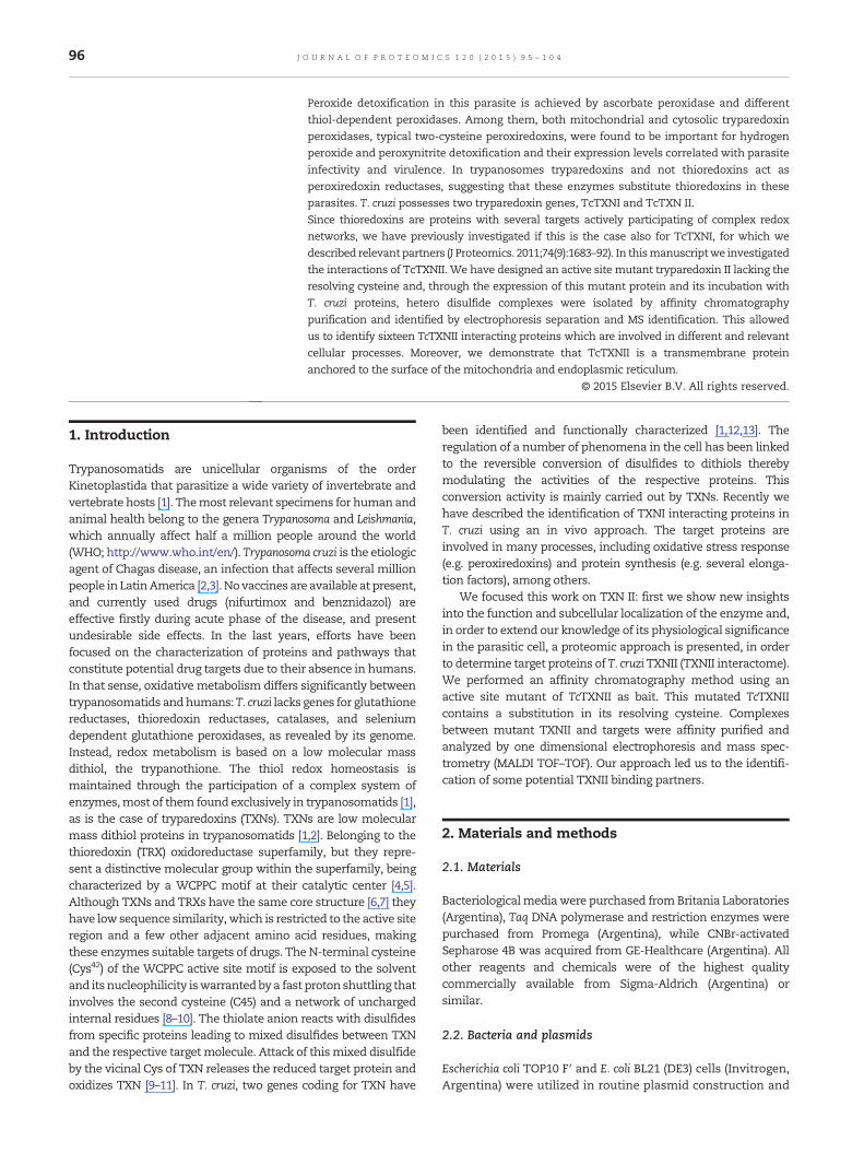
T. cruzi epimastigotes (5·108 cells) were pelleted andsuspended in 1 ml of 10 mM Tris–HCl, pH 7.5, and 1 mMEDTA (plus cocktail of protease inhibitors) and disruptedunder hypotonic conditions by passage through a narrowhypodermic needle. The lysate was fractionated by serialdifferential centrifugation (from 500 ×g to 18,000 ×g) for30 min at 4 °C. The last soluble fraction was treated with8 mM CaCl2 (for vesicles and microsomes precipitation [19])and newly centrifuged for 30 min at 18,000 ×g. The obtainedpellets (each centrifugation step) were mixed with SDS-PAGEsample buffer, boiled for 5 min and stored at −20 °C. Aliquotswere analyzed by western blot using rabbit polyclonalantisera against TcTXNII, TcTXNI, TcmTXNPx, TccTXNPx,TcGlcK, TcCruzipain and TcAPx. HRP-conjugated anti-rabbitantibodies (Life Technologies) were employed as secondaryantibody. Bands were visualized using the ECL Westernblotting detections reagents (Thermo Scientific).
3. Results and discussion
3.1. The Cys45 of TcTXNII is essential to protein disulfidereduction, but not for low molecular mass disulfide reduction
As previously described [9–11], the thiolate anion of N-terminalCys reactswith disulfides from specific proteins. This event leads
to mixed disulfides between TXN and the respective targetmolecule. Then, the attack of this mixed disulfide by the vicinalCys (C-terminal, Cys45 of TcTXNII) of TXN releases the reducedtarget protein and oxidizes TXN to disulfide.We tested the abilityof TcTXNIIC45SΔ22C to catalyze the reduction of T. cruziperoxiredoxins (cTXNPx or mTXNPx) by T(SH)2. This wasevaluated by the coupled assay for trypanothione-dependentt-bOOH reduction via a tryparedoxin/peroxidase-mediated reac-tion [15]. As positive control, TcTXNIIΔ22Cwas used in equivalentconditions. Fig. 1-A shows that TcTXNIIΔ22CC45S was notactive for transferring reducing equivalents to cytoplasmatic(TccTXNPx) or mitochondrial (TcmTXNPx) 2-Cys peroxiredoxins.However, when 2-Cys peroxiredoxins were replaced by GSSG orGSNO as final acceptor in the coupled assay, TcTXNIIC45SΔ22Cexhibited identical catalytic efficiency to TcTXNIIΔ22C. Theseresults indicate that reduced TcTXNIIC45SΔ22C [plus T(SH)2]exhibited a capacity to reduce low molecular mass disulfides viamonothiol mechanism (similar to monothiol glutaredoxins[15,20,21]). This result was consistent with the formationof adducts between low molecular mass disulfides withmonothiolic TXNII version (TcTXNIIC45SΔ22C, Fig. 1-B). In addi-tion, themutantTXNII showeda slight tendency to formcovalentdimers in the presence of physiologic oxidant substrates,compared to the wild type version (Fig. 1-B). These resultsindicate the essential role of C-terminal cysteine (Cys45 inTcTXNII) in the resolution of protein mixed disulfides, whichcorrelates with previously published data on the catalyticmechanism of TXNs [15,18,22].
99J O U R N A L O F P R O T E O M I C S 1 2 0 ( 2 0 1 5 ) 9 5 – 1 0 4
3.2. TcTXNII is a transmembrane protein anchored to thesurface of the mitochondria and endoplasmic reticulum
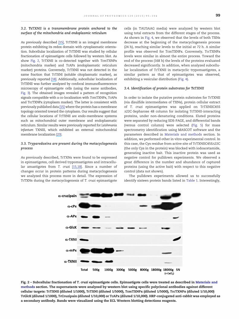
As previously described [15], TcTXNII is an integral membraneprotein exhibiting its redox domain with cytoplasmatic orienta-tion. Subcellular localization of TcTXNII was studied by cellularfractionation of epimastigote cells followed by western blot. Asshow Fig. 2, TcTXNII is co-detected together with TcmTXNPx(mitochondria marker) and TcAPx (endoplasmatic reticulummarker) proteins. Conversely, TcTXNII was not detected in thesame fraction that TcTXNI (soluble citoplasmatic marker), aspreviously reported [18]. Additionally, subcellular localization ofTcTXNII was further analyzed by confocal immunofluorescencemicroscopy of epimastigote cells (using the same antibodies,Fig. 3). The obtained images revealed a pattern of recognitionsignals compatible with a co-localization with TcmTXNPx, TcAPxand TccTXNPx (cytoplasm marker). The latter is consistent withpreviouslypublisheddata [15]where theproteinhas amembranetopology oriented toward the cytoplasm. Our results suggest thatthe cellular locations of TcTXNII are endo-membrane systemssuch as mitochondrial outer membrane and endoplasmaticreticulum. Similar resultswere previously reported for Leishmaniainfantum TXNIII, which exhibited an external mitochondrialmembrane localization [23].
3.3. Tryparedoxins are present during the metacyclogenesisprocess
As previously described, TcTXNs were found to be expressedin epimastigotes, cell derived trypomastigotes and intracellu-lar amastigotes from T. cruzi [15,18]. Since a number ofchanges occur in protein patterns during metacyclogenesiswe analyzed this process more in detail. The expression ofTcTXNs during the metacyclogenesis of T. cruzi epimastigote
Fig. 2 – Subcellular fractionation of T. cruzi epimastigote cells. Epmethods section. The supernatants were analyzed by western bcellular targets: TcTXNII (diluted 1/1000), TcTXNI (diluted 1/5000)TcGlcK (diluted 1/1000), TcCruzipain (diluted 1/10,000) or TcAPx (da secondary antibody. Bands were visualized using the ECL Wes
cells (in TAU3AAG media) were analyzed by western blotusing total extracts from the different stages of the process.As shown in Fig. 4, we observed that the levels of both TXNsdecrease at the beginning of the metacyclogenesis process(24 h), reaching similar levels to the initial at 72 h. A similarprofile was observed for TcmTXNPx. Conversely, TccTXNPxlevels were similar in almost the entire process. Toward theend of the process (168 h) the levels of the proteins evaluateddecreased significantly. In addition, when analyzed subcellu-lar localization of TcTXNII in metacyclic trypomastigotes, asimilar pattern as that of epimastigotes was observed,exhibiting a vesicular distribution (Fig. 4).
3.4. Identification of protein substrates for TcTXNII
In order to isolate the putative protein substrates for TcTXNII(via disulfide intermediates of TXNs), protein cellular extractof T. cruzi epimastigotes was applied on TcTXNIIC45SΔ22C-Sepharose 4B column for isolating TcTXNII-interactingproteins, under non-denaturing conditions. Eluted proteinswere separated by reducing SDS-PAGE, and differential bands(versus control column) were selected (Fig. 5) for massspectrometry identification using MASCOT software and theparameters described in Materials and methods section. Inaddition, we performed other in vitro experimental control. Inthis case, the Cys residue from active site of TcTXNIIC45SΔ22C(the only Cys in the protein) was blocked with iodoacetamide,generating inactive bait. This inactive protein was used asnegative control for pulldown experiments. We observed agreat difference in the number and abundance of capturedproteins (using the active bait) with respect to this negativecontrol (data not shown).
The pulldown experiments allowed us to successfullyidentify sixteen protein bands listed in Table 1. Interestingly,
imastigote cells were treated as described in Materials andlot using specific polyclonal antibodies against different, TcmTXNPx (diluted 1/5000), TccTXNPx (diluted 1/10,000)iluted 1/10,000). HRP-conjugated anti-rabbit was employed astern blotting detections reagents.
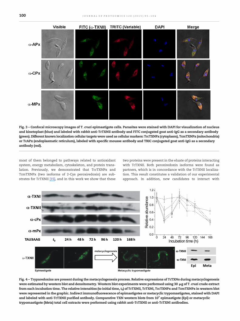
Fig. 3 – Confocal microscopy images of T. cruzi epimastigote cells. Parasites were stained with DAPI for visualization of nucleusand kinetoplast (blue) and labeled with rabbit anti-TcTXNII antibody and FITC conjugated goat anti-IgG as a secondary antibody(green). Different known localization cellular targetswereused as cellularmarkers:TccTXNPx (cytoplasm), TcmTXNPx (mitochondria)or TcAPx (endoplasmatic reticulum), labeled with specific mousse antibody and TRIC conjugated goat anti-IgG as a secondaryantibody (red).
100 J O U R N A L O F P R O T E O M I C S 1 2 0 ( 2 0 1 5 ) 9 5 – 1 0 4
most of them belonged to pathways related to antioxidantsystem, energy metabolism, cytoskeleton, and protein trans-lation. Previously, we demonstrated that TccTXNPx andTcmTXNPx (two isoforms of 2-Cys peroxiredoxin) are sub-strates for TcTXNII [15], and in this work we show that these
Fig. 4 – Tryparedoxins are present during themetacyclogenesis prowere estimated bywestern blot and densitometry.Western blot exfromeach incubation time. The relative intensities (to initial time, twere represented in the graphic. Indirect immunofluorescence of eand labeled with anti-TcTXNII purified antibody. Comparative TXNtrypomastigote (Meta) total cell extracts were performed using rab
two proteins were present in the eluate of proteins interactingwith TcTXNII. Both peroxiredoxin isoforms were found aspartners, which is in concordance with the TcTXNII localiza-tion. This result constitutes a validation of our experimentalapproach. In addition, new candidates to interact with
cess. Relative expressions of TcTXNs duringmetacyclogenesisperimentswere performedusing 30 μg of T. cruzi crude extract0) ofTcTXNII, TcTXNI,TccTXNPx and TcmTXNPx inwestern blotpimastigotes ormetacyclic trypomastigotes, stainedwithDAPIwestern blots from 107 epimastigote (Epi) or metacyclic
bit anti-TcTXNII or anti-TcTXNI antibodies.
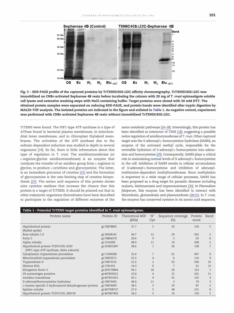
Fig. 5 – SDS-PAGE profile of the captured proteins by TcTXNIIC45SΔ22C affinity chromatography. TcTXNIIC45SΔ22C wasimmobilized on CNBr-activated Sepharose 4B resin before incubating the column with 20 mg of T. cruzi epimastigote solublecell lysate and extensive washing steps with NaCl-containing buffer. Target proteins were eluted with 50 mM DTT. Theobtained protein samples were separated on reducing SDS-PAGE, and protein bands were identified after tryptic digestion byMALDI–TOF analysis. The isolated proteins are indicated in the figure and enlisted in Table 1. As negative control, experimentwas performed with CNBr-activated Sepharose 4B resin without immobilized TcTXNIIC45SΔ22C.
101J O U R N A L O F P R O T E O M I C S 1 2 0 ( 2 0 1 5 ) 9 5 – 1 0 4
TcTXNII were found. The F0F1-type ATP synthase is a type ofATPase found in bacterial plasma membranes, in mitochon-drial inner membranes, and in chloroplast thylakoid mem-branes. The activation of the ATP synthase due to theredoxin-dependent reduction was studied in depth in severalorganisms [24]. So far, there is little information about thistype of regulation in T. cruzi. The amidinotransferase (orL-arginine:glycine amidinotransferase) is an enzyme thatcatalyzes the transfer of an amidino group from L-arginine toglycine, to produce L-ornithine and glycocyamine. The latter,is an immediate precursor of creatine [25] and the formationof glycocyamine is the rate-limiting step of creatine biosyn-thesis [25]. The amino acid sequence of this protein showsnine cysteine residues that increase the chance that thisprotein is a target of TcTXNII. It should be pointed out that inother eukaryotic organisms thioredoxins have been describedto participate in the regulation of different enzymes of the
Table 1 – Potential TcTXNII target proteins identified in T. cruzi
Protein name Protein ID
Hypothetical protein(Radial spoke)
gi:70878892
Beta tubulin 1.9 gi:18568141Porin 3 gi:70864370Alpha-tubulin gi:1314208Hypothetical protein TCSYLVIO_6332(F0F1-type ATP synthase, delta subunit)
gi:322821007
Cytoplasmic tryparedoxin peroxidase gi:71396508Mitochondrial tryparedoxin peroxidase gi:70870571Tryparedoxin II gi:70872315Histone H2A gi:1781355Elongation factor 2 gi:205278864I/6 autoantigen putative gi:407859521Amidino transferase gi:407851021S-adenosylhomocysteine hydrolase gi:70873492D-isomer specific 2-hydroxyacid dehydrogenase-protein gi:70876009Epsilon-tubulin gi:407398727Hypothetical protein TCSYLVIO_000159 gi:407867802
samemetabolic pathways [26–28]. Interestingly, this protein hasbeen identified as interactor of TXNI [18], suggesting a possibleredox regulation of amidinotransferase of T. cruzi. Other capturedtarget was the S-adenosyl-L-homocysteine hydrolase (SAHH), anenzyme of the activated methyl cycle, responsible for thereversible hydration of S-adenosyl-L-homocysteine into adeno-sine and homocysteine [29]. Consequently, SAHH plays a criticalrole in maintaining normal levels of S-adenosyl-L-homocysteinein the cell. Inhibition of SAHH results in cellular accumulationof S-adenosyl-L-homocysteine and inhibition of adenosyl-methionine-dependent methyltransferases. Since methylationis important in a wide range of cellular processes, SAHH hasbeen proposed as a drug target for parasitic diseases includingmalaria, leishmaniasis and trypanosomiasis [30]. In Plasmodiumfalciparum, this enzyme has been identified to interact withthioredoxin, glutaredoxin and plasmoredoxin [28,31]. In T. cruzi,the enzyme has conserved cysteine in its amino acid sequence,
epimastigotes.
Theoretical MW(kDa)
N°Cys
Sequence coverage(%)
Proteinscore
Band
37.7 5 32 159 5
49.7 12 29 202 229.6 9 3 63 648.9 11 10 208 228.6 1 26 138 7
22.4 7 61 497 925.5 4 6 110 921.9 2 25 258 1014.3 0 7 61 1294.1 18 26 153 123.0 4 21 101 1143.1 9 41 125 448.4 11 3 70 338.5 5 33 87 527.9 5 48 151 834.2 5 14 129 5

102 J O U R N A L O F P R O T E O M I C S 1 2 0 ( 2 0 1 5 ) 9 5 – 1 0 4
constituting good candidates to be attacked by TcTXNII. Anothercaptured protein was the D-isomer specific 2-hydroxyaciddehydrogenase (or R-lactate dehydrogenase). This enzymecatalyzes the NAD-dependent oxidation of R-lactate to pyruvateand is part of the detoxification pathway of methylglyoxal(dependent of glyoxalase I, glyoxalase II and low molecularmass thiols) in trypanosomatids [32]. In a previous study theenzymes form Trypanosoma conorhini and Crithidia fasciculata havebeen characterized, and it was observed that the enzymaticactivity of this protein was activated in the presence of cysteine[33]. These previous results and those obtained here stronglysuggest a redox regulation of this enzyme in trypanosomatids.
A group of TXNII partners related to cytoskeleton wereidentified: α-, β-, ε-tubulin, I/6 autoantigen (an internallyrepetitive cytoskeletal protein [34]) and radial spoke protein.In the case of tubulin, it has been observed that the assemblyand polymerization of this protein depend on redox changes[35–37]. For example, in mammalian microtubule [38], cyste-ine oxidation to disulfides altered the ability of the proteins topromote the assembly of microtubules from tubulin. Thetreatment with reducing molecule (such as thioredoxin) fullyrestores the ability to promote microtubule assembly [38]. Sofar, in trypanosomes is unclear the dependence of redox stateand polymerization (and assembly) of tubulin. On the otherhand, the radial spoke is a multi-unit protein structure foundin the axonemes of eukaryotic cilia and flagella [39]. Theflagellum of Trypanosoma is a multifunctional organelle withcritical roles in motility and other aspects of the trypanosomelife cycle [40]. In Trypanosoma brucei, RNA interference toablate expression of radial spoke protein and central paircomponents presented the first evidence that flagellar beatingis important for cell division (cytokinesis) and proposed thedrive flagellar beat as drug targets for the treatment of Africansleeping sickness [40]. So far, in trypanosomes is unclear thedependence of redox state and polymerization (and assembly)of tubulin on the function of flagellar proteins.
A component of the protein biosynthesis was identified asa potential target. Here we identified the eukaryotic elonga-tion factor 2 (eEF2) as a target of TcTXNII. The eEF2 catalyzesthe translocation of tRNA and mRNA down the ribosome atthe end of each round of polypeptide elongation [41]. Previousstudies reported that oxidative stress inhibits protein synthe-sis at the elongation step in mammals [42]. Moreover theelongation factor in chloroplasts has been captured by thechloroplast Trx [43]. The interaction of eEF2 with thioredoxinhas also been reported in Arabidopsis thaliana [44], Dictyosteliumdiscoideum [45], Entamoeba histolytica [46], E. coli thioredoxin [47]and P. falciparum [31]. Finally, it was observed a regulation oftranslation by the redox state of the elongation factor inSynechocystis sp. [48]. Taken together these reports indicate astrong relationship between elongation of peptides and redoxstate, with a relevant participation of thioredoxins. Inconcordance with this finding, T. cruzi eEF2 present eighteenhighly conserved cysteine residues in its primary structure;this would allow interaction via disulfide bridges TXNII.Interestingly, in a previous report, no interaction was ob-served between TXNI and eEF2. The redox interaction wasonly detected with the eukaryotic initiation factor 4a [18]which strongly suggests a possible specific interaction ofTXNII with eEF2.
A protein target of TcTXNII that was interesting due to thecellular localization which might occur is porin 3, a voltage-dependent anion channel of the outer mitochondrial mem-brane. The voltage-dependent anion channel regulates the fluxof mostly anionic metabolites through the outer mitochondrialmembrane [49]. The T. cruzi protein has high identity (98%) withthe protein from T. brucei, which is the main metabolitetransporter in the outer mitochondrial membrane in thisorganism [49]. The single T. brucei mitochondrial porin isessential only under growth conditions that depend onoxidative phosphorylation [49]. In plants, it has been shownthat thioredoxin interacts with mitochondrial porin via thiol/disulfide exchange reaction [24]. In addition, in mammaliancells, the voltage sensor of the mitochondrial permeabilitytransition pore is tuned by the oxidation-reduction state ofvicinal thiols (increase of the gating potential by oxidants andits reversal by reducing agents) [50]. This result was extremelyinteresting given the cellular location of TcTXNII on the outermitochondrial membrane.
Ahypothetical protein (TCSYLVIO_000159)was isolate by thismethodology as TXNII-partner. Although we could not identifyconservation in this protein, it has four Cys residues, of whichthe residue Cys202 is conserved in all trypanosomatids (data notshown). Future experiments will be needed to determine thefunction of this protein.
Other unrelated protein that was a target of TcTXNII wasthe histone H2A. It must be pointed out that, despite havingused a method based on capture of partners through thegeneration of heterodisulfide dimers, false positives cannot bediscarded. That seems to be the case for the histone H2A,which has no cysteine residues, and so its capture by affinitychromatography could be due to co-purification with any ofthe other targets isolated. However, these results stimulate usto further investigations.
4. Conclusions
In trypanosomatids, TXNs transfer reducing equivalents fromtrypanothione to redox pathways involving thiol/disulfide ex-change [4,9]. Herein, we analyze the expression and cellularlocalization of TcTXNII. By means of specific antibodies designedagainst TcTXNII, we evidenced the occurrence of this proteinthrough of metacyclogenesis process of T. cruzi epimastigotes.The protein showed cellular co-localization with TcmTXNPx(mitochondria marker) and TcAPx (endoplasmatic reticulummarker) proteins. In addition, the identity of TcTXNII as a trueintegral membrane protein with a cytoplasmatic orientation ofthe redox domain was previously demonstrated [15]. With thispreviously data, together with the results here obtained, wemayconclude that TcTXNII is a transmembrane protein anchored tothe surface of the mitochondria and endoplasmic reticulum.
In order to deepen the studies about the redox interactomeof T. cruzi TXNs, an in vitro proteomic approach allowed us toidentify putative partners of TXNII in this parasite. Aftercloning and expression the TcTXNIIC45SΔ22C, the observed invitro redox activity indicated the incapacity of mutant proteinto catalyze the thiol/disulfide exchange with protein sub-strates. This result validates the use of this protein for thepreparation of the affinity column for isolation of potential
103J O U R N A L O F P R O T E O M I C S 1 2 0 ( 2 0 1 5 ) 9 5 – 1 0 4
interactors. After performing affinity chromatography, puri-fied proteins were isolated and identified by SDS-PAGE andMS. By using this approach with TcTXNII, sixteen putativepartner proteins were identified, belonging to four mainprocesses: antioxidant system, energy metabolism, cytoskel-eton, and protein translation. Most of the proteins arelocalized (or predicted to be) in the cytosol and, interestinglymost of them have either homologues that have beendescribed to interact with TRX in other organisms. This invitro approach led us to the discovery of several putativelyTcTXNII-interacting proteins thereby contributing to ourunderstanding of the redox interactome in T. cruzi. Theinvolvement of TXNs in several parasite physiological pro-cesses suggests novel insights about the protein involvementin redox signaling.
Transparency document
The Transparency document associated with this article canbe found, in the online version.
Acknowledgments
We thank Magdalena Portela (UBYPA, Institut Pasteur deMontevideo) for mass spectrometry analysis. This workwas supported by Agencia Nacional de Investigación eInnovación (ANII-Uruguay) grant DCI-ALA/2011/023-502“Contrato de apoyo a las políticas de innovación y cohesiónterritorial”, FOCEM (MERCOSUR Structural ConvergenceFund), COF 03/11, UNL (CAI + D Orientados & Redes 2011),CONICET (PIP112-2011-0100439, PIP114-2011-0100168) andANPCyT (PICT2012-2439, PICT2013-00253). M.D.P and C.R.are researchers from the Sistema Nacional Investigadores(SNI-ANII), Uruguay. D.G.A., S.A.G. and A.A.I. are investiga-tor career members from CONICET, Argentina.
Appendix A. Supplementary data
Supplementary data to this article can be found online athttp://dx.doi.org/10.1016/j.jprot.2015.03.001.
R E F E R E N C E S
[1] Irigoin F, Cibils L, Comini MA, Wilkinson SR, Flohé L, Radi R.Insights into the redox biology of Trypanosoma cruzi:trypanothione metabolism and oxidant detoxification. FreeRadic Biol Med 2008;45:733–42.
[2] Krauth-Siegel RL, CominiMA. Redox control in trypanosomatids,parasitic protozoa with trypanothione-based thiol metabolism.Biochim Biophys Acta 2008;1780:1236–48.
[3] PineyroMD, Parodi-Talice A, Arcari T, Robello C. Peroxiredoxinsfrom Trypanosoma cruzi: virulence factors and drug targets fortreatment of Chagas disease? Gene 2008;408:45–50.
[4] Gommel DU, Nogoceke E, Morr M, Kiess M, Kalisz HM, Flohé L.Catalytic characteristics of tryparedoxin. Eur J Biochem 1997;248:913–8.
[5] LüdemannH,DormeyerM, SticherlingC, StallmannD, FollmannH, Krauth-Siegel RL. Trypanosoma brucei tryparedoxin, athioredoxin-like protein in African trypanosomes. FEBS Lett1998;431:381–5.
[6] Kalisz HM, Hofmann B, Nogoceke E, Gommel DU, Flohé L,Hecht HJ. Crystallisation of tryparedoxin I from Crithidiafasciculata. Biofactors 2000;11:73–5.
[7] Guerrero SA, Montemartini M, Spallek R, Hecht HJ, Steinert P,Flohé L, et al. Cloning and expression of tryparedoxin I fromCrithidia fasciculata. Biofactors 2000;11:67–9.
[8] MontemartiniM, Nogoceke E, Gommel DU, SinghM, Kalisz HM,Steinert P, et al. Tryparedoxin and tryparedoxin peroxidase.Biofactors 2000;11:71–2.
[9] Flohé L, Steinert P, Hecht HJ, Hofmann B. Tryparedoxin andtryparedoxin peroxidase. Methods Enzymol 2002;347:244–58.
[10] Reckenfelderbaumer N, Krauth-Siegel RL. Catalytic properties,thiol pK value, and redox potential of Trypanosoma bruceitryparedoxin. J Biol Chem 2002;277:17548–55.
[11] Steinert P, Plank-Schumacher K, Montemartini M, Hecht HJ,Flohé L. Permutation of the active site motif of tryparedoxin2. Biol Chem 2000;381:211–9.
[12] El-Sayed NM, Myler PJ, Bartholomeu DC, Nilsson D, AggarwalG, Tran AN, et al. The genome sequence of Trypanosoma cruzi,etiologic agent of Chagas disease. Science 2005;309:409–15.
[13] Wilkinson SR, Meyer DJ, Taylor MC, Bromley EV, Miles MA,Kelly JM. The Trypanosoma cruzi enzymeTcGPXI is a glycosomalperoxidase and can be linked to trypanothione reduction byglutathione or tryparedoxin. J Biol Chem 2002;277:17062–71.
[14] Maniatis T, Fritsch EF, Sambrook J. Molecular cloning: alaboratory manual. N. Y. — USA: Cold Spring Harhor; 1982.
[15] Arias DG, Marquez VE, Chiribao ML, Gadelha FR, Robello C,Iglesias AA, et al. Redox metabolism in Trypanosoma cruzi:functional characterization of tryparedoxins revisited. FreeRadic Biol Med 2013;63:65–77.
[16] Gomez ML, Erijman L, Arauzo S, Torres HN, Tellez-Inon MT.Protein kinase C in Trypanosoma cruzi epimastigote forms:partial purification and characterization. Mol BiochemParasitol 1989;36:101–8.
[17] Contreras VT, Salles JM, Thomas N, Morel CM, Goldenberg S.In vitro differentiation of Trypanosoma cruzi under chemicallydefined conditions. Mol Biochem Parasitol 1985;16:315–27.
[18] Pineyro MD, Parodi-Talice A, Portela M, Arias DG, GuerreroSA, Robello C. Molecular characterization and interactomeanalysis of Trypanosoma cruzi tryparedoxin 1. J Proteomics2011;74:1683–92.
[19] Schenkman JB, Cinti DL. Preparation of microsomes withcalcium. Methods Enzymol 1978;52:83–9.
[20] Marquez VE, Arias DG, Chiribao ML, Faral-Tello P, Robello C,Iglesias AA, et al. Redox metabolism in Trypanosoma cruzi.Biochemical characterization of dithiol glutaredoxin dependentcellular pathways. Biochimie 2014;106:56–67.
[21] Lillig CH, Berndt C, Holmgren A. Glutaredoxin systems.Biochim Biophys Acta 2008;1780:1304–17.
[22] Fueller F, Jehle B, Putzker K, Lewis JD, Krauth-Siegel RL. Highthroughput screening against the peroxidase cascade ofAfrican trypanosomes identifies antiparasitic compoundsthat inactivate tryparedoxin. J Biol Chem 2012;287:8792–802.
[23] Castro H, Romao S, Carvalho S, Teixeira F, Sousa C, TomasAM. Mitochondrial redox metabolism in trypanosomatids isindependent of tryparedoxin activity. PLoS One 2010;5:e12607.
[24] Balmer Y, Vensel WH, Tanaka CK, Hurkman WJ, Gelhaye E,Rouhier N, et al. Thioredoxin links redox to the regulation offundamental processes of plantmitochondria. Proc Natl AcadSci U S A 2004;101:2642–7.
[25] HummA, Fritsche E, Steinbacher S, Huber R. Crystal structureand mechanism of human L-arginine:glycineamidinotransferase: a mitochondrial enzyme involved increatine biosynthesis. EMBO J 1997;16:3373–85.

104 J O U R N A L O F P R O T E O M I C S 1 2 0 ( 2 0 1 5 ) 9 5 – 1 0 4
[26] Marchand CH, Vanacker H, Collin V, Issakidis-Bourguet E,Marechal PL, Decottignies P. Thioredoxin targets inArabidopsis roots. Proteomics 2010;10:2418–28.
[27] Vignols F, Brehelin C, Surdin-Kerjan Y, Thomas D, Meyer Y. Ayeast two-hybrid knockout strain to explore thioredoxin-interacting proteins in vivo. Proc Natl Acad Sci U S A 2005;102:16729–34.
[28] Sturm N, Jortzik E, Mailu BM, Koncarevic S, Deponte M,Forchhammer K, et al. Identification of proteins targeted bythe thioredoxin superfamily in Plasmodium falciparum. PLoSPathog 2009;5:e1000383.
[29] Hu Y, Komoto J, Huang Y, Gomi T, Ogawa H, Takata Y, et al.Crystal structure of S-adenosylhomocysteine hydrolase fromrat liver. Biochemistry 1999;38:8323–33.
[30] Cai S, Li QS, Borchardt RT, Kuczera K, Schowen RL. Theantiviral drug ribavirin is a selective inhibitor ofS-adenosyl-L-homocysteine hydrolase from Trypanosomacruzi. Bioorg Med Chem 2007;15:7281–7.
[31] Kawazu S, Takemae H, Komaki-Yasuda K, Kano S. Targetproteins of the cytosolic thioredoxin in Plasmodium falciparum.Parasitol Int 2009;59:298–302.
[32] Greig N, Wyllie S, Vickers TJ, Fairlamb AH.Trypanothione-dependent glyoxalase I in Trypanosoma cruzi.Biochem J 2006;400:217–23.
[33] Bacchi CJ, Ciaccio EI, O'Connell KM, Hutner SH. Biochemicalproperties of trypanosomatid lactate dehydrogenases. J Bacteriol1970;102:826–34.
[34] Detmer E, Hemphill A, Muller N, Seebeck T. The Trypanosomabrucei autoantigen I/6 is an internally repetitive cytoskeletalprotein. Eur J Cell Biol 1997;72:378–84.
[35] Landino LM, Skreslet TE, Alston JA. Cysteine oxidation of tauand microtubule-associated protein-2 by peroxynitrite:modulation of microtubule assembly kinetics by thethioredoxin reductase system. J Biol Chem 2004;279:35101–5.
[36] Livanos P, Galatis B, Apostolakos P. The interplay between ROSand tubulin cytoskeleton in plants. Plant Signal Behav 2014;9.
[37] Clark HM, Hagedorn TD, Landino LM. Hypothiocyanousacid oxidation of tubulin cysteines inhibits microtubulepolymerization. Arch Biochem Biophys 2014;541:67–73.
[38] Khan IA, Luduena RF. Possible regulation of the in vitroassembly of bovine brain tubulin by the bovine thioredoxinsystem. Biochim Biophys Acta 1991;1076:289–97.
[39] Yang P, Diener DR, Yang C, Kohno T, Pazour GJ, Dienes JM,et al. Radial spoke proteins of Chlamydomonas flagella. J Cell Sci2006;119:1165–74.
[40] Ralston KS, Lerner AG, Diener DR, Hill KL. Flagellar motilitycontributes to cytokinesis in Trypanosoma brucei and ismodulated by an evolutionarily conserved dynein regulatorysystem. Eukaryot Cell 2006;5:696–711.
[41] Smulski CR, Longhi SA, Ayub MJ, Edreira MM, Simonetti L,Gomez KA, et al. Interaction map of the Trypanosoma cruziribosomal P protein complex (stalk) and the elongation factor2. J Mol Recognit 2011;24:359–70.
[42] Ayala A, Parrado J, Bougria M, Machado A. Effect of oxidativestress, produced by cumene hydroperoxide, on the varioussteps of protein synthesis. Modifications of elongation factor-2.J Biol Chem 1996;271:23105–10.
[43] Balmer Y, Koller A, del Val G, Manieri W, Schurmann P,Buchanan BB. Proteomics gives insight into the regulatoryfunction of chloroplast thioredoxins. Proc Natl Acad Sci U S A2003;100:370–5.
[44] Yamazaki D, Motohashi K, Kasama T, Hara Y, Hisabori T.Target proteins of the cytosolic thioredoxins in Arabidopsisthaliana. Plant Cell Physiol 2004;45:18–27.
[45] Brodegger T, Stockmann A, Oberstrass J, NellenW, Follmann H.Novel thioredoxin targets inDictyostelium discoideum identified bytwo-hybrid analysis: interactions of thioredoxin with elongationfactor 1alpha and yeast alcohol dehydrogenase. Biol Chem 2004;385:1185–92.
[46] Schlosser S, Leitsch D, Duchene M. Entamoeba histolytica:identification of thioredoxin-targeted proteins and analysisof serine acetyltransferase-1 as a prototype example.Biochem J 2013;451:277–88.
[47] Kumar JK, Tabor S, Richardson CC. Proteomic analysis ofthioredoxin-targeted proteins in Escherichia coli. Proc NatlAcad Sci U S A 2004;101:3759–64.
[48] Kojima K, Motohashi K, Morota T, Oshita M, Hisabori T,Hayashi H, et al. Regulation of translation by the redox stateof elongation factor G in the cyanobacterium Synechocystis sp.PCC 6803. J Biol Chem 2009;284:18685–91.
[49] Pusnik M, Charriere F, Maser P, Waller RF, Dagley MJ, LithgowT, et al. The single mitochondrial porin of Trypanosoma bruceiis the mainmetabolite transporter in the outer mitochondrialmembrane. Mol Biol Evol 2009;26:671–80.
[50] Petronilli V, Costantini P, Scorrano L, Colonna R, Passamonti S,Bernardi P. The voltage sensor of themitochondrial permeabilitytransition pore is tuned by the oxidation-reduction state ofvicinal thiols. Increase of the gating potential by oxidants and itsreversal by reducing agents. J Biol Chem 1994;269:16638–42.
Copyright © 2022 FDOKUMEN