Benzoic acid and pyridine derivatives as inhibitors of Trypanosoma cruzi trans-sialidase
14
Benzoic acid and pyridine derivatives as inhibitors of Trypanosoma cruzi trans-sialidase Joa ˜o Neres, a Pascal Bonnet, a, Philip N. Edwards, a Pravin L. Kotian, c Alejandro Buschiazzo, b Pedro M. Alzari, b Richard A. Bryce a and Kenneth T. Douglas a, * a School of Pharmacy and Pharmaceutical Sciences, University of Manchester, Manchester, M13 9PL, UK b Unite ´ de Biochimie Structurale, Institut Pasteur, 25 rue du Dr. Roux, 75724 Paris, France c BioCryst Pharmaceuticals, Inc., 2190 Parkway Drive, Birmingham, AL 35244, USA Received 3 October 2006; revised 7 December 2006; accepted 13 December 2006 Available online 15 December 2006 Abstract—Benzoic acid and pyridine derivatives inhibit recombinant trans-sialidase from Trypanosoma cruzi with I 50 values between 0.4 and 1 mM. The best compounds, 4-acetylamino-3-hydroxymethylbenzoic acid and 5-acetylamino-6-aminopyridine-2-carboxylic acid, provide new leads to inhibitors not containing the synthetically complex sialic acid structure. The weak inhibition by such compounds contrasts with their much stronger inhibition of neuraminidase from Influenza virus. Ó 2007 Elsevier Ltd. All rights reserved. 1. Introduction Chagas’ disease, widely distributed in Central and South America with 13 million persons infected and 100 mil- lion people at risk, is estimated to cause 14,000 deaths annually. This disease is caused by Trypanosoma cruzi, a protozoan parasite transmitted to humans by haemat- ophagous bugs or directly by transfusion of infected blood. 1 The only established drugs for the acute phase of infection are nifurtimox and benznidazole, with few other candidates yet on the clinical horizon. 2 The prob- lem of resistance, both of the parasite to drug at molec- ular level and of the vector to insecticide, further adds to the gloomy prospects. 3 With this in mind there has been considerable interest in trying to develop novel approaches and targets for drug design. A key problem in the pathogenicity of T. cruzi is its abil- ity to evade host immune responses. Surface sialylation plays a central role in this and in the host cell adhesion/ invasion mechanism, and provides a possible entry to novel therapeutics against this disease. This parasite is unable to synthesise sialic acids de novo. Trans-sialidase (TcTS), a parasite membrane-associated protein which catalyses the transfer of sialic acid molecules from host cell-surface glycoconjugates to its own surface mucin- like glycoproteins, seems to be a key enzyme to T. cruzi infective/invasive ability. 4–6 Thus, the developmental stage-specific expression of trans-sialidase is an impor- tant virulence factor of T. cruzi. As the trans-sialidases are unique to the parasite, TcTS has been discussed as a potential target for anti-Chagas drug design. 7,8 It is not possible to test the potential validity of this enzyme as a drug target by gene-knockout experiments as the TcTS protein family is coded for by 140 genes in the genome. 9 However, there are good biochemical reasons to believe that processes involving TcTS are vital to the parasite. TcTS specifically (and preferentially) transfers sialic acid linked to saccharides or glycoconjugates by a-2,3 bonds to terminal b-galactosyl residues in acceptor molecules. However, in the absence of an appropriate acceptor, TcTS can also act as a sialidase, hydrolysing the donor substrate and releasing free sialic acid. Both activities (sialidase and trans-sialidase) are associated with the same active site in TcTS, therefore assays based on sialic acid transfer or hydrolysis can be used to monitor TcTS activity. 10–13 There are no known specific chemical inhibitors of TcTS, nor drugs known to act against it. Thus, chemical 0968-0896/$ - see front matter Ó 2007 Elsevier Ltd. All rights reserved. doi:10.1016/j.bmc.2006.12.024 Keywords: Trans-sialidase; Inhibitors; Chagas disease; Neuraminidase. * Corresponding author. Tel.: +44 161 275 2371; fax: +44 161 275 2481; e-mail: [email protected] Present address: Johnson & Johnson Pharmaceutical Research & Development, Turnhoutseweg 30, 2340 Beerse, Belgium. Bioorganic & Medicinal Chemistry 15 (2007) 2106–2119
-
Upload
independent -
Category
Documents
-
view
0 -
download
0
Transcript of Benzoic acid and pyridine derivatives as inhibitors of Trypanosoma cruzi trans-sialidase
Bioorganic & Medicinal Chemistry 15 (2007) 2106–2119
Benzoic acid and pyridine derivatives as inhibitors ofTrypanosoma cruzi trans-sialidase
Joao Neres,a Pascal Bonnet,a,� Philip N. Edwards,a Pravin L. Kotian,c
Alejandro Buschiazzo,b Pedro M. Alzari,b Richard A. Brycea and Kenneth T. Douglasa,*
aSchool of Pharmacy and Pharmaceutical Sciences, University of Manchester, Manchester, M13 9PL, UKbUnite de Biochimie Structurale, Institut Pasteur, 25 rue du Dr. Roux, 75724 Paris, France
cBioCryst Pharmaceuticals, Inc., 2190 Parkway Drive, Birmingham, AL 35244, USA
Received 3 October 2006; revised 7 December 2006; accepted 13 December 2006
Available online 15 December 2006
Abstract—Benzoic acid and pyridine derivatives inhibit recombinant trans-sialidase from Trypanosoma cruzi with I50 values between0.4 and 1 mM. The best compounds, 4-acetylamino-3-hydroxymethylbenzoic acid and 5-acetylamino-6-aminopyridine-2-carboxylicacid, provide new leads to inhibitors not containing the synthetically complex sialic acid structure. The weak inhibition by suchcompounds contrasts with their much stronger inhibition of neuraminidase from Influenza virus.� 2007 Elsevier Ltd. All rights reserved.
1. Introduction
Chagas’ disease, widely distributed in Central and SouthAmerica with 13 million persons infected and 100 mil-lion people at risk, is estimated to cause 14,000 deathsannually. This disease is caused by Trypanosoma cruzi,a protozoan parasite transmitted to humans by haemat-ophagous bugs or directly by transfusion of infectedblood.1 The only established drugs for the acute phaseof infection are nifurtimox and benznidazole, with fewother candidates yet on the clinical horizon.2 The prob-lem of resistance, both of the parasite to drug at molec-ular level and of the vector to insecticide, further adds tothe gloomy prospects.3 With this in mind there has beenconsiderable interest in trying to develop novelapproaches and targets for drug design.
A key problem in the pathogenicity of T. cruzi is its abil-ity to evade host immune responses. Surface sialylationplays a central role in this and in the host cell adhesion/invasion mechanism, and provides a possible entry tonovel therapeutics against this disease. This parasite isunable to synthesise sialic acids de novo. Trans-sialidase
0968-0896/$ - see front matter � 2007 Elsevier Ltd. All rights reserved.
doi:10.1016/j.bmc.2006.12.024
Keywords: Trans-sialidase; Inhibitors; Chagas disease; Neuraminidase.* Corresponding author. Tel.: +44 161 275 2371; fax: +44 161 275
2481; e-mail: [email protected]� Present address: Johnson & Johnson Pharmaceutical Research &
Development, Turnhoutseweg 30, 2340 Beerse, Belgium.
(TcTS), a parasite membrane-associated protein whichcatalyses the transfer of sialic acid molecules from hostcell-surface glycoconjugates to its own surface mucin-like glycoproteins, seems to be a key enzyme to T. cruziinfective/invasive ability.4–6 Thus, the developmentalstage-specific expression of trans-sialidase is an impor-tant virulence factor of T. cruzi. As the trans-sialidasesare unique to the parasite, TcTS has been discussed asa potential target for anti-Chagas drug design.7,8 It isnot possible to test the potential validity of this enzymeas a drug target by gene-knockout experiments as theTcTS protein family is coded for by 140 genes in thegenome.9 However, there are good biochemical reasonsto believe that processes involving TcTS are vital to theparasite.
TcTS specifically (and preferentially) transfers sialic acidlinked to saccharides or glycoconjugates by a-2,3 bondsto terminal b-galactosyl residues in acceptor molecules.However, in the absence of an appropriate acceptor,TcTS can also act as a sialidase, hydrolysing the donorsubstrate and releasing free sialic acid. Both activities(sialidase and trans-sialidase) are associated with thesame active site in TcTS, therefore assays based on sialicacid transfer or hydrolysis can be used to monitor TcTSactivity.10–13
There are no known specific chemical inhibitors ofTcTS, nor drugs known to act against it. Thus, chemical

J. Neres et al. / Bioorg. Med. Chem. 15 (2007) 2106–2119 2107
evidence of the enzyme as a target is lacking, and thepresent contribution reports initial studies to discovernovel inhibitory molecular architectures based on theavailability of the crystal structure for this enzyme.10
The crystal structure of TcTS shows two domains; theN-terminal catalytic domain is connected through a longa-helix to a C-terminal, lectin-like domain. The activesite shows several of the conserved features of microbialsialidases, the main features being the arginine triad thatbinds the carboxylate group and a hydrophobic pocketthat binds the N-acetyl group in sialic acid.10
The few inhibitors reported for TcTS are weak and havecomplex chemical structures. 2-Deoxy-2,3-didehydro-DD-N-acetylneuraminic acid (DANA, 1, Fig. 1), a potentinhibitor of Influenza neuraminidase, inhibits TcTS witha reported Ki of 12.3 mM.14 The GM3 ganglioside 2 is agood donor substrate for TcTS, but when the sialic acidresidue is modified, namely at C4 (deoxy or methoxy),C7 (deoxy) and C8 (deoxy or methoxy), it becomes apartial inhibitor of the trans-sialidase reaction, at rela-tively high inhibitor/substrate ratios.13
2,3-Difluorosialic acid (3) inactivates TcTS time-depen-dently, through a covalent bond with the hydroxylgroup of Tyr342. However, complete inactivation re-quires very high concentrations (20 mM) and the en-zyme spontaneously recovers activity after removal ofexcess inactivator.15 3-Fluoro-sialosyl fluoride alsocovalently labels tyrosine for the sialidase fromT. rangeli (�70% sequence identity for 640 aminoac-ids).16 Although the enzymes from T. cruzi andT. rangeli show some similar features they behave dis-tinctly from each other in their structural acceptanceof various inhibitor frameworks.17 Lactitol (4) and otherlactose (acceptor substrate) analogues were reported toinhibit TcTS activity towards conventional substratesboth in vitro and in vivo, again at relatively high concen-trations of lactitol (mM range). However, these com-pounds do not inhibit the catalytic activity of TcTSbut act as lpreferential acceptors when compared withthe conventional b-galactosides.18,19 Recently, twocyclohexenephosphonate monoalkyl esters 5 (I50 =4.7 mM) and 6 (I50 = 5.7 mM) were also reported asweak inhibitors.20
Figure 1. Structure of reported TcTS inhibitors.
Away from complex, sugar frameworks, the only reportof TcTS inhibitors of which we are aware is pyridoxalphosphate (7, non-competitive inhibition, Ki 7.3 mM).21
It has been pointed out that although strong inhibitorshave been discovered against Influenza virus enzymebased on DANA, inhibitor design against the sialidasesor trans-sialidases of bacteria or trypanosomes has beenmuch less straightforward.22 In view of this we now re-port the first generation of simple, non-sugar basedinhibitors of TcTS that may lead to biological tools orpotential leads for drug design. We address at this stagethe sialic acid-binding site in TcTS.
One initial approach was to use a framework that hadproved successful in the inhibition of Influenza virusneuraminidase.23–27 This involved the replacement ofthe tetrahydropyran ring of sialic acid by simpler cyclicstructures such as benzene or pyridine. With this ap-proach, the relative chemical sensitivity and complexstereochemistry of the sialic acid or DANA-derivedclass of inhibitors is avoided, and one can begin to ad-dress issues such as bioavailability and other biologicallyimportant molecular properties. The compounds detect-ed in this way also serve as initial frameworks to subse-quently decorate with acceptor site ligand structures. Wereport TcTS inhibition data for a series of benzoic acidand pyridine-carboxylic acid derivatives (I and II, fullstructures are given in Tables 1 and 2, respectively),along with other carboxylic acid frameworks.
2. Results
2.1. Molecular modelling
2.1.1. Structure-based de novo design. The GRIDprogram28 was used to identify favourable energy

Table 1. Inhibition of TcTS by benzoic acid derivatives I
O
HO
R1
R2
R3I
Code R1 R2 R3 Activity
% inhibition at 1 mM (±SD) I50 (mM)
28 H NHCOCH3 H 0 —
29 CH2OH NHCOCH3 H — 0.54
30 CH2CH2OH NHCOCH3 H 16 ± 5 —
31a CH2CH(OH)CH2OH NHCOCH3 H 17 ± 2 —
32a CH2CH(OH)CH2OH NHCOCH3 H 0 —
33 CH2CH2NH2 NHCOCH3 H 25 ± 5 —
34 CH2CONH2 NHCOCH3 H 0 —
35 CH@NOH NHCOCH3 H 24 ± 6 —
36 N@C(NH2)2 NHCOCH3 H 41 ± 7 —
37 N@C(NH2)2 NHCOCH3 CH2CH2OH 0 —
38 N@C(NH2)2 CONHCH3 H — 0.76
39 N@C(NH2)2 NHSOCH3 H 29 ± 9 —
19 NH2 NHCOCH2Ph H — 1.2
18 N@C(NH2)2 NHCOCH2Ph H 54 ± 6b —
8 N@C(NH2)2 NHCH2Ph H 9 ± 4 —
40 N@C(NH2)2 H CH2CH2NH2c —
41 N@C(NH2)2 NHSO2CH3 CH2OH 11 ± 4 —
42 H NHSO2CH3 CH(OH)CH2NH2 17 ± 2 —
43 N@C(NH2)2 NHSO2CH3 C@NOH 46 ± 7 —
44 N@C(NH2)2 H N@C(NH2)2d —
45 N@C(NH2)2 H C@NOH 35 ± 4 —
46 N@C(NH2)2 NH2 CH2CH2OH 0 —
47 H NHCOCH2 N O H 0 —
48 NHCOCH3 N O H — 0.58
49 NH2 NHCH2
NH — 0.74
24 N@C(NH2)2 SCH2Ph H 43 ± 6e —
25 NH2 SCH2Ph H — 0.70
27 NHCOCH2NH2 SCH2Ph H — 1.0
a Compounds 31 and 32 are enantiomers.b Concentration tested was 0.5 mM.c Apparent activation (19% increase in activity).d Idem (31%).e Concentration tested was 0.4 mM.
Table 2. Inhibition of TcTS by pyridine-2-carboxylic acid derivatives II
NO
HO
R1
R2
R3II
Code R1 R2 R3 Activity
% inhibition at 1 mM (±SD) I50 (mM)
50 H NHCOCH3 H 22 ± 6 —
51 H NHCOCH3 NH2 14 ± 6 —
52 NH2 NHCOCH3 H — 0.44
53 H NHSO2CH3 H 25 ± 12 —
54 H NHCSCH3 H — 0.54
2108 J. Neres et al. / Bioorg. Med. Chem. 15 (2007) 2106–2119
J. Neres et al. / Bioorg. Med. Chem. 15 (2007) 2106–2119 2109
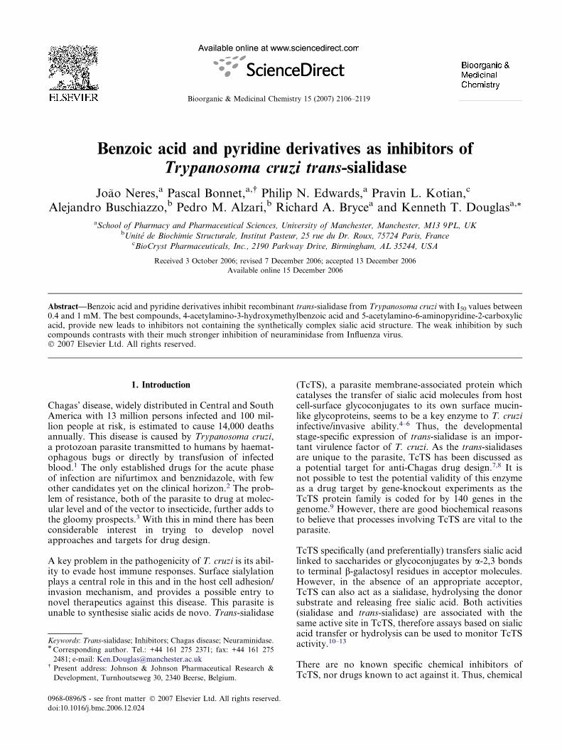
interaction regions in the active site between differentfunctional group probes. As expected, the very stronginteraction (�19 kcal/mol) of the carboxylate probewas detected in the region close to the arginine triad,as well as a favourable interaction with the cationic sidechain of Arg93, as shown in Figure 2A where the struc-ture of 2-deoxy-2,3-didehydro-DD-N-acetylneuraminicacid (DANA) is superimposed in the crystallographicconformation in complex with TcTS.10 The hydropho-bic pocket formed by Trp120, Tyr113 and Val95 wasclearly shown by various hydrophobic probes, includingthe methyl probe (Fig. 2B), and the DRY (hydrophobicprobe) or the aromatic CH probe (not shown). This isthe region which accommodates the N-acetyl group ofDANA or sialic acid. Free space is available in thehydrophobic pocket, apparently enough to enclosegroups as large as a phenyl ring.
The positively charged amidine probe, like other proton-ated nitrogen probes, gave essentially two regions with astrong interaction energy of �13 kcal/mol, between res-idues Glu230 and Asp96, and near Asp51 (Fig. 2C).Favourable binding positions at approximately�10 kcal/mol (Fig. 2D) were predicted for neutral NHor NH2 probes in several regions of the active site,namely close to residues Glu230, Asp96 and Gln195(binding region for the glycerol chain of DANA).
Figure 2. Favourable interaction energy surfaces (shown in green) obtained
probe (�19.0 kcal/mol) also showing DANA (crystallographic pose;10) (B) me
(D) cationic amidine probe (�13.5 kcal/mol).
These results indicated target regions for positivelycharged groups, such as guanidino or protonated ami-no-acylamino groups (e.g., glycylamino), and also forhydrophobic groups to fit into the hydrophobic pocket.Benzoic acid derivatives were designed, replacing the su-gar ring of sialic acid by a more synthetically accessiblebenzene ring, which would allow easier variation of thedifferent substituent groups. This approach had provensuccessful for Influenza neuraminidase, with severalbenzoic acid-based sialic acid mimetics inhibiting the en-zyme with I50 values in the low micromolar range, someeven stronger.23,29,30 Therefore, benzoic acid-derivedstructures were designed to contain these two functionsmeta and para to the carboxylate group. The structuresof the designed compounds and all other compoundstested during the present work were docked into the ac-tive site using DOCK 4.0,31 with ligand flexibility. Gen-erally, the functional groups in the top-ranked dockedposes obtained corresponded to the respective regionspredicted by the GRID program.
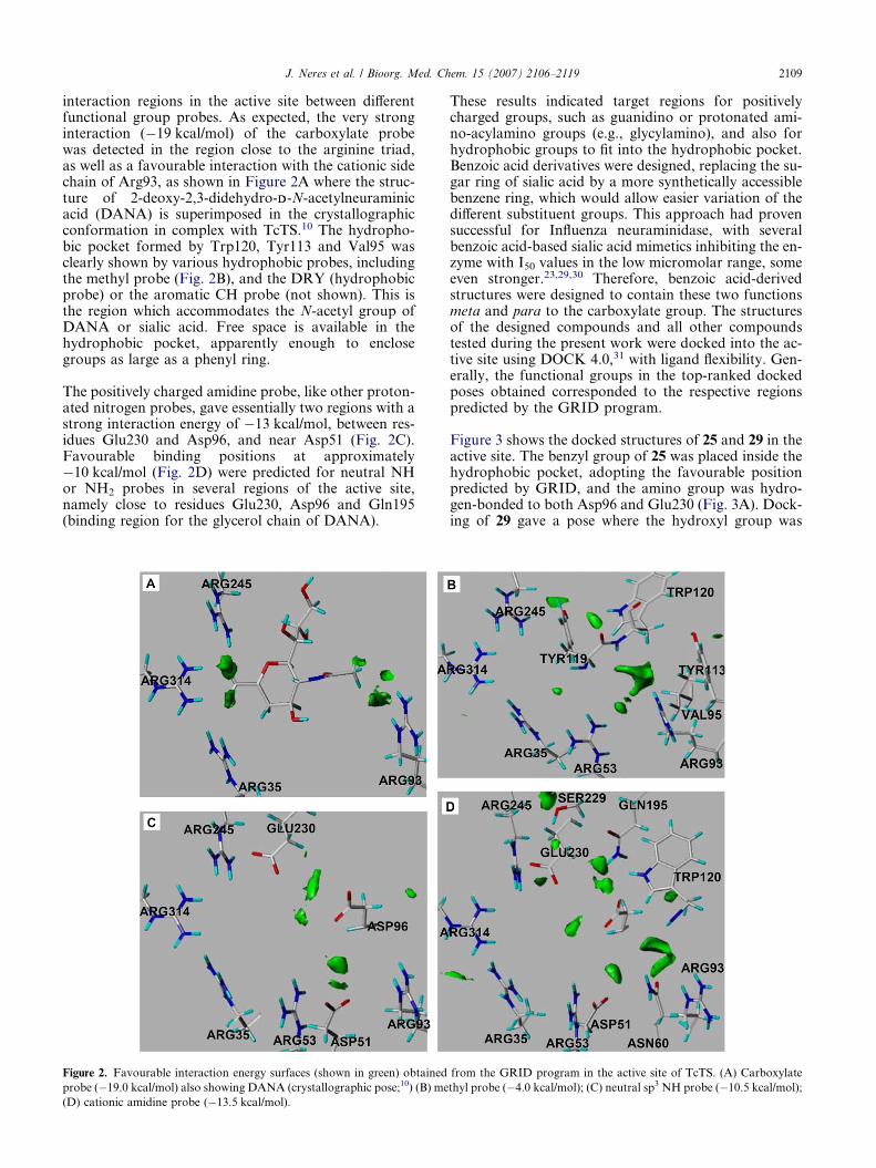
Figure 3 shows the docked structures of 25 and 29 in theactive site. The benzyl group of 25 was placed inside thehydrophobic pocket, adopting the favourable positionpredicted by GRID, and the amino group was hydro-gen-bonded to both Asp96 and Glu230 (Fig. 3A). Dock-ing of 29 gave a pose where the hydroxyl group was
from the GRID program in the active site of TcTS. (A) Carboxylate
thyl probe (�4.0 kcal/mol); (C) neutral sp3 NH probe (�10.5 kcal/mol);
Figure 3. Docked conformation of compounds 25 (A), superimposed
with the GRID favourable contour energy surface for the aromatic CH
probe at �3.7 kcal/mol) and 29 (B) in the active site of TcTS.
2110 J. Neres et al. / Bioorg. Med. Chem. 15 (2007) 2106–2119
hydrogen-bonded to both Gln195 and Glu230 and theamide nitrogen with Asp96 (Fig. 3B). Commonly tothe observed for most benzoic acid derivatives studiedin this work, in both structures the carboxylate groupwas in close interaction with the three arginine residuesin the triad.
2.1.2. Virtual screening. Virtual screening of the Asinexdatabase using DOCK produced numerous structurallydiverse hits potentially containing negatively chargedgroups, such as carboxylate, sulfonamide and sulfonefunctions, predominantly positioned close to the argi-nine triad. Many also contained a positively chargedgroup, usually interacting with one of the anionic resi-dues in the active site, and/or a hydrophobic moiety,which was located in the hydrophobic pocket. Thus,the results of the screen are in line with the types ofinteractions seen in the preceding modelling work. Thir-ty-two highly ranked compounds with varied structuralfeatures were purchased from Asinex, with special atten-tion given to benzoic acid and benzimidazole deriva-tives. However, only five of these compounds inhibitedTcTS to some extent at 1 mM, namely 48, 49 (Table 1)and 58–60 (Fig. 5). Results from virtual screening hitsother than those shown in Table 1 (47–49) and Figure5 (57–60) are not shown.
2.2. Synthesis
Benzoic acids 8, 18 and 19 were synthesised from4-amino-3-nitrobenzoic acid (9), according to
Scheme 1. The methyl ester 10 was prepared to avoidside-reactions of the carboxylate group. Compound 11was successfully synthesised in good yield (77%) byrefluxing 10 in DMF for 70 h in the presence of ex-cess benzyl bromide, even though the amine in thestarting material was strongly deactivated by the o-ni-tro and p-carboxylic ester. Synthesis of 15 by reacting10 with phenylacetyl chloride was more straightfor-ward. Reduction of the nitro groups of 11 and 15proceeded without problem using transfer hydrogena-tion, following the procedure described by Singhet al.32 Whereas 17 was synthesised by reaction of16 with cyanamide and HCl with 52% yield, the syn-thesis of 14 was performed with the strerically moredemanding reagent, 1,3-bis(tert-butoxycarbonyl)-2-methyl-2-thiopseudourea, due to the presence of theadditional benzyl-substituted amine group in 12. Thet-Boc groups of 13 were efficiently removed using tri-ethylsilane and TFA in reasonable yield (68%),according to a method previously developed in ourlaboratory.33 Hydrolysis of the methyl esters inNaOH yielded the target compounds in the neutralform (zwitterionic for 8 and 18).
Synthesis of 24, 25 and 27 (Scheme 2) started from themethyl ester of 4-chloro-3-nitrobenzoic acid (20) withintroduction of the benzylmercaptyl group. Compound24 was obtained after introduction of the guanidinogroup using the conditions used for 18, followed bymethyl ester hydrolysis. Fmoc-protected glycine wasused to synthesise 26 from 22 in the presence of DCC.Fmoc group removal with diethylamine followed by es-ter hydrolysis gave 27.
2.3. TcTS inhibition
TcTS inhibition screening results including I50 values fora series of substituted benzoic acid and pyridinecarboxy-lic acid derivatives are given in Tables 1 and 2, respec-tively. Inhibition results and structures for othercompounds tested, including furosemide (55), are shownin Figure 5. The percentage inhibition at 1 mM concen-tration is the average of at least three independentexperiments.
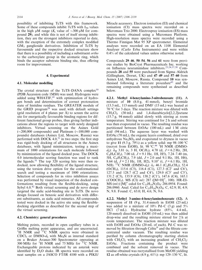
The inhibition by 29 was studied in greater detail todetermine the inhibition pattern of diagnostic plots.The data are shown in Figure 4A–C. The lines, drawnby linear least squares regression analysis, are not fit-ted to either mixed or non-competitive kinetic inhibi-tion models for this display. Fitting the data bynon-linear least squares analysis to the respectiveequations for non-competitive and mixed inhibitionmodels gave the following results, which were notsignificantly different, according to the statistical Ftest, performed in GRAFIT.34 Non-competitiveinhibition: Ki 0.33 ± 0.03 mM, Km 0.73 ± 0.11, Vmax
10,806 ± 870 fluorescence units (reduced v2 71,671);mixed inhibition: Ki 0.37 ± 0.17 mM, K 0i 0.31 ±0.13 mM, Km 0.75 ± 0.17, Vmax 11,011 ± 1352fluorescence units (reduced v2 75,068). Either modelconfirms the weak inhibition with Ki values around300 lM.

Scheme 1. Reagents and conditions: (a) MeOH, concd H2SO4, D; (b) PhCH2Br, DMF, D; (c) PhCH2COCl, CHCl3, D; (d) H2NNH2, 10% Pd/C,
EtOH; (e) H2NCN, concd HCl, EtOAc, D; (f) 1—1 M NaOH; 2—HCl; (g) H3CSC[@NCO2C(CH3)3]NHCO2C(CH3)3, Et3N, HgCl2, DMF; (h) TFA,
Et3SiH, DCM.
J. Neres et al. / Bioorg. Med. Chem. 15 (2007) 2106–2119 2111
3. Discussion
Sialic acid derivatives/mimetics were highly successful inthe design of inhibitors for the related Influenza neur-aminidase, with the discovery of potent inhibitors, twoof which are now in the market for the treatment ofInfluenza virus infections.35,36 However, we have foundin the present studies that the behaviour of the enzymesfrom Influenza and trypanosome is very different in re-spect of recognition of inhibitor structures.
In the present work, we used a sialic acid hydrolysis as-say, previously validated in our laboratory,11 to assessthe inhibition of TcTS by the described compounds.We found a substantial difference in the behaviours ofthe substituted aromatic carboxylic acids from Tables1 and 2 towards TcTS compared with the Influenza
neuraminidase, with no apparent correlation betweenthe two. For example, the stronger Influenza enzymeinhibitors 36, 38, 43 and 51 had I50 values of 2.5, 5, 80and 70 lM, respectively, against this enzyme.23,24 How-ever with TcTS, inhibition was much weaker, with 36and 43 inhibiting 40–50% of the activity at 1 mM and51 only 14% inhibition at the same concentration, and38 with an I50 of 0.76 mM. Conversely, stronger TcTSinhibitors 29 and 52, with I50 of approximately0.5 mM, were weaker inhibitors of the Influenza en-zyme.23,24 Compound 54 had a similar I50 for both en-zymes (�0.4–0.5 mM).24 In general, the stronginhibition that some aromatic carboxylates couldachieve for the Influenza enzyme (with I50 values aslow as 2.5 lM for 36) was not observed for TcTS. Thestrongest TcTS inhibitors here described only showedI50 values in 0.4–1 mM region. The remaining aromatic
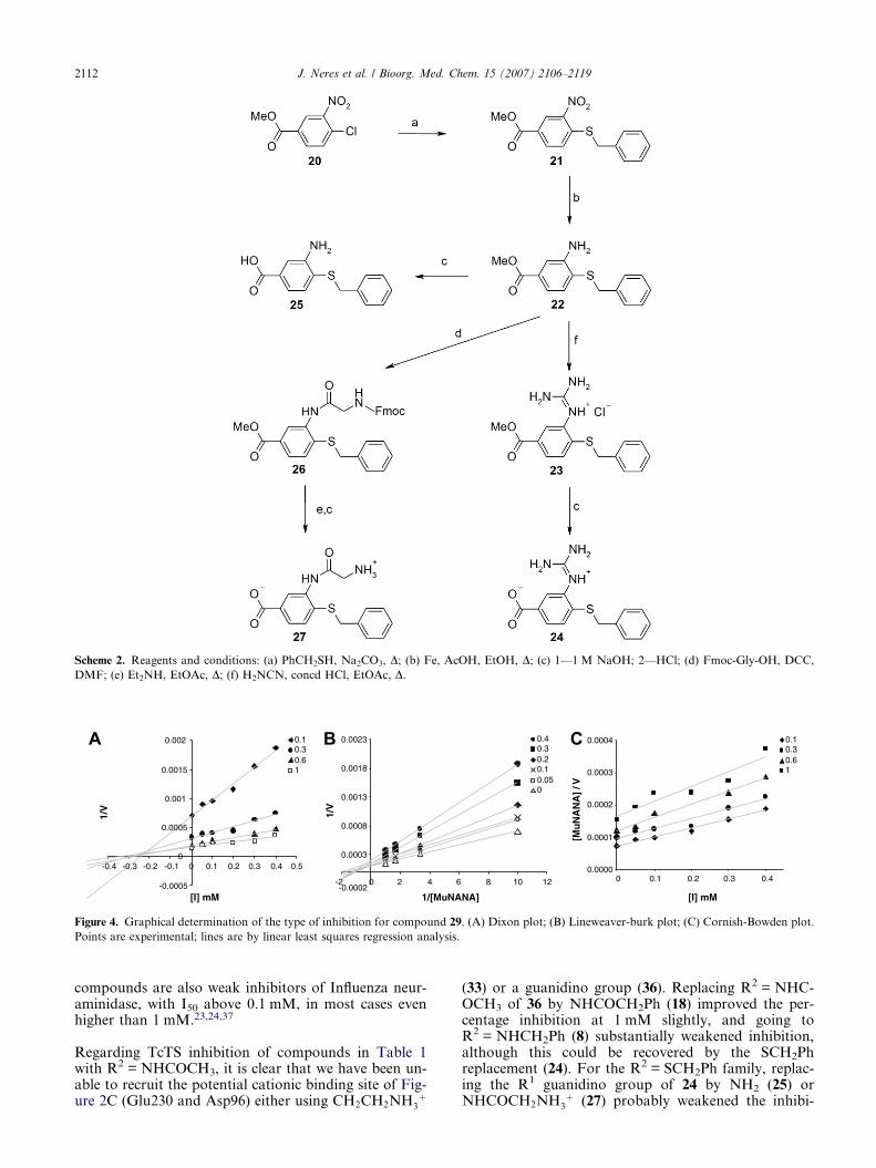
Scheme 2. Reagents and conditions: (a) PhCH2SH, Na2CO3, D; (b) Fe, AcOH, EtOH, D; (c) 1—1 M NaOH; 2—HCl; (d) Fmoc-Gly-OH, DCC,
DMF; (e) Et2NH, EtOAc, D; (f) H2NCN, concd HCl, EtOAc, D.
-0.0005
0
0.0005
0.001
0.0015
0.002
-0.4 -0.3 -0.2 -0.1 0 0.1 0.2 0.3 0.4 0.5
[I] mM
1/V
0.10.30.61
-0.0002
0.0003
0.0008
0.0013
0.0018
0.0023
-2 0 2 4 6 8 10 12
1/[MuNANA]
1/V
0.40.30.20.10.050
0.0000
0.0001
0.0002
0.0003
0.0004
0 0.1 0.2 0.3 0.4
[I] mM
[Mu
NA
NA
] / V
0.10.30.61
A B C
Figure 4. Graphical determination of the type of inhibition for compound 29. (A) Dixon plot; (B) Lineweaver-burk plot; (C) Cornish-Bowden plot.
Points are experimental; lines are by linear least squares regression analysis.
2112 J. Neres et al. / Bioorg. Med. Chem. 15 (2007) 2106–2119
compounds are also weak inhibitors of Influenza neur-aminidase, with I50 above 0.1 mM, in most cases evenhigher than 1 mM.23,24,37
Regarding TcTS inhibition of compounds in Table 1with R2 = NHCOCH3, it is clear that we have been un-able to recruit the potential cationic binding site of Fig-ure 2C (Glu230 and Asp96) either using CH2CH2NH3
þ
(33) or a guanidino group (36). Replacing R2 = NHC-OCH3 of 36 by NHCOCH2Ph (18) improved the per-centage inhibition at 1 mM slightly, and going toR2 = NHCH2Ph (8) substantially weakened inhibition,although this could be recovered by the SCH2Phreplacement (24). For the R2 = SCH2Ph family, replac-ing the R1 guanidino group of 24 by NH2 (25) orNHCOCH2NH3
þ (27) probably weakened the inhibi-

N
N
NH2NH
HOOC
HOOC
O
O
O
NH O
HOOC Cl
SO2NH2
HOOC
O
O
OH
NH
NHOOC
OH
S
OH
NH
NH2
O
O
OH HN
NH
NH2
NH
O
HO
O17±5% inhibition at 1 mM55
I50 0.67 mM56
No inhibition at 1 mM57
58 59 60
14±8% inhibition at 1 mM 20±2% inhibition at 1 mM31±2% inhibition at 1 mM
61
No inhibition at 1 mM
Figure 5. Structures and TcTS inhibition of other compounds tested.
J. Neres et al. / Bioorg. Med. Chem. 15 (2007) 2106–2119 2113
tion slightly (with I50 values of 0.70 mM for 25 and1.0 mM for 27).
The inhibition by 29 is consistent with either non-com-petitive inhibition (Ki 0.33 ± 0.03 mM) or mixed inhibi-tion (Ki 0.37 ± 0.17 mM, K 0i 0.31 ± 0.13 mM) with reallyvery little to discriminate the models. In either case theinhibition is weak with Ki values around 300 lM only.
The introduction of a pyridine (Table 2) instead of abenzene nucleus (Table 1) made little difference to thestrength of inhibition of TcTS (compare 28 and 50).Compound 51 inhibits the Influenza enzyme with anI50 of 0.07 mM24 but is much weaker with TcTS. Com-pound 61 is very strong against the Influenza enzyme(with I50 in the low nanomolar range), indeed reachingphase III clinical trials,38 but was only a poor inhibitorof TcTS.
From the above data, it is clear that the Influenza andTcTS enzymes respond very differently to inhibitorstructure. This can be explained not only by the struc-tural differences between the two enzymes, but also bya different dynamic behaviour of sialidases and trans-sialidases.14,17,39 Comparison of the crystal structuresof Influenza neuraminidase (1F8B40) and TcTS showsmany common features in the sialic acid-binding region,namely the arginine triad and Asp/Glu residues inapproximately the same regions of the active site. How-ever, the active site of the Influenza enzyme contains agreater number of negatively charged residues(Glu119, Asp151, Glu227, Glu276 and Glu277) whencompared with TcTS (Asp59, Asp96, Glu230), plusthere is one extra arginine residue (Arg53) in the latter.Furthermore, the active site of the Influenza enzyme(and indeed those of all structurally characterized sialid-ases, including the closely related T. rangeli enzyme) iseasily accessible, with a greater solvent exposure thanthat in TcTS, where the active site is more hydrophobic.This is probably one key feature that distinguishes thedifferent nature of the enzymatic activities, namely sialic
acid hydrolysis for neuraminidase and sialic acid trans-fer for TcTS.10 Another remarkable difference is thedynamic behaviour of TcTS during substrate bindingand catalysis, where occupancy of the sialic acid-bindingsite was demonstrated to promote a conformationalchange that modulates the affinity for the acceptor sugarsubstrate.10 Similar conformational changes are expect-ed to take place upon inhibitor binding to the sialic acid-binding site. Although the nature and extent of theseinhibitor-induced changes cannot be assessed in the ab-sence of experimental structural information on TcTS-inhibitor complexes, it is plausible to suggest that theycould be responsible for the lower sensitivity of trans-sialidase for the general sialidase inhibitor DANA14
and might explain, at least in part, why it is difficult toobtain good TcTS inhibitors.
In particular, both the low accessibility and the flexiblenature of the active site may account for the low activityof the aromatic carboxylate derivatives (Tables 1 and 2),since their docked structures fitted well in the active siteof TcTS, with the guanidino and other positivelycharged groups interacting with the side chains ofGlu230 and Asp96. Furthermore, these structures donot explore the acceptor substrate binding site in TcTS,which has been discussed as a potential target for TcTSinhibition and is targeted by lactitol and its derivatives,which prevent the sialylation of lactose or lactosamineby TcTS.10,18,22
The clinically used diuretic compound 55 (furosemide,Lasix�) also inhibits TcTS, but, with an I50 of0.67 mM, any TcTS-based dose would be unacceptablein man. However, the furan ring region of 55 is dockedagainst the acceptor substrate site in our docking analy-sis, thus providing an extra interaction with the activesite compared with the other aromatic derivatives; fur-ther exploration of the SAR in this region is desirable.
In conclusion, the range of aromatic derivatives evaluat-ed in the present work as TcTS inhibitors has shown the
2114 J. Neres et al. / Bioorg. Med. Chem. 15 (2007) 2106–2119
possibility of inhibiting TcTS with this framework.Some of these compounds inhibit TcTS with I50 valuesin the high lM range (Ki value of �300 lM for com-pound 29), and while this is not of itself strong inhibi-tion, they are the strongest inhibitors reported to date,with the exception of the extremely complex modifiedGM3 ganglioside derivatives. Inhibition of TcTS byfurosemide and the respective docked structure showthat there is a possibility of including a substituent orthoto the carboxylate group in the aromatic ring, whichbinds the acceptor substrate binding site, thus offeringroom for improvement.
4. Experimental
4.1. Molecular modelling
The crystal structure of the TcTS–DANA complex10
(PDB Accession code 1MS8) was used. Hydrogens wereadded using WHATIF,41 with optimization of hydro-gen bonds and determination of correct protonationstate of histidine residues. The GREATER module ofthe GRID program28 was used with default settings(grid resolution of 4 planes per A) to screen the activesite for energetically favourable binding regions for dif-ferent functional group probes, thus giving further indi-cations about the regions to target with newly designedinhibitors. Virtual screening on the Asinex Gold(�200,000 compounds) and Platinum (�100,000 com-pounds) databases (Asinex Ltd, Moscow, Russia) wasperformed with DOCK 4.0,31 in two steps: the first stepwas rigid-body docking of all structures in the Asinexdatabases, with ligand minimization, testing a maxi-mum of 1000 orientations for each molecule followedby minimization of the ligand docked pose. The DOCK4.0 intermolecular scoring function was used to rankthe ligands.31 The top 320 scoring hits were then re-docked, now allowing flexibility of the ligand structure,using the torsion drive option for the conformationalsearch and testing a maximum of 1000 orientations.Selection of compounds for in vitro inhibition assayswas performed by visual inspection of the docked con-formations resulting from the flexible-docking, usingSybyl 6.8.42 Both virtual screening and de novo designtargeted the sialic acid-binding site in TcTS. De novodesign focused on benzoic acid derivatives with differ-ent substituents, as sialic acid mimetics. All compoundstested were docked in the active site using the flexible-docking algorithm as described for the second step ofthe virtual screening.
4.2. Chemistry: general procedures
Melting points, recorded in open capillary tubes in aGriffin melting point apparatus, and are uncorrected.1H NMR and 13C NMR spectra were obtained inCDCl3 or DMSO-d6 with Me4Si as internal standardon a Bruker Avance-300 Spectrometer operating at300 MHz for 1H NMR and 75 MHz for 13C NMR.Exchangeable protons indicated by an asterisk wereidentified by D2O shake. IR spectra were recorded onneat samples on a JASCO FT/IR 4100 with a PIKE/
Miracle accessory. Electron ionisation (EI) and chemicalionisation (CI) mass spectra were recorded on aMicromass Trio 2000. Electrospray ionisation (ES) massspectra were obtained using a Micromass Platform.High-resolution mass spectra were recorded using aThermo Finnigan Mat 95 XP spectrometer. Elementalanalyses were recorded on an EA 1108 ElementalAnalyser (Carlo Erba Instruments) and were within0.4% of the calculated values unless otherwise noted.
Compounds 29–46, 50–54, 56 and 61 were from previ-ous studies by BioCryst Pharmaceuticals Inc. workingon Influenza neuraminidase inhibitors.23,24,37,38 Com-pounds 28 and 55 were purchased from Sigma–Aldrich(Gillingham, Dorset, UK) and 47–49 and 57–60 fromAsinex Ltd, Moscow, Russia. Compound 10 was syn-thesised following a literature procedure43 and theremaining compounds were synthesised as describedbelow.
4.2.1. Methyl 4-benzylamino-3-nitrobenzoate (11). Amixture of 10 (8.0 g, 41 mmol), benzyl bromide(13.5 mL, 113 mmol) and DMF (15 mL) was heated at70 �C for 3 days. The reaction mixture was diluted withCH2Cl2 (30 mL) and 1,4-diazabicyclo[2,2,2]octane(15.7 g, 94 mmol) added slowly with stirring at roomtemperature. Stirring was continued for 2 h and solventwas then evaporated in vacuo. The resulting residue waspartitioned between EtOAc (100 mL) and 1 M citricacid (94 mL). The aqueous layer was washed withEtOAc (70 mL), the organic layers combined, dried overanhydrous Na2SO4 and evaporated to dryness in vacuoto give 11 (9.3 g, 79%) as a yellow solid: mp 98–100 �C(recryst from EtOH), lit. 98 �C.44 1H NMR (DMSO-d6): dH 3.81 (s, 3 H, OCH3), 4.70 (d, J = 6.2 Hz, 2H,C6H5CH2), 7.00 (d, J = 9.1 Hz, 1H, H5), 7.24–7.40 (m,5H, C6H5CH2), 7.9 (dd, J = 2.0 and 9.1 Hz, 1H, H6),8.64 (d, J = 2.1 Hz, 1H, H2), 9.10* (t, J = 6.1 Hz, 1H,NH). 13C NMR (DMSO-d6): dC 46.1 (C6H5CH2), 52.4(OCH3), 115.6 (C5), 116.4 (C1), 127.3 (C2 0 and C6 0),127.5 and 128.7 (C2 and C4 0), 129.0 (C3 0 and C5 0),131.2 (C3), 135.9 (C6), 138.2 (C1 0), 147.6 (C4), 165.1(COOCH3). MS (CI) m/z 287 ([M+H]+, 100). HR-EI-MS (m/z) [M]+ calcd for C15H14N2O4: 286.0954. Found:286.0960. Anal. Calcd for C15H14N2O4: C, 62.9; H, 4.9;N, 9.8. Found: C, 63.0; H, 4.6; N, 9.6.
4.2.2. Methyl 3-amino-4-benzylaminobenzoate (12). Asuspension of 11 (9 g, 31.4 mmol) in EtOH (25 mL)was added to a mixture of 10% Pd–C (3 g) and 5%HCl (7 mL). Hydrazine hydrate (85%, 4.4 mL,120 mmol) dissolved in EtOH (10 mL) was then addeddrop-wise and the resulting mixture stirred for 2 h atroom temperature. The reaction mixture was dilutedwith EtOH and EtOAc (20 mL of each), Pd–C was re-moved by filtration through Celite� and the filtrate con-centrated under vacuum. The resulting residue waspurified by column chromatography starting elutionwith CH2Cl2 with an increasing gradient of CH2Cl2:EtOAc. Fractions containing the product werecombined and the solvent removed in vacuo. Theresidue obtained was recrystallised from MeOH yielding12 as off-white crystals (4.9 g, 61%): mp 129–130 �C, lit.
J. Neres et al. / Bioorg. Med. Chem. 15 (2007) 2106–2119 2115
125 �C.44 1H NMR (DMSO-d6): dH 3.70 (s, 3H, OCH3),4.38 (d, J = 5.8 Hz, 2H, C6H5CH2NH), 4.85* (s, 2H,NH2), 5.96* (br t, J = 5.7 Hz, 1H, C6H5CH2NH), 6.35(d, J = 8.3 Hz, 1H, H5), 7.10 (dd, J = 1.9 and 8.3 Hz,1H, H6), 7.19 (d, J = 2.0 Hz, 1H, H2), 7.21–7.36 (m,5H, C6H5CH2NH). 13C NMR (DMSO-d6): dC 46.6(C6H5CH2NH), 51.5 (OCH3), 108.9 and 114.8 (C2 andC5), 117.3 (C1), 120.9 (C6), 127.1 (C4 0), 127.5 (C2 0,C6 0), 128.7 (C3 0 and C5 0), 134.4 (C3), 139.9 and 140.6(C4 and C1 0), 167.1 (COOCH3). MS (ES+) m/z 311([M+Na+MeOH]+, 100), 279 ([M+Na]+, 10), 257([M+H]+, 23). Anal. Calcd for C15H16N2O2: C, 70.3;H, 6.3; N, 10.9. Found: C, 69.9; H, 6.2; N, 10.9.
4.2.3. Methyl 4-benzylamino-3-[N 0,N00-di(tert-butoxycar-bonyl)guanidino]benzoate (13). To an ice-cold suspensionof 12 (1 g, 3.9 mmol) in dry DMF (7 mL) were addedwith stirring triethylamine (1.1 mL, 7.8 mmol), 1,3-bis-(tert-butoxycarbonyl)-2-methyl-2-thiopseudourea (1.7 g,5.9 mmol) and HgCl2 (1.6 g, 5.9 mmol). The ice bathwas removed after 20 min and stirring continued atroom temperature for 7 h. The reaction mixture wasdiluted with EtOAc (20 mL) and filtered through Cel-ite�. Solvent was removed in vacuo and the residue ob-tained purified by column chromatography, startingwith CH2Cl2 with an increasing gradient of CH2Cl2:EtOAc. Fractions containing the product were com-bined and solvent was removed in vacuo to give 13 asa white solid (1.2 g, 62%): mp 135–138 �C. 1H NMR(CDCl3): dH 1.44 (s, 9H, C(CH3)3), 1.54 (s, 9H,C(CH3)3), 3.84 (s, 3H, OCH3), 4.43 (d, J = 5.8 Hz, 2H,C6H5CH2NH), 5.64* (t, J = 5.7 Hz, 1H, C6H5CH2NH),6.64 (d, J = 8.4 Hz, 1H, H5), 7.25–7.37 (m, 5H,C6H5CH2NH), 7.76–7.81 (m, 2H, H2 and H6), 9.97*
(s, 1H, NHC(@NR)NHR), 11.61* (s, 1H,NHC(@NR)NHR). 13C NMR (CDCl3): dC 28.5 and28.6 [2· (CH3)3], 48.1 (C6H5CH2), 52.1 (OCH3), 80.2and 84.4 [2· C(CH3)3], 112.2 (C5), 119.0 (C1), 122.2(C3), 127.6 (C4 0), 127.8 (C2 0 and C6 0), 128.7 (C6),129.1 (C3 0 and C5 0), 130.5 (C3), 138.8 (C1 0), 147.7(C4), 153.7 and 155.0 (2· C@O t-Boc), 163.5(NHC(@NR)NHR), 167.2 (COOCH3). MS (ES+) m/z499 ([M+H]+, 100). Anal. Calcd for C26H34N4O6Æ0.25-H2O: C, 62.1; H, 6.9; N, 11.1. Found: C, 62.2; H, 6.5;N, 11.1.
4.2.4. Methyl 4-benzylamino-3-guanidino-benzoate (14).To a solution of 13 (250 mg, 0.5 mmol) in dichlorometh-ane (2 mL) were added trifluoroacetic acid (1 mL,13 mmol) and triethylsilane (0.4 mL, 2.5 mmol). Thereaction mixture was stirred at room temperature for6 h with exclusion of moisture. Solvent was removedin vacuo, the residue triturated with chloroform and col-lected by filtration, and dried giving 14 as a white solid(0.14 g, 68%): mp 119–121 �C (recryst from MeOH/Et2O). 1H NMR (DMSO-d6): dH 3.75 (s, 3H, OCH3),4.45 (d, J = 6.0 Hz, 2H, C6H5CH2NH), 6.61 (d,J = 8.4 Hz, 1H, H5), 7.00* (t, J = 6.0 Hz, 1H,C6H5CH2NH), 7.21–7.36 (m, 9H, C6H5 and NH@C(NH2)2), 7.56 (d, J = 1.8 Hz, 1H, H2), 7.68 (dd,J = 1.8 and 9.0 Hz, 1H, H6), 9.05* (s, 1H, NH@C(NH2)2). 13C NMR (CDCl3): dC 45.4 (C6H5CH2),51.4 (OCH3), 110.6 (C5), 116.2 and 118.5 (C1 and C3),
126.7 (C2 0, C4 0 and C6 0), 128.2 (C3 0 and C5 0), 130.3and 130.9 (C2 and C6), 138.9 (C1 0), 149.1 (C4), 156.8(NH@C(NH2)2), 165.7 (COOCH3). MS (ES+) m/z 299([M+H]+, 100). HR-ES-MS (m/z) [M+H]+ calcd forC16H19N4O2: 299.1503. Found: 299.1500.
4.2.5. 4-Benzylamino-3-guanidino-benzoic acid (8). Amixture of 14 (80 mg, 0.19 mmol), 1 M NaOH (10 mL)and THF (1 mL) was stirred at 40 �C for 3 h. The resultingsolution was concentrated in vacuo, diluted with waterand the pH was adjusted to�7 with 1 N HCl. The precip-itate that formed was collected by filtration and dried,yielding 8 as a white solid (0.040 g, 73%). 1H NMR(DMSO-d6): dH 4.37 (br d, 2H, C6H5CH2NH), 6.14* (brs, 1H, C6H5CH2NH), 6.44 (d, J = 8.5 Hz, 1H, H5), 7.19(t, J = 7.3 Hz, 1H, H4 0), 7.28 (t, J = 7.3 Hz, 2H, H3 0 andH5 0), 7.40 (d, J = 7.3 Hz, 2H, H2 0 and H6 0), 7.53–7.56(m, 2H, H2 and H6), 7.92* (br s, 4H, NH@C(NH2)2),11.32* (br s, 1 H, NH@C(NH2)2). 13C NMR (DMSO-d6): dC 45.9 (C6H5CH2), 109.7 (C5), 119.9 and 126.4 (C1and C3), 126.5 (C4 0), 126.9 (C2 0 and C6 0), 128.1 (C3 0
and C5 0), 128.6 and 129.2 (C2 and C6), 139.6 (C1 0),145.4 (C4), 157.3 (NH@C(NH2)2), 170.9 (COOH). MS(ES+) m/z 285 ([M+H]+, 100), 308 ([M+Na]+, 48). HR-ES-MS (m/z) [M+H]+ calcd for C15H17N4O2: 285.1346.Found: 285.1347.
4.2.6. Methyl 3-nitro-4-phenylacetylaminobenzoate (15).A mixture of 10 (1 g, 5.1 mmol) and phenylacetyl chlo-ride (1.35 mL, 10.2 mmol) in CHCl3 (2 mL) was stirredat 100 �C for 2 h. Acetonitrile (6 mL) was added to thereaction mixture, which was heated until the suspendedsolid dissolved. Water (1.5 mL) was added and the solu-tion partitioned between CH2Cl2 (15 mL) and saturatedNaHCO3 solution (15 mL). The organic layer was sepa-rated, washed with water (2· 15 mL), dried over anhy-drous Na2SO4 and solvent removed in vacuo. Theresidue was recrystallised from MeOH, yielding 15(0.96 g, 60%) as a yellow solid: mp 129–130 �C. 1HNMR (CDCl3): dH 3.86 (s, 2H, CH2C6H5), 3.94 (s,3H, OCH3), 7.35–7.47 (m, 5H, CH2C6H5), 8.25 (dd,J = 2.0 and 8.9 Hz, 1H, H6), 8.83 (d, J = 2.0 Hz, 1H,H2), 8.94 (d, J = 8.9 Hz, 1H, H5), 10.48* (s, 1H,CONH). 13C NMR (CDCl3): dC 46.30 (CH2C6H5),53.02 (OCH3), 121.92 (C5), 125.42 (C1), 127.86 and128.60 (C2 and C4 0), 129.88 (C3 0 and C5 0), 130.07(C2 0 and C6 0), 133.21 and 135.93 (C3 and C4), 136.81(C5), 138.52 (C1 0), 165.04 (COOCH3), 170.87 (CONH).MS (CI) m/z 315 ([M+H]+, 100). HR-EI-MS (m/z) [M]+
calcd for C16H14N2O5: 314.0903. Found: 314.0908.Anal. Calcd for C16H14N2O5: C, 61.1; H, 4.5; N, 8.9.Found: C, 61.2; H, 4.3; N, 8.9.
4.2.7. Methyl 3-amino-4-phenylacetylaminobenzoate (16).A suspension of 15 (0.7 g, 2.23 mmol) in EtOH (20 mL)was added to a mixture of 10% Pd–C (0.7 g) and 5%HCl (0.51 mL). Hydrazine hydrate (64%, 0.44 mL,9.16 mmol) dissolved in ethanol (3 mL) was addeddrop-wise, and the mixture stirred for 1 h at room tem-perature, diluted with ethanol and ethyl acetate (20 mLof each) and the Pd–C removed by filtration throughCelite�. The filtrate was evaporated in vacuo to give16 as a white solid (0.52 g, 82%): mp 179–181 �C (recryst

2116 J. Neres et al. / Bioorg. Med. Chem. 15 (2007) 2106–2119
from MeOH). 1H NMR (DMSO-d6): dH 3.70 (s, 2H,CH2C6H5), 3.79 (s, 3H, OCH3), 5.22* (s, 2H, NH2),7.15 (dd, J = 1.7 and 8.3 Hz, 1H, H6), 7.38 (d,J = 1.7 Hz, 1H, H2), 7.48 (d, J = 8.3 Hz, 1H, H5),7.24–7.35 (m, 5H, CH2C6H5), 9.46* (s, 1H, CONH).13C NMR (DMSO-d6): dC 42.7 (CH2C6H5), 51.7(OCH3), 116.3 and 117.2 (C2 and C6), 123.8 (C5),126.2 (C1), 126.4 (C1 and C4 0), 127.7 (C4), 128.2 (C3 0
and C5 0), 129.1 (C2 0 and C6 0), 135.9 (C1 0), 140.8 (C3),166.2 (COOCH3), 169.3 (CONH). MS (CI) m/z 285([M+H]+, 49). HR-EI-MS (m/z) [M]+ calcd forC16H16N2O3: 284.1161. Found: 284.1160. Anal. Calcdfor C16H16N2O3: C, 67.6; H, 5.7; N, 9.9. Found: C,67.8; H, 5.5; N, 9.7.
4.2.8. Methyl 3-guanidino-4-phenylacetylaminobenzoate(17). A mixture of 16 (0.47 g, 1.65 mmol), cyanamide(1.4 g, 33 mmol) and concd HCl (0.15 mL) in EtOAc(15 mL) was refluxed for 6 h. The reaction mixturewas diluted with EtOAc (30 mL) and partitioned withK2CO3 solution (15 mL, half of saturation concentra-tion). The organic layer was washed with water, driedover Na2SO4, filtered and the solvent evaporated in vac-uo. The residue obtained was recrystallised from EtOHyielding 17 (0.28 g, 52%) as a white solid: mp 182–184 �C. 1H NMR (DMSO-d6): dH 3.73 (s, 2H,CH2C6H5), 3.77 (s, 3H, OCH3), 5.48* (s, 4H,N@C(NH2)2), 7.24–7.40 (m, 6H, H6 and CH2C6H5),7.46 (d, J = 1.9 Hz, 1H, H2), 8.23 (d, J = 8.4 Hz, 1H,H5), 9.10* (s, 1H, CONH). 13C NMR (DMSO-d6): dC
43.98 (CH2C6H5), 51.56 (OCH3), 116.75, 121.18 and121.21 (C2, C5 and C6), 123.65 (C1), 126.72 (C4 0),128.50 (C3 0 and C5 0), 129.28 (C2 0 and C6 0), 135.25and 136.11 (C4 and C1 0), 139.06 (C3), 154.39(N@C(NH2)2), 166.35 (COOCH3), 168.62 (CONH).MS (CI) m/z 327 ([M+H]+, 100), 267 ([M�NH@C(NH2)2]+, 45). HR-EI-MS (m/z) [M]+ calcd forC17H18N4O3: 326.1379. Found: 326.1386. Anal. Calcdfor C17H18N4O3Æ0.6H2O: C, 60.4; H, 6.0; N, 16.6.Found: C, 60.2; H, 5.5; N, 16.9.
4.2.9. 3-Guanidino-4-phenylacetylaminobenzoic acid (18).A mixture of 17 (80 mg, 0.18 mmol), NaOH 1 M(0.6 mL) and THF (0.4 mL) was stirred at room temper-ature for 2 h. The pH of the resulting clear solution wasadjusted to �7 with 1 M HCl, and 18 precipitated as awhite solid, which was collected by filtration and dried(0.059 g, 77%): mp 236–237 �C. 1H NMR (DMSO-d6):dH 3.73 (s, 2H, C6H5CH2), 7.21–7.36 (m, 5H, C6H5),7.71–7.79 (m, 3H, H2, H5, H6), 8.05* (br s, 4H,N@C(NH2)2), 10.0* (s, 1H, NHCOCH2C6H5), 10.73*
(br, COOH). MS (ES+) m/z 313 ([M+H]+, 100), 335([M+Na]+, 52). HR-ES-MS (m/z) [M+H]+ calcd forC16H17N4O3: 313.1295. Found: 313.1307.
4.2.10. 3-Amino-4-phenylacetylaminobenzoic acid (19). Amixture of 16 (20 mg, 0.070 mmol), NaOH 0.5 M (1 mL)and THF (0.5 mL) was stirred at room temperature for16 h. The resulting suspension was filtered, the filtratediluted with 2 mL of water and the pH adjusted to�7 with1 M HCl. The precipitate thus formed was collected by fil-tration and dried, giving 19 as a fine white powder(0.011 g, 58%): mp 168–170 �C. 1H NMR (DMSO-d6):
dH 3.70 (s, 2H, C6H5CH2CONH), 5.18* (s, 2H, NH2),7.12 (d, J = 8.2 Hz, 1H, H5), 7.23–7.34 (m, 6H,H2 and C6H5CH2CONH), 7.43 (d, J = 8.2 Hz, 1H,H6), 9.50* (s, 1H, C6H5CH2CONH), 12.56* (br s, 1H,COOH). 13C NMR (DMSO-d6): dC 42.7 (C6H5COCH2),116.6, 117.4 and 123.7 (C2, C5 and C6), 126.4 (C4 0),127.2 and 127.8 (C1 and C4), 128.2 (C2 0 and C6 0),129.0 (C3 0 and C5 0), 136.0 (C1 0), 140.7 (C3), 167.4(C6H5CH2CONH), 169.2 (COOH). MS (ES�) m/z 269([M�H]�, 100). HR-EI-MS (m/z) [M+Na]+ calcdfor C15H14N2O3Na: 293.0897. Found: 293.0893.
4.2.11. Methyl 3-nitro-4-benzylmercaptobenzoate (21).To a mixture of benzyl mercaptan (3.3 mL, 27.8 mmol),Na2CO3 (3.25 g, 30.6 mmol) and water (10 mL) wasadded methyl 4-chloro-3-nitrobenzoate (5 g, 23.2 mmol)in ethanol (40 mL). The mixture was refluxed for 2 h anddiluted with water (40 mL). The solid thus formed wasfiltered off, washed with n-hexane and dried, yielding21 as a yellow solid (6.56 g, 93%): mp 136–138 �C (re-cryst from MeOH), lit. 138 �C.45 1H NMR (DMSO-d6): dH 3.90 (s, 3H, OCH3), 4.45 (s, 2H, C6H5CH2S),7.28–7.40 (m, 3H, H2 0, H4 0 and H6 0), 7.47–7.50 (m,2H, H3 0 and H5 0), 7.90 (d, J = 8.6 Hz, 1H, H5), 8.17(dd, J = 1.9 and 8.6 Hz, 1H, H6), 8.64 (d, 1H,J = 1.9 Hz, H2). 13C NMR (DMSO-d6): dC 36.0(C6H5CH2S), 52.6 (OCH3), 126.1 (C1), 126.4, 127.6and 127.8 (C2, C5 and C4 0), 128.6 (C2 0 and C6 0),129.3 (C3 0 and C5 0), 133.4 (C6), 134.9 (C4), 142.8(C1 0), 144.5 (C3) and 164.3 (COOCH3). MS (CI) m/z321 ([M+NH4]+, 100), (EI) m/z 303 ([M]+, 21). Anal.Calcd for C15H13NO4S: C, 59.4; H, 4.3; N, 4.6; S,10.6. Found: C, 59.1; H, 4.3; N, 4.6; S, 10.3.
4.2.12. Methyl 3-amino-4-benzylmercaptobenzoate (22).A solution of 21 (5 g, 16.5 mmol) in EtOH (10 mL) wastreated with iron dust (7.9 g, 137 mmol) and 20% aque-ous acetic acid (2.8 mL) refluxed for 30 min with vigor-ous stirring. The reaction mixture was diluted withEtOAc (40 mL) and filtered over Celite�. The filteredsolution was washed with 5% aqueous NaHCO3
(40 mL), followed by water (2· 40 mL), and dried overanhydrous Na2SO4. The solvent was evaporated in vac-uo and the residue obtained was recrystallised fromMeOH to give 22 as an off-white solid (4.0 g, 88%): mp76–77 �C lit. 76–78 �C.45 1H NMR (DMSO-d6): dH 3.79(s, 3H, OCH3), 4.10 (s, 2H, C6H5CH2S), 5.46* (s, 2H,NH2), 7.05 (dd, J = 1.9 and 9.0 Hz, 1H, H6), 7.19–7.28(m, 6H, H5 and C6H5CH2S), 7.32 (d, J = 1.9 Hz, 1H,H2). 13C NMR (DMSO-d6): dC 36.3 (C6H5CH2S), 51.8(OCH3), 114.3 and 116.6 (C2 and C6), 122.7 (C4),127.0 (C4 0), 128.2 (C2 0 and C6 0), 128.8 (C3 0 and C5 0),128.9 (C1), 131.7 (C5), 137.4 (C1 0), 147.7 (C3), 166.3(COOCH3). MS (CI) m/z 274 ([M+H]+, 100). Anal.Calcd for C15H15NO2S: C, 65.9; H, 5.5; N, 5.1; S, 11.7.Found: C, 66.0; H, 5.6; N, 5.1; S, 11.4.
4.2.13. Methyl 4-benzylmercapto-3-guanidino-benzoatehydrochloride (23). A mixture of 22 (0.7 g, 2.6 mmol),cyanamide (2.7 g, 64 mmol) and concd HCl (0.33 mL)in EtOAc (20 mL) was refluxed for 2 h. The reaction mix-ture was cooled to room temperature and a solid crystal-lised. This solid was filtered, washed with EtOAc and
J. Neres et al. / Bioorg. Med. Chem. 15 (2007) 2106–2119 2117
dried, yielding 23 as a white solid (0.8 g, 88%): mp 245–246 �C (recryst from EtOH). 1H NMR (DMSO-d6): dH
3.85 (s, 3H, OCH3), 4.36 (s, 2H, C6H5CH2S), 7.26–7.48(m, 9H, decreased to 5H after D2O shake, C6H5CH2S,NH@C(NH2)2), 7.62 (d, J = 8.4 Hz, 1H, H5), 7.73 (d,J = 1.8 Hz, 1H, H2), 7.90 (dd, J = 1.8 and 8.4 Hz, 1H,H6), 9.66* (br s, 1H, NH@C(NH2)2). 13C NMR(DMSO-d6): dC 34.8 (C6H5CH2S), 52.2 (OCH3), 126.5(C2), 126.8 (C1), 127.4 (C4 0), 128.5 (C2 0 and C6 0),128.89–128.93 (C5, C6, and C3 0 and C5 0), 131.2 (C4),135.8 (C1 0), 143.5 (C3), 156.4 (NH@C(NH2)2), 165.3(COOCH3). MS (ES+) m/z 316 ([M]+, 100), (ES�) m/z350 ([M�H+Cl]�, 100). Anal. Calcd forC16H18N3O2SCl: C, 54.6; H, 5.2; N, 11.9; S, 9.1; Cl,10.1. Found: C, 54.7; H, 5.1; N, 12.0; S, 8.9; Cl, 10.4.
4.2.14. 4-Benzylmercapto-3-guanidino-benzoic acid (24).A solution of 23 (0.3 g, 0.85 mmol) in THF (1 mL) wasadded to 1 M NaOH (4 mL) and the mixture heated to70 �C for 1 h. The solvent was evaporated, and the res-idue obtained re-dissolved in water (3 mL) and the pHadjusted to 6–7 with 1 M HCl. A solid slowly cameout of solution, which was filtered and dried, yielding24 as a white solid, (0.22 g, 87%): mp >260 �C (recrystfrom EtOH/water). 1H NMR (DMSO-d6): dH 4.23 (s,2H, C6H5CH2S), 7.21–7.41 (m, 6H, H5 and C6H5CH2S),7.62 (d, J = 1.5 Hz, 1H, H2), 7.76 (dd, J = 1.5 and8.1 Hz, 1H, H6), 8.00* (br s, 4H, NH@C(NH2)2),11.36* (br s, 1H, NH@C(NH2)2). 13C NMR (DMSO-d6): dC 35.6 (C6H5CH2S), 126.9 (C2), 127.2 (C4 0),128.4 (C2 0 and C6 0), 128.7 (C5 and C6), 128.9 (C3 0
and C5 0), 131.7 (C1), 136.5, 137.2, 137.3 (C3, C4, C1 0)156.4 (NH@C(NH2)2), 169.5 (COOH). MS (ES+) m/z302 ([M+H]+, 100), (ES�) m/z 300 ([M�H]�, 100). Anal.Calcd for C15H15N3O2SÆH2O: C, 56.4; H, 5.4; N, 13.2; S,10.0. Found: C, 56.3; H, 5.0; N, 13.2; S, 9.8.
4.2.15. Synthesis of 3-amino-4-benzylmercapto-benzoicacid (25). A mixture of 22 (0.42 g, 1.54 mmol), 1 MNaOH (3 mL) and MeOH (0.5 mL) was stirred at40 �C for 8 h. The volume of the resulting solutionwas reduced to one third and the pH adjusted to 4 with1 M HCl. The solution was extracted with EtOAc(30 mL) and the organic phase dried over anhydrousNa2SO4 and evaporated in vacuo, giving 25 as an off-white solid (0.35 g, 88%): mp 155–157 �C (recryst fromMeOH). 1H NMR (CDCl3): dH 4.10 (s, 2H,SCH2NC6H5), 5.45* (s, 2H, NH2), 4.44 (2H,J = 5.6 Hz, C6H5CH2NH), 7.03 (dd, J = 1.8 and8.0 Hz, 1H, H6), 7.17–7.29 (m, 6H, C6H5CH2S andH5), 7.31 (d, J = 1.8 Hz, 1H, H2), 12.71* (br s, 1H,COOH). 13C NMR (DMSO-d6): dC 36.4 (SCH2NC6H5)114.6 and 116.9 (C1 and C6), 122.0 (C4), 126.9 (C4 0),128.2 (C2 0 and C6 0), 128.8 (C3 0 and C5 0), 130.1 (C1),131.2 (C5), 137.4 (C1 0), 147.7 (C3), 167.4 (COOH).MS (ES�) m/z 258 ([M�H]�, 100). Anal. Calcd forC14H13NO2S: C, 64.8; H, 5.1; N, 5.4; S, 12.4. Found:C, 64.7; H, 5.0; N, 5.3; S, 12.2.
4.2.16. Methyl 4-benzylmercapto-3-[2-(9H-fluoren-9-ylmethoxycarbonylamino)-acetylamino] benzoate (26).To a solution of 22 (1 g, 3.7 mmol) in anhydrous DMF(5 mL) cooled in an ice bath were added Fmoc-Gly-OH
(1.74 g, 5.9 mmol) and DCC (1.1 g, 5.5 mmol) and themixture stirred for a few minutes on the ice bath and thenat room temperature for 16 h. The suspended solid wasremoved by filtration and the filtrate evaporated in vacuo.The resulting residue was suspended in EtOAc (50 mL)and the insoluble material filtered out (solid A). The fil-trate was chromatographed over a silica-gel column,starting with CH2Cl2 with an increasing gradient ofCH2Cl2: EtOAc (up to 30% EtOAc). Fractions contain-ing the product were combined with solid A and solventremoved in vacuo. Successive recrystallisations of the res-idue from EtOAc and EtOH yielded 26 as a white solid(0.65 g, 33%): mp 193–195 �C. 1H NMR (DMSO-d6):dH 3.83–3.88 (br s, 5H, OCH3 and NHCOCH2NHFmoc),4.24–4.33 (m, 5H, C6H5CH2S; CH and CH2 Fmoc), 7.20–7.35 (m, 7H, C6H5CH2S and 2 Car H Fmoc), 7.42 (t,J = 7.3 Hz, 2H, 2 Car H Fmoc), 7.54 (d, J = 8.3 Hz, 1H,H5), 7.69–7.74 (m, 3H, H6 and 2 Car H Fmoc), 7.79*
(app t, 1H, NHCOCH2NHFmoc), 7.90 (d, J = 7.4 Hz,2H, 2 Car H Fmoc), 8.11 (s, 1H, H2), 9.47* (s, 1H,NHCOCH2NHFmoc). 13C NMR (DMSO-d6): dC 36.1(C6H5CH2S), 43.9 (NHCOCH2NHFmoc), 46.5 (CHFmoc), 52.0 (OCH3), 65.8 (CH2 Fmoc), 120.0 (CHFmoc), 124.8 (C2), 125.1 (CH Fmoc), 125.9 (C6), 127.0(CH Fmoc), 127.2 (C4 0), 127.5 (CH Fmoc), 128.3 (C2 0
and C5 0), 128.6 (C5), 128.9 (C3 0 and C5 0), 135.22,135.24, 136.2, 136.6 (C1, C3, C4 and C1 0), 140.6 and143.7 (2 · Cquat Fmoc), 156.5 (CO Fmoc), 165.6(COOCH3), 168.4 (NHCOCH2NHFmoc). MS (ES+) m/z 575 ([M+Na]+, 100); (ES�) m/z 551 ([M�H]�, 100).HR-ES-MS (m/z) [M+Na]+ calcd for C32H28N2O5SNa:575.1611. Found: 575.1607.
4.2.17. 3-[(Aminoacetyl)amino]-4-benzylmercapto-benzo-ic acid (27). A solution of 26 (0.125 g, 0.23 mmol) inEtOAc (5 mL) and diethylamine (5 mL) was refluxedfor 1 h and the resulting solution evaporated to drynessin vacuo. The resulting residue was suspended in MeOH(30 mL) and filtered. The filtrate was evaporated to dry-ness and the residue dissolved in THF (2 mL) and 1 MNaOH (4 mL), and refluxed for 1 h. The mixture was fil-tered and the pH adjusted to �7 with 1 M HCl. The sol-id formed was collected by filtration and dried, giving 27as a white solid (0.026 g, 36%): mp colour change todark brown at 210 �C; melted at 240–241 �C. 1HNMR (DMSO-d6, TFA): dH 3.81–3.83 (br q, 2H,NHCOCH 2NH3
þ), 4.33 (s, 2H, C6H5CH2S), 7.25–7.43(m, 5H, C6H5CH2S), 7.56 (d, J = 8.4 Hz, 1H, H5) 7.76(dd, J = 1.7 and 8.4, 1H, H6), 7.94 (d, J = 1.6 Hz, 1H,H2), 8.17* (br s, 3H, NHCOCH2NH 3
þ), 10.07* (s, 1H,NHCOCH2NH3
þ). 13C NMR (DMSO-d6): dC 35.9(C6H5CH2S), 40.9 (NHCOC H2 NH3
þ), 126.9, 127.4and 127.6 (C2, C5, C6, C4 0), 128.2 (C2 0 and C6 0),129.2 (C3 0 and C5 0), 134.0, 136.4 and 138.5 (C3, C4,C1 0), 165.9 (NHCOCH2NH3
þ), 166.8 (COOH). MS(ES�) m/z 315 ([M�H]�, 100). HR-ES-MS (m/z)[M�H]� calcd for C16H15N2O3S: 315.0802. Found:315.0809.
4.3. Expression and purification of recombinant TcTS
The protocol for expression and purification of recombi-nant TcTS was adapted from Buschiazzo et al.46
2118 J. Neres et al. / Bioorg. Med. Chem. 15 (2007) 2106–2119
Competent E. coli BL21(DE3)pLysS (Promega, South-ampton, UK) cells were transformed with a plasmid con-taining the TcTS gene (pTrcTS611/247) and grownovernight in LB broth containing 100 lg/mL ampicillinat 37 �C. The culture was diluted 1:50 with TB broth con-taining 100 lg/mL ampicillin and incubation continuedunder the same conditions to A600 � 1.2. The tempera-ture was adjusted to 18 �C and bacteria induced toover-express recombinant TcTS by adding IPTG(1 mM). Induction was maintained for 14–16 h, afterwhich cells were harvested and frozen. After one thaw/freeze cycle, lysis was achieved in the presence of20 mM Tris–HCl, pH 8.5, 30 mM NaCl, 0.5% TritonX-100, 5 lg/mL DNAse I, protease inhibitor cocktailand lysozyme produced by the cells, followed by sonica-tion (6 · 30 s pulses of ultrasound). The lysate was centri-fuged at 16,000 rpm for 45 min at 4 �C and thesupernatant subjected to iminodiacetic metal affinitychromatography (HisTrap FF 1 mL), on an AKTA-FPLC system (GE Healthcare, Little Chalfont, UK),after adjustment of the NaCl concentration to 0.5 Mand pH to 8.5. After applying the lysate, the columnwas washed with buffer containing 20 mM Tris–HCl,pH 8.5, 0.5 M NaCl and 10 mM imidazole, and the pro-tein eluted with a linear gradient of imidazole (10–500 mM) in the same buffer. Fractions containing theprotein were desalted (HiPrep 26/10 desalting column)and further purification was achieved by FPLC anionicexchange (MonoQ 5/50 GL) applying a linear elution gra-dient of NaCl (0–0.5 M). Purified protein was quantifiedusing the Bradford method, using BSA as a standard.48
4.4. Inhibition studies
Enzyme inhibition was assessed at I50 level using a con-tinuous assay developed in our laboratory and describedelsewhere.11 The assay mixture, containing 20 mM Tris–HCl, pH 7.5, buffer, TcTS (10 lL of 0.33 mg/mL) andinhibitor solution (10 lL), was incubated for 10 min at25 �C and reaction initiated by addition of MuNANA(10 lL of a 1 mM solution). The fluorescence of the re-leased product (Mu) was monitored at 25 �C for 10 min,with excitation and emission wavelengths of 322 and448 nm, respectively. Values of I50, the concentration re-quired to give 50% inhibition under the assay conditionsdescribed above, were determined by interpolation froma plot of assay velocity (in percentage of the activity inabsence of inhibitors) versus inhibitor concentration, fit-ting to the IC50 0–100 equation in Grafit34 (V = 100%/{1+([I]/I50)s}), where s is a slope factor.
Ki determinations were performed using a discontinuousversion of the assay. Briefly, the reaction mixture, con-taining 20 mM Tris–HCl, pH 7.5 (150 lL), enzyme solu-tion (20 lL of 0.33 mg/mL) and, when studied, inhibitorsolution (20 lL), was incubated for 10 min at 25 �C andreaction initiated by addition of MuNANA solution(10 lL of 2–20 mM MuNANA solution). After incuba-tion (2, 3, 4 and 5 min), aliquots (40 lL) of the reactionmixture were taken and enzymatic reaction quenched byadding to 0.2 M Na2CO3 solution at pH 10 (100 lL) in a96-well plate. The fluorescence of the released product(Mu) was measured at 25 �C with excitation and emis-
sion wavelengths of 365 and 442 nm, respectively. Inhi-bition type was assessed by analysing the patterns ofthree diagnostic classes of plot: 1/vo versus 1/[So] for var-ious [I]; 1/vo versus [I] for various [So]; [So]/vo versus [I]at various [So]. Values of Ki and K 0i for mixed inhibitionwere determined by direct weighted (1=v2
o for weighting)least squares non-linear regression analysis of the rawdata using the appropriate equation using the Grafitprogram,34 viz., for mixed inhibition, v ¼ V max½So�=ð½So�ð1þ ½I�=K 0iÞ þ Kmð1þ ½1�=K iÞ. Values of Vmax andKm were obtained by least squares non-linear regressionanalysis using Grafit.
Acknowledgments
We are grateful to Professor A. Carlos C. Frasch andLaura Ratier (Instituto de Investigaciones Biotecnolog-icas, Universidad Nacional de San Martın, Argentina)for general advice and to the Portuguese Foundationfor Science and Technology (F.C.T.) for financialsupport.
References and notes
1. The World Health Report 2004: Changing history. 2004.Geneva, World Health Organization.
2. Cerecetto, H.; Gonzalez, M. Curr. Top. Med. Chem. 2002,2, 1187–1213.
3. Vassena, C. V.; Picollo, M. I.; Zerba, E. N. Med. Vet.Entomol. 2000, 14, 51–55.
4. Schenkman, S.; Jiang, M. S.; Hart, G. W.; Nussenzweig,V. Cell 1991, 65, 1117–1125.
5. Schenkman, S.; Eichinger, D.; Pereira, M. E. A.; Nus-senzweig, V. Annu. Rev. Microbiol. 1994, 48, 499–523.
6. Ming, M.; Chuenkova, M.; Ortegabarria, E.; Pereira, M.E. A. Mol. Biochem. Parasitol. 1993, 59, 243–252.
7. Buschiazzo, A.; Tavares, G. A.; Campetella, O.; Spinelli,S.; Cremona, M. L.; Paris, G.; Amaya, M. F.; Frasch, A.C. C.; Alzari, P. M. EMBO J. 2000, 19, 16–24.
8. Cross, G. A. M.; Takle, G. B. Annu. Rev. Microbiol. 1993,47, 385–411.
9. Cremona, M. L.; Campetella, O.; Sanchez, D. O.; Frasch,A. C. C. Glycobiology 1999, 9, 581–587.
10. Buschiazzo, A.; Amaya, M. F.; Cremona, M. L.; Frasch,A. C.; Alzari, P. M. Mol. Cell 2002, 10, 757–768.
11. Neres, J.; Buschiazzo, A.; Alzari, P. M.; Walsh, L.;Douglas, K. T. Anal. Biochem. 2006, 357, 302–304.
12. Schenkman, S.; de Carvalho, L. P.; Nussenzweig, V.J. Exp. Med. 1992, 175, 567–575.
13. Vandekerckhove, F.; Schenkman, S.; de Carvalho, L. P.;Tomlinson, S.; Kiso, M.; Yoshida, M.; Hasegawa, A.;Nussenzweig, V. Glycobiology 1992, 2, 541–548.
14. Paris, G.; Ratier, L.; Amaya, M. F.; Nguyen, T.; Alzari, P.M.; Frasch, A. C. C. J. Mol. Biol. 2005, 345, 923–934.
15. Watts, A. G.; Damager, I.; Amaya, M. L.; Buschiazzo, A.;Alzari, P.; Frasch, A. C.; Withers, S. G. J. Am. Chem. Soc.2003, 125, 7532–7533.
16. Watts, A. G.; Oppezzo, P.; Withers, S. G.; Alzari, P. M.;Buschiazzo, A. J. Biol. Chem. 2006, 281, 4149–4155.
17. Amaya, M. F.; Buschiazzo, A.; Nguyen, T.; Alzari, P. M.J. Mol. Biol. 2003, 325, 773–784.
18. Agusti, R.; Paris, G.; Ratier, L.; Frasch, A. C. C.;de Lederkremer, R. M. Glycobiology 2004, 14, 659–670.
19. Previato, J. O. Glycobiology 2004, 14, 25G.

J. Neres et al. / Bioorg. Med. Chem. 15 (2007) 2106–2119 2119
20. Streicher, H.; Busse, H. Bioorg. Med. Chem. 2006, 14,1047–1057.
21. Ferrero-Garcia, M. A.; Sanchez, D. O.; Frasch, A. C. C.;Parodi, A. J. An. Asoc. Quim. Argent 1993, 81, 127–132.
22. Streicher, H. Curr. Med. Chem.: Anti-infective Ag 2004, 3,149–161.
23. Chand, P.; Babu, Y. S.; Bantia, S.; Chu, N. M.; Cole, L.B.; Kotian, P. L.; Laver, W. G.; Montgomery, J. A.;Pathak, V. P.; Petty, S. L.; Shrout, D. P.; Walsh, D. A.;Walsh, G. W. J. Med. Chem. 1997, 40, 4030–4052.
24. Chand, P.; Kotian, P. L.; Morris, P. E.; Bantia, S.; Walsh,D. A.; Babu, Y. S. Bioorg. Med. Chem. 2005, 13, 2665–2678.
25. Howes, P. D.; Cleasby, A.; Evans, D. N.; Feilden, H.;Smith, P. W.; Sollis, S. L.; Taylor, N.; Wonacott, A. J.Eur. J. Med. Chem. 1999, 34, 225–234.
26. Jedrzejas, M. J.; Singh, S.; Brouillette, W. J.; Laver, W. G.;Air, G. M.; Luo, M. Biochemistry 1995, 34, 3144–3151.
27. Williams, M.; Bischofberger, N.; Swaminathan, S.; Kim,C. U. Bioorg. Med. Chem. Lett. 1995, 5, 2251–2254.
28. Goodford, P. J. J. Med. Chem. 1985, 28, 849–857.29. Atigadda, V. R.; Brouillette, W. J.; Duarte, F.; Ali, S. M.;
Babu, Y. S.; Bantia, S.; Chand, P.; Chu, N.; Montgomery,J. A.; Walsh, D. A.; Sudbeck, E. A.; Finley, J.; Luo, M.;Air, G. M.; Laver, G. W. J. Med. Chem. 1999, 42, 2332–2343.
30. Brouillette, W. J.; Atigadda, V. R.; Luo, M.; Air, G. M.;Babu, Y. S.; Bantia, S. Bioorg. Med. Chem. Lett. 1999, 9,1901–1906.
31. Ewing, T. J. A.; Makino, S.; Skillman, A. G.; Kuntz, I. D.J. Comput. Aided Mol. Des. 2001, 15, 411–428.
32. Singh, S.; Jedrzejas, M. J.; Air, G. M.; Luo, M.; Laver, W.G.; Brouillette, W. J. J. Med. Chem. 1995, 38, 3217–3225.
33. Mehta, A.; Jaouhari, R.; Benson, T. J.; Douglas, K. T.Tetrahedron Lett. 1992, 33, 5441–5444.
34. Grafit 5.0. Erithacus Software Limited. Horley, Surrey,UK, 2005.
35. Kim, C. U.; Lew, W.; Williams, M. A.; Liu, H. T.; Zhang,L. J.; Swaminathan, S.; Bischofberger, N.; Chen, M. S.;Mendel, D. B.; Tai, C. Y.; Laver, W. G.; Stevens, R. C.J. Am. Chem. Soc. 1997, 119, 681–690.
36. von Itzstein, M.; Wu, W. Y.; Kok, G. B.; Pegg, M. S.;Dyason, J. C.; Jin, B.; Phan, T. V.; Smythe, M. L.; White,H. F.; Oliver, S. W.; Colman, P. M.; Varghese, J. N.;Ryan, D. M.; Woods, J. M.; Bethell, R. C.; Hotham, V. J.;Cameron, J. M.; Penn, C. R. Nature 1993, 363, 418–423.
37. Sudbeck, E. A.; Jedrzejas, M. J.; Singh, S.; Brouillette, W.J.; Air, G. M.; Laver, W. G.; Babu, Y. S.; Bantia, S.;Chand, P.; Chu, N.; Montgomery, J. A.; Walsh, D. A.;Luo, M. J. Mol. Biol. 1997, 267, 584–594.
38. Babu, Y. S.; Chand, P.; Bantia, S.; Kotian, P.; Dehghani,A.; El Kattan, Y.; Lin, T. H.; Hutchison, T. L.; Elliott, A.J.; Parker, C. D.; Ananth, S. L.; Horn, L. L.; Laver, G.W.; Montgomery, J. A. J. Med. Chem. 2000, 43, 3482–3486.
39. Amaya, M. F.; Watts, A. G.; Damager, T.; Wehenkel, A.;Nguyen, T.; Buschiazzo, A.; Paris, G.; Frasch, A. C.;Withers, S. G.; Alzari, P. M. Structure 2004, 12, 775–784.
40. RCSB Protein Data Bank, http://www.rcsb.org/.41. Vriend, G. J. Mol. Graph. 1990, 8, 52–56.42. Sybyl 6.8. Tripos Inc., 2002.43. Naim, S. S.; Singh, S. K.; Sharma, S.; Gupta, S.; Fatma,
N.; Chatterjee, R. K.; Katiyar, J. C. Indian J. Chem. B1988, 27, 1106–1109.
44. Goker, H.; Tebrizli, E.; Abbasoglu, U. Il Farmaco 1996,51, 53–58.
45. Donleavy, J. J.; Condit, P. C. J. Am. Chem. Soc. 1947, 69,1781–1784.
46. Buschiazzo, A.; Frasch, A. C. C.; Campetella, O. Cell.Mol. Biol. 1996, 42, 703–710.
47. Buschiazzo, A.; Campetella, O.; Frasch, A. C. C. Glyco-biology 1997, 7, 1167–1173.
48. Bradford, M. M. Anal. Biochem. 1976, 72, 248–254.