
Microstructure-property relationships in a tissue-engineering scaffold
Upload
fukuoka-eduCategory
view
2download
0
This document is downloaded at: 2012-04-28T14:35:22Z
Title A novel small molecule fluorescent sensor for Zn2+ based onpyridine‒pyridone scaffold
Author(s) Hagimori, Masayori; Mizuyama, Naoko; Yamaguchi, Yasuchika; Saji,Hideo; Tominaga, Yoshinori
Citation Talanta, 83(5), pp.1730-1735; 2011
Issue Date 2011-02-15
URL http://hdl.handle.net/10069/24564
Right Copyright © 2010 Elsevier B.V. All rights reserved.
NAOSITE: Nagasaki University's Academic Output SITE
http://naosite.lb.nagasaki-u.ac.jp
1
A Novel Small Molecule Fluorescent Sensor for Zn2+ Based on Pyridine-
Pyridone Scaffold
Masayori Hagimori,a Naoko Mizuyama,b Yasuchika Yamaguchi,a Hideo Saji,c and Yoshinori
Tominagad*
aFaculty of Pharmaceutical Sciences, Nagasaki International University, 2825-7 Huis Ten Bosch,
Sasebo 859-3298, Japan, bDepartment of Hospital Pharmacy, Saga University Hospital, 5-1-1
Nabeshima, Saga 849-8521, Japan, cGraduate School of Pharmaceutical Sciences, Kyoto University,
46–29 Yoshida-Shimoadachi-cho, Sakyo-ku, Kyoto 606–8501, Japan, dFaculty of Environmental Studies,
Nagasaki University,1-14 Bunkyo-machi, Nagasaki 852-8521, Japan
Abstract
The development of a water-soluble and small molecular weight fluorescent probe, 3-(4-
methoxyphenyl)-4-(methylsulfanyl)-6-(pyridin-2-yl)pyridin-2(1H)-one (3), for detecting Zn2+ based on
pyridine-pyridone skeleton is reported. We observed a clear chelation enhanced fluorescence effect of 3
in the presence of Zn2+. Other fluorescent properties of 3 are discussed.
Keywords
Zn2+, Fluorescent sensor, Pyridine-pyridone, Small molecule, Water-soluble
1. Introduction
Zinc is an essential element for life and is known to play important roles in biological processes
including gene expression, apoptosis, enzyme regulation, immune system and neurotransmission [1–6].
Most of Zn2+ in living cells are bound to the proteins, which are called as metalloprotein [7–9]. Zinc
metalloproteases employ Zn2+ as the catalysis center and a most well-known example in this class is
angiotensin converting enzyme (ACE) [10–12]. Zinc chelation also contributes to the structure
stabilization of proteins. For example, the zinc finger proteins form a stable three dimensional structure
to interact a specific DNA sequence for controlling gene functions [13].
2
There also are free or chelatable Zn2+ in living cells, which may be involved in both physiology and
disease states. In pancreatic β-cells, Zn2+ participates in controlling synthesis, storage and secretion of
insulin. When Zn2+ is short, blood sugar level rises by a secretion delay of the insulin [14,15]. In central
nervous system, free or chelatable Zn2+ are co-localized with glutamate in presynaptic vesicles of the
mammalian hippocampus [16,17].
To investigate physiological roles of free or chelatable Zn2+ in living cells, we should know their
concentration in each tissue and in different (patho-) physiological states. In this context, a number of
fluorescent sensors have recently been developed based on elegant ideas [18–20]. Although they
contributed great extends to understand Zn2+ roles in physiology and particularly in the field of
neurochemistry [16,17], its mechanisms of action is not well understood in comparison with other
cations such as Na+, K+, Ca2+, etc.
We realized that Zn2+ selective fluorescent probes have mainly used three core structures, namely
quinoline, BF2 chelated dipyrromethene and fluorescein [21–30]. After modification of the core
structure in getting selectivity toward Zn2+, the molecule obviously became bigger and more
hydrophobic. Therefore most of the fluorescent probes prepared so far have undesirable characteristics
in terms of aqueous solubility. This problem should be overcome somehow to develop a new simple
fluorescent probe based upon different chemical backbone structures. We thought that if we could find a
new and small molecular weight (MW = ca. 300) fluorescent core structure having certain selectivity
toward Zn2+, such molecules would become an advantageous starting point for designing a new
fluorescent sensor. If the initial core structure is small enough, the fluorescent probes may still be
molecular weight below 500 with desirable physico-chemical properties, even after the modifications
[31]. We have recently published a new and simple synthetic method for preparing pyridine-pyridone
derivative 3 [32]. Since then we have been interested in the molecule characters of this class of
heterocyclic compounds. In this paper, we report the fluorescent characteristics of pyridine-pyridone
derivative 3, MW = 324, that can potentially be used as a lead structure for finding a new fluorescent
sensor for Zn2+.
2. Experimental
2.1. Materials and instruments
All the solvents were of analytic grade and used as received. The solutions of metal ions were prepared
from NaCl, KCl, BaCl2, MgCl2·6H2O, CaCl2, FeCl3·6H2O, CoCl2·6H2O, NiCl2·6H2O, ZnCl2,
CdCl2·2.5H2O, CuCl2, MnCl2·4H2O, AlCl3·6H2O, respectively, and were dissolved in distilled water. 1H NMR was measured on a JEOL-GX-400 (400MHz) with chemical shifts reported as ppm (in
3
DMSO-d6). HRMS was measured on a JMS-T100LP mass spectrometer. Mass spectra (MS) were
recorded on a JEOL-DX-303 mass spectrometer. Fluorescence spectra were determined on a Jasco FP-
6200 spectrofluorometer. Ultraviolets (UVs) absorption spectra were determined in 95% ethanol on a
Hitachi 323 spectrometer. Infrared (IR) spectra were recorded in potassium bromide pellets on JASCO
810.
2.2. Synthesis of 3-(4-methoxyphenyl)-4-(methylsulfanyl)-6-(pyridin-2-yl)pyridin-2(1H)-one (3)
Powdered sodium hydroxide (0.40 g, 10.0 mmol) was added to a solution of 1.13 g (5.0 mmol) of 3,3-
bis-methylsulfanyl-1-pyridin-2-yl-propenone (1) [32] and 0.88 g (6.0 mmol) of 2 in 50 mL of DMSO,
and the mixture was stirred for 2 h at room temperature. The reaction was poured into 300 mL of ice
water and neutralized with a 10% hydrochloric acid solution. The mixture was extracted with 100 mL of
dichloromethane three times. The combined organic extracts were washed with water, dried over
anhydrous sodium sulfate, and concentrated under reduced pressure. A mixture of the residue and a 1%
hydrochloric acid solution was refluxed for 1 h. After evaporation, the residual solid was recrystallized
from methanol to give 3 (1.01 g, 62%) as yellow leaflets. Mp: 260-261 °C. IR (KBr, cm-1): 3456, 1605,
1510, 1275, 1175. 1H NMR (DMSO-d6, 400 MHz) δ 2.48 (s, 3H), 3.80 (s, 3H), 6.96 (d, J = 8.8 Hz, 2H),
7.20 (d, J = 8.8 Hz, 2H), 7.50 (ddd, J = 2.9, 4.9, 7.8 Hz, 1H), 7.97 (ddd, J = 1.5, 2.0, 7.8 Hz, 1H), 8.25
(d, J = 7.8 Hz, 1H), 8.25 (d, J = 7.8 Hz, 1H), 8.72 (ddd, J = 1.0, 3.9, 4.9 Hz, 1H), 11.09 (brs, 1H). 13C
NMR (DMSO-d6, 100 MHz) δ 14.46, 55.06, 113.41, 120.93, 124.73, 127.09, 131.35, 137.63, 149.41,
151.54, 158.62, 159.71. Ms m/z: 325 (M++1, 12), 324 (M+, 36), 310 (11), 309 (42), 294 (7), 278 (10),
149 (11), 84 (19), 66 (21), 57 (11), 44 (100). Anal. Calcd. for C18H16N2O2S: C, 66.64; H, 4.97; N,
8.64%. Found: C, 66.51, H, 5.07; N, 8.51%. HRMS calcd. for C18H16N2O2S: 324.0932; found: 324.0949.
2.3. Synthesis of 6-methoxy-5-(4-methoxyphenyl)-4-(methylsulfanyl)-2,2'-bipyridine (4)
Powdered sodium hydroxide (0.20 g, 5.0 mmol) was added to a solution of 0.56 g (2.5 mmol) of 1 and
0.37 g (2.5 mmol) of 2 in 25 mL of DMSO, and the mixture was stirred for 2 h at room temperature.
The reaction was poured into 300 mL of ice water and neutralized with a 10% hydrochloric acid
solution. The mixture was extracted with 100 mL of dichloromethane three times. The combined
organic extracts were washed with water, dried over anhydrous sodium sulfate, and concentrated under
reduced pressure. A mixture of the residue in 100 mL of methanol was refluxed for 6 h. After
evaporation, the residual solid was recrystallized from methanol to give 4 (0.38 g, 45%) as yellow
leaflets. Mp: 149-150 °C. IR (KBr, cm-1): 3425, 2940, 1610, 1570, 1540, 1510, 1450, 1430, 1350, 1270,
1250, 1170, 1105, 1035. 1H NMR (DMSO-d6, 400 MHz) δ 2.47 (s, 3H), 3.81 (s, 3H), 3.90 (s, 3H), 7.00
(d, J = 7.8 Hz, 2H), 7.20 (d, J = 8.3 Hz, 2H), 7.45 (dd, J = 5.4, 6.3 Hz, 1H), 7.95 (dd, J = 7.3, 8.3 Hz,
2H), 8.41 (d, J = 7.8 Hz, 1H), 8.71 (d, J = 4.4 Hz, 1H). 13C NMR (DMSO-d6, 100 MHz) δ 14.18, 53.35,
4
55.07, 108.54, 113.77, 120.57, 120.67, 124.18, 126.03, 131.14, 137.32, 149.22, 151.00, 152.19, 154.74,
158.85, 159.90. Ms m/z: 339 (M+ + 1, 24), 338 (M+, 100), 337 (29), 323 (19), 293 (8), 291 (7), 161 (6),
121 (5), 105 (5), 44 (4). Anal. Calcd. for C19H18N2SO2: C, 67.43; H, 5.36; N, 8.28%. Found: C, 67.61; H,
5.50; N, 8.10%. HRMS calcd. for C19H18N2O2S: 338.1089; found: 338.1098.
2.4. Synthesis of 3-(4-methoxyphenyl)-4-(methylsulfanyl)-6-phenylpyridin-2(1H)-one (6)
Powdered sodium hydroxide (0.40 g, 10.0 mmol) was added to a solution of 1.13 g (5.0 mmol) of 3,3-
bis-methylsulfanyl-1-phenyl-propenone (5) and 0.88 g (6.0 mmol) of 2 in 50 mL of DMSO, and the
mixture was stirred for 2 h at room temperature. The reaction was poured into 300 mL of ice water and
neutralized with a 10% hydrochloric acid solution. The mixture was extracted with 100 mL of
dichloromethane three times. The combined organic extracts were washed with water, dried over
anhydrous sodium sulfate, and concentrated under reduced pressure. A mixture of the residue and a 1%
hydrochloric acid solution was refluxed for 1 h. After evaporation, the residual solid was recrystallized
from methanol to give 6 (0.38 g, 24%) as yellow leaflets. Mp: 238-240 °C. IR (KBr, cm-1): 3056, 1568,
1527, 1285, 1176. 1H NMR (DMSO-d6, 400 MHz) δ 2.45 (s, 3H), 3.79 (s, 3H), 6.56 (s, 1H), 6.95 (d, J =
8.8 Hz, 2H), 7.18 (d, J = 8.8 Hz, 2H), 7.49 (m, 3H), 7.82 (dd, J = 3.9, 7.8 Hz, 2H), 11.75 (brs, 1H). 13C
NMR (DMSO-d6, 100 MHz) δ 14.31, 55.04, 101.25, 113.37, 123.56, 127.04, 127.34, 128.74, 129.72,
131.44, 133.78, 151.43, 158.50, 160.63. HRMS calcd. for C19H17NO2S: 323.0980; found: 323.0940.
2.5. Spectral measurement
The compounds 3, 4 and 6 stock solution (1×10⁻² M) was prepared by directly dissolving in DMSO.
The UV/vis spectrum of 3 (10-4 M) was measured in HEPES buffer (100 mM, 5% DMSO, pH = 7.4), as
shown in Fig.1. For the fluorescence analysis, 3 (10-6 M) upon addition of Zn2+ in the form of
perchlorate salt was measured in HEPES buffer (100 mM, 5% DMSO, pH 7.4). The binding
stoichiometry of 3 to Zn2+ was investigated by Job’s plot. We measured the fluorescence intensity of 3
in the following buffers: 100 mM glycine - HCl buffer (pH 2.0), 100 mM citrate buffer (pH 3.0), 100
mM acetate buffer (pH 4.0 – 5.0), 100 mM Phosphate buffer (pH 6.0), 100 mM HEPES buffer (pH 7.0 –
8.0), 100 mM tris-HCl buffer (pH 9.0). The dissociation constant (Kd) of 3 in HEPES buffer was
determined by plotting the fluorescence intensity to free Zn2+ concentration. The metal selectivity of 3
was investigated in HEPES buffer (100 mM, 5% DMSO, pH 7.4) and that of 4 and 6 were investigated
in aqueous EtOH solution (1:1 EtOH/water (v/v)). The cational solutions were prepared from NaCl, KCl,
BaCl2, MgCl2·6H2O, CaCl2, FeCl3·6H2O, CoCl2·6H2O, NiCl2·6H2O, ZnCl2, CdCl2·2.5H2O, CuCl2,
MnCl2·4H2O, AlCl3·6H2O (10-3 M), respectively. The measurements were carried out at 298 K. The
5
fluorescence quantum yield values were measured with respect to quinine sulfate solution (Φ = 0.54) as
standard.
3. Results and discussion
The synthesis of 3 is described in Scheme 1. The reaction of 3,3-bis-methylsulfanyl-1-pyridin-2-yl-
propenone (1) with 2-(4-methoxyphenyl)acetonitrile (2) in the presence of powdered sodium hydroxide
in DMSO followed by treatment with 1% HCl gave 3-(4-methoxyphenyl)-4-(methylsulfanyl)-6-
(pyridin-2-yl)pyridin-2(1H)-one (3) in 62% yield [32]. The structure was established by 1H/13C NMR,
MS and elemental analysis.
Scheme 1. Synthesis of compound 3.
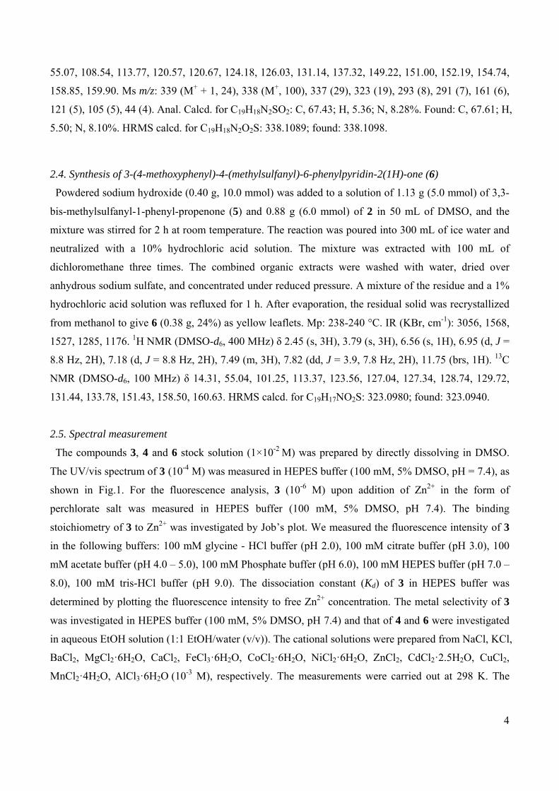
Fig. 1 shows the UV/vis spectrum of 3 in HEPES buffer (100 mM, 5% DMSO, pH 7.4). Zn2+ addition
changed the UV/vis spectrum of 3 in a Zn2+ concentration dependent manner. Namely the absorption
peaks at 310 nm and 342 nm decreased and contrary those at 350 nm increased after addition of Zn2+.
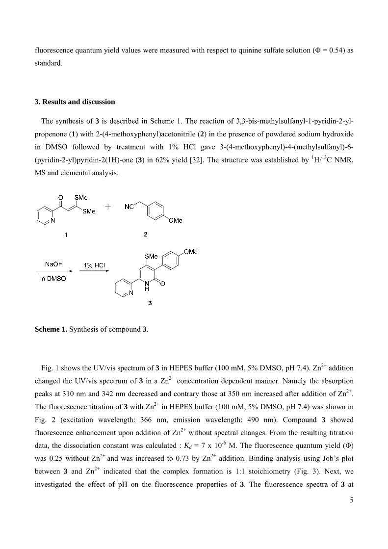
The fluorescence titration of 3 with Zn2+ in HEPES buffer (100 mM, 5% DMSO, pH 7.4) was shown in
Fig. 2 (excitation wavelength: 366 nm, emission wavelength: 490 nm). Compound 3 showed
fluorescence enhancement upon addition of Zn2+ without spectral changes. From the resulting titration
data, the dissociation constant was calculated : Kd = 7 x 10⁻⁶ M. The fluorescence quantum yield (Φ)
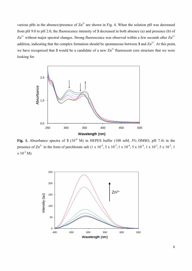
was 0.25 without Zn2+ and was increased to 0.73 by Zn2+ addition. Binding analysis using Job’s plot
between 3 and Zn2+ indicated that the complex formation is 1:1 stoichiometry (Fig. 3). Next, we
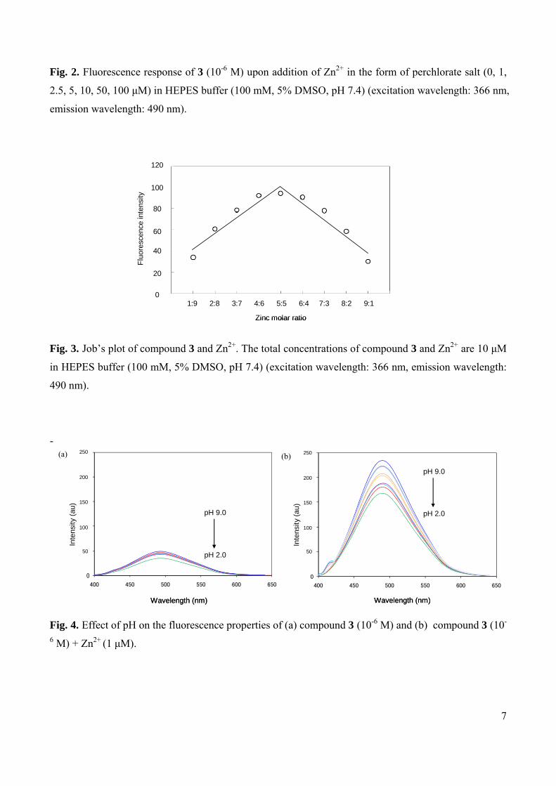
investigated the effect of pH on the fluorescence properties of 3. The fluorescence spectra of 3 at
6
various pHs in the absence/presence of Zn2+ are shown in Fig. 4. When the solution pH was decreased
from pH 9.0 to pH 2.0, the fluorescence intensity of 3 decreased in both absence (a) and presence (b) of
Zn2+ without major spectral changes. Strong fluorescence was observed within a few seconds after Zn2+
addition, indicating that the complex formation should be spontaneous between 3 and Zn2+. At this point,
we have recognized that 3 would be a candidate of a new Zn2+ fluorescent core structure that we were
looking for.
Fig. 1. Absorbance spectra of 3 (10-4 M) in HEPES buffer (100 mM, 5% DMSO, pH 7.4) in the
presence of Zn2+ in the form of perchlorate salt (1 x 10⁻⁵, 5 x 10⁻⁵, 1 x 10⁻⁴, 5 x 10⁻⁴, 1 x 10⁻³, 5 x 10⁻³, 1
x 10-2 M).
0.0
1.0
2.0
250 300 350 400 450 500
Abs
orb
an
ce
Wavelength (nm)
0.0
1.0
2.0
250 300 350 400 450 500
Abs
orb
an
ce
Wavelength (nm)
0.0
1.0
2.0
250 300 350 400 450 500
Abs
orb
an
ce
Wavelength (nm)
0
50
100
150
200
250
400 450 500 550 600 650
Zn2+
Inte
nsity
(au
)
Wavelength (nm)
0
50
100
150
200
250
400 450 500 550 600 650
Zn2+
Inte
nsity
(au
)
Wavelength (nm)
7
Fig. 2. Fluorescence response of 3 (10-6 M) upon addition of Zn2+ in the form of perchlorate salt (0, 1,
2.5, 5, 10, 50, 100 μM) in HEPES buffer (100 mM, 5% DMSO, pH 7.4) (excitation wavelength: 366 nm,
emission wavelength: 490 nm).
Fig. 3. Job’s plot of compound 3 and Zn2+. The total concentrations of compound 3 and Zn2+ are 10 μM
in HEPES buffer (100 mM, 5% DMSO, pH 7.4) (excitation wavelength: 366 nm, emission wavelength:
490 nm).
-
Fig. 4. Effect of pH on the fluorescence properties of (a) compound 3 (10-6 M) and (b) compound 3 (10-
6 M) + Zn2+ (1 μM).
1:9 2:8 3:7 4:6 5:5 6:4 7:3 8:2 9:1
Zinc molar ratio
120
100
80
60
40
20
0
Flu
ores
cenc
e in
tens
ity
1:9 2:8 3:7 4:6 5:5 6:4 7:3 8:2 9:1
Zinc molar ratio
120
100
80
60
40
20
0
Flu
ores
cenc
e in
tens
ity
(a) (b)
0
50
100
150
200
250
400 450 500 550 600 650
Wavelength (nm)
Inte
nsity
(au
)
250
200
150
100
50
pH 9.0
pH 2.0
0
50
100
150
200
250
400 450 500 550 600 650
Wavelength (nm)
Inte
nsity
(au
)
250
200
150
100
50
pH 9.0
pH 2.0
0
30
60
90
120
150
400 450 500 550 600 650
Wavelength (nm)
Inte
nsity
(au
)
250
200
150
100
50
pH 9.0
pH 2.0
0
30
60
90
120
150
400 450 500 550 600 650
Wavelength (nm)
Inte
nsity
(au
)
250
200
150
100
50
pH 9.0
pH 2.0
8
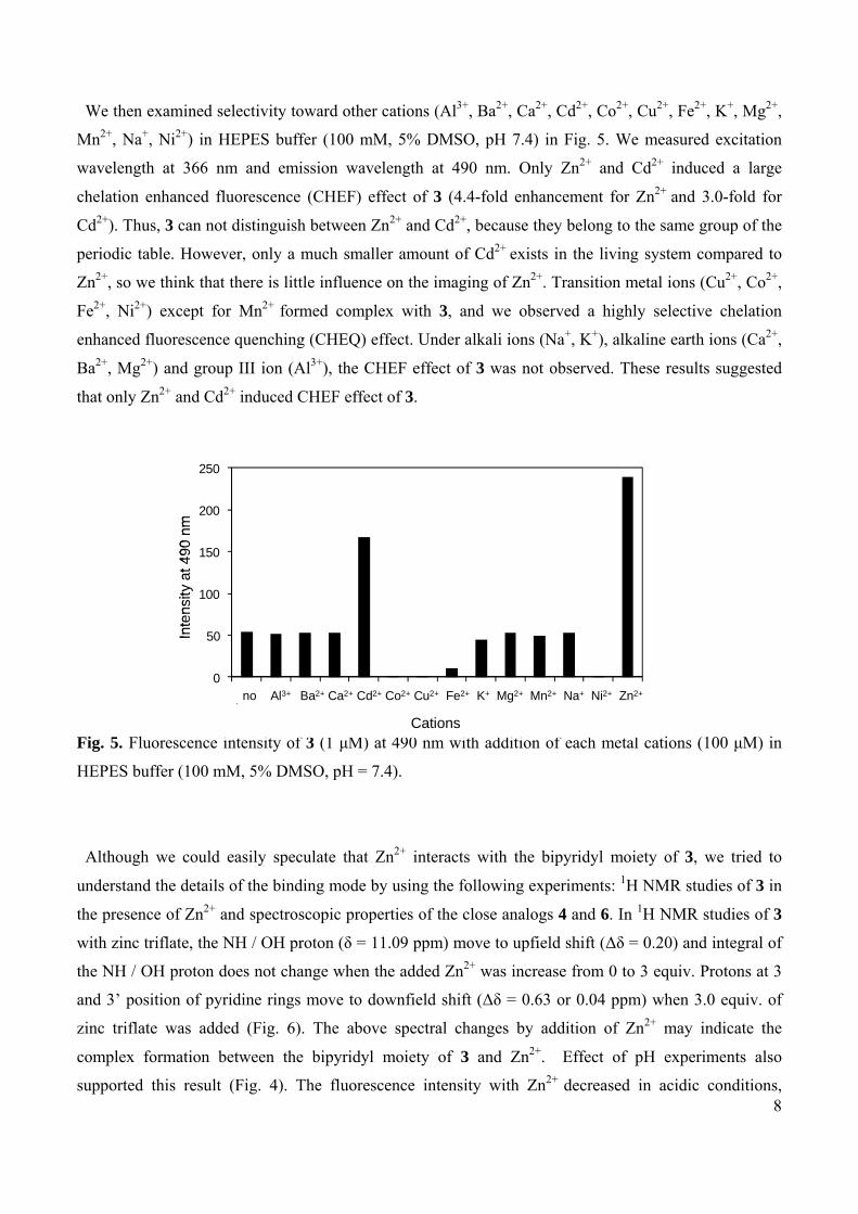
We then examined selectivity toward other cations (Al3+, Ba2+, Ca2+, Cd2+, Co2+, Cu2+, Fe2+, K+, Mg2+,
Mn2+, Na+, Ni2+) in HEPES buffer (100 mM, 5% DMSO, pH 7.4) in Fig. 5. We measured excitation
wavelength at 366 nm and emission wavelength at 490 nm. Only Zn2+ and Cd2+ induced a large
chelation enhanced fluorescence (CHEF) effect of 3 (4.4-fold enhancement for Zn2+ and 3.0-fold for
Cd2+). Thus, 3 can not distinguish between Zn2+ and Cd2+, because they belong to the same group of the
periodic table. However, only a much smaller amount of Cd2+ exists in the living system compared to
Zn2+, so we think that there is little influence on the imaging of Zn2+. Transition metal ions (Cu2+, Co2+,
Fe2+, Ni2+) except for Mn2+ formed complex with 3, and we observed a highly selective chelation
enhanced fluorescence quenching (CHEQ) effect. Under alkali ions (Na+, K+), alkaline earth ions (Ca2+,
Ba2+, Mg2+) and group III ion (Al3+), the CHEF effect of 3 was not observed. These results suggested
that only Zn2+ and Cd2+ induced CHEF effect of 3.
Fig. 5. Fluorescence intensity of 3 (1 μM) at 490 nm with addition of each metal cations (100 μM) in
HEPES buffer (100 mM, 5% DMSO, pH = 7.4).
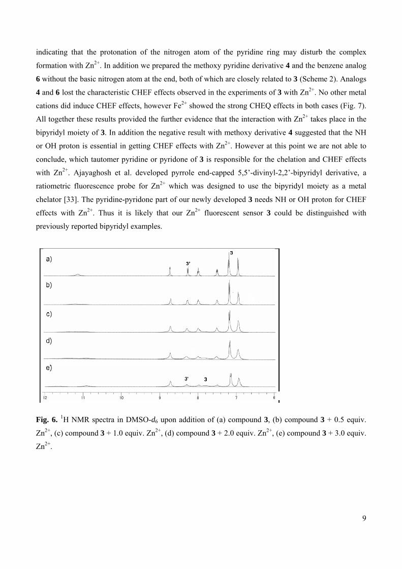
Although we could easily speculate that Zn2+ interacts with the bipyridyl moiety of 3, we tried to
understand the details of the binding mode by using the following experiments: 1H NMR studies of 3 in
the presence of Zn2+ and spectroscopic properties of the close analogs 4 and 6. In 1H NMR studies of 3
with zinc triflate, the NH / OH proton (δ = 11.09 ppm) move to upfield shift (Δδ = 0.20) and integral of
the NH / OH proton does not change when the added Zn2+ was increase from 0 to 3 equiv. Protons at 3
and 3’ position of pyridine rings move to downfield shift (Δδ = 0.63 or 0.04 ppm) when 3.0 equiv. of
zinc triflate was added (Fig. 6). The above spectral changes by addition of Zn2+ may indicate the
complex formation between the bipyridyl moiety of 3 and Zn2+. Effect of pH experiments also
supported this result (Fig. 4). The fluorescence intensity with Zn2+ decreased in acidic conditions,
0
50
100
150
200
250
no Al3+ Ba2+ Ca2+ Cd2+ Co2+ Cu2+ Fe2+ K+ Mg2+ Mn2+ Na+ Ni2+ Zn2+
Inte
nsity
at 4
90 n
m
Cations
0
50
100
150
200
250
no Al3+ Ba2+ Ca2+ Cd2+ Co2+ Cu2+ Fe2+ K+ Mg2+ Mn2+ Na+ Ni2+ Zn2+
Inte
nsity
at 4
90 n
m
Cations
9
indicating that the protonation of the nitrogen atom of the pyridine ring may disturb the complex
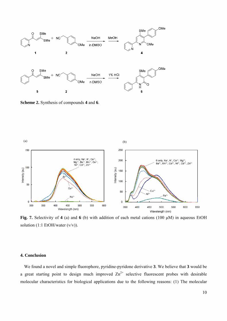
formation with Zn2+. In addition we prepared the methoxy pyridine derivative 4 and the benzene analog
6 without the basic nitrogen atom at the end, both of which are closely related to 3 (Scheme 2). Analogs
4 and 6 lost the characteristic CHEF effects observed in the experiments of 3 with Zn2+. No other metal
cations did induce CHEF effects, however Fe2+ showed the strong CHEQ effects in both cases (Fig. 7).
All together these results provided the further evidence that the interaction with Zn2+ takes place in the
bipyridyl moiety of 3. In addition the negative result with methoxy derivative 4 suggested that the NH
or OH proton is essential in getting CHEF effects with Zn2+. However at this point we are not able to
conclude, which tautomer pyridine or pyridone of 3 is responsible for the chelation and CHEF effects
with Zn2+. Ajayaghosh et al. developed pyrrole end-capped 5,5’-divinyl-2,2’-bipyridyl derivative, a
ratiometric fluorescence probe for Zn2+ which was designed to use the bipyridyl moiety as a metal
chelator [33]. The pyridine-pyridone part of our newly developed 3 needs NH or OH proton for CHEF
effects with Zn2+. Thus it is likely that our Zn2+ fluorescent sensor 3 could be distinguished with
previously reported bipyridyl examples.
Fig. 6. 1H NMR spectra in DMSO-d6 upon addition of (a) compound 3, (b) compound 3 + 0.5 equiv.
Zn2+, (c) compound 3 + 1.0 equiv. Zn2+, (d) compound 3 + 2.0 equiv. Zn2+, (e) compound 3 + 3.0 equiv.
Zn2+.
10
Scheme 2. Synthesis of compounds 4 and 6.
Fig. 7. Selectivity of 4 (a) and 6 (b) with addition of each metal cations (100 μM) in aqueous EtOH
solution (1:1 EtOH/water (v/v)).
4. Conclusion
We found a novel and simple fluorophore, pyridine-pyridone derivative 3. We believe that 3 would be
a great starting point to design much improved Zn2+ selective fluorescent probes with desirable
molecular characteristics for biological applications due to the following reasons: (1) The molecular
(b)(a)
11
weight (MW = 324) is small enough. (2) Bipyridyl moiety as the chelating functionality for Zn2+ nicely
shares with the fluorescent part, so the molecular weight can be kept minimal. (3) Despite its simplicity
3 already shows reasonable fluorescent properties and Zn2+ selectivity as a starting point. (4) Synthesis
we used is simple, so this allows us to prepare a number of structurally related compounds. We are
currently studying the next generation of bipyridyl based fluorescent probes related to 3 and will report
on these results in due course.
References
1 K.H. Falchuk, Mol. Cell. Biochem. 188 (1998) 41-48.
2 D.K. Perry, M.J. Smyth, H.R. Stennicke, G.S. Salvesen, P. Duriez, G.G. Poirier, Y.A. Hannun, J.
Biol. Chem. 272 (1997) 18530-18533.
3 P.D. Zalewski, I.J. Forbes, W.H. Betts, Biochem. J. 296 (1993) 403-408.
4 W. Maret, Y. Li, Chem. Rev. 109 (2009) 4682-4707.
5 H. Kitamura, H. Morikawa, H. Kamon, M. Iguchi, S. Hojyo, T. Fukada, S. Yamashita, T. Kaisho, S.
Akira, M. Murakami, T. Hirano, Nat Immunol. 7 (2006) 971-977.
6 M. Hershfinkel, K. Kandler, M.E. Knoch, M. Dagan-Rabin, M.A. Aras, C. Abramovitch-Dahan, I.
Sekler, E. Aizenman, Nat. Neurosci. 12 (2009) 725-727.
7 C.J. Frederickson, J.Y. Koh, A.I. Bush, Nat. Rev. Neurosci. 6 (2005) 449-462.
8 A.M.L. Edstrom, J. Malm, B. Frohm, J.A. Martellini, A. Giwercman, M. Morgelin, A.M. Cole,
O.E. Sorensen, J. Immunol. 181 (2008) 3413-3421.
9 K.J. Waldron, J.C. Rutherford, D. Ford, N.J. Robinson, Nature 460 (2009) 823-830.
10 A.S. Rahman, M. Kimura, Y. Itokawa, Biol. Trace Elem. Res. 67 (1999) 29-36.
11 E.G. Erdos, R.A. Skidgel, Lab. Invest. 56 (1987) 345-348.
12 L. Wei, F. Alhenc-Gelas, P. Corvol, E. Clauser, J. Biol. Chem. 266 (1991) 9002-9008.
13 Y. Choo, A. Klug, Proc. Natl. Acad. Sci. U. S. A. 91 (1994) 11168-11172.
14 W.J. Qian, C.A. Aspinwall, M.A. Battiste, R.T. Kennedy, Anal. Chem. 72 (2000) 711-717.
15 K.R. Gee, Z.L. Zhou, W.J. Qian, R. Kennedy, J. Am. Chem. Soc. 124 (2002) 776-778.
16 S.C. Burdette, S.J. Lippard, Proc. Natl. Acad. Sci. U. S. A. 100 (2003) 3605-3610.
17 E.M. Nolan, S.J. Lippard, Acc. Chem. Res. 42 (2009) 193-203.
18 K.L. Haas, K.J. Franz, Chem. Rev. 109 (2009) 4921-4960.
19 E.L. Que, D.W. Domaille, C.J. Chang, Chem. Rev. 108 (2008) 1517-1549.
20 K. Kikuchi, K. Komatsu, T. Nagano, Curr. Opin. Chem. Biol. 8 (2004) 182-191.
12
21 K. Soroka, R.S. Vithanage, D.A. Phillips, B. Walker, P.K. Dasgupta, Anal. Chem. 59 (1987) 629-
636.
22 S.G. Schulman, L.B. Sanders, Anal. Chim. Acta. 56 (1971) 83-89.
23 L.V. Meervelt, M. Goethals, N. Leroux, Th. Zeegers-Huyskens, J. Phys. Org. Chem. 10 (1997)
680-686.
24 C.J. Frederickson, E.J. Kasarskis, D. Ringo, R.E. Frederickson, J. Neurosci. Methods 20 (1987) 91-
103.
25 H. Ito, M. Matsuoka, Y. Ueda, M. Takuma, Y. Kudo, K. Iguchi, Tetrahedron 65 (2009) 4235-4238.
26 A. Ojida, T. Sakamoto, M. Inoue, S. Fujishima, G. Lippens, I. Hamachi, J. Am. Chem. Soc. 131
(2009) 6543-6548.
27 G.K. Walkup, S.C. Burdette, S.J. Lippard, R.Y. Tsien, J. Am. Chem. Soc. 122 (2000) 5644-5645.
28 S.C. Burdette, G.K. Walkup, B. Spingler, R.Y. Tsien, S.J. Lippard, J. Am. Chem. Soc. 123 (2001)
7831-7841.
29 C.J. Chang, J. Jaworski, E.M. Nolan, M. Sheng, S.J. Lippard, Proc. Natl. Acad. Sci. U. S. A. 101
(2004) 1129-1134.
30 T. Hirano, K. Kikuchi, Y. Urano, T. Nagano, J. Am. Chem. Soc. 124 (2002) 6555-6562.
31 C.A. Lipinski, F. Lombardo, B.W. Dominy, P.J. Feeney, Adv. Drug Deliv. Rev. 46 (2001) 3-26.
32 N. Mizuyama, Y. Tominaga, S. Kohra, K. Ueda, S. Hirayama, Y. Shigemitsu, Bull. Chem. Soc. Jpn.
79 (2006) 602-611.
33 A. Ajayaghosh, P. Carol, S. Sreejith, J. Am. Chem. Soc. 127 (2005) 14962-14963.
Copyright © 2022 FDOKUMEN