Synthesis of PEGylated lactose analogs for inhibition studies on T.cruzi trans-sialidase
11
Synthesis of PEGylated lactose analogs for inhibition studies on T.cruzi trans-sialidase M. Eugenia Giorgi & Laura Ratier & Rosalía Agusti & Alberto C. C. Frasch & Rosa M. de Lederkremer Received: 4 May 2010 / Revised: 29 June 2010 / Accepted: 30 June 2010 # Springer Science+Business Media, LLC 2010 Abstract Trypanosoma cruzi, the agent of Chagas disease, expresses a unique enzyme, the trans-sialidase (TcTS) involved in the transfer of sialic acid from host glyco- conjugates to mucins of the parasite. The enzyme is shed to the medium and may affect the immune system of the host. We have previously described that lactose derivatives effectively inhibited the transfer of sialic acid to N- acetyllactosamine. Lactitol also prevented the apoptosis caused by the TcTS, although it is rapidly eliminated from the circulatory system. In this paper we report covalent conjugation of polyethylene glycol (PEG) with lactose, lactobionolactone and benzyl β-D-galactopyranosyl- (1→6)-2-amino-2-deoxy-α-D-glucopyranoside (1) with the hope to improve the bioavailability, though retaining their inhibitory properties. Different conjugation methods have been used and the behavior of the PEGylated products in the TcTS reaction was studied. Keywords Trans-sialidase . PEGylation . Inhibitors . Trypanosoma cruzi Introduction The need to maintain drugs in blood is one of the main goals in therapeutic applications, which are often hampered by the short in vivo half-life of administered compounds. Many factors are involved in the removal of substances from the circulation, being renal excretion and inactivation reactions by blood enzymes the main causes for the brief plasma residence time of low-molecular mass non-protein drugs [1]. Modification of biological molecules by covalent conjugation with polyethylene glycol (PEG) has been used to improve the bioavailability of drugs [2]. PEG is a linear polyether diol with many useful properties such as biocompatibility, solubility in aqueous and organic media, lack of toxicity and very low immunogenicity. Probably, the most important feature of PEG modification is that it greatly extends the half-life (t 1/2 ) of most proteins, and results in a greatly increased plasma presence. This can be attributed, in part, to the increase in molecular weight of the conjugate beyond the limits of renal filtration and reduced proteolysis of the conjugate [3]. PEG has been coupled to antibodies or antibody fragments to prolong the circulating half-lives in vivo [4]. Selective alkylation and acylation of amino groups in a somatostatin analogue using two different PEG reagents has been described [5]. Also, low molecular-weight drugs have been PEGylated in order to prolong the in vivo action [6] or for targeting drug delivery [7]. There are few reports on the PEGylation of carbohydrate molecules and these have been focused on polysaccharides or on carbohydrates linked to proteins. Thus, the linear glycan chitosan was coupled by amide bond formation between the aminogroups and a carboxylic acid function- alized PEG [8]. N-hydroxysuccinimide (NHS) esters are frequently used for derivatization of primary amino groups. M. E. Giorgi : R. Agusti : R. M. de Lederkremer (*) CIHIDECAR, Departamento de Química Orgánica, Facultad de Ciencias Exactas y Naturales, Universidad de Buenos Aires, Ciudad Universitaria, Pabellón II, 1428 Buenos Aires, Argentina e-mail: [email protected] L. Ratier : A. C. C. Frasch Universidad Nacional de General San Martín y Consejo Nacional de Investigaciones Científicas y Técnicas, INTI, Av. General Paz 5445, Edificio 24, Casilla de correo 30, 1650 General San Martín, Argentina Glycoconj J DOI 10.1007/s10719-010-9300-7
-
Upload
conicet-ar -
Category
Documents
-
view
2 -
download
0
Transcript of Synthesis of PEGylated lactose analogs for inhibition studies on T.cruzi trans-sialidase

Synthesis of PEGylated lactose analogs for inhibition studieson T.cruzi trans-sialidase
M. Eugenia Giorgi & Laura Ratier & Rosalía Agusti &Alberto C. C. Frasch & Rosa M. de Lederkremer
Received: 4 May 2010 /Revised: 29 June 2010 /Accepted: 30 June 2010# Springer Science+Business Media, LLC 2010
Abstract Trypanosoma cruzi, the agent of Chagas disease,expresses a unique enzyme, the trans-sialidase (TcTS)involved in the transfer of sialic acid from host glyco-conjugates to mucins of the parasite. The enzyme is shed tothe medium and may affect the immune system of the host.We have previously described that lactose derivativeseffectively inhibited the transfer of sialic acid to N-acetyllactosamine. Lactitol also prevented the apoptosiscaused by the TcTS, although it is rapidly eliminated fromthe circulatory system. In this paper we report covalentconjugation of polyethylene glycol (PEG) with lactose,lactobionolactone and benzyl β-D-galactopyranosyl-(1→6)-2-amino-2-deoxy-α-D-glucopyranoside (1) withthe hope to improve the bioavailability, though retainingtheir inhibitory properties. Different conjugation methodshave been used and the behavior of the PEGylated productsin the TcTS reaction was studied.
Keywords Trans-sialidase . PEGylation . Inhibitors .
Trypanosoma cruzi
Introduction
The need to maintain drugs in blood is one of the maingoals in therapeutic applications, which are often hamperedby the short in vivo half-life of administered compounds.Many factors are involved in the removal of substancesfrom the circulation, being renal excretion and inactivationreactions by blood enzymes the main causes for the briefplasma residence time of low-molecular mass non-proteindrugs [1]. Modification of biological molecules by covalentconjugation with polyethylene glycol (PEG) has been usedto improve the bioavailability of drugs [2]. PEG is a linearpolyether diol with many useful properties such asbiocompatibility, solubility in aqueous and organic media,lack of toxicity and very low immunogenicity. Probably, themost important feature of PEG modification is that itgreatly extends the half-life (t1/2) of most proteins, andresults in a greatly increased plasma presence. This can beattributed, in part, to the increase in molecular weight of theconjugate beyond the limits of renal filtration and reducedproteolysis of the conjugate [3]. PEG has been coupled toantibodies or antibody fragments to prolong the circulatinghalf-lives in vivo [4]. Selective alkylation and acylation ofamino groups in a somatostatin analogue using twodifferent PEG reagents has been described [5]. Also, lowmolecular-weight drugs have been PEGylated in order toprolong the in vivo action [6] or for targeting drug delivery[7].
There are few reports on the PEGylation of carbohydratemolecules and these have been focused on polysaccharidesor on carbohydrates linked to proteins. Thus, the linearglycan chitosan was coupled by amide bond formationbetween the aminogroups and a carboxylic acid function-alized PEG [8]. N-hydroxysuccinimide (NHS) esters arefrequently used for derivatization of primary amino groups.
M. E. Giorgi : R. Agusti : R. M. de Lederkremer (*)CIHIDECAR, Departamento de Química Orgánica, Facultad deCiencias Exactas y Naturales, Universidad de Buenos Aires,Ciudad Universitaria, Pabellón II,1428 Buenos Aires, Argentinae-mail: [email protected]
L. Ratier :A. C. C. FraschUniversidad Nacional de General San Martín y Consejo Nacionalde Investigaciones Científicas y Técnicas,INTI, Av. General Paz 5445, Edificio 24, Casilla de correo 30,1650 General San Martín, Argentina
Glycoconj JDOI 10.1007/s10719-010-9300-7
A heterodifunctional PEG with a protected amino group atone end and a NHS ester at the other was used to locateactive molecules at the distal end of the PEG chain linkedto chitosan. An amino functionalized α-mannoside wasintroduced in this way [9]. Chitosan partially amidated withlactobionic acid was PEGylated for DNA carrier studies[10]. These PEG-chitosan copolymers have shown im-proved biocompatibility. Specific PEGylation of the carbo-hydrate of ricin A-chain was accomplished by periodateoxidation followed by hydrazide formation with a hydra-zine derivative of PEG [11]. The method was applied toproteins previously glycosylated with oligosaccharides[12]. Also, a protein was modified by sequential enzymatictransfer of galactose and PEGylated sialic acid from therespective nucleotides [13]. However, few reports describedirect PEGylation of small oligosaccharides. Diels-Alderchemistry was used to immobilize several different mono-saccharides bearing a cyclopentadiene group, attached througha polyethylene glycol (PEG) linker, to a monolayer presentingbenzoquinone groups on a gold surface [14]. A recent paper[15] describes the preparation of monosaccharide-cappedPEGylated quantum dots (PEG-QDs) by reaction of thePEG-QD with a glycosidic thiol.
Glycans have been recognized as candidates for chemo-therapy [16]. In this respect, Trypanosoma cruzi, the agentof American trypanosomiasis [17], expresses a uniqueenzyme, the trans-sialidase (TcTS) involved in the transferof sialic acid from host glycoconjugates to mucins of theparasite [18, 19]. This is the only way trypanosomes mayincorporate sialic acid, instead of using the correspondingnucleotide sugar as donor. Oligosaccharides of the mucinshave been synthesized and their acceptor and inhibitoryproperties were studied [20, 21]. Recombinant TcTS hasalso been used for the preparation of sialylated oligosac-charides [22]. Sialic acid on the surface of the parasite isinvolved in host cell invasion and in protection of theparasite against complement [23] and from killing by antiα-galactosyl antibodies [24]. Also, in Trypanosoma brucei,the agent of sleeping sickness, incorporation of sialic acidthrough a trans-sialidase is essential for the parasite tosurvive in the insect vector [25].Trans-sialidase, being avirulence factor in the mammal and/or insect stages oftrypanosomes and being specific for the parasite, with noequivalent in the human host, is an interesting target fordrug design. The 3D structure of TcTS shows two (sub)sitesin the active center: the sialic acid-binding site and thegalactose-binding site [26, 27]. Potential inhibitors areusually classified depending on the active site region theytarget. In the last years several laboratories have been activein seeking inhibitors for TcTS, mainly directed to the sialicacid binding site [28, 29]. In particular, some naturalproducts or synthetically modified natural products provedto be good inhibitors in vitro of the TcTS but the authors do
not report studies on toxicity in animals [30, 31]. Inhibitorsof TcTS directed to the β-galactosyl acceptor site should bemore specific, as other sialidases lack this interaction.Lactose derivatives effectively inhibited the transfer ofsialic acid to N-acetyllactosamine, lactitol being the best[32]. The lactose analogs are only moderate inhibitors.However, they are non-toxic to animals [33]. In fact, Mucciet al. proved that intravenous administration of 10 mg oflactitol inhibited TcTS in mice blood by 95–98%. Fastclearance from blood was solved using lactitol releasingpellets which prevented apoptosis of spleen cells.
Here, we report PEGylation of lactose derivatives and ofbenzyl β-D-galactopyranosyl-(1→6)-2-amino-2-deoxy-α-D-glucopyranoside (1) with the hope to improve thebioavailability, though retaining their inhibitory properties.Different conjugation methods have been used and thebehavior of the PEGylated products in the TcTS reactionwas studied.
Materials and methods
Materials and general methods
Methoxypolyethylene glycol amine 750 (MW: 750)(CH3O-PEG750-NH2), Methoxypolyethylene glycol amine 5000(MW: 5000)(CH3O-PEG5000-NH2) and Methoxypolyethy-lene glycol 5000 acetic acid N-hydroxysuccinimidyl ester(MW: 5000)(CH3O-PEG5000-NHS ester) were purchasedfrom Fluka (Steinheim, Germany), Methoxypolyethyleneglycol N-hydroxysuccinimidyl ester (MW: 685.71) (CH3
O-PEG12-NHS ester) was purchased from Pierce Biotech-nology (Rockford, USA). Lactobionic acid, lactose and 3’-sialyllactose were purchased from Sigma Chemical Co.[D-glucose-1-14C] lactose was purchased from AmershamBiosciences. NMR spectra were recorded with a BrukerAM 500 spectrometer at 500 MHz (1H) and 125 MHz (13C)at 30°C. Assignments were supported by 2D HSQCexperiments. High resolution electrospray ionization massspectra (ESI-TOF) were recorded on BRUKER microTOF-Q II ESI-Qq-TOF spectrometer. MALDI-TOF spectra werecollected on a PE BIOSYSTEMS DE-STR MALDI-TOFspectrometer. Optical rotations were measured with Perkin-Elmer 343 polarimeter. Analytical TLC was performed on0.2 mm Silica Gel 60 F254 (Merck) aluminium supportedplates. Detection was effected by spraying with 10% (v/v)sulfuric acid in ethanol containing 0.5% p-anisaldehyde andcharring. Column chromatography was performed on SilicaGel 60 (230–400 mesh, Merck). Melting points weredetermined with a Fisher-Johns apparatus. FT-IR spectrawere determined with a 510 P Nicolet FT-IR spectropho-tometer (Thermo Fisher Scientific, Inc., Waltham, MA,USA) using the KBr disk method, at 4,000−250 cm-1; 32–
Glycoconj J
64 scans were taken with a resolution of 2–4 cm-1.Radioactivity was measured on WinSpectral 1414 liquidscintillation counter (Wallac).
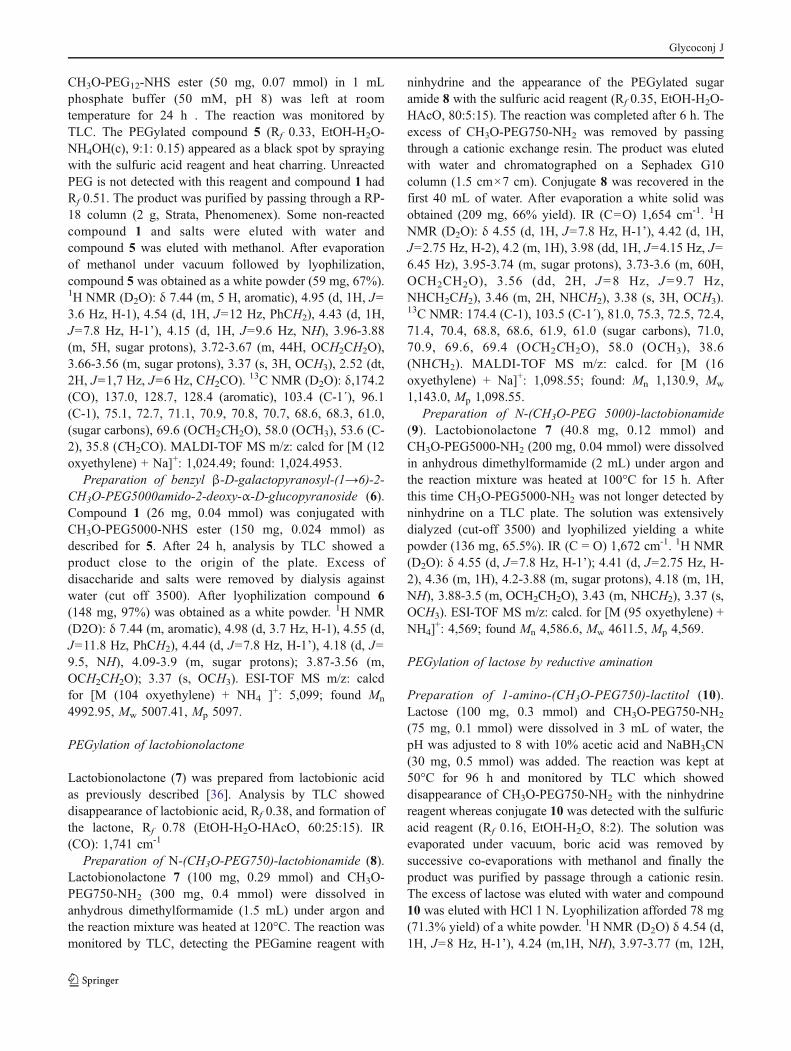
Synthesis of benzyl β-D-galactopyranosyl-(1→6)-2-amino-2-deoxy-α-D-glucopyranoside (1).
Synthesis of benzyl 2-acetamido-2-deoxy-α-D-glucopyr-anoside (2). To a solution of N-acetyl-D-glucosamine (3 g,13,56 mmol) in benzyl alcohol (21 mL), BF3 diethyl ethercomplex (320 μl, 2.7 mmol) was added and the mixturewas heated with stirring at 80°C for 4 h. After this time,TLC showed disappearance of the starting compound andthe product was precipitated by addition of diethyl ether(100 mL). The benzyl glycosides were obtained in 83%yield as a mixture of α and β anomers (8:2 ratio) (Rf 0.66and 0.60 respectively, nPrOH-EtOH-H2O, 7:1:2).
1H NMR(D2O, 500 MHz):δ anomeric region 4.83 (d, 0.8 H, J=3.5 Hz, H-1α), 4.42(d, 0.2 H, J=8.5 Hz, H-1β) Pure benzyl2-acetamido-2-deoxy-α-D-glucopyranoside was obtainedby fractional crystallization from ethanol. The meltingpoint and optical rotation were in agreement with thosepreviously reported [34].
Synthesis of benzyl (2,3,4,6-tetra-O-benzoyl-β-D-galactopyranosyl)-(1→6)-2-acetamido-2-deoxy-α-D-gluco-pyranoside (3).To an externally cooled (0°C) solution of1,2,3,4,6-penta-O-benzoyl-β-D-galactopyranose [35](1.67 g, 2.36 mmol) in dry Cl2CH2 (6 mL) under argon,tin(IV) chloride (0.27 mL, 2.31 mmol) was added. After15 min of stirring at 0°C, benzyl 2-acetamido-2-deoxy-α-D-glucopyranoside (0.60 g, 1.42 mmol) and dry CH3CN(0.6 mL) were added, and stirring was continued for 16 h atroom temperature. TLC showed a main compound of Rf
0.69, minor amounts of less polar compounds and remain-ing starting material (15% CH3OH in CH2Cl2). The mixturewas diluted with Cl2CH2 (40 mL) and poured into coldsaturated aqueous NaHCO3 with vigorous stirring. Theaqueous layer was extracted with Cl2CH2 (2×50 mL), andthe combined organic solutions were washed with wateruntil pH 7, dried (MgSO4), filtered, and concentrated underreduced pressure. The product was purified by columnchromatography (toluene-EtOAc,1:15) to give compound 3(731 mg, 42.4%), mp 118–120°C; [α]D:+86,8º (c1,CHCl3).
1H NMR (CDCl3): δ 8.05-7.2 (m, 25 H, aromatic),6.0 (dd,1H, J3´,4´=3.5 Hz, J4´,5´=1 Hz, H-4’), 5.91 (d, 1H,J=8.5 Hz, NH), 5.85 (dd, 1H, J2´,3´=10.5 Hz, J1´,2´=7.8 Hz, H-2’), 5.64 (dd, 1H, H-3’), 4.93 (d, 1H, H-1’), 4.7(d, 1H, J1,2=3.5 Hz, H-1), 4.67, 4.42 (2dd, 2H, J6´a,6´b=11.5 Hz, J6´a,5´=6.5 Hz, J6´b,5´=6.5 Hz, H-6´a, H-6´b), 4.57,4.19 (2d, 2H, J=11.7 Hz, PhCH2), 4.35 (ddd, 1H, H-5´),4.27 (dd, 1H, J5,6a=8.5 Hz, J6a,6b=11.5 Hz, H-6a), 3.88(ddd, 1H, J4,5=9 Hz, J5,6b=4 Hz, H-5), 3.83-3.77 (2dd, 2H,H-2, H-6), 3.58 (dd, 1H, J2,3=10.2, J3,4=9 Hz, H-3), 3.39(dd, 1H, H-4), 1.90 (s, 3H, CH3).
13C NMR (CDCl3): δ172.1(CONH), 166.1-165.3 (PhCO), 136-128 (aromatic),
102.4 (C-1’), 96.2 (C-1), 74.3 (C-3), 71.5-71.4 (C-4, C-3´,C-5´), 70.5 (C-2), 69.8 (C-2´), 69.6 (C-6), 69.2 (PhCH2),68.1 (C-4´), 61.9 (C-6’), 53.6 (C-5), 23.1 (CH3CO). Anal.Calcd for C49H47NO15: C, 66.13; H, 5.32; N, 1.57. Found:C, 65.74; H, 5.31; N, 1.68. ESI-TOF MS m/z: calcd for[M + H]+: 890.3018, found: 890.3036. Calcd for [M +Na]+: 912.2838, found: 912.2847.
Synthesis of benzyl β-D-galactopyranosyl-(1→6)-2-acetamido-2-deoxy-α-D-glucopyranoside (4). To a solutionof compound 3 (731 mg, 0.82 mmol) in anhydrous MeOH(10 mL) at 0°C, 0.5 M NaOMe in MeOH (10 mL) wasadded. After stirring for 1 h at 0°C and 2 h at roomtemperature, TLC examination showed only one compoundmore polar than 3. The solution was passed through acolumn of Amberlite IR 120 (plus) H+ resin eluting theproduct with methanol. The solvent was evaporated and theremaining methyl benzoate was removed by successive co-evaporations with water, to afford 291 mg of 4 as a whitesolid (75 % yield). Crystallization from EtOH gave mp234-235°C. The physical constants and spectra were thesame as previously reported [35].
Synthesis of benzyl β-D-galactopyranosyl-(1→6)-2-amino-2-deoxy-α-D-glucopyranoside (1). Compound 4(203 mg, 0.43 mmol) was dissolved in 1.2 mL of 1 MKOH and heated at 100°C for 16 h. After this time, TLCshowed that the reaction was complete, (Rf 0.36, n-PrOH-EtOH-H2O, 7:1:2). The solution was passed through acolumn (1.5 cm×6 cm) containing Amberlite IR 120 (plus)H+ resin and eluted with 0.1 N HCl to afford 110 mg of 1(0.29 mmol, 67% yield).[α]D:+45,4º (c1, H2O).
1H NMR(D2O): δ 7.47 (m, 5H, aromatic), 5.24 (d, 1H, J1,2=3.7 Hz,H-1), 4.81, 4.66 (2d, 2H, J=11.6 Hz, PhCH2), 4.43 (d, 1H,J1´,2´=7.9 Hz, H-1’), 4.10 (dd, 1H, J6a,6b=11.2 Hz, J5,6a=1.7 Hz, H-6a), 3.93 (dd, 1H, J4´,5´=0.7 Hz, J3´,4´=3.5 Hz,H-4´), 3.89 (dd, 1H, J2,3=10.5 Hz, J3,4=9.1, H-3), 3.89 (dd,1H, J5,6b=4.3 Hz, H-6b), 3.87 (ddd, 1H, J4,5=9.5 Hz, H-5),3.79 (dd, 1H, J5´,6´a=7.9 Hz, J6´a,6´b=11.7 Hz, H-6´a), 3.75(dd, 1H, J5´,6´b=4.3 Hz, H-6´b), 3.7 (ddd, 1H, H-5´), 3.66(dd, 1H, J2´,3´=10.0 Hz, H-3´), 3.64 (dd, 1H, H-4), 3.56(dd, 1H, H-2´), 3.37 (dd, 1H, H-2). 13C NMR (D2O): δ136.5, 128.8, 128.6, 128.5 (aromatic), 103.4 (C-1’), 94.5 (C-1), 75.1 (C-5’), 72.7 (C-3´), 71.2 (C-5), 70.7 (C-2´), 70.0(PhCH2), 69.9 (C-3), 69.1 (C-4), 68.6 (C-4´), 67.8 (C-6),60.9 (C-6’), 53.8 (C-2). ESI-TOF MS m/z: calcd for [M +H]+:464.1684 found:432.1863. Calcd for [M + Na]+: 454.1684 found: 454.1677.
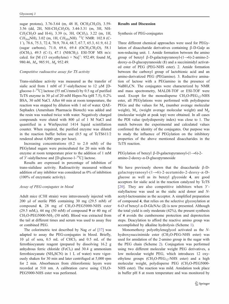
PEGylation of compound 1
Preparation of benzyl β-D-galactopyranosyl-(1→6)-2-CH3O-PEG12amido-2-deoxy-α-D-glucopyranoside (5). Asolution of benzyl β-D-galactopyranosyl-(1→6)-2-amino-2-deoxy-α-D-glucopyranoside (1) (38 mg, 0.09 mmol) and
Glycoconj J
CH3O-PEG12-NHS ester (50 mg, 0.07 mmol) in 1 mLphosphate buffer (50 mM, pH 8) was left at roomtemperature for 24 h . The reaction was monitored byTLC. The PEGylated compound 5 (Rf 0.33, EtOH-H2O-NH4OH(c), 9:1: 0.15) appeared as a black spot by sprayingwith the sulfuric acid reagent and heat charring. UnreactedPEG is not detected with this reagent and compound 1 hadRf 0.51. The product was purified by passing through a RP-18 column (2 g, Strata, Phenomenex). Some non-reactedcompound 1 and salts were eluted with water andcompound 5 was eluted with methanol. After evaporationof methanol under vacuum followed by lyophilization,compound 5 was obtained as a white powder (59 mg, 67%).1H NMR (D2O): δ 7.44 (m, 5 H, aromatic), 4.95 (d, 1H, J=3.6 Hz, H-1), 4.54 (d, 1H, J=12 Hz, PhCH2), 4.43 (d, 1H,J=7.8 Hz, H-1’), 4.15 (d, 1H, J=9.6 Hz, NH), 3.96-3.88(m, 5H, sugar protons), 3.72-3.67 (m, 44H, OCH2CH2O),3.66-3.56 (m, sugar protons), 3.37 (s, 3H, OCH3), 2.52 (dt,2H, J=1,7 Hz, J=6 Hz, CH2CO).
13C NMR (D2O): δ,174.2(CO), 137.0, 128.7, 128.4 (aromatic), 103.4 (C-1´), 96.1(C-1), 75.1, 72.7, 71.1, 70.9, 70.8, 70.7, 68.6, 68.3, 61.0,(sugar carbons), 69.6 (OCH2CH2O), 58.0 (OCH3), 53.6 (C-2), 35.8 (CH2CO). MALDI-TOF MS m/z: calcd for [M (12oxyethylene) + Na]+: 1,024.49; found: 1,024.4953.
Preparation of benzyl β-D-galactopyranosyl-(1→6)-2-CH3O-PEG5000amido-2-deoxy-α-D-glucopyranoside (6).Compound 1 (26 mg, 0.04 mmol) was conjugated withCH3O-PEG5000-NHS ester (150 mg, 0.024 mmol) asdescribed for 5. After 24 h, analysis by TLC showed aproduct close to the origin of the plate. Excess ofdisaccharide and salts were removed by dialysis againstwater (cut off 3500). After lyophilization compound 6(148 mg, 97%) was obtained as a white powder. 1H NMR(D2O): δ 7.44 (m, aromatic), 4.98 (d, 3.7 Hz, H-1), 4.55 (d,J=11.8 Hz, PhCH2), 4.44 (d, J=7.8 Hz, H-1’), 4.18 (d, J=9.5, NH), 4.09-3.9 (m, sugar protons); 3.87-3.56 (m,OCH2CH2O); 3.37 (s, OCH3). ESI-TOF MS m/z: calcdfor [M (104 oxyethylene) + NH4 ]+: 5,099; found Mn
4992.95, Mw 5007.41, Mp 5097.
PEGylation of lactobionolactone
Lactobionolactone (7) was prepared from lactobionic acidas previously described [36]. Analysis by TLC showeddisappearance of lactobionic acid, Rf 0.38, and formation ofthe lactone, Rf 0.78 (EtOH-H2O-HAcO, 60:25:15). IR(CO): 1,741 cm-1
Preparation of N-(CH3O-PEG750)-lactobionamide (8).Lactobionolactone 7 (100 mg, 0.29 mmol) and CH3O-PEG750-NH2 (300 mg, 0.4 mmol) were dissolved inanhydrous dimethylformamide (1.5 mL) under argon andthe reaction mixture was heated at 120°C. The reaction wasmonitored by TLC, detecting the PEGamine reagent with
ninhydrine and the appearance of the PEGylated sugaramide 8 with the sulfuric acid reagent (Rf 0.35, EtOH-H2O-HAcO, 80:5:15). The reaction was completed after 6 h. Theexcess of CH3O-PEG750-NH2 was removed by passingthrough a cationic exchange resin. The product was elutedwith water and chromatographed on a Sephadex G10column (1.5 cm×7 cm). Conjugate 8 was recovered in thefirst 40 mL of water. After evaporation a white solid wasobtained (209 mg, 66% yield). IR (C=O) 1,654 cm-1. 1HNMR (D2O): δ 4.55 (d, 1H, J=7.8 Hz, H-1’), 4.42 (d, 1H,J=2.75 Hz, H-2), 4.2 (m, 1H), 3.98 (dd, 1H, J=4.15 Hz, J=6.45 Hz), 3.95-3.74 (m, sugar protons), 3.73-3.6 (m, 60H,OCH2CH2O), 3.56 (dd, 2H, J=8 Hz, J=9.7 Hz,NHCH2CH2), 3.46 (m, 2H, NHCH2), 3.38 (s, 3H, OCH3).13C NMR: 174.4 (C-1), 103.5 (C-1´), 81.0, 75.3, 72.5, 72.4,71.4, 70.4, 68.8, 68.6, 61.9, 61.0 (sugar carbons), 71.0,70.9, 69.6, 69.4 (OCH2CH2O), 58.0 (OCH3), 38.6(NHCH2). MALDI-TOF MS m/z: calcd. for [M (16oxyethylene) + Na]+: 1,098.55; found: Mn 1,130.9, Mw
1,143.0, Mp 1,098.55.Preparation of N-(CH3O-PEG 5000)-lactobionamide
(9). Lactobionolactone 7 (40.8 mg, 0.12 mmol) andCH3O-PEG5000-NH2 (200 mg, 0.04 mmol) were dissolvedin anhydrous dimethylformamide (2 mL) under argon andthe reaction mixture was heated at 100°C for 15 h. Afterthis time CH3O-PEG5000-NH2 was not longer detected byninhydrine on a TLC plate. The solution was extensivelydialyzed (cut-off 3500) and lyophilized yielding a whitepowder (136 mg, 65.5%). IR (C = O) 1,672 cm-1. 1H NMR(D2O): δ 4.55 (d, J=7.8 Hz, H-1’); 4.41 (d, J=2.75 Hz, H-2), 4.36 (m, 1H), 4.2-3.88 (m, sugar protons), 4.18 (m, 1H,NH), 3.88-3.5 (m, OCH2CH2O), 3.43 (m, NHCH2), 3.37 (s,OCH3). ESI-TOF MS m/z: calcd. for [M (95 oxyethylene) +NH4]
+: 4,569; found Mn 4,586.6, Mw 4611.5, Mp 4,569.
PEGylation of lactose by reductive amination
Preparation of 1-amino-(CH3O-PEG750)-lactitol (10).Lactose (100 mg, 0.3 mmol) and CH3O-PEG750-NH2
(75 mg, 0.1 mmol) were dissolved in 3 mL of water, thepH was adjusted to 8 with 10% acetic acid and NaBH3CN(30 mg, 0.5 mmol) was added. The reaction was kept at50°C for 96 h and monitored by TLC which showeddisappearance of CH3O-PEG750-NH2 with the ninhydrinereagent whereas conjugate 10 was detected with the sulfuricacid reagent (Rf 0.16, EtOH-H2O, 8:2). The solution wasevaporated under vacuum, boric acid was removed bysuccessive co-evaporations with methanol and finally theproduct was purified by passage through a cationic resin.The excess of lactose was eluted with water and compound10 was eluted with HCl 1 N. Lyophilization afforded 78 mg(71.3% yield) of a white powder. 1H NMR (D2O) δ 4.54 (d,1H, J=8 Hz, H-1’), 4.24 (m,1H, NH), 3.97-3.77 (m, 12H,
Glycoconj J
sugar protons), 3.76-3.64 (m, 48 H, OCH2CH2O), 3.59-3.56 (dd, 2H, NH-CH2CH2O), 3.44-3.31 (m, 3H, NH-CH2CH2O and H-6), 3.39 (s, 3H, OCH3), 3.22 (m, 1H,CH2(a)NH), 3.02 (m, 1H, CH2(b)NH).
13C NMR: 102.8 (C-1´), 78.6, 75.3, 72.4, 70.9, 70.4, 68.7, 67.7, 65.3, 61.9, 61.2(sugar carbons), 71.0, 69.6, 69.4 (OCH2CH2O), 58.1(OCH3), 49.5 (C-1), 47.1 (NHCH2). ESI-TOF MS m/z:calcd. for [M (13 oxyethylene) + Na]+: 952.49; found Mn
980.46, Mw 985.91, Mp 952.49.
Competitive radioactive assay for TS activity
Trans-sialidase activity was measured as the transfer ofsialic acid from 1 mM of 3’-sialyllactose to 12 μM [D-glucose-1-14C] lactose (55 mCi/mmol) by 0.5 ng of purifiedTcTS enzyme in 30 μl of 20 mM Hepes-Na (pH 7.5), 0.2%BSA, 30 mM NaCl. After 60 min at room temperature, thereaction was stopped by dilution with 1 ml of water. QAE-Sephadex (Amersham Pharmacia Biotech) was added andthe resin was washed twice with water. Negatively chargedcompounds were eluted with 800 μl of 1 M NaCl andquantified in a WinSpectral 1414 liquid scintillationcounter. When required, the purified enzyme was dilutedin the reaction buffer before use (0.5 ng of TcTS611/2rendered about 4,000 cpm per hour).
Increasing concentrations (0.2 to 2.0 mM) of thePEGylated sugars were preincubated for 20 min with theenzyme at room temperature prior to the addition of 1 mMof 3’-sialyllactose and [D-glucose-1-14C] lactose.
Results are expressed in percentage of inhibition oftrans-sialidase activity. Radioactivity measured withoutaddition of any inhibitor was considered as 0% of inhibition(100% of enzymatic activity).
Assay of PEG-conjugates in blood
Adult mice (C3H strain) were intravenously injected with200 μl of sterile PBS containing 30 mg (29.5 mM) ofcompound 6, 28 mg of CH3O-PEG5000-NHS ester(29.5 mM,), 44 mg (50 mM) of compound 9 or 40 mg ofCH3O-PEG5000-NH2 (50 mM). Blood was extracted fromthe tail at different times and serum was used to assay freeor combined PEG.
The colorimetric test described by Nag et al [37] wasadapted to assay the PEG-conjugates in blood. Briefly,10 μl of sera, 0.5 mL of CHCl3 and 0.5 mL of theferrothiocyanate reagent (prepared by dissolving 16.2 ganhydrous ferric chloride (FeCl3) and 30.4 g ammoniumferrothiocyanate (NH4SCN) in 1 L of water) were vigor-ously shaken for 30 min and later centrifuged at 5,000 rpmfor 2 min. Absorbances from chloroformic layers wererecorded at 510 nm. A calibration curve using CH3O-PEG5000-NHS ester was performed.
Results and Discussion
Synthesis of PEG-conjugates
Three different chemical approaches were used for PEGy-lation of disaccharide derivatives containing β-D-Galp asnon-reducing unit. 1. Amide formation between the aminogroup of benzyl β-D-galactopyranosyl-(1→6)-2-amino-2-deoxy-α-D-glucopyranoside (1) and a succinimidyl activat-ed ester of PEG (PEG-NHS ester). 2. Amide formationbetween the carboxyl group of lactobionic acid and anamino-derivatized PEG (PEGamino). 3. Reductive amina-tion of lactose with a PEGamino in the presence ofNaBH3CN. The conjugates were characterized by NMRand mass spectrometry, MALDI-TOF or ESI-TOF wereused. Except for the monodisperse CH3O-PEG12-NHSester, all PEGylations were performed with polydispersePEGs and the values for Mn (number average molecularweight), Mw (weight average molecular weight) and Mp
(molecular weight at peak top) were obtained. In all casesthe PDI value (polydispersity index) was close to 1. Thematch between the experimental and calculated valuesconfirmed the identity of the conjugates. Our purpose wasto study the influence of PEGylation on the inhibitoryproperties of the above mentioned disaccharides in theTcTS reaction.
PEGylation of benzyl β-D-galactopyranosyl-(1→6)-2-amino-2-deoxy-α-D-glucopyranoside
We have previously shown that the disaccharide β-D-galactopyranosyl-(1→6)-2-acetamido-2-deoxy-α-D-glucose as well as its benzyl glycoside 4, are goodacceptors for sialic acid in the reaction catalyzed by TcTS[20]. They are also competitive inhibitors when 3’-sialyllactose was used as the sialic acid donor and N-acetyl-lactosamine as the acceptor. A simplified preparationof compound 4, that relies on the selective glycosylation at6-O of benzyl α-D-GlcNAc (2) is now presented. Althoughthe total yield is only moderate (42%), the present synthesisof 4 avoids the cumbersome protection and deprotectionsteps. Deacylation to afford the reactive amino group wasaccomplished by alkaline hydrolysis (Scheme 1).
Monomethoxy polyethyleneglycol activated as the N-hydroxysuccinimide ester (CH3O-PEG-NHS ester) wasused for amidation of the 2-amino group in the sugar withthe PEG chain (Scheme 2). Conjugation was performedusing two different molecular weight PEG derivatives, alow molecular weight PEG, which introduces 12 oxy-ethylene groups (CH3O-PEG12-NHS ester) and a highmolecular weight, polydisperse PEG (CH3O-PEG5000-NHS ester). The reaction was mild. Amidation took placein buffer pH 8 at room temperature and was monitored by
Glycoconj J
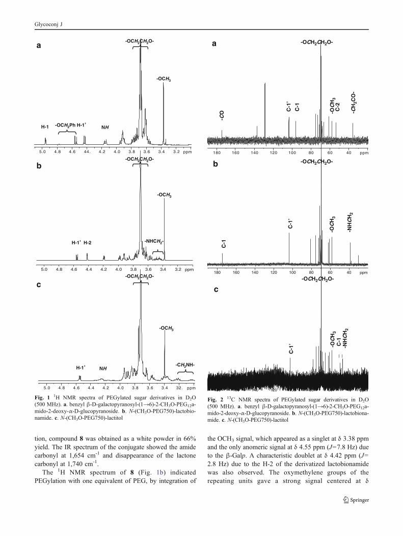
TLC being detected as a black spot after heating with thesulfuric acid reagent. The starting CH3O-PEG-NHS esterdid not stain with this reagent. After purification by passingthrough a RP-18 cartridge or by dialysis against water andlyophilization, in the case of the high molecular weightproduct, compounds 5 and 6 were obtained as whitepowders in 61% and 97% yield, respectively. The 1HNMR spectrum of 5 (Fig. 1a) indicated PEGylation withone equivalent of PEG, by integration of the only OCH3
signal, which appeared as a singlet at δ 3.37 ppm and thetwo anomeric signals at δ 4.95 ppm (J1,2=3.6 Hz) for theα-GlcN and at δ 4.43 ppm (J1’,2’=7.8 Hz) due to the β-Galp. One of the protons of the PhCH2, substituent in thesugar, was shown at δ 4.54 ppm with the characteristic highcoupling constant J=12 Hz, the other proton was sup-pressed together with the DHO signal. The PEG oxy-methylene protons appeared as a strong signal at δ3.70 ppm, which integrated to about 11 oxyethylenegroups. The methylene linked to the amide carbonylappeared at higher field (δ 2.52 ppm). The 13C NMRspectrum (Fig. 2a) showed among others the anomericsignals at δ 103.4 ppm (β-Galp) and δ 96.1 ppm (α-GlcN),and the C-2 signal of the amino sugar at δ 53.6 ppm. Forthe PEG moiety the OCH3 signal appeared at 58.0 ppm, themethylene next to the carbonyl sugar amide appeared at35.8 ppm and the oxymethylene groups of the PEG chainappeared as a strong peak at 69.6 ppm. MALDI-TOF massspectral analysis showed a main peak of [M + Na]+ at 1024corresponding to the conjugate 5 with 12 oxyethylenegroups.
Accordingly, the higher molecular weight PEG-conjugate 6 showed signals with similar chemical shifts(not shown). ESI-TOF mass spectral analysis of 6 revealeda series of 44 Da (OCH2CH2) spaced peaks correspondingto the NH4
+ adducts. In this case, ammonium acetate wasadded to help ionization. A main peak of [M + NH4]
+ at5,097 was observed. This value corresponded to conjugate6 with 104 oxyethylene groups.
PEGylation of lactobionolactone
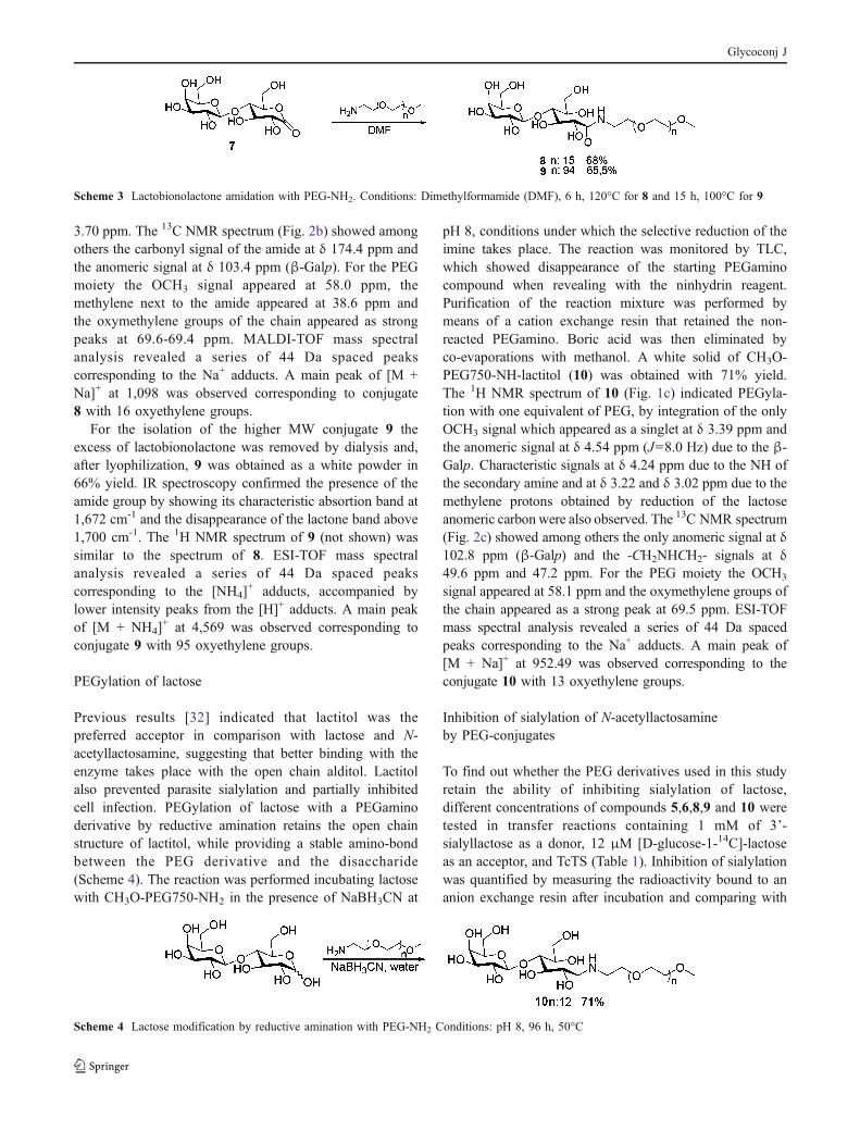
We had previously determined that lactobionic acid was agood acceptor of sialic acid and inhibitor of TcTS [32]. Inthis work we took advantage of the carboxyl group alreadypresent in this disaccharide to form an amide linkage withan amino functionalized PEG (Scheme 3). Again, we usedtwo different molecular weight derivatives, CH3O-PEG750-NH2 and CH3O-PEG5000-NH2. In the case of the highMW PEG, a molecular excess of the sugar was used since itcould be easily removed by dialysis after reaction. Thelactobionic acid was activated as lactobionolactone [36] andthen heated with the corresponding PEGamino in anhy-drous dimethylformamide. The reaction could be monitoredby TLC. Whereas the PEGamino starting material wasdetected with the ninhydrin reagent, the sugar conjugatewas only revealed with the sulfuric acid reagent. In the caseof the lower MW conjugate 8, purification from excessPEGamino was accomplished by cation exchange chroma-tography, which retained excess reagent, followed bySephadex G-10 column chromatography. After lyophiliza-
Scheme 1 Synthesis of benzyl β-D-galactopyranosyl-(1→6)-2-amino-2-deoxy-α-D-glucopyranoside (1). Conditions: a 16 h, r.t.; b 1 h, 0°C→2 hr.t.; (c) 1 M KOH, 16 h, 100°C
Scheme 2 Synthesis of N-PEGylated derivatives of benzylβ-D-galactopyranosyl-(1→6)-2-amino-2-deoxy-α-D-glucopyra-noside. Conditions: 24 h, r.t
Glycoconj J
tion, compound 8 was obtained as a white powder in 66%yield. The IR spectrum of the conjugate showed the amidecarbonyl at 1,654 cm-1 and disappearance of the lactonecarbonyl at 1,740 cm-1.
The 1H NMR spectrum of 8 (Fig. 1b) indicatedPEGylation with one equivalent of PEG, by integration of
the OCH3 signal, which appeared as a singlet at δ 3.38 ppmand the only anomeric signal at δ 4.55 ppm (J=7.8 Hz) dueto the β-Galp. A characteristic doublet at δ 4.42 ppm (J=2.8 Hz) due to the H-2 of the derivatized lactobionamidewas also observed. The oxymethylene groups of therepeating units gave a strong signal centered at δ
3.23.43.63.84.04.244.4.64.85.0 ppm
-OCH2CH2O-
-OCH2CH2O-
-OCH2CH2O-
-OCH2Ph H-1’
-OCH3
-OCH3
-OCH3
-NHCH2-
-CH2NH-
NH
NH
H-1
a
3.23.43.63.84.04.24.44.64.85.0 ppm
b
32.3.43.63.84.04.24.44.64.85.0 ppm
c
H-1’
H-1’
H-2
Fig. 1 1H NMR spectra of PEGylated sugar derivatives in D2O(500 MHz). a. benzyl β-D-galactopyranosyl-(1→6)-2-CH3O-PEG12a-mido-2-deoxy-α-D-glucopyranoside. b. N-(CH3O-PEG750)-lactobio-namide. c. N-(CH3O-PEG750)-lactitol
406080100120140160180 ppm
-OC
H3
_ C
-1
406080100120140160180 ppm
-CO
-OC
H3
C-1
’
C-2
-OCH2CH2O-
C-1
-CH
2CO
-
a
406080100120140160180 ppm
C-1
C-1
’
-OC
H3
-NH
CH
2
-OCH2CH2O- b
C-1
’ -NH
CH
2
-OCH2CH2O-
c
_
Fig. 2 13C NMR spectra of PEGylated sugar derivatives in D2O(500 MHz). a. benzyl β-D-galactopyranosyl-(1→6)-2-CH3O-PEG12a-mido-2-deoxy-α-D-glucopyranoside. b. N-(CH3O-PEG750)-lactobiona-mide. c. N-(CH3O-PEG750)-lactitol
Glycoconj J
3.70 ppm. The 13C NMR spectrum (Fig. 2b) showed amongothers the carbonyl signal of the amide at δ 174.4 ppm andthe anomeric signal at δ 103.4 ppm (β-Galp). For the PEGmoiety the OCH3 signal appeared at 58.0 ppm, themethylene next to the amide appeared at 38.6 ppm andthe oxymethylene groups of the chain appeared as strongpeaks at 69.6-69.4 ppm. MALDI-TOF mass spectralanalysis revealed a series of 44 Da spaced peakscorresponding to the Na+ adducts. A main peak of [M +Na]+ at 1,098 was observed corresponding to conjugate8 with 16 oxyethylene groups.
For the isolation of the higher MW conjugate 9 theexcess of lactobionolactone was removed by dialysis and,after lyophilization, 9 was obtained as a white powder in66% yield. IR spectroscopy confirmed the presence of theamide group by showing its characteristic absortion band at1,672 cm-1 and the disappearance of the lactone band above1,700 cm-1. The 1H NMR spectrum of 9 (not shown) wassimilar to the spectrum of 8. ESI-TOF mass spectralanalysis revealed a series of 44 Da spaced peakscorresponding to the [NH4]
+ adducts, accompanied bylower intensity peaks from the [H]+ adducts. A main peakof [M + NH4]
+ at 4,569 was observed corresponding toconjugate 9 with 95 oxyethylene groups.
PEGylation of lactose
Previous results [32] indicated that lactitol was thepreferred acceptor in comparison with lactose and N-acetyllactosamine, suggesting that better binding with theenzyme takes place with the open chain alditol. Lactitolalso prevented parasite sialylation and partially inhibitedcell infection. PEGylation of lactose with a PEGaminoderivative by reductive amination retains the open chainstructure of lactitol, while providing a stable amino-bondbetween the PEG derivative and the disaccharide(Scheme 4). The reaction was performed incubating lactosewith CH3O-PEG750-NH2 in the presence of NaBH3CN at
pH 8, conditions under which the selective reduction of theimine takes place. The reaction was monitored by TLC,which showed disappearance of the starting PEGaminocompound when revealing with the ninhydrin reagent.Purification of the reaction mixture was performed bymeans of a cation exchange resin that retained the non-reacted PEGamino. Boric acid was then eliminated byco-evaporations with methanol. A white solid of CH3O-PEG750-NH-lactitol (10) was obtained with 71% yield.The 1H NMR spectrum of 10 (Fig. 1c) indicated PEGyla-tion with one equivalent of PEG, by integration of the onlyOCH3 signal which appeared as a singlet at δ 3.39 ppm andthe anomeric signal at δ 4.54 ppm (J=8.0 Hz) due to the β-Galp. Characteristic signals at δ 4.24 ppm due to the NH ofthe secondary amine and at δ 3.22 and δ 3.02 ppm due to themethylene protons obtained by reduction of the lactoseanomeric carbon were also observed. The 13C NMR spectrum(Fig. 2c) showed among others the only anomeric signal at δ102.8 ppm (β-Galp) and the -CH2NHCH2- signals at δ49.6 ppm and 47.2 ppm. For the PEG moiety the OCH3
signal appeared at 58.1 ppm and the oxymethylene groups ofthe chain appeared as a strong peak at 69.5 ppm. ESI-TOFmass spectral analysis revealed a series of 44 Da spacedpeaks corresponding to the Na+ adducts. A main peak of[M + Na]+ at 952.49 was observed corresponding to theconjugate 10 with 13 oxyethylene groups.
Inhibition of sialylation of N-acetyllactosamineby PEG-conjugates
To find out whether the PEG derivatives used in this studyretain the ability of inhibiting sialylation of lactose,different concentrations of compounds 5,6,8,9 and 10 weretested in transfer reactions containing 1 mM of 3’-sialyllactose as a donor, 12 μM [D-glucose-1-14C]-lactoseas an acceptor, and TcTS (Table 1). Inhibition of sialylationwas quantified by measuring the radioactivity bound to ananion exchange resin after incubation and comparing with
Scheme 4 Lactose modification by reductive amination with PEG-NH2 Conditions: pH 8, 96 h, 50°C
Scheme 3 Lactobionolactone amidation with PEG-NH2. Conditions: Dimethylformamide (DMF), 6 h, 120°C for 8 and 15 h, 100°C for 9
Glycoconj J
the radioactivity bound in the absence of inhibitor. ThePEGylated amides (5 and 6) of the aminodisaccharide 1showed inhibition values similar to those of the originaldisaccharide, even better for the shorter PEG conjugate.These results indicate that the presence of a polyoxy-ethylene chain attached to the glucoside unit do not impairtheir ability to interact with the enzyme. However, the PEGderivatives of the open chain sugars (8-10) gave lowerinhibition values for the TcTS reaction than thecorresponding precursors, being insignificant for the PEGy-lated amine 10.
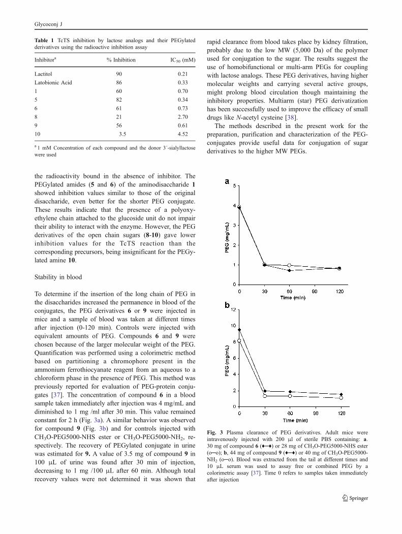
Stability in blood
To determine if the insertion of the long chain of PEG inthe disaccharides increased the permanence in blood of theconjugates, the PEG derivatives 6 or 9 were injected inmice and a sample of blood was taken at different timesafter injection (0-120 min). Controls were injected withequivalent amounts of PEG. Compounds 6 and 9 werechosen because of the larger molecular weight of the PEG.Quantification was performed using a colorimetric methodbased on partitioning a chromophore present in theammonium ferrothiocyanate reagent from an aqueous to achloroform phase in the presence of PEG. This method waspreviously reported for evaluation of PEG-protein conju-gates [37]. The concentration of compound 6 in a bloodsample taken immediately after injection was 4 mg/mL anddiminished to 1 mg /ml after 30 min. This value remainedconstant for 2 h (Fig. 3a). A similar behavior was observedfor compound 9 (Fig. 3b) and for controls injected withCH3O-PEG5000-NHS ester or CH3O-PEG5000-NH2, re-spectively. The recovery of PEGylated conjugate in urinewas estimated for 9. A value of 3.5 mg of compound 9 in100 μL of urine was found after 30 min of injection,decreasing to 1 mg /100 μL after 60 min. Although totalrecovery values were not determined it was shown that
rapid clearance from blood takes place by kidney filtration,probably due to the low MW (5,000 Da) of the polymerused for conjugation to the sugar. The results suggest theuse of homobifunctional or multi-arm PEGs for couplingwith lactose analogs. These PEG derivatives, having highermolecular weights and carrying several active groups,might prolong blood circulation though maintaining theinhibitory properties. Multiarm (star) PEG derivatizationhas been successfully used to improve the efficacy of smalldrugs like N-acetyl cysteine [38].
The methods described in the present work for thepreparation, purification and characterization of the PEG-conjugates provide useful data for conjugation of sugarderivatives to the higher MW PEGs.
Fig. 3 Plasma clearance of PEG derivatives. Adult mice wereintravenously injected with 200 μl of sterile PBS containing: a.30 mg of compound 6 (♦─♦) or 28 mg of CH3O-PEG5000-NHS ester(o─o); b, 44 mg of compound 9 (♦─♦) or 40 mg of CH3O-PEG5000-NH2 (o─o). Blood was extracted from the tail at different times and10 μL serum was used to assay free or combined PEG by acolorimetric assay [37]. Time 0 refers to samples taken immediatelyafter injection
Table 1 TcTS inhibition by lactose analogs and their PEGylatedderivatives using the radioactive inhibition assay
Inhibitora % Inhibition IC50 (mM)
Lactitol 90 0.21
Latobionic Acid 86 0.33
1 60 0.70
5 82 0.34
6 61 0.73
8 21 2.70
9 56 0.61
10 3.5 4.52
a 1 mM Concentration of each compound and the donor 3´-sialyllactosewere used
Glycoconj J
The PEG-conjugates are potentially useful for studies oninhibition of other galactose binding proteins. In this respect,amino derivatives of lactulose have been synthesized and theirfunction as galectin inhibitors has been described [39].
Acknowledgments This work was supported by grants fromAgencia Nacional de Promoción Científica y Tecnológica (ANPCyT),National Research Council (CONICET) and Universidad de BuenosAires. A. C. C. Frasch, R. M. de Lederkremer and R. Agustí areresearch members of CONICET and M. E. Giorgi is a fellow fromCONICET. The work of A. C. C. Frasch was partially supported byInternational Research Scholar grants from the Howard HughesMedical Institute.
References
1. Mongardini, C., Veronese, F.M.: Stabilization of substances incirculation. Bioconjugate Chem. 9, 418–450 (1998)
2. Veronese, F.M., Mero, A.: The impact of PEGylation onbiological therapies. BioDrugs. 22, 315–329 (2008)
3. Greenwald, R.B.: PEG drugs: an overview. J. Control. Release.74, 159–171 (2001)
4. Chapman, A.P.: PEGylated antibodies and antibody fragments forimproved therapy: a review. Adv. Drug. Deliv. Res. 54, 531–545(2002)
5. Morpurgo, M., Monfardini, C., Hofland, L.J., Sergi, M., Orsolini,P., Dumont, J.M., Veronese, F.M.: Selective alkylation andacylation of α and ∈ amino group with PEG in a somatostatinanalogue: tailored chemistry for optimized bioconjugates. Bio-conjugate Chem. 13, 1238–1243 (2002)
6. Marcus, Y., Sasson, K., Fridkin, M., Shechter, Y.: Turning low-molecular-weight drugs into prolonged acting prodrugs byreversible pegylation: a study with gentamicin. J. Med. Chem.51, 4300–4305 (2008)
7. Dixit, V., Van den Bossche, J., Sherman, D.M., Thompson, D.H.,Andres, R.P.: Synthesis and grafting of thioctic acid-PEG-folateconjugates onto Au nanoparticles for selective targeting of folatereceptor-positive tumor cells. Bioconjugate Chem. 117, 603–609(2006)
8. Prego, C., Torres, D., Fernandez-Megia, E., Novoa-Carballal, R.,Quiñoá, E., Alonso, M.J.: Chitosan-PEG nanocapsules as newcarriers for oral peptide delivery. Effect of chitosan pegylationdegree. J. Controlled Release. 111, 299–308 (2006)
9. Fernandez-Megia, E., Novoa-Carballal, R., Quiñoá, E., Riguera,R.: Conjugation of bioactive ligands to PEG-grafted chitosan atthe distal end of PEG. Biomacromolecules 8, 833–842 (2007)
10. Park, I.L., Kim, T.H., Park, Y.H., Shin, B.A., Choi, E.S.,Chowdhury, E.H., Akaike, T., Cho, C.S.: Galactosylatedchitosan-graft-poly(ethylene glycol) as hepatocyte-targetingDNA carrier. J. Controlled Release 76, 349–362 (2001)
11. Youn, Y.S., Na, D.H., Yoo, S.D., Song, S.C., Lee, K.C.:Carbohydrate-specifically polyethylene glycol-modified ricin A-chain with improved therapeutic potential. Int. J. Biochem. Cell.Biol. 37, 1525–1533 (2005)
12. Salmaso, S., Semenzato, A., Bersani, S., Mastrotto, F., Scomparin,A., Caliceti, P.: Site-selective protein glycation and PEGylation.Euro. Polym. J. 44, 1378–1389 (2008)
13. DeFrees, S., Wang, Z.G., Xing, R., Scott, A.E., Wang, J., Zopf,D., Gouty, D.L., Sjoberg, E.R., Panneerselvam, K., Brinkman-Vandel Linden, E.C., Bayer, R.J., Tarp, M.A., Clausen, H.: Glyco-PEGylation of recombinant therapeutic proteins produced inEscherichia coli. Glycobiology 16, 833–843 (2006)
14. Houseman, B.T., Mrksich, M.: Carbohydrate arrays for theevaluation of protein binding and enzymatic modification. ChemBiol 9, 443–454 (2002)
15. Kikkeri, R., Lepenies, B., Adibekian, A., Laurino, P., Seeberger, P.H.: In vitro imaging and in vivo liver targeting with carbohydratecapped quantum dots. J. Am. Chem. Soc. 131, 2110–2112 (2009)
16. Kawasaki, N., Itoh, S., Hashii, N., Takakura, D., Qin, Y., Huang,X., Yamaguchi, T.: The significance of glycosylation analysis indevelopment of biopharmaceuticals. Biol. Pharm. Bull. 32, 796–800 (2009)
17. Moncayo, A.: Chagas disease: current epidemiological trends alter theinterruption of vectorial and transfusional transmission in the SouthernCone countries. Mem. Inst. Oswaldo. Cruz. 98, 577–591 (2003)
18. Schenkman, S., Jiang, M.S., Hart, G.W., Nussenzweig, V.: Anovel cell surface trans-sialidase of Trypanosoma cruzi generatesa stage-specific epitope required for invasion of mammalian cells.Cell 65, 1117–1125 (1991)
19. Frasch, A.C.C.: Functional diversity in the tans-sialidase andmucin families in Trypanosoma cruzi. Parasitol. Today 16, 282–286 (2000)
20. Agustí, R., Giorgi, M.E., Mendoza, V.M., Gallo-Rodriguez, C.,Lederkremer, R.M.: Comparative rate of sialylation by recombinanttrans-sialidase and inhibitor properties of synthetic oligosaccharidesfrom Trypanosoma cruzi mucins-containing galactofuranose andgalactopyranose. Bioorg. Med. Chem. 15, 2611–2616 (2007)
21. Lederkremer, R.M., Agustí, R.: Glycobiology of Trypanosomacruzi. Adv. Carbohydr. Chem. Biochem. 62, 311–366 (2009)
22. Kröger, L., Scudlo, A., Thiem, J.: Subsequent enzymatic galactosy-lation and sialylation towards sialylated Thomsen-Friedenreichantigen components. Adv. Synth. Catal. 348, 1217–1227 (2006)
23. Tomlinson, S., de Carvalho LC, Pontes, Vandekerckhove, F.,Nussenzweig, V.: Role of sialic acid in the resistance ofTrypanosoma cruzi trypomastigotes to complement. J. Immunol.153, 3141–3147 (1994)
24. Pereira-Chioccola, V.L., Acosta-Serrano, A., Correia de Almeida,I., Ferguson, M.A., Souto-Padron, T., Rodrigues, M.M., Trav-assos, L.R., Schenkman, S.: Mucin-like molecules form anegatively charged coat that protects Trypanosoma cruzi trypo-mastigotes from killing by human anti-alpha-galactosyl anti-bodies. J Cell Sci 113, 1299–1307 (2000)
25. Nagamune, K., Acosta-Serrano, A., Uemura, H., Brun, R., Kunz-Renggli, C., Maeda, Y., Ferguson, M.A., Kinoshita, T.: Surfacesialic acids taken from the host allow trypanosome survival intsetse fly vectors. J Exp Med. 199, 1445–1450 (2004)
26. Amaya, F.M., Buschiazzo, A., Nguyen, T., Alzari, P.M.: The highresolution structures of free and inhibitor-bound Trypanosomarangeli sialidase and its camparison with T. cruzi trans-sialidase. J.Mol. Biol. 325, 773–784 (2003)
27. Buschiazzo, A., Amaya, M.F., Cremona, M.L., Frasch, A.C.C.,Alzari, P.M.: The crystal structure and mode of action of trans-sialidase, a key enzyme in Trypanosoma cruzi pathogenesis. Mol.Cell. 10, 757–768 (2002)
28. Neres, J., Bryce, R.J., Douglas, K.T.: Rational drug design inparasitology: trans-sialidase as a case study for Chagas disease.Drug Discov. Today 13, 110–117 (2008)
29. Buchini, S., Buschiazzo, A., Withers, S.G.: A new generation ofspecific Trypanosoma cruzi trans-sialidase inhibitors. Angew.Chem. Int. Ed. Engl. 47, 2700–2703 (2008)
30. Kim, J.H., Ryu, H.W., Shim, J.H., Park, K.H., Withers, S.G.:Development of new and selective Trypanosoma cruzi trans-sialidase inhibitors from sulfonamide chalcones and their deriva-tives. Chembiochem 10, 2475–2479 (2009)
31. Arioka, S., Sakagami, M., Uematsu, R., Yamaguchi, H., Togame,H., Takemoto, H., Hinou, H., Nishimura, S.: Potent inhibitorscaffold against Trypanosoma cruzi trans-sialidase. Bioorg. Med.Chem. 18, 1633–1640 (2010)
Glycoconj J
32. Agustí, R., Páris, G., Ratier, L., Frasch, A.C.C., Lederkremer, R.M.: Lactose derivatives are inhibitors of Trypanosoma cruzi trans-sialidase activity toward conventional substrates in vitro and invivo. Glycobiology 14, 659–670 (2004)
33. Mucci, J., Risso, M.G., Leguizamón, M.S., Frasch, A.C.C.,Campetella, O.: The trans-sialidase from Trypanosoma cruzitriggers apoptosis by target cell sialylation. Cell. Microbiol. 8,1086–1095 (2006)
34. Kuhn, R., Baer, H.H., Seelinger, A.: Zur Methylierung Von N-Acetylglucosamin-Derivaten. Liebigs Ann. Chem. 611, 236–241(1958)
35. Gallo-Rodriguez, C., Varela, O., Lederkremer, R.M.: One-potsynthesis of β-D-Galf(1→4)[β-D-Galp(1→6)]-D-GlcNAc, a‘core’ trisaccharide linked O-glycosidically in glycoproteins ofTrypanosoma cruzi. Carbohydr. Res. 305, 163–170 (1998)
36. Isbell, H.S., Frush, H.L.: Lactonization of aldonic acids. Meth.Carbohydr. Chem. 2, 16–18 (1963)
37. Nag, A., Mitra, G., Ghosh, P.C.: A colorimetric assay for estimationof polyethylene glycol and polyethylene glycolated protein usingammonium ferrothiocyanate. Anal. Biochem. 237, 224–231 (1996)
38. Navath, R.S., Wang, B., Kannan, S., Romero, R., Kannan, R.M.:Stimuli-responsive star poly(ethylene glycol) drug conjugates forimproved intracellular delivery of the drug in neuroinflammation.J. Control. Release. 142, 447–456 (2010)
39. Rabinovich, G.A., Cumashi, A., Bianco, G.A., Ciavardelli, D.,Iurisci, I., D’Egidio, M., Piccolo, E., Tinari, N., Nifantiev, N.,Iacobelli, S.: Synthetic lactulose amines: Novel class of anticanceragents that induce tumor cell apoptosis and inhibit galectin-mediated homotypic cell aggregation and endothelial cell mor-phogenesis. Glycobiology 16, 210–220 (2006)
Glycoconj J