Avalanche Photodiodes Performance Parameters Estimation under Thermal Irradiation Fields
Upload
independentCategory
view
1download
0
Research ArticlePegylated G-CSF Inhibits Blood Cell Depletion, IncreasesPlatelets, Blocks Splenomegaly, and Improves Survival afterWhole-Body Ionizing Irradiation but Not after IrradiationCombined with Burn
Juliann G. Kiang,1,2,3 Min Zhai,1 Pei-Jyun Liao,1 David L. Bolduc,1
Thomas B. Elliott,1 and Nikolai V. Gorbunov1
1 Radiation Combined Injury Program, Armed Forces Radiobiology Research Institute, Bethesda, MD 20889, USA2Department of Radiation Biology, Uniformed Services University of the Health Sciences, Bethesda, MD 20814, USA3Department of Medicine, Uniformed Services University of the Health Sciences, Bethesda, MD 20814, USA
Correspondence should be addressed to Juliann G. Kiang; [email protected]
Received 18 November 2013; Accepted 22 January 2014; Published 5 March 2014
Academic Editor: Pranela Rameshwar
Copyright © 2014 Juliann G. Kiang et al. This is an open access article distributed under the Creative Commons AttributionLicense, which permits unrestricted use, distribution, and reproduction in any medium, provided the original work is properlycited.
Exposure to ionizing radiation alone (radiation injury, RI) or combinedwith traumatic tissue injury (radiation combined injury, CI)is a crucial life-threatening factor in nuclear and radiological accidents. As demonstrated in animal models, CI results in greatermortality than RI. In our laboratory, we found that B6D2F1/J female mice exposed to 60Co-𝛾-photon radiation followed by 15%total-body-surface-area skin burns experienced an increment of 18%highermortality over a 30-day observation period compared toirradiation alone; that was accompanied by severe cytopenia, thrombopenia, erythropenia, and anemia. At the 30th day after injury,neutrophils, lymphocytes, and platelets still remained very low in surviving RI andCImice. In contrast, their RBC, hemoglobin, andhematocrit were similar to basal levels. Comparing CI and RI mice, only RI induced splenomegaly. Both RI and CI resulted in bonemarrow cell depletion. It was observed that only the RI mice treated with pegylated G-CSF after RI resulted in 100% survival overthe 30-day period, and pegylated G-CSF mitigated RI-induced body-weight loss and depletion of WBC and platelets. Peg-G-CSFtreatment sustained RBC balance, hemoglobin levels, and hematocrits and inhibited splenomegaly after RI.The results suggest thatpegylated G-CSF effectively sustained animal survival by mitigating radiation-induced cytopenia, thrombopenia, erythropenia,and anemia.
1. Introduction
Injuries induced by ionizing radiation alone (RI) or in com-bination with trauma from blast and thermal energy expo-sure (CI) are expected after the detonation of radiationdispersal devices or nuclear weapons. In vivo [1] and invitro [2, 3] studies indicate that RI induced DNA double-strand breaks (DSBs), activated signal transduction path-ways, elevated cytokine/chemokine concentrations in theperipheral blood, and increased systemic bacterial infection,thereby leading to cell death and multiple-organ dysfunctionand failure [1, 4–6]. Traumatic injury followed by RI (i.e.,CI) enhanced histopathological responses to RI, thereby
increasing the mortality [1, 5–7]. Because the responses to RIand CI occur at molecular, cellular, tissue, and system levels,the complexity of the responses makes it difficult to identifycountermeasures for prophylaxis, mitigation, or therapy.
RI and CI remarkably increased granulocyte colony stim-ulating factor (G-CSF) in mouse blood for more than 7 days[7]. The increase was initially believed to be a self-defensiveresponse, but its appearance was probably too late to par-ticipate in repairing bone marrow damage. Bone marrowinjury usually occurred within hours after RI [1, 2]. Withthis consideration, G-CSF and its modified form, that is,pegylated G-CSF (peg-G-CSF), have been used clinically totreat radiation-injured patients [8]. It is reported that this
Hindawi Publishing CorporationOxidative Medicine and Cellular LongevityVolume 2014, Article ID 481392, 10 pageshttp://dx.doi.org/10.1155/2014/481392
2 Oxidative Medicine and Cellular Longevity
growth factor decreased the period of neutropenia or aplasiain the limited number of radiation accident victims studiedand also enhanced neutrophil recovery following anticancertherapy [8]. The cytokine activates or primes neutrophilsto enhance their function [9]. The peg-G-CSF formulationhas a much longer biological half-life than G-CSF [10],thus avoiding the necessity of daily injections, which aredeleterious in irradiated mice. The drug has no toxic oradverse effects in mice at the doses used. Akin to G-CSFpeptide, peg-G-CSF initiates proliferation and differentiationof myeloid progenitors into mature granulocytes and induceshematopoietic stem cell mobilization from the bone marrowinto the bloodstream. It is involved in recovery from infection[11, 12] and wound healing [13]. Peg-G-CSF, when combinedwith stem cell factor and erythropoietin, was successfullyused in saving a hospital technician who had accidentallyentered a 60Co-irradiation therapy room and received a 4.5-Gy dose of radiation [14].
This report, which is intended to stimulate interest inadvancing research on peg-G-CSF in support of approvalfor treatment of radiation-induced neutropenia or aplasiaby U.S. Food and Drug Administration, provides data froman experimental animal model designed to demonstrate theefficacy of peg-G-CSF as an effective radiomitigator.
2. Materials and Methods
Research was conducted in a facility accredited by theAssociation for Assessment and Accreditation of Labora-tory Animal Care International (AAALACI). All proceduresinvolving animals were reviewed and approved by the AFRRIInstitutional Animal Care and Use Committee. Euthanasiawas carried out in accordancewith the recommendations andguidance of the American Veterinary Medical Association[15, 16]
2.1. Animals. B6D2F1/J femalemice (The JacksonLaboratory,Bar Harbor, ME) were maintained in a facility accredited bythe Association for Assessment and Accreditation of Labora-tory Animal Care International in plastic microisolator cageson hardwood chip bedding. Commercial rodent chow andacidified tap water were provided ad libitum at 12 to 20 weeksof age. Animal holding rooms were maintained at 21∘C ± 1∘Cwith 50% ± 10% relative humidity using at least 10 changes/hof 100% conditioned fresh air. A 12-h 0600 (light) to 1800(dark) full-spectrum lighting cycle was used. The AFRRIInstitutional Animal Care and Use Committee approved allanimal procedures.
2.2. Gamma Irradiation. Mice were given 9.5Gy [1] whole-body bilateral 60Co gamma-photon radiation, delivered ata dose rate of 0.4Gy/min, while held in vertically stacked,ventilated, four-compartment, and acrylic plastic boxes thatprovided electron equilibrium during irradiation. Emptycompartments within the boxes were filled with 3-inch-long, 1-inch-diameter acrylic phantoms to ensure uniformelectron scattering. The mapping of the radiation field wasperformed with alanine/EPR dosimetry [17] using standard
alanine calibration sets from NIST and National PhysicalLaboratory of UK.Themapping provided dose rates to waterin the core of the acrylic phantom (3 inches long, 1 inch indiameter) in each compartment of the mouse rack on the dayof themapping.Thefieldwas uniformwithin±1.8%over all ofthe 120 compartments.The exposure time for each irradiationwas determined from the mapping data; corrections for the60Co decay and the small differences in the mass energyabsorption coefficients for water and soft tissue were applied.The accuracy of the actual dose delivery was verified with anionization chamber adjacent to the mouse rack, which hadbeen calibrated in terms of dose to soft tissue in the cores ofmice.
2.3. Skin Injury. Skin surface injuries were performed onthe shaved dorsal surface of mice. Animals receiving skinburns were anesthetized by methoxyflurane inhalation. A15% total-body-surface-area skin burn was performed within1 h after irradiation using a 1 × 1-in custom designed templatepositioned centrally over the shaved dorsal skin surface.Micereceived a 12-s burn from ignited 95% ethanol (0.25mL,[5, 6]). All mice subjected to the skin injury were given0.5mL sterile 0.9% NaCl intraperitoneally (i.p.), which con-tained 150mg/kg of acetaminophen (AmerisourceBergen,Glen Allen, VA) and 0.05mg/kg of buprenorphine immedi-ately after skin injury to alleviate pain. Four hours later, micewere given a second dose of 150mg/kg of acetaminophen.For animals receiving skin wounds, a 15% total body-surface-area skin wound was performed within 1 h after irradiation[5, 6]. Skin-wounded mice received one dose of 150mg/kg ofacetaminophen immediately after skin injury.
2.4. Pegylated G-CSF. Peg-G-CSF (Neulasta; NDC: 555-13-019001) is a polyethylene glycol pharmaceutical-formulated-grade drug, also known as pegfilgrastim, and was purchasedfrom AmerisourceBergen Corporation (Valley Forge, PA). Adose of 1000 𝜇g/kg was administered by s.c. injection [18, 19]in a volume of 0.2mL 24 h, 8 d, and 15 d after RI or CI, thatis, 25 𝜇g/25-g mouse. Neulasta is supplied in 0.6mL prefilledsyringes for subcutaneous injection. Each syringe contains6mgPeg-G-CSF in a sterile, clear, colorless, and preservative-free solution containing 0.35mg acetate, 0.02mg polysorbate20, 0.02mg sodium, and 30mg sorbitol in water for injection,USP. Peg-G-CSF was studied in mice with sham-operation,burns, radiation, or radiation combined with burns.
2.5. G-CSF. G-CSF (Neupogen;Amgen, Inc.,ThousandOaks,CA, NDC: 555-13-546-10). A dose of 10 𝜇g/kg was injected s.c.[20] in a volume of 0.2mL on day 1 at 24 h and thereafteronce daily on days 2–14 after RI or CI. The vehicle givento control mice was sterile 0.9% sodium chloride solutionfor injection, USP. G-CSF was studied in mice with sham-operation, wounds, radiation, or radiation combined withwounds.
2.6. Antimicrobial Agents. Gentamicin sulfate cream, 0.1%(generic, E. Fougera and Co., Melville, NY, NDC 0168-007-15), was applied daily for 10 days to the skin injuries on days
Oxidative Medicine and Cellular Longevity 3
1–10. Levofloxacin (LVX) (generic, Aurobindo Pharma, Ltd.,Mahaboob Nagar, India, NDC 65862-537-50), 100mg/kg in0.2mL/mouse, was administered p.o. daily for 14 days on days3–16. Briefly, a 500-mg tablet was crushed bymortar and pes-tle.TheLVX in the powderwas dissolved in a volumeof sterilewater approximately one-third the total volume required toprepare the concentration needed for the average body massof the mice to be treated. The suspension was centrifugedto remove the particulate filler and the supernatant solutionwas passed through a 0.45-𝜇m membrane filter into a sterileamber bottle, which was sealed with a sterile rubber stopper.
2.7. Survival and Body Weight. Animals were monitored atleast twice daily for their general health and survival for 30days. Their body weights were measured on days 0, 1, 3, 7, 14,21, and 28.
2.8. Assessment of Blood Cell Profile in Peripheral Blood.Blood sampleswere collected in EDTA tubes at day 30 after RIor CI and assessed with the ADVIA 2120 Hematology System(Siemens, Deerfield, IL). Differential analysis was conductedusing the peroxidase method and the light scattering tech-niques recommended by the manufacturer.
2.9.Measurements of SpleenWeights and Splenocytes. Spleenswere collected from each euthanized mouse at day 30 afterRI or CI. Each specimen was weighed and then homog-enized in a cell strainer (BD Falcon, Bedford, MA) with1X Hank’s Balanced Salt Solution (Invitrogen, Grand Island,NY). Splenocytes in the buffer were washed with 1X ACKlysis buffer (Invitrogen) to lyse RBC, mixed by vortexing,and centrifuged at 800×g. Splenocytes were collected andcounted using a hemocytometer.
2.10. Measurements of BoneMarrow Cells. Bonemarrow cellsfrom femurs were collected at day 30 after RI or CI andwashed with 10mL 1X phosphate-buffered saline (PBS). Thecells were then centrifuged at 800×g, resuspended in 10mL1X PBS buffer, and then counted using a hemocytometer.
2.11. Statistical Analysis. Parametric data are expressed as themean ± s.e.m. For each survival experiment, 20–22 mice pergroup were tested on an individual basis. Survival analyseswere performed using the log-rank test. For cell analysis, one-way ANOVA, two-way ANOVA, studentized-range test, andStudent’s 𝑡-test were used for comparison of groups, with 5%as a significant level.
3. Results
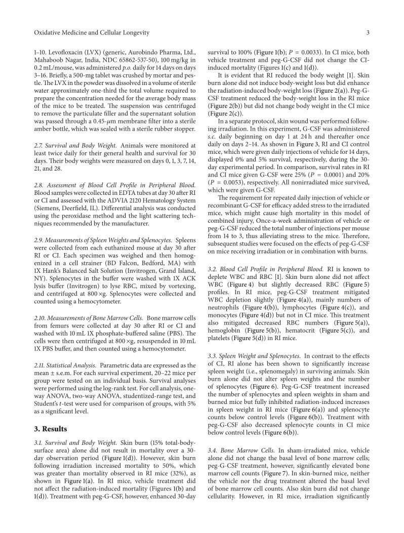
3.1. Survival and Body Weight. Skin burn (15% total-body-surface area) alone did not result in mortality over a 30-day observation period (Figure 1(d)). However, skin burnfollowing irradiation increased mortality to 50%, whichwas greater than mortality observed in RI mice (32%), asshown in Figure 1(a). In RI mice, vehicle treatment didnot affect the radiation-induced mortality (Figures 1(b) and1(d)). Treatment with peg-G-CSF, however, enhanced 30-day
survival to 100% (Figure 1(b); 𝑃 = 0.0033). In CI mice, bothvehicle treatment and peg-G-CSF did not change the CI-induced mortality (Figures 1(c) and 1(d)).
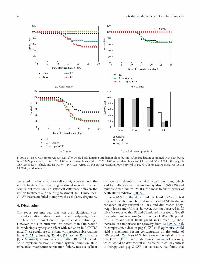
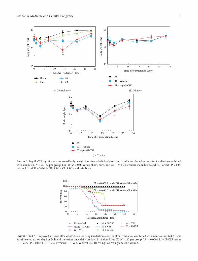
It is evident that RI reduced the body weight [1]. Skinburn alone did not induce body-weight loss but did enhancethe radiation-induced body-weight loss (Figure 2(a)). Peg-G-CSF treatment reduced the body-weight loss in the RI mice(Figure 2(b)) but did not change body weight in the CI mice(Figure 2(c)).
In a separate protocol, skin woundwas performed follow-ing irradiation. In this experiment, G-CSF was administereds.c. daily beginning on day 1 at 24 h and thereafter oncedaily on days 2–14. As shown in Figure 3, RI and CI controlmice, which were given daily injections of vehicle for 14 days,displayed 0% and 5% survival, respectively, during the 30-day experimental period. In comparison, survival rates in RIand CI mice given G-CSF were 25% (𝑃 = 0.0001) and 20%(𝑃 = 0.0053), respectively. All nonirradiated mice survived,which were given G-CSF.
The requirement for repeated daily injection of vehicle orrecombinant G-CSF for efficacy added stress to the irradiatedmice, which might cause high mortality in this model ofcombined injury, Once-a-week administration of vehicle orpeg-G-CSF reduced the total number of injections permousefrom 14 to 3, thus alleviating stress to the mice. Therefore,subsequent studies were focused on the effects of peg-G-CSFon mice receiving irradiation or in combination with burns.
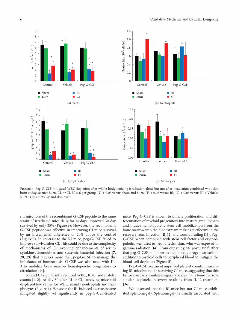
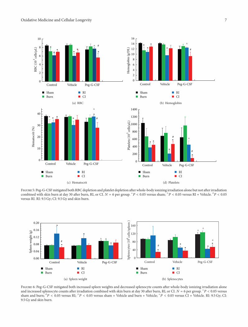
3.2. Blood Cell Profile in Peripheral Blood. RI is known todeplete WBC and RBC [1]. Skin burn alone did not affectWBC (Figure 4) but slightly decreased RBC (Figure 5)profiles. In RI mice, peg-G-CSF treatment mitigatedWBC depletion slightly (Figure 4(a)), mainly numbers ofneutrophils (Figure 4(b)), lymphocytes (Figure 4(c)), andmonocytes (Figure 4(d)) but not in CI mice. This treatmentalso mitigated decreased RBC numbers (Figure 5(a)),hemoglobin (Figure 5(b)), hematocrit (Figure 5(c)), andplatelets (Figure 5(d)) in RI mice.
3.3. Spleen Weight and Splenocytes. In contrast to the effectsof CI, RI alone has been shown to significantly increasespleen weight (i.e., splenomegaly) in surviving animals. Skinburn alone did not alter spleen weights and the numberof splenocytes (Figure 6). Peg-G-CSF treatment increasedthe number of splenocytes and spleen weights in sham andburned mice but fully inhibited radiation-induced increasesin spleen weight in RI mice (Figure 6(a)) and splenocytecounts below control levels (Figure 6(b)). Treatment withpeg-G-CSF also decreased splenocyte counts in CI micebelow control levels (Figure 6(b)).
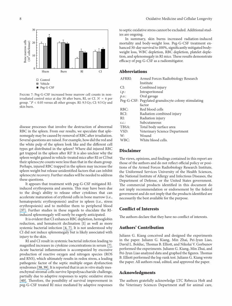
3.4. Bone Marrow Cells. In sham-irradiated mice, vehiclealone did not change the basal level of bone marrow cells;peg-G-CSF treatment, however, significantly elevated bonemarrow cell counts (Figure 7). In skin-burned mice, neitherthe vehicle nor the drug treatment altered the basal levelof bone marrow cell counts. Also skin burn did not changecellularity. However, in RI mice, irradiation significantly
4 Oxidative Medicine and Cellular Longevity
0
20
40
60
80
100
120
0 5 10 15 20 25 30
ShamBurn
RICI
Surv
ival
(%)
Time after irradiation (days)
∗
∗∗
(a) Control mice
0
20
40
60
80
100
120
0 5 10 15 20 25 30Time after irradiation (days)
Surv
ival
(%)
∗∗P = 0.0033
RIRI + VehicleRI + peg-G-CSF
(b) RI mice
Time after irradiation (days)
0
20
40
60
80
100
120
0 5 10 15 20 25 30
Surv
ival
(%)
∗
CICI + VehicleCI + peg-G-CSF
(c) CI mice
0
20
40
60
80
100
120
ControlVehiclePeg-G-CSF
Sham Burn RI CI
Surv
ival
(%)
(d) Vehicle versus peg-G-CSF
Figure 1: Peg-G-CSF improved survival after whole-body ionizing irradiation alone but not after irradiation combined with skin burn.𝑁 = 20–22 per group. For (a) ∗𝑃 < 0.05 versus sham, burn, and CI; ∗∗𝑃 < 0.05 versus sham burn and CI. For (b) ∗𝑃 = 0.0033 RI + peg-G-CSF versus RI + Vehicle and RI. For (c) ∗𝑃 < 0.05 versus CI. For (d) representing 100% survival in peg-G-CSF treated RI mice. RI: 9.5 Gy;CI: 9.5 Gy and skin burn.
decreased the bone marrow cell count, whereas both thevehicle treatment and the drug treatment increased the cellcounts, but there was no statistical difference between thevehicle treatment and the drug treatment. In CI-mice, peg-G-CSF treatment failed to improve the cellularity (Figure 7).
4. Discussion
This report presents data that skin burn significantly in-creased radiation-induced mortality and body-weight loss.The latter was thought due to injured small intestines [7].However, the skin burn was less potent than skin woundin producing a synergistic effect with radiation in B6D2F1/Jmice. These results are consistent with previous observationsin rat [21, 22], guinea pig [23], dog [24], swine [25], and mice[1, 5, 6, 26–29]. Consequences of either RI or CI includeacute myelosuppression, immune system inhibition, fluidimbalance, macro/microcirculation failure, massive cellular
damage, and disruption of vital organ functions, whichlead to multiple-organ dysfunction syndrome (MODS) andmultiple-organ failure (MOF), the most frequent causes ofdeath after irradiation [30–32].
Peg-G-CSF at the dose used displayed 100% survivalin sham-operated and burned mice. Peg-G-CSF treatmentenhanced 30-day survival to 100% and diminished body-weight losses after RI; this, however, was not observed in CImice.We reported that RI andCI induced increases in G-CSFconcentrations in serum (on the order of 100–1,000 pg/mLin RI mice and 2,000–10,000 pg/mL in CI mice [7]. Theseincreases are important for recovery from RI [29, 33, 34].In comparison, a dose of peg-G-CSF at 25𝜇g/mouse wouldyield a maximum serum concentration on the order of1,000 pg/mL [35]. Peg-G-CSF has a longer biological half-lifethanG-CSF [10].Therefore, daily injections are not necessary,which would be detrimental in irradiated mice. In contrastto therapy with peg-G-CSF, our laboratory has found that
Oxidative Medicine and Cellular Longevity 5
15
20
25
0 5 10 15 20 25 30
Body
wei
ght (
gm)
Time after irradiation (days)
ShamBurn
RICI
∗
∗∗
(a) Control mice
15
20
25
0 5 10 15 20 25 30
Body
wei
ght (
gm)
Time after irradiation (days)
RIRI + VehicleRI + peg-G-CSF
∗
(b) RI mice
15
20
25
0 5 10 15 20 25 30
Body
wei
ght (
gm)
Time after irradiation (days)
CICI + VehicleCI + peg-G-CSF
(c) CI mice
Figure 2: Peg-G-CSF significantly improved body-weight loss after whole-body ionizing irradiation alone but not after irradiation combinedwith skin burn.𝑁 = 20–22 per group. For (a) ∗𝑃 < 0.05 versus sham, burn, and CI; ∗∗𝑃 < 0.05 versus sham, burn, and RI. For (b) ∗𝑃 < 0.05versus RI and RI + Vehicle. RI: 9.5 Gy; CI: 9.5 Gy and skin burn.
0
20
40
60
80
100
120
0 5 10 15 20 25 30 35
Surv
ival
(%)
Postirradiation time (days)
Sham + Veh
W + VehRI + Veh
CI + VehSham + G-CSF
W + G-CSF
RI + G-CSFCI + G-CSF
∗
∗P = 0.0001
∗P = 0.0053
RI + G-CSF versus RI + Veh
CI + G-CSF versus CI + Veh
Figure 3: G-CSF improved survival after whole-body ionizing irradiation alone or after irradiation combined with skin wound. G-CSF wasadministered s.c. on day 1 at 24 h and thereafter once daily on days 2–14 after RI or CI. 𝑁 = 20 per group. ∗𝑃 = 0.0001 RI + G-CSF versusRI + Veh; ∗𝑃 = 0.0053 CI + G-CSF versus CI + Veh. Veh: vehicle; RI: 9.5 Gy; CI: 9.5 Gy and skin wound.
6 Oxidative Medicine and Cellular Longevity
∗
^
∗
#∗ #
∗∗
∗
0
1
2
3
4
5
6
7
8
9
Control Vehicle Peg-G-CSF
ShamBurn
RICI
WBC
(103 ce
lls/𝜇
L)
(a) WBC
^
∗
∗
ShamBurn
RICI
0.0
0.2
0.4
0.6
0.8
1.0
1.2
Control Vehicle Peg-G-CSF
Neu
troph
ils (1
03 cells
/𝜇L)
(b) Neutrophils
∗ ∗
^∗ #
∗∗
#∗
ShamBurn
RICI
0
1
2
3
4
5
6
7
8
Control Vehicle Peg-G-CSF
Lym
phoc
ytes
(103
cells
/𝜇L)
(c) Lymphocytes
∗
^∗
∗∗
ShamBurn
RICI
0.00
0.05
0.10
0.15
0.20
0.25
Control Vehicle Peg-G-CSF
Mon
ocyt
es (1
03 cells
/𝜇L)
(d) Monocytes
Figure 4: Peg-G-CSF mitigated WBC depletion after whole-body ionizing irradiation alone but not after irradiation combined with skinburn at day 30 after burn, RI, or CI.𝑁 = 6 per group. ∗𝑃 < 0.05 versus sham and burn; #𝑃 < 0.05 versus RI; ∧𝑃 < 0.05 versus RI + Vehicle.RI: 9.5 Gy; CI: 9.5 Gy and skin burn.
s.c. injections of the recombinant G-CSF peptide to the samestrain of irradiated mice daily for 14 days improved 30-daysurvival by only 25% (Figure 3). However, the recombinantG-CSF peptide was effective in improving CI mice survivalby an incremental difference of 20% above the control(Figure 3). In contrast to the RI mice, peg-G-CSF failed toimprove survival after CI.This could be due to the complexityof mechanisms of CI involving enhancements of serumcytokines/chemokines and systemic bacterial infection [7,28, 29] that requires more than peg-G-CSF to manage theimbalance of homeostasis. G-CSF was also used with IL-3 to mobilize bone marrow hematopoietic progenitors tocirculation [14].
RI and CI significantly reduced WBC, RBC, and plateletcounts [1, 2]. At day 30 after RI or CI, surviving mice stilldisplayed low values for WBC, mainly neutrophils and lym-phocytes (Figure 4). However, the RI-induced decreases weremitigated slightly yet significantly in peg-G-CSF-treated
mice. Peg-G-CSF is known to initiate proliferation and dif-ferentiation of myeloid progenitors into mature granulocytesand induce hematopoietic stem cell mobilization from thebone marrow into the bloodstream making it effective in therecovery from infection [11, 12] and wound healing [13]. Peg-G-CSF, when combined with stem cell factor and erythro-poietin, was used to treat a technician, who was exposed togamma radiation [14]. From our study, we postulate furtherthat peg-G-CSF mobilizes hematopoietic progenitor cells inaddition to myeloid cells to peripheral blood to mitigate theblood-cell depletion (Figure 5).
Peg-G-CSF treatment improved platelet counts in surviv-ing RI-mice but not in surviving CI-mice, suggesting that thisfactor also can stimulatemegakaryocytes in the bonemarrow,similar to platelet recovery resulting from IL-12 treatment[36].
We observed that the RI mice but not CI mice exhib-ited splenomegaly. Splenomegaly is usually associated with
Oxidative Medicine and Cellular Longevity 7
∗∗
∗∗
#
∗
^∗
0
2
4
6
8
10
Control Vehicle Peg-G-CSF
RBC
(10 3
cells
/𝜇L)
ShamBurn
RICI
(a) RBC
#
02468
10121416
Control Vehicle Peg-G-CSF
Hem
oglo
bin
(g/D
L)
∗∗
∗
∗
^
ShamBurn
RICI
(b) Hemoglobin
#∗
^∗
∗∗
0
10
20
30
40
Control Vehicle Peg-G-CSF
Hem
atoc
rit (%
)
∗
ShamBurn
RICI
(c) Hematocrit
#∗
0
200
400
600
800
1000
1200
1400
Control Vehicle Peg-G-CSF
Plat
elet
s (10
3ce
lls/𝜇
L)
∗^
#∗
∗
∗
ShamBurn
RICI
(d) Platelets
Figure 5: Peg-G-CSFmitigated bothRBCdepletion and platelet depletion afterwhole-body ionizing irradiation alone but not after irradiationcombined with skin burn at day 30 after burn, RI, or CI.𝑁 = 6 per group. ∗𝑃 < 0.05 versus sham; ∧𝑃 < 0.05 versus RI + Vehicle. #𝑃 < 0.05versus RI. RI: 9.5 Gy; CI: 9.5 Gy and skin burn.
0.00
0.04
0.08
0.12
0.16
0.20
Control Vehicle Peg-G-CSF
Sple
en w
eigh
t (g)
∗
∗
^ ^#∗
∗
ShamBurn
RICI
(a) Spleen weight
0
40
80
120
160
Control Vehicle Peg-G-CSF
Sple
nocy
tes (
10 6 ce
lls/s
plee
n )
^ ^
∗∗#
∗∗
+∗
ShamBurn
RICI
(b) Splenocytes
Figure 6: Peg-G-CSF mitigated both increased spleen weights and decreased splenocyte counts after whole-body ionizing irradiation aloneand increased splenocyte counts after irradiation combined with skin burn at day 30 after burn, RI, or CI.𝑁 = 6 per group. ∗𝑃 < 0.05 versussham and burn; #𝑃 < 0.05 versus RI; ∧𝑃 < 0.05 versus sham + Vehicle and burn + Vehicle; +𝑃 < 0.05 versus CI + Vehicle. RI: 9.5 Gy; CI:9.5 Gy and skin burn.
8 Oxidative Medicine and Cellular Longevity
0
10
20
30
ControlVehiclePeg-G-CSF
Sham Burn RI CI
Bone
mar
row
cells
(106 ce
lls/fe
mur
)
∗
Figure 7: Peg-G-CSF increased bone marrow cell counts in non-irradiated control mice at day 30 after burn, RI, or CI. 𝑁 = 6 pergroup. ∗𝑃 < 0.05 versus all other groups. RI: 9.5 Gy; CI: 9.5 Gy andskin burn.
disease processes that involve the destruction of abnormalRBC in the spleen. From our results, we speculate that sple-nomegalymay be caused by removal of RBC after irradiation.Several questions are raised. For example, howdid the red andthe white pulp of the spleen look like and the different celltypes get distributed in the spleen? Where did injured RBCget trapped in the spleen after RI? It is also unclear why thespleenweight gained in vehicle-treatedmice after RI orCI buttheir splenocyte counts were less than that in the sham group.Perhaps, injured RBC trapped in the spleen may increase thespleen weight but release unidentified factors that can inhibitsplenocyte recovery. Further studies will be needed to addressthese questions.
It appears that treatment with peg-G-CSF mitigated RI-induced erythropenia and anemia. This may have been dueto the drug’s ability to release other cytokines that canaccelerate maturation of erythroid cells in bone marrow (i.e.,hematopoietic erythropoiesis) and/or in spleen (i.e., stresserythropoiesis) and to mobilize them to peripheral blood[37]. Further studies in these regards to elucidate the RI-induced splenomegaly will surely be eagerly anticipated.
It is evident that CI enhances RBC depletion, hemoglobinreduction, and hematocrit declination [1] as well as moresystemic bacterial infection [4, 7]. It is not understood whyCI did not induce splenomegaly but is likely associated withinjury to the skin.
RI and CI result in systemic bacterial infection leading tomagnified increases in cytokine concentrations in serum [7].Acute bacterial inflammation is accompanied by excessiveproduction of reactive oxygen and nitrogen species (ROSand RNS), which ultimately results in redox stress, a leadingpathogenic factor of the septic multiple organ dysfunctionsyndromes [38, 39]. It is reported that in an in vitro studymes-enchymal stromal cells survive lipopolysaccharide challenge,partially due to adaptive responses to septic oxidative stress[40]. Therefore, the possibility of survival improvement inpeg-G-CSF treated RI mice mediated by adaptive responses
to septic oxidative stress cannot be excluded. Additional stud-ies are ongoing.
In summary, skin burns increased radiation-inducedmortality and body-weight loss. Peg-G-CSF treatment en-hanced 30-day survival to 100%, significantlymitigated body-weight loss, WBC depletion, RBC depletion, platelet deple-tion, and splenomegaly in RImice.These results demonstrateefficacy of peg-G-CSF as a radiomitigator.
Abbreviations
AFRRI: Armed Forces Radiobiology ResearchInstitute
CI: Combined injuryi.p.: Intraperitonealp.o.: Oral gavagePeg-G-CSF: Pegylated granulocyte colony stimulating
factorRBC: Red blood cellsRCI: Radiation combined injuryRI: Radiation injurys.c.: SubcutaneousTBSA: Total body surface areaVSD: Veterinary Science DepartmentW: WoundWBC: White blood cells.
Disclaimer
The views, opinions, and findings contained in this report arethose of the authors and do not reflect official policy or posi-tions of the Armed Forces Radiobiology Research Institute,the Uniformed Services University of the Health Sciences,the National Institute of Allergy and Infectious Diseases, theDepartment of Defense, or the United States government.The commercial products identified in this document donot imply recommendation or endorsement by the federalgovernment and do not imply that the products identified arenecessarily the best available for the purpose.
Conflict of Interests
The authors declare that they have no conflict of interests.
Authors’ Contribution
Juliann G. Kiang conceived and designed the experimentsin the paper. Juliann G. Kiang, Min Zhai, Pei-Jyun Liao,David L. Bolduc,Thomas B. Elliott, and Nikolai V. Gorbunovperformed the experiments. Juliann G. Kiang, Min Zhai, andPei-Jyun Liao analyzed data and graphed the figures.ThomasB. Elliott performed the log-rank test. Juliann G. Kiang wrotethe paper. All authors read, edited, and approved the paper.
Acknowledgments
The authors gratefully acknowledge LTC Rebecca Holt andthe Veterinary Sciences Department staff for animal care,
Oxidative Medicine and Cellular Longevity 9
Dr. VitalyNagy andRadiationDosimetry staff for conductingwhole-body irradiation, and Dr. Xinyue Lu, Dr. DilberNurmemet, Marsha Anderson, HM1, USN, Ms. Joan Smith,Mr. True Burns, and Dr. Risaku Fukumoto for their technicalassistance. Research was supported by NIH/NIAID YI-AI-5045-04 to JuliannG.Kiang andAFRRIRAB32164 toThomasB. Elliott and Juliann G. Kiang.
References
[1] J. G. Kiang, B. R. Garrison, T. M. Burns et al., “Wound traumaalters ionizing radiation dose assessment,” Cell and Bioscience,vol. 2, no. 1, article 20, 12 pages, 2012.
[2] J. G. Kiang, B. R. Garrison, R. Fukumoto, and T. B. Elliott,“Ciprofloxacin inhibits gamma radiation-induced increases in𝛾-H2AX, p53 phosphorylation in human tumor cells and p53phosphorylation, Gadd45a, bax, and Bcl-2 gene expression inhuman peripheral blood cells,” in Proceedings of the 56th Radi-ation Research Society Annual Meeting, p. 188, Maui, Hawaii,USA, September 2010.
[3] R. Fukumoto and J. G. Kiang, “Geldanamycin analog 17-DMAG limits apoptosis in human peripheral blood cells byinhibition of p53 activation and its interaction with heat-shockprotein 90 kDa after exposure to ionizing radiation,” RadiationResearch, vol. 176, no. 3, pp. 333–345, 2011.
[4] R. Fukumoto, L. H. Cary, N. V. Gorbunov, T. B. Elliott, and J. G.Kiang, “Ciprofloxacin modulates cytokine profiles, acceleratesbone marrow recovery and mitigates ileum injury after radia-tion combined with wound trauma,” PLoS ONE, vol. 8, no. 3,Article ID e58389, 11 pages, 2013.
[5] G. D. Ledney and T. B. Elliott, “Combined injury: factors withpotential to impact radiation dose assessments,”Health Physics,vol. 98, no. 2, pp. 145–152, 2010.
[6] J. G. Kiang and G. D. Ledney, “Skin injuries reduce survival andmodulate corticosterone, C-reactive protein, complement com-ponent 3, IgM, and prostaglandin E2 after whole-body reactor-produced mixed field (n + 𝛾-photons) irradiation,” OxidativeMedicine and Cellular Longevity, vol. 2013, Article ID 821541, 10pages, 2013.
[7] J. G. Kiang, W. Jiao, L. H. Cary et al., “Wound trauma increasesradiation-inducedmortality by activation of iNOS pathway andelevation of cytokine concentrations and bacterial infection,”Radiation Research, vol. 173, no. 3, pp. 319–332, 2010.
[8] M. E. Berger, D. M. Christensen, P. C. Lowry, O. W. Jones, andA. L.Wiley, “Medical management of radiation injuries: currentapproaches,” Occupational Medicine, vol. 56, no. 3, pp. 162–172,2006.
[9] J. K. Waselenko, T. J. MacVittie, W. F. Blakely et al., “Medicalmanagement of the acute radiation syndrome: recommen-dations of the strategic national stockpile radiation workinggroup,” Annals of Internal Medicine, vol. 140, no. 12, pp. 1037–1051, 2004.
[10] G. Molineux, “The design and development of pegfilgras-tim (PEG-rmetHuG-CSF, Neulasta),” Current PharmaceuticalDesign, vol. 10, no. 11, pp. 1235–1244, 2004.
[11] D. Metcalf, “The role of the colony-stimulating factors in thetreatment of infections,” in Frontiers of Infectious Diseases: NewAntibacterial Strategies: Proceedings of an International Sympo-sium Sponsored by Glaxo Research, Brocker Hall, Hertfordshire30 June-3 July 1990, H. C. Neu, Ed., Churchill Livingstone, NewYork, NY, USA, 1990.
[12] D. Metcalf, “Hematopoietic cytokines,” Blood, vol. 111, no. 2, pp.485–491, 2008.
[13] E. V. Badiavas, M. Abedi, J. Butmarc, V. Falanga, and P.Quesenberry, “Participation of bone marrow derived cells incutaneous wound healing,” Journal of Cellular Physiology, vol.196, no. 2, pp. 245–250, 2003.
[14] J. M. Bertho, N. M. Griffiths, and P. Gourmelon, “The medicaldiagnosis and treatment of radiation overexposed people,” 2003,http://irpa11.irpa.net/pdfs/RC-7a.pdf.
[15] C. A. Montgomery, “Oncologic and toxicologic research: alle-viation and control of pain and distress in laboratory animals,”Cancer Bulletin, vol. 42, pp. 230–237, 1990.
[16] S. P. Tomasivic, L. G. Coghlan, K. N. Gray, A. J. Mastromarino,and E. L. Travis, “IACUC evaluation of experiments requiringdeath as an end point: a cancer center’s recommendations,” LabAnimal, pp. 31–34, 1988.
[17] International Standardization Organization and ASTM Inter-national, Standard Practice for Use of an Alanine-EPRDosimetrySystem, Standard 51607:2004(E), ISO, Geneva, Switzerland;ASTM International, West Conshohocken, Pa, USA, 2004.
[18] B. I. Lord, L. B. Woolford, and G. Molineux, “Kinetics ofneutrophil production in normal and neutropenic animalsduring the response to filgrastim (r-metHuG-CSF) or filgrastimSD/01 (PEG-r-metHu G-CSF),” Clinical Cancer Research, vol. 7,no. 7, pp. 2085–2090, 2001.
[19] A. B. van Spriel, I. E. van den Herik-Oudijk, and J. G. J. van deWinkel, “A single injection of polyethylene-glycol granulocytecolony-stimulating factor strongly prolongs survival of micewith systemic candidiasis,” Cytokine, vol. 12, no. 6, pp. 666–670,2000.
[20] K. J. Neelis, S. C. C. Hartong, T. Egeland, G. R. Thomas, D. L.Eaton, andG.Wagemaker, “The efficacy of single-dose adminis-tration of thrombopoietinwith coadministration of either gran-ulocyte/macrophage or granulocyte colony-stimulating factorin myelosuppressed rhesus monkeys,” Blood, vol. 90, no. 7, pp.2565–2573, 1997.
[21] E. L. Alpen and G. E. Sheline, “The combined effects ofthermal burns and whole-body X-radiation on survival timeand mortality,” Annuls of Surgery, vol. 140, no. 1, pp. 113–118,1954.
[22] F. A. Valeriote and D. G. Baker, “The combined effects of ther-mal trauma and X-irradiation on early,” Radiation Research, vol.22, pp. 693–702, 1964.
[23] B. Korlof, “Infection of burns. I. A bacteriological and clinicalstudy of 99 cases. II. Animal experiments: burns and total bodyx-irradiation,” Acta Chirurgica Scandinavica, Supplementum,vol. 209, pp. 1–144, 1956.
[24] J. W. Brooks, E. I. Evans, W. T. Ham Jr., and J. D. Reid, “Theinfluence of external body radiation on mortality from thermalburns,” Annals of Surgery, vol. 136, no. 3, pp. 533–545, 1952.
[25] H. Baxter, J. A. Drummond, L. G. Stephens-Newsham, and R.G. Randall, “Studies on acute total body irradiation in animals.I. Effect of streptomycin following exposure to a thermal burnand irradiation,”Plastic Reconstruction Surgery, vol. 12, no. 6, pp.439–445, 1953.
[26] G. D. Ledney, T. B. Elliott, and M. M. Moore, “Modulations ofmortality by tissue trauma and sepsis in mice after radiationinjury,” in The Biological Basis of Radiation Protection Practice,K. L. Mossman and W. A. Mills, Eds., pp. 202–217, Williams &Wilkins, Baltimore, Md, USA, 1992.
[27] G. D. Ledney, D. A. Stewart, E. D. Exum, and P. A. Sheehy,“Skin wound-enhanced survival and myelocytopoiesis in mice
10 Oxidative Medicine and Cellular Longevity
after whole-body irradiation,” Acta Radiologica: Oncology, vol.20, no. 1, pp. 29–38, 1981.
[28] J. L. Palmer, C. R. Deburghgraeve,M. D. Bird,M. Hauer-Jensen,and E. J. Kovacs, “Development of a combined radiation andburn injury model,” Journal of Burn Care and Research, vol. 32,no. 2, pp. 317–323, 2011.
[29] A. Jacob, K. G. Shah, R. Wu, and P. Wang, “Ghrelin as a noveltherapy for radiation combined injury,”MolecularMedicine, vol.16, no. 3-4, pp. 137–143, 2010.
[30] Z. Zou, H. Sun, Y. Su, T. Cheng, and C. Luo, “Progress inresearch on radiation combined injury in China,” RadiationResearch, vol. 169, no. 6, pp. 722–729, 2008.
[31] K. L. Koenig, R. E. Goans, R. J. Hatchett et al., “Medicaltreatment of radiological casualties: current concepts,” Annalsof Emergency Medicine, vol. 45, no. 6, pp. 643–652, 2005.
[32] Z. Lausevic, M. Lausevic, J. Trbojevic-Stankovic, S. Krstic, andB. Stojimirovic, “Predicting multiple organ failure in patientswith severe trauma,” Canadian Journal of Surgery, vol. 51, no.2, pp. 97–102, 2008.
[33] T. J. MacVittie, A. M. Farese, and W. Jackson III, “Defining thefull therapeutic potential of recombinant growth factors in thepost radiation-accident environment: the effect of supportivecare plus administration of G-CSF,” Health Physics, vol. 89, no.5, pp. 546–555, 2005.
[34] V. K. Singh, O. O. Fatanmi, P. K. Singh, and M. H. Whitnall,“Role of radiation-induced granulocyte colony-stimulating fac-tor in recovery fromwhole body gamma-irradiation,” Cytokine,vol. 58, no. 3, pp. 406–414, 2012.
[35] M. Scholz, M. Ackermann, C. Engel, F. Emmrich, M. Loeffler,and M. Kamprad, “A pharmacokinetic model of filgrastim andpegfilgrastim application in normal mice and those withcyclophosphamide-induced granulocytopaenia,” Cell Prolifera-tion, vol. 42, no. 6, pp. 813–822, 2009.
[36] T. Chen, K. A. Burke, Y. Zhan, X. Wang, D. Shibata, and Y.Zhao, “IL-12 facilitates both the recovery of endogenous hem-atopoiesis and the engraftment of stem cells after ionizingradiation,” Experimental Hematology, vol. 35, no. 2, pp. 203–213,2007.
[37] S. Millot, V. Andrieu, P. Letteron et al., “Erythropoietin stim-ulates spleen BMP4-dependent stress erythropoiesis and par-tially corrects anemia in a mouse model of generalized inflam-mation,” Blood, vol. 116, no. 26, pp. 6072–6081, 2010.
[38] J. A. Russell, “Management of sepsis,”The New England Journalof Medicine, vol. 355, no. 16, pp. 1699–1713, 2006.
[39] M. Perl, C. Chung, R. Swan, andA. Ayala, “Role of programmedcell death in the immunopathogenesis of sepsis,”DrugDiscoveryToday, vol. 4, no. 4, pp. 223–230, 2007.
[40] N. V. Gorbunov, B. R. Garrison, D. P. McDaniel et al., “Adaptiveredox response of mesenchymal stromal cells to stimulationwith lipopolysaccharide inflammagen:mechanisms of remodel-ing of tissue barriers in sepsis,”Oxidative Medicine and CellularLongevity, vol. 2013, Article ID 186795, 16 pages, 2013.

Submit your manuscripts athttp://www.hindawi.com
Stem CellsInternational
Hindawi Publishing Corporationhttp://www.hindawi.com Volume 2014
Hindawi Publishing Corporationhttp://www.hindawi.com Volume 2014
MEDIATORSINFLAMMATION
of
Hindawi Publishing Corporationhttp://www.hindawi.com Volume 2014
Behavioural Neurology
International Journal of
EndocrinologyHindawi Publishing Corporationhttp://www.hindawi.com
Volume 2014
Hindawi Publishing Corporationhttp://www.hindawi.com Volume 2014
Disease Markers
BioMed Research International
Hindawi Publishing Corporationhttp://www.hindawi.com Volume 2014
OncologyJournal of
Hindawi Publishing Corporationhttp://www.hindawi.com Volume 2014
Hindawi Publishing Corporationhttp://www.hindawi.com Volume 2014
Oxidative Medicine and Cellular Longevity
PPARRe sea rch
Hindawi Publishing Corporationhttp://www.hindawi.com Volume 2014
The Scientific World JournalHindawi Publishing Corporation http://www.hindawi.com Volume 2014
Immunology ResearchHindawi Publishing Corporationhttp://www.hindawi.com Volume 2014
Journal of
ObesityJournal of
Hindawi Publishing Corporationhttp://www.hindawi.com Volume 2014
Hindawi Publishing Corporationhttp://www.hindawi.com Volume 2014
Computational and Mathematical Methods in Medicine
OphthalmologyJournal of
Hindawi Publishing Corporationhttp://www.hindawi.com Volume 2014
Diabetes ResearchJournal of
Hindawi Publishing Corporationhttp://www.hindawi.com Volume 2014
Hindawi Publishing Corporationhttp://www.hindawi.com Volume 2014
Research and TreatmentAIDS
Hindawi Publishing Corporationhttp://www.hindawi.com Volume 2014
Gastroenterology Research and Practice
Parkinson’s DiseaseHindawi Publishing Corporationhttp://www.hindawi.com Volume 2014
Evidence-Based Complementary and Alternative Medicine
Volume 2014Hindawi Publishing Corporationhttp://www.hindawi.com
Copyright © 2022 FDOKUMEN