Building Lasting Growth from Viral Moments - NEO Philanthropy
Upload
independentCategory
view
0download
0
Long-Lasting Molecular Changes in Human Skin after Repetitivein situ UV Irradiation
Michaela Brenner1, Sergio G. Coelho2, Janusz Z. Beer3, Sharon A. Miller3, Rainer Wolber4,Christoph Smuda4, and Vincent J. Hearing21 Department of Dermatology, Ludwig-Maximilians-University of Munich, Munich, Germany
2 Laboratory of Cell Biology, National Cancer Institute, National Institutes of Health, Bethesda, MD, USA
3 Center for Devices and Radiological Health, Food and Drug Administration, Silver Spring, MD, USA
4 R&D, Skin Research, Beiersdorf AG, Hamburg, Germany
AbstractIt is known that UV modulates the expression of paracrine factors that regulate melanocyte functionin the skin. We have investigated the consequences of repetitive UV exposure of human skin inbiopsies of 10 subjects with phototypes 2-3.5 taken 1-4 yr later. The expression of melanogenicfactors (TYR, MART1, MITF), growth factors/receptors (SCF/KIT, bFGF/FGFR1, ET1/EDNRB,HGF, GM-CSF), adhesion molecules (β-catenin, E-cadherin, N-cadherin), cell cycle proteins(PCNA, cyclins D1, E2) as well as Bcl-2, DKK1 and DKK3, were analyzed byimmunohistochemistry. Most of those markers showed no detectable changes at ≥1 yr after therepetitive UV irradiation. While increased expression of EDNRB protein was detected in 3 of 10UV-irradiated subjects, there was no detectable change in the expression of ET1 protein or in EDNRBmRNA levels. In summary, only the expression of TYR, MART1 and/or EDNRB, and only in somesubjects, was elevated at ≥1 yr post-UV irradiation. Thus the long-term effects of repetitive UVirradiation on human skin did not lead to significant changes in skin morphology and there isconsiderable subject-to-subject variation in responses. The possibility that changes in the expressionand function of EDNRB triggers downstream activation of abnormal melanocyte proliferation anddifferentiation deserves further investigation.
Keywordsultraviolet; skin; long-term changes; long-lasting pigmentation
INTRODUCTIONThe incidence of melanoma has tripled over the past 3 decades probably as a result, at least inpart, from lifestyle changes that have led to an increased exposure of the fair-skinned populationto ultraviolet radiation (UV) [NCI Seer Statistics, http://seer.cancer.gov/]. Epidemiological andlaboratory data provide strong evidence that solar exposure is a major causative factor inmelanomagenesis (Gilchrest et al., 1999). Emerging evidence suggests that there is a clear link
Address correspondence to: Dr. Vincent J. Hearing, Laboratory of Cell Biology, National Cancer Institute, National Institutes of Health,Building 37, Room 2132, MSC 4256, Bethesda, MD 20892, USA; Tel.: 301-496-1564; FAX: 301-402-8787; E-mail: E-mail:[email protected] OF INTERESTThe authors state no conflict of interest.
NIH Public AccessAuthor ManuscriptJ Invest Dermatol. Author manuscript; available in PMC 2010 April 1.
Published in final edited form as:J Invest Dermatol. 2009 April ; 129(4): 1002–1011. doi:10.1038/jid.2008.325.
NIH
-PA Author Manuscript
NIH
-PA Author Manuscript
NIH
-PA Author Manuscript

between tanning devices and malignant melanoma (Clough-Gorr et al., 2008; InternationalAgency for Research on Cancer, 2007; Ting et al., 2007). Pathomechanisms for thedevelopment of non-melanocytic skin cancers have already been revealed (Matsumura andAnanthaswamy, 2002; Stary et al., 1997), however, the exact mechanism(s) of UV-inducedmelanomagenesis in the skin in situ remains unknown. Since UV-fingerprint mutations arerare in melanomas, it has been speculated that UV causes melanoma development by indirecteffects, for example by dysregulation of growth factors in the skin, as proposed in a humanskin graft model for UV-induced melanomas (Berking et al., 2004). We have recently shownthat UV modulates the production (by keratinocytes and by fibroblasts in vitro) of growthfactors regulating melanocyte function (Brenner et al., 2005). UV is presently the only knownenvironmental carcinogen for melanomas, however it is still controversial which wavelengthsare critical. UVA has been shown to promote melanoma in a hybrid fish melanoma model(Setlow et al., 1993), while UVB promotes melanoma in several transgenic mouse models(Noonan et al., 2003).
There is an urgent need to determine the specific mechanisms that are involved inphotocarcinogenesis of UV-irradiated human skin in situ. Numerous studies have examinedthe effects of UV on skin cells in vitro, however that experimental setting does not allow oneto explore inter-individual differences such as skin phototype or minimal erythema dose(MED). The examination of UV-induced changes in UV-irradiated skin in situ, with respectto different wavelengths and doses, provides a more suitable approach to characterize UVphotocarcinogenesis in the skin, particularly with respect to early events in the malignantcascade.
Our group has previously reported several studies on the acute effects of UV radiation on humanskin of varying skin pigmentation phenotypes. So far, we have shown that even a single,relatively low (1 MED) UV dose (60% UVA/40% UVB) causes significant damage to DNAin epidermal cells and stimulates the production of photoprotective melanin (Tadokoro et al.,2003). Most UV-induced DNA damage is removed relatively quickly (within days), however,some UV-induced molecular changes might persist and could initiate malignant transformationin the skin. Our earlier studies examined only the short-term effects of UV on human skin insitu. To extend our understanding of the long-lasting and potentially photocarcinogenic effectsof UV, we examined biopsies from sites exposed to repetitive UV irradiation several years ago.In particular, we characterized the long-lasting effects of UV on melanocytes and onkeratinocytes. We examined various potential morphologic and molecular markers ofmalignant transformation in 2 different UV-irradiated groups. One group of 6 subjects hadbeen repeatedly irradiated over several weeks at 3 different cumulative doses with Sunlampsemitting a UV spectrum commonly used in tanning salons (≥95% UVA/≤5% UVB) while inanother group, 4 different subjects had been repeatedly irradiated over a period of 2 weeks witha solar simulator (SS, ≥90% UVA/≤10% UVB). The molecular markers examined can bedivided into 6 subgroups and were selected due to their possible involvement in melanocytetransformation and/or because many previously published studies have analyzed theirresponses in human skin within 1 month of UV exposure:
a) Melanin content and several melanocyte-specific proteins: microphthalmiatranscription factor (MITF), melanoma antigen recognized by T-cells (MART1),tyrosinase (TYR).
b) growth factors and their receptors: stem cell factor (SCF)/KIT, basic fibroblast growthfactor/fibroblast growth factor receptor 1 (bFGF/FGFR1), endothelin 1/endothelin Breceptor (ET1/EDNRB), granulocyte macrophage colony stimulating factor (GM-CSF)and hepatocyte growth factor (HGF).
c) cell cycle proteins: proliferating cell nuclear antigen (PCNA), cyclins D1 and E2.
Brenner et al. Page 2
J Invest Dermatol. Author manuscript; available in PMC 2010 April 1.
NIH
-PA Author Manuscript
NIH
-PA Author Manuscript
NIH
-PA Author Manuscript

d) adhesion molecules: E- and N-cadherins, β-catenin.
e) the anti-apoptotic factor: B-cell lymphoma 2 (Bcl-2).
f) Wnt-pathway regulator dickkopf 1 (DKK1) and its homologue dickkopf 3 (DKK3).
RESULTSMorphological changes in epidermal structure
Subjects (listed in Table 1) from 2 independent studies (Miller et al., 2008;Wolber et al.,2008) were invited to return for reexamination >1 yr after their initial UV exposures. Areaspreviously irradiated were identified using photos taken at the time of initial exposure,considering surface features and in some cases, pigmented areas that still remained.
Skin specimens from all subjects were stained with H&E and were carefully examined by lightmicroscopy for morphological changes in the structure of the epidermis and/or the dermis. Nomorphological changes were seen in any UV-irradiated skin specimens or from the adjacentunirradiated skin specimens [Figure 1]. There were no signs that could be correlated to anyclinical aspect of a localized sunburn reaction, such as a superficial inflammatory infiltrate inthe dermis or the presence of sunburn cells in the epidermis. Special attention was paid to detectpossible changes in the density, distribution and/or localization of melanocytes (e.g. suprabasallocalization of melanocytes, formation of melanocyte cell nests). No such changes weredetected, although there was a significant increase in melanocyte density in some subjects (seebelow).
Visible pigmentation, melanin content and expression of melanogenic proteins after UVirradiation
To evaluate long-lasting changes in melanin content, the UV-irradiated skin areas and thecorresponding unirradiated control tissues were examined visually and were biopsied after >1yr post-exposure. Repetitive UV irradiation with the Sunlamp or with the solar simulatorproduced good early tanning reactions in all subjects. However, only 1 subject (T50) in theSunlamp-irradiated group showed long-lasting pigmentation (LLP) in the areas exposed to theintermediate or high cumulative UV doses (Miller et al., 2008). Figure 2 shows suchpigmentation at 520 d post-irradiation. None of the subjects in the SS-irradiated group hadvisibly increased pigmentation in the irradiated areas at the time of biopsy [Table 1].
The melanin content in each skin specimen was determined by quantitative densitometry ofthe Fontana-Masson stained sections [Figure 3]. Only one subject (T50) in the Sunlamp-irradiated group and one subject (B6) in the SS-irradiated group showed significantly (p<0.05)increased total amounts of melanin [Table 2].
There were no significant differences in the numbers of MITF-positive cells compared to theunirradiated controls in the UV-irradiated subjects. Although no difference could be foundbetween the UV-irradiated and the control specimens in the number of MART1-positive cellsin the Sunlamp-irradiated group, 2 subjects (B6 and B8) showed significantly higher (p<0.05)numbers of MART1-positive cells in the SS-irradiated group. In the Sunlamp-irradiated group,a significant increase in the number of TYR-positive cells compared to the unirradiated controlwas only found in subject T50, who was the only subject in either study group with visiblepigmentation remaining in the previously UV-irradiated areas. In the SS-irradiated group, 3out of 4 subjects (B2, B6 and B8) showed significant (p<.05) increases in the density of TYR-positive cells [Table 2, Figure 3], although no visibly increased pigmentation was noted inthose previously UV-irradiated areas.
Brenner et al. Page 3
J Invest Dermatol. Author manuscript; available in PMC 2010 April 1.
NIH
-PA Author Manuscript
NIH
-PA Author Manuscript
NIH
-PA Author Manuscript

Expression patterns of markers in the skin after UV irradiationTable 3 summarizes the molecular markers examined and their long-term responses to UV-irradiation and examples of the staining patterns are shown in Figure 4.
Growth factors and their receptors—In the skin specimens examined, SCF, bFGF andET1 were expressed in the basal epidermal layer. Between different samples, the stainingintensity ranged from weak to moderate, but overall, no marked differences in expression couldbe detected between UV-irradiated and control specimens from either protocol. KIT expressionwas detected in all samples and showed a strong regular pattern of staining in the basal layersof the epidermis. FGFR1 staining was weak and was restricted to the basal epidermal layer.Expression of HGF could not be detected in the epidermis of any skin sections examined. GM-CSF expression in the skin showed irregular patterns, with some specimens showing basalexpression and others showing dispersed expression throughout the whole epidermis [Figure4]. Two of the 6 subjects in the Sunlamp-irradiated group (T35 and T47) and 1 of the 4 subjectsin the SS-irradiated group (B6) showed marked differences in the expression of EDNRB in thebasal epidermal layers compared to the unirradiated controls. We used a tissue array with 39melanoma samples as a positive control for EDNRB [Figure 5].
Tissue-in situ hybridization for EDNRB mRNA in the Sunlamp- or SS- irradiated skin showedcytoplasmic staining mainly in the basal epidermal layers, but no marked difference in EDNRBmRNA expression between the UV-irradiated and control skin [Figure 5].
Adhesion molecules—All skin specimens were examined for expression of variousadhesion molecules (β-catenin, E- and N-cadherins). β-Catenin showed a regular strongcytoplasmic staining pattern throughout the whole epidermis with no changes in the expressionbetween UV-irradiated and unirradiated skin. No nuclear staining pattern could be detected.In all skin specimens, E-cadherin was expressed in a regular strong cytoplasmic patternthroughout the epidermis and no marked differences between UV-irradiated and controlspecimens were found. N-cadherin expression could not be detected [Figure 6].
Cell cycle proteins—Skin specimens were analyzed for expression of the proliferative indexof PCNA, cyclins D1 and E2. Neither cyclin D1 nor E2 showed a positive staining. PCNAexpression showed no differences between UV-irradiated and control specimens, except for 1subject (B6) in the SS-irradiated group. A clearly higher nuclear staining for PCNA was foundin that specimen, however, double staining with TYR revealed that mainly keratinocytes, butnot melanocytes, showed the increased expression of PCNA [Figure 6].
DKK1, DKK3 and Bcl-2—DKK1 could not be detected in any skin specimens examined,UV-irradiated or unirradiated. DKK3 expression showed a regular and strong expressionpattern in the upper epidermal layers but did not exhibit any significant changes in responseto UV irradiation. Bcl-2 immunoreactivity was found in melanocytes in the basal cell layer ofthe skin as expected, but no marked differences in the expression of Bcl-2 was noted betweenUV-irradiated and unirradiated specimens [Figure 6].
DISCUSSIONIn the past, many in vitro and in vivo experiments have been conducted to characterize thecarcinogenic effects of UV radiation on human skin cells. In vitro studies with humanmelanocytes (Bittner et al., 2000; Clark et al., 2000; Hipfel et al., 2000; Valery et al., 2001;Young et al., 1998b) and in vivo studies on human skin have examined the effects of short-term UV irradiation (Hachiya et al., 2004; Mass et al., 2003; Tadokoro et al., 2003; Tronnieret al., 1997; Young et al., 1998a) but so far there have been no studies about the long-lasting
Brenner et al. Page 4
J Invest Dermatol. Author manuscript; available in PMC 2010 April 1.
NIH
-PA Author Manuscript
NIH
-PA Author Manuscript
NIH
-PA Author Manuscript

effects of UV. Some in vivo studies have examined molecular changes of atypical nevi(melanoma precursor lesions) (Wang et al., 1996), but it has been estimated that melanomasmainly arise in the skin de novo and in only 30% of melanocytic nevi (Wolff et al., 2005).
In our study, we tried to identify long-lasting molecular changes in human skin in situ manymonths after the initial repetitive UV irradiation. The analysis was mainly based onimmunohistochemistry techniques and was completed by tissue in situ hybridization for theanalysis of EDNRB mRNA. While this approach allowed the analysis of a considerable numberof molecular factors it does not permit quantitative analysis of protein and/or RNA expression.We analyzed melanin content using Fontana-Masson staining and melanocyte density usingantibodies to melanocyte-specific markers, such as MITF, MART1 and TYR. Earlier, wereported the results of 3 clinical studies measuring melanocyte density after UV irradiation thatshowed significant increases to levels about 3-fold higher than in unirradiated skin at up to 5weeks after irradiation and that increase was consistent with the increased melanin content(Tadokoro et al., 2003; Tadokoro et al., 2005; Yamaguchi et al., 2006). In this study, asignificant increase in MART1-positive melanocytes in 2 of 10 subjects and a significantincrease in TYR-positive melanocytes in 4 of 10 subjects was found, although only 1 of thosesubjects still showed a visibly increased pigmentation in the UV-irradiated area. This indicatesthat the activation of melanocytes might persist much longer than the UV-induced pigmentationor the acute UV-induced changes in cell morphology.
As there is a wide variety of other possible genes of interest and their corresponding encodedproteins, we focused our analysis on representatives of 5 major groups of proteins wherechanges in expression could be involved in the transformation of melanocytes. The growthfactors (and their cognate receptors) SCF/KIT, bFGF/FGFR1, ET1/EDNRB, GM-CSF/GM-CSFR and HGF/MET, are part of the paracrine network between melanocytes andkeratinocytes/fibroblasts (Imokawa, 2004; Yamaguchi et al., 2007; Brenner and Hearing,2008). SCF and ET1 are highly mitogenic for human melanocytes (Grichnik et al., 1995;1998; Imokawa et al., 1996a). The expression of KIT, the cognate receptor for SCF, is down-regulated with progression in melanoma cell lines and tumors (Montone et al., 1997). Recently,it was shown that SCF and KIT are frequently expressed by melanomas and by dysplastic nevisuggesting an autocrine growth factor mechanism (Giehl et al., 2007). Also reported recentlyare studies showing that bFGF, SCF and ET3 may be involved in melanomagenesis (Berkinget al., 2004). FGFR1 is the cognate receptor for bFGF and the co-expression of FGFR1 andbFGF in melanoma cells was reported to be associated with increased microvessel density(Straume and Akslen, 2002).
In contrast to KIT expression, which is gradually lost during the transformation of melanocytesto malignant melanoma, EDNRB expression is greatly enhanced and can serve as a marker ofmelanoma progression (Bittner et al., 2000; Demunter et al., 2001; Loftus et al., 1999). Thoseresults and the finding that ET1 production by keratinocytes is increased following UVirradiation (Ahn et al., 1998) suggest that EDNRB as well as its ligand, ET1, contribute tomelanomagenesis and progression (Demunter et al., 2001; Lahav, 2005). Increased EDNRBexpression, probably coupled with epidermal hyper-secretion of ET1, could be a first step togenerate local populations of melanocytes with the potential to escape from the epidermis iffurther changes occur (e. g. loss of E-cadherin). EDNRB has been recently reported to be up-regulated in solar lentigos (Aoki et al., 2007), which is consistent with the findings of our study,and suggests that over-expression of that receptor may result from chronic UV exposure andmay be involved in the increased pigmentation of human skin following UV. One study inhuman skin in vivo showed an early increase in SCF and KIT mRNA expression and asubsequent increase in the expression of TYR, ET1 and EDNRB mRNA and protein 7 to 10d after 2 MED UVB irradiation (Hachiya et al., 2004). HGF is a multifunctional cytokine,which, among other activities, acts as a growth factor for melanocytes. It has been shown that
Brenner et al. Page 5
J Invest Dermatol. Author manuscript; available in PMC 2010 April 1.
NIH
-PA Author Manuscript
NIH
-PA Author Manuscript
NIH
-PA Author Manuscript

UVB irradiation leads to melanomas in neonatal HGF-transgenic mice (Noonan et al., 2001).HGF, which is expressed by fibroblasts and by melanoma cells, but not by normal melanocytes(Iyer et al., 1990; Li et al., 2001; Rosen et al., 1994) could not be detected in the epidermis ofour skin specimens. GM-CSF has been shown to be secreted from keratinocytes after UVA(Imokawa et al., 1996b) or UVB irradiation (Hirobe et al., 2004) and might play an importantrole in regulating the proliferation and differentiation of human melanocytes after UVirradiation.
In our study, we could not detect any marked differences between the UV-irradiated skin andthe unirradiated controls in long-term follow-up specimens regarding the expression of SCF,KIT, GM-CSF, bFGF, FGFR1 or ET1 in the epidermis. However, in 3 of 10 subjects, a clearlyincreased expression of EDNRB protein was detected in the irradiated skin; however, that wasnot detected at the mRNA level. Whether the increased expression of EDNRB in some of oursubjects might predict an increased risk for skin cancer further in the future deserves a longerterm follow-up study.
β-Catenin is a potent mediator of growth for melanoma cells and plays a dual role in cells, incadherin-mediated cell adhesion (Steinberg and McNutt, 1999) and in the Wnt signalingpathway (Morin, 1999). E-cadherin, which is a tumor invasion suppressor, is down-regulatedin most melanoma cells, and this renders them refractory to keratinocyte-mediated control andenhances their capability for invasion (Hsu et al., 2000). Staining for E-cadherin and for β-catenin resulted in strong staining patterns; however, no differences in their expression in UV-irradiated skin and in unirradiated controls were detected. Also, no expression of N-cadherincould be detected, which might indicate that changes in adhesion molecules occur in late stagesof melanomagenesis and are signs of the invasive behavior of malignant skin cells.
Dysregulation of the cell cycle is a hallmark of tumor progression and increased proliferativeactivity of tumor cells is an important prognostic marker in many human cancers. Proliferationis regulated by the formation, activation and degradation of a series of cyclins. Widely-usedimmunohistochemical markers to assess cell proliferation include PCNA and Ki-67 (Lindenet al., 1992). In 1 of our 10 UV-irradiated skin specimens, a strong increase in PCNA expressionin keratinocytes, but not in melanocytes, was detected. We could not detect any changes in theexpression of cyclin D1 or cyclin E2 in the UV-irradiated skin, which is possibly due to thefact that UV-induced changes in cell cycle proteins are early events that might already beremoved at this late time-point (>1 yr).
DKK1, a secreted protein preferentially expressed by palmoplantar fibroblasts in the dermisinhibits melanocyte growth and differentiation through canonical Wnt signaling, β-cateninexpression and MITF function (Yamaguchi et al., 2004). In contrast, DKK3, which is expressedat higher levels in fibroblasts in nonpalmoplantar dermis has no effect on melanocyte growthand function (Yamaguchi et al., 2005). The potential role(s) of DKK proteins in melanoma istotally unknown. An analysis of the expression profile of UVB responses in normal humanmelanocytes detected a >2-fold induction of DKK1 gene expression after UVB irradiation;however, in many melanoma cell lines a loss of DKK1 expression has been reported (Yang etal., 2006). A recent study found a strong reduction of DKK3 expression in human primarymelanomas and metastases compared to normal skin (Kuphal et al., 2006). In our study, DKK1could not be detected in the nonpalmoplantar skin that was UV-irradiated (confirming ourearlier studies) while DKK3 expression showed a regular pattern in the upper epidermal layersand did not exhibit any significant changes in response to UV exposure.
We characterized the expression of Bcl-2 as it represents the prominent marker within the largeBcl-2 family and numerous studies have been published regarding the effects of UV irradiationon human skin with respect to Bcl-2 expression. In our study, Bcl-2 immunoreactivity was
Brenner et al. Page 6
J Invest Dermatol. Author manuscript; available in PMC 2010 April 1.
NIH
-PA Author Manuscript
NIH
-PA Author Manuscript
NIH
-PA Author Manuscript

found predominantly in the basal cell layer of the skin specimens. One study that analyzedhuman skin in situ 6 h after a single SS-irradiation found a transient reduction of ∼40% in theexpression of Bcl-2 (Isoherranen et al., 1999). We could not detect any significant differencesin the expression of Bcl-2 between UV-irradiated and unirradiated skin. Regarding theexpression pattern of Bcl-2 after UV exposure, the literature is contradictory. One in vitro studydescribed an induction of Bcl-2 at the mRNA level, but not at the protein level after UVB(Bivik et al., 2005). In vitro studies on human melanocytes revealed no changes in expressionof Bcl-2 after UVA or UVB irradiation (Kim et al., 2000; Zhang and Rosdahl, 2003), whileother groups reported that Bcl-2 levels decrease after UV irradiation (Im et al., 1998;Isoherranen et al., 1999; Kadekaro et al., 2003).
In summary, among the 20 factors examined, only MART1, TYR and EDNRB proteinexpression, and only in some subjects, were elevated at ≥1 yr post-UV irradiation. Thesefindings suggest that the long-term effects of repetitive UV irradiation on human skin in thesestudies do not lead to significant changes in skin morphology and there is considerable subject-to-subject variation in effects on skin pigmentation. The possibility that changes in theexpression of the EDNRB protein trigger downstream activation of abnormal melanocyteproliferation and, considering the key role of the melanocortin system in regulating human skinpigmentation, the long-lasting UV-induced molecular changes in the expression of factors suchas proopiomelanocortin (POMC), POMC-derived peptides and melanocortin 1 receptor(MC1R), also deserves further investigation.
MATERIALS AND METHODSStudy subjects, UV irradiation and dosimetry
This study involved volunteer subjects with skin phototypes 2-3.5, as shown in Table 1, andwas approved by the Research Involving Human Subjects Committees of the U.S. Food andDrug Administration and Beiersdorf AG. Written informed consent was obtained from eachdonor. Each subject's MED was determined prior to the start of the experiment, as detailed in(Miller et al., 2006;2008;Wolber et al., 2008;Yamaguchi et al., 2008b). Subjects did not receivesignificant UV exposure between the times of the initial experimental irradiation and the long-term biopsies.
Group 1 - Sunlamp—Six subjects (T11, T16, T32, T35, T47 and T50), from the studydetailed in (Miller et al., 2008), were studied with long-term follow-up. For the repetitiveSunlamp-irradiation protocol, a 12-lamp tanning bed canopy (≥95%UVA, ≤5%UVB) was usedand monitored as previously detailed (Miller et al., 2008). Three different repeated UVexposure regimens were used over the course of 3-4 weeks on the back of each subject.Cumulative doses were 1,900 J/m2, 2,900 J/m2 and 4,200 J/m2 for low, medium and high doseexposure regimens, respectively.
Group 2 - SS—Four subjects (B1, B2, B6 and B8, from (Wolber et al., 2008), were studiedwith long-term follow-up. For the repetitive SS-irradiation protocol, an Oriel 1600W SS wasused and doses were monitored as previously detailed (Schlenz et al., 2005; Miyamura et al.,2007; Wolber et al., 2008). Irradiations were given a total of 10 times over 2 weeks on 5consecutive days with cumulative doses that ranged between 333 and 1030 J/m2. Irradiationdoses varied between individuals depending on each subject's MED to induce comparabletanning.
Biopsies and Immunohistochemical analysisSkin biopsies were taken from the control and UV-exposed sites at various times after the lastUV exposure. Each biopsy was placed dermis side down on a Millipore filter and was then
Brenner et al. Page 7
J Invest Dermatol. Author manuscript; available in PMC 2010 April 1.
NIH
-PA Author Manuscript
NIH
-PA Author Manuscript
NIH
-PA Author Manuscript

fixed in 4% formaldehyde, embedded in paraffin, sectioned at 3 μm-thickness, mounted onsilane-coated glass slides, and then stained using immunohistochemistry, as previouslydescribed (Yamaguchi et al., 2004; 2006). Briefly, specimens were deparaffinized twice withxylene for 5 min and were then dehydrated with a graduated series of ethanol, followed byantigen retrieval via boiling in antigen unmasking solution (Vector Laboratories, Inc.Burlingame, CA, USA) for 12 min. For antigens expressed in low abundance, 1 mM EDTA-heat antigen retrieval was used. Specimens were subsequently incubated with 10% goat, 10%horse or 10% donkey serum (Vector Laboratories) for 30 min at 37°C, as appropriate, and thenwith primary antibodies in 5% goat serum (5% horse or 5% donkey serum respectively) at 4°C overnight. Secondary antibodies were used as appropriate for the primary antibody, AlexaFluor 488/594 anti-mouse, anti-rabbit or anti-goat IgG (H+L) (at 1:500 dilution, MolecularProbes Inc., Eugene, OR, USA). Staining was analyzed using a Leica DMRB/DMLDfluorescence microscope, and an internal control was used each time to control for reproducibleantibody staining. Negative controls omitting the primary antibody were performed each time.
Protein expression using the following antibodies was measured: SCF antibody (at 1:10dilution; R&D Systems, Minneapolis, MN, USA), SCF R/KIT antibody (at 1:100 dilution;R&D Systems), bFGF antibody (at 1:10 dilution; R&D Systems), FGFR1 antibody (at 1:100dilution; Abcam Inc, Cambridge, MA, USA), ET-1 antibody (at 1:200 dilution; Abcam Inc),EDNRB antibody (at 1:100 dilution; Abcam Inc), GM-CSF antibody (at 1:20 dilution; R&DSystems), HGF antibody (at 1:10 dilution; R&D Systems), PCNA antibody (at 1:200 dilution;Dako Inc, Carpinteria, CA, USA), cyclin D1 antibody (at 1:20 dilution; Zymed Laboratories,South San Francisco, CA, USA), cyclin E2 antibody (at 1:20 dilution; Cell SignalingTechnology, Beverly, MA, USA), E-cadherin antibody (at 1:100 dilution; Takara Bio Inc,Shiga, Japan), N-cadherin antibody (at 1:10 dilution; Dako Inc), β-catenin antibody (at 1:100dilution; Cell Signaling Technology), Bcl-2 antibody (at 1:20 dilution; Santa CruzBiotechnology Inc, Santa Cruz, CA, USA), DKK-1 antibody (at 1:50 dilution; R&D Systems)and DKK-3 antibody (at 1: 100 dilution; R&D Systems).
Tissue in Situ HybridizationTissue in situ hybridization was performed as previously detailed (Passeron et al., 2007a;2007b; Valencia et al., 2006). Oligonucleotide probes specific for human EDNRB weredesigned, and target sites were selected based on the analysis of sequence matches andmismatches BLAST (GenBank). Probes showed no evidence of cross-reaction with sequencesof other genes including other ETR family genes. Optimal results were obtained with thefollowing probes: sense primer 5′-CATACGATTTAGGTGACACTATAGgccatttggagctgagatgt -3′; antisense primer 5′-GCGCGTAATACGACTCACTATAGGGgacaaggaccaggcaaaaga -3′. The probes were 3′tailed with digoxigenin-11-dUTP with a DIG RNA labeling kit (Roche, Basel, Switzerland),according to recommendations of the manufacturer. Briefly, after deparaffinization andrehydration, skin sections were immersed in antigen retrieval solution and heated in amicrowave for 12 min then cooled for 20 min. Slides were then washed in glycine solution (2mg/ml in PBS) for 10 min, washed twice in PBS, and then placed in 200 ml acetylation buffer(0.1 M triethylamine, pH 8.0, containing 0.25% acetic anhydride) for 15 min. After furtherwashing in 4X SSC for 10 min, samples were incubated in prehybridization solution (2X SSC,50% deionized formamide) for 1 h at 47°C. After overnight hybridization at 47°C, sampleswere placed in hybridization solution (Mutsuga et al., 2004) containing 10 μl purified DIG-labeled antisense riboprobe. Samples then were incubated in 10 mM Tris-HCl, 0.5 M NaCl,and 0.25 mM EDTA (TNE) buffer, treated with RNaseA for 30 min, and returned to TNEbuffer for 3 min, all at 37°C. After washing in 0.1X SSC for 15 min at 47°C, samples wereblocked for 30 min and incubated with anti-DIG/HRP conjugate (DAKO) for 40 min at roomtemperature. The tyramide signal amplification system (GenePoint kit, DAKO) was used with
Brenner et al. Page 8
J Invest Dermatol. Author manuscript; available in PMC 2010 April 1.
NIH
-PA Author Manuscript
NIH
-PA Author Manuscript
NIH
-PA Author Manuscript

VIP solution (Vector Laboratories) according to the manufacturers' instructions. Samples wereobserved and photographed in a Leica DMRB microscope.
Melanocyte Density and Melanin ContentMelanocytes were counted following staining for tyrosinase, MART1 and MITF, and theirdensity along the epidermal:dermal border was determined as cells/mm. Primary antibodiesused were αPEP7h (at 1:750 dilution) to detect tyrosinase (Yamaguchi et al., 2004) and Ab3(at 1:100 dilution, NeoMarkers, Fremont, CA, USA) to detect MART1. Fluorescenceintensities for antibodies detecting melanocyte-specific markers were normalized againstDAPI staining.
For measurements of melanin content, specimens were stained by the Fontana-Masson method(Bancroft and Stevens, 1982). Transmitted light intensity was measured by the Leica DMRB[unk]DMLD microscope and ScionImage software was used to analyze melanin quantity fromintegrated density in the skin sections, as previously described (Yamaguchi et al., 2006;2008a). Five randomly selected areas of each specimen were photographed and quantitated.
Statistical analysesStatistical analysis was performed using Microsoft Office Excel Analysis Toolpak. Statisticaldifferences between irradiated specimens and controls were determined by Student's t-test witha two-tailed distribution (two-sample unequal variance). A p< 0.05 is defined as significant.
ACKNOWLEDGEMENTSThis research was supported by the Intramural Research Program of the National Cancer Institute at NIH, and by theOffice of Science, Office of Women's Health and the Center for Devices and Radiological Health, Food and DrugAdministration (FDA). The authors wish to thank Drs. Thierry Passeron and Itaru Suzuki for their advice and help onthe TISH analysis.
AbbreviationsBcl-2, B-cell lymphoma 2bFGF, basic fibroblast growth factorDKK, dickkopfEDNRB, endothelin receptor BET, endothelinGM-CSF, granulocyte macrophage colony stimulating factorFGFR, fibroblast growth factor receptorHGF, hepatocyte growth factorMART1, melanoma antigen recognized by T-cellsMED, minimal erythema doseMITF, microphthalmia transcription factorPCNA, proliferating cell nuclear antigenSCF, stem cell factorSS, solar simulatorTYR, tyrosinaseUV, ultraviolet radiationLLP, long-lasting pigmentation
Brenner et al. Page 9
J Invest Dermatol. Author manuscript; available in PMC 2010 April 1.
NIH
-PA Author Manuscript
NIH
-PA Author Manuscript
NIH
-PA Author Manuscript

REFERENCESAhn GY, Butt KI, Jindo T, Yaguchi H, Tsuboi R, Ogawa H. The expression of endothelin-1 and its
binding sites in mouse skin increased after ultraviolet B irradiation or local injection of tumor necrosisfactor alpha. J Dermatol 1998;25:78–84. [PubMed: 9563273]
Aoki H, Moro O, Tagami H, Kishimoto J. Gene expression profiling analysis of solar lentigo in relationto immunohistochemical characteristics. Br J Dermatol 2007;156:1214–1223. [PubMed: 17419692]
Bancroft, JD.; Stevens, A. Theory and Practice of Histological Techniques. Churchill Livingstone; NewYork: 1982.
Berking C, Takemoto R, Satyamoorthy K, Shirakawa T, Eskandarpour M, Hansson J, VanBelle PA, ElderDE, Herlyn M. Induction of melanoma phenotypes in human skin by growth factors and ultravioletB. Cancer Res 2004;64:807–811. [PubMed: 14871803]
Bittner M, Meltzer P, Chen Y, Jiang Y, Seftor E, Hendrix M, Radmacher M, Simon R, Yakhini Z, Ben-Dor A, Sampas N, Dougherty E, Wang E, Marincola F, Gooden C, Lueders J, Glatfelter A, PollockP, Carpten J, Gillanders E, Leja D, Dietrich K, Beaudry C, Berens M, Alberts D, Sondak V. Molecularclassification of cutaneous malignant melanoma by gene expression profiling. Nature 2000;406:536–540. [PubMed: 10952317]
Bivik CA, Anderssn EB, Rosdahl IK. Wavelength-specific effects on UVB-induced apoptosis inmelanocytes. A study of Bcl-2/Bax expression and keratinocyte rescue effects. Melanoma Res2005;15:7–13. [PubMed: 15714115]
Brenner M, Degitz K, Besch R, Berking C. Differential expression of melanoma-associated growthfactors in keratinocytes and fibroblasts by ultraviolet A and ultraviolet B radiation. Brit J Dermatol2005;153:733–739. [PubMed: 16181453]
Brenner M, Hearing VJ. Modifying skin pigmentation - approaches through intrinsic biochemistry andexogenous agents. Drug Discovery Today - Disease Mechanisms 2008;5in press
Clark EA, Golub TR, Lander ES, Hynes RO. Genomic analysis of metastasis reveals an essential rolefor RhoC. Nature 2000;406:532–535. [PubMed: 10952316]
Clough-Gorr KM, Titus-Ernstoff L, Perry AE, Spencer SK, Ernstoff MS. Exposure to sunlamps, tanningbeds, and melanoma risk. Cancer Causes Control. 2008
Demunter A, De Wolf-Peeters C, Degreef H, Stas M, van den Oord JJ. Expression of the endothelin-Breceptor in pigment cell lesions of the skin. Evidence for its role as tumor progression marker inmalignant melanoma. Virchows Arch 2001;438:485–491. [PubMed: 11407477]
Giehl KA, Nagele U, Volkenandt M, Berking C. Protein expression of melanocyte growth factors (bFGF,SCF) and their receptors (FGFR-1, c-kit) in nevi and melanoma. J Cutan Pathol 2007;34:7–14.[PubMed: 17214848]
Gilchrest BA, Eller MS, Geller AC, Yaar M. The pathogenesis of melanoma induced by ultravioletradiation. New Eng J Med 1999;340:1341–1348. [PubMed: 10219070]
Grichnik JM, Burch JA, Burchette J, Shea CR. The SCF/KIT pathway plays a critical role in the controlof normal human melanocyte homeostasis. J Invest Dermatol 1998;111:233–238. [PubMed:9699723]
Grichnik JM, Crawford J, Jimenez F, Kurtzberg J, Buchanan M, Blackwell S, Clark RE, Hitchcock MG.Human recombinant stem-cell factor induces melanocytic hyperplasia in susceptible patients. J AmAcad Dermatol 1995;33:577–583. [PubMed: 7545704]
Hachiya A, Kobayashi A, Yoshida Y, Kitahara T, Takema Y, Imokawa G. Biphasic expression of twoparacrine melanogenic cytokines, stem cell factor and endothelin-1, in ultraviolet B-induced humanmelanogenesis. Am J Pathol 2004;165:2099–2109. [PubMed: 15579452]
Hipfel R, Schittek B, Bodingbauer Y, Garbe C. Specifically regulated genes in malignant melanomatissues identified by subtractive hybridization. Br J Cancer 2000;82:1149–1157. [PubMed:10735498]
Hirobe T, Furuya R, Hara E, Horii I, Tsunenaga M, Ifuku O. Granulocyte-macrophage colony-stimulatingfactor (GM-CSF) controls the proliferation and differentiation of mouse epidermal melanocytes frompigmented spots induced by ultraviolet radiation. B. Pigment Cell Res 2004;17:230–240.
Brenner et al. Page 10
J Invest Dermatol. Author manuscript; available in PMC 2010 April 1.
NIH
-PA Author Manuscript
NIH
-PA Author Manuscript
NIH
-PA Author Manuscript

Hsu MY, Meier FE, Nesbit M, Hsu J-Y, Van Belle P, Elder DE, Herlyn M. E-cadherin expression inmelanoma cells restores keratinocyte-mediated growth control and down-regulates expression ofinvasion-related adhesion receptors. Amer J Pathol 2000;156:1515–1525. [PubMed: 10793063]
Im S, Moro M, Peng F, Medrano EE, Cornelius J, Babcock G, Nordlund JJ, Abdel-Malek ZA. Activationof the cyclic AMP pathway by a-melanotropin mediates the response of human melanocytes toultraviolet B radiation. Cancer Res 1998;58:47–54. [PubMed: 9426056]
Imokawa G. Autocrine and paracrine regulation of melanocytes in human skin and in pigmentarydisorders. Pigment Cell Res 2004;17:96–110. [PubMed: 15016298]
Imokawa G, Yada Y, Kimura M. Signalling mechanisms of endothelin-induced mitogenesis andmelanogenesis in human melanocytes. Biochem J 1996a;314:305–312. [PubMed: 8660299]
Imokawa G, Yada Y, Kimura M, Morisaki N. Granulocyte/macrophage colony-stimulating factor is anintrinsic keratinocyte-derived growth factor for human melanocytes in UVA-induced melanosis.Biochem J 1996b;313(Pt 2):625–631. [PubMed: 8573102]
International Agency for Research on Cancer. The association of use of sunbeds with cutaneous malignantmelanoma and other skin cancers: A systematic review. Int J Cancer 2007;120:1116–1122. [PubMed:17131335]
Isoherranen K, Sauroja I, Jansen C, Punnonen K. UV irradiation induces downregulation of bcl-2expression in vitro and in vivo. Arch Dermatol Res 1999;291:212–216. [PubMed: 10335918]
Iyer A, Kmiecik TE, Park M, Daar I, Blair D, Dunn KJ, Sutrave P, Ihle JN, Bodescot M, Vande WoudeGF. Structure, tissue-specific expression, and transforming activity of the mouse met protooncogene.Cell Growth Differ 1990;1:87–95. [PubMed: 2085463]
Kadekaro AL, Kavanagh R, Wakamatsu K, Ito S, Pipitone MA, Abdel-Malek ZA. Cutaneousphotobiology. The melanocyte versus the sun: who will win the final round? Pigment Cell Res2003;16:434–447. [PubMed: 12950718]
Kim, YG.; Kim, HJ.; Kim, DS.; Kim, SD.; Han, WS.; Kim, KH.; Chung, JH.; Park, KC. Up-regulationand redistribution of Bax in ultraviolet B-irradiated melanocytes. Vol. 13 ed. 2000. p. 352-357.
Kuphal S, Lodermeyer S, Bataille F, Schuierer M, Hoang BH, Bosserhoff AK. Expression of Dickkopfgenes is strongly reduced in malignant melanoma. Oncogene 2006;25:5027–5036. [PubMed:16568085]
Lahav R. Endothelin receptor B is required for the expansion of melanocyte precursors and malignantmelanoma. Int J Dev Biol 2005;49:173–180. [PubMed: 15906230]
Li G, Schaider H, Satyamoorthy K, Hanakawa Y, Hashimoto K, Herlyn M. Downregulation of E-cadherinand Desmoglein 1 by autocrine hepatocyte growth factor during melanoma development. Oncogene2001;20:8125–8135. [PubMed: 11781826]
Linden MD, Torres FX, Kubus J, Zarbo RJ. Clinical application of morphologic andimmunocytochemical assessments of cell proliferation. Am J Clin Pathol 1992;97:S4–13. [PubMed:1575220]
Loftus SK, Chen Y, Gooden G, Ryan JF, Birznieks G, Hilliard M, Baxevanis AD, Bittner M, Meltzer P,Trent JM, Pavan WJ. Informatic selection of a neural crest-melanaocyte cDNA set for microarrayanalysis. Proc Natl Acad Sci USA 1999;96:9277–9280. [PubMed: 10430933]
Mass P, Hoffmann K, Gambichler T, Altmeyer P, Mannherz HG. Premature keratinocyte death andexpression of marker proteins of apoptosis in human skin after UVB exposure. Arch Dermatol Res2003;295:71–79. [PubMed: 12756586]
Matsumura Y, Ananthaswamy HN. Short-term and long-term cellular and molecular events followingUV irradiation of skin: implications for molecular medicine. Expert Rev Mol Med 2002;4:1–22.[PubMed: 14585163]
Miller SA, Coelho SG, Zmudzka BZ, Beer JZ. Reduction of the UV burden to indoor tanners throughnew exposure schedules: a pilot study. Photodermatol Photoimmunol Photomed 2006;22:59–66.[PubMed: 16606410]
Miller SA, Coelho SG, Zmudzka BZ, Bushar HF, Yamaguchi Y, Hearing VJ, Beer JZ. Dynamics ofpigmentation induction by repeated UV exposures: dose, dose interval and UV spectrum dependence.Brit J Dermatol. 2008in press
Brenner et al. Page 11
J Invest Dermatol. Author manuscript; available in PMC 2010 April 1.
NIH
-PA Author Manuscript
NIH
-PA Author Manuscript
NIH
-PA Author Manuscript
Miyamura Y, Coelho SG, Wolber R, Miller SA, Wakamatsu K, Zmudzka BZ, Ito S, Smuda C, PasseronT, Choi W, Batzer J, Yamaguchi Y, Beer JZ, Hearing VJ. Regulation of human skin pigmentationand responses to ultraviolet radiation. Pigment Cell Res 2007;20:2–13. [PubMed: 17250543]
Montone KT, van BP, Elenitsas R, Elder DE. Proto-oncogene c-kit expression in malignant melanoma:protein loss with tumor progression. Mod Pathol 1997;10:939–944. [PubMed: 9310959]
Morin PJ. b-Catenin signaling and cancer. Bioessays 1999;21:1021–1030. [PubMed: 10580987]Mutsuga N, Shahar T, Verbalis JG, Brownstein MJ, Xiang CC, Bonner RF, Gainer H. Selective gene
expression in magnocellular neurons in rat supraoptic nucleus. J Neurosci 2004;24:7174–7185.[PubMed: 15306651]
Noonan FP, Dudek J, Merlino G, De Fabo EC. Animal models of melanoma: an HGF/SF transgenicmouse model may facilitate experimental access to UV initiating events. Pigment Cell Res2003;16:16–25. [PubMed: 12519121]
Noonan FP, Recio JA, Takayama H, Duray P, Anver MR, Rush WL, De Fabo EC, Merlino G. Neonatalsunburn and melanoma in mice. Nature 2001;413:271–272. [PubMed: 11565020]
Passeron T, Coelho SG, Miyamura Y, Takahashi K, Hearing VJ. Immunohistochemistry and tissue insitu hybridization in the study of human skin melanocytes. Exp Dermatol 2007a;16:162–170.[PubMed: 17286807]
Passeron T, Valencia JC, Bertolotto C, Hoashi T, Takahashi K, Le Pape E, Ballotti R, Hearing VJ. SOX9is a key player in UVB-induced melanocyte differentiation and pigmentation. Proc Natl Acad SciUSA 2007b;104:13984–13989. [PubMed: 17702866]
Rosen EM, Nigam SK, Goldberg ID. Scatter factor and the c-met receptor: a paradigm for mesenchymal/epithelial interaction. J Cell Biol 1994;127:1783–1787. [PubMed: 7806559]
Schlenz K, Smuda C, Batzer J, Stab F, Wenck H, Elsaesser HP, Wolber R. Pigmentation mechanismsinduced by different wavelengths of UV light. Pigment Cell Res 2005;18:S33.
Setlow RB, Grist E, Thompson K, Woodhead AD. Wavelengths effective in induction of malignantmelanoma. Proc Natl Acad Sci USA 1993;90:6666–6670. [PubMed: 8341684]
Stary A, Robert C, Sarasin A. Deleterious effects of ultraviolet A radiation in human cells. Mutat Res1997;383:1–8. [PubMed: 9042414]
Steinberg MS, McNutt PM. Cadherins and their connections: adhesion junctions have broader functions.Curr Opin Cell Biol 1999;11:554–560. [PubMed: 10508659]
Straume O, Akslen LA. Importance of vascular phenotype by basic fibroblast growth factor, and influenceof the angiogenic factors basic fibroblast growth factor/fibroblast growth factor receptor-1 andephrin-A1/EphA2 on melanoma progression. Am J Pathol 2002;160:1009–1019. [PubMed:11891198]
Tadokoro T, Kobayashi N, Zmudzka BZ, Ito S, Wakamatsu K, Yamaguchi Y, Korossy KS, Miller SA,Beer JZ, Hearing VJ. UV-induced DNA damage and melanin content in human skin differing inracial/ethnic origin and photosensitivity. FASEB J 2003;17:1177–1179. [PubMed: 12692083]
Tadokoro T, Yamaguchi Y, Batzer J, Coelho SG, Zmudzka BZ, Miller SA, Wolber R, Beer JZ, HearingVJ. Mechanisms of skin tanning in different racial/ethnic groups in response to ultraviolet radiation.J Invest Dermatol 2005;124:1326–1332. [PubMed: 15955111]
Ting W, Schultz K, Cac NN, Peterson M, Walling HW. Tanning bed exposure increases the risk ofmalignant melanoma. Int J Dermatol 2007;46:1253–1257. [PubMed: 18173518]
Tronnier M, Alexander M, Wolff HH. Adhesion molecule expression in normal skin and melanocyticlesions. Role of UV-irradiation and architectural characteristics in nevi. J Cutan Pathol 1997;24:278–285. [PubMed: 9194580]
Valencia JC, Watabe H, Chi A, Rouzaud F, Chen KG, Vieira WD, Takahashi K, Yamaguchi Y, BerensW, Nagashima K, Shabanowitz J, Hunt DF, Appella E, Hearing VJ. Sorting of Pmel17 to melanocytesthrough the plasma membrane by AP1 and AP2: evidence for the polarized nature of melanocytes.J Cell Sci 2006;119:1080–1091. [PubMed: 16492709]
Valery C, Grob JJ, Verrando P. Identification by cDNA microarray technology of genes modulated byartificial ultraviolet radiation in normal human melanocytes: relation to melanocarcinogenesis. JInvest Dermatol 2001;117:1471–1482. [PubMed: 11886511]
Brenner et al. Page 12
J Invest Dermatol. Author manuscript; available in PMC 2010 April 1.
NIH
-PA Author Manuscript
NIH
-PA Author Manuscript
NIH
-PA Author Manuscript

Wang Y, Rao U, Mascari R, Richards TJ, Panson AJ, Edington HD, Shipe-Spotloe JM, Donnelly SS,Kirkwood JM, Becker D. Molecular analysis of melanoma precursor lesions. Cell Growth Differ1996;7:1733–1740. [PubMed: 8959342]
Wolber R, Schlenz K, Wakamatsu K, Smuda C, Nakanishi Y, Hearing VJ, Ito S. Pigmentation effects ofsolar simulated radiation as compared with UVA and UVB radiation. Pigment Cell Res. 2008in press
Wolff, K.; Johnson, RA.; Suurmond, D. Fitzpatrick's Color Atlas and Synopsis of Clinical Dermatology.McGraw-Hill; New York: 2005.
Yamaguchi Y, Beer JZ, Hearing VJ. Melanin mediated apoptosis of epidermal cells damaged byultraviolet radiation: factors influencing the incidence of skin cancer. Arch Dermatol Res 2008a;300(Suppl 1):S43–S50. [PubMed: 17985102]
Yamaguchi Y, Brenner M, Hearing VJ. The regulation of skin pigmentation. J Biol Chem2007;282:27557–27561. [PubMed: 17635904]
Yamaguchi Y, Coelho SG, Zmudzka BZ, Takahashi K, Beer JZ, Hearing VJ, Miller SA. Cyclobutanepyrimidine dimer formation and p53 production in human skin after repeated UV irradiation. ExpDermatol 2008b;17in press
Yamaguchi Y, Hearing VJ, Itami S, Yoshikawa K, Katayama I. Mesenchymal-epithelial interactions inthe skin: aiming for site-specific tissue regeneration. J Dermatol Sci 2005;40:1–9. [PubMed:16157476]
Yamaguchi Y, Itami S, Watabe H, Yasumoto K, Abdel-Malek ZA, Kubo T, Rouzaud F, Tanemura A,Yoshikawa K, Hearing VJ. Mesenchymal-epithelial interactions in the skin: Increased expression ofdickkopf1 by palmoplantar fibroblasts inhibits melanocyte growth and differentiation. J Cell Biol2004;165:275–285. [PubMed: 15117970]
Yamaguchi Y, Takahashi K, Zmudzka BZ, Kornhauser A, Miller SA, Tadokoro T, Berens W, Beer JZ,Hearing VJ. Human skin responses to UV radiation: Pigment in the upper epidermis protects againstDNA damage in the lower epidermis and facilitates apoptosis. FASEB J 2006;20:1486–1488.[PubMed: 16793869]
Yang G, Zhang G, Pittelkow MR, Ramoni M, Tsao H. Expression profiling of UVB response inmelanocytes identifies a set of p53-target genes. J Invest Dermatol 2006;126:2490–2506. [PubMed:16888633]
Young AR, Chadwick CA, Harrison GI, Nikaido O, Ramsden J, Potten CS. The similarity of actionspectra for thymine dimers in human epidermis and erythema suggests that DNA is the chromophorefor erythema. J Invest Dermatol 1998a;111:982–988. [PubMed: 9856805]
Young AR, Potten CS, Nikaido O, Parsons PG, Boenders J, Ramsden JM, Chadwick CA. Humanmelanocytes and keratinocytes exposed to UVB or UVA in vivo show comparable levels of thyminedimers. J Invest Dermatol 1998b;111:936–940. [PubMed: 9856799]
Zhang H, Rosdahl I. Ultraviolet A and B differently induce intracellular protein expression in humanskin melanocytes - a speculation of separate pathways in initiation of melanoma. Carcinogenesis2003;24:1929–1934. [PubMed: 14514652]
Brenner et al. Page 13
J Invest Dermatol. Author manuscript; available in PMC 2010 April 1.
NIH
-PA Author Manuscript
NIH
-PA Author Manuscript
NIH
-PA Author Manuscript

Figure 1.A) Histologically, no morphological changes were detectable in the UV-irradiated skinspecimens (left) and the unirradiated controls (right); specimens from B6 are shown as anexample. B) Inter-individual differences in skin morphology and melanin content were seen(subjects B2 and B6 are shown as examples, top and bottom, respectively).
Brenner et al. Page 14
J Invest Dermatol. Author manuscript; available in PMC 2010 April 1.
NIH
-PA Author Manuscript
NIH
-PA Author Manuscript
NIH
-PA Author Manuscript

Figure 2.Residual increased pigmentation was visible in only 1 subject (T50, photo at 520 d afterexposure) after the repetitive Sunlamp-irradiation (in medium and high exposure areas).Subject T47 is shown as an example where no residual pigmentation was observed (photo takenat 427 d after exposure).
Brenner et al. Page 15
J Invest Dermatol. Author manuscript; available in PMC 2010 April 1.
NIH
-PA Author Manuscript
NIH
-PA Author Manuscript
NIH
-PA Author Manuscript

Figure 3.Significant increases were noted in melanin content in the skin of 4 of 10 subjects followingUV-irradiation, while differences in the density of MART1-positive cells were seen in 2 of 10subjects, in the density of TYR-positive cells in 4 of 10 subjects, but no differences were seenin the density of MITF-positive cells (B6 is shown as an example for all 4 markers) bars = 50μm.
Brenner et al. Page 16
J Invest Dermatol. Author manuscript; available in PMC 2010 April 1.
NIH
-PA Author Manuscript
NIH
-PA Author Manuscript
NIH
-PA Author Manuscript

Figure 4.Staining intensity ranged from weak to moderate, but overall there were no marked differencesin the expression of SCF, KIT, bFGF, FGFR1, ET1 or GM-CSF between UV-irradiated andunirradiated control specimens (B6 is shown as an example for all 6 markers) bars = 50 μm.
Brenner et al. Page 17
J Invest Dermatol. Author manuscript; available in PMC 2010 April 1.
NIH
-PA Author Manuscript
NIH
-PA Author Manuscript
NIH
-PA Author Manuscript
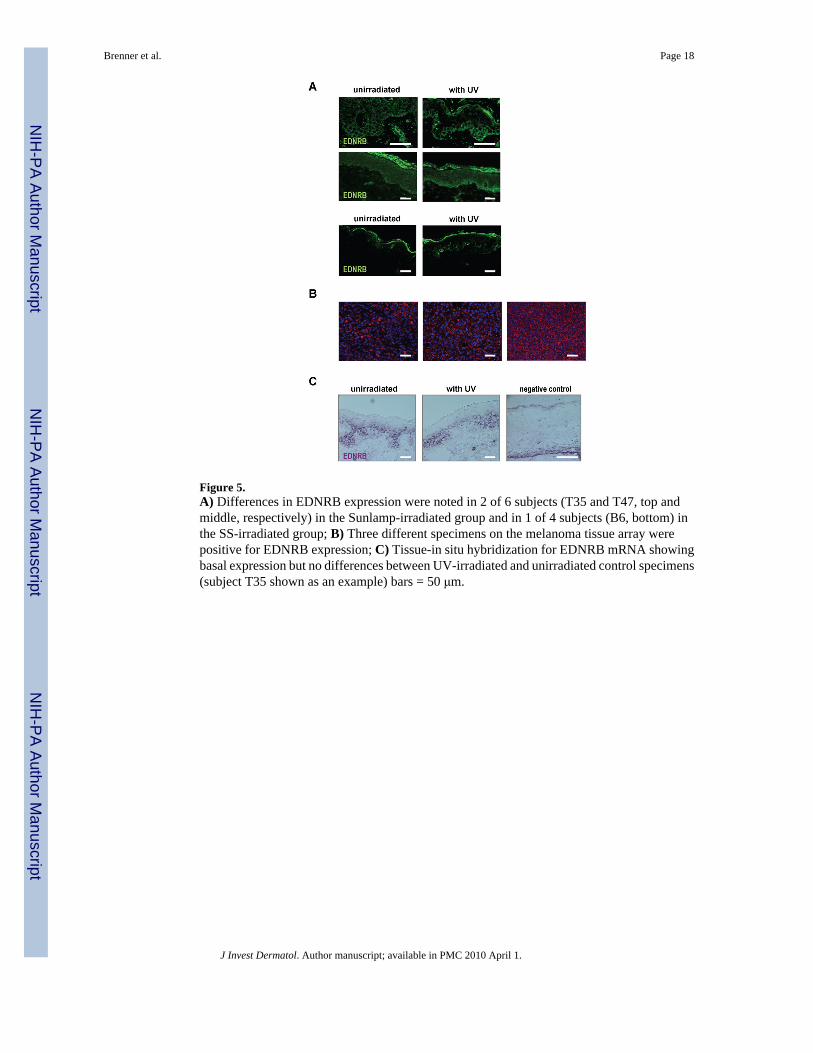
Figure 5.A) Differences in EDNRB expression were noted in 2 of 6 subjects (T35 and T47, top andmiddle, respectively) in the Sunlamp-irradiated group and in 1 of 4 subjects (B6, bottom) inthe SS-irradiated group; B) Three different specimens on the melanoma tissue array werepositive for EDNRB expression; C) Tissue-in situ hybridization for EDNRB mRNA showingbasal expression but no differences between UV-irradiated and unirradiated control specimens(subject T35 shown as an example) bars = 50 μm.
Brenner et al. Page 18
J Invest Dermatol. Author manuscript; available in PMC 2010 April 1.
NIH
-PA Author Manuscript
NIH
-PA Author Manuscript
NIH
-PA Author Manuscript

Figure 6.Examples of staining patterns in UV-irradiated specimens (right) and in unirradiated controls(left). ß-Catenin (subject B6), E-cadherin (subject B6), PCNA (subject B1), PCNA (subjectB6, double-stained for TYR in red on far right), Bcl-2 (subject T47) and DKK3 (subject B2)bars = 50 μm.
Brenner et al. Page 19
J Invest Dermatol. Author manuscript; available in PMC 2010 April 1.
NIH
-PA Author Manuscript
NIH
-PA Author Manuscript
NIH
-PA Author Manuscript

NIH
-PA Author Manuscript
NIH
-PA Author Manuscript
NIH
-PA Author Manuscript
Brenner et al. Page 20Ta
ble
1V
isib
le L
ong-
last
ing
Pigm
enta
tion
Follo
win
g U
V Ir
radi
atio
n
Gro
up 1
-Su
nlam
pD
ays b
etw
een
final
UV
Exp
osur
ean
d B
iops
y
Lon
g-la
stin
gV
isib
lePi
gmen
tatio
n
Gro
up 2
-SS
Day
s bet
wee
nfin
al U
V E
xpos
ure
and
Bio
psy
Lon
g-la
stin
gV
isib
lePi
gmen
tatio
n
T11
1310
No
B1
406
No
T16
1282
No
B2
402
No
T32
751
No
B6
402
No
T35
730
No
B8
406
No
T47
427
No
T50
520
Yes
Gro
up 1
as d
etai
led
in (M
iller
et a
l., 2
008)
Gro
up 2
as d
etai
led
in (W
olbe
r et a
l., 2
008)
J Invest Dermatol. Author manuscript; available in PMC 2010 April 1.

NIH
-PA Author Manuscript
NIH
-PA Author Manuscript
NIH
-PA Author Manuscript
Brenner et al. Page 21
Table 2Characteristics of Skin Pigmentation Following UV Irradiation
Differences in Group 1 -Sunlamp-irradiated
Group 2 -SS-irradiated
Melanin Content integrated density stain/area (mean) (control/UV)
1 / 6T11 (8.0/8.1)T16 (8.9/9.4)T32 (4.5/5.5)T35 (10.2/10.9)T47 (5.4/5.1)T50 (6.1/9.5)*
1 / 4B1 (6.2/7.1)B2 (16.3/15.1)B6 (13.0/18.2)*B8 (8.1/7.4)
Density of MITF-positive cells per mm (average) (control/UV)
0 / 6T11 (46/50)T16 (35/30)T32 (34/39)T35 (36/37)T47 (19/22)T50 (23/26)
0 / 6B1 (17/12)B2 (18/32)B6 (62/71)B8 (44/32)
Density of MART1-positive cells per mm (average) (control/UV)
0 / 6T11 (45/44)T16 (36/37)T32 (43/41)T35 (34/36)T47 (20/18)T50 (24/22)
2 / 4B1 (25/27)B2 (27/27)B6 (62/111)*
B8 (18/41)*
Density of TYR-positive cells per mm (average) (control/UV)
1 / 6T11 (39/42)T16 (31/34)T31 (41/39)T35 (35/33)T47 (22/20)T50 (16/28)*
3 / 4B1 (34/35)B2 (23/36)*
B6 (51/83)*
B8 (21/43)*
*= p <0.05
J Invest Dermatol. Author manuscript; available in PMC 2010 April 1.

NIH
-PA Author Manuscript
NIH
-PA Author Manuscript
NIH
-PA Author Manuscript
Brenner et al. Page 22
Table 3Summary of Staining Patterns of Various Markers after UV-irradiation
Group 1 -Sunlamp control
Group 2 -SS control
Ligands & Receptors
SCF + + + +
KIT ++ ++ ++ ++
bFGF + + + +
FGFR1 + + + +
ET1 ++ ++ ++ ++
EDNRB ++ (2/6) / + ± ++ (1/4) / + ±
HGF − − − −
GM-CSF * ++ * ++ * ++ * ++ *
Adhesion Molecules
β-catenin +++ +++ +++ +++
E-cadherin +++ +++ +++ +++
N-cadherin − − − −
Cell Cycle Proteins
PCNA ++ ++ +++ (B6) / ++ ++
Cyclin D1 − − − −
Cyclin E2 − − − −
Other Proteins
Bcl-2 + + + +
DKK1 − − − −
DKK3 ++ ++ ++ ++All tissue sections were read by the same investigator (MB). The Table shows an overall picture of the staining patterns with various markers after UVirradiation. Staining intensity and extent was graded as: − (no staining), + (weak staining intensity and small area stained), ++ (moderate staining intensityand middle-size area stained) or +++ (high staining intensity and large area stained). The cut-off to classify staining as positive was 1%, i.e. at least 1%of the epidermis stained positively as detected by immunohistochemistry.
*=irregular staining pattern
J Invest Dermatol. Author manuscript; available in PMC 2010 April 1.
Copyright © 2022 FDOKUMEN