Localized final solutions: Nazi extermination process(es) from ...
Upload
independentCategory
view
4download
0
Membrane association and subcellular localization of sialidase NEU3
1
Sialidase NEU3 is a peripheral membrane protein localized on the cell surface and in
endosomal structures
Gabriele ZANCHETTI*, Paolo COLOMBI*, Marta MANZONI*, Luigi ANASTASIA†, Luigi
CAIMI*, Giuseppe BORSANI*, Bruno VENERANDO†, Guido TETTAMANTI†, Augusto
PRETI*, Eugenio MONTI* and Roberto BRESCIANI*1
ADDRESSES
*Department of Biomedical Sciences and Biotechnology, School of Medicine, University of
Brescia, Viale Europa, 11, 25123 Brescia, Italy; †Department of Medical Chemistry, Biochemistry
and Biotechnology, L.I.T.A.-Segrate, School of Medicine, University of Milano, Via F.lli Cervi, 93,
20090 Segrate, Italy.
CORRESPONDING AUTHOR 1Correspondence should be addressed to: Roberto Bresciani, Department of Biomedical Sciences
and Biotechnology, School of Medicine, University of Brescia, Viale Europa, 11, 25123 Brescia,
Italy, Phone: +39-030-3717-545 (-553), Fax: +39-030-3701157, e-mail: [email protected].
KEYWORDS: sialidase NEU3, plasma membrane, gangliosides, endosomes, peripheral membrane
protein.
ABBREVIATIONS:
MmNEU3: Mus musculus membrane-associated sialidase NEU3
HsNEU3: Homo sapiens membrane-associated sialidase NEU3
4MU-NeuAc: 4-Methylumbelliferyl-N-acetyl-α-D-neuraminic acid
[3H]GD1a: isotopically tritium-labeled ganglioside GD1a at position 3 of the sphingosine moiety
GPI: glycosyl-phosphatidyl-inositol
Stag
e 2(
a) P
OST
-PR
INT
Biochemical Journal Immediate Publication. Published on 21 Aug 2007 as manuscript BJ20070503
TH
IS IS
NO
T T
HE
FIN
AL
VE
RS
ION
- s
ee d
oi:1
0.10
42/B
J200
7050
3
Licenced copy. Copying is not permitted, except with prior permission and as allowed by law.
© 2007 The Authors Journal compilation © 2007 Biochemical Society
Membrane association and subcellular localization of sialidase NEU3
2
ABSTRACT
Sialidase NEU3 is also known as the plasma membrane-associated form of mammalian sialidases,
exhibiting a high substrate specificity toward gangliosides. In this respect, sialidase NEU3
modulates cell surface biological events and plays a pivotal role in different cellular processes,
including cell adhesion, recognition and differentiation. At the moment, no detailed studies
concerning the subcellular localization of NEU3 are available and the mechanism of its association
to cellular membranes is still unknown. Here we demonstrate that sialidase NEU3, besides its
localization at the plasma membrane, is present in intracellular structures at least partially
represented by a subset of the endosomal compartment. Moreover, we show that NEU3 present at
the plasma membrane is internalized and locates then to the recycling endosomal compartment. The
enzyme is associated to the outer leaflet of the plasma membrane, as shown by selective cell surface
protein biotinylation. This evidence is in agreement with the ability of NEU3 in degrading
gangliosides inserted in the plasma membrane of adjacent cells. Moreover, the association
mechanism of the protein to the lipid bilayer was elucidated by carbonate extraction. Under these
experimental conditions we succeeded in solubilizing NEU3, thus demonstrating that the enzyme is
a peripheral membrane protein. In addition, Triton X-114 phase separation further demonstrates the
hydrophilic nature of the protein. Overall, these results provide important information about the
biology of NEU3, the most studied member of the mammalian sialidase family.
Stag
e 2(
a) P
OST
-PR
INT
Biochemical Journal Immediate Publication. Published on 21 Aug 2007 as manuscript BJ20070503
TH
IS IS
NO
T T
HE
FIN
AL
VE
RS
ION
- s
ee d
oi:1
0.10
42/B
J200
7050
3
Licenced copy. Copying is not permitted, except with prior permission and as allowed by law.
© 2007 The Authors Journal compilation © 2007 Biochemical Society

Membrane association and subcellular localization of sialidase NEU3
3
INTRODUCTION
Sialidases or neuraminidases (EC 3.2.1.18) are glycohydrolytic enzymes that remove sialic acid
residues from sialoglycoconjugates, such as gangliosides, sialoglycoproteins and
sialooligosaccharides. They are widely distributed in nature, from viruses and bacteria to vertebrates
[1]. In mammals, four different forms of sialidases have been cloned so far and their classification is
mainly based on their subcellular distribution: the lysosomal NEU1, the cytosolic NEU2, the
plasma membrane-associated NEU3 and the lysosomal/mitochondrial NEU4 [2]. Among them, the
plasma membrane-associated sialidase NEU3 has been shown to be preferentially active toward
gangliosidic substrates [3-8]. Moreover, NEU3 is present within lipid rafts [9, 10] that represent
specialized functional areas of the membranes, highly enriched in cholesterol and sphingolipids [11,
12]. Sphingolipids, and among them gangliosides, are known to affect different biological events
including tumorigenic transformation [13-18], cell differentiation [19, 20] and motility [21-25]. The
evidence that the over-expression of sialidase NEU3 induces dramatic changes in the cellular
ganglioside pattern [6] is of particular interest because of the relevance of these amphiphilic
molecules in mediating important cellular behaviours [26]. Indeed, sialidase NEU3 has been
reported to play a pivotal role in different normal cellular processes including axonal growth and
regeneration [27], and tumorigenic transformation [28]. Importantly, the activity of sialidase NEU3
is exerted also on gangliosides exposed on the extracellular leaflet of the plasma membrane of
adjacent cells by cell-to-cell interaction [6]. This evidence has suggested the hypothesis that the
enzyme is located at the external leaflet of the plasma membrane, facing the extracellular
environment, thus allowing its interaction with the gangliosidic substrates exposed on the plasma
membrane of neighbouring cells.
Detailed studies concerning the membrane anchoring of sialidase NEU3 have not been reported so
far. Analysis of NEU3 aminoacid sequence reveals a striking similarity with the soluble counterpart
NEU2. Indeed, based on the crystal structure of human NEU2 [29] and on the aminoacid sequence
homology, a modeling of human NEU3 was performed, suggesting a common β-propeller folding
for both enzymes [30]. The possible presence of canonical aminoacid motif(s) necessary for post-
translational modification(s) potentially involved in NEU3 membrane anchoring can be explored by
bioinformatic analysis [31-33]. In order to gain more information about the subcellular distribution
of sialidase NEU3 and its mechanism of association to the lipid bilayer, we expressed the mouse
protein (MmNEU3) in HeLa and COS-7 cells. We found that expressed sialidase NEU3 is present
both at the plasma membrane and in intracellular tubulo-vesicular structures that represent a subset
of the endosomal compartment. In addition, experiments of cell surface protein biotinylation and Stag
e 2(
a) P
OST
-PR
INT
Biochemical Journal Immediate Publication. Published on 21 Aug 2007 as manuscript BJ20070503
TH
IS IS
NO
T T
HE
FIN
AL
VE
RS
ION
- s
ee d
oi:1
0.10
42/B
J200
7050
3
Licenced copy. Copying is not permitted, except with prior permission and as allowed by law.
© 2007 The Authors Journal compilation © 2007 Biochemical Society
Membrane association and subcellular localization of sialidase NEU3
4
indirect immunofluorescence gave the first direct evidence that sialidase NEU3 is associated to the
external leaflet of the plasma membrane. Finally, using different extraction and solubilization
methods, we provide evidences that sialidase NEU3 has hydrophilic characteristics and behaves as a
peripheral membrane protein.
Stag
e 2(
a) P
OST
-PR
INT
Biochemical Journal Immediate Publication. Published on 21 Aug 2007 as manuscript BJ20070503
TH
IS IS
NO
T T
HE
FIN
AL
VE
RS
ION
- s
ee d
oi:1
0.10
42/B
J200
7050
3
Licenced copy. Copying is not permitted, except with prior permission and as allowed by law.
© 2007 The Authors Journal compilation © 2007 Biochemical Society
Membrane association and subcellular localization of sialidase NEU3
5
EXPERIMENTAL
Cell culture and transfection
COS-7 and HeLa cells were cultured in Dulbecco’s Modified Eagle’s Medium (DMEM) containing
4 mM L-glutamine, 100 U/ml penicillin, 100 µg/ml streptomycin and 10% (v/v) fetal bovine serum
and maintained at 37°C and 5% CO2 in a humidified incubator. Cells were transiently transfected
with a C-terminal HA-tagged form of mouse NEU3 (MmNEU3-HA), subcloned into the
pcDNA1/Amp (Invitrogen) vector. Transfection was performed in serum-free medium (OptiMEM;
Gibco-BRL) employing FuGENE 6 (Roche) and incubating the cells at 37°C for 6 h. After
transfection, cells were grown in DMEM for 36 h and then processed.
Antibodies
For indirect immunofluorescence and immunoblotting experiments the following primary
antibodies were used: rabbit anti-HA (Sigma), mouse anti-EEA1 (Transduction Laboratories),
mouse anti-TfR (Zymed), mouse anti-PDI (Stressgene), mouse anti-LBPA (from Dr. J. Gruenberg,
Department of Biochemistry, University of Geneva, Switzerland) and rabbit anti-caveolin-1 (Santa
Cruz). Detection of the human NEU3 in immunoblotting experiments was achieved using a rabbit
anti-human NEU3, (from Dr. N. Stamatos, Institute of Human Virology, University of Maryland,
Baltimore, U.S.A.). For immunofluorescence experiments, donkey anti-rabbit Alexa 488 and
donkey anti-mouse Alexa 555 (Molecular Probes) secondary antibodies were used. For
immunoblotting experiments, donkey anti-rabbit and sheep anti-mouse HRP-conjugated secondary
antibodies (GE Healthcare) were used.
Indirect immunofluorescence and confocal microscopy analysis
Indirect immunofluorescence experiments were performed as already described with minor
modifications [6]. COS-7 or HeLa cells were seeded onto glass coverslips and transfected with
MmNEU3-HA. At 36 h post-transfection cells were fixed with 3% (w/v) paraformaldehyde in PBS
for 15 min at room temperature. Paraformaldehyde was then quenched incubating the samples with
50 mM NH4Cl in PBS for 15 min. After three washes with PBS, cells were permeabilized with
0.5% saponin in PBS (PBS/Sap) for 30 min and incubated with primary antibodies diluted in
PBS/Sap for 1 h. Subsequently, cells were washed three times with PBS/Sap and incubated with
secondary antibodies diluted in PBS/Sap for the same period. Finally, after three washes with
PBS/Sap followed by washes with PBS, specimens were mounted using DakoCytomation
Fluorescent Mounting Medium (DakoCytomation) and analyzed with confocal system LSM-510 Stag
e 2(
a) P
OST
-PR
INT
Biochemical Journal Immediate Publication. Published on 21 Aug 2007 as manuscript BJ20070503
TH
IS IS
NO
T T
HE
FIN
AL
VE
RS
ION
- s
ee d
oi:1
0.10
42/B
J200
7050
3
Licenced copy. Copying is not permitted, except with prior permission and as allowed by law.
© 2007 The Authors Journal compilation © 2007 Biochemical Society
Membrane association and subcellular localization of sialidase NEU3
6
META (Carl Zeiss). For indirect immunofluorescence of non permeabilized cells, the same
procedure was used substituting 0.5% saponin with 1% BSA. Images were processed with LSM
Image Browser (Carl Zeiss) and Adobe Photoshop software.
Antibody uptake
COS-7 cells seeded onto glass coverslips were transfected with MmNEU3-HA as above. After 36 h
of transfection, cells were incubated for 3 h at 37°C in growth medium supplemented with rabbit
anti-HA antibody at 2.5 µg/ml final concentration. Cells were then chilled on ice, washed three
times with ice-cold PBS and fixed with methanol. After saturation with 1% BSA in PBS
(BSA/PBS), specimens were incubated with mouse anti-TfR antibody diluted in BSA/PBS for 1 h.
After extensive washes with BSA/PBS, donkey anti-rabbit Alexa 488 and donkey anti-mouse Alexa
555 secondary antibodies were used. Specimens were then mounted and analyzed as above.
Immunoblotting
Proteins were separated by 12% SDS-PAGE and transferred to a PVDF membrane (Hybond-P;
Amersham Biosciences). Membranes were then blocked with 5% (w/v) not-fat dry milk in PBS,
washed three times with PBS containing 0.1% (v/v) Tween 20 (PBST) and incubated with primary
antibody diluted in PBST containing 1% (w/v) milk for 2 h at room temperature. After three washes
with PBST, membranes were incubated with HRP-conjugated secondary antibody diluted in PBST
for 1 h at room temperature. Detection of the immunocomplexes was performed by enhanced
chemiluminescent-based system (SuperSignal West Pico Chemiluminescent Substrate; Pierce),
followed by densitometric analysis using GelPro 3.1 software (Media Cybernatics).
Cell surface protein biotinylation and purification
For biotinylation experiments all steps were carried out at 4°C. Surface proteins of subconfluent
cultured COS-7 and HeLa cells expressing MmNEU3-HA for 36 h were labeled with 0.5 mg/ml of
membrane-impermeable sulfosuccinimidyl-2-(biotinamido)ethyl-1,3-dithiopropionate (Sulfo-NHS-
SS-biotin; Pierce) in PBS for 30 min with gentle agitation, as indicated by the manufacturer’s
instructions. Cells were then washed three times with PBS and free biotin was quenched by one
wash with 25 mM Tris-HCl pH 8.0, followed by three washes with PBS. Cells were then scraped in
PBS and then lysed in RIPA buffer (25 mM Tris-HCl pH 7.4, 0.15 M NaCl, 0.1% SDS, 1% Triton
X-100, 1% sodium deoxycholate) by sonication. After lysis, total cell extracts were clarified by
centrifugation at 800 x g for 10 min and biotinylated proteins were separated from non-biotinylated Stag
e 2(
a) P
OST
-PR
INT
Biochemical Journal Immediate Publication. Published on 21 Aug 2007 as manuscript BJ20070503
TH
IS IS
NO
T T
HE
FIN
AL
VE
RS
ION
- s
ee d
oi:1
0.10
42/B
J200
7050
3
Licenced copy. Copying is not permitted, except with prior permission and as allowed by law.
© 2007 The Authors Journal compilation © 2007 Biochemical Society
Membrane association and subcellular localization of sialidase NEU3
7
proteins using Immobilized Monomeric Avidin resin (Pierce). Fractions were adjusted to the same
final volume and analyzed by immunoblotting.
Bioinformatic analysis
Analysis of the protein motifs involved in post-translational modifications was performed using the
aminoacid sequence of MmNEU3 (accession number Q9JMH7) [34] and HsNEU3 (accession
number Q9UQ49) [5] with public domain software. Prediction of GPI-anchor motifs was performed
using the following softwares: big-PI Predictor - GPI Modification Site Prediction:
http://mendel.imp.ac.at/sat/gpi/gpi_server.html, DGPI - Prediction of GPI-anchor and cleavage
sites: http://129.194.185.165/dgpi/index_en.html, GPI-SOM - Identification of GPI-anchor signals
by a Kohonen Self Organizing Map: http://gpi.unibe.ch/. Prediction of fatty acyl- or prenyl-anchor
motifs was performed with the following softwares: Myristoylator Prediction of N-terminal
myristoylation by neural networks: http://www.expasy.org/tools/myristoylator/, NMT - Prediction
of N-terminal N-myristoylation: http://mendel.imp.ac.at/myristate/SUPLpredictor.htm, PrePS -
Prenylation Prediction Suite: http://mendel.imp.ac.at/sat/PrePS/index.html. For the prediction of
transmembrane sequences the following softwares were used: HMMTOP - Prediction of
transmembrane helices and topology of proteins: http://www.enzim.hu/hmmtop/, SOSUI -
Prediction of transmembrane regions: http://sosui.proteome.bio.tuat.ac.jp/sosuiframe0.html,
TMHMM - Prediction of transmembrane helices in proteins:
http://www.cbs.dtu.dk/services/TMHMM-2.0/, TMpred - Prediction of transmembrane regions and
protein orientation: http://www.ch.embnet.org/software/TMPRED_form.html. Finally, we analyzed
the aminoacid sequences of MmNEU3 with AmphipaSeeK (http://npsa-pbil.ibcp.fr/cgi-
bin/npsa_automat.pl?page=/NPSA/npsa_amphipaseek.html), a monotopic membrane protein
prediction tool.
Membrane protein extraction
HeLa or COS-7 cells 36 h after transfection with MmNEU3-HA were broken by bundle sonication
in ice-cold lysis buffer (10 mM Tris-HCl pH 7.5 containing 0.032 µg/ml aprotinin, 0.032 µg/ml
leupeptin, 0.008 µg/ml pepstatin A, 0.06 µg/ml chymostatin and 0.2 mM PMSF as protease
inhibitors). After cell lysis, samples were clarified by centrifugation at 800 x g for 10 min at 4°C
and the resulting supernatant (total cell extract) was centrifuged at 100,000 x g for 1 h at 4°C in
order to obtain a total cell membranes fraction. The resulting pellet was washed once with ice-cold
distilled water, resuspended in the lysis buffer and split into identical aliquots. Extraction of
peripheral proteins was performed by the exposure of total cell membranes either to pH 11.5 or 1 M
Stag
e 2(
a) P
OST
-PR
INT
Biochemical Journal Immediate Publication. Published on 21 Aug 2007 as manuscript BJ20070503
TH
IS IS
NO
T T
HE
FIN
AL
VE
RS
ION
- s
ee d
oi:1
0.10
42/B
J200
7050
3
Licenced copy. Copying is not permitted, except with prior permission and as allowed by law.
© 2007 The Authors Journal compilation © 2007 Biochemical Society
Membrane association and subcellular localization of sialidase NEU3
8
KCl or 0.025 M EGTA and incubated on ice for 30 min. The above extraction conditions were
achieved by the addition to the samples of an equal volume of 0.2 M Na2CO3 pH 12 or 2 M KCl or
0.05 M EGTA, respectively. As control sample, membranes were incubated in presence of lysis
buffer alone and processed as above. Finally, solubilized and non-extractable proteins were
separated by centrifugation at 100,000 x g and carbonate-containing samples were immediately
neutralized to pH 7.5 by the addition of acetic acid. Soluble and membrane fractions were then
adjusted to the same final volume before proteins repartition analysis by sialidase activity and/or
immunoblotting assay.
Triton X-114 phase separation
Triton X-114 phase separation was performed as described [35]. Briefly, at 36 h post-transfection
with MmNEU3-HA, HeLa cells were chilled at 4°C. After three washes with cold PBS, cells were
scraped and pelleted at 800 x g for 10 min. Cells were then resuspended in 10 mM Tris-HCl pH 7.4,
lyzed by sonication and the resulting total cell extracts were diluted to a protein concentration
corresponding to 1.0 mg/ml in 0.1 ml in the same buffer. Proteins were extracted by adding 0.1 ml
of 2% (v/v) precondensed Triton X-114 (Sigma) and incubating the sample for 1 h on ice.
Detergent-extracted samples (200 µl) were then layered onto 6% (w/v) sucrose cushion (300 µl),
incubated at 30°C for 3 min and finally centrifuged at 300 x g for 3 min. After centrifugation, the
upper aqueous phase was removed, re-extracted with 1% Triton X-114 and subjected to a second
separation through the same sucrose cushion. The detergent and aqueous phases were adjusted to
the same final volume and MmNEU3-HA repartition, together with the endogenous protein
markers, was analyzed by immunoblotting.
Sialidase activity assay
The enzymatic activity of MmNEU3-HA was determined as previously described [6] using
radioactive GD1a ganglioside ([3H]GD1a), prepared according to Ghidoni et al. [36] (specific
radioactivity, 1.2 Ci/mmol; homogeneity > 99%), and 4MU-NeuAc (Sigma) as substrates, in 100
mM sodium-citrate/phosphate buffer and performed at pH 3.8. Reaction mixtures were adjusted to
the appropriate final concentrations of buffered-carbonate or Triton X-114 before the addition of the
enzymatic source. Samples were incubated at 37°C for 30 min. For NEU3 enzymatic stability at
alkaline pH, total membranes were incubated at pH 11.5 for different time periods (up to 30 min),
neutralized to pH 7.5 and assayed as above without ultracentrifugation. NEU3 activity related to
Triton X-114 fractions was measured both in the single aqueous and detergent phases, as well as in
the pooled phases. Stag
e 2(
a) P
OST
-PR
INT
Biochemical Journal Immediate Publication. Published on 21 Aug 2007 as manuscript BJ20070503
TH
IS IS
NO
T T
HE
FIN
AL
VE
RS
ION
- s
ee d
oi:1
0.10
42/B
J200
7050
3
Licenced copy. Copying is not permitted, except with prior permission and as allowed by law.
© 2007 The Authors Journal compilation © 2007 Biochemical Society
Membrane association and subcellular localization of sialidase NEU3
9
RESULTS
Subcellular localization of sialidase NEU3.
In order to study the subcellular distribution and localization of sialidase NEU3, the chimeric form
of the mouse protein (MmNEU3) bearing the haemagglutinin epitope (HA) at the C-terminus
(MmNEU3-HA) was transiently expressed in both HeLa and COS-7 cells. The expression of this
construct gives rise to a fusion protein that is catalytically active and has already been extensively
used in biochemical and functional studies [6-8, 10]. After 36 h expression, cells were fixed,
permeabilized with saponin and MmNEU3-HA subsequently detected using anti-HA antibodies.
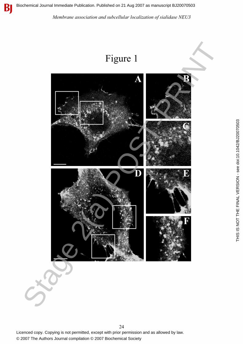
Laser confocal microscopy analysis clearly shows that, irrespective to the cell type considered
(Figures 1A and 1D), MmNEU3-HA localizes both at the plasma membrane (Figures 1B and 1E)
and in intracellular vesicular structures that concentrate in the juxtanuclear region of the cells
(Figures 1C and 1F). While the presence of NEU3 at the plasma membrane was already
demonstrated [3, 5-7], an intracellular localization of the enzyme was only once reported without
giving any information about the precise nature of this intracellular labelling [8].
In order to better characterize the intracellular distribution of NEU3, co-localization experiments
were carried out using markers of different cellular compartments. When MmNEU3-HA
distribution was related either to the endoplasmic reticulum marker protein disulfide isomerase
(PDI), as well as to the Golgi complex marker GM-130, no co-localization was observed (data not
shown). Instead, co-localization between MmNEU3-HA and markers of the endosomal
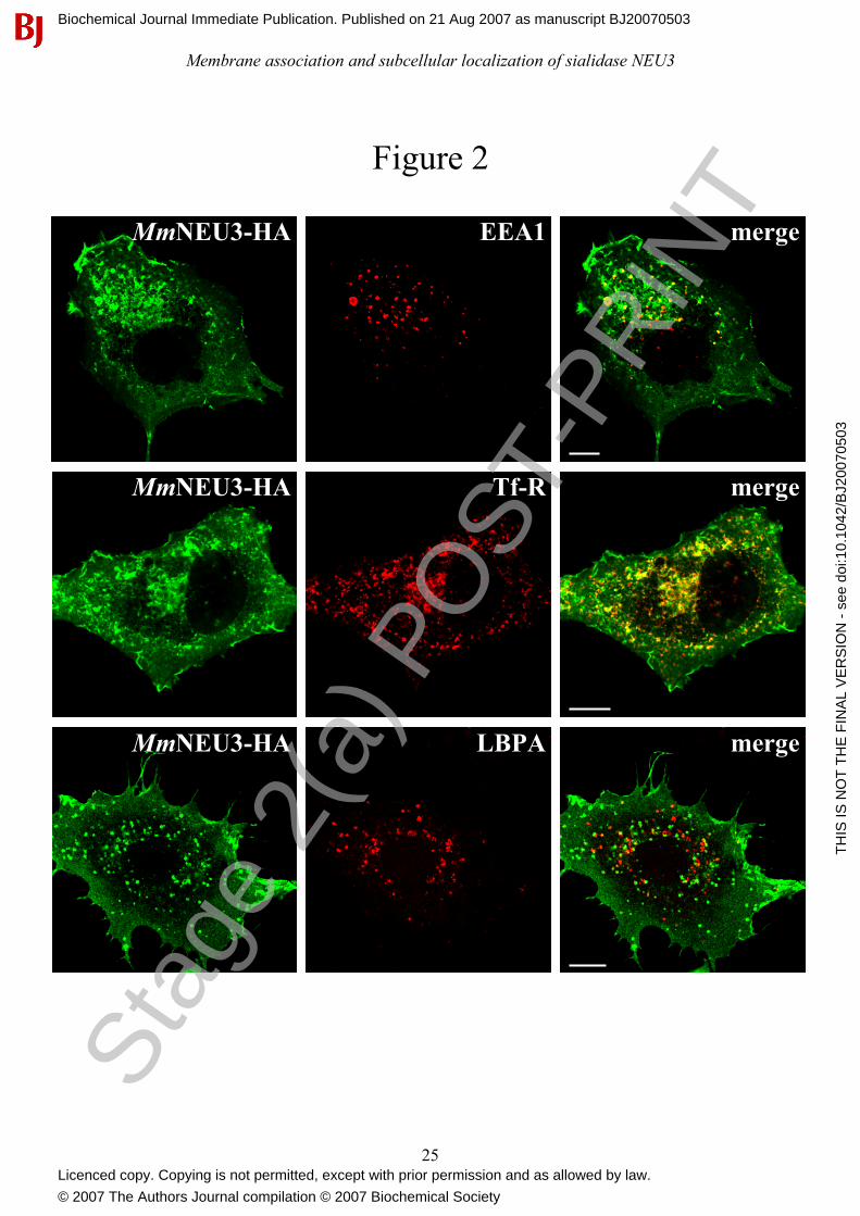
compartment was observed. As shown in Figure 2, COS-7 cells showed intracellular vesicular
structures labelled for both MmNEU3-HA and the early endosome antigen 1 (EEA1), a marker of
the early-endosomes (Figure 2, upper row). Co-localization of MmNEU3-HA is even more striking
with the juxtanuclear transferrin receptor (Tf-R) positive structures, representing the recycling-
endosomal compartment (Figure 2, middle row). Interestingly, MmNEU3-HA does not colocalize
with the late-endosomes markers lyso-bisphosphatidic acid (LBPA) (Figure 2, bottom row), as well
as with the lysosomal associated membrane protein-1 (LAMP-1) (data not shown). Taken together
these results indicate a restrict localization of sialidase NEU3 to a subset of the endosomal
compartment. Moreover, the protein does not follow the degradative lysosomal pathway as
demonstrated by the lack of co-localization with both LBPA and LAMP-1. A similar intracellular
distribution and co-localization pattern was observed also in HeLa cells (data not shown). Overall,
we can conclude that MmNEU3-HA, besides its localization at the plasma membrane, is present
also in intracellular membranous structures that are mainly represented by the early- and recycling-
endosomes. Sta
ge 2
(a) P
OST
-PR
INT
Biochemical Journal Immediate Publication. Published on 21 Aug 2007 as manuscript BJ20070503
TH
IS IS
NO
T T
HE
FIN
AL
VE
RS
ION
- s
ee d
oi:1
0.10
42/B
J200
7050
3
Licenced copy. Copying is not permitted, except with prior permission and as allowed by law.
© 2007 The Authors Journal compilation © 2007 Biochemical Society
Membrane association and subcellular localization of sialidase NEU3
10
Sialidase NEU3 is associated to the external leaflet of the plasma membrane.
In order to confirm the presence of sialidase NEU3 both at the plasma membrane and in
intracellular compartments, COS-7 cells expressing MmNEU3-HA were subjected to cell surface
protein biotinylation at 4°C, and labelled proteins were isolated by affinity chromatography using
an avidin resin. Distribution of MmNEU3-HA between avidin-unbound and avidin-bound fractions
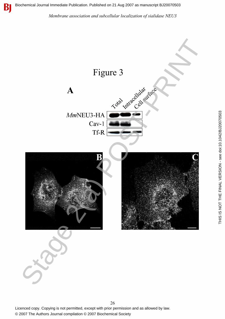
was then analyzed by Western-blot. As shown in Figure 3(A), MmNEU3-HA was found both in the
non-biotinylated and in the biotinylated fractions, confirming that the protein is present both in
intracellular compartments and at the cell surface. Densitometric analysis of the immunoblotting
indicates that 23.8% ± 5.4 (n= 3) of MmNEU3-HA localizes to the cell surface while the remaining
portion of the protein is localized intracellularly. We also analyzed the distribution of two other
proteins, i.e. Caveolin-1 (Cav-1), a protein known to be associated to the internal leaflet of the
plasma membrane [37, 38], and Tf-R, a transmembrane protein that cycles between the plasma
membrane and intracellular compartments [39]. As shown in Figure 3(A), in the same cell extracts
used for the detection and distribution of MmNEU3-HA, Cav-1 was detected only in the non-
biotinylated fraction, as expected, confirming that under these experimental conditions
intracellularly localized proteins are not accessible to biotin. Conversely, Tf-R was detected in both
the biotinylated and non-biotinylated fractions, as expected. Superimposable results were obtained
also using HeLa cells (data not shown). Based on this evidence we can reasonably assume that the
portion of MmNEU3-HA that is bound to the plasma membrane is accessible to biotinylation, hence
exposed to the extracellular environment. In other words, the enzyme is associated to the external
leaflet of the plasma membrane. In order to confirm this finding, non permeabilized COS-7 and
HeLa cells expressing MmNEU3-HA were processed for indirect immunofluorescence using, at the
same time, anti-HA antibodies and, as control, anti-PDI antibodies. Under these experimental
conditions no labelling for the intracellular endoplasmic reticulum marker PDI was observed at all
(data not shown), demonstrating the integrity of the plasma membrane, while a specific labelling for
MmNEU3-HA was detected (Figures 3B and 3C). This result further strenghtens the notion that
sialidase NEU3 is associated to the external leaflet of the plasma membrane.
NEU3 is internalized from the cell surface to the recycling compartment.
As described above, we could demonstrate that sialidase NEU3 localizes to the external leaflet of
the plasma membrane and in the endosomal compartment. We then tried to directly assess the
propensity of the protein to move from the surface to the cell interior. For this purpose, cultured
COS-7 cells transfected with MmNEU3-HA were incubated for 3 h in presence of anti-HA Stag
e 2(
a) P
OST
-PR
INT
Biochemical Journal Immediate Publication. Published on 21 Aug 2007 as manuscript BJ20070503
TH
IS IS
NO
T T
HE
FIN
AL
VE
RS
ION
- s
ee d
oi:1
0.10
42/B
J200
7050
3
Licenced copy. Copying is not permitted, except with prior permission and as allowed by law.
© 2007 The Authors Journal compilation © 2007 Biochemical Society

Membrane association and subcellular localization of sialidase NEU3
11
antibody. Cells were then fixed, permeabilazed and the distribution of the MmNEU3-HA/anti-HA
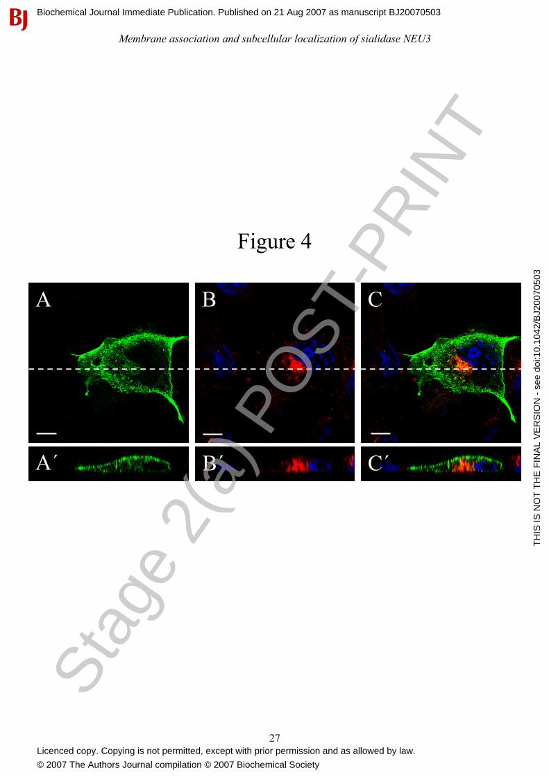
antibody immuno-complexes was analyzed and compared to Tf-R distribution. As shown in Figure
4(A), in transfected cells a significant labelling for NEU3 could be detected at the plasma
membrane but also in vesicular structures that are supposed to be located intracellularly. Co-
localization between the anti-HA antibody signal and Tf-R (Figure 4B) is observed in the
juxtanuclear region of the cell (Figure 4C), suggesting that the MmNEU3-HA/anti-HA antibody
immunocomplexes were internalized from the cell surface and localize to the recycling endosomal
compartment. The evidence that the anti-HA antibody is internalized and localizes intracellularly is
given by the direct vertical confocal sections shown in Figure 4(A’ – C’). Indeed, a significant
labelling for MmNEU3-HA both at the surface and in intracellular structures that concentrate at the
juxtanuclear region of the cell is evidenced. Notably, these structures co-localize with the
intracellular labelling for Tf-R. Taken together these results clearly indicate that NEU3 internalizes
from the plasma membrane and that internalized molecules reach the recycling endosomal
compartment.
Sialidase NEU3 is a peripheral membrane-associated protein.
The aminoacid sequence of MmNEU3 was analyzed for the prediction of trasmembrane domain(s)
that would anchor the protein to the lipid bilayer. Only TMpred program (see Experimental)
predicts the presence of a single hydrophobic aminoacid stretch, spanning between residues A178
and S203. If we assume this stretch as a transmembrane portion of the enzyme, the protein would
additionally feature an intracellular and an extracellular domain. This structural model is unlikely
because, in this configuration, putative aminoacid residues essential for the catalytic activity would
be located at opposite sides of the membrane, that would be clearly inconsistent with catalytic
activity [2]. Moreover, based on the crystal stucture of NEU2 [29] and considering the high
homology between the two proteins, a common β-propeller fold for the human NEU3 was recently
suggested [30]. Bioinformatic analysis of MmNEU3 aminoacid sequence excludes also the
possibility that the protein may associate to membranes as a monotopic protein [40]. Finally,
sequence analysis does not reveal the presence of any aminoacid motifs for post-translational
modifications that would anchor the protein to the lipid bilayer, such as GPI-, fatty acyl- or prenyl-
linkages. On this basis, we took into consideration the possibility that sialidase NEU3 would
associate to cellular membranes as a peripheral protein. For this purpose, total cell membranes
obtained from HeLa and COS-7 cells expressing MmNEU3-HA were incubated for 30 min in
presence of 0.1 M sodium carbonate at pH 11.5 and then soluble and membrane fractions were
obtained by ultracentrifugation [41-43]. The resulting fractions were then adjusted to pH 7.5 and Stag
e 2(
a) P
OST
-PR
INT
Biochemical Journal Immediate Publication. Published on 21 Aug 2007 as manuscript BJ20070503
TH
IS IS
NO
T T
HE
FIN
AL
VE
RS
ION
- s
ee d
oi:1
0.10
42/B
J200
7050
3
Licenced copy. Copying is not permitted, except with prior permission and as allowed by law.
© 2007 The Authors Journal compilation © 2007 Biochemical Society
Membrane association and subcellular localization of sialidase NEU3
12
analyzed for their enzyme content by Western-blot and sialidase activity. As control, total cell
membranes were incubated for the same time period in 0.01 M Tris/HCl pH 7.5 and processed as
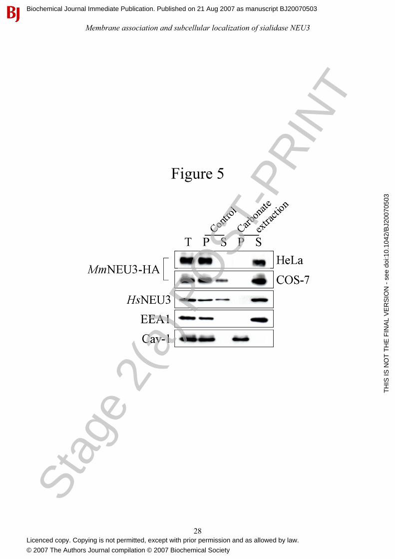
above. As expected, incubation of membranes under control conditions, had no significant effect on
the association of MmNEU3-HA to the membranes (Figure 5) and the sialidase activity was almost
completely recovered in the particulate fraction (82.5-87.3%, depending on the substrate used) (see
Table 1). The small amount of MmNEU3-HA activity in the soluble fraction may derive by a
spontaneous release of the protein, as a consequence of membrane manipulation. On the contrary, a
remarkable, almost complete, solubilization of MmNEU3-HA was achieved when membranes were
incubated under alkaline conditions, irrespective to the cell line taken in consideration (Figure 5).
Under these experimental conditions, the peripheral membrane protein EEA1 was completely
recovered in the soluble fraction, as expected [44]. Noteworthy, carbonate treatment preserved the
lipid structure without affecting the association of integral proteins to the lipid bilayer, as confirmed
by the analysis of the repartition of Cav-1 in the same cell extracts. In fact, Cav-1 remained always
associated to the membranous fractions.
In order to investigate whether alkaline extraction of MmNEU3-HA could be somehow ascribed to
the over-expression of the heterologous protein, solubilization of the human endogenous protein
HsNEU3 was analyzed in non transfected (Mock) HeLa cells. For this purpose, we took advantage
of a rabbit anti-HsNEU3 antiserum that specifically recognizes the human protein in
immunoblotting experiments [45]. The endogenous enzyme showed a roughly superimposable
behaviour compared to MmNEU3-HA, with a complete extraction of the protein only after alkaline
treatment (Figure 5). However, using both the artificial 4MU-NeuAc and the [3H]GD1a ganglioside
as substrates, almost no sialidase activity could be detected in the particulate and in the soluble
fractions after 30 min carbonate treatment, although samples were adjusted to pH 7.5 immediately
after ultracentrifugation (Table 1). Indeed, the enzymatic activity measured after carbonate
extraction was markedly lower compared to control experimental conditions (3-7%, when assayed
with [3H]GD1a). Loss of activity might be ascribed either to the presence of carbonate in the
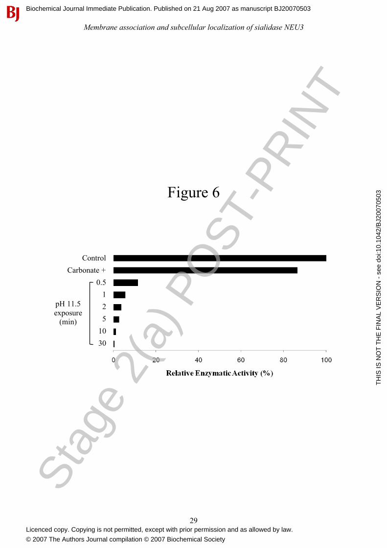
enzyme assay mixture or to exposure of the protein to the alkaline pH. We therefore determined the
sialidase activity of total membranes by adding buffered-carbonate (see Experimental), at the
appropriate final concentration, to the enzymatic assay mixture before the addition of the enzyme
source. A small reduction in sialidase activity was observed as compared to control conditions
(Figure 6), indicating that the presence of carbonate per se in the reaction mixture is compatible
with the catalytic activity. We then subjected total membranes to alkaline pH treatment for different
time intervals and, after adjustment to pH 7.5, we tested the sialidase activity. As shown in Figure
6, already after 1 min exposure to alkaline pH, more than 90% of the enzymatic activity was lost. Stag
e 2(
a) P
OST
-PR
INT
Biochemical Journal Immediate Publication. Published on 21 Aug 2007 as manuscript BJ20070503
TH
IS IS
NO
T T
HE
FIN
AL
VE
RS
ION
- s
ee d
oi:1
0.10
42/B
J200
7050
3
Licenced copy. Copying is not permitted, except with prior permission and as allowed by law.
© 2007 The Authors Journal compilation © 2007 Biochemical Society
Membrane association and subcellular localization of sialidase NEU3
13
By considering that the enzymatic activity was determined without separation of the soluble and
membranous fractions, we can conclude that loss of catalytic activity can be ascribed to exposure to
alkaline pH. To gain further information about the mechanism of NEU3 membrane anchoring,
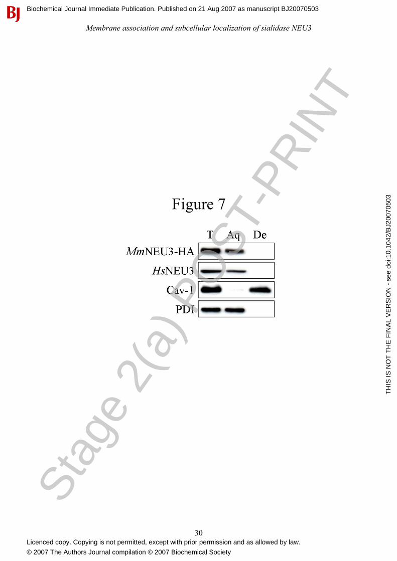
HeLa cells expressing MmNEU3-HA were subjected to Triton X-114 extraction followed by
aqueous/detergent phase separation, and the distribution of the protein was analyzed by Western-
blot. These experimental conditions allow the separation of hydrophylic peripheral membrane
proteins from transmembrane, GPI-anchored and fatty acyl- or prenyl-anchored membrane proteins
[35, 46]. After phase separation, we observed a significant enrichment or a complete segregation of
MmNEU3-HA in the aqueous phase (Figure 7). The same results were obtained for the endogenous
HsNEU3 in HeLa cells. On the other hand, Cav-1 that is associated to membranes by a hydrophobic
domain and palmitoylations [38], segregates exclusively in the detergent phase. As in the case of
carbonate extraction, we were not able to recover any activity either in the single or in the
reconstituted fractions after Triton X-114 phase separation, regardless of the substrate used in the
assay. In addition to these findings, treatment of membranes derived from transfected cells with
high ionic strength (1 M KCl) or with divalent cation chelator (25 mM EGTA) did not result in any
solubilization of sialidase NEU3 (data not shown).
Overall, these findings demonstrate that: (i) NEU3 behaves as a hydrophilic peripheral protein; (ii)
its biological activity is highly sensitive to alkaline pH; (iii) the interaction of the protein with the
membrane does not seem to depend either on direct or calcium-mediated electrostatic interactions
with phospholipids.
Stag
e 2(
a) P
OST
-PR
INT
Biochemical Journal Immediate Publication. Published on 21 Aug 2007 as manuscript BJ20070503
TH
IS IS
NO
T T
HE
FIN
AL
VE
RS
ION
- s
ee d
oi:1
0.10
42/B
J200
7050
3
Licenced copy. Copying is not permitted, except with prior permission and as allowed by law.
© 2007 The Authors Journal compilation © 2007 Biochemical Society
Membrane association and subcellular localization of sialidase NEU3
14
DISCUSSION
The plasma membrane-associated sialidase NEU3 is known to be highly active toward gangliosidic
substrates [3, 5, 6, 34] and to be involved in several biological processes, possibly through the
modulation of sialic acid content at the cell surface. Sialidase NEU3 is present in lipid rafts and co-
fractionates with the lipid raft marker Cav-1 [9, 10]. These organized subsets of the membrane
represent specialized areas where cell-to-cell interactions and signal transduction events take place
[13, 26]. Up-regulation of sialidase NEU3 has been correlated to apoptosis suppression in human
colon carcinoma [46], in renal carcinoma [25], as well as in other tumor cell lines [28]. In addition,
it has been recently demonstrated that silencing of NEU3 results in the activation of apoptosis
mechanisms in tumor derived cells [48]. Also, sialidase NEU3 plays an important role in normal
cellular processes like axonal growth and regeneration [27], as well as axonal fate in unpolarized
neurons [48]. Transfection and over-expression of sialidase NEU3 induces dramatic changes in the
ganglioside composition of transfected cells and in neighbouring non-transfected cells [6],
supporting the hypothesis that the protein is associated to the external leaflet of the plasma
membrane.
Here we provide the first direct demonstration that sialidase NEU3 is topologically associated to the
outer leaflet of the plasma membrane, where its natural gangliosidic substrates are present. In fact,
after cell surface protein biotinylation, sialidase NEU3 is recovered in the biotinylated fraction.
However, we also found that most sialidase NEU3 was not accessible to biotin, suggesting that the
protein is present in intracellular compartments too. We confirmed this hypothesis by indirect
immunofluorescence experiments coupled to laser confocal analysis where we observed a diffused
cytoplasmic labelling for sialidase NEU3, with a specific localization in vesicular structures. The
presence of sialidase NEU3 in intracellular structures was already observed by Yamaguchi K. et al.
[8], but no precise topological identification was given. We could demonstrate that a subset of the
intracellular labelling corresponds to the endosomal compartment since intracellular NEU3-positive
structures were found to co-localize with EEA1 and the Tf-R. These results were independent from
the cell type taken into consideration (HeLa and COS-7 cells), and from the expression level of the
protein, indicating that the presence of sialidase NEU3 in the endosomal compartment is not a
consequence of any possible impairment of intracellular trafficking and distribution of the protein,
due to transfection. Possibly, sialidase NEU3 is present in different cellular pools in a dynamic
equilibrium with each other and such a possibility is strongly suggested by the specific
internalization of exogenously administrated antibody recognizing the HA epitope of the transfected
protein. Interestingly, internalized antibody co-localize with Tf-R at the recycling endosomal Stag
e 2(
a) P
OST
-PR
INT
Biochemical Journal Immediate Publication. Published on 21 Aug 2007 as manuscript BJ20070503
TH
IS IS
NO
T T
HE
FIN
AL
VE
RS
ION
- s
ee d
oi:1
0.10
42/B
J200
7050
3
Licenced copy. Copying is not permitted, except with prior permission and as allowed by law.
© 2007 The Authors Journal compilation © 2007 Biochemical Society
Membrane association and subcellular localization of sialidase NEU3
15
compartment, further indicating a possible dynamic equilibrium between these two pools of protein.
It has been recently demonstrated that sialidase NEU3 dynamically accumulates at peripheral areas
of the plasma membrane upon stimulation of starved cells with EGF [8]. Under these experimental
conditions, sialidase NEU3 co-localizes with Rac-1, a small GTPase protein that is involved in cell
motility events. This indicates that sialidase NEU3 undergoes a specific recruitment to membrane
ruffles upon external stimuli. Nothing is known concerning the possibility that sialidase NEU3 may
traffic between the plasma membrane and intracellular compartments and whether this event is
dependent on the presence of extracellular factors. Our finding that at steady-state the protein is
present both at the plasma membrane and in the recycling endosomal compartment and that NEU3
internalizes from the plasma membrane, strongly supports such a possibility. The molecular
mechanisms by which sialidase NEU3 is internalized and factors that may regulate this
phenomenon are under investigation.
Moreover, the mechanism of sialidase NEU3 anchorage to the lipid bilayer was studied. The
bioinformatic analysis of MmNEU3 aminoacid sequence performed, excludes the possibility that
the protein is a member of the GPI-anchored protein family or is a fatty acylated or prenylated
protein because none of the consensus sequences necessary for these post-translational
modifications are found. This evidence adds to the already established lack of transmembrane
domains in sialidase NEU3 [2]. Therefore, we considered the possibility that sialidase NEU3 would
associate to the lipid bilayer as a peripheral membrane protein. These are hydrophilic proteins that
associate to membranes usually by ionic and/or hydrogen interactions between the protein itself and
either phospholipids (protein-lipid interactions) or with integral membrane proteins (protein-protein
interactions). In any case, peripheral membrane proteins can be released by different treatments
such as extreme pH, high ionic strength and chaotropic agents, without disruption of the lipid
bilayer organization [50]. Extracted peripheral proteins can be then isolated from intrinsic
membrane proteins by ultracentrifugation. Under control conditions sialidase NEU3 remained
associated to the particulate fraction and fully maintained its catalytic activity. Alkaline treatment
[41] resulted in the extraction of the peripheral membrane protein EEA1 [44], as well as of both the
murine and human sialidases NEU3, as assessed by Western-blot analysis. The same treatment did
not affect at all the association to membranes of Cav-1, an integral membrane protein. We were not
able to recover appreciable activity in the soluble and particulate fractions obtained after carbonate
extraction, although the incubating conditions for the enzyme assay were optimal. Loss of activity
can not be ascribed either to carbonate itself or to the separation between the soluble and the
particulate fractions. Presumably, the enzyme is highly sensitive to exposure to alkaline pH,
becoming then inactive. Stag
e 2(
a) P
OST
-PR
INT
Biochemical Journal Immediate Publication. Published on 21 Aug 2007 as manuscript BJ20070503
TH
IS IS
NO
T T
HE
FIN
AL
VE
RS
ION
- s
ee d
oi:1
0.10
42/B
J200
7050
3
Licenced copy. Copying is not permitted, except with prior permission and as allowed by law.
© 2007 The Authors Journal compilation © 2007 Biochemical Society
Membrane association and subcellular localization of sialidase NEU3
16
In order to further support the hydrophilic nature of NEU3 we analyzed the partitioning of the
protein between the aqueous and detergent phases obtained after protein extraction in the presence
of Triton X-114, followed by temperature-induced phase separation. By this method, hydrophilic
proteins partition in the aqueous phase, while proteins that associate to biological membranes via
fatty acyl- or prenyl-chains, GPI-anchors or transmembrane domains distribute into the detergent
phase [35, 46]. Triton X-114 treatment allows the enrichment of both the murine and the human
sialidase NEU3 in the aqueous phase, thus demonstrating the hydrophilic nature of the two proteins.
However, also in this case, phase separation results in a complete lack of NEU3 enzymatic activity.
A possible explanation can reside in the segregation of the enzyme in the aqueous phase and a
putative cofactor, important for catalytic activity, in the detergent phase, although upon pooling the
two phases the enzymatic activity could not be recovered. Possibly, the lack of activity in the
reconstituted system can be ascribed to the Triton X-114 insolubility at temperatures higher than
0°C. In addition, when membranes were incubated in presence of high salt concentrations or treated
with a divalent cation chelator (data not shown), sialidase NEU3 was not released from the lipid
bilayer. As a whole, these observations clearly demonstrate that NEU3 is a hydrophilic peripheral
membrane protein.
In summary, here we give the first direct demonstration that sialidase NEU3 is a peripheral
membrane protein that resides on the external leaflet of the plasma membrane. Moreover, the
protein is localized also in intracellular compartments that are in relation with the
endocytic/recycling pathway. Presence of the protein in these two different cellular compartments
suggests a possible dynamic equilibrium between the different pools of NEU3. This hypothesis is
suggested by the evidence that NEU3 can internalize from the cell surface and then localizes to the
recycling compartment. Finally, given the hydrophilic nature of the protein and the phospholipids-
independent mechanism of association to membrane, it is possible that NEU3 may interact with an
integral protein partner. The latter may represent also the driving force for the recycling of NEU3 in
relation to extracellular stimuli, thus regulating the presence of sialidase NEU3 on the cell surface.
The latter aspect may be critical in relation to the role of NEU3 and gangliosides in cell growth and
malignant transformation.
Stag
e 2(
a) P
OST
-PR
INT
Biochemical Journal Immediate Publication. Published on 21 Aug 2007 as manuscript BJ20070503
TH
IS IS
NO
T T
HE
FIN
AL
VE
RS
ION
- s
ee d
oi:1
0.10
42/B
J200
7050
3
Licenced copy. Copying is not permitted, except with prior permission and as allowed by law.
© 2007 The Authors Journal compilation © 2007 Biochemical Society
Membrane association and subcellular localization of sialidase NEU3
17
ACKNOWLEDGMENTS
The authors are thankful to Dr. Nicholas Stamatos (Institute of Human Virology, University of
Maryland, Baltimore, Maryland 21201) for providing the anti-human NEU3 antiserum and Dr. Jean
Gruenberg (Biochemistry Department, University of Geneva, Switzerland) for the anti-LBPA
antibody. We also thank Mario P. Franguelli for technical support.
This work was supported by FIRB 2001 to AP and MIUR-PRIN 2004 to EM and GT.
Stag
e 2(
a) P
OST
-PR
INT
Biochemical Journal Immediate Publication. Published on 21 Aug 2007 as manuscript BJ20070503
TH
IS IS
NO
T T
HE
FIN
AL
VE
RS
ION
- s
ee d
oi:1
0.10
42/B
J200
7050
3
Licenced copy. Copying is not permitted, except with prior permission and as allowed by law.
© 2007 The Authors Journal compilation © 2007 Biochemical Society
Membrane association and subcellular localization of sialidase NEU3
18
REFERENCES
1 Saito, M. Y., R.K. (1995) Biochemistry and function of Sialidases. In Biology of the Sialic Acids (Rosenberg, A., ed.). pp. 261-313, Plenum Press, New York 2 Monti, E., Preti, A., Venerando, B. and Borsani, G. (2002) Recent development in mammalian sialidase molecular biology. Neurochem. Res. 27, 649-663 3 Miyagi, T., Wada, T., Iwamatsu, A., Hata, K., Yoshikawa, Y., Tokuyama, S. and Sawada, M. (1999) Molecular cloning and characterization of a plasma membrane-associated sialidase specific for gangliosides. J. Biol. Chem. 274, 5004-5011 4 Wada, T., Yoshikawa, Y., Tokuyama, S., Kuwabara, M., Akita, H. and Miyagi, T. (1999) Cloning, expression, and chromosomal mapping of a human ganglioside sialidase. Biochem. Biophys. Res. Commun. 261, 21-27 5 Monti, E., Bassi, M. T., Papini, N., Riboni, M., Manzoni, M., Venerando, B., Croci, G., Preti, A., Ballabio, A., Tettamanti, G. and Borsani, G. (2000) Identification and expression of NEU3, a novel human sialidase associated to the plasma membrane. Biochem. J. 349, 343-351 6 Papini, N., Anastasia, L., Tringali, C., Croci, G., Bresciani, R., Yamaguchi, K., Miyagi, T., Preti, A., Prinetti, A., Prioni, S., Sonnino, S., Tettamanti, G., Venerando, B. and Monti, E. (2004) The plasma membrane-associated sialidase MmNEU3 modifies the ganglioside pattern of adjacent cells supporting its involvement in cell-to-cell interactions. J. Biol. Chem. 279, 16989-16995 7 Valaperta, R., Chigorno, V., Basso, L., Prinetti, A., Bresciani, R., Preti, A., Miyagi, T. and Sonnino, S. (2006) Plasma membrane production of ceramide from ganglioside GM3 in human fibroblasts. FASEB J. 20, 1227-1229 8 Yamaguchi, K., Hata, K., Wada, T., Moriya, S. and Miyagi, T. (2006) Epidermal growth factor-induced mobilization of a ganglioside-specific sialidase (NEU3) to membrane ruffles. Biochem. Biophys. Res. Commun. 346, 484-490 9 Kalka, D., von Reitzenstein, C., Kopitz, J. and Cantz, M. (2001) The plasma membrane ganglioside sialidase cofractionates with markers of lipid rafts. Biochem. Biophys. Res. Commun. 283, 989-993 10 Wang, Y., Yamaguchi, K., Wada, T., Hata, K., Zhao, X., Fujimoto, T. and Miyagi, T. (2002) A close association of the ganglioside-specific sialidase Neu3 with caveolin in membrane microdomains. J. Biol. Chem. 277, 26252-26259 11 Simons, K. and Ikonen, E. (1997) Functional rafts in cell membranes. Nature 387, 569-572 12 Munro, S. (2003) Lipid rafts: elusive or illusive? Cell 115, 377-388 13 Hakomori, S. and Handa, K. (2002) Glycosphingolipid-dependent cross-talk between glycosynapses interfacing tumor cells with their host cells: essential basis to define tumor malignancy. FEBS Lett. 531, 88-92. 14 Toledo, M. S., Suzuki, E., Handa, K. and Hakomori, S. (2004) Cell growth regulation through GM3-enriched microdomain (glycosynapse) in human lung embryonal fibroblast WI38 and its oncogenic transformant VA13. J. Biol. Chem. 279, 34655-34664 15 Bektas, M. and Spiegel, S. (2004) Glycosphingolipids and cell death. Glycoconj. J. 20, 39-47 16 Bieberich, E. (2004) Integration of glycosphingolipid metabolism and cell-fate decisions in cancer and stem cells: review and hypothesis. Glycoconj. J. 21, 315-327 17 Mitsuzuka, K., Handa, K., Satoh, M., Arai, Y. and Hakomori, S. (2005) A specific microdomain ("glycosynapse 3") controls phenotypic conversion and reversion of bladder cancer cells through GM3-mediated interaction of alpha3beta1 integrin with CD9. J. Biol. Chem. 280, 35545-35553 18 d'Azzo, A., Tessitore, A. and Sano, R. (2006) Gangliosides as apoptotic signals in ER stress response. Cell Death Differ. 13, 404-414 St
age
2(a)
PO
ST-P
RIN
T
Biochemical Journal Immediate Publication. Published on 21 Aug 2007 as manuscript BJ20070503
TH
IS IS
NO
T T
HE
FIN
AL
VE
RS
ION
- s
ee d
oi:1
0.10
42/B
J200
7050
3
Licenced copy. Copying is not permitted, except with prior permission and as allowed by law.
© 2007 The Authors Journal compilation © 2007 Biochemical Society
Membrane association and subcellular localization of sialidase NEU3
19
19 Sonnino, S. and Chigorno, V. (2000) Ganglioside molecular species containing C18- and C20-sphingosine in mammalian nervous tissues and neuronal cell cultures. Biochim. Biophys. Acta 1469, 63-77. 20 Malisan, F. and Testi, R. (2002) GD3 ganglioside and apoptosis. Biochim. Biophys. Acta 1585, 179-187 21 Ono, M., Handa, K., Sonnino, S., Withers, D. A., Nagai, H. and Hakomori, S. (2001) GM3 ganglioside inhibits CD9-facilitated haptotactic cell motility: coexpression of GM3 and CD9 is essential in the downregulation of tumor cell motility and malignancy. Biochemistry 40, 6414-6421 22 Lang, Z., Guerrera, M., Li, R. and Ladisch, S. (2001) Ganglioside GD1a enhances VEGF-induced endothelial cell proliferation and migration. Biochem. Biophys. Res. Commun. 282, 1031-1037 23 Kawakami, Y., Kawakami, K., Steelant, W. F., Ono, M., Baek, R. C., Handa, K., Withers, D. A. and Hakomori, S. (2002) Tetraspanin CD9 is a "proteolipid," and its interaction with alpha 3 integrin in microdomain is promoted by GM3 ganglioside, leading to inhibition of laminin-5-dependent cell motility. J. Biol. Chem. 277, 34349-34358 24 Wang, X. Q., Sun, P. and Paller, A. S. (2005) Gangliosides inhibit urokinase-type plasminogen activator (uPA)-dependent squamous carcinoma cell migration by preventing uPA receptor/alphabeta integrin/epidermal growth factor receptor interactions. J. Invest. Dermatol. 124, 839-848 25 Ueno, S., Saito, S., Wada, T., Yamaguchi, K., Satoh, M., Arai, Y. and Miyagi, T. (2006) Plasma membrane-associated sialidase is up-regulated in renal cell carcinoma and promotes interleukin-6-induced apoptosis suppression and cell motility. J. Biol. Chem. 281, 7756-7764 26 Hakomori, S. (2002) Inaugural Article: The glycosynapse. Proc. Natl. Acad. Sci. U.S.A. 99, 225-232. 27 Rodriguez, J. A., Piddini, E., Hasegawa, T., Miyagi, T. and Dotti, C. G. (2001) Plasma membrane ganglioside sialidase regulates axonal growth and regeneration in hippocampal neurons in culture. J. Neurosci. 21, 8387-8395 28 Miyagi, T., Wada, T., Yamaguchi, K. and Hata, K. (2004) Sialidase and malignancy: a minireview. Glycoconj. J. 20, 189-198 29 Chavas, L. M., Tringali, C., Fusi, P., Venerando, B., Tettamanti, G., Kato, R., Monti, E. and Wakatsuki, S. (2005) Crystal structure of the human cytosolic sialidase Neu2. Evidence for the dynamic nature of substrate recognition. J. Biol. Chem. 280, 469-475 30 Magesh, S., Suzuki, T., Miyagi, T., Ishida, H. and Kiso, M. (2006) Homology modeling of human sialidase enzymes NEU1, NEU3 and NEU4 based on the crystal structure of NEU2: Hints for the design of selective NEU3 inhibitors. J. Mol. Graph. Model. 25, 196-207 31 Tusnady, G. E. and Simon, I. (1998) Principles governing amino acid composition of integral membrane proteins: application to topology prediction. J. Mol. Biol.. 283, 489-506 32 Eisenhaber, B., Bork, P. and Eisenhaber, F. (1999) Prediction of potential GPI-modification sites in proprotein sequences. J. Mol. Biol. 292, 741-758 33 Resh, M. D. (1999) Fatty acylation of proteins: new insights into membrane targeting of myristoylated and palmitoylated proteins. Biochim. Biophys. Acta. 1451, 1-16 34 Hasegawa, T., Yamaguchi, K., Wada, T., Takeda, A., Itoyama, Y. and Miyagi, T. (2000) Molecular cloning of mouse ganglioside sialidase and its increased expression in neuro2a cell differentiation. J. Biol. Chem.. 275, 14778 35 Bordier, C. (1981) Phase separation of integral membrane proteins in Triton X-114 solution. J. Biol. Chem. 256, 1604-1607 36 Ghidoni, R., Sonnino, S., Masserini, M., Orlando, P. and Tettamanti, G. (1981) Specific tritium labeling og gangliosides at the 3-position of sphingosine. J. Lpid Res., 22, 1286-1295 37 Schlegel, A. and Lisanti, M. P. (2000) A molecular dissection of caveolin-1 membrane attachment and oligomerization. Two separate regions of the caveolin-1 C-terminal domain mediate membrane binding and oligomer/oligomer interactions in vivo. J. Biol. Chem. 275, 21605-21617
Stag
e 2(
a) P
OST
-PR
INT
Biochemical Journal Immediate Publication. Published on 21 Aug 2007 as manuscript BJ20070503
TH
IS IS
NO
T T
HE
FIN
AL
VE
RS
ION
- s
ee d
oi:1
0.10
42/B
J200
7050
3
Licenced copy. Copying is not permitted, except with prior permission and as allowed by law.
© 2007 The Authors Journal compilation © 2007 Biochemical Society
Membrane association and subcellular localization of sialidase NEU3
20
38 Parton, R. G., Hanzal-Bayer, M. and Hancock, J. F. (2006) Biogenesis of caveolae: a structural model for caveolin-induced domain formation. J. Cell Sci. 119, 787-796 39 Trowbridge, I. S. (1991) Endocytosis and signals for internalization. Curr. Opin. Cell Biol. 3, 634-641 40 Sapay, N., Guermeur, Y. and Deleage, G. (2006) Prediction of amphipathic in-plane membrane anchors in monotopic proteins using a SVM classifier. BMC Bioinformatics. 7, 255 41 Fujiki, Y., Hubbard, A. L., Fowler, S. and Lazarow, P. B. (1982) Isolation of intracellular membranes by means of sodium carbonate treatment: application to endoplasmic reticulum. J. Cell Biol. 93, 97-102 42 Funaki, T., Fujiwara, T., Hong, H. S., Misumi, Y., Nishioka, M. and Ikehara, Y. (1996) Identification and characterization of a 230-kDa Golgi-associated protein recognized by autoantibodies from a patient with HBV hepatitis. Cell Struct. Funct. 21, 63-72 43 Morel-Huaux, V. M., Pypaert, M., Wouters, S., Tartakoff, A. M., Jurgan, U., Gevaert, K. and Courtoy, P. J. (2002) The calcium-binding protein p54/NEFA is a novel luminal resident of medial Golgi cisternae that traffics independently of mannosidase II. Eur. J. Cell Biol. 81, 87-100 44 Mu, F. T., Callaghan, J. M., Steele-Mortimer, O., Stenmark, H., Parton, R. G., Campbell, P. L., McCluskey, J., Yeo, J. P., Tock, E. P. and Toh, B. H. (1995) EEA1, an early endosome-associated protein. EEA1 is a conserved alpha-helical peripheral membrane protein flanked by cysteine "fingers" and contains a calmodulin-binding IQ motif. J. Biol. Chem. 270, 13503-13511 45 Stamatos, N. M., Liang, F., Nan, X., Landry, K., Cross, A. S., Wang, L. X. and Pshezhetsky, A. V. (2005) Differential expression of endogenous sialidases of human monocytes during cellular differentiation into macrophages. FEBS J. 272, 2545-2556 46 Hooper, N. M. and Bashir, A. (1991) Glycosyl-phosphatidylinositol-anchored membrane proteins can be distinguished from transmembrane polypeptide-anchored proteins by differential solubilization and temperature-induced phase separation in Triton X-114. Biochem. J. 280 ( Pt 3), 745-751 47 Kakugawa, Y., Wada, T., Yamaguchi, K., Yamanami, H., Ouchi, K., Sato, I. and Miyagi, T. (2002) Up-regulation of plasma membrane-associated ganglioside sialidase (Neu3) in human colon cancer and its involvement in apoptosis suppression. Proc. Natl. Acad Sci. U S A. 99, 10718-10723 48 Wada, T., Hata, K., Yamaguchi, K., Shiozaki, K., Koseki, K., Moriya, S. and Miyagi, T. (2007) A crucial role of plasma membrane-associated sialidase in the survival of human cancer cells. Oncogene 26, 2483-2490 49 Da Silva, J. S., Hasegawa, T., Miyagi, T., Dotti, C. G. and Abad-Rodriguez, J. (2005) Asymmetric membrane ganglioside sialidase activity specifies axonal fate. Nat. Neurosci. 8, 606-615 50 Jennings, M. L. (1989) Topography of membrane proteins. Annu. Rev. Biochem. 58, 999-1027
Stag
e 2(
a) P
OST
-PR
INT
Biochemical Journal Immediate Publication. Published on 21 Aug 2007 as manuscript BJ20070503
TH
IS IS
NO
T T
HE
FIN
AL
VE
RS
ION
- s
ee d
oi:1
0.10
42/B
J200
7050
3
Licenced copy. Copying is not permitted, except with prior permission and as allowed by law.
© 2007 The Authors Journal compilation © 2007 Biochemical Society
Membrane association and subcellular localization of sialidase NEU3
21
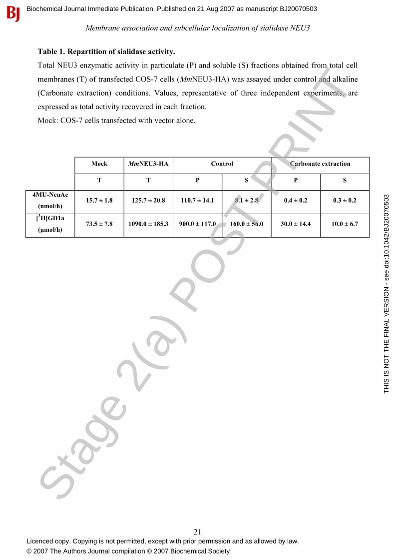
Table 1. Repartition of sialidase activity.
Total NEU3 enzymatic activity in particulate (P) and soluble (S) fractions obtained from total cell
membranes (T) of transfected COS-7 cells (MmNEU3-HA) was assayed under control and alkaline
(Carbonate extraction) conditions. Values, representative of three independent experiments, are
expressed as total activity recovered in each fraction.
Mock: COS-7 cells transfected with vector alone.
Mock MmNEU3-HA Control Carbonate extraction
T T P S P S
4MU-NeuAc
(nmol/h) 15.7 ± 1.8 125.7 ± 20.8 110.7 ± 14.1 8.1 ± 2.8 0.4 ± 0.2 0.3 ± 0.2
[3H]GD1a
(µmol/h) 73.5 ± 7.8 1090.0 ± 185.3 900.0 ± 117.0 160.0 ± 56.0 30.0 ± 14.4 10.0 ± 6.7
Stag
e 2(
a) P
OST
-PR
INT
Biochemical Journal Immediate Publication. Published on 21 Aug 2007 as manuscript BJ20070503
TH
IS IS
NO
T T
HE
FIN
AL
VE
RS
ION
- s
ee d
oi:1
0.10
42/B
J200
7050
3
Licenced copy. Copying is not permitted, except with prior permission and as allowed by law.
© 2007 The Authors Journal compilation © 2007 Biochemical Society

Membrane association and subcellular localization of sialidase NEU3
22
LEGENDS TO FIGURE
Figure 1: Sialidase NEU3 localizes both at the plasma membrane and intracellularly. COS-7
(A) and HeLa (D) cells transfected with MmNEU3-HA were fixed and, after permeabilization,
subjected to indirect immunofluorescence using anti-HA antibody. Transfectants were then
analyzed by laser confocal microscopy. Details of the plasma membrane (B and E) and intracellular
labelling (C and F) are given for both cell types. Single confocal planes are reported. Bars: 10 µm.
Figure 2: Sialidase NEU3 colocalizes with endosomal markers. COS-7 cells were transfected
with MmNEU3-HA and subjected to immunofluorescence for co-localization experiments.
Subcellular distribution of MmNEU3-HA was related to EEA1 (top panel), Tf-R (middle panel) and
LBPA (bottom panel), markers of the endocytic/recycling pathway. Single confocal planes are
shown in figure. Bars: 10 µm.
Figure 3: Sialidase NEU3 is associated with the extracellular leaflet of the plasma membrane.
(A) Cell surface proteins of COS-7 cells transfected with MmNEU3-HA were selectively
biotinylated and isolated by avidin-biotin affinity chromatography. Equal volumes of the total cell
extract (Total), avidin-unbound (Intracellular) and avidin-bound (Cell surface) fractions were
analyzed by immunoblotting in order to reveal MmNEU3-HA and the endogenous markers Cav-1
and Tf-R. Non-permeabilized HeLa (B) and COS-7 (C) cells transfected with MmNEU3-HA were
subjected to indirect immunofluorescence using anti-HA antibody. Projections of sequential
confocal planes (0.37 µm distance each plane) acquired through the cell thickness are shown. Bars:
10 µm.
Figure 4: Cell surface sialidase NEU3 internalizes to the recycling endosomal compartment.
MmNEU3-HA transfected COS-7 cells were subjected to anti-HA antibody uptake for 3 h,
processed for indirect immunofluorescence and analyzed by laser confocal microscopy as desribed
in the Experimental section. Subcellular distribution of MmNEU3-HA/anti-HA antibody
immunocomplexes (A, A’) was related to Tf-R staining (B, B’). Nuclei are evidenced by DAPI
staining. Merge is given in (C, C’). The white lines in A-C represent the plane through which the
corresponding direct vertical confocal sections(A’-C’) were acquired. Bars: 10 µm.
Figure 5: Sialidase NEU3 is a peripheral membrane-associated protein. A crude preparation of
cell membranes from COS-7 and HeLa cells transfected with MmNEU3-HA were extracted with
Stag
e 2(
a) P
OST
-PR
INT
Biochemical Journal Immediate Publication. Published on 21 Aug 2007 as manuscript BJ20070503
TH
IS IS
NO
T T
HE
FIN
AL
VE
RS
ION
- s
ee d
oi:1
0.10
42/B
J200
7050
3
Licenced copy. Copying is not permitted, except with prior permission and as allowed by law.
© 2007 The Authors Journal compilation © 2007 Biochemical Society
Membrane association and subcellular localization of sialidase NEU3
23
Tris buffer (Control) or with sodium carbonate (Carbonate extraction). After treatment, membranes
were collected by ultracentrifugation and equal volumes corresponding to the starting crude cell
membranes (T), membranes (P) and soluble (S) fractions were analyzed by Western-blot using anti-
HA, anti-HsNEU3, anti-EEA1 and anti-Cav-1 antibodies.
Figure 6: NEU3 enzymatic activity is highly sensitive to alkaline pH. NEU3 enzymatic activity
toward the artificial substrate 4MU-NeuAc was assayed using total cell membranes in presence of
buffered-carbonate in the reacion mixture (Carbonate +) or following exposure to alkaline pH for
different time periods (0.5 to 30 min). Values are given as percentage of recovered activity related
to control conditions. Data are representative of two independent experiments.
Figure 7: Sialidase NEU3 is a hydrophilic protein. Total cell extracts derived from HeLa cells
transfected with MmNEU3-HA were extracted and subjected to phase separation with Triton X-114.
Equal aliquots of the aqueous (Aq) and detergent (De) phases were then analyzed by Western-blot
using anti-HA, anti-HsNEU3, anti-Cav-1 and anti-PDI antibodies.
Stag
e 2(
a) P
OST
-PR
INT
Biochemical Journal Immediate Publication. Published on 21 Aug 2007 as manuscript BJ20070503
TH
IS IS
NO
T T
HE
FIN
AL
VE
RS
ION
- s
ee d
oi:1
0.10
42/B
J200
7050
3
Licenced copy. Copying is not permitted, except with prior permission and as allowed by law.
© 2007 The Authors Journal compilation © 2007 Biochemical Society
Membrane association and subcellular localization of sialidase NEU3
24
Figure 1
B
C
E
F
A
D
Stag
e 2(
a) P
OST
-PR
INT
Biochemical Journal Immediate Publication. Published on 21 Aug 2007 as manuscript BJ20070503
TH
IS IS
NO
T T
HE
FIN
AL
VE
RS
ION
- s
ee d
oi:1
0.10
42/B
J200
7050
3
Licenced copy. Copying is not permitted, except with prior permission and as allowed by law.
© 2007 The Authors Journal compilation © 2007 Biochemical Society
Membrane association and subcellular localization of sialidase NEU3
25
Figure 2
MmNEU3-HA EEA1 merge
MmNEU3-HA
MmNEU3-HA
merge
merge
Tf-R
LBPA
Stag
e 2(
a) P
OST
-PR
INT
Biochemical Journal Immediate Publication. Published on 21 Aug 2007 as manuscript BJ20070503
TH
IS IS
NO
T T
HE
FIN
AL
VE
RS
ION
- s
ee d
oi:1
0.10
42/B
J200
7050
3
Licenced copy. Copying is not permitted, except with prior permission and as allowed by law.
© 2007 The Authors Journal compilation © 2007 Biochemical Society
Membrane association and subcellular localization of sialidase NEU3
26
Figure 3
A
C B
Stag
e 2(
a) P
OST
-PR
INT
Biochemical Journal Immediate Publication. Published on 21 Aug 2007 as manuscript BJ20070503
TH
IS IS
NO
T T
HE
FIN
AL
VE
RS
ION
- s
ee d
oi:1
0.10
42/B
J200
7050
3
Licenced copy. Copying is not permitted, except with prior permission and as allowed by law.
© 2007 The Authors Journal compilation © 2007 Biochemical Society
Membrane association and subcellular localization of sialidase NEU3
27
Figure 4
C
C´
B
B´
A
A´
Stag
e 2(
a) P
OST
-PR
INT
Biochemical Journal Immediate Publication. Published on 21 Aug 2007 as manuscript BJ20070503
TH
IS IS
NO
T T
HE
FIN
AL
VE
RS
ION
- s
ee d
oi:1
0.10
42/B
J200
7050
3
Licenced copy. Copying is not permitted, except with prior permission and as allowed by law.
© 2007 The Authors Journal compilation © 2007 Biochemical Society
Membrane association and subcellular localization of sialidase NEU3
28
Figure 5
Stag
e 2(
a) P
OST
-PR
INT
Biochemical Journal Immediate Publication. Published on 21 Aug 2007 as manuscript BJ20070503
TH
IS IS
NO
T T
HE
FIN
AL
VE
RS
ION
- s
ee d
oi:1
0.10
42/B
J200
7050
3
Licenced copy. Copying is not permitted, except with prior permission and as allowed by law.
© 2007 The Authors Journal compilation © 2007 Biochemical Society
Membrane association and subcellular localization of sialidase NEU3
29
Control Carbonate +
0.5 1 2 5
10 30
pH 11.5 exposure
(min)
Figure 6
Stag
e 2(
a) P
OST
-PR
INT
Biochemical Journal Immediate Publication. Published on 21 Aug 2007 as manuscript BJ20070503
TH
IS IS
NO
T T
HE
FIN
AL
VE
RS
ION
- s
ee d
oi:1
0.10
42/B
J200
7050
3
Licenced copy. Copying is not permitted, except with prior permission and as allowed by law.
© 2007 The Authors Journal compilation © 2007 Biochemical Society
Membrane association and subcellular localization of sialidase NEU3
30
Figure 7
Stag
e 2(
a) P
OST
-PR
INT
Biochemical Journal Immediate Publication. Published on 21 Aug 2007 as manuscript BJ20070503
TH
IS IS
NO
T T
HE
FIN
AL
VE
RS
ION
- s
ee d
oi:1
0.10
42/B
J200
7050
3
Licenced copy. Copying is not permitted, except with prior permission and as allowed by law.
© 2007 The Authors Journal compilation © 2007 Biochemical Society
Copyright © 2022 FDOKUMEN