Localized Maternal Proteins in XenopusRevealed by Subtractive Immunization
Upload
independentCategory
view
0download
0
Experimental Cell Research 281, 50–62 (2002)doi:10.1006/excr.2002.5651
Human Heparanase Is Localized within Lysosomes in a Stable FormOrit Goldshmidt,*,3 Liat Nadav,§,3 Helena Aingorn,* Cohen Irit,* Naomi Feinstein,� Neta Ilan,¶,‡ Eli Zamir,**
Benjamin Geiger,** Israel Vlodavsky,*,‡,1 and Ben-Zion Katz§,2
*Department of Oncology, Hadassah-Hebrew University Hospital, Jerusalem; §Department of Hematology, Tel-Aviv Sourasky MedicalCenter, Tel-Aviv; �Electron Microscopy Unit, Institute of Life Sciences, Hebrew University of Jerusalem; ¶InSight Ltd., Rehovot;
**Department of Cellular and Molecular Biology, Weizmann Institute of Science, Rehovot; and ‡Vascular Biology
Heparanase is an endo-�-D-glucuronidase involvedin degradation of heparan sulfate (HS) and extracellu-lar matrix (ECM) of a wide range of cells of vertebrateand invertebrate tissues. The enzymatic activity ofheparanase is characterized by specific intrachaincleavage of glycosidic bonds with a hydrolase mecha-nism. This enzyme facilitates cell invasion and henceplays a role in tumor metastasis, angiogenesis, inflam-mation, and autoimmunity. Although the expressionpattern and molecular properties of heparanase havebeen characterized, its subcellular localization hasnot been unequivocally determined. We have previ-ously suggested that heparanase subcellular localiza-tion is a major determinant in regulating the enzyme’sbiological functions. In the present study we examinedheparanase localization in three different cell types,utilizing immunofluorescent staining and electron mi-croscopy. Our results indicate that heparanase is lo-calized primarily within lysosomes and the Golgi ap-paratus. A construct composed of heparanase cDNAfused to green fluorescent protein, utilized in order tovisualize the enzyme within living cells, confirmed itslocalization in acidic vesicles. We suggest that follow-ing synthesis, heparanase is transported into the Golgiapparatus and subsequently accumulates in a stableform within the lysosomes, where it functions in HSturnover. The lysosomal compartment may also serveas a site for heparanase confinement within the cells,limiting its secretion and uncontrolled extracellularactivities associated with tumor metastasis and angio-genesis. © 2002 Elsevier Science (USA)
Key Words: heparan sulfate proteoglycans; hepara-nase; heparanase-GFP; lysosomes; signal peptide;MCF7 breast carcinoma.
1 To whom correspondence and reprint requests should be addressedat Department of Oncology, Hadassah Hospital, POB 12000, Jerusalem91120, Israel. Fax: 972-2-6422794. E-mail: [email protected].
2 Dedicated to the memory of Professor Amiram Eldor (who diedNovember 2001 in a tragic airplane crash), whose inspiration, wis-dom, and encouragement contributed to the accomplishment of thisstudy.
3 These authors contributed equally to this study.
500014-4827/02 $35.00© 2002 Elsevier Science (USA)All rights reserved.
INTRODUCTION
Heparan sulfate proteoglycans (HSPGs) on cell sur-faces and in basement membranes and extracellularmatrices (ECM) affect biological processes by interact-ing with various ECM components and a large numberof biologically active molecules [1, 2]. HSPGs can thusinfluence a variety of normal and pathological pro-cesses, involving cell adhesion, migration, and invasion[1–4]. The importance and multifunctional roles ofHSPGs in the physiology of cells and tissues maketheir cleavage an essential factor in the regulation ofthe integrity and functional state of organs. Enzymaticdegradation of heparan sulfate (HS) is therefore likelyto be involved in fundamental biological processesranging from pregnancy, morphogenesis, and normaldevelopment to inflammation, angiogenesis, and can-cer metastasis [5–10]. Despite earlier reports on theexistence of several distinct mammalian HS degradingendoglycosidases (heparanases), the cloning of thesame gene by several groups suggests that mammaliancells express predominantly a single functionalheparanase enzyme [6, 11–13]. The HS chains arecleaved by heparanase at only a few sites, yielding HSfragments of still appreciable size (10–20 sugar units)[14]. The heparanase mRNA and protein are preferen-tially expressed in metastatic cell lines and humantumor tissues [6, 8–12, 15]. Moreover, enhancedheparanase mRNA expression correlates with reducedpostoperative survival of cancer patients [16, 17].Overexpression of the heparanase cDNA in low-meta-static tumor cells conferred a high metastatic potentialin experimental animals, resulting in an increased rateof mortality [6]. The heparanase enzyme also releasesECM-resident angiogenic factors in vitro and its over-expression induces an angiogenic response in vivo [18].While several studies have reported heparanase clon-ing, molecular properties, expression, and involvementin cancer progression, heparanase regulation and sub-cellular localization were not thoroughly investigated.Heparanase activity was first isolated from rat liver
Research Center, the Bruce Rappaport Faculty of Medicine, Technion, Haifa, 31096, Israel
lysosomes [19] and has been demonstrated in both

FIG. 1. Heparanase activity and cellular localization in transfected C6 glioma cells. (A) Cell lysates of C6 glioma cells transfected withhuman heparanase (H-hpa) (F) or mock transfected (E) were incubated (24 h, pH 6.0, 2 � 106 cells) on sulfate-labeled ECM. Labeleddegradation fragments released into the incubation medium were analyzed by gel filtration on Sepharose 6B, as described under Materialsand Methods. (B) Pooled populations of C6 glioma cells transfected with H-hpa cDNA were subjected to indirect immunofluorescence stainingwith monoclonal antiheparanase antibodies (mAb 130) followed by Cy3-conjugated goat antimouse antibody, as described under Materialsand Methods. Cells transfected with H-hpa cDNA displayed primarily a perinuclear granular staining (arrow). Mock-transfected C6 gliomacells were used as control and showed no staining (not shown).
51LYSOSOMAL LOCALIZATION OF HEPARANASE

lysosomal and endosomal cellular compartments [20–23]. Previous studies identified heparanase activity intertiary granules within neutrophiles, where it colocal-ized with gelatinase [24, 25]. In these early experi-ments the heparanase cellular localization was char-acterized by cell fractionation and by using antibodiesthat identified a 96-kDa protein [24]. Cloning and ex-pression of the mammalian heparanase, however, re-vealed molecular weights of 65 kDa for the proenzymeand 50 kDa for the processed, highly active heparanase[26], questioning the specificity of these antibodies.The availability of recombinant heparanase enabledthe development of monoclonal and polyclonal anti-heparanase antibodies, which were utilized to identifyheparanase expression in cell lines and human tissues[6, 15–17], and the investigation of its localization intransfected cells [27]. The objective of the presentstudy was to determine the subcellular localization ofheparanase and better elucidate its routing within thecells. Using confocal and electron microscopies, as wellas heparanase-GFP fusion protein, we have identifiedheparanase predominantly within lysosomes of bothnormal and malignant cells. We have also identifiedheparanase within the Golgi apparatus and demon-strated that the integrity of the Golgi is essential forthe generation of an active enzyme.
MATERIALS AND METHODS
Antibodies. Monoclonal mouse antihuman heparanase antibod-ies (mAb 130) directed against the C-terminus of the 50-kDa activeenzyme were obtained (Insight Ltd., Rohovot, Israel) as previouslydescribed [6]. Cy2-, Cy3-, or Alexa-conjugated goat antimouse anti-bodies were from Jackson Laboratories (Bar Harbor, ME).
Generation of heparanase-GFP fusion protein. Heparanase-GFPfusion protein was designed using standard molecular biology meth-ods [28] and generated through the insertion of a unique EcoRVrestriction site at the 3� end of heparanase cDNA (upstream thetermination codon) into the already described heparanase expressionvector pHep2 [6]. Heparanase C-terminus was amplified by PCR,using pHep2 as a template. The sense primer, 5�-AGCCGAGGTTAC-CCTATCCTTTTTC-3�, contained the BstEII restriction site, followedby a complementary sequence encoding for heparanase downstreamto the restriction site. The antisense primer, 5�-ACCGCCCTCGAGT-TCTGGTCAGATATCGATGCAAGCAGCAACTTT-3�, contained theXhoI restriction site, termination codon, and EcoRV restriction site,followed by a complementary sequence coding for heparanase up-stream to the termination codon. The PCR product and the pHep2plasmid were both restricted with XhoI and BstEII, extracted from apreparative gel, and ligated. The recombinant plasmid was identifiedby the restriction enzymes EcoRV, XhoI, and BstEII (restricting theinsert) and gel electrophoresis. GFP cDNA was amplified with theaddition of two restriction sites, EcoRV and XhoI, using a previouslydescribed GFP expression vector as a template [29]. The senseprimer, 5�-CGCATCGATATCATGGTGAGCAAGGGCGAG-3�, con-tained the EcoRV restriction site, followed by a complementary se-quence coding for GFP downstream to its initiation codon. The an-tisense primer, 5�-TGTGCCCTCGAGTTACTTGTACAGTGCGTC-3�,contained the XhoI restriction site followed by a complementarysequence coding for GFP upstream to its termination codon. ThePCR product and the plasmid coding for heparanase were both
restricted with XhoI and EcoRV and then extracted from a prepar-ative gel. After ligation, the recombinant plasmid was identified byrestriction analysis with XhoI, BstEII, and EcoRV. The sequence ofthe pHep-GFP construct was confirmed by automated nucleotidesequencing. The resulting pHep-GFP is driven by the CMV promoterand it expresses in frame the complete heparanase cDNA followed bythe GFP coding sequence. The previously described plasmids pHep2and pGFP were utilized as controls [6, 29].
Cells and transfection procedures. Human foreskin fibroblastswere kindly provided by Dr. Susan S. Yamada (NIDCR, NIH, Be-thesda, MD). Cells were maintained in Dulbecco’s modified Eagle’smedium (DMEM, 1 g of glucose/liter) supplemented with 1 mMglutamine, 50 �g/ml of streptomycin, 50 U/ml of penicillin, and 10%heat-inactivated bovine serum (Biological Industries, Beit-Haemek,Israel) at 37°C in a 5% humidified incubator. Electroporation of thecells was performed at 170 V and 950 �F utilizing a Bio–Rad Gene-Pulser (Hercules, CA), with a thymidine block, as previously de-scribed [30]. Transient transfection was performed using 30 �g ofDNA for each plasmid, and protein expression was examined 48 hlater. C6 rat glioma cells were cultured in DMEM (4.5 g of glucose/liter) supplemented with glutamine, antibiotics, and 10% fetal calfserum (FCS). MCF7 and MDA-231 human breast carcinoma cellswere cultured in RPMI medium supplemented with 10% FCS, glu-tamine, and antibiotics. For transfection, H-hpa cDNA was sub-cloned into pcDNA3 plasmid (Invitrogen, NV Leek, Netherlands) atthe EcoRI site as described [6, 27]. C6 rat glioma cells (5 � 106
cells/10-cm dish) were incubated (48–72 h, 37°C) with a total of 1–2�g of DNA and 6 �l of FuGene transfection reagent (Boehringer,Mannheim, Germany) in 94 �l of Optimem (GibcoBRL, Rockville,MD). Transfected cells were selected with 800 �g/ml of G418 andstable populations of heparanase expressing cells were obtained [27].Cultures of bovine corneal endothelial cells were established fromsteer eyes and maintained in DMEM (1 g of glucose/liter) supple-mented with 5% newborn calf serum and 10% FCS, as described [31].
Preparation of dishes coated with ECM. Bovine corneal endothe-lial cells were plated into 35-mm tissue culture dishes and culturedas described above, except that 4% dextran T-40 was included in thegrowth medium [31, 32]. Na2
35SO4 (25 �Ci/ml) (Amersham, Bucking-hamshire, UK) was added on days 2 and 5 after seeding and thecultures were incubated with the label without medium change. Onday 12, the subendothelial ECM was exposed by dissolving the celllayer with PBS containing 0.5% Triton X-100 and 20 mM NH4OH,followed by four washes with PBS [31, 32]. The ECM remainedintact, free of cellular debris, and firmly attached to the entire areaof the tissue culture dish.
Heparanase activity. Cells (2 � 106 cells/ml) were lysed by threecycles of freezing and thawing in heparanase reaction solution (0.15M NaCl, 20 mM phosphate-citrate buffer, pH 5.8, 1 mM dithiothre-itol (DTT), 1 mM CaCl2). Cell lysates were incubated (24 h, 37°C, pH5.8) with 35S-labeled ECM, the incubation medium was centrifuged,and the supernatant containing sulfate-labeled degradation frag-ments was analyzed by gel filtration on a Sepharose CL-6B column(0.9 � 30 cm). Fractions (0.2 ml) were eluted with PBS and theirradioactivity was counted in a �-scintillation counter [6, 32]. Degra-dation fragments of HS side chains were eluted from Sepharose 6B at0.5 � Kav � 0.8 (fractions 15–35, peak II). Nearly intact HSPGs wereeluted just after the V0 (Kav � 0.2, fractions 1–10, peak I) [6, 32].Each experiment was performed at least three times and the varia-tion in elution positions (Kav values) did not exceed � 15%.
Indirect immunofluorescence. Cultured primary human foreskinfibroblasts and C6 rat glioma and MDA-231 cells were stained byindirect immunofluorescence as previously described [27, 33]. Cellswere fixed with either acetone (�20°C, 5 min) or 4% formaldehyde inPBS for 20 min and, when stated, permeabilized with 0.5% TritonX-100 in PBS for 5 min. The cells were then incubated with theindicated antibodies in PBS. Antiheparanase monoclonal antibodies
52 GOLDSHMIDT ET AL.

were diluted to 10 �g/ml followed by staining with secondary Alexa-,Cy2-, or Cy3-conjugated goat antimouse antibodies, diluted 1:200.
Immunogold labeling. C6 glioma and MDA-231 breast carcinomacells, cultured as described above and reaching 80% confluence, werewashed (�2) with PBS and fixed (1% glutaraldehyde in 0.1 M caco-dylate buffer, pH 7.4) for 10 min at room temperature. Fixative wasreplaced with fresh glutaraldehyde solution in the same buffer fol-lowed by 30 min incubation at RT. Fixed cells were briefly washedand fixed again with 0.5% osmium tetraoxide (OsO4) for 30 min at RT[34]. Following a brief wash with the same cacodylate buffer, cellswere dehydrated with 25, 50, 70, and 90% ethanol for 10 min each, 90and 95% ethanol for 20 min, and 100% ethanol (�2) for 20 min. Cellswere then scraped from the plate, collected into 1.5-ml tubes, equil-ibrated with propylene oxide, and embedded in embedding resin, asdescribed [34]. Thin sections (60 nm) were then incubated (5 h, RT)with 10 �g/ml of antiheparanase mAb 130 and washed (�5) withTris-buffered saline (TBS), pH 8.2, containing 20 mM Tris base, 0.9%NaCl, 0.5% BSA, 0.5% Tween 20, and 0.13% NaN3. Sections werethen incubated (30 min, RT) with antimouse IgG-conjugated gold (12nm) (Sigma, St. Louis, MO), washed (�2) with TBS and (�5) withDDW, and examined with a CM 12 Philips transmission electronmicroscope (TEM).
Labeling intracellular organelles. Following transfection of hu-man foreskin fibroblasts with pHep2 and pHep-GFP, the cells wereincubated with LysoTracker DNA-99 (Molecular Probes, Eugene,OR) at 50 nM in growth medium for 1.5 h at 37°C. Cells were thenwashed with growth medium and visualized without fixation, asdescribed [35]. Alternatively, cells were fixed and permeabilized withacetone (�20°C, 5 min), followed by immunofluorescent staining ofheparanase, as described above.
Digital fluorescence imaging analysis of heparanase subcellulardistribution. Images of stained cells were acquired at 3-min inter-vals for 6 h using an Axioscope microscope (Zeiss, Oberkochen,Germany) equipped with a charge-coupled device (CCD) camera(Model C220, Photometrics Co., Tucson, AZ) with a Texas Instru-ments 1024 � 1024 pixels chip readout generating 12-bit digitaldata. The location of individual lysosomes was determined at twotime points 9 min apart. This system for computerized microscopyand fluorescence ratio imaging was described in detail elsewhere[36]. In the present study, cells were examined with a 100X/1.3-NAplan Neofluar objective (Zeiss) resulting in a pixel length of 0.118�m. Corrections for nonhomogenous illumination and pixel-to-pixelvariations in CCD sensitivities, as well as aligning the LysoTrackerand the GFP images, were routinely performed. The images were
FIG. 2. Transmission electron micrographs of C6 glioma and MDA-231 breast carcinoma cells. (A) Rat C6 glioma cells transfected withH-hpa. (B–D) MDA-231 human breast carcinoma cells expressing endogenous heparanase. Cultured cells were processed for electronmicroscopy and heparanase was visualized using antihuman heparanase mAb 130 followed by antimouse IgG-conjugated gold particles, asdescribed under Materials and Methods. Heparanase (arrows) is localized within lysosomes (A–C) and in the Golgi apparatus (D). Bars, 200nm (B), 100 nm (C and D), and 200 nm (insets).
53LYSOSOMAL LOCALIZATION OF HEPARANASE

then high pass filtered with a box size of 4.7 � 4.7 �m and thresh-olded to eliminate the background fluorescence.
RESULTS
Expression and Localization of Human Heparanase
In order to evaluate the subcellular localization ofheparanase in transfected cells, nonmetastatic C6 rat
glioma cells that lack heparanase expression and ac-tivity were stable transfected with the full-length hu-man heparanase (H-hpa) cDNA. Cell lysates were thenassayed for their ability to degrade a biosyntheticallylabeled ECM (Fig. 1A). Cells transfected with an in-sert-free pcDNA3 plasmid were used as control. Fol-lowing incubation on 35S-labeled ECM, the mediumwas analyzed by gel filtration on Sepharose 6B, as
FIG. 3. Heparanase localization in acidic vesicles. (A) Primary human fibroblasts; primary human fibroblasts were transfected withH-hpa cDNA and 48 h later subjected to staining of heparanase and lysosomes. (Left) Cells were fixed, permeabilized, and applied to indirectimmunofluorescence staining with monoclonal antiheparanase antibodies (mAb 130) followed by Alexa-conjugated goat antimouse antibody(green fluorescence). (Middle) Intact cells were live-stained for 1 h with LysoTracker, labeling acidic organelles, predominantly lysosomes(red fluorescence). (Right) Living cells were first labeled with LysoTracker, followed by fixation, permeabilization, and staining withantiheparanase antibodies. Note the colocalization of heparanase and acidic vesicles (yellow). (B) Localization of endogenous heparanase inMDA-231 cells. (Left) Cells stained with antiheparanase antibodies (mAb 130) followed by Alexa-conjugated goat antimouse antibody (greenfluorescence). (Middle) cells live-stained with LysoTracker (red fluorescence). (Right) Cells were labeled with LysoTracker followed bystaining with antiheparanase antibodies, as described in A. Note the colocalization of heparanase and acidic vesicles (yellow fluorescence).
54 GOLDSHMIDT ET AL.
described [32]. As demonstrated in Fig. 1A, labeledmaterial released from the ECM by mock-transfectedcells eluted just after the void volume (V0) (peak I,fractions 1–10, Kav � 0.2) and consisted almost entirelyof nearly intact, high-molecular-weight proteoglycans.In contrast, lysates of H-hpa-transfected cells de-graded the ECM HSPGs as indicated by release oflow-molecular-weight labeled degradation fragmentsthat eluted toward the Vt of the column (peak II, frac-tions 15–30, 0.5 � Kav � 0.75) (Fig. 1A). Labeled frag-ments eluted in peak II were shown to be degradationproducts of HS, as they were five- to sixfold smallerthan intact HS side chains, resistant to further diges-tion with papain and chondroitinase ABC, and suscep-tible to deamination by nitrous acid [32]. Next, weevaluated the subcellular localization of heparanase inthe transfected cells by immunofluorescent staining,utilizing antiheparanase mAb 130. In permeabilizedcells, heparanase was detected predominantly in a pe-rinuclear, granular pattern (Fig. 1B). In contrast, nostaining was detected when fixed unpermeablized cellswere incubated with the same antibodies, indicatinglittle or no cell surface localization of the enzyme (notshown). In a subsequent experiment, H-hpa-trans-fected C6 glioma cells and nontransfected MDA-231breast carcinoma cells expressing endogenous hepara-nase were subjected to immunogold labeling of hepara-nase and TEM. Specific heparanase labeling was de-tected within lysosomes (Figs. 2A–2C, arrows) and theGolgi apparatus (Fig. 2D) of both transfected and non-transfected cells. To further confirm heparanase sub-cellular localization, we utilized an independent iden-tification method and a different cell type. Primaryhuman foreskin fibroblasts (HFF) that lacked hepara-nase activity were transfected with H-hpa. Forty-eighthours later, the transfected cells were stained by indi-rect immunofluorescence with antiheparanase anti-bodies and Alexa-conjugated goat antimouse antibod-ies. Specific heparanase staining was found exclusivelyin a granular pattern (Fig. 3A, left). No heparanasestaining was detected in nonpermeabilized HFF cells,indicating accumulation of the enzyme in cytoplasmicgranules (not shown). In order to determine whetherheparanase is indeed localized within lysosomes, intra-cellular organelles of HFF living cells were first labeledwith LysoTracker (a specific fluorescent label of acidicvesicles, predominantly lysosomes) (Fig. 3A, middle).Fixation of these cells followed by indirect immunoflu-orescent staining with antiheparanase antibodies re-sulted in colocalization of the LysoTracker withheparanase in the same cytoplasmic granules (Fig. 3A,right), indicating that heparanase is indeed storedwithin lysosomes. We have also examined heparanaselocalization in cells that endogenously express the en-zyme. For this purpose, nonmetastatic (MCF7) andmoderately metastatic (MDA-231) human breast car-
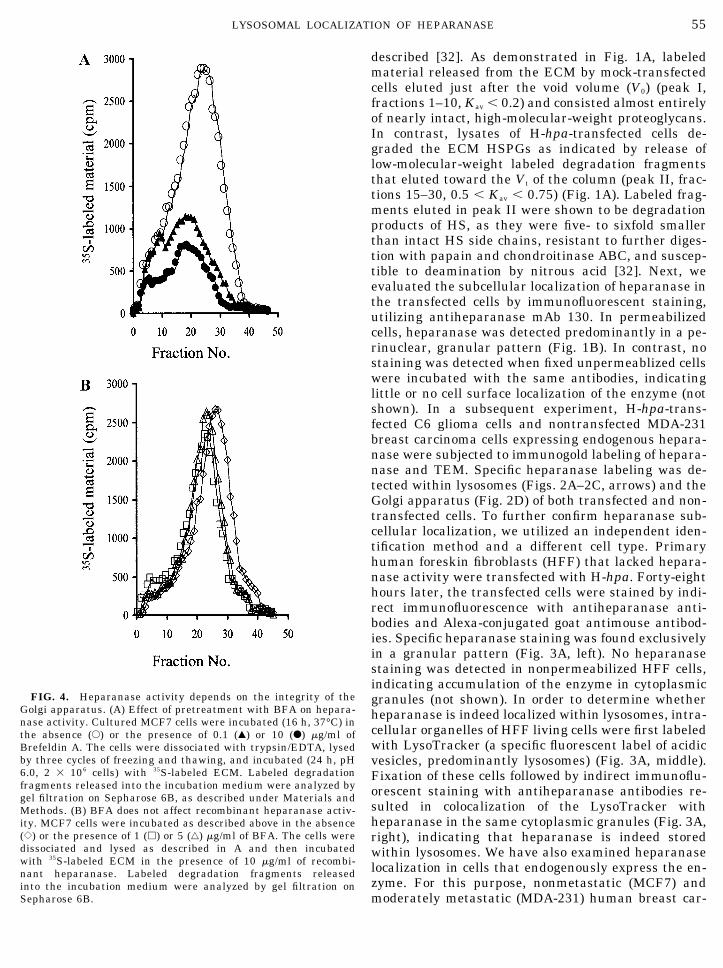
FIG. 4. Heparanase activity depends on the integrity of theGolgi apparatus. (A) Effect of pretreatment with BFA on hepara-nase activity. Cultured MCF7 cells were incubated (16 h, 37°C) inthe absence (E) or the presence of 0.1 (Œ) or 10 (F) �g/ml ofBrefeldin A. The cells were dissociated with trypsin/EDTA, lysedby three cycles of freezing and thawing, and incubated (24 h, pH6.0, 2 � 106 cells) with 35S-labeled ECM. Labeled degradationfragments released into the incubation medium were analyzed bygel filtration on Sepharose 6B, as described under Materials andMethods. (B) BFA does not affect recombinant heparanase activ-ity. MCF7 cells were incubated as described above in the absence({) or the presence of 1 (�) or 5 (‚) �g/ml of BFA. The cells weredissociated and lysed as described in A and then incubatedwith 35S-labeled ECM in the presence of 10 �g/ml of recombi-nant heparanase. Labeled degradation fragments releasedinto the incubation medium were analyzed by gel filtration onSepharose 6B.
55LYSOSOMAL LOCALIZATION OF HEPARANASE
cinoma cells were immunostained with antihepara-nase mAb 130. A perinuclear granular staining wasobserved in both MCF7 (not shown) and MDA-231 (Fig.3B, left). Staining of living MCF7 and MDA-231 cellswith LysoTracker yielded the same cytoplasmic gran-ular staining pattern (Fig. 3B, middle), as also indi-cated by the colocalization of heparanase and Lyso-Tracker observed in double-stained cells (Fig. 3B,right). Similar results were obtained with the non-metastatic MCF7 breast carcinoma cells, expressinglower heparanase activity [37]. In both the MCF7 andMDA-231 cells, the cytoplasm is occupied mostly by thenucleus.
Heparanase Activity Depends on the Integrity of theGolgi Apparatus
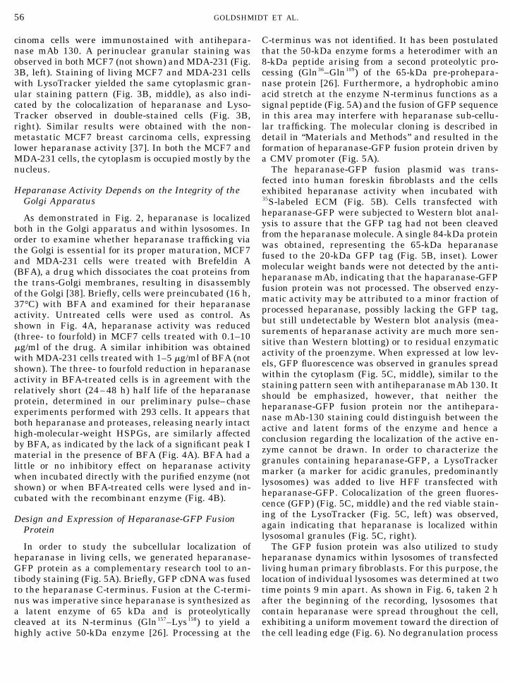
As demonstrated in Fig. 2, heparanase is localizedboth in the Golgi apparatus and within lysosomes. Inorder to examine whether heparanase trafficking viathe Golgi is essential for its proper maturation, MCF7and MDA-231 cells were treated with Brefeldin A(BFA), a drug which dissociates the coat proteins fromthe trans-Golgi membranes, resulting in disassemblyof the Golgi [38]. Briefly, cells were preincubated (16 h,37°C) with BFA and examined for their heparanaseactivity. Untreated cells were used as control. Asshown in Fig. 4A, heparanase activity was reduced(three- to fourfold) in MCF7 cells treated with 0.1–10�g/ml of the drug. A similar inhibition was obtainedwith MDA-231 cells treated with 1–5 �g/ml of BFA (notshown). The three- to fourfold reduction in heparanaseactivity in BFA-treated cells is in agreement with therelatively short (24–48 h) half life of the heparanaseprotein, determined in our preliminary pulse–chaseexperiments performed with 293 cells. It appears thatboth heparanase and proteases, releasing nearly intacthigh-molecular-weight HSPGs, are similarly affectedby BFA, as indicated by the lack of a significant peak Imaterial in the presence of BFA (Fig. 4A). BFA had alittle or no inhibitory effect on heparanase activitywhen incubated directly with the purified enzyme (notshown) or when BFA-treated cells were lysed and in-cubated with the recombinant enzyme (Fig. 4B).
Design and Expression of Heparanase-GFP FusionProtein
In order to study the subcellular localization ofheparanase in living cells, we generated heparanase-GFP protein as a complementary research tool to an-tibody staining (Fig. 5A). Briefly, GFP cDNA was fusedto the heparanase C-terminus. Fusion at the C-termi-nus was imperative since heparanase is synthesized asa latent enzyme of 65 kDa and is proteolyticallycleaved at its N-terminus (Gln157–Lys158) to yield ahighly active 50-kDa enzyme [26]. Processing at the
C-terminus was not identified. It has been postulatedthat the 50-kDa enzyme forms a heterodimer with an8-kDa peptide arising from a second proteolytic pro-cessing (Gln36–Gln109) of the 65-kDa pre-prohepara-nase protein [26]. Furthermore, a hydrophobic aminoacid stretch at the enzyme N-terminus functions as asignal peptide (Fig. 5A) and the fusion of GFP sequencein this area may interfere with heparanase sub-cellu-lar trafficking. The molecular cloning is described indetail in “Materials and Methods” and resulted in theformation of heparanase-GFP fusion protein driven bya CMV promoter (Fig. 5A).
The heparanase-GFP fusion plasmid was trans-fected into human foreskin fibroblasts and the cellsexhibited heparanase activity when incubated with35S-labeled ECM (Fig. 5B). Cells transfected withheparanase-GFP were subjected to Western blot anal-ysis to assure that the GFP tag had not been cleavedfrom the heparanase molecule. A single 84-kDa proteinwas obtained, representing the 65-kDa heparanasefused to the 20-kDa GFP tag (Fig. 5B, inset). Lowermolecular weight bands were not detected by the anti-heparanase mAb, indicating that the haparanase-GFPfusion protein was not processed. The observed enzy-matic activity may be attributed to a minor fraction ofprocessed heparanase, possibly lacking the GFP tag,but still undetectable by Western blot analysis (mea-surements of heparanase activity are much more sen-sitive than Western blotting) or to residual enzymaticactivity of the proenzyme. When expressed at low lev-els, GFP fluorescence was observed in granules spreadwithin the cytoplasm (Fig. 5C, middle), similar to thestaining pattern seen with antiheparanase mAb 130. Itshould be emphasized, however, that neither theheparanase-GFP fusion protein nor the antihepara-nase mAb-130 staining could distinguish between theactive and latent forms of the enzyme and hence aconclusion regarding the localization of the active en-zyme cannot be drawn. In order to characterize thegranules containing heparanase-GFP, a LysoTrackermarker (a marker for acidic granules, predominantlylysosomes) was added to live HFF transfected withheparanase-GFP. Colocalization of the green fluores-cence (GFP) (Fig. 5C, middle) and the red viable stain-ing of the LysoTracker (Fig. 5C, left) was observed,again indicating that heparanase is localized withinlysosomal granules (Fig. 5C, right).
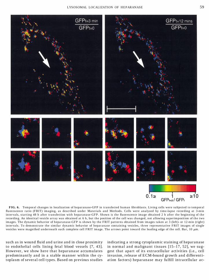
The GFP fusion protein was also utilized to studyheparanase dynamics within lysosomes of transfectedliving human primary fibroblasts. For this purpose, thelocation of individual lysosomes was determined at twotime points 9 min apart. As shown in Fig. 6, taken 2 hafter the beginning of the recording, lysosomes thatcontain heparanase were spread throughout the cell,exhibiting a uniform movement toward the direction ofthe cell leading edge (Fig. 6). No degranulation process
56 GOLDSHMIDT ET AL.
or change in fluorescence intensity of the heparanase-GFP was observed, even following a prolonged (6 h)observation. We therefore suggest that human hepara-nase storage within lysosomes is apparently stable.
DISCUSSION
Heparan sulfate proteoglycans are major compo-nents of the extracellular microenvironment, cell sur-faces, and basement membranes. These ubiquitousmolecules interact with elements of the basementmembrane and provide the ECM with the ability tobind and store growth factors, cytokines, and enzymes[1–4, 39–41]. The extracellular activity of heparanasemodulates the molecular structure of the ECM andsubendothelial basement membrane, thereby enablingthe trans-endothelial migration of normal hematopoi-etic cells [42] and blood-borne tumor cells [5–10]. Inaddition, heparanase facilitates the release of heparin-binding growth promoting factors from the ECM [18,40], thus also acting as a pro-angiogenic factor [9, 18,43–45]. The extracellular function of heparanase inregulating cellular trafficking and growth factor bio-availability requires its secretion from the cell. How-ever, a heparanase export mechanism(s) should betightly regulated, since uncontrolled overexpression ofthe enzyme and the associated inadvertent cleavage ofHS may promote tissue damage and pathological pro-cesses, such as tumor invasion and metastasis [45], orautoimmune disorders [5, 8–10, 42]. It appears thatcellular localization of heparanase plays a role in itsactivation, regulation, and bioavailability. We have ob-served that when heparanase is directed to the cellsurface, it enhances tumor angiogenesis and the met-astatic potential of malignant cells [45]. Similarly, thesubcellular localization of cathepsin D and B in breastand bladder cancer cells changes from within lyso-somes to the plasma membrane with increasing meta-static potential [46, 47]. Interestingly, we have recentlydemonstrated that exogenously added recombinantheparanase is readily internalized by human foreskinfibroblasts and accumulates primarily within endoso-mal structures [48].
Analysis of the human heparanase amino acid se-quence indicates the presence of a hydrophobic 35-amino-acid stretch at the N-terminus [6, 11, 27], whichmay function as a signal peptide, directing the proteinto the Golgi apparatus and subsequent secretion. How-ever, our recent immunostaining studies indicated thathuman heparanase is localized primarily within thecell in a perinuclear granular pattern and is not readilysecreted [27]. Replacing the signal peptide of the hu-man heparanase with that of the chicken enzyme re-sulted in its cell surface localization and efficient se-cretion [27]. An analysis of the human heparanasesignal peptide sequence revealed a significant (�65%,
Fig. 7) homology to the signal peptide of the mannose-6-phosphate receptor (M6P-R), responsible for lysoso-mal trafficking of enzymes [49]. On the other hand,there was a relatively low level of homology (�30%)between the human and chicken heparanase signalpeptides (Fig. 7). These data suggest that the signalpeptide is essential for subcellular trafficking ofheparanase. The homology between the signal se-quences of the human enzyme and the M6P-R and theoccurrence of a putative small transmembrane se-quence at the C-terminus of the human heparanasemay imply that the human enzyme could be initiallymembrane bound. In fact heparanase activity has beenpreviously shown to be partially membrane associated,as revealed by immunostaining [15] and displacementby mannose-6-phosphate [50]. The observed 65% ho-mology between the M6P-R and human heparanasesignal sequences may imply that heparanase gets toendosomes by a pathway similar to that of M6P-R andthen to the lysosomes by another mechanism. An en-dogenous, most likely heparanase activity is present inI-cell patient cells [22], implying its lysosomal entry bya M6P-independent pathway.
In the present study, we have identified the localiza-tion of heparanase within the lysosomes of three celltypes, utilizing three independent methods: immuno-gold labeling and electron microscopy, colocalization ofthe enzyme and fluorescent labeled lysosomes, and lo-calization of a fluorescent fusion enzyme within lyso-somes of living cells. We have also demonstrated thatthe integrity of the Golgi apparatus is essential forheparanase activity in MCF7 and MDA-231 breast car-cinoma cells, endogenously expressing the enzyme.These results suggest a possible trafficking mechanismin which following synthesis, the enzyme is trans-ported into the Golgi apparatus and subsequently ac-cumulates within lysosomes. Once accumulated,heparanase is apparently stored within these or-ganelles in a stable form, since its activity was readilydetected in cell lysates, but was not secreted into theconditioned medium of the transfected cells. In addi-tion, there was no degranulation or trafficking of theenzyme to the cell surface during several hours ofobservation of living cells expressing fluorescentheparanase (GFP fusion protein) in their lysosomes.Apparently, the acidic microenvironment within lyso-somes provides heparanase with suitable conditionsfor storage and expression of optimal enzymatic activ-ity [19, 20, 51]. Moreover, lysosomes contain a varietyof proteolytic enzymes (e.g., cathepsins) that may fa-cilitate the conversion of heparanase from its 65-kDalatent form to the active 50-kDa enzyme and/or theproposed 58-kDa heterodimer heparanase protein [6,10, 26]. It is therefore suggested that lysosomes mayserve as a primary site for heparanase activity andstorage. The predominant function attributed to
57LYSOSOMAL LOCALIZATION OF HEPARANASE

heparanase is degradation of heparan sulfate proteo-glycans in the ECM and basement membranes [7, 9,10], a function that clearly requires the presence of cell
surface-bound and extracellular heparanase. Indeed,heparanase was occasionally detected on the cell sur-faces [11, 15] and in several extracellular locations,
FIG. 5. Generation and expression of heparanase-GFP fusion protein. (A) Heparanase-GFP fusion protein; GFP cDNA was fused to theheparanase C-terminus, as described under Materials and Methods. The resulting expression vector (designated pHep-GFP) contains a CMVpromoter, full-length human heparanase cDNA fused in frame with GFP cDNA. Note the position of the signal peptide. (B) Heparanase-GFPactivity; primary human fibroblasts transfected with heparanase-GFP (F) or mock transfected (‚) were incubated (24 h, pH 6.0, 2 � 106 cells)on 35S-labeled ECM. Labeled degradation fragments released into the incubation medium were analyzed by gel filtration on Sepharose 6B.(Inset) Western blot analysis; primary human fibroblasts were transfected with heparanase-GFP and subjected to immunoblot analysis usingantiheparanase mAb-130. Lane 1, recombinant human heparanase showing both the 65- and 50-kDa forms; lane 2, 84-kDa heparananse-GFPfusion protein. (C) Heparanase-GFP expression and localization; primary human fibroblasts were transfected with pHep-GFP. Forty eighthours later, the cells were live-stained for 1 h with LysoTracker. Digital images of GFP-heparanase (middle image, green) or LysoTracker(left image, red) were recorded and superimposed (right image, yellow). Note the colocalization of GFP-heparanase and the acidic vesicles.To exemplify the localization of heparanase in acidic vesicles, an image of several vesicles was magnified and is presented as an inset in eachpanel.
58 GOLDSHMIDT ET AL.
such as in wound fluid and urine and in close proximityto endothelial cells lining fetal blood vessels [7, 43].However, we show here that heparanase accumulatespredominantly and in a stable manner within the cy-toplasm of several cell types. Based on previous studies
indicating a strong cytoplasmic staining of heparanasein normal and malignant tissues [15–17, 52], we sug-gest that apart of its extracellular activities (i.e., cellinvasion, release of ECM-bound growth and differenti-ation factors) heparanase may fulfill intracellular ac-
FIG. 6. Temporal changes in localization of heparanase-GFP in transfected human fibroblasts. Living cells were subjected to temporalfluorescence ratio (FRIT) imaging, as described under Materials and Methods. Cells were analyzed by time-lapse recording at 3-minintervals, starting 48 h after transfection with heparanase-GFP. Shown is the fluorescence image obtained 2 h after the beginning of therecording. An identical vesicle array was obtained at 6 h, but the position of the cell was changed, not allowing superimposition of the twoimages. The dynamic behavior of heparanase-GFP is shown by the FRIT patterns obtained from images taken at 3 (left)- or 12-min (right)intervals. To demonstrate the similar dynamic behavior of heparanase containing vesicles, three representative FRIT images of singlevesicles were magnified underneath each complete cell FRIT image. The arrows point toward the leading edge of the cell. Bar, 10 �m.
59LYSOSOMAL LOCALIZATION OF HEPARANASE

tivities, such as regulation of HSPG turnover and me-tabolism, known to be associated with lysosomalfunctions [20, 21]. Also, as indicated in previous stud-ies, acidic pH is essential for optimal heparanase ac-tivity [51, 53]. It has been previously proposed that HSis degraded stepwise in endosomal (chloroquine insen-sitive) and then further in lysosomal compartments[54, 55]. A more recent study, demonstrated, however,that HS degradation occurs only in lysosomes and lateendosomes [56], supporting our observations. It shouldbe pointed out, however, that both the LysoTracker dyeand electron microscopy analysis used in this study failto distinguish between late endosomes and lysosomes.Hence, the actual sequence and sites of HS degradationcannot be determined based on the present study.Taken together, it is conceivable that the lysosomalcompartment may serve as the predominant subcellu-lar site for heparanase confinement within the cell,preventing its uncontrolled extracellular activity asso-ciated, for example, with cancer progression and auto-immune disorders.
We thank Dr. I. Pecker, Dr. Orom Yacoby-Zeevi (InSight Ltd.,Rehovot, Isarel), Dr. M. Trashis, and Dr. E. Rachamim (Hadassah-University Medical School) for their helpful suggestions and excel-lent assistance and Insight Ltd. for providing the antihuman hepara-nase antibodies (mAb 130). This work was supported by the IsraelScience Foundation (Grant 503/98); the Association for InternationalCancer Research, UK; the NIH (R21 CA87085); and the U.S. Army(Grant 0278).
REFERENCES
1. Bernfield, M., Gotte, M., Park, P. W., Reizes, O., Fitzgerald,M. L., Lincecum, J., and Zako, M. (1999). Functions of cellsurface heparan sulfate proteoglycans. Annu. Rev. Biochem. 68,729–777.
2. Vlodavsky, I., Bar-Shavit, R., Korner, G., and Fuks, Z. (1993).Extracellular matrix-bound growth factors, enzymes andplasma proteins. In “Molecular and Cellular Aspects of Base-ment Membranes” (D. H. Rohrbach and R. Timpl, Eds.), pp.327–343, Academic Press, Orlando, Fl.
3. Kjellen, L., and Lindahl, U. (1991). Proteoglycans: Structuresand interactions. Annu. Rev. Biochem. 60, 443–475.
4. Iozzo, R. V. (1998). Matrix proteoglycans: from molecular designto cellular function. Annu. Rev. Biochem. 67, 609–652.
5. Vlodavsky, I., Mohsen, M., Lider, O., Svahn, C. M., Ekre, H. P.,Vigoda, M., Ishai-Michaeli, R., and Peretz, T. (1994). Inhibition
of tumor metastasis by heparanase inhibiting species of hepa-rin. Invasion Metastasis 14, 290–302.
6. Vlodavsky, I., Friedmann, Y., Elkin, M., Aingorn, H., Atzmon,R., Ishai-Michaeli, R., Bitan, M., Pappo, O., Peretz, T., Michal,I., Spector, L., and Pecker, I. (1999). Mammalian heparanase:Gene cloning, expression and function in tumor progression andmetastasis. Nature Med. 5, 793–802.
7. Dempsey, L. A., Plummer, T. B., Coombes, S. L., and Platt, J. L.(2000). Heparanase expression in invasive trophoblasts andacute vascular damage. Glycobiology 10, 467–475.
8. Nakajima, M., Irimura, T., and Nicolson, G. L. (1988). Hepara-nases and tumor metastasis. J. Cell Biochem. 36, 157–167.
9. Vlodavsky, I., and Friedmann, Y. (2001). Molecular propertiesand involvement of heparanase in cancer metastasis and angio-genesis. J. Clin. Invest. 108, 341–347.
10. Parish, C. R., Freeman, C., and Hulett, M. D. (2001). Hepara-nase: A key enzyme involved in cell invasion. Biochim. Biophys.Acta 1471, M99–108.
11. Hulett, M. D., Freeman, C., Hamdorf, B. J., Baker, R. T., Har-ris, M. J., and Parish, C. R. (1999). Cloning of mammalianheparanase, an important enzyme in tumor invasion and me-tastasis. Nat. Med. 5, 803–809.
12. Kussie, P. H., Hulmes, J. D., Ludwig, D. L., Patel, S., Navarro,E. C., Seddon, A. P., Giorgio, N. A., and Bohlen, P. (1999).Cloning and functional expression of a human heparanasegene. Biochem. Biophys. Res. Commun. 261, 183–187.
13. Toyoshima, M., and Nakajima, M. (1999). Human heparanase.Purification, characterization, cloning, and expression. J. Biol.Chem. 274, 24153–24160.
14. Pikas, D. S., Li, J. P., Vlodavsky, I., and Lindahl, U. (1998).Substrate specificity of heparanases from human hepatoma andplatelets. J. Biol. Chem. 273, 18770–18777.
15. Friedmann, Y., Vlodavsky, I., Aingorn, H., Aviv, A., Peretz, T.,Pecker, I., and Pappo, O. (2000). Expression of heparanase innormal, dysplastic, and neoplastic human colonic mucosa andstroma. Evidence for its role in colonic tumorigenesis. Am. J.Pathol. 157, 1167–1175.
16. Gohji, K., Okamoto, M., Kitazawa, S., Toyoshima, M., Dong, J.,Katsuoka, Y., and Nakajima, M. (2001). Heparanase proteinand gene expression in bladder cancer. J. Urol. 166, 1286–1290.
17. Koliopanos, A., Friess, H., Kleeff, J., Shi, X., Liao, Q., Pecker, I.,Vlodavsky, I., Zimmermann, A., and Buchler, M. W. (2001).Heparanase expression in primary and metastatic pancreaticcancer. Cancer Res. 61, 4655–4659.
18. Elkin, M., Ilan, N., Ishai-Michaeli, R., Friedmann, Y., Papo, O.,Pecker, I., and Vlodavsky, I. (2001). Heparanase as mediator ofangiogenesis: Mode of action. FASEB J. 15, 1661–1663.
FIG. 7. Signal peptide sequence alignment. Heparanase 5� cDNA encodes for a 35-amino-acid putative signal peptide. There is arelatively high homology (�65%) between the human heparanase signal peptide and the signal peptide of the mannose-6-phosphate receptor(mannose-6-PR), responsible for lysosomal trafficking of enzymes, vs a relatively low level of homology (�30%) with the chicken heparanasesignal peptide.
60 GOLDSHMIDT ET AL.

19. Hook, M., Wasteson, A., and Oldberg, A. (1975). A heparansulfate-degrading endoglycosidase from rat liver tissue. Bio-chem. Biophys. Res. Commun. 67, 1422–1428.
20. Kjellen, L., Pertoft, H., Oldberg, A., and Hook, M. (1985). Oli-gosaccharides generated by an endoglucuronidase are interme-diates in the intracellular degradation of heparan sulfate pro-teoglycans. J. Biol. Chem. 260, 8416–8422.
21. Yanagishita, M., and Hascall, V. C. (1992). Cell surface heparansulfate proteoglycans. J. Biol. Chem. 267, 9451–9454.
22. Brauker, J. H., and Wang, J. L. (1987). Nonlysosomal process-ing of cell-surface heparan sulfate proteoglycans. Studies ofI-cells and NH4Cl-treated normal cells. J. Biol. Chem. 262,13093–13101.
23. Bame, K. J. (2001). Heparanases: Endoglycosidases that de-grade heparan sulfate proteoglycans. Glycobiology 11, 91R–98R.
24. Mollinedo, F., Nakajima, M., Llorens, A., Barbosa, E., Callejo,S., Gajate, C., and Fabra, A. (1997). Major co-localization of theextracellular-matrix degradative enzymes heparanase and ge-latinase in tertiary granules of human neutrophils. Biochem. J.327, 917–923.
25. Matzner, Y., Vlodavsky, I., Bar-Ner, M., Ishai-Michaeli, R., andTauber, A. I. (1992). Subcellular localization of heparanase inhuman neutrophils. J. Leukocyte Biol. 51, 519–524.
26. Fairbanks, M. B., Mildner, A. M., Leone, J. W., Cavey, G. S.,Mathews, W. R., Drong, R. F., Slightom, J. L., Bienkowski,M. J., Smith, C. W., Bannow, C. A., and Heinrikson, R. L.(1999). Processing of the human heparanase precursor andevidence that the active enzyme is a heterodimer. J. Biol.Chem. 274, 29587–29590.
27. Goldshmidt, O., Zcharia, E., Aingorn, H., Guatta-Rangini, Z.,Atzmon, R., Michal, I., Pecker, I., Mitrani, E., and Vlodavsky, I.(2001). Expression pattern and secretion of human and chickenheparanase are determined by their signal peptide sequence. J.Biol. Chem. 276, 29178–29187.
28. Sambrook, J., Fritsch, E. F., and Maniatis, T. (1989). “Molecu-lar Cloning, A Laboratory Manual,” Cold Spring Harbor Labo-ratory Press, Cold Spring Harbor, NY.
29. Katz, B. Z., Krylov, D., Aota, S., Olive, M., Vinson, C., andYamada, K. M. (1998). Green fluorescent protein labeling ofcytoskeletal structures—Novel targeting approach based onleucine zippers. Biotechniques 25, 298–302.
30. LaFlamme, S. E., Thomas, L. A., Yamada, S. S., and Yamada,K. M. (1994). Single subunit chimeric integrins as mimics andinhibitors of endogenous integrin functions in receptor localiza-tion, cell spreading and migration, and matrix assembly. J. CellBiol. 126, 1287–1298.
31. Vlodavsky, I. (1999). Preparation of extracellular matrices pro-duced by cultured corneal endothelial and PF-HR9 endodermalcells. In “Current Protocols in Cell Biology,” Vol. 1, pp. 10.4.1–10.4.14, Wiley, New York.
32. Vlodavsky, I., Fuks, Z., Bar-Ner, M., Ariav, Y., and Schirrma-cher, V. (1983). Lymphoma cell-mediated degradation of sul-fated proteoglycans in the subendothelial extracellular matrix:Relationship to tumor cell metastasis. Cancer Res. 43, 2704–2711.
33. Katz, B. Z., Levenberg, S., Yamada, K. M., and Geiger, B.(1998). Modulation of cell-cell adherens junctions by surfaceclustering of the N-cadherin cytoplasmic tail. Exp. Cell Res.243, 415–424.
34. Castel, M., Belenky, M., Cohen, S., Ottersen, O. P., and Storm-Mathisen, J. (1993). Glutamate-like immunoreactivity in reti-nal terminals of the mouse suprachiasmatic nucleus. Eur.J. Neurosci. 5, 368–381.
35. Zamir, E., Katz, M., Posen, Y., Erez, N., Yamada, K. M., Katz,B. Z., Lin, S., Lin, D. C., Bershadsky, A., Kam, Z., and Geiger,B. (2000). Dynamics and segregation of cell-matrix adhesions incultured fibroblasts. Nat. Cell Biol. 2, 191–196.
36. Zamir, E., Katz, B. Z., Aota, S., Yamada, K. M., Geiger, B., andKam, Z. (1999). Molecular diversity of cell-matrix adhesions.J. Cell Sci. 112, 1655–1669.
37. Zcharia, E., Metzger, S., Chajek-Shaul, T., Friedmann, Y.,Pappo, O., Aviv, A., Elkin, M., Pecker, I., Peretz, T., and Vlo-davsky, I. (2001). Molecular properties and involvement ofheparanase in cancer progression and mammary gland mor-phogenesis. J. Mamm. Gland Biol. Neoplasia 6, 311–322.
38. Ward, T. H., Polishchuk, R. S., Caplan, S., Hirschberg, K., andLippincott-Schwartz, J. (2001). Maintenance of Golgi structureand function depends on the integrity of ER export. J. Cell Biol.155, 557–570.
39. Wight, T. N., Kinsella, M. G., and Qwarnstrom, E. E. (1992).The role of proteoglycans in cell adhesion, migration and pro-liferation. Curr. Opin. Cell Biol. 4, 793–801.
40. Vlodavsky, I., Bar-Shavit, R., Ishai-Michaeli, R., Bashkin, P.,and Fuks, Z. (1991). Extracellular sequestration and release offibroblast growth factor: A regulatory mechanism? Trends Bio-chem. Sci. 16, 268–271.
41. Gupta, P., Oegema, T. R., Jr., Brazil, J. J., Dudek, A. Z., Slun-gaard, A., and Verfaillie, C. M. (1998). Structurally specific hepa-ran sulfates support primitive human hematopoiesis by formationof a multimolecular stem cell niche. Blood 92, 4641–4651.
42. Vlodavsky, I., Eldor, A., Haimovitz-Friedman, A., Matzner, Y.,Ishai-Michaeli, R., Lider, O., Naparstek, Y., Cohen, I. R., andFuks, Z. (1992). Expression of heparanase by platelets andcirculating cells of the immune system: Possible involvement indiapedesis and extravasation. Invasion Metastasis 12, 112–127.
43. Vlodavsky, I., Miao, H.-Q., Benezra, M., Lider, O., Bar-Shavit,R., Schmidt, A., and Peretz, T. (1997). Involvement of the ex-tracellular matrix, heparan sulfate proteoglycans and heparansulfate degrading enzymes in angiogenesis and metastasis. In“Tumor Angiogenesis” (C. E. Lewis, R. Bicknell, and N. Ferrara,Eds.), pp. 125–140, Oxford Univ. Press, Oxford UK.
44. Vlodavsky, I., Miao, H. Q., Medalion, B., Danagher, P., and Ron,D. (1996). Involvement of heparan sulfate and related mole-cules in sequestration and growth promoting activity of fibro-blast growth factor. Cancer Metastasis Rev. 15, 177–186.
45. Goldhsmidt, O., Zcharia, E., Abramovitch, A., Metzger, S., Ain-gorn, H., Friedmann, Y., Schirrmacher, V., Mitrani, E., andVlodavsky, I. (2002). Cell surface expression and secretion ofheparanase markedly promote tumor angiogenesis and metas-tasis. Proc. Nat. Acad. Sci. USA 99, 10031–10036.
46. Rochefort, H., Liaudet, E., and Garcia, M. (1996). Alterationsand role of human cathepsin D in cancer metastasis. EnzymeProtein 49, 106–116.
47. Sloane, B. F., Moin, K., Sameni, M., Tait, L. R., Rozhin, J., andZiegler, G. (1994). Membrane association of cathepsin B can beinduced by transfection of human breast epithelial cells withc-Ha-ras oncogene. J. Cell Sci. 107, 373–384.
48. Nadav. L., Eldor, A., Yacoby-Zeevi, O., Zamir, E., Pecker, I.,Ilan, N., Vlodavsky, I., and Katz, B. (2002). Activation, process-ing and trafficking of extracellular heparanase by primary hu-man fibroblasts. J. Cell Sci. 115, 2179–2187.
49. Wendland, M., von Figura, K., and Pohlmann, R. (1991). Mu-tational analysis of disulfide bridges in the Mr 46,000 mannose6-phosphate receptor. Localization and role for ligand binding.J. Biol. Chem. 266, 7132–7136.
50. Bartlett, M. R., Cowden, W. B., and Parish, C. R. (1995). Dif-ferential effects of the anti-inflammatory compounds heparin,
61LYSOSOMAL LOCALIZATION OF HEPARANASE

mannose-6-phosphate, and castanospermine on degradation ofthe vascular basement membrane by leukocytes, endothelialcells, and platelets. J. Leukocyte Biol. 57, 207–213.
51. Gilat, D., Hershkoviz, R., Goldkorn, I., Cahalon, L., Korner, G.,Vlodavsky, I., and Lider, O. (1995). Molecular behavior adaptsto context: heparanase functions as an extracellular matrix-degrading enzyme or as a T cell adhesion molecule, dependingon the local pH. J. Exp. Med. 181, 1929–1934.
52. Bernard, D., Mehul, B., Delattre, C., Simonetti, L., Thomas-Collignon, A., and Schmidt, R. (2001). Purification and charac-terization of the endoglycosidase heparanase 1 from humanplantar stratum corneum: a key enzyme in epidermal physiol-ogy? J. Invest. Dermatol. 117, 1266–1273.
53. Nakajima, M., Irimura, T., Di Ferrante, D., Di Ferrante, N.,and Nicolson, G. L. (1983). Heparan sulfate degradation: Rela-tion to tumor invasive and metastatic properties of mouse B16melanoma sublines. Science 220, 611–613.
54. Iozzo, R. (1987). Turnover of heparan sulfate proteoglycan inhuman colon carcinoma cells. A quantitative biochemical andautoradiographic study. J. Biol. Chem. 262, 1888–1900.
55. Yanagishita, M. (1985). Inhibition of intracellular degradationof proteoglycans by leupeptin in rat ovarian granulose cells.J. Biol. Chem. 260, 11075–11082.
56. Egeberg, M. (2001). Internalization and stepwise degradation ofheparan sulfate proteoglycans in rat hepatocytes. Biochim. Bio-phys. Acta 1541, 135–149.
Received March 11, 2002Revised version received August 19, 2002Published online October 11, 2002
62 GOLDSHMIDT ET AL.
Copyright © 2022 FDOKUMEN