
Implementing a Visualization System suited to Localized Documents

DEVELOPMENTAL BIOLOGY 192, 446–454 (1997)ARTICLE NO. DB978773
Localized Maternal Proteins in Xenopus Revealedby Subtractive Immunization
James M. Denegre, Erich R. Ludwig, and Kimberly L. MowryDepartment of Molecular Biology, Cell Biology & Biochemistry,Brown University, Providence, Rhode Island 02912
It has long been appreciated that the localization of cytoplasmic determinants in the egg can provide the foundation forpatterning in the embryo. Differences in cell fate among the early blastomeres are thus a consequence of asymmetricdistributions of informational molecules prior to fertilization. The frog egg has a single axis of asymmetry present priorto fertilization, the animal/vegetal axis, and the localization of developmental information appears to be polarized alongthis axis. Such developmental information can be localized as either RNA or protein; localized RNAs are well documentedin the Xenopus oocyte, and some are thought to play roles in axial patterning. While it is apparent that not all of thelocalized maternal components are RNAs, much less is known about maternal proteins that might be localized in the egg.In the present study, we have taken a novel approach to identify localized maternal proteins within the Xenopus egg.Using a subtractive immunization strategy, we have generated monoclonal antibodies which recognize antigens that arerestricted to the vegetal cortex of fertilized eggs. Analysis of biogenesis during oogenesis reveals two distinct patterns oflocalization to the cortex. At least three of these localized antigens are proteins, and these localized proteins could representmaternal determinants with roles in patterning. q 1997 Academic Press
INTRODUCTION rial believed to contain protein and RNA (Williams andSmith, 1971). Early in oogenesis, germ plasm accumulates
The detailed studies of cell lineage by early cytologists in the mitochondrial cloud, or Balbiani body, and movessuch as C. O. Whitman, E. B Wilson, and E. G. Conklin to the vegetal cortex after Balbiani body breakdown during(Whitman, 1878; Conklin, 1905; reviewed in Wilson, 1925), stage II of oogenesis (Heasman et al., 1984). In the egg, germhave led to the proposal that the localization of maternal plasm is restricted to the vegetal cortex and is inheritedfactors in the egg provides the basis for pattern formation in preferentially by the primordial germ cells during cleavage,the embryo. According to this view, maternal components suggesting that it acts as a localized maternal germlinebecome regionally localized in the oocyte during oogenesis determinant (Blackler, 1958).and are subsequently inherited differentially during cleavage. Maternal determinants can be localized as either RNA or
The localization of maternal factors in the frog egg can protein. In the frog egg, mRNAs have been identified thatbe inferred from an analysis of the developmental potential localize to either the animal pole or the vegetal pole, andalong the animal/vegetal axis (AV). The egg has a single certain of these mRNAs have been proposed to be criticalaxis of asymmetry present prior to fertilization, the AV for patterning of the embryo (reviewed in St. Johnston,axis, along which the localization of macromolecules ap- 1995). Much less is known, however, about maternal pro-pears to be polarized (reviewed in Klymkowsky and Karnov- teins that might be localized in the frog egg. An early studysky, 1994). The developmental potential along the AV axis (Moen and Namenwirth, 1977), looking at AV differencesis recapitulated in the fate map of the three primary germ in nonyolk proteins by gel electrophoresis, was limited bylayers, the ectoderm, the mesoderm, and the endoderm. the fractionation methods, in that only 4% of the egg’s totalThe animal hemisphere gives rise to ectodermal cell types, nonyolk proteins could be loaded on a gel. Even withinwhile the vegetal hemisphere contains prospective endo- this small subset of maternal proteins, a significant numberderm, and mesoderm results from an inductive signal ema- appeared to be differentially distributed along the AV axis.nating from the vegetal blastomeres received by the overly- Independent attempts have been made to raise monoclonaling cells in the marginal zone (Nieuwkoop, 1969). Also antibodies (MAbs) to vegetal hemisphere nonyolk proteinslocalized to the vegetal hemisphere is the germ plasm, (Smith et al., 1986) and animal hemisphere nonyolk cyto-
plasm (Suzuki et al., 1991). Surprisingly, both approacheswhich is an aggregation of mitochondria and granular mate-
446
0012-1606/97 $25.00Copyright q 1997 by Academic Press
All rights of reproduction in any form reserved.
AID DB 8773 / 6x34$$$141 12-05-97 10:34:32 dbal

447Localized Maternal Proteins in Xenopus
Mannheim), 20 mg/ml aprotinin (Sigma), 20 mg/ml leupeptinidentified MAbs which react with animal hemisphere anti-(Sigma), 1 mM EDTA, in phosphate-buffered saline (PBS)], and thegens. A possible explanation for this is that the fractionationprotein concentration was determined by the Bradford assay (Bio-methods used to remove yolk proteins may have also re-Rad). Antigens were suspended in RIBI adjuvant (Immunochemicalmoved proteins bound to the vegetal cortex and/or cytoskel-Research) to a final protein concentration of Ç500–700 mg/ml, justeton (Elinson et al., 1993).prior to immunization.
In the present study, we have taken an novel immunologi- Balb/cJ mice (Jackson Laboratories) were injected (i.p.) with 200cal approach to identify localized maternal proteins within mg of animal hemisphere extract, followed immediately by cyclo-the frog egg. A subtractive library of MAbs against proteins phosphamide (Cytoxan, Mead Johnson, 100 mg/kg body wt). Cyclo-localized to the vegetal hemisphere was constructed. The phosphamide treatment was repeated 24 and 48 h postimmuniza-
tion. Cyclophosphamide was cleared for 2 weeks following the finalvegetal hemisphere was targeted due to the abundance ofapplication, and mice were immunized with vegetal hemispheredevelopmental information residing there. Subtractive im-extract (300 mg protein in RIBI, i.p.). A booster immunization fol-munization was used to remove the representation of ani-lowed 2 weeks later (300 mg protein in RIBI, i.p.), with a final boostmal hemisphere antigens in the hybridoma population.the following 2 weeks (150 mg in 0.15 M NaCl, given via tail vein).Characterization by immunocytochemistry and WesternSpleens from selected mice were harvested 3 days after the finalblotting indicates that we have identified at least three pro-boost.
teins which are localized to the vegetal hemisphere. These The first fusion (D4) and hybridoma selection was performed aslocalized proteins may represent maternal determinants in Hixson and Allison (1985), using the parental myeloma cell linewith roles in patterning and polarity. PAI. The second fusion (D5) and hybridoma selection was per-
formed using the cell line X63Ag8.653 and the ClonaCell-HY hy-bridoma cloning kit (Stem Cell Technologies). Selection and clon-ing were performed on semisolid medium (Davis et al., 1982).MATERIALS AND METHODS
Oocytes, Eggs, and EmbryosWhole-Mount Immunocytochemistry
Oocytes were obtained surgically from adult female XenopusOocytes and eggs were fixed in methanol at room temperaturelaevis frogs (Xenopus I) and defolliculated by treatment with 0.2%
or at 0207C overnight. Eggs were fixed between 0.66 and 0.75 ofcollagenase (Sigma, Type I) in OR20 (82 mM NaCl, 2.5 mM KCl,the first cell cycle. Samples were rehydrated in a methanol series1.5 mM Na2HPO4, 50 mM Hepes, pH 7.6). Fertilized eggs wereand washed three times for 20 min each in PBS with 0.1% Tweenobtained from adult females that were induced to spawn with 80020 (PBS/T20). Samples were incubated overnight at 47C in primaryunits of human chorionic gonadatropin (HCG, Sigma), delivered toantibody plus an equal volume of PBS/T20 with 2% fetal calf se-the dorsal lymph sac. In some cases, females were primed with 30rum, followed by two washes of PBS/T20 over 1 h. Secondary anti-units of pregnant mare serum gonadatropin (Sigma), 3 to 4 days inbody [fluoresceinated anti-mouse IgG (Sigma) or Cy3-labeled anti-advance of HCG treatment. Eggs were manually squeezed frommouse IgG (Jackson ImmunoResearch)] was applied diluted in PBS/spawning females into a dry petri dish. Testes were dissected fromT20 plus 2% fetal calf serum. Secondary antibodies were washeda male and held in MMR (100 mM NaCl, 2 mM KCl, 2 mM CaCl2,with two changes of PBS/T20 at room temperature. For vital stain-1 mM MgCl2, 5 mM Hepes, pH 7.4) plus 0.2 mg/ml gentamicining of germ plasm, fertilized eggs were bathed in the mitochon-at 47C, until needed. For fertilization, one-quarter of a testis wasdrion-selective dye Red CMXRos (MitoTracker, Molecular Probes),macerated in MMR in a microcentrifuge tube and held on ice. A1–2 nM in MMR/3 for 1 h followed by several washes in MMR/3,few drops of testis suspension were placed on spawned eggs, andas in Robb et al. (1996), and samples were fixed in methanol. Whenthe petri dish was flooded with one-third strength MMR (MMR/required, samples were cleared for optical sectioning by two washes3). Approximately 10 min after fertilization, eggs were dejellied inof 100% ethanol followed by Murray’s clear (1:2, benzyl alcohol:be-2.5% cysteine in MMR/3 (pH 8.0), with gentle shaking. Cysteinenzyl benzoate). Whole-mounts were analyzed on a Zeiss IM in-was removed with three washes in MMR/3. Fertilizations and de-verted microscope or a Zeiss LSM 410 inverted confocal micro-velopment of embryos occurred at 18 to 217C. Oocytes were stagedscope. Eggs were oriented with the aid of a dissecting microscopeas in Dumont (1971), and embryos were staged according to Nieuw-mounted over the stage. For analysis of regional localization alongkoop and Faber (1967).the AV axis, eggs were optically sectioned through the animal andvegetal hemispheres, to a depth of 200–300 mm past the surface.
Immunosubtraction and Immunization
Antigen was prepared by sectioning frozen eggs into animal and Western Blottingvegetal thirds. Individual fertilized eggs, taken throughout the sec-ond half of the first cell cycle, were oriented on glass slides with Fertilized eggs, stored in ethanol, were resuspended directly in
sample buffer (5% b-mercaptoethanol; 4% SDS; 5% glycerol; 50the AV axis perpendicular, and excess buffer was removed. Theslide was placed on a crushed dry ice surface under a dissecting mM Tris, pH 6.8) and boiled. Total proteins (100 mg) were separated
by SDS–PAGE and transferred to nitrocellulose as in Towbin etmicroscope. As the egg froze, the vegetal third or animal third ofthe egg was cut off with a scalpel. Because only one-third of the al. (1979). Filters were washed in blotto [50 mM Tris (pH 7.5), 185
mM NaCl, 0.05% Tween 20, 3% (w/v) nonfat dry milk] and probedegg was removed, the animal/vegetal fractions were derived fromsister eggs, not the same egg. Egg fractions were held at0807C until (2 h at room temperature) with MAbs (diluted 1:1 in blotto). After
washing for 1 h in blotto, peroxidase-conjugated sheep a-mouseuse, then thawed at room temperature and sheared by pipetting andvortexing to aid antigen presentation. Samples were diluted in 2 (Cappel) was used as secondary antibody, and detection was by
enhanced chemiluminescence (Genius 7, Boehringer).vol of protease inhibitor buffer [0.5 mM Pefabloc (Boehringer-
Copyright q 1997 by Academic Press. All rights of reproduction in any form reserved.
AID DB 8773 / 6x34$$$141 12-05-97 10:34:32 dbal

448 Denegre, Ludwig, and Mowry
FIG. 1. Immunosuppression reduces the immune response to nonlocalized antigens. Shown are vegetal cortical views of whole-mountpreparations of fertilized eggs, stained with (A) preimmune serum; (B) serum from a mouse that was immunized with animal hemisphereextract, and immunosuppressed; and (C) serum from a mouse that was immunized with animal hemisphere extract, no immunosuppression.The photographs were taken on a Zeiss IM inverted microscope using a 401 objective.
could block the response to antigens shared between theRESULTSanimal and vegetal hemispheres (nonlocalized antigens).Preimmune serum (Fig. 1A) exhibits very little staining.A general problem in generating MAbs to regionalizedThe serum shown in Fig. 1B was from a mouse that hadantigens is that they may be missed in the background ofbeen challenged with animal hemisphere extract, then im-nonlocalized antigens or underrepresented in the presencemunosuppressed, and demonstrates staining equivalent toof immunodominant proteins such as yolk. For targetingthat observed with preimmune serum (Fig. 1A). In contrast,proteins localized to the vegetal hemisphere, this difficultyhigh general staining is evident in Fig. 1C, which showswas overcome by using a subtractive immunization strategystaining with serum from a mouse that was challenged withto selectively eliminate antibody production against anti-animal hemisphere extract, but not subjected to immuno-gens that are shared between the animal and vegetal hemi-suppression. Sera from 6 of 10 mice, in three separate trials,sphere. In this strategy, mice are challenged with animalgave no humoral response to antigen presentation after im-hemisphere cytoplasm, then immunosuppressed with themunosuppression. Sera from the other 4 mice showed onlydrug cyclophosphamide. Cyclophosphamide blocks thea weak background staining. The immunosuppressed lym-clonal proliferation of lymphocytes in response to antigenphocyte population can also recover with time. Those im-presentation. After clearing of the immunosuppressant, themunosuppressed mice not used for further immunizationssame animal is then challenged with vegetal hemispherewere maintained for 6 months, and their sera were retestedcytoplasm. The animal is capable of responding to only theagainst frog egg whole-mounts. All sera tested exhibitedunique antigens of the vegetal hemisphere, and the resultingpositive staining of the vegetal cortex (data not shown).hybridomas are in effect a subtractive library of MAbs, spe-
These results indicate that immunosuppression is suc-cific to vegetal hemisphere antigens. The technique cancessful in eliminating the response to antigens common toresult in a significant increase in selection of MAbs againstboth animal and vegetal hemispheres of the Xenopus egg.specific antigens (Matthew and Sandrock, 1987; Suzue etOnly those mice demonstrating successful subtraction (asal., 1990; Carnahan and Patterson, 1991). However, this ap-with Fig. 1B) were chosen for subsequent immunizationproach had not been used for the identification of a largewith vegetal hemisphere extract.class of localized antigens, and it remains to be determined
if immunosubtraction would be effective in a complex,yolk-rich cell such as the frog egg. Region-Specific Hybridomas Can Be Generated
after ImmunosuppressionImmunosuppression Is Effective in Eliminating Two subtraction schemes were completed, followed byResponse to Nonlocalized Antigens two hybridoma fusions. Initial screening of hybridomas was
limited to examination of the vegetal hemisphere in whole-To assess immunosuppressed mice for successful subtrac-tion, mice were immunized with animal hemisphere cyto- mount preparations. For the screening, 20 eggs pooled from
three females were scored for each colony. Hybridomas ex-plasm, and a subset were immunosuppressed by treatmentwith cyclophosphamide. Sera from the mice were then hibiting positive staining of the vegetal cortex were stored
frozen until the initial screen was complete and furthertested against frog eggs by whole-mount immunocytochem-istry. Shown in Fig. 1 is a direct comparison of sera from analysis could be performed. For the first fusion, D4, pri-
mary screening revealed positive staining in 23 of the 288suppressed versus unsuppressed mice. Vegetal views of eggsare presented to ascertain whether immunosuppression primary colonies. For the second fusion, D5, colonies were
Copyright q 1997 by Academic Press. All rights of reproduction in any form reserved.
AID DB 8773 / 6x34$$$141 12-05-97 10:34:32 dbal
449Localized Maternal Proteins in Xenopus
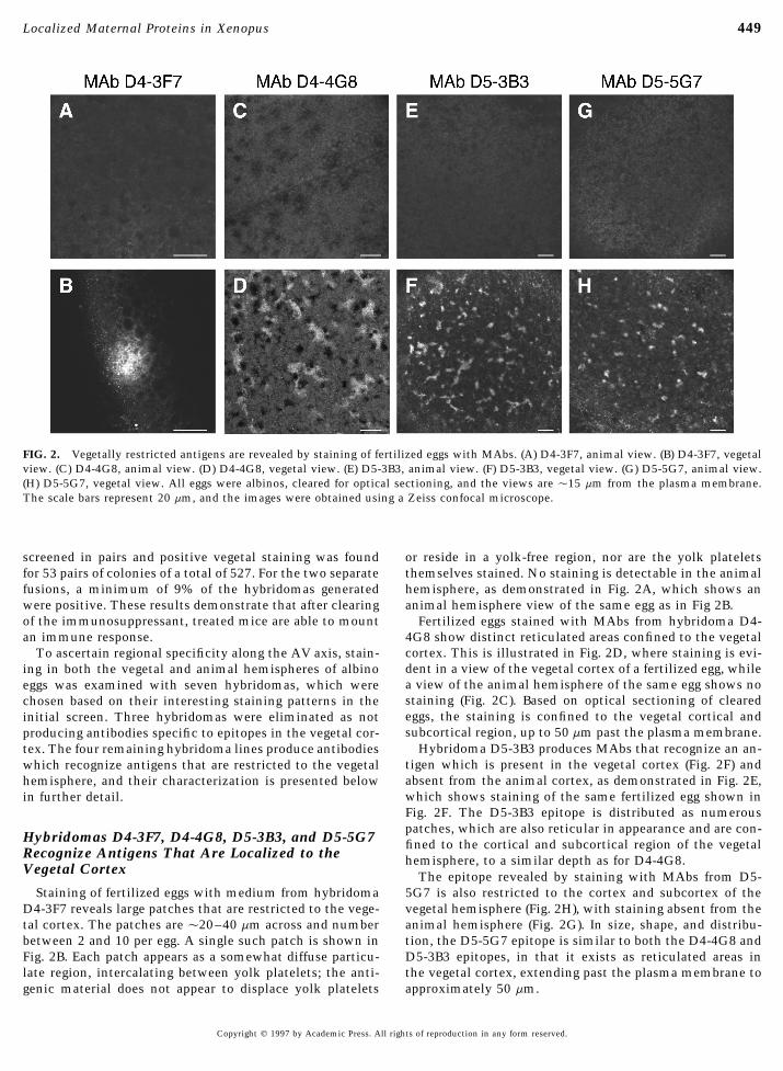
FIG. 2. Vegetally restricted antigens are revealed by staining of fertilized eggs with MAbs. (A) D4-3F7, animal view. (B) D4-3F7, vegetalview. (C) D4-4G8, animal view. (D) D4-4G8, vegetal view. (E) D5-3B3, animal view. (F) D5-3B3, vegetal view. (G) D5-5G7, animal view.(H) D5-5G7, vegetal view. All eggs were albinos, cleared for optical sectioning, and the views are Ç15 mm from the plasma membrane.The scale bars represent 20 mm, and the images were obtained using a Zeiss confocal microscope.
screened in pairs and positive vegetal staining was found or reside in a yolk-free region, nor are the yolk plateletsthemselves stained. No staining is detectable in the animalfor 53 pairs of colonies of a total of 527. For the two separate
fusions, a minimum of 9% of the hybridomas generated hemisphere, as demonstrated in Fig. 2A, which shows ananimal hemisphere view of the same egg as in Fig 2B.were positive. These results demonstrate that after clearing
of the immunosuppressant, treated mice are able to mount Fertilized eggs stained with MAbs from hybridoma D4-4G8 show distinct reticulated areas confined to the vegetalan immune response.cortex. This is illustrated in Fig. 2D, where staining is evi-To ascertain regional specificity along the AV axis, stain-dent in a view of the vegetal cortex of a fertilized egg, whileing in both the vegetal and animal hemispheres of albinoa view of the animal hemisphere of the same egg shows noeggs was examined with seven hybridomas, which werestaining (Fig. 2C). Based on optical sectioning of clearedchosen based on their interesting staining patterns in theeggs, the staining is confined to the vegetal cortical andinitial screen. Three hybridomas were eliminated as notsubcortical region, up to 50 mm past the plasma membrane.producing antibodies specific to epitopes in the vegetal cor-
Hybridoma D5-3B3 produces MAbs that recognize an an-tex. The four remaining hybridoma lines produce antibodiestigen which is present in the vegetal cortex (Fig. 2F) andwhich recognize antigens that are restricted to the vegetalabsent from the animal cortex, as demonstrated in Fig. 2E,hemisphere, and their characterization is presented belowwhich shows staining of the same fertilized egg shown inin further detail.Fig. 2F. The D5-3B3 epitope is distributed as numerouspatches, which are also reticular in appearance and are con-
Hybridomas D4-3F7, D4-4G8, D5-3B3, and D5-5G7 fined to the cortical and subcortical region of the vegetalRecognize Antigens That Are Localized to the hemisphere, to a similar depth as for D4-4G8.Vegetal Cortex The epitope revealed by staining with MAbs from D5-
5G7 is also restricted to the cortex and subcortex of theStaining of fertilized eggs with medium from hybridomaD4-3F7 reveals large patches that are restricted to the vege- vegetal hemisphere (Fig. 2H), with staining absent from the
animal hemisphere (Fig. 2G). In size, shape, and distribu-tal cortex. The patches are Ç20–40 mm across and numberbetween 2 and 10 per egg. A single such patch is shown in tion, the D5-5G7 epitope is similar to both the D4-4G8 and
D5-3B3 epitopes, in that it exists as reticulated areas inFig. 2B. Each patch appears as a somewhat diffuse particu-late region, intercalating between yolk platelets; the anti- the vegetal cortex, extending past the plasma membrane to
approximately 50 mm.genic material does not appear to displace yolk platelets
Copyright q 1997 by Academic Press. All rights of reproduction in any form reserved.
AID DB 8773 / 6x34$$$141 12-05-97 10:34:32 dbal

450 Denegre, Ludwig, and Mowry
MAbs D5-3B3, D4-4G8, and D5-5G7 Recognize body localization seen for these antigens, no epitope couldbe detected by immunocytochemistry with MAb D4-4G8Antigens Which Colocalize with Germ Plasmuntil midoogenesis. An example of the lack of staining in
The patterns of staining revealed by MAbs D4-4G8, D5- a stage I oocyte is shown in Fig. 4E, followed by an example3B3, and D5-5G7 in fertilized eggs are reminiscent of re- of cortical staining in a stage III oocyte in Fig. 4F, the earliestcently published images of germ plasm in the frog egg (Robb stage at which the epitope is detectable by immunocyto-et al., 1996). To examine the possibility that the vegetal- chemistry.specific MAbs recognize a component localized to the samesubcellular compartment as the germ plasm, fertilized eggs
MAbs D4-4G8, D4-3F7, and D5-5G7 Recognizewere labeled with the vital dye, MitoTracker, which stainsDifferent Protein Antigensthe germ plasm (Robb et al., 1996). The labeled eggs were
then fixed and costained with MAbs. Staining with each of To determine whether the vegetal-specific MAbs recog-the three MAbs was seen to colocalize with the germ plasm, nize protein antigens, Western blot analysis was performedas shown in Fig. 3, where the green images (Figs. 3A, 3D, against total fertilized egg extracts. As shown in Fig. 5,and 3G) show staining with the MAbs, the red images show MAbs D4-3F7, D4-4G8, and D5-5G7 each recognize a singleMitoTracker labeling (Figs. 3B, 3E, and 3H), and the overlap protein antigen of Ç50 kDa for D4-4G8 (lane 1), Ç40 kDais shown by yellow (Figs. 3C, 3F, and 3I). In the egg, colocal- for D4-3F7 (lane 2), and Ç67 kDa for D5-5G7 (lane 3). Theization with islands of germ plasm is striking, but not per- D5-3B3 antigen is undetectable on Western blots (notfectly coherent for D5-3B3 (Fig. 3C), D4-4G8 (Fig. 3F), and shown), suggesting either that D5-3B3 does not recognize aD5-5G7 (not shown). Regions of overlap (yellow) predomi- protein epitope or that it cannot recognize the antigen afternate, but small regions of contiguous nonoverlapping stain- denaturation. The Western blotting results with MAbs D4-ing (green or red) are present as well. This pattern may 3F7, D4-4G8, and D5-5G7 demonstrate that these three dif-indicate a complex of two overlapping compartments. Dur- ferent hybridoma lines produce antibodies against differenting the first cell cycle, islands of germ plasm coalesce into vegetally localized proteins.larger islands, which eventually ingress inward along earlycleavage furrows (Ressom and Dixon, 1988; Savage and Dan-ilchik, 1993). All three epitopes display such behavior, DISCUSSIONwhich is demonstrated with MAb D5-5G7 (Figs. 3G–3I), inan eight-cell embryo showing a large coalesced island of Reasoning that localized maternal molecules play a sig-germ plasm adjacent to a cleavage furrow, costained with nificant role in development, we created a subtractive li-D5-5G7 (Fig. 3G) and MitoTracker (Fig. 3H). Colocalization, brary of MAbs against proteins specific to the vegetal hemi-as demonstrated by an image showing both channels (Fig. sphere of the frog egg. Most current analyses of maternally3I), is very coherent at this stage and suggests that there derived patterning in the frog embryo have been the resultmay be remodeling of the D5-5G7, D4-4G8, and D5-3B3 of a top-down strategy, wherein determined cell types areepitopes during aggregation. the starting point for a reverse mapping of the signaling
pathways involved (reviewed in Kessler and Melton, 1994;Hogan, 1996; Kuhl and Wedlich, 1997). This approach hasTwo Patterns of Biogenesis Are Revealed by MAbssignificantly increased our understanding of early pat-D5-3B3, D5-5G7, and D4-4G8terning in the embryo, but mapping has led back to the eggin only a few cases (see for examples, Ku and Melton, 1993;The colocalization of D4-3B3, D4-4G8, and D5-5G7 anti-
gens with germ plasm in eggs led us to ask if the biogenesis Heasman et al., 1994; Yost et al., 1995; Larabell et al., 1997).Complementary approaches to directly identify determi-of these antigens is similar to the pattern reported for germ
plasm during oogenesis. In very young (stage I) oocytes, nants within the egg or oocyte have also been successful(Rebagliati et al., 1985; Mosquera et al., 1993), but havegerm plasm is localized to the Balbiani body (Heasman et
al., 1984). The epitopes recognized by MAbs D5-3B3 and focused attention exclusively on localized RNAs ratherthan proteins. To search for localized maternal proteinsD5-5G7 are both present as early as stage I of oogenesis and
are highly localized to the Balbiani body. This is demon- with potential roles as determinants, we have used the eggas a starting point for a novel immunological approach tostrated in a stage I oocyte stained with D5-5G7 in Figs. 4A
and 4B, showing serial sections through the Balbiani body. this problem. We have discovered at least three proteinswhich are polarized along the AV axis, all three being highlyMAb D5-3B3 exhibits the same pattern, as seen in Fig. 4C.
During stage II of oogenesis, the Balbiani body breaks down localized to the vegetal cortex.The means to uncover potentially rare, localized antigensand its contents migrate to the future vegetal cortex (Heas-
man et al., 1984). The epitopes for MAbs D5-3B3 and D5- against a background of abundant unlocalized proteins hasbeen provided by an immunosubtraction strategy (Matthew5G7 follow this pattern, as demonstrated for D5-3B3 in Fig.
4D. In this stage II oocyte the Balbiani body is no longer and Sandrock, 1987). It was not immediately obvious, how-ever, that such a scheme could be successfully applied to apresent, and the staining material now extends between the
GV and the vegetal cortex. MAb D5-5G7 demonstrates the large, yolk-rich cell such as the frog egg, which has exten-sive stockpiles of maternal proteins. Therefore, it was im-same pattern (data not shown). In contrast to the Balbiani
Copyright q 1997 by Academic Press. All rights of reproduction in any form reserved.
AID DB 8773 / 6x34$$$141 12-05-97 10:34:32 dbal

451Localized Maternal Proteins in Xenopus
FIG. 3. Colocalization of vegetal antigens with germ plasm. Shown are vegetal views of fertilized eggs (A–F) or an eight-cell embryo (G–I) labeled with MitoTracker and stained with the following MAbs: (A–C) D5-3B3, (B–F) D4-4G8, and (G–I) D5-5G7. MAb staining isshown as green, germ plasm is shown in red, and regions of colocalization appear as yellow. All eggs and embryos were cleared for opticalsectioning, and the images were obtained using a Zeiss confocal microscope. The scale bars represent 20 mm.
portant to derive a minimum estimate of the effectiveness vegetal hemisphere. Also, in the initial screen 8 of the total815 hybridomas could be eliminated as not vegetally spe-for the immunosubtraction. Our results indicate that sub-
traction was effective, but not complete, since three of the cific, since staining was strong enough to be seen even inthe pigmented animal hemisphere. These data indicate thatseven MAbs examined in detail were not specific to the
Copyright q 1997 by Academic Press. All rights of reproduction in any form reserved.
AID DB 8773 / 6x34$$$141 12-05-97 10:34:32 dbal

452 Denegre, Ludwig, and Mowry
FIG. 4. Two distinct patterns of biogenesis during early oogenesis. Oocytes were stained with the indicated MAbs and imaged using aZeiss confocal microscope. Scale bars for all panels represent 50 mm. (A, B) Serial sections of a stage I oocyte, stained with D5-5G7. (C)Stage I oocyte stained with D5-3B3. (D) Stage II oocyte stained with D5-3B3. (E) Stage I oocyte stained with D4-4G8. (F) Stage III oocytestained with D4-4G8.
a minimum of 0.5% (4/815) of the hybridomas generated between 300 and 600 colonies, a minimum result of 0.5%subtracted positives will yield at least two to three MAbsthrough immunosubtraction are genuine products of sub-
traction. Since spleen cell/myeloma fusions typically yield specific to the region of interest, a success rate which sup-ports the use of this strategy to identify specific localizedproteins.
We have identified four hybridomas whose MAbs recog-nize epitopes showing strong localization to the vegetal cor-tex of the frog egg. One MAb, D4-3F7, recognizes a proteinwith a previously uncharacterized distribution in the vege-tal cortex. The other three MAbs recognize epitopes whichcolocalize to islands of germ plasm, a long-studied cyto-plasmic localization thought to be a germ cell determinant.Two of the epitopes which colocalize with the germ plasm,those recognized by D5-3B3 and D5-5G7, are present in theBalbiani body of early oocytes, while epitope D4-4G8 is notidentifiable until stage III of oogenesis. Based on Westernblot analysis, the MAbs recognize at least three distinctproteins ofÇ40 (D4-3F7),Ç 50 (D4-4G8), andÇ67 kDa (D5-5G7). In spite of the similarities in staining patterns seen bywhole-mount immunocytochemistry with MAbs D4-4G8,
FIG. 5. Protein antigens are revealed by Western blot analysis. EggD5-3B3, and D5-5G7, it is apparent from Western blots,extracts were subjected to SDS–PAGE, transferred to nitrocelluloseand the differences in their biogenesis, that MAb D4-4G8filters, and probed with D4-4G8 (lane 1), D4-3F7, (lane 2), or D5-recognizes a different protein than MAb D5-5G7. The stain-5G7 (lane 3). The positions of the proteins recognized by the MAbsing patterns, however, would suggest that the two proteinsare indicated by arrowheads, and the sizes of molecular weight
standards are shown at the right. are localized to the same compartment in the egg. MAb D5-
Copyright q 1997 by Academic Press. All rights of reproduction in any form reserved.
AID DB 8773 / 6x34$$$141 12-05-97 10:34:32 dbal

453Localized Maternal Proteins in Xenopus
3B3 may also recognize a unique antigen which localizes lapping domains within the vegetal cortex has been de-scribed for RNA molecules (Forristall et al., 1995; Kloc andto this compartment, or it may recognize a different epitope
of the same protein as recognized by MAb D5-5G7. Etkin, 1995), and our results indicate that the organizationalcomplexity of the vegetal cortex may be even greater thanThe identity of the proteins recognized by the vegetal-
specific MAbs remains to be established. There is nothing previously appreciated.It is evident that many maternal components of the frogimmediately obvious about the nature of the D4-3F7 epi-
tope as seen by immunohistology. No previously described egg are localized in a manner critical to the correct pat-terning of the future embryo. The vegetally localized mater-structures in the vegetal cortex of the frog egg are known
to us to be comparable, nor does the size of the protein nal proteins that we have identified may contribute to this,and it will next be important to determine what role theserecognized by Western blot recommend an identity. Further
characterization of the biogenesis of the protein, and its proteins may play in early embryogenesis. Our results sug-gest that the analysis of localized maternal proteins in thepatterning in embryogenesis, will be necessary to provide
clues to its possible function or identity. frog egg is a productive area for exploring potential determi-nants.The colocalization of the D4-4G8, D5-3B3, and D5-5G7
epitopes with islands of germ plasm is intriguing. Germplasm is historically defined as electron-dense, granular–fibrillar material embedded in aggregates of mitochondria, ACKNOWLEDGMENTSwith no limiting membrane (Smith, 1975). This picture isbecoming more complex. From the work of Robb et al.
We gratefully acknowledge Doug Hixson and Jeanne Brown at(1996), it is clear that the compartments of mitochondriaRhode Island Hospital for sharing their expertise in hybridoma pro-stained by MitoTracker encompass other molecules, in par-duction and maintenance, Gary Wessel for helpful discussions andticular a kinesin-like protein (Xklp1), vimentin, and micro-use of a Zeiss 401 objective, John Coleman for use of the Zeiss
tubules. The presence of these molecules, which are not IM, and Sean Conner for Western blotting conditions. Additionalunique to the germ plasm, indicates that islands of germ gratitude goes to the reviewers for comments in strengthening theplasm may contain more than the classic electron-dense manuscript. This work was supported in part by an NRSA Postdoc-material surrounded by mitochondria. Additional prece- toral Fellowship to J.M.D. (HD07966).dence for this is provided by Terasaki et al. (1984), whoshowed that staining of mitochondria with the lipophilicdye DiOC6(3) also reveals a strong association with the
REFERENCESendoplasmic reticulum. In Caenorhabditis elegans, tran-sient associations between the germ plasm and specific
Blackler, A. W. (1958). Contribution to the study of germ-cells inproteins have been demonstrated. Both MEX-3, a cyto-the Anuran. J. Embryol. Exp. Morphol. 6, 491–503.plasmic putative RNA binding protein, and PIE-1, a poten-
Carnahan, J. F., and Patterson, P. H. (1991). The generation oftial repressor of transcription, associate with the germ-line- monoclonal antibodies that bind preferentially to adrenal chro-specific P granules only during early development (Draper maffin cells and the cells of embryonic sympathetic ganglia. J.et al., 1996; Mello et al., 1996). MEX-1, which is similar Neurosci. 11, 3493–3506.to PIE-1 in structure, associates with P granules only during Conklin, E. G. (1905). The organization and cell lineage of the ascid-
ian egg. J. Acad. Natl. Sci. Phil. 13, 1–119.early cleavages and may play a role in anchoring P granulesDavis, J. M., Pennington, J. E., Kubler, A.-M., and Conscience, J. F.for proper partitioning (Guedes and Priess, 1997). A similar
(1982). A simple, single-step technique for selecting and cloningtransient association with germ plasm may explain ourhybridomas for the production of monoclonal antibodies. J. Im-results.munol. Methods 50, 161–171.We do not believe that MAbs D4-4G8, D5-3B3, and D5-
Draper, B. W., Mello, C. C., Bowerman, B., Hardin, J., and Priess,57 recognize germ plasm components in the classic sense,J. R. (1996). MEX-3 is a KH domain protein that regulates blasto-
but instead that the staining patterns seen with these MAbs mere identity in early C. elegans embryos. Cell 87, 205–216.reveal overlapping subcompartments. The first appearance Dumont, J. N. (1971). Oogenesis in Xenopus laevis (Daudin). I.of D4-4G8 staining during stage III of oogenesis strongly Stages of oocyte development in laboratory maintained animals.argues against its identity as a germ plasm component, since J. Morphol. 136, 153–179.
Elinson, R. P., King, M. L., and Forristall, C. (1993). Isolated vegetalno such pattern has been described for any germ plasm ele-cortex from Xenopus oocytes selectively retains localizedment. While the early biogenesis of the D5-3B3 and D5-mRNAs. Dev. Biol. 160, 554–562.5G7 epitopes is similar to that described for germ plasm,
Forristall, C., Pondel, M., Chen, L., and King, M. L. (1995). Patternscostaining with MitoTracker in the fertilized egg is not per-of localization and cytoskeletal association of two vegetally lo-fectly coherent, suggesting that they are not germ plasmcalized RNAs, Vg1 and Xcat-2. Development 121, 201–208.components. Rather, the staining patterns seen with MAbs
Guedes, S., and Priess, J. R. (1997). The C. elegans MEX-1 proteinD4-4G8, D5-3B3, and D5-57 and MitoTracker reveal two is present in germline blastomeres and is a P granule component.closely associated compartments in the vegetal cortex of Development 124, 731–739.the egg. It is only during cleavage, as the germ plasm islands Heasman, J., Crawford, A., Goldstone, K., Garner-Hamrick, P.,aggregate, that these compartments appear to coalesce into Gumbiner, B., McCrea, P., Kintner, C., Noro, C. Y., and Wylie,
C. (1994). Overexpression of cadherin and underexpression ofone coherent compartment. Localization to distinct, over-
Copyright q 1997 by Academic Press. All rights of reproduction in any form reserved.
AID DB 8773 / 6x34$$$141 12-05-97 10:34:32 dbal

454 Denegre, Ludwig, and Mowry
b-catenin inhibit dorsal mesoderm induction in early Xenopus Rebagliati, M. R., Weeks, D. L., Harvey, R. P., and Melton, D. A.(1985). Identification and cloning of localized maternal mRNAsembryos. Cell 79, 791–803.from Xenopus eggs. Cell 42, 769–777.Heasman, J., Quarmby, J., and Wylie, C. C. (1984). The mitochon-
Ressom, R. E., and Dixon, K. E. (1988). Relocation and reorganiza-drial cloud of Xenopus oocytes: The source of germinal granuletion of germ plasm in Xenopus embryos after fertilization. Devel-material. Dev. Biol. 105, 458–469.opment 103, 507–518.Hixson, D. D., and Allison, J. P. (1985). Monoclonal antibodies rec-
Robb, D. L., Heasman, J., Raats, J., and Wylie, C. (1996). A kinesin-ognizing oval cells induced in the liver of rats by N-2-fluorenyla-like protein is required for germ-plasm aggregation in Xenopus.cetamide or ethionine in a choline-deficient diet. Cancer Res.Cell 87, 823–831.45, 3750–3760.
Savage, R. M., and Danilchik, M. V. (1993). Dynamics of germHogan, B. L. M. (1996). Bone morphogenetic proteins: Multifunc-plasm localization and its inhibition by ultraviolet irradiation intional regulators of vertebrate development. Genes Dev. 10,early cleavage Xenopus embryos. Dev. Biol. 157, 371–382.1580–1594.
Smith, L. D., Williams, M. (1975). Germinal plasm and determina-Kessler, D. S., and Melton, D. A. (1994). Vertebrate embryonic in-tion of the primordial germ cells. In ‘‘The Developmental Biologyduction: Mesodermal and neural patterning. Science 266, 596–of Reproduction’’ (C. L. Markert and J. Papaconstantinou, Eds.),604.pp. 3–24. Academic Press, New York.Kloc, M., and Etkin, L. (1995). Two distinct pathways for the local-
Smith, R. C., Neff, A. W., and Malacinski, G. M. (1986). Accumula-ization of RNAs at the vegetal cortex in Xenopus oocytes. Devel-tion, organization and deployment of oogenetically derived Xeno-opment 121, 287–297.pus yolk/nonyolk proteins. Development Suppl. 97, 45–64.Klymkowsky, M. W., and Karnovsky, A. K. (1994). Morphogenesis
St. Johnston, D. (1995). The intracellular localization of messengerand the cytoskeleton: Studies of the Xenopus embryo. Dev. Biol.RNAs. Cell 81, 161–170.165, 372–384.
Suzue, T., Kaprielian, A., and Patterson, P. H. (1990). A monoclonalKu, M., and Melton, D. A. (1993). Xwnt-11: A novel maternallyantibody that defines rostrocaudal gradients in the mammalianexpressed Xenopus wnt gene. Development 119, 1161–1173.nervous system. Neuron 5, 421–431.Kuhl, M., and Wedlich, D. (1997). Wnt signalling goes nuclear.
Suzuki, A. S., Manabe, J., and Hiracawa, A. (1991). Dynamic distri-Bioessays 19, 101–104.bution of region-specific maternal protein during oogenesis andLarabell, C. A., Torres, M., Rowning, B. A., Yost, C., Miller, J. R.,early embryogenesis of Xenopus laevis. Roux’s Arch. Dev. Biol.Wu, M., Kimelman, D., and Moon, R. T. (1997). Establishment200, 213–222.of the dorso-ventral axis in Xenopus embryos is presaged by early
Terasaki, M., Song, J., Wong, J. R., Weiss, M. J., and Chen, L. B.asymmetries in b-catenin that are modulated by the wnt signal-(1984). Localization of endoplasmic reticulum in living and glu-ing pathway. J. Cell Biol. 136, 1123–1136.taraldehyde fixed cells with fluorescent dyes. Cell 38, 101–108.Matthew, W. D., and Sandrock, A. W. (1987). Cyclophosphamide
Towbin, H., Staehlin, T., and Gordon, J. (1979). Electrophoretictreatment used to manipulate the immune response for the pro-transfer of proteins from polyacrylamide gels to nitrocelluloseduction of monoclonal antibodies. J. Immunol. Methods 100, 73.sheets: Procedure and some applications. Proc. Natl. Acad. Sci.Mello, C. C., Schubert, C., Draper, B., Zhang, W., Lobel, R., andUSA 76, 4350–4354.Priess, J. R. (1996). The PIE-1 protein and germline specification
Whitman, C. O. (1878). The embryology of Clepsine. Q. J. Microsc.in C. elegans embryos. Nature 382, 710–712.Sci. 18, 215.Moen, T. L., and Namenwirth, M. (1977). The distribution of solu-
Williams, M. A., and Smith, L. D. (1971). Ultrastructure of the ‘‘ger-ble proteins along the animal–vegetal axis of frog eggs. Dev. Biol.minal plasm’’ during maturation and early cleavage of Rana pi-58, 1–10.piens. Dev. Biol. 25, 568–580.Mosquera, L., Forristall, C., Zhou, Y., and King, M. L. (1993). A
Wilson, E. B. (1925). ‘‘The Cell in Development and Heredity.’’mRNA localized to the vegetal cortex of Xenopus oocytes en-Macmillan, New York.codes a protein with a nanos-like zinc finger. Development 117,
Yost, H. J., Phillips, C. R., Boore, J. L., Bertman, J., Whalon, B., and377–386.Danilchik, M. V. (1995). Relocation of mitochondria to the pro-Nieuwkoop, P. D. (1969). The formation of mesoderm in urodeleanspective dorsal marginal zone during Xenopus embryogenesis.amphibians. I. Induction by the endoderm. Roux’s Arch. En-Dev. Biol. 170(1), 83–90.twicklungsmech. Org. 162, 341–373.
Nieuwkoop, P. D., and Faber, J. (1967). ‘‘Normal Table of Xenopus Received for publication July 18, 1997Accepted October 3, 1997laevis (Daudin).’’ North Holland, Amsterdam.
Copyright q 1997 by Academic Press. All rights of reproduction in any form reserved.
AID DB 8773 / 6x34$$$141 12-05-97 10:34:32 dbal
Copyright © 2022 FDOKUMEN