Human copper transporter 2 is localized in late endosomes and lysosomes and facilitates cellular...
11
Biochem. J. (2007) 407, 49–59 (Printed in Great Britain) doi:10.1042/BJ20070705 49 Human copper transporter 2 is localized in late endosomes and lysosomes and facilitates cellular copper uptake Peter V. E. VAN DEN BERGHE*, Dineke E. FOLMER*, Helga E. M. MALINGR ´ E*, Ellen VAN BEURDEN*, Adriana E. M. KLOMP*, Bart VAN DE SLUIS*†, Maarten MERKX‡, Ruud BERGER* and Leo W. J. KLOMP* 1 *Department of Metabolic and Endocrine Diseases, University Medical Center Utrecht, 3584 EA Utrecht, The Netherlands, †Complex Genetics Section, Department of Medical Genetics, University Medical Center Utrecht, 3508 TA Utrecht, The Netherlands, and ‡Department of Biomedical Engineering, Technical University Eindhoven, 5600 MB Eindhoven, The Netherlands High-affinity cellular copper uptake is mediated by the CTR (copper transporter) 1 family of proteins. The highly homologous hCTR (human CTR) 2 protein has been identified, but its function in copper uptake is currently unknown. To characterize the role of hCTR2 in copper homoeostasis, epitope-tagged hCTR2 was transiently expressed in different cell lines. hCTR2–vsvG (vesi- cular-stomatitis-virus glycoprotein) predominantly migrated as a 17 kDa protein after imunoblot analysis, consistent with its pre- dicted molecular mass. Chemical cross-linking resulted in the de- tection of higher-molecular-mass complexes containing hCTR2– vsvG. Furthermore, hCTR2–vsvG was co-immunoprecipitated with hCTR2–FLAG, suggesting that hCTR2 can form multimers, like hCTR1. Transiently transfected hCTR2–eGFP (enhanced green fluorescent protein) was localized exclusively to late endo- somes and lysosomes, and was not detected at the plasma mem- brane. To functionally address the role of hCTR2 in copper meta- bolism, a novel transcription-based copper sensor was developed. This MRE (metal-responsive element)–luciferase reporter con- tained four MREs from the mouse metallothionein 1A promoter upstream of the firefly luciferase open reading frame. Thus the MRE–luciferase reporter measured bioavailable cytosolic copper. Expression of hCTR1 resulted in strong activation of the reporter, with maximal induction at 1 µM CuCl 2 , consistent with the K m of hCTR1. Interestingly, expression of hCTR2 significantly induced MRE–luciferase reporter activation in a copper-dependent manner at 40 and 100 µM CuCl 2 . Taken together, these results identify hCTR2 as an oligomeric membrane protein localized in lysosomes, which stimulates copper delivery to the cytosol of human cells at relatively high copper concentrations. This work suggests a role for endosomal and lysosomal copper pools in the maintenance of cellular copper homoeostasis. Key words: copper biosensor, copper transporter 1 (Ctr1), copper transporter 2 (Ctr2), copper uptake, metallothionein, zinc. INTRODUCTION Copper is a trace element which is essential for all organisms that utilize oxygen as an electron acceptor during respiration. As a transition metal, copper serves as a cofactor in a number of redox enzymes. The same redox-active properties also render the metal potentially toxic, as it can induce free radical formation and cause direct damage to proteins, lipids and DNA. To maintain copper homoeostasis, remarkably efficient mechanisms have evolved to regulate copper import and export and cellular distribution of the metal. The proteins involved in cellular copper homoeostasis are largely conserved in both prokaryotes and eukaryotes [1–3]. These proteins form intricate metabolic pathways aimed at maintaining an extremely low free copper concentration, while ensuring that sufficient copper is bioavailable to sustain essential copper- dependent cellular processes [4]. As one example, a significant proportion of cellular copper is bound to copper chaperones, which deliver copper to specific target enzymes or transporters by regulated protein–protein interactions [5,6]. A second example, metallothioneins are small ubiquitously expressed proteins that scavenge cytosolic copper and other metals such as zinc and cadmium [7], and the expression of metallothionein genes is transcriptionally regulated in a copper-dependent fashion. Severe disorders may arise as a result of disruption of copper homoeostasis. Recessive mutations in the gene encoding ATP7A (copper-transporting P 1B -type ATPase) result in Menkes disease, a fatal X-linked neurodevelopmental disorder [8,9]. ATP7A deficiency results in a lack of copper transport through the placenta and the intestinal epithelium, leading to systemic copper deficiency and the failure to provide essential cuproenzymes with the metal. The calamity mutant of Danio rerio, the zebrafish, is a recently characterized animal model for Menkes disease [10], which displays a marked reduction in pigment formation and a strikingly altered notochord development in comparison with wild-type zebrafish. Interestingly, the calamity mutant is phenocopied by copper deficiency, thus illustrating further the essential role of copper in physiology and development. Copper uptake is facilitated by the CTR (copper transporter) family of proteins, which are highly conserved [11]. In Saccharo- myces cerevisiae, high-affinity uptake of copper is mediated by yCtr1p and yCtr3p [12,13]. A third Ctr protein expressed in S. cerevisiae, yCtr2p, is thought to mediate low-affinity copper uptake. In mammals, a high-affinity copper permease, CTR1, facilitates copper import, with a K m of approx. 1–5 µM [14]. hCTR (human CTR) 1 is predominantly localized to intra- cellular vesicles which are in close apposition to the Golgi Abbreviations used: ATP7A, copper-transporting P 1B -type ATPase; BCS, bathocuproinedisulfonic acid; CTR, copper transporter; DFO, desferrioxamine; DMEM, Dulbecco’s modified Eagle’s medium; eGFP, enhanced green fluorescent protein; EGS, ethylene glycolbis(succinimidylsuccinate); FCS, foetal calf serum; hCTR, human CTR; HEK-293T cell, human embryonic kidney cell expressing the large T-antigen of SV40 (simian virus 40); LAMP, lysosome- associated membrane protein; MEF, mouse embryonic fibroblast; MRE, metal-responsive element; MTF, metal responsive transcription factor; MTT, 3-(4,5-dimethylthiazol-2-yl)-2,5-diphenyl-2H-tetrazolium bromide; RLU, relative light units; TfR, transferrin receptor; TGN, trans-Golgi network; vsvG, vesicular-stomatitis-virus glycoprotein. 1 To whom correspondence should be addressed (email [email protected]). c The Authors Journal compilation c 2007 Biochemical Society
Transcript of Human copper transporter 2 is localized in late endosomes and lysosomes and facilitates cellular...

Biochem. J. (2007) 407, 49–59 (Printed in Great Britain) doi:10.1042/BJ20070705 49
Human copper transporter 2 is localized in late endosomes and lysosomesand facilitates cellular copper uptakePeter V. E. VAN DEN BERGHE*, Dineke E. FOLMER*, Helga E. M. MALINGRE*, Ellen VAN BEURDEN*, Adriana E. M. KLOMP*,Bart VAN DE SLUIS*†, Maarten MERKX‡, Ruud BERGER* and Leo W. J. KLOMP*1
*Department of Metabolic and Endocrine Diseases, University Medical Center Utrecht, 3584 EA Utrecht, The Netherlands, †Complex Genetics Section, Department of Medical Genetics,University Medical Center Utrecht, 3508 TA Utrecht, The Netherlands, and ‡Department of Biomedical Engineering, Technical University Eindhoven, 5600 MB Eindhoven,The Netherlands
High-affinity cellular copper uptake is mediated by the CTR(copper transporter) 1 family of proteins. The highly homologoushCTR (human CTR) 2 protein has been identified, but its functionin copper uptake is currently unknown. To characterize the roleof hCTR2 in copper homoeostasis, epitope-tagged hCTR2 wastransiently expressed in different cell lines. hCTR2–vsvG (vesi-cular-stomatitis-virus glycoprotein) predominantly migrated as a17 kDa protein after imunoblot analysis, consistent with its pre-dicted molecular mass. Chemical cross-linking resulted in the de-tection of higher-molecular-mass complexes containing hCTR2–vsvG. Furthermore, hCTR2–vsvG was co-immunoprecipitatedwith hCTR2–FLAG, suggesting that hCTR2 can form multimers,like hCTR1. Transiently transfected hCTR2–eGFP (enhancedgreen fluorescent protein) was localized exclusively to late endo-somes and lysosomes, and was not detected at the plasma mem-brane. To functionally address the role of hCTR2 in copper meta-bolism, a novel transcription-based copper sensor was developed.This MRE (metal-responsive element)–luciferase reporter con-
tained four MREs from the mouse metallothionein 1A promoterupstream of the firefly luciferase open reading frame. Thusthe MRE–luciferase reporter measured bioavailable cytosoliccopper. Expression of hCTR1 resulted in strong activation ofthe reporter, with maximal induction at 1 µM CuCl2, consistentwith the Km of hCTR1. Interestingly, expression of hCTR2significantly induced MRE–luciferase reporter activation in acopper-dependent manner at 40 and 100 µM CuCl2. Takentogether, these results identify hCTR2 as an oligomeric membraneprotein localized in lysosomes, which stimulates copper deliveryto the cytosol of human cells at relatively high copperconcentrations. This work suggests a role for endosomal andlysosomal copper pools in the maintenance of cellular copperhomoeostasis.
Key words: copper biosensor, copper transporter 1 (Ctr1), coppertransporter 2 (Ctr2), copper uptake, metallothionein, zinc.
INTRODUCTION
Copper is a trace element which is essential for all organisms thatutilize oxygen as an electron acceptor during respiration. As atransition metal, copper serves as a cofactor in a number of redoxenzymes. The same redox-active properties also render the metalpotentially toxic, as it can induce free radical formation and causedirect damage to proteins, lipids and DNA. To maintain copperhomoeostasis, remarkably efficient mechanisms have evolved toregulate copper import and export and cellular distribution of themetal. The proteins involved in cellular copper homoeostasis arelargely conserved in both prokaryotes and eukaryotes [1–3]. Theseproteins form intricate metabolic pathways aimed at maintainingan extremely low free copper concentration, while ensuringthat sufficient copper is bioavailable to sustain essential copper-dependent cellular processes [4]. As one example, a significantproportion of cellular copper is bound to copper chaperones,which deliver copper to specific target enzymes or transporters byregulated protein–protein interactions [5,6]. A second example,metallothioneins are small ubiquitously expressed proteins thatscavenge cytosolic copper and other metals such as zinc andcadmium [7], and the expression of metallothionein genes istranscriptionally regulated in a copper-dependent fashion.
Severe disorders may arise as a result of disruption of copperhomoeostasis. Recessive mutations in the gene encoding ATP7A(copper-transporting P1B-type ATPase) result in Menkes disease,a fatal X-linked neurodevelopmental disorder [8,9]. ATP7Adeficiency results in a lack of copper transport through theplacenta and the intestinal epithelium, leading to systemic copperdeficiency and the failure to provide essential cuproenzymes withthe metal. The calamity mutant of Danio rerio, the zebrafish,is a recently characterized animal model for Menkes disease[10], which displays a marked reduction in pigment formationand a strikingly altered notochord development in comparisonwith wild-type zebrafish. Interestingly, the calamity mutant isphenocopied by copper deficiency, thus illustrating further theessential role of copper in physiology and development.
Copper uptake is facilitated by the CTR (copper transporter)family of proteins, which are highly conserved [11]. In Saccharo-myces cerevisiae, high-affinity uptake of copper is mediated byyCtr1p and yCtr3p [12,13]. A third Ctr protein expressed inS. cerevisiae, yCtr2p, is thought to mediate low-affinity copperuptake. In mammals, a high-affinity copper permease, CTR1,facilitates copper import, with a Km of approx. 1–5 µM [14].hCTR (human CTR) 1 is predominantly localized to intra-cellular vesicles which are in close apposition to the Golgi
Abbreviations used: ATP7A, copper-transporting P1B-type ATPase; BCS, bathocuproinedisulfonic acid; CTR, copper transporter; DFO, desferrioxamine;DMEM, Dulbecco’s modified Eagle’s medium; eGFP, enhanced green fluorescent protein; EGS, ethylene glycolbis(succinimidylsuccinate); FCS, foetalcalf serum; hCTR, human CTR; HEK-293T cell, human embryonic kidney cell expressing the large T-antigen of SV40 (simian virus 40); LAMP, lysosome-associated membrane protein; MEF, mouse embryonic fibroblast; MRE, metal-responsive element; MTF, metal responsive transcription factor; MTT,3-(4,5-dimethylthiazol-2-yl)-2,5-diphenyl-2H-tetrazolium bromide; RLU, relative light units; TfR, transferrin receptor; TGN, trans-Golgi network; vsvG,vesicular-stomatitis-virus glycoprotein.
1 To whom correspondence should be addressed (email [email protected]).
c© The Authors Journal compilation c© 2007 Biochemical Society

50 P. V. E. van den Berghe and others
complex, but a fraction is also present at the plasma membrane[15,16]. The proportion of cell-surface-exposed hCTR1 is de-pendent on the cell type [15], and elevated concentrations of cop-per have been shown to result in the internalization of cell-surfacehCTR1, possibly as a rapid adaptive response to limit copperuptake [17]. hCTR1 contains three membrane-spanning regionsand is thought to assemble into a trimer which forms an aqueouspore to enable copper transport [18,19].
The physiological role of the Ctr1 family of proteins hasbeen addressed elegantly using knockout mouse models. Ctr1-knockout mice died during mid-gestation as a result of copperdeficiency [20,21]. Conditional knockout mice, lacking Ctr1specifically in the intestinal epithelium, were born healthy, butdisplayed markedly impaired dietary copper uptake, resulting ina severe systemic copper deficiency comparable with Menkesdisease patients [22]. This knockout mouse work established theessential role of Ctr1 in embryonic development and in dietarycopper uptake. However, these animals display some residualcellular copper uptake, suggesting that additional pathways ofcopper import do exist. Furthermore, MEFs (mouse embryonicfibroblasts) obtained from Ctr1-knockout embryos survived inculture without the addition of extra copper to their medium, andwere able to take up copper in a low-affinity manner [23]. Theproteins mediating Ctr1-independent low-affinity copper importhave currently not been characterized.
A candidate protein which may participate in such alternativecopper import routes is hCTR2. Alignment of the primaryamino acid sequences of hCTR1 and CTR2 from multiplespecies revealed marked homology in the transmembrane regions,suggesting that hCTR2 contains three putative transmembraneregions, similar to hCTR1 [14,24] (Figure 1A). Extrapolatingfrom hCTR1, the N-terminus of hCTR2 is probably located extra-cytoplasmically [14,24]. The highly conserved MXXXM (Met-Xaa-Xaa-Xaa-Met) motif in the second transmembrane regionof hCTR1, which has been shown to be critical for copper co-ordination during copper uptake [25], is also present in hCTR2.All amino acid residues that have been shown to be criticalfor oligomerization and copper transport activity of hCTR1are conserved in hCTR2: the GXXXG (Gly-Xaa-Xaa-Xaa-Gly)motif in the third putative transmembrane region of hCTR1[26], one methionine residue, approx. 20 residues upstream ofthe first transmembrane domain (Figure 1A), and the MXXXMmotif in the second transmembrane domain [27]. There are alsonotable differences between hCTR2 and hCTR1. hCTR2 lacksthe MXXXM motif and histidine-rich regions that are importantfor high-affinity copper transport, and does not contain theappropriate consensus sites for N-glycosylation [27–29]. Theseobservations support the hypothesis that hCTR2 does transportcopper, but suggest that this occurs with a lower affinity thanthat of hCTR1. However, the function of hCTR2 has not beenexperimentally addressed and its annotation as a CTR has beenbased only on the alignment with other CTR sequences [30].In the present study, we have characterized the role of hCTR2in copper homoeostasis using a combination of biochemicalanalyses, cellular localization studies and a novel copper sensor.The results provide evidence that hCTR2 is an intracellular proteininvolved in copper uptake in human cells.
EXPERIMENTAL
Cell culture and MTT [3-(4,5-dimethylthiazol-2-yl)-2,5-diphenyl-2H-tetrazolium bromide] viability assay
The HEK-293T cell [human embryonic kidney cell expressingthe large T-antigen of SV40 (simian virus 40)] line, HeLa cell
line and U2OS cell line were purchased from the A.T.C.C.Cells were cultured at 37 ◦C under humidified air containing5% CO2, and were maintained in DMEM (Dulbecco’s modi-fied Eagle’s medium) GlutaMAXTM (Invitrogen) containing 10%(v/v) FCS (foetal calf serum) (Invitrogen), 100 µg/ml penicil-lin (Invitrogen) and 100 µg/ml streptomycin (Invitrogen). Forsome experiments, cells were cultured in DMEM containing10% FCS, 100 µg/ml penicillin and 100 µg/ml streptomycinin the presence of different concentrations of CuCl2, ZnCl2,MgCl2, FeCl3, MnCl2 (Merck Chemicals) or a copper chelator,BCS (bathocuproinedisulfonic acid) (Sigma). MTT viabilityassays [31] were performed after incubation of the cells withdifferent concentrations of metals in 96-well microtitre plates(Corning). Cells were incubated with 0.4 mg/ml MTT (Sigma)in DMEM containing 10% (v/v) FCS, 100 µg/ml penicillin and100 µg/ml streptomycin for 30 min at 37 ◦C. Prior to lysis, cellswere rinsed twice with PBS (25 mM sodium phosphate bufferand 140 mM NaCl, pH 7.4) and lysed in 100 µl of 100% propan-2-ol containing 40 mM HCl. Conversion of MTT into formazanwas measured at 595 nm using a multi-plate reader model 550spectrophotometer (Bio-Rad), and the viability of the cells wascalculated relative to untreated cells.
Plasmids
pCL-NEO RabIP4-vsvG (vesicular-stomatitis-virus glycopro-tein) was kindly provided by Dr P. J. van de Sluijs (Departmentof Cell Biology, University Medical Center Utrecht, Utrecht, TheNetherlands). pcDNA3.1-cullin1-FLAG was amplified from livercDNA using FLAG–cullin forward and reverse primers (Table 1).The PCR fragment was cloned into pCRII (Invitrogen) andsubsequently cloned into pcDNA3.1/ZEO. hCTR2–vsvG cDNAwas amplified from cDNA by PCR using hCTR2–vsvG forwardand hCTR2–vsvG reverse primers, and subsequently subclonedinto pZeoSV2. The reverse primer contained a sequence toadd the vsvG tag at the C-terminus of hCTR2 as describedpreviously for hCTR1–vsvG [15]. To construct pcDNA3.1/Zeo-hCTR1-vsvG (hCTR1–vsvG) and pcDNA3.1/Zeo-hCTR2-vsvG(hCTR2–vsvG), hCTR1–vsvG and hCTR2–vsvG fragments wereisolated from pZeoV2-hCTR1-vsvG [15] or pZeoV2-hCTR2-vsvG after digestion with BamHI and XbaI. The fragmentswere ligated into the BamHI and XbaI sites of pcDNA3.1/Zeo(Invitrogen). To generate C-terminal eGFP (enhanced greenfluorescent protein)-tagged hCTR1 and hCTR2, hCTR1 andhCTR2 fragments were amplified from cDNA by PCR using theAdvantage cDNA polymerase mix (Clontech), with hCTR1 andhCTR2 forward and reverse primers respectively (Table 1). Thefragments were ligated into the pCRII vector using the TA-cloningkit (Invitrogen). hCTR1 and hCTR2 fragments were isolatedfrom pCRII after EcoRI digestion and subsequently cloned intothe EcoRI site of pEGFP-N3 (Clontech), resulting in peGFP-N3-hCTR1 (hCTR1–eGFP) and peGFP-N3-hCTR2 (hCTR2–eGFP). To generate the pGL3-E1b-TATA-4MRE construct [MRE(metal-responsive element)–luciferase reporter], four MREs wereamplified by PCR from the 4 × MRE(d) construct [7,32] using4MRE-F/HindIII and 4MRE-R/PstI primers (Table 1). Thefragment was digested by HindIII and PstI, and subsequentlysubcloned into the E1b-TATA pGL3 vector (kindly providedby Dr Eric Kalkhoven, University Medical Centre Utrecht,Utrecht, The Netherlands). hCTR1 and hCTR2 constructs witha C-terminal FLAG tag were generated by PCR of hCTR1–vsvG and hCTR2–vsvG template constructs using Pfu Turbopolymerase (Stratagene) and the corresponding BamHI forwardand NotI reverse primers for hCTR1 and hCTR2 respectively.These PCR fragments were digested with BamHI and NotI,
c© The Authors Journal compilation c© 2007 Biochemical Society

hCTR2 facilitates copper uptake in mammalian cells 51
Table 1 Primers
fw, forward; rev, reverse.
Name Sequence (5′ → 3′)
FLAG–cullin1 rev tcacttgtcgtcatcgtcttgtagtcagccaagtaactgtaggtgtcFLAG–cullin1 fw atggactacaaagacgatgacgacaagtcgtcaacccggagccaghCTR2–vsvG fw atggcgatgcatttcatctthCTR2–vsvG rev tcactttcctagtcggttcatctcgatgtcagtgtaagctgtgctgagaagtgghCTR1 fw tggatcattcccaccatatghCTR1 rev atgggcaatgctctgtgatatchCTR2 fw atggcgatgcatttcatctthCTR2 rev agctgtgctgagaagtgggt4MRE-F/HindIII cccaagcttgactcgaggagctctgcac4MRE-R/PstI ctgcagtatgccaaggtcgacgggchCTR2 BamHI fw ctaggatccaccatggcgatgcatttcatcttchCTR2 NotI rev gtaagcggccgcagctgtgctgagaagtgggtaaghCTR1 BamHI fw ggatccgccaccatggatcattcccaccatatghCTR1 NotI rev gcggccgcatggcaatgctctgtgatatchCTR1 IXXXI fw cataagctacttcctcattctcatcttcattacctacaacgggtachCTR1 IXXXI rev gtacccgttgtaggtaatgaagatgagaatgaggaagtagcttatghCTR2 IXXXI fw catcggctacttcatcattctggccgtaatttcctacaacacctghCTR2 IXXXI fw caggtgttgtaggaaattacggccagaatgatgaagtagccgatg
and the fragments were ligated into the BamHI and NotIsites in pEBB-FLAG (kindly provided by Dr C.S. Duckett,University of Michigan Medical School, Ann Arbor, MI,U.S.A.), resulting in pEBB-hCTR1-FLAG and pEBB-hCTR2-FLAG constructs. pEBB-hCTR1-FLAG IXXXI (Ile-Xaa-Xaa-Xaa-Ile) and pEBB-hCTR2-FLAG IXXXI were generated byQuikChange® site-directed mutagenesis (Stratagene), using thehCTR1 IXXXI forward and reverse primers and the hCTR2IXXXI forward and reverse primers. All constructs were verifiedby sequence analysis.
Transfection, chemical cross-linking, immunoprecipitationand immunoblot analysis
HEK-293T cells were transiently transfected with either hCTR1–vsvG or hCTR2–vsvG using the calcium phosphate precipitationprotocol [33]. DNA was diluted to a maximum concentrationof 10 µg/ml in 244 mM CaCl2 solution. The DNA mixturewas then diluted with an equal volume of 2 × HBSS (to afinal concentration of 6.275 mM Hepes, 190 µM Na2HPO4,68.5 mM NaCl and 122 mM CaCl2, pH 7.5). After incubatingfor 20 min at 20 ◦C, the transfection mixture was added tothe cells in 10 ml DMEM, and the cells were harvested after2 days. Chemical cross-linking was performed by incubationof transfected HEK-293T cells for 30 min at room temperature(20 ◦C) with PBS containing 1% DMSO in 0 mM, 0.5 mM or1 mM EGS [ethylene glycolbis(succinimidylsuccinate)] (Pierce).For cross-linking experiments and immunoprecipitation, HEK-293T cells were lysed in RIPA buffer [50 mM Tris/HCl, pH 7.4,0.1% (v/v) SDS, 1% (v/v) Nonidet P40, 150 mM NaCl, 10 mg/ml sodiumdeoxychalate, 2 mM EDTA and 1 mM NaVO3] supple-mented with CompleteTM protease inhibitor cocktail (Roche), andlysates were solubilized by passage seven times through a needle(23 gauge). For immunoprecipitation, 1 mg of total protein, 20 µlof Protein A–agarose beads (Sigma) and 0.6 µg of rabbit anti-vsvG antibody (Abcam) were incubated overnight at 4 ◦C in avolume of 500 µl while rotating. For co-immunoprecipitationexperiments, 1 ml of the cell lysate was divided into twoprecipitation reaction mixtures with either 20 µl of Protein A–agarose beads and 0.6 µg of rabbit anti-vsvG antibody or 20 µlof anti-FLAG M2–agarose (Sigma), which were incubated for4 h at 4 ◦C with rotation. Immunocomplexes were precipitated
by microcentrifugation at 2000 g for 3 min at 4 ◦C and washedfour times in RIPA buffer. Immunoprecipitates were resuspendedin SDS/PAGE sample buffer [62.5 mM Tris/HCl, 4% (v/v)SDS, 5% (v/v) glycerol, 0.01% (w/v) Bromophenol Blueand 2% (v/v) 2-mercaptoethanol, pH 6.8] and heated at 95 ◦Cfor 5 min prior to resolution by SDS/PAGE (12 % gels).Proteins were transferred on to Hybond-P PVDF membranes(Amersham Biosciences) by standard immunoblot procedures.The membranes were blocked in Tris-buffered saline (25 mMTris/HCl, pH 7.4, 137 mM NaCl and 2.7 mM KCl) containing5% (w/v) non-fat dried milk (Sigma) and 0.1% (v/v) Tween20. Immunoblots were probed with rabbit anti-vsvG antibody(0.6 µg/ml) (Abcam), mouse anti-(vsvG hybridoma) supernatantfrom clone P5D4 (1:250 dilution) [24] or horseradish-peroxidase-conjugated mouse anti-FLAG M2 antibody (Sigma) for 1 h.Reactivity was detected using horseradish-peroxidase-conjugatedsecondary antibodies (1 ng/ml; Pierce) and ECL® (enhancedchemiluminescence) (Amersham Biosciences), according to themanufacturer’s instructions.
Indirect immunofluorescence and confocal laser-scanningmicroscopy
HEK-293T cells, U2OS cells and HeLa cells were transientlytransfected with hCTR1–eGFP, hCTR2–eGFP or hCTR2–vsvG,using LipofectamineTM 2000 (Invitrogen) according to the manu-facturer’s protocol. At 1 day after transfection, cells were treatedwith trypsin and seeded on to coverslips (Paul Marienfeld,GmbH & Co.). After 24 h, cells were rinsed with ice-coldPBS and fixed in 3.7% (w/v) paraformaldehyde in PBS for20 min at 4 ◦C. Cells were rinsed three times with 0.02%(v/v) Triton X-100 in PBS, and non-specific binding wasblocked with blocking buffer [5% (w/v) BSA in PBS and0.02% (v/v) Triton X-100] for 30 min at room temperature.Immunolabelling was performed in blocking buffer for 1 h withthe antibodies indicated below. Cells were rinsed three timeswith 0.02% (v/v) Triton X-100 in PBS, and secondary labellingwas performed with affinipure Alexa Fluor® 568-conjugatedgoat anti-(mouse IgG) or goat anti-(rabbit IgG) antibodies(10 µg/ml; Molecular Probes). Coverslips were mounted inFluorsave (VWR International), and confocal laser-scanningmicroscopy was performed using a Leica TCS 4D microscopeequipped with a Plan APO ×63 oil immersion objective(numerical aperture 1.4) and dedicated imaging software (LeicaTCSNT version 1.6.587). hCTR1–vsvG and hCTR2–vsvG werelabelled with rabbit anti-vsvG antibodies (0.6 µg/ml) (Abcam).For double-labelling experiments, monoclonal antibodies againstp230 (230 kDa protein) [a TGN (trans-Golgi network) marker](1.25 µg/ml; BD Biosciences), human TfR (transferrin receptor)(1 µg/ml; Molecular Probes), CD63 (a late endosomal marker)(75 ng/ml; Zymed Laboratories) and the lysosomal markersCD107a [LAMP (lysosome-associated membrane protein)-1]and CD107b (LAMP-2) (5 µg/ml; BD Biosciences) were used.
Luciferase reporter assays
For luciferase reporter assays, HEK-293T cells were seeded in96-well plates and transiently transfected using the calcium phos-phate transfection protocol. Each well was co-transfected with35 ng of the MRE–luciferase reporter, 0.25 ng of pRL-TK vector(Promega Benelux BV) and 3.5 ng of hCTR1–vsvG, 3.5 ngof hCTR2–vsvG, 0.35 ng of pEBB-hCTR1-FLAG or 0.35 ng ofpEBB-hCTR2-FLAG constructs as indicated. After incubationfor 24 h, the cells were rinsed with PBS and subsequentlymaintained for 24 h in DMEM in the presence or absenceof different metals. To investigate the response to hypoxia,
c© The Authors Journal compilation c© 2007 Biochemical Society
52 P. V. E. van den Berghe and others
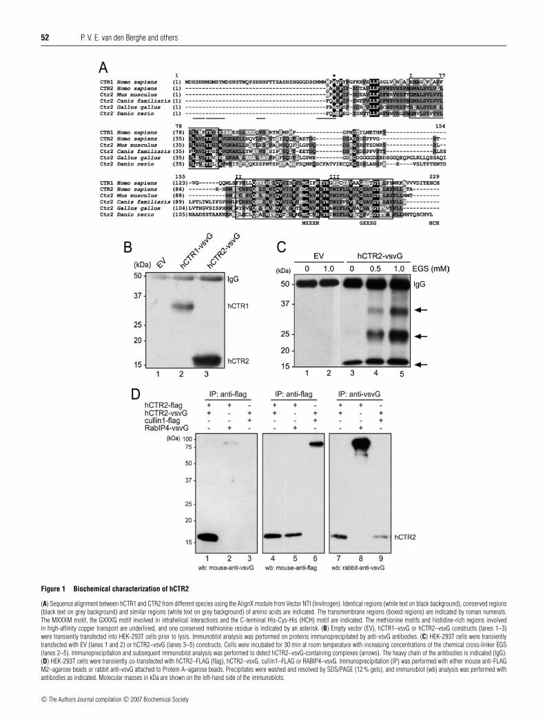
Figure 1 Biochemical characterization of hCTR2
(A) Sequence alignment between hCTR1 and CTR2 from different species using the AlignX module from Vector NTI (Invitrogen). Identical regions (white text on black background), conserved regions(black text on grey background) and similar regions (white text on grey background) of amino acids are indicated. The transmembrane regions (boxed regions) are indicated by roman numerals.The MXXXM motif, the GXXXG motif involved in intrahelical interactions and the C-terminal His-Cys-His (HCH) motif are indicated. The methionine motifs and histidine-rich regions involvedin high-affinity copper transport are underlined, and one conserved methionine residue is indicated by an asterisk. (B) Empty vector (EV), hCTR1–vsvG or hCTR2–vsvG constructs (lanes 1–3)were transiently transfected into HEK-293T cells prior to lysis. Immunoblot analysis was performed on proteins immunoprecipitated by anti-vsvG antibodies. (C) HEK-293T cells were transientlytransfected with EV (lanes 1 and 2) or hCTR2–vsvG (lanes 3–5) constructs. Cells were incubated for 30 min at room temperature with increasing concentrations of the chemical cross-linker EGS(lanes 2–5). Immunoprecipitation and subsequent immunoblot analysis was performed to detect hCTR2–vsvG-containing complexes (arrows). The heavy chain of the antibodies is indicated (IgG).(D) HEK-293T cells were transiently co-transfected with hCTR2–FLAG (flag), hCTR2–vsvG, cullin1–FLAG or RABIP4–vsvG. Immunoprecipitation (IP) was performed with either mouse anti-FLAGM2–agarose beads or rabbit anti-vsvG attached to Protein A–agarose beads. Precipitates were washed and resolved by SDS/PAGE (12 % gels), and immunoblot (wb) analysis was performed withantibodies as indicated. Molecular masses in kDa are shown on the left-hand side of the immunoblots.
c© The Authors Journal compilation c© 2007 Biochemical Society

hCTR2 facilitates copper uptake in mammalian cells 53
cells were transiently transfected with 10 ng of the hypoxia-responsive reporter vector with 5× HRE [34] and 0.25 ng ofpRL-TK vector. After 24 h, cells were incubated for a further24 h with 100 µM DFO (desferrioxamine), an iron chelator.After incubation, the cells were rinsed in PBS, harvested inpassive lysis buffer (Promega) according to the manufacturer’sprotocol and assayed by luminometry (Berthold Technologies)using the Dual-Luciferase® reporter assay system (Promega) forfirefly luciferase activity and Renilla luciferase activity, accordingto the manufacturer’s protocol. The RLU (relative light units)were calculated by dividing firefly luciferase measurements byRenilla luciferase measurements. All values were expressed asfold inductions relative to empty vector control incubations (setat 1). Statistical analysis was performed on RLU data for thedifferent incubations. A two-tailed Student’s t test was used toanalyse the statistical differences between different data points.
RESULTS
Biochemical characterization of hCTR2
To characterize hCTR2, hCTR1–vsvG and hCTR2–vsvG wereexpressed in HEK-293T cells, immunoprecipitated and subjectedto immunoblot analysis. No precipitated proteins were detectedin cells transfected with the empty vector, whereas hCTR1–vsvG was observed to migrate on SDS/PAGE at approx. 35 kDa(Figure 1B). This was consistent with previous studies [15],which indicated that the fully glycosylated mature hCTR1 wasexpressed. hCTR2–vsvG was detected to migrate with an apparentmolecular mass of approx. 17 kDa (Figure 1B), in agreementwith its size as predicted from amino acid sequence analysis(17000 Da). Crude cell fractionation studies indicated that hCTR2was associated with cellular membranes (results not shown). Thisbiochemical analysis indicated that hCTR2–vsvG was expressedas a stable membrane-associated protein and was most probablynot glycosylated.
Chemical cross-linking and two-dimensional crystallographyexperiments conducted previously revealed that hCTR1 assem-bles as a trimer [16,18]. Thus we considered the possibility thathCTR2 also exists as an oligomeric complex. The amine-reactivechemical cross-linker EGS was used to covalently link cellularinteracting proteins. Chemical cross-linking with EGS in HEK-293T cells transiently transfected with hCTR2–vsvG resultedin the detection of discrete vsvG immunopositive bands whichmigrated at apparent molecular masses of approx. 24 and 35 kDa.The intensity of these putative hCTR2–vsvG oligomers increasedin an EGS concentration-dependent manner (Figure 1C; arrows).At high EGS concentrations (1 mM), additional complexes ofeven higher molecular mass (>200 kDa) were also observed(results not shown). These results suggested that hCTR2 maybe capable of forming oligomeric complexes, similar to thoseobserved for hCTR1 [16].
To investigate further this possibility, hCTR2 constructs weregenerated with a FLAG tag present at the C-terminus. Theseconstructs were co-expressed with hCTR2–vsvG in HEK-293Tcells, and cell lysates were subjected to co-immunoprecipitationwith either anti-FLAG M2–agarose beads or Protein A–agarosebeads coupled to rabbit anti-vsvG antibodies. Immunoprecipi-tation of both vsvG-tagged proteins (Figure 1D, lanes 7–9) andFLAG-tagged proteins (Figure 1D, lanes 4–6) was observed.Co-immunoprecipitation of hCTR2–vsvG with hCTR2–FLAGwas observed (Figure 1D, lane 1), but no interaction was notedwith RabIP4–vsvG (Figure 1D, lane 2) or with cullin1–FLAG(Figure 1D, lane 3), which were used as negative controls.Taken together, these experiments suggest that hCTR2 monomers
mutually interact, and support the hypothesis that CTR proteinsform trimeric complexes to enable copper transport.
Intracellular localization of hCTR2
To investigate the cellular localization of hCTR2, we generatedconstructs encoding hCTR2 with a C-terminal eGFP tag. Similarlyto hCTR2–vsvG, hCTR2–eGFP expressed in HEK-293T cellsalso migrated at its expected molecular mass of 40 kDa andcould be detected in higher-molecular-mass oligomers (resultsnot shown). The cellular localization of hCTR2 was determinedafter transient transfection of hCTR2–eGFP into HEK-293T cells,U2OS cells and HeLa cells (Figure 2A). hCTR2–eGFP was notdetected at the plasma membrane, but was primarily localized inrelatively large vesicles in all three cell types (Figure 2A). Weset out to exclude the possibility that the inserted tag could haveinterfered with the localization of transiently expressed hCTR2.Independently transfected hCTR2–vsvG and hCTR2–FLAG werealso detected in large vesicular compartments in multiple celltypes (results not shown), and co-expression of hCTR2–vsvGwith hCTR2–eGFP resulted in strong co-localization betweenthe two constructs (Figure 2B). The latter result indicates that thecellular localization of hCTR2 was independent of the tag addedto the protein.
Consistent with previous results, hCTR1–eGFP was detectablein HeLa cells at the plasma membrane, but was primarily localizedin intracellular vesicles, and hCTR1-eGFP also partly co-localizedwith hCTR2 (Figure 3A) [15,16]. Cell-surface hCTR1 is knownto be rapidly and specifically internalized upon incubation ofHEK-293T cells with increasing concentrations of copper [17].To determine whether copper affects hCTR2 localization, HEK-293T cells were transiently transfected with hCTR2–eGFP. Cellswere incubated with different copper concentrations or withthe copper-chelating agent, BCS, for 1 h. No apparent copper-dependent relocalization of hCTR2 was observed (Figure 3B),which is in accordance with previous results suggesting thatthe intracellular fraction of hCTR1 does not undergo copper-dependent relocalization [15].
The nature of the vesicular localization of hCTR2 in HEK-293Tcells was investigated further. HEK-293T cells were transientlytransfected with hCTR2–eGFP, and immunofluorescence labell-ing of different cellular marker proteins was performed. No signi-ficant co-localization was observed between hCTR2–eGFP andTfR, an early endosomal marker, or between hCTR2–eGFPand p230, a TGN-resident protein (Figure 4). However, hCTR2was partly co-localized with late endosomes, identified bylabelling with an anti-CD63 antibody, and marked co-localizationwas observed with the lysosomal markers LAMP-1 and LAMP-2 (Figure 4, arrowheads). Similar results were observed in HeLacells (results not shown), indicating that the localization of hCTR2in late endosomes and lysosomes was not cell-type-specific.
A novel transcription-based copper sensor to measure cytosolicbioavailable copper pools
To address whether hCTR2 could facilitate copper uptake, a noveltranscription-based copper biosensor was designed. The design ofthis sensor was based on the endogenous capacity of cells to trans-activate metallothionein genes in response to elevated cytosoliccopper availability. An increase in the concentration of copperin the cytosol results in the displacement of zinc frommetallothioneins. The released zinc binds to and activates MTF(metal responsive transcription factor)-1, which subsequentlyinduces gene expression by binding to MREs in the promoterregion of specific genes [7,35]. Four MREs from the murinemetallothionein 1A promoter [7,32] were cloned upstream of
c© The Authors Journal compilation c© 2007 Biochemical Society

54 P. V. E. van den Berghe and others
Figure 2 hCTR2 is localized in intracellular vesicles
(A) Localization of hCTR2 was assessed by direct confocal laser-scanning microscopy in HEK-293T, U2OS and HeLa cells after transient transfection with hCTR2–eGFP. (B) HeLa cells were transientlyco-transfected with both hCTR2–vsvG and hCTR2–eGFP constructs. hCTR2–vsvG was immunolabelled with rabbit anti-vsvG antibodies, and secondary labelling was performed with Alexa Fluor®568 conjugated-goat anti-rabbit and goat anti-mouse antibodies. hCTR2–eGFP was visualized by direct confocal microscopy. Co-localization is indicated by arrowheads.
the firefly luciferase open reading frame in the pGL3-E1b-TATA vector (Figure 5A), and this resulted in the formation ofthe MRE–luciferase reporter. A construct encoding the metal-insensitive TK (thymidine kinase)–Renilla luciferase was usedto correct for differences in transfection efficiencies. Similarlyto the endogenous metallothionein 1A promoter, the MRE–luciferase reporter was predicted to be transactivated in a metal-concentration-dependent fashion by MTF-1 [7]. In this way, theMRE–luciferase reporter measured bioavailable cytosolic copper.To test the validity of this approach, the MRE–luciferase reporterwas transiently transfected into HEK-293T cells (Figure 5C) andU2OS cells (results not shown), and cells were incubated withdifferent copper concentrations for 24 h. At the concentrationof copper normally present in the medium, reporter activitywas slightly increased compared with cells transfected with acontrol vector containing no MREs. A linear concentration-dependent induction of the activity of the MRE–luciferase reporterwas noted when cells were incubated with 20–100 µM CuCl2,an effect that was not observed in cells transfected with theempty vector only (Figure 5C). The induction of the MRE–luciferase reporter activity was approx. 5-fold increased at aconcentration of 100 µM CuCl2, in comparison with the activityobserved at the concentration of basal copper present in themedium. Higher concentrations of copper did not result infurther induction of MRE–luciferase reporter activity. A moredetailed examination of MRE–luciferase reporter activities at lowcopper concentrations revealed that the sensor was not effectivelyinduced by copper concentrations below 20 µM in these cells(results not shown). Since the reporter is responsive to cytosoliccopper, an increase in reporter activity was interpreted as anindication that copper import and transport across a cellularmembrane had occurred. Quantitative RT (reverse transcription)–PCR experiments revealed that induction of the endogenous
metallothionein 1A promoter also occurred at the same copperconcentrations as the MRE–luciferase reporter activity wasinduced (results not shown). As different metals are known toinduce MTF-1-mediated transcription [7], the specificity of theMRE–luciferase reporter for several other biologically importantmetals was tested. Zinc is especially relevant in this respect, sinceactivation of MTF-1 is caused by incorporation of cellular zincinto MTF-1 after displacement of zinc from metallothioneins byother metals, such as copper [7]. Initially, metal toxicity wasdetermined by MTT viability assays to calculate LD50 valuesin our cells (Figure 5B). Subsequently, HEK-293T cells weretransiently transfected with the MRE–luciferase reporter, and,24 h after transfection, cells were incubated with either a low or ahigh metal concentration that was still below the lethal concentra-tion (Figure 5D). CuCl2 and ZnCl2 resulted in the induction ofreporter activities in a concentration-dependent manner, whereasMgCl2, FeCl3 and MnCl2 failed to activate the MRE–luciferasereporter. MTF-1-mediated transcription may also be inducedby other factors, such as oxidative stress and hypoxia [7,36].However, no MRE–luciferase reporter induction was observedas a result of oxidative stress. This was examined by incubationof transfected cells with 100 µM hydrogen peroxide or 100 µMparaquat for 24 h (results not shown) [37]. To mimic hypoxia,cells were treated with DFO [38], which resulted in the inductionof the hypoxia-responsive 5× HRE reporter (Figure 5E). TheMRE–luciferase reporter activity was only slightly induced uponDFO treatment, whereas CuCl2 strongly induced the MREreporter, but not the 5× HRE reporter (Figure 5D). Consideredtogether with the idea that all experiments in the presentstudy were performed under normoxic conditions, we concludedthat the MRE–luciferase reporter specifically detects elevatedcopper and zinc concentrations under these defined experimentalconditions.
c© The Authors Journal compilation c© 2007 Biochemical Society

hCTR2 facilitates copper uptake in mammalian cells 55
Figure 3 hCTR2 is partially co-localized with hCTR1
(A) Confocal laser-scanning microscopy was performed on HeLa cells which were transientlytransfected with hCTR1–eGFP and hCTR2–vsvG constructs. hCTR2–vsvG was immunolabelledwith rabbit anti-vsvG antibodies, and secondary labelling was performed using Alexa Fluor®568-conjugated antibodies. hCTR1–eGFP was visualized by direct confocal microscopy. Plasmamembrane localization of hCTR1–eGFP is indicated with arrows, and co-localization witharrowheads. (B) HEK-293T cells were transiently transfected with the hCTR2–eGFP construct.Prior to analysis, cells were incubated for 1 h with either BCS or CuCl2 at the concentrationsindicated.
hCTR1 and hCTR2 both facilitate copper uptake
Next, we tested whether this approach could be used to effectivelyassess perturbations in the known copper import pathway. Toinvestigate this, hCTR1–FLAG was co-transfected into HEK-293T cells together with the MRE–luciferase reporter. Withoutsupplementing the medium with additional CuCl2, this resultedin a significant increase (approx. 3-fold) in reporter activity whencompared with cells transfected with the empty vector (Figures 6Aand 6B). This increase in reporter activity could be preventedcompletely by incubation with the copper chelator, BCS (resultsnot shown), indicating that exogenous expression of hCTR1–FLAG resulted in a significant increase in the cytosolic copperconcentration in the presence of cell culture medium containinga low concentration of copper. Addition of CuCl2 (up to 2.5 µM)resulted in a marked and significant increase in the reporteractivity, with a maximal 10-fold induction observed at 2.5 µMCuCl2 (Figure 6A). Since the MRE–luciferase reporter was activ-ated by zinc as well as by copper, one possible explanation forthe reporter induction is that exogenous expression of hCTR1–FLAG resulted in the mobilization of sequestered vesicular zincpools under conditions of excess cytosolic copper. To addressthis possibility, the effects of transient transfection of hCTR1–FLAG on zinc-induced reporter activity were assessed. In trans-
Figure 4 hCTR2 co-localized with late endosomal and lysosomal vesicles
HEK-293T cells were transiently transfected with hCTR2–eGFP prior to labelling with antibodiesagainst different vesicular markers: the TGN marker p230, TfR (early endosomal marker), CD63(late endosomal marker) and the lysosomal markers LAMP-1 and LAMP-2. Secondary labellingwas performed using Alexa Fluor® 568-conjugated antibodies before being visualized by directconfocal microscopy. Green and red channels were merged to determine the co-localization ofhCTR2–eGFP with the different compartmental markers (arrowheads).
fected cells containing the empty vector, no induction of theMRE–luciferase reporter activity was observed when incubatedwith ZnCl2 at the same concentrations as used in the copperexperiments. In cells expressing exogenous hCTR1–FLAG, thebasal reporter activity was raised approx. 3-fold as shown above(Figures 6A and 6B), but the MRE–luciferase reporter activity wasnot induced further as a result of additional ZnCl2 (Figure 6B).These results strongly indicate that expression of hCTR1–FLAGresulted in an increase in the bioavailability of copper, but not zinc.As a control, immunoblot analysis of hCTR1–FLAG was used todetermine protein expression under these conditions (Figure 6E).Together, these data indicated the feasibility of using the MRE–luciferase reporter to specifically assess changes in the expressionof copper import proteins on copper uptake. Furthermore, theseresults confirmed previous observations that the expression ofhCTR1 was a limiting factor in copper import, at least at relativelylow copper concentrations [14].
c© The Authors Journal compilation c© 2007 Biochemical Society

56 P. V. E. van den Berghe and others
Figure 5 Characterization of a novel MRE–luciferase reporter assay
(A) The MRE–luciferase reporter was constructed by cloning four MREs upstream of the firefly luciferase open reading frame of the pGL3-TATA construct as indicated. (B) HEK-293T cells weretransiently transfected with the MRE–luciferase reporter or the control vector, pGL3-TATA. After transfection, cells were incubated for 24 h with different concentrations of CuCl2. (C) Metal toxicitywas measured using MTT viability assays after incubation for 24 h with different metal concentrations, and LD50 values were also determined. (D) HEK-293T cells were transiently transfected withthe MRE–luciferase reporter, and, 24 h after transfection, cells were incubated with a low or a high sub-lethal metal concentration. (E) HEK-293T cells were transiently transfected with either theMRE–luciferase reporter or a HRE–luciferase reporter. After transfection, cells were incubated with 100 µM CuCl2 or 100 µM of DFO (to mimic hypoxia). Luciferase reporter activities were measuredand normalized for Renilla luciferase activities. Values are expressed as fold induction relative to control incubations +−S.E.M. (see the Experimental section) (n � 3).
Next, the potential role of hCTR2 in copper import wasstudied in a similar manner to that of hCTR1 by co-transfectionof hCTR2–FLAG and MRE–luciferase reporter in HEK-293Tcells. Expression of hCTR2–FLAG was verified by immunoblotanalysis (Figure 6E). In cells transfected with the empty vector,reporter activity was induced in the presence of 10–20 µMCuCl2 (results not shown). However, hCTR2–FLAG expressionresulted in a significantly higher reporter induction comparedwith cells transfected with the empty vector. At concentrations of40 and 100 µM CuCl2, expression of hCTR2–FLAG resulted ina >2-fold increase (Figure 6C). The increased hCTR2–FLAG-dependent MRE–luciferase reporter activation was copper-specific, as no differences were observed on incubation withZnCl2 (Figure 6D). To demonstrate further that the functionof hCTR2 was copper-specific, we mutated the methionineresidues in the MXXXM motif to isoleucine. Previous findingsby Eisses and Kaplan [25] revealed a marked reduction inhCTR1-mediated copper uptake after disruption of the analogousmethionine residues present in the MXXXM motif of hCTR1.Whereas expression of wild-type hCTR1–FLAG or hCTR2–FLAG resulted in a significant copper-dependent induction ofthe MRE–luciferase reporter, substitution of the MXXXM motifto IXXXI completely abolished the induction of the reporter byhCTR1–FLAG and by hCTR2–FLAG (Figures 6A and 6C). Thelack of induction was a result of the amino acid substitution,as all the constructs were shown to be expressed successfully(Figure 6E). No differences were observed after incubation withZnCl2 (Figures 6B and 6D). Taken together, expression of hCTR2facilitates cellular copper uptake with a lower affinity comparedwith hCTR1.
DISCUSSION
Since the initial characterization of hCTR1 almost a decade ago[30], elegant studies have addressed the structure and localizationof mammalian Ctr1, its transport kinetics, regulation by copperand the role in embryonic development and dietary copper uptakein mice [14–18,20–27,29,39,40]. Although these studies havegreatly increased our knowledge of the mechanisms of cellularcopper uptake, they also established that Ctr1-independent copperimport pathways exist. MEFs obtained from Ctr1-knockoutembryos displayed low-affinity copper uptake [23]. On the basisof the evidence of the present study, we propose that hCTR2, aprotein with structural similarity to hCTR1, but with a previouslyunknown function, mediates an alternative low-affinity copperimport pathway in human cells.
To be able to characterize the function of hCTR2, we con-structed a novel metal sensor. This sensor allowed sensitiveand high-throughput assessment of copper homoeostasis. It isimportant to note that the MRE–luciferase reporter measurescytosolic bioavailable copper and was based on a cell-intrinsiccopper-sensing mechanism, removing the need to add exogenouscompounds that bind copper directly and might potentially inter-fere with the copper homoeostatic mechanisms present in the cell[41]. By performing control experiments using zinc, the sensorallowed determination of the specific effects of exogenouslyexpressed proteins on copper metabolism. Our data strongly in-dicated that hCTR2 facilitates cellular copper import with a loweraffinity than that mediated by hCTR1. At copper concentrationsbelow 10 µM, at which hCTR1-dependent MRE–luciferase re-porter activity was maximally induced, no effect of hCTR2
c© The Authors Journal compilation c© 2007 Biochemical Society

hCTR2 facilitates copper uptake in mammalian cells 57
Figure 6 Cellular copper uptake is facilitated by hCTR2 and is dependent on the CTR-specific MXXXM motif
The MXXXM motifs were mutated to form IXXXI in pEBB-hCTR1-FLAG and pEBB-hCTR2-FLAG. HEK-293T cells were transiently transfected with the MRE–luciferase reporter in combination withwild-type pEBB-hCTR1-FLAG (A,B), pEBB-hCTR2-FLAG (C,D) or the IXXXI mutant motif constructs. After transfection, cells were incubated for 24 h with increasing amounts of CuCl2 (A,C) orZnCl2 (B,D). MRE–luciferase reporter activities were measured and RLU were calculated after normalization for Renilla luciferase activity. Values are expressed as fold induction relative to controlincubations (see the Experimental section) +− S.E.M. (n = 3). Statistical differences between empty vector (EV)- and hCTR1- or hCTR2-transfected cells are indicated (*P < 0.01). (E) Lysates fromthe MRE–luciferase assay were resuspended in SDS/PAGE sample buffer, resolved by SDS/PAGE and subjected to immunoblot analysis using the mouse anti-FLAG antibody. Molecular masses areindicated in kDa.
expression on reporter activity could be detected. In contrast,exogenous expression of hCTR2 resulted in significantly highercopper-dependent MRE–luciferase activity compared with emptyvector controls. This effect was copper-specific, since mutation ofconserved methionine residues known to be essential for coppertransport in CTR proteins completely abolished the activation ofthe reporter. Therefore hCTR2 expression resulted in a specificincrease in cytosolic copper availability at high concentrations
of copper in the extracellular medium. Since the copper sensordoes not measure the transport of copper directly, further studiesare necessary to address the kinetics of hCTR2-mediated copperimport in more detail.
The apparent steady-state localization of this protein in theendosomal and lysosomal compartments is notable. Lysosomes inmammalian cells and vacuoles in yeast have equivalent functions.In Saccharomyces cerevisiae, the hCTR2 orthologue, yCtr2p, is
c© The Authors Journal compilation c© 2007 Biochemical Society

58 P. V. E. van den Berghe and others
localized to the vacuolar membrane and can mobilize vacuolarcopper pools [42]. A similar vacuolar membrane localizationwas observed for Ctr6, the hCTR2 orthologue in the fissionyeast, Schizosaccharomyces pombe [43]. Combined with our data,we suggest that the endosomal and lysosomal localization andmobilization of intracellular copper stores are common propertiesof yeast and mammalian CTR2/Ctr6 proteins. Our data suggestthat the observed intracellular localization of hCTR2 is correct,since it was observed in multiple cell types and appeared to occurindependently of the tags present. Moreover, expressed proteinswere demonstrated to stimulate functional copper uptake activity.A similar localization of endogenous CTR2 in large vesicularcompartments, reminiscent of late endosomes and lysosomes,was observed in human and monkey cells in other independentstudies (J. Bertinato, personal communication). There is ampleprecedence for the localization of CTRs in intracellular compart-ments. Under basal copper levels, ATP7A resides in the Golgi-network and is only distributed towards peripheral vesicles and theplasma membrane in response to an increase in cytosolic copperconcentrations [44,45]. In addition, hCTR1 also resides predo-minantly in intracellular vesicles, but has been shown to recycleconstitutively between this vesicular compartment and the plasmamembrane [17,24]. In fact, part of the intracellular pool of hCTR2resided in vesicular compartments that also contained hCTR1,but further studies are required to clarify the implications of thisintriguing observation.
Our results suggest that hCTR2 is an intracellular oligomericmembrane protein which is rate-limiting for the delivery of copperinto the cytosol at high copper concentrations. We have proposed amodel of cellular copper uptake by hCTR2. In mammalian cells,extracellular copper and cuproenzymes destined for lysosomaldegradation may be endocytosed or pinocytosed by CTR-depend-ent or -independent mechanisms. Copper may subsequently beconcentrated into lysosomes, prior to mobilization by hCTR2.This process may require high extracellular concentrations ofcopper or prolonged concentration of copper in lysosomal vesi-cles, consistent with our observation that hCTR2 stimulatescopper uptake at relatively high copper concentrations. Thismodel fits with the current knowledge of the biology of dietarycopper import and intracellular copper sequestration. Intestinalepithelium of gut-specific Ctr1-null mice unexpectedly accumul-ated dietary copper [22]. The failure to express Ctr1 did not leadto reduced cellular copper uptake, but rather caused an increase inthe intracellular sequestration of copper, presumably in lysosomalvesicles. If mouse Ctr2 could function to mobilize copper fromintracellular pools in gut epithelium, this activity is clearly notsufficient to prevent the severe systemic copper deficiency presentin intestine-specific Ctr1-null mice [22]. Although the existence ofvesicular copper stores in mammalian cells remains speculative,extensive copper accumulation has indeed been observed inlysosomes of Wilson disease patients [46], in LEC (Long-EvansCinnamon) rats (a model for Wilson disease) [47] and in theliver cells of Bedlington terriers affected with copper toxicosis[48]. Finally, several other metal transport systems utilize theendocytic machinery to import metals into the cytosol. In yeast,zinc transporters regulate vacuolar zinc stores to modulate zinchomoeostasis [49]. The mouse zinc transporters ZIP1, ZIP3 andZIP4 are localized in intracellular vesicles, and may translocate tothe plasma membrane to allow zinc uptake [50,51]. Furthermore,the DMT (divalent metal transporter) 1 protein is involved in thetransfer of endocytosed iron into the cytosol [52,53]. Thereforecellular copper uptake may require a combination of transportacross the plasma membrane, endocytosis of the metal andmobilization from intracellular pools, as observed for the uptakeof other transition metals.
In summary, these results suggest that hCTR2 is predominantlylocalized in the endosomal system and stimulates copper uptakewith a relatively low affinity to make it bioavailable in the cytosol,and suggest that hCTR2 may be responsible for the residual copperuptake activity observed in cells obtained from Ctr1-null embryos.Since this work is the first study on the function and localizationof hCTR2, many questions remain unanswered. It will beimportant to examine further the nature and function of vesicularcopper sequestration in copper homoeostasis, and to identifythe mechanisms that are responsible for import of copper intoendosomes and lysosomes. Ultimately, studying the impact oncopper homoeostasis of knocking out Ctr2 from the mousegenome at the level of the entire organism will be requiredto obtain a complete understanding of the regulation of copperuptake under different conditions and cellular demands.
We thank Dr Walter Schaffner for providing the 4 × MRE(d) construct, and Dr P. J. vande Sluijs for providing the pCL-NEO RabIP4-vsvG construct. We are grateful to Dr EricKalkhoven and Professor Dr Cisca Wijmenga for critical evaluation of the manuscript, andthe members the Klomp–Wijmenga group for valuable discussions and assistance. Thiswork was supported by a grant from the Netherlands Organization for Scientific Research(NWO: 912-04-106).
REFERENCES
1 Culotta, V. C., Lin, S. J., Schmidt, P., Klomp, L. W., Casareno, R. L. and Gitlin, J. (1999)Intracellular pathways of copper trafficking in yeast and humans. Adv. Exp. Med. Biol.448, 247–254
2 de Bie, P., van de Sluis, B., Klomp, L. and Wijmenga, C. (2005) The many faces of thecopper metabolism protein MURR1/COMMD1. J. Hered. 96, 803–811
3 Himelblau, E. and Amasino, R. M. (2000) Delivering copper within plant cells.Curr. Opin. Plant Biol. 3, 205–210
4 Rae, T. D., Schmidt, P. J., Pufahl, R. A., Culotta, V. C. and O’Halloran, T. V. (1999)Undetectable intracellular free copper: the requirement of a copper chaperone forsuperoxide dismutase. Science 284, 805–808
5 Casareno, R. L., Waggoner, D. and Gitlin, J. D. (1998) The copper chaperone CCS directlyinteracts with copper/zinc superoxide dismutase. J. Biol. Chem. 273, 23625–23628
6 Hamza, I., Schaefer, M., Klomp, L. W. and Gitlin, J. D. (1999) Interaction of the copperchaperone HAH1 with the Wilson disease protein is essential for copper homeostasis.Proc. Natl. Acad. Sci. U.S.A. 96, 13363–13368
7 Zhang, B., Georgiev, O., Hagmann, M., Gunes, C., Cramer, M., Faller, P., Vasak, M. andSchaffner, W. (2003) Activity of metal-responsive transcription factor 1 by toxic heavymetals and H2O2 in vitro is modulated by metallothionein. Mol. Cell. Biol. 23, 8471–8485
8 Mercer, J. F., Livingston, J., Hall, B., Paynter, J. A., Begy, C., Chandrasekharappa, S.,Lockhart, P., Grimes, A., Bhave, M., Siemieniak, D. et al. (1993) Isolation of a partialcandidate gene for Menkes disease by positional cloning. Nat. Genet. 3, 20–25
9 Vulpe, C., Levinson, B., Whitney, S., Packman, S. and Gitschier, J. (1993) Isolation of acandidate gene for Menkes disease and evidence that it encodes a copper-transportingATPase. Nat. Genet. 3, 7–13
10 Mendelsohn, B. A., Yin, C., Johnson, S. L., Wilm, T. P., Solnica-Krezel, L. and Gitlin, J. D.(2006) Atp7a determines a hierarchy of copper metabolism essential for notochorddevelopment. Cell Metab. 4, 155–162
11 Petris, M. J. (2004) The SLC31 (Ctr) copper transporter family. Pflugers Arch. 447,752–755
12 Dancis, A., Yuan, D. S., Haile, D., Askwith, C., Eide, D., Moehle, C., Kaplan, J. andKlausner, R. D. (1994) Molecular characterization of a copper transport protein in S.cerevisiae: an unexpected role for copper in iron transport. Cell 76, 393–402
13 Knight, S. A., Labbe, S., Kwon, L. F., Kosman, D. J. and Thiele, D. J. (1996) A widespreadtransposable element masks expression of a yeast copper transport gene. Genes Dev. 10,1917–1929
14 Eisses, J. F. and Kaplan, J. H. (2002) Molecular characterization of hCTR1, the humancopper uptake protein. J. Biol. Chem. 277, 29162–29171
15 Klomp, A. E., Tops, B. B., Van Denberg, I. E., Berger, R. and Klomp, L. W. (2002)Biochemical characterization and subcellular localization of human copper transporter 1(hCTR1). Biochem. J. 364, 497–505
16 Lee, J., Pena, M. M., Nose, Y. and Thiele, D. J. (2002) Biochemical characterization of thehuman copper transporter Ctr1. J. Biol. Chem. 277, 4380–4387
c© The Authors Journal compilation c© 2007 Biochemical Society

hCTR2 facilitates copper uptake in mammalian cells 59
17 Petris, M. J., Smith, K., Lee, J. and Thiele, D. J. (2003) Copper-stimulated endocytosisand degradation of the human copper transporter, hCtr1. J. Biol. Chem. 278,9639–9646
18 Aller, S. G. and Unger, V. M. (2006) Projection structure of the human copper transporterCTR1 at 6-A resolution reveals a compact trimer with a novel channel-like architecture.Proc. Natl. Acad. Sci. U.S.A. 103, 3627–3632
19 Nose, Y., Rees, E. M. and Thiele, D. J. (2006) Structure of the Ctr1 copper trans‘PORE’terreveals novel architecture. Trends Biochem. Sci. 31, 604–607
20 Lee, J., Prohaska, J. R. and Thiele, D. J. (2001) Essential role for mammalian coppertransporter Ctr1 in copper homeostasis and embryonic development. Proc. Natl.Acad. Sci. U.S.A. 98, 6842–6847
21 Kuo, Y. M., Gybina, A. A., Pyatskowit, J. W., Gitschier, J. and Prohaska, J. R. (2006)Copper transport protein (Ctr1) levels in mice are tissue specific and dependent oncopper status. J. Nutr. 136, 21–26
22 Nose, Y., Kim, B. E. and Thiele, D. J. (2006) Ctr1 drives intestinal copper absorption andis essential for growth, iron metabolism, and neonatal cardiac function. Cell Metab. 4,235–244
23 Lee, J., Petris, M. J. and Thiele, D. J. (2002) Characterization of mouse embryonic cellsdeficient in the ctr1 high affinity copper transporter. Identification of a Ctr1-independentcopper transport system. J. Biol. Chem. 277, 40253–40259
24 Klomp, A. E., Juijn, J. A., van der Gun, L. T., van den Berg, I. E., Berger, R. and Klomp,L. W. (2003) The N-terminus of the human copper transporter 1 (hCTR1) is localizedextracellularly, and interacts with itself. Biochem. J. 370, 881–889
25 Eisses, J. F. and Kaplan, J. H. (2005) The mechanism of copper uptake mediated byhuman CTR1: a mutational analysis. J. Biol. Chem. 280, 37159–37168
26 Aller, S. G., Eng, E. T., De Feo, C. J. and Unger, V. M. (2004) Eukaryotic CTR copperuptake transporters require two faces of the third transmembrane domain for helixpacking, oligomerization, and function. J. Biol. Chem. 279, 53435–53441
27 Puig, S., Lee, J., Lau, M. and Thiele, D. J. (2002) Biochemical and genetic analyses ofyeast and human high affinity copper transporters suggest a conserved mechanism forcopper uptake. J. Biol. Chem. 277, 26021–26030
28 Pena, M. M., Lee, J. and Thiele, D. J. (1999) A delicate balance: homeostatic control ofcopper uptake and distribution. J. Nutr. 129, 1251–1260
29 Puig, S. and Thiele, D. J. (2002) Molecular mechanisms of copper uptake anddistribution. Curr. Opin. Chem. Biol. 6, 171–180
30 Zhou, B. and Gitschier, J. (1997) hCTR1: a human gene for copper uptake identified bycomplementation in yeast. Proc. Natl. Acad. Sci. U.S.A. 94, 7481–7486
31 Mosmann, T. (1983) Rapid colorimetric assay for cellular growth and survival: applicationto proliferation and cytotoxicity assays. J. Immunol. Methods 65, 55–63
32 Westin, G., Gerster, T., Muller, M. M., Schaffner, G. and Schaffner, W. (1987) OVEC, aversatile system to study transcription in mammalian cells and cell-free extracts.Nucleic Acids Res. 15, 6787–6798
33 de Bie, P., van de Sluis, B., Burstein, E., Duran, K. J., Berger, R., Duckett, C. S.,Wijmenga, C. and Klomp, L. W. (2006) Characterization of COMMD protein–proteininteractions in NF-κB signalling. Biochem. J. 398, 63–71
34 Shibata, T., Giaccia, A. J. and Brown, J. M. (2000) Development of a hypoxia-responsivevector for tumor-specific gene therapy. Gene Ther. 7, 493–498
35 Palmiter, R. D. (1994) Regulation of metallothionein genes by heavy metals appears to bemediated by a zinc-sensitive inhibitor that interacts with a constitutively activetranscription factor, MTF-1. Proc. Natl. Acad. Sci. U.S.A 91, 1219–1223
36 Murphy, B. J., Andrews, G. K., Bittel, D., Discher, D. J., McCue, J., Green, C. J.,Yanovsky, M., Giaccia, A., Sutherland, R. M., Laderoute, K. R. and Webster, K. A. (1999)Activation of metallothionein gene expression by hypoxia involves metal responseelements and metal transcription factor-1. Cancer Res. 59, 1315–1322
37 Poss, K. D. and Tonegawa, S. (1997) Reduced stress defense in heme oxygenase1-deficient cells. Proc. Natl. Acad. Sci. U.S.A. 94, 10925–10930
38 Bianchi, L., Tacchini, L. and Cairo, G. (1999) HIF-1-mediated activation of transferrinreceptor gene transcription by iron chelation. Nucleic Acids Res. 27, 4223–4227
39 Eisses, J. F., Chi, Y. and Kaplan, J. H. (2005) Stable plasma membrane levels of hCTR1mediate cellular copper uptake. J. Biol. Chem. 280, 9635–9639
40 Kuo, Y. M., Zhou, B., Cosco, D. and Gitschier, J. (2001) The copper transporter CTR1provides an essential function in mammalian embryonic development. Proc. Natl. Acad.Sci. U.S.A. 98, 6836–6841
41 Zeng, L., Miller, E. W., Pralle, A., Isacoff, E. Y. and Chang, C. J. (2006) A selective turn-onfluorescent sensor for imaging copper in living cells. J. Am. Chem. Soc. 128, 10–11
42 Rees, E. M., Lee, J. and Thiele, D. J. (2004) Mobilization of intracellular copper stores bythe ctr2 vacuolar copper transporter. J. Biol. Chem. 279, 54221–54229
43 Bellemare, D. R., Shaner, L., Morano, K. A., Beaudoin, J., Langlois, R. and Labbe, S.(2002) Ctr6, a vacuolar membrane copper transporter in Schizosaccharomyces pombe.J. Biol. Chem. 277, 46676–46686
44 Petris, M. J., Mercer, J. F., Culvenor, J. G., Lockhart, P., Gleeson, P. A. and Camakaris, J.(1996) Ligand-regulated transport of the Menkes copper P-type ATPase efflux pump fromthe Golgi apparatus to the plasma membrane: a novel mechanism of regulated trafficking.EMBO J. 15, 6084–6095
45 Yamaguchi, Y., Heiny, M. E., Suzuki, M. and Gitlin, J. D. (1996) Biochemicalcharacterization and intracellular localization of the Menkes disease protein. Proc. Natl.Acad. Sci. U.S.A. 93, 14030–14035
46 Sternlieb, I., Van den Hamer, C. J., Morell, A. G., Alpert, S., Gregoriadis, G. andScheinberg, I. H. (1973) Lysosomal defect of hepatic copper excretion in Wilson’s disease(hepatolenticular degeneration). Gastroenterology 64, 99–105
47 Terada, K., Aiba, N., Yang, X. L., Iida, M., Nakai, M., Miura, N. and Sugiyama, T. (1999)Biliary excretion of copper in LEC rat after introduction of copper transporting P-typeATPase, ATP7B. FEBS Lett. 448, 53–56
48 Haywood, S., Fuentealba, I. C., Foster, J. and Ross, G. (1996) Pathobiology ofcopper-induced injury in Bedlington terriers: ultrastructural and microanalytical studies.Anal. Cell Pathol. 10, 229–241
49 MacDiarmid, C. W., Gaither, L. A. and Eide, D. (2000) Zinc transporters that regulatevacuolar zinc storage in Saccharomyces cerevisiae. EMBO J. 19, 2845–2855
50 Kim, B. E., Wang, F., Dufner-Beattie, J., Andrews, G. K., Eide, D. J. and Petris, M. J.(2004) Zn2+-stimulated endocytosis of the mZIP4 zinc transporter regulates its location atthe plasma membrane. J. Biol. Chem. 279, 4523–4530
51 Wang, F., Dufner-Beattie, J., Kim, B. E., Petris, M. J., Andrews, G. and Eide, D. J. (2004)Zinc-stimulated endocytosis controls activity of the mouse ZIP1 and ZIP3 zinc uptaketransporters. J. Biol. Chem. 279, 24631–24639
52 Fleming, M. D., Romano, M. A., Su, M. A., Garrick, L. M., Garrick, M. D. and Andrews,N. C. (1998) Nramp2 is mutated in the anemic Belgrade (b) rat: evidence of a role forNramp2 in endosomal iron transport. Proc. Natl. Acad. Sci. U.S.A. 95, 1148–1153
53 Touret, N., Furuya, W., Forbes, J., Gros, P. and Grinstein, S. (2003) Dynamic trafficthrough the recycling compartment couples the metal transporter Nramp2 (DMT1) withthe transferrin receptor. J. Biol. Chem. 278, 25548–25557
Received 29 May 2007/4 July 2007; accepted 6 July 2007Published as BJ Immediate Publication 6 July 2007, doi:10.1042/BJ20070705
c© The Authors Journal compilation c© 2007 Biochemical Society