Host cell invasion by trypanosomes requires lysosomes and microtubule/kinesin-mediated transport
14
Host Cell Invasion by Trypanosomes Requires Lysosomes and Microtubule/Kinesin-mediated Transport Ana Rodriguez, Erika Samoff, Marc G. Rioult, Albert Chung, and Norma W. Andrews Department of Cell Biology, Yale University School of Medicine, New Haven, Connecticut 06510 Abstract. Invasion of mammalian cells by the proto- zoan parasite Trypanosoma cruzi occurs by an actin- independent mechanism distinct from phagocytosis. Clusters of host lysosomes are observed at the site of parasite attachment, and lysosomal markers are de- tected in the vacuolar membrane at early stages of the entry process. These observations led to the hy- pothesis that the trypanosomes recruit host lysosomes to their attachment site, and that lysosomal fusion serves as a source of membrane to form the parasito- phorous vacuole. Here we directly demonstrate directional migration of lysosomes to the parasite entry site, using time-lapse video-enhanced microscopy of L6E 9 myoblasts exposed to T. cruzi trypomastigotes. BSA-gold-loaded lysos- omes moved towards the cell periphery, in the direction of the parasite attachment site, but only when their original position was less than 11-12 Ixm from the inva- sion site. Lysosomes more distant from the invasion area exhibited only the short multi-directional saltatory movements previously described for lysosomes, regard- less of their proximity to the cell margins. Specific depletion of peripheral lysosomes was ob- tained by microinjection of NRK cells with antibodies against the cytoplasmic domain of lgp 120, a treatment that aggregated lysosomes in the perinuclear area and inhibited T. cruzi entry. The microtubule-binding drugs nocodazole, colchicine, vinblastine, and taxol also in- hibited invasion, in both NRK and LrE 9 cells. Further- more, microinjection of antibodies to the heavy chain of kinesin blocked the acidification-induced, microtu- bule-dependent redistribution of lysosomes to the host cell periphery, and reduced trypomastigote entry. Our results therefore demonstrate that during T. cruzi invasion of host cells lysosomes are mobilized from the immediately surrounding area, and that avail- ability of lysosomes at the cell periphery and microtu- bule/kinesin-mediated transport are requirements for parasite entry. T RYPANOSOMA cruzi, the protozoan parasite that causes Chagas' disease in humans, invades and mul- tiplies in a wide variety of vertebrate cells. The inva- sion process is unusual, in the sense that it is independent of actin polymerization, and involves the recruitment and fusion of host cell lysosomes at the site of parasite entry (Schenkman et al., 1991; Tardieux et al., 1992; Andrews, 1995). This conclusion is supported by the presence of ly- sosomal markers in the membrane of partially formed par- asite-containing vacuoles (Hall et al., 1992; Tardieux et al., 1992), the colocalization of extracellularly attached para- sites with lysosome clusters, and the observation that treatments inducing redistribution of lysosomes from the perinuclear area to the cell periphery enhance invasion. Furthermore, conditions that deplete cells of peripheral ly- sosomes or impair lysosomal fusion capacity reduce try- panosome entry (Tardieux et al., 1992). Address all correspondence to N.W. Andrews, Department of Cell Biol- ogy, Yale University School of Medicine, 333 Cedar Street, New Haven, CT 06510. Tel.: (203) 785-4314. Fax: (203) 785-7226. The recruitment of lysosomes, a process apparently trig- gered by association of T. cruzi with host cells before inva- sion, suggests that the parasite might activate signal trans- duction pathways involved in lysosome mobilization. Recent reports showed that T. cruzi invasion of mammalian cells requires the activation of at least two signaling pathways, one involving phospholipase C-dependent phosphoinosit- ide hydrolysis, with inositol 1,4,5-trisphosphate (IP3)1 for- mation and Ca 2÷ mobilization from intracellular stores (Rodriguez et al., 1995), and another involving the TGF-13 I and II receptors (Ming et al., 1995). The IP3-dependent pathway is triggered by a proteolytically generated trypo- mastigote factor (PGTF) (Burleigh and Andrews, 1995; Rodriguez et al., 1995) and induces rapid intracellular free Ca 2÷ concentration ([Ca2+]i) transients in mammalian cells (Tardieux et al., 1994; Rodriguez et al., 1995). The rela- tionship between these parasite-induced signals and the 1. Abbreviations used in this paper: [Ca2+]i, intracellular free Ca 2÷ concen- tration; IP3, inositol 1,4,5-trisphosphate; lgp, lysosomal membrane glyco- protein; NRK, normal rat kidney; PGTF, proteolytically generated trypo- mastigote factor. © The Rockefeller University Press, 0021-9525/96/07/349/14 $2.00 The Journal of Cell Biology, Volume 134, Number 2, July 1996 349-362 349 on November 12, 2014 jcb.rupress.org Downloaded from Published July 15, 1996
-
Upload
independent -
Category
Documents
-
view
0 -
download
0
Transcript of Host cell invasion by trypanosomes requires lysosomes and microtubule/kinesin-mediated transport

Host Cell Invasion by Trypanosomes Requires Lysosomes and Microtubule/Kinesin-mediated Transport Ana Rodriguez, Erika Samoff, Marc G. Rioult, Albert Chung, and Norma W. Andrews
Department of Cell Biology, Yale University School of Medicine, New Haven, Connecticut 06510
Abstract. Invasion of mammalian cells by the proto- zoan parasite Trypanosoma cruzi occurs by an actin- independent mechanism distinct from phagocytosis. Clusters of host lysosomes are observed at the site of parasite attachment, and lysosomal markers are de- tected in the vacuolar membrane at early stages of the entry process. These observations led to the hy- pothesis that the trypanosomes recruit host lysosomes to their attachment site, and that lysosomal fusion serves as a source of membrane to form the parasito- phorous vacuole.
Here we directly demonstrate directional migration of lysosomes to the parasite entry site, using time-lapse video-enhanced microscopy of L6E 9 myoblasts exposed to T. cruzi trypomastigotes. BSA-gold-loaded lysos- omes moved towards the cell periphery, in the direction of the parasite attachment site, but only when their original position was less than 11-12 Ixm from the inva- sion site. Lysosomes more distant from the invasion area exhibited only the short multi-directional saltatory
movements previously described for lysosomes, regard- less of their proximity to the cell margins.
Specific depletion of peripheral lysosomes was ob- tained by microinjection of NRK cells with antibodies against the cytoplasmic domain of lgp 120, a treatment that aggregated lysosomes in the perinuclear area and inhibited T. cruzi entry. The microtubule-binding drugs nocodazole, colchicine, vinblastine, and taxol also in- hibited invasion, in both NRK and LrE 9 cells. Further- more, microinjection of antibodies to the heavy chain of kinesin blocked the acidification-induced, microtu- bule-dependent redistribution of lysosomes to the host cell periphery, and reduced trypomastigote entry.
Our results therefore demonstrate that during T. cruzi invasion of host cells lysosomes are mobilized from the immediately surrounding area, and that avail- ability of lysosomes at the cell periphery and microtu- bule/kinesin-mediated transport are requirements for parasite entry.
T RYPANOSOMA cruzi, the protozoan parasite that causes Chagas' disease in humans, invades and mul- tiplies in a wide variety of vertebrate cells. The inva-
sion process is unusual, in the sense that it is independent of actin polymerization, and involves the recruitment and fusion of host cell lysosomes at the site of parasite entry (Schenkman et al., 1991; Tardieux et al., 1992; Andrews, 1995). This conclusion is supported by the presence of ly- sosomal markers in the membrane of partially formed par- asite-containing vacuoles (Hall et al., 1992; Tardieux et al., 1992), the colocalization of extracellularly attached para- sites with lysosome clusters, and the observation that treatments inducing redistribution of lysosomes from the perinuclear area to the cell periphery enhance invasion. Furthermore, conditions that deplete cells of peripheral ly- sosomes or impair lysosomal fusion capacity reduce try- panosome entry (Tardieux et al., 1992).
Address all correspondence to N.W. Andrews, Department of Cell Biol- ogy, Yale University School of Medicine, 333 Cedar Street, New Haven, CT 06510. Tel.: (203) 785-4314. Fax: (203) 785-7226.
The recruitment of lysosomes, a process apparently trig- gered by association of T. cruzi with host cells before inva- sion, suggests that the parasite might activate signal trans- duction pathways involved in lysosome mobilization. Recent reports showed that T. cruzi invasion of mammalian cells requires the activation of at least two signaling pathways, one involving phospholipase C-dependent phosphoinosit- ide hydrolysis, with inositol 1,4,5-trisphosphate (IP3) 1 for- mation and Ca 2÷ mobilization from intracellular stores (Rodriguez et al., 1995), and another involving the TGF-13 I and II receptors (Ming et al., 1995). The IP3-dependent pathway is triggered by a proteolytically generated trypo- mastigote factor (PGTF) (Burleigh and Andrews, 1995; Rodriguez et al., 1995) and induces rapid intracellular free Ca 2÷ c o n c e n t r a t i o n ([Ca2+]i) transients in mammalian cells (Tardieux et al., 1994; Rodriguez et al., 1995). The rela- tionship between these parasite-induced signals and the
1. Abbreviat ions used in this paper: [Ca2+]i, intracellular free Ca 2÷ concen- tration; IP3, inositol 1,4,5-trisphosphate; lgp, lysosomal membrane glyco- protein; NRK, normal rat kidney; PGTF, proteolytically generated trypo- mastigote factor.
© The Rockefeller University Press, 0021-9525/96/07/349/14 $2.00 The Journal of Cell Biology, Volume 134, Number 2, July 1996 349-362 349
on Novem
ber 12, 2014jcb.rupress.org
Dow
nloaded from
Published July 15, 1996
recruitment and fusion of host lysosomes is not fully un- derstood, although we have suggested that caa÷-triggered cortical actin rearrangements may facilitate lysosome ac- cess to the invasion site (Rodriguez et al., 1995).
The movement of lysosomes in mammalian ceils is known to be mediated by microtubules, and to be inde- pendent of intermediate filaments and microfilament net- works (Matteoni and Kreis, 1987). Drugs that affect mi- crotubule stability, such as nocodazole and taxol, inhibit lysosome movement (Herman and Albertini, 1984; Mat- teoni and Kreis, 1987; Heuser, 1989). Lysosomes and late endosomes are mainly clustered in the perinuclear area, near the slow-growing (minus) end of microtubules (Mat- teoni and Kreis, 1987; Cole and Lippincott-Schwartz, 1995). Lysosome dispersion towards the cell periphery is observed during mitosis, and can be induced experimen- tally by microtubule-depolymerizing drugs, or by cytoplas- mic acidification. This redistribution is reversible, with rapid reclustering occurring by migration of the lysosomes towards the minus ends of microtubules (Matteoni and Kreis, 1987; Heuser, 1989). Microtubule-associated motor proteins that use the energy of ATP hydrolysis to translo- cate organelles have been identified, and movements to- wards the plus and minus ends of microtubules have been attributed, respectively, to kinesin and cytoplasmic dynein (Schroer and Sheetz, 1991).
In this study we use video microscopy to visualize the distribution of lysosomes in living cells during the T. cruzi cell entry process. We found that before invasion there is a directional movement of lysosomes located in the vicinity of the entry site towards the parasite attachment site at the cell periphery. Antibody-mediated clustering of lysosomes in the perinuclear area inhibits invasion, suggesting that availability of lysosomes for fusion at the cell periphery is a requirement for parasite entry. In addition, by treating the host cells with drugs that affect microtubule stability, and by microinjecting antibodies that block kinesin func- tion, we show that T. cruzi invasion requires an intact mi- crotubule/kinesin-mediated transport apparatus.
Materials and Methods
Materials Vinblastine, BSA, protein A-Sepharose, protein G-Sepharose, thrombin, and gold chloride were obtained from Sigma Chem. Co. (St. Louis, MO); colchicine, nocodazole, and taxol were from Calbiochem (La Jolla, CA). Lucifer yellow was from Molecular Probes, Inc. (Eugene, OR), and key- hole limpet hemocyanin was from Boehringer Mannheim (Indianapolis, IN).
Mammalian Cells and Parasites
Normal rat kidney (NRK) fibroblasts and I_~E 9 rat myoblasts were grown in DMEM containing 10% FBS, at 37°C with 5% CO2. Trypomastigotes from the T. cruzi Y strain were obtained from the supernatant of infected LLC-MK2 cells (Andrews et al., 1987). Parasites were washed in Hepes- buffered Ringer's solution (Heuser, 1989) and resuspended in the same solution before exposure to cell monolayers.
Time Lapse Video-enhanced Microscopy LtE9 cells were plated on glass coverslips at 104 cells/cm 2 and cultured for 48 h. Lysosomes were labeled by the internalization of BSA adsorbed to 15-nm-gold particles, prepared according to published procedures (Slot and Geuze, 1985). Cells were incubated with BSA-gold (OD 4.0 at 255 nm) for 3 h at 37°C, washed, and chased for 2 h. Coverslips were then mounted
in a perfusion chamber (Warner Instruments, New Haven, CT), placed on the heated stage of a Zeiss Axiovert 135 microscope (Carl Zeiss, Inc., Thornwood, NY) and perfused with T. cruzi trypomastigotes (5 × 10⁷/ml). Bright-field images were collected using a 100× oil immersion objective, and recorded with a video camera (CCD-C72: Dage/MTI, Michigan City, IN) on an optical memory disc recorder (TQ-3031F, Panasonic, Seeaucus, N J). A computer-controlled shutter system was used to acquire time lapse images at two frames/s (Metamorph software, Universal Imaging, West Chester, PA). Images were transferred to Adobe Photoshop (Adobe Sys- tems, Inc., Mountain View, CA), and composed and printed in a XLS 8600PS high resolution printer (Kodak, Rochester, NY). Lysosome motil- ity tracks were constructed by manually transferring the position of indi- vidual lysosomes from the monitor screen onto transparent acetate sheets, during frame by frame playback of the video recording.
Microinjected Antibodies
Anti-lysosomal membrane glycoprotein (lgp) 120 cytoplasmic tail: poly- clonal sera were raised by immunizing rabbits with the synthetic peptide R-K-R-S-H-A-G-Y-Q-T-I, corresponding to the conserved hydrophilic COOH-terminal sequence of lgp 120 (Howe et al., 1988; Viitala et al., 1988), coupled to keyhole limpet hemocyanin. IgG isolated from the im- mune sera with protein A-Sepharose was affinity-purified on the peptide immobilized to ECH Sepharose 4B (Pharmacia Biotech Inc., Pistacaway, NJ). Anti-kinesin: a mouse hybridoma line producing the SUK-4 IgG1 mAb against kinesin (Ingold et al,, 1988) was obtained from the Develop- mental Studies Hybridoma Bank (Iowa City, Iowa). Culture supernatants were concentrated by ammonium sulfate precipitation and the antibodies affinity purified on protein G-Sepharnse.
Microinjection NRK cells were plated on gridded coverslips (Cellocate, Eppendorf, Mad- ison, WI) at 2 × 10⁴ cell]ml and cultured for 24 h. Coverslips were trans- ferred to DMEM with 1% BSA, 20 mM Hepes, and kept at 37°C on the heated microscope stage during microinjection, which was performed with a microinjector 5242 and micromanipulator 5170 (Eppendorf, Madison, WI). Antibody solutions had a final concentration of 4 mg/ml in microin- jection buffer (27 mM KzHPO4, 8 mM Na2HPO4 and 26 mM KH2PO4, pH 7.2). An unrelated IgG1 mouse mAb was used as a control for SUK-4, and normal rabbit IgG was used as a control for the anti-lgp 120 tail antibod- ies. Approximately 500 cells were microinjected in each experiment. After injection, the cells were incubated in DMEM 10% FBS at 37°C with 5% CO2 for 2 h before assays were performed. For acidification-induced re- distribution of lysosomes, NRK cells microinjected with SUK-4 mAb or a control IgGz mouse mAb were exposed to acetate Ringer's buffer at pH 6.6 (Heuser, 1989) for 15 rain before fixation. Lucifer yellow uptake into NRK cells microinjected with anti-lgp 120 tail antibodies was performed by incubation with 10 mg/ml of the tracer in DMEM 10% FBS for 2 h, fol- lowed by washes, a chase of 2 h, and fixation.
Cell Invasion Assay Trypomastigotes were washed in Hepes-buffered Ringer's solution (Heuser, 1989) and resuspended in the same solution at 5 x 10⁷/ml or 2.5 × 10⁷/ml for infection of NRK or L~E9 cells, respectively. Coverslips with mammalian cells plated at 2 × 10⁴/cm z 24 h before were incubated with trypomastigotes for 20 min at 37°C and fixed in 2% paraformaldehyde (PFA) for 20 min. Microtubule inhibitor experiments: host cells were pre- treated with the different microtubule inhibitors in DMEM without FBS for 1 h at 37°C. In the case of nocodazole, the cells were preincubated with the drug for 30 n'tin at 4°C, followed by I h at 37°C. After infection the cells were fixed and incubated for 30 min with 10 Ixg/ml IgG from a rabbit immunized with T. cruzi trypomastigotes. After washing, rhodamine-con- jugated anti-rabbit IgG antibodies (Boehringher Mannheim) were added for 30 min, followed by washing and staining of host cell and parasite DNA with 2.5 ixg/ml DAPI (Sigma). With this procedure, only extracellu- lar parasites are stained with rhodamine, while all parasites are stained with DAPI (Tardieux et al., 1992). DAPI-positive parasites with negative or faint rhodamine immunolabeling were counted as intraeellular, whereas parasites strongly labeled with anti-T, cruzi antibodies were con- sidered extracellular and not counted. More than 200 host cells were ana- lyzed in triplicate for each experiment. Microinjection experiments: NRK cells were infected 2 h after microinjection. After fixation, extracellular parasites were stained with 10 Ixg/ml IgG from a rabbit immunized with
The Journal of Cell Biology, Volume 134, 1996 350
on Novem
ber 12, 2014jcb.rupress.org
Dow
nloaded from
Published July 15, 1996

Figure 1. Recruitment of lysosomes during invasion of an L6E 9 myoblast by T. cruzi. (a-k) Time lapse images of a trypomastigote enter- ing a cell pre-incubated with BSA-gold for 3 h, followed by a chase of 2 h. Sequential frames are shown approximately every 0.8 min (time indicated in the upper right comer) covering a total period of 8 min. The arrow in a points to a fixed reference structure. Black ar- rowheads (a; 1 and 2) point to two lysosomes that show directional movement towards the parasite attachment site. White arrowheads (a; 3 and 4) point to two lysosomes that did not show directional movement, k corresponds to the point at which internalization was completed. (/, upper panels) Selected areas from/" and k showing the intracellular trypomastigote; (/, lower panels) schematic representa- tion of the same areas showing BSA-gold-loaded lysosomes associated with the intracellular parasite. Bar, 10 ixm.
Rodriguez et al. Lysosome Recruitment during Trypanosome Invasion 351
on Novem
ber 12, 2014jcb.rupress.org
Dow
nloaded from
Published July 15, 1996
o <
t J1
?
V
~
v
V
V
v V
¥
V
4,
• Y
on November 12, 2014jcb.rupress.orgDownloaded from
Pub
lishe
d Ju
ly 1
5, 1
996

T. cruzi trypomastigotes followed by incubation with rhodamine-conju- gated anti-rabbit IgG antibodies (SUK-4 and control experiments) or with the trypomastigote-specific mAb 2A1 (Andrews et al., 1987) fol- lowed by rhodamine-conjugated anti-mouse IgG antibodies (anti-lgp 120 tail and control experiments). After permeabilization with 0.0l % saponin, the cells were incubated with fluorescein-conjugated anti-mouse (SUK-4 experiments) or anti-rabbit (anti-lgp 120 tail experiments) IgG (Boering- her Mannheim, Indianapolis, IN) to stain the microinjected antibody. Only parasites associated with microinjected ceils were counted; as de- scribed above, parasites stained by DAPI and not by extracellularly added anti-T, cruzi antibodies were considered intracellular.
Staining for Lysosomes, Endosomes, and Microtubules
Cells were fixed with 2% paraformaldehyde for 15 min, washed in TBS and permeabilized with 0.01% saponin in TBS. To stain microtubules, coverslips were incubated with the TUB 2.1 mAb anti-rat 13-tubulin (Sigma), followed by incubation with rhodamine-conjugated anti-mouse IgG antibodies (Boehringher Mannheim) for 30 min. To stain lysosomes, coverslips were incubated with a 1:50 dilution of supernatants from a mouse hybridoma line (LYIC6) producing antibodies to rat lgp 120 (kindly provided by I. Mellman, Department of Cell Biology, Yale Uni- versity School of Medicine), followed by fluorescein-conjugated anti- mouse IgG antibodies (anti-lgp 120 tail experiments), or affinity-purified anti-lgp 120 tail antibodies followed by fluorescein-conjugated anti-rabbit antibodies (SUK-4 experiments). Microinjected antibodies were stained with either rhodamine-conjugated anti-rabbit IgG or rhodamine-conju- gated anti-mouse IgG antibodies. To stain early endosomes, coverslips were incubated with the mAb H68.4 to the cytosolic domain of the human transferrin receptor (White et al., 1992), a gift from I. Trowbridge (Salk Institute, La Jolla, CA), followed by fluorescein-conjugated anti-mouse IgG antibodies. Preparations were visualized in a Zeiss Axiovert 135 fluo- rescence microscope.
Ca 2+ Signaling Soluble extracts of T. cruzi trypomastigotes containing the signaling factor PGTF (Rodriguez et al., 1995) were prepared according to published pro- cedures (Burleigh and Andrews, 1995). Spectrofluorometric measure- ments of [Ca2+]; in NRK cells were also performed as described previ- ously (Burleigh and Andrews, 1995).
Results
Host Cell Peripheral Lysosomes Move DirectionaUy Toward the T. cruzi Invasion Site
To study the process of lysosome recruitment by T. cruzi trypomastigotes, we selected the L6E9 myoblast cell line, since its flat morphology facilitates video-microscopy anal- ysis. Lysosomes were loaded with BSA-gold by fluid-phase endocytosis, and visualized according to the method of De Brabander et al. (1986). When viewed by bright-field mi- croscopy and video-enhanced contrast, lysosomes contain- ing BSA-gold appeared as black spherical structures of relatively uniform size (Figs. 1 and 3). No similar struc-
Table I. Effect of Proximity to the Parasite Entry Site on Lysosome Motility
Distance traveled (in p.m) by individual lysosomes analyzed in Fig. 2
48"* I '36" 2'24" 3'12" 4' 4'48" 5'36" 6'24" 7'12" 8'
3.5~ 1.5 1.3 0.7 ~ 0.6 1.6 1.7 0.8 0.2 0.4 4.0 0.6 1.4 1.1 1.1 1.8 [ ~ 0.3 2.4 0.8 0.2 7.5 [ ~ 1.9 0.2 2.6 0.2 0.6 0.5 0.8 0.1 0.6 8.0 0.5 0.8 1.8 0.9 0.1 1.5 0.0 1.1 0.1 0.0 9.0 0.5 0.4 1.3 0.0 0.8 0.1 2.6 0.0 0.1 1.0 9.0 0.5 0.4 0.5 0.5 1.7 2.2 0.5 0.3 0.1 1.9 9.5 0.6 1.3 0.0 1.1 1.8 0.1 0.1 0.0 1.5 2.0
1.8 1.6 ~ 1.4 1.1 0.1 0.1 1.9 0.0 10.0 2.3 10.0 [ ~ 0.5 1.2 1.6 [ ~ [ ~ 0.4 1.5 0.1 1.1 10.0 0.3 1.0 0.8 ~ 1.2 1.5 0.5 0.1 0.5 0.3 11.0 1.1 1.5 0.9 1.9 0.8 0.8 0.7 0.1 0.5 0.7 11.0 1.1 1.6 1.2 1.8 0.1 1.0 0.8 0.2 0.0 1.0 11.5 0.1 1.0 1.1 0.8 0.9 2.3 1.2 1.1 ~ 0.9 12.5 0.8 1.2 0.7 1.0 1.0 0.0 0.1 [ ~ 0.0 0.9 15.5 0.6 0.6 1.2 0.5 1.2 0.9 0.2 2.0 0.5 0.5 16.0 0.5 0.5 0.6 0.5 1.6 1.3 1.2 0.6 1.1 1.5 16.0 0.6 0.8 0.8 0.8 0.7 0.6 0.7 0.6 0.3 0.7 16.5 0.6 0.5 1.2 1.3 1.5 0.1 1.3 1.9 1.3 1.2 17.0 0.3 0.6 2.1 0.8 1.6 0.5 0.8 0.6 2.9 1.4 18.5 1.2 1.0 0.7 0.7 0.5 0.3 0.0 0.5 0.1 0.5 18.5 0.6 0.3 0.6 0.6 1.8 1.1 1.6 1.0 0.0 0.3 19.0 1.4 1.6 6.0 0.5 1.5 1.1 1.1 1.0 0.7 0.8 19.5 0.6 0.1 0.3 1.5 2.1 0.6 0.2 0.1 0.8 0.7 19.5 0.6 0.2 2.0 0.1 2.1 1.9 1.9 0.1 0.6 0.5 19.5 1.2 0.3 0.3 1.1 1.9 0.7 0.7 1.2 0.6 1.6 20.0 0.6 0.3 0.4 0.3 0.7 1.1 1.1 0.7 1.6 1.2 20.0 2.1 1.7 0.3 0.5 1 . 7 " 1 . 6 1.6 1.9 1.4 1.4 20.0 0.7 0.3 1.6 0.6 1.6 0.7 1.3 0.9 1.1 1.1 21.0 0.6 0.5 [ ~ 0.6 0.1 0.8 0.4 0.0 0.6 0.6 25.0 0.3 0.2 0.4 0.5 0.4 1.0 1.0 0.1 0.3 0.3
*Time intervals in which the distance of each lysosome from its position in the previ- ous time frame was measured, starting with the frame shown in Fig. 1 a. The total pc- hod analyzed was 8 min. ~Original distance of each individual lysosome to the parasite attachment site, as indi- cated in Fig. 2. Boxed numbers indicate distances equal to or longer than 3 p,m.
tures were observed in cells not preincubated with BSA- gold (not shown). Trypomastigotes were introduced into the perfusion chamber and kept for 5 min in contact with the cells on the heated microscope stage. The chamber was then perfused with Ringer's solution to wash away unat- tached parasites, and a field containing trypomastigotes associated with host cell margins was selected for time lapse video recording.
Fig. I shows selected frames from a time-lapse recording of one complete sequence of trypomastigote invasion into a n L6E 9 myoblast cell. The invasion process lasted ,',~8 min;
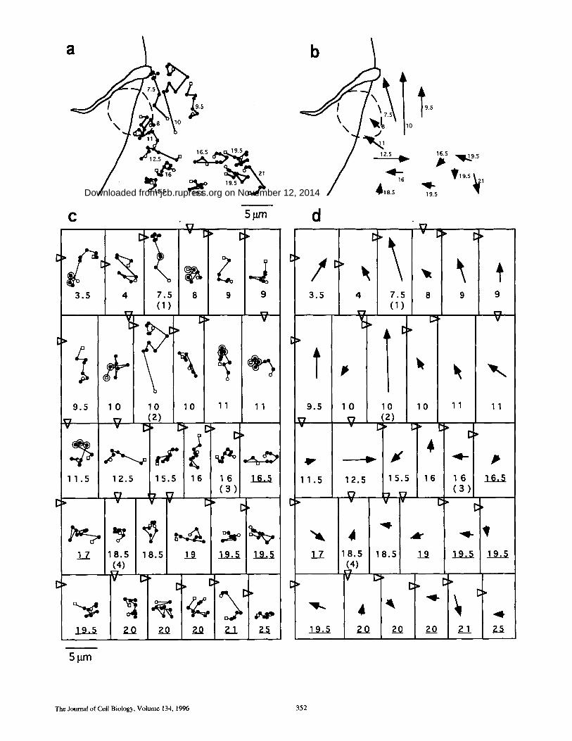
Figure 2. Direct ional m o v e m e n t of B S A - g o l d - l o a d e d lysosomes towards the T. cruzi invasion site. (a) Diagram showing the f rame- to- f rame t rajectory of th i r teen se lec ted lysosomes, during the 8-rain t ime interval cor responding to the invasion sequence shown in Fig. 1, a-k. Small open circles and squares represent , respectively, the initial and final pos i t ion of each lysosome. The dis tance in i~m f rom each lysosome to the parasi te a t t achment site in the initial f rame (Fig. 1 a) is indicated next to each track. The do t t ed circle indicates the area into which the parasi te m o v e d w h e n cell invasion was ini t iated (Fig. 1, f -h) . b, same as a, except that the f rame- to - f rame tracks were re- p laced by arrows indicat ing the di rect ion of m o v e m e n t of each lysosome; the base and tip of the arrows correspond, respectively, to the initial and final pos i t ion of each lysosome. (c and d) Diagrams showing the l inear t racks and cor responding direct ions of m o v e m e n t of all thirty individual lysosomes analyzed. Large open circles r ep resen t the f rames in which lysosomes colocalized with the parasite. The dis tance in p~m f rom each lysosome to the parasi te a t t achment site in the initial f rame (Fig. 1 a) is indicated be low each track; under l ined numbers indicate lysosomes originally located at more than 10 ixm f rom the cell margin. N u mb er s in pa ren theses indicate the lysosomes shown by a r rowheads in Fig. 1 a. O p e n tr iangles r ep resen t the initial pos i t ion of the parasi te in re la t ion to each lysosome (Fig. 1 a).
Rodriguez et al. Lysosome Recruitment during Trypanosome Invasion 353
on Novem
ber 12, 2014jcb.rupress.org
Dow
nloaded from
Published July 15, 1996

frames are shown approximately every 0.8 min (Fig. 1, a-k). During this period, the movements of all lysosomes that were continuously in focus were tracked by recording their position in relation to a reference structure (Fig. 1 a, ar- row). Black arrowheads in Fig. 1 point to two lysosomes (#1, #2) that showed significant directional movement, covering a distance of more than 7 ~m towards the para- site attachment site (Fig. 1, a-e). White arrowheads point to two lysosomes (#3, #4) that did not show directional movement during the same period.
Analysis of the position of lysosomes #1 and #2 in rela- tion to the reference structure (Fig. 1, arrow) shows that the most significant directional movement occurred be- tween frames a and d, during a period of ~2.5 min that fol- lowed the stable attachment of the trypomastigote to the host cell margin, and before cell invasion was initiated. Mean velocities of 0.055 ixrn/s and 0.135 ixm/s were calculated for lysosomes #1 and #2, respectively, during this period. These values are within the range reported previously for the velocity of intracellular vesicles traveling on microtu- bules (Herman and Albertini, 1984; Matteoni and Kreis, 1987). After lysosomes #1 and #2 reached the cell periph- ery, their movement was significantly reduced (Fig. 1, f, black arrowheads), and their motility pattern in the re- maining frames (Fig. 1, g-k, black arrowheads) resembled
the short-range saltatory movement showed by lysosomes #3 and #4 throughout the whole invasion sequence (Fig. 1, a-k, white arrowheads). Lysosome #1 associated tran- siently with the invading trypomastigote (Fig. 1, c-e), al- though this particular lysosome did not fuse with the na- scent parasitophorous vacuole and continued to move forward (Fig. 1, f-k) . However, some gold-loaded lyso- somes did appear to fuse with the parasitophorous vacu- ole, as indicated by the accumulation of BSA-gold parti- cles observed around the parasite at later stages of the entry process (Fig. 1 l).
For a more quantitative assessment of lysosome move- ment in relation to the invading parasite, the movements of all lysosomes continuously in focus throughout the com- plete invasion sequence shown in Fig. 1 were individually tracked. The trajectory of some of these lysosomes in rela- tion to the attached parasite is shown in Fig. 2 a. The initial and final positions and the direction of movement of the same lysosomes are represented by the arrows in Fig. 2 b. Fig. 2, c and d shows trajectories and direction of move- ment for all lysosomes analyzed in this video sequence, a total of thirty. Lysosomes originally located closer to the invasion site tended to move towards the host cell periph- ery, in direction of the parasite attachment site (Fig. 2, b and d). The arrows indicating the initial and final position
e
C ~ C> v I::" I~:"
2 . 5 4 4 2 . 5 4 4 ( 1 ) ( 1 )
V V V V
7 8 9 . 5 7 8 9 . 5 (2 ) (2)
v ¥
11 12 12 11 12 12
V v V V v
' ' / 4 4 . 5 5 4 4 . 5 5
( 1 ) ( 2 ) ( 1 ) ( 2 )
v v ~ t e v ~ v v
5 . 5 8 8 5 . 5 8 8 ~ ~-7 ~
8 . 5 11 11 8 . 5 11 11
Figure 3. Parasite attachment not followed by invasion does not induce lysosome recruitment. Initial (a and c) and final (b and d) frames of two independent time lapse image recordings of trypomastigote attachment events that did not result in invasion. L6E9 myo- blasts were pre-incubated with BSA-gold for 3 h, followed by a chase of 2 h. The a-b sequence covered a period of 9 rain, and the c-d sequence a period of 15 rain. Black lines indicate the initial (a and c) and final (b and d) positions of two lysosomes (1 and 2) in each sequence. The arrows point to the trypomastigote attachment site. (e and f) Trajectory and direction of movement of nine individual ly- sosomes during the a-b (e) and c-d O0 recorded sequences, as described in the legend of Fig. 2. All lysosomes in both video recordings were originally <10 ~m from the cell margin. Bar, 10 Ixm.
The Journal of Cell Biology, Volume 134, 1996 354
on Novem
ber 12, 2014jcb.rupress.org
Dow
nloaded from
Published July 15, 1996

of each lysosome (Fig. 2 d) show that when initial dis- tances to the invasion site were shorter than 12 ixm (dis- tance in p~m is indicated by the numbers below each track/ arrow), eleven out of thirteen lysosomes underwent direc- tional movement.
From the eleven video frames analyzed (Fig. 1, a-k), the initial five (Fig. 1, a-e) correspond to events occurring be- fore the trypomastigote entered the host cell. We can therefore conclude that long stretches of directional lyso- some movement occurred before invasion, while the para- site was still attached to the host cell margin (Table I, time frames 48"3'12"). Measurements of the distance traveled during regular time intervals (every 0.8 min) revealed nine instances of dislocations longer than 3 p~m, for lysosomes originally at less than 12 ~m from the invasion site. In con- trast, when lysosomes originally at more than 12 Ixm from the invasion site were analyzed, only two were found to move more than 3 p~m between selected frames (Table I, boxed numbers).
The lysosomes located at >12 p~m from the invasion site showed mostly the short saltatory movements previously described for these organelles (Herman and Albertini, 1984; Matteoni and Kreis, 1987). This motility pattern ap- peared to be independent of the distance of the lysosomes to the host cell margin, as can be verified in Fig. 2 (where underlined numbers designate lysosomes originally at more than 10 ~m from the cell margin) and Fig. 3 (where all lysosomes analyzed were at less than 10 tLm from the cell margin).
These findings strongly suggest that the recruitment of lysosomes occurring before and during T. cruzi invasion involves only lysosomes initially located at the host cell pe- riphery, in the vicinity of the entry site. Furthermore, tran- sient attachment of trypomastigotes to host cells that do not result in internalization do not appear to affect the motility of lysosomes in the surrounding area. This can be observed in Fig. 3, which shows the analysis of lysosome motility in two independent video recordings of parasite attachments that did not result in invasion (a-b and c-d correspond to the first and last frame of each recording). Lysosomes in the vicinity of the parasite attachment site showed only random saltatory movements with no prefer- ential direction (Fig. 3, e and f). In addition, distances trav- eled by individual lysosomes in these cells every 0.8 min were all shorter than 3 p,m, similar to what was observed for distant lysosomes in the "productive" invasion se- quence (Table II, compare with Table I).
Antibodies to the Cytoplasmic Tail of lgp 120 Induce Aggregation of Lysosomes and Inhibit T. cruzi Invasion
The time lapse video microscopy observations described above showed that peripheral lysosomes are gradually mo- bilized to the vicinity of the trypomastigote invasion site. To obtain direct evidence that this recruitment of lyso- somes is required for T. cruzi invasion, we sought an irre- versible method of removing lysosomes from the host cell periphery without affecting the distribution of other or- ganelles. For this we took advantage of the high concen- tration of the glycoprotein lgp 120 in lysosomes. Lgp 120, together with the related lgp 110, accounts for ~50% of the total protein content of lysosomal membranes. It has a
Table II. Lysosome Motility in Cells with Attached but Noninvading Parasites
Distance traveled (in p,m) by individual lysosomes analyzed in Fig. 3 e.
48"* 1'36" 2'24" 3'12" 4' 4'48" 5'36" 6'24" 7'12' 8'
2.5* 0.5 0.3 0.0 0.1 0.2 0.0 0.1 0.0 0.5 0.1 4.0 0.1 0.5 0.1 0.1 0.2 0.0 0.3 0.5 0.0 0.0 4.0 0.6 0.0 0.3 0.0 0.1 0.1 0.0 0.1 0.1 0.7 7.0 0.7 0.2 0.4 0.0 0.1 0.0 0.6 0.0 0.2 0.7 8.0 1.0 0.1 0.5 0.0 0.1 0.2 0.0 0.0 0.2 0.3 9.5 0.6 0.1 0.1 0.2 0.3 0.0 0.2 0.1 0.0 0.2
11.0 0.8 0.5 0.0 0.5 0.1 0.5 0.3 0.3 0.0 0.1 12.0 0.2 0.0 0.1 0.0 0.0 0.3 0.0 0.3 0.0 0.0 12.0 0.0 0.2 0.0 0.2 0.0 0.0 0.2 0.2 0.1 0.3
Distance traveled (in o,m) by each individual lysosome analyzed in Fig. 3f.
48"* 1'36" 2'24" 3'12" 4' 4'48" 5'36" 6'24" 7'12" 8'
4.0* 0.6 1.0 2.6 0.1 1.1 0.7 0.2 0.4 0.1 0.3 4.5 1.1 0.2 0.3 0.0 0.5 0.2 0.0 0.1 0.2 0.4 5.0 1.5 0.0 0.7 0.1 1.1 0.0 0.1 0.3 0.1 0.0 5.5 1.2 0.9 0.0 0.2 0.1 0.2 0.0 0.0 0.0 0.8 8.0 0.1 0.7 0.0 0.7 0.3 0.1 0.1 0.2 0.3 1.3 8.0 0.9 0.1 0.0 0.0 0.0 1.2 1.1 0.3 0.0 0.0 8.5 0.5 0.1 0.1 0.0 0.6 0.1 0.5 0.2 0.0 0.1
11.0 0.0 0.1 0.1 0.6 0.3 0,0 0.1 1.0 0.1 0.1 11.0 0.5 0.0 1.3 0.1 0.0 0.2 0.1 0.0 0.1 0.0
* Time intervals in which the distance of each lysosome from its position in the previ- ous time frame was measured, starting with the frames shown in Fig. 3 a (top table) or 3 c (bottom table). The total period analyzed was 8 min. Original distance of each individual lysosome to the parasite attachment site, as indi-
cated in Fig. 3, e andf. No dislocation was longer than 3 p~m.
heavily glycosylated lumenal domain, a single transmem- brane anchor, and a short, highly conserved eleven amino acid cytoplasmic tail (Kornfeld and Mellman, 1989). We prepared affinity-purified polyclonal antibodies against a synthetic peptide corresponding to the cytoplasmic tail of lgp 120, and microinjected these antibodies into NRK fi- broblasts. As a control, normal rabbit IgG was injected into a separate group of cells.
Immunofluorescence with anti-rabbit antibodies was used to visualize the microinjected IgG (Fig. 4, a and b). To stain lysosomes of both injected and noninjected cells, labeling with a mAb specific for the lumenal domain of rat lgp 120 was performed on the same preparations (Fig. 4, c and d). Cells injected with normal IgG showed a diffuse distribution of the microinjected antibody throughout the cytoplasm (Fig. 4 a). The general distribution of lysosomes in these cells appeared normal, indistinguishable from noninjected cells in the same field (Fig. 4 c). In contrast, in cells injected with anti-lgp 120 tail IgG, the antibodies were visualized associated with large structures (Fig. 4 b), which also stained with the mAb against the lumenal do- main of lgp 120 (Fig. 4 d). Thus, the introduction of anti- lgp 120 tail antibodies induced a dramatic reorganization in the distribution of lysosomes, with the formation of large clusters in the perinuclear area. A similar aggrega- tion of lysosomes by microinjected anti-lgp 120 tail anti- bodies was observed in ErE 9 rat myoblasts and in HeLa cells (not shown).
Since trace amounts of lgp 120 have been detected in early endosomes (Rabinowitz et al., 1992), we investigated
Rodriguez et al. Lysosome Recruitment during Trypanosome Invasion 355
on Novem
ber 12, 2014jcb.rupress.org
Dow
nloaded from
Published July 15, 1996
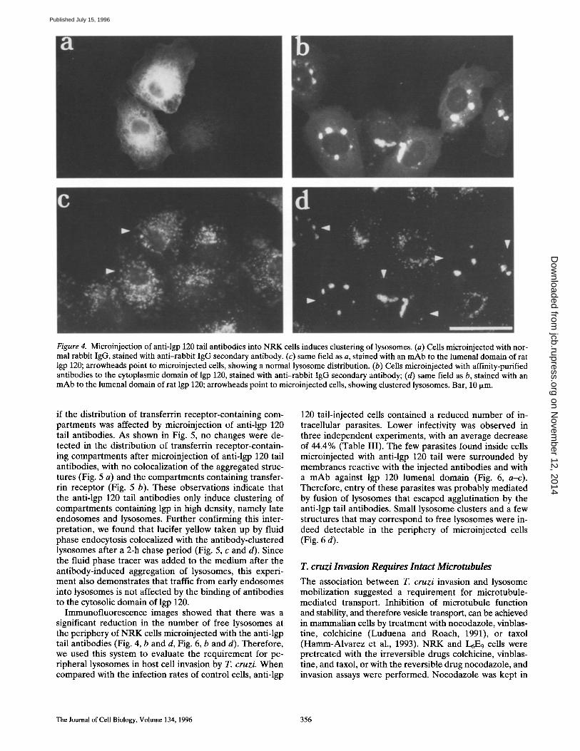
Figure 4. Microinjection of anti-lgp 120 tail antibodies into NRK cells induces clustering of lysosomes. (a) Cells microinjected with nor- mal rabbit IgG, stained with anti-rabbit IgG secondary antibody. (c) same field as a, stained with an mAb to the lumenal domain of rat lgp 120; arrowheads point to microinjected cells, showing a normal lysosome distribution. (b) Ceils microinjected with affinity-purified antibodies to the cytoplasmic domain of lgp 120, stained with anti-rabbit IgG secondary antibody; (d) same field as b, stained with an mAb to the lumenal domain of rat lgp 120; arrowheads point to microinjected cells, showing clustered lysosomes. Bar, 10 ixm.
if the distribution of transferrin receptor-containing com- partments was affected by microinjection of anti-lgp 120 tail antibodies. As shown in Fig. 5, no changes were de- tected in the distribution of transferrin receptor-contain- ing compartments after microinjection of anti-lgp 120 tail antibodies, with no colocalization of the aggregated struc- tures (Fig. 5 a) and the compartments containing transfer- rin receptor (Fig. 5 b). These observations indicate that the anti-lgp 120 tail antibodies only induce clustering of compartments containing lgp in high density, namely late endosomes and lysosomes. Further confirming this inter- pretation, we found that lucifer yellow taken up by fluid phase endocytosis colocalized with the antibody-clustered lysosomes after a 2-h chase period (Fig. 5, c and d). Since the fluid phase tracer was added to the medium after the antibody-induced aggregation of lysosomes, this experi- ment also demonstrates that traffic from early endosomes into lysosomes is not affected by the binding of antibodies to the cytosolic domain of lgp 120.
Immunofluorescence images showed that there was a significant reduction in the number of free lysosomes at the periphery of NRK cells microinjected with the anti-lgp tail antibodies (Fig. 4, b and d, Fig. 6, b and d). Therefore, we used this system to evaluate the requirement for pe- ripheral lysosomes in host cell invasion by T. cruzi. When compared with the infection rates of control cells, anti-lgp
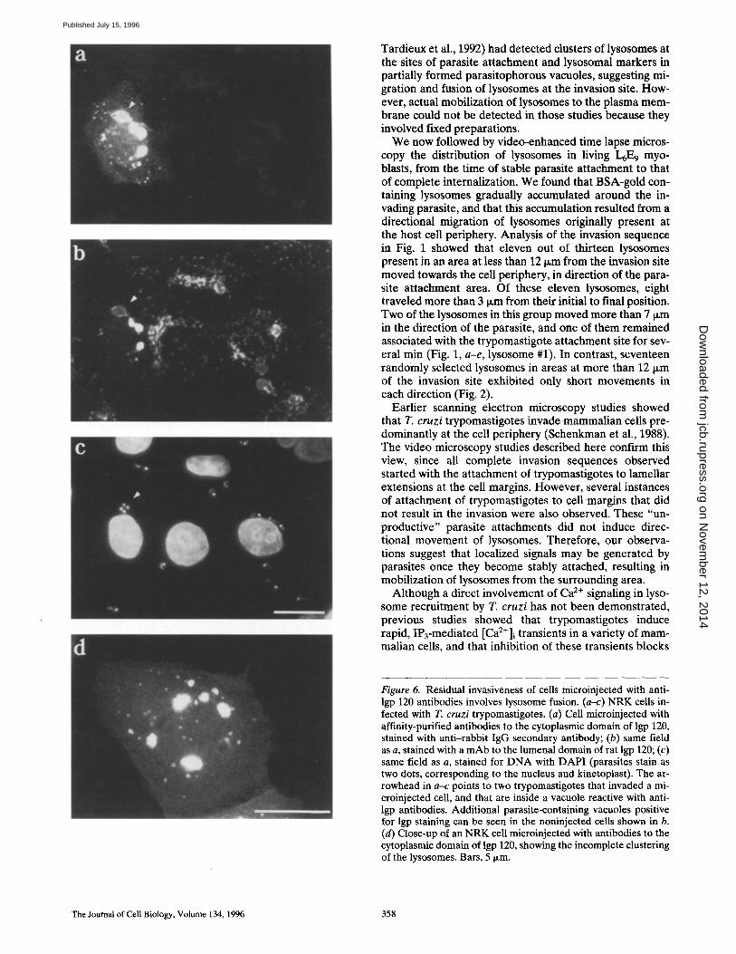
120 tail-injected cells contained a reduced number of in- tracellular parasites. Lower infectivity was observed in three independent experiments, with an average decrease of 44.4% (Table III). The few parasites found inside cells microinjected with anti-lgp 120 tail were surrounded by membranes reactive with the injected antibodies and with a mAb against lgp 120 lumenal domain (Fig. 6, a-c). Therefore, entry of these parasites was probably mediated by fusion of lysosomes that escaped agglutination by the anti-lgp tail antibodies. Small lysosome clusters and a few structures that may correspond to free lysosomes were in- deed detectable in the periphery of microinjected cells (Fig. 6 d).
T. cruzi Invasion Requires Intact Microtubules
The association between T. cruzi invasion and lysosome mobilization suggested a requirement for microtubule- mediated transport. Inhibition of microtubule function and stability, and therefore vesicle transport, can be achieved in mammalian cells by treatment with nocodazole, vinblas- tine, colchicine (Luduena and Roach, 1991), or taxol (Hamm-Alvarez et al., 1993). NRK and L6E 9 cells were pretreated with the irreversible drugs colchicine, vinblas- tine, and taxol, or with the reversible drug nocodazole, and invasion assays were performed. Nocodazole was kept in
The Journal of Cell Biology. Volume 134, 1996 356
on Novem
ber 12, 2014jcb.rupress.org
Dow
nloaded from
Published July 15, 1996

Figure 5. Anti-lgp 120 tail antibody induces clustering of lyso- somes but not of early endosomes. (a and b) The distribution of transferrin receptor-containing early endosomes is not altered in NRK cells with clustered lysosomes: (a) cells microinjected with anti-lgp 120 tail antibodies, stained with anti-rabbit secondary antibody; (b) same field as a, stained with an mAb to the transfer- rin receptor; arrowheads point to microinjected cells. (c and d) Endocytosed lucifer yellow is delivered to clustered lysosomes af- ter a 2-h chase: (c) cells microinjected with anti-lgp 120 tail anti- bodies, stained with anti-rabbit secondary antibody; (d) same field as c, showing lucifer yellow fluorescence in the clustered lysosomes; arrowheads point to microinjected cells. Bar, 5 Ixm.
the medium during the invasion assay, since it is known not to affect trypanosome microtubules (Tardieux et al., 1992; Robinson et al., 1995).
In both NRK and L6E9 cells, the number of trypomasti- gotes invading drug-treated cells was significantly reduced in relation to untreated cells (Fig. 7). The inhibition in- duced by colchicine was dose-dependent, and correlated well with the degree of microtubule depolymerization ob- served in the cells by immunofluorescence with anti-J3 tu- bulin (not shown).
Table IlL Anti-lgp 120 Tail Antibodies Inhibit Host Cell Invasion by T. cruzi
Control Anti-lgp tail Inhibition of (parasites/100 cells) (parasites/100 cells) invasion
%
18.8 6 68.0
18.7 11.2 40.2
16.0 11.9 25.3 mean 44.4 ± 21 .6SD
T. cruzi invasion was quantitated in ~200 microinjected cells, identified by labeling microinjected antibodies with fluorescein-coojugated anti-rabbit IgG. Total parasites were localized by DAPI staining and intracellular parasites by absence of staining, be- fore cell permeabilization, with a mAb to a trypomastigote membrane protein fol- lowed by rhodamine-conjugated anti-mouse IgG. Only parasites associated with mi- croinjected cells were counted. SD, standard deviation.
To verify if treatment with microtubule-active drugs under our experimental conditions affected the availability of signaling receptors at the plasma membrane, NRK cells treated with 10 IxM colchicine for 1 h were tested in a Ca 2÷ signaling assay. The agonists used were thrombin and T. cruzi soluble extracts containing the Ca2+-signaling factor PGTF, previously implicated in host cell invasion (Burleigh and Andrews, 1995; Rodriguez et al., 1995). We found that colchicine treatment did not block the generation of [Ca2+]i transients in NRK cells after exposure to either one of the factors (not shown).
Anti-kinesin Antibodies Inhibit T. cruzi Invasion
The observed migration of lysosomes to the T. cruzi inva- sion site at the cell periphery (Figs. 1 and 2) suggested the involvement of a microtubule-based, plus-end directed motor such as kinesin. Inhibition of kinesin-driven motility of organelles has been achieved with mAbs to the kinesin heavy chain, either in vitro (Ingold et al., 1988; Brady et al., 1990) or after introduction of the antibodies into intact cells (Hollenbeck and Swanson, 1990). To investigate if in- hibitory antibodies to kinesin interfered with lysosome an- terograde movement and T. cruzi invasion, NRK cells were microinjected with SUK-4 mAb (Ingold et al., 1988) or an unrelated IgG1 mAb.
To demonstrate that the microinjected SUK-4 antibod- ies were effective in inhibiting kinesin-dependent lysoso- mal movements under our conditions, we used cytosolic acidification as an assay for microtubule-dependent antero- grade transport (Heuser, 1989). Kinesin heavy chain has been shown to be involved in the acidification-induced re- distribution of lysosomes to the periphery of mammalian cells (Feiguin et al., 1994; Nakata et al., 1995). In cells in- jected with control antibodies, immunofluorescent stain- ing of lysosomes revealed the expected redistribution of lysosomes to the cell periphery after acidification (Fig. 8, e and f). In contrast, in cells microinjected with the SUK-4 anti-kinesin mAb the lysosomes remained clustered in the perinuclear area after acetate treatment (Fig. 8, g-j). Mi- croinjection of either control (Fig. 8, a and b) or SUK-4 (Fig. 8, c and d) mAbs had no effect on the normal perinu- clear organization of lysosomes in cells kept at neutral pH.
Susceptibility to infection by T. cruzi was measured af- ter microinjection of control and SUK-4 anti-kinesin anti- bodies in NRK cells. Five different experiments were per- formed, and inhibition ranging from 35.2 to 69.4% was observed, when controls were compared with SUK-4- injected cells (Table IV). Also consistent with a role for the ATP-driven motor kinesin in T. cruzi invasion, we ob- served a 52% reduction in parasite entry into NRK cells pretreated with sodium azide (10 mM, 30 min at 37°C). This treatment has been described to deplete in 90% the cellular pool of ATP, and to completely arrest the salta- tory motion of intracellular vesicles (De Brabander et al., 1988).
Discussion
We have characterized the recruitment of lysosomes that occurs during the invasion of mammalian cells by the pro- tozoan parasite T. cruzi. Previous studies (Hall et al., 1992;
Rodriguez et al. Lysosome Recruitment during Trypanosome Invasion 357
on Novem
ber 12, 2014jcb.rupress.org
Dow
nloaded from
Published July 15, 1996
Tardieux et al., 1992) had detected clusters of lysosomes at the sites of parasite attachment and lysosomal markers in partially formed parasitophorous vacuoles, suggesting mi- gration and fusion of lysosomes at the invasion site. How- ever, actual mobilization of lysosomes to the plasma mem- brane could not be detected in those studies because they involved fixed preparations.
We now followed by video-enhanced time lapse micros- copy the distribution of lysosomes in living L6E 9 myo- blasts, from the time of stable parasite attachment to that of complete internalization. We found that BSA-gold con- taining lysosomes gradually accumulated around the in- vading parasite, and that this accumulation resulted from a directional migration of lysosomes originally present at the host cell periphery. Analysis of the invasion sequence in Fig. 1 showed that eleven out of thirteen lysosomes present in an area at less than 12 ~m from the invasion site moved towards the cell periphery, in direction of the para- site attachment area. Of these eleven lysosomes, eight traveled more than 3 ~m from their initial to final position. Two of the lysosomes in this group moved more than 7 ~m in the direction of the parasite, and one of them remained associated with the trypomastigote attachment site for sev- eral rain (Fig. 1, a-e, lysosome #1). In contrast, seventeen randomly selected lysosomes in areas at more than 12 ~m of the invasion site exhibited only short movements in each direction (Fig. 2).
Earlier scanning electron microscopy studies showed that T. cruzi trypomastigotes invade mammalian cells pre- dominantly at the cell periphery (Schenkman et al., 1988). The video microscopy studies described here confirm this view, since all complete invasion sequences observed started with the attachment of trypomastigotes to lamellar extensions at the cell margins. However, several instances of attachment of trypomastigotes to cell margins that did not result in the invasion were also observed. These "un- productive" parasite attachments did not induce direc- tional movement of lysosomes. Therefore, our observa- tions suggest that localized signals may be generated by parasites once they become stably attached, resulting in mobilization of lysosomes from the surrounding area.
Although a direct involvement of Ca 2÷ signaling in lyso- some recruitment by T. cruzi has not been demonstrated, previous studies showed that trypomastigotes induce rapid, IP3-mediated [Ca2÷]i transients in a variety of mam- malian cells, and that inhibition of these transients blocks
Figure 6. Residual invasiveness of cells microinjected with anti- lgp 120 antibodies involves lysosome fusion. (a-c) NRK cells in- fected with T. cruzi trypomastigotes. (a) Cell microinjected with affinity-purified antibodies to the cytoplasmic domain of lgp 120, stained with anti-rabbit IgG secondary antibody; (b) same field as a, stained with a mAb to the lumenal domain of rat lgp 120; (c) same field as a, stained for DNA with DAP! (parasites stain as two dots, corresponding to the nucleus and kinetoplast). The ar- rowhead in a-c points to two trypomastigotes that invaded a mi- croinjected cell, and that are inside a vacuole reactive with anti- lgp antibodies. Additional parasite-containing vacuoles positive for lgp staining can be seen in the noninjected cells shown in b. (d) Close-up of an NRK cell microinjected with antibodies to the cytoplasmic domain of lgp 120, showing the incomplete clustering of the lysosomes. Bars, 5 ~m.
The Journal of Cell Biology, Volume 134, 1996 358
on Novem
ber 12, 2014jcb.rupress.org
Dow
nloaded from
Published July 15, 1996

u
O O
N
2 Gt
NRK
20
10
0 Control Nocodazole Taxol Vinblastine Colchicine
200
"6 U
O
~, 100
2 U
L6E9
Control Nocodazole Taxo~ Vinblastine Cotchicine
Figure 7. Irthibitors of host cell microtubule dynamics reduce T. cruzi invasion. The number of intracellular para- sites in N RK (a) or L6E9 cells (b) pretreated with microtu- bule inhibitors was deter- mined after a 20-min infection period. Drug concentrations in a and b were 10 I~M no- codazole, 4 ixM taxol, 1 I~M vinblastine, and 10 I~M colch- icine. The data represent the average of triplicate determi- nations +-SD.
parasite entry (Tardieux et al., 1994; Rodriguez et al., 1995). Interestingly, a localized area of elevated [Ca2+]i was detected around recently internalized T. cruzi trypo- mastigotes in L6E 9 myoblasts (Moreno et al., 1994). Since a soluble CaZ+-signaling factor extracted from trypomas- tigotes (PGTF) is capable of inducing a rapid and revers- ible disassembly of the cortical actin cytoskeleton of NRK cells (Rodriguez et al., 1995), it is conceivable that a local- ized, Ca 2÷ triggered microfilament rearrangement induced by live parasites is a required step for migration and fusion of lysosomes during formation of the T. cruzi-containing vacuole.
The observation that only lysosomes in the vicinity of the invasion site are recruited may be related to the spatial dynamics of locally induced second messengers. Although the cytosolic range of action for Ca 2÷ is very short (~0.08 Ixm), IP3 has a longer diffusion length, estimated in the cy- toplasm of Xenopus oocytes to be ~17 Ixm (Kasai and Pe- tersen, 1994). This value is compatible with the 11-12 Ixm range of action for lysosome mobilization detected in our study, further reinforcing a possible relationship between the T. cruzi-induced, IP3-mediated Ca 2÷ signal (Rodriguez et al., 1995) and lysosome recruitment, cAMP, in contrast, has been reported to have a diffusion length of 30-220 Ixm (Kasai and Petersen, 1994), significantly longer than the range of directional lysosome motility observed during T. cruzi invasion.
Several lines of evidence suggest that tysosomes ap- proaching the trypomastigote invasion site gradually fuse with the plasma membrane, contributing to formation of the parasitophorous vacuole. In addition to the gradual ac- cumulation of BSA-gold around invading trypanosomes observed in this study, lgp 120 and 3-h chased horseradish peroxidase were detected in partially formed vacuoles, when portions of the parasites were still extracellular (Hall et al., 1992; Tardieux et al., 1992). In this context, the be- havior of lysosome #1 in the time lapse invasion sequence shown in Fig. 1 was intriguing. It moved with an average velocity of 0.05 ixrn/s towards the parasite attachment site, where it remained for ~2 min before detaching and reas- suming a saltatory pattern of movement at a location above the invasion site. This transient association followed by detachment is very similar to what was reported for the interaction of BSA-gold-containing late endosomes with phagosomes in macrophages (Desjardins et al., 1994). One possibility is that these observations are related to the
loading of lysosomes with BSA-gold, since the presence of certain indigestible substances within lysosomes and phago- lysosomes can interfere with their fusion capacity (Mont- gomery et al., 1991; Oh and Swanson, 1996). On the other hand, differential rates of transfer of distinct soluble fluo- rescent tracers from lysosomes to phagolysosomes have been described (Wang and Goren, 1987), suggesting that transient fusion-fission events, referred to as "kiss and run" (Desjardins, 1995), might occur. Since fluorescent tracers can only be used to follow the movement of intra- cellular organelles for a limited period of time, due to the damage of vesicular membranes that occurs upon illumi- nation (De Brabander et al., 1986; Swanson et al., 1992), we did not attempt to repeat our invasion studies using flu- orescent markers.
To interfere specifically with the availability of lyso- somes for fusion at the cell periphery and test its effect on trypanosome invasion, we microinjected NRK cells with antibodies against the conserved, eleven amino acid cyto- plasmic tail of the major lysosome membrane glycoprotein lgp 120. These antibodies induced a dramatic clustering of lysosomes in the perinuclear area, significantly reducing the number of isolated lgp-positive structures detectable at the cell periphery. This is, to our knowledge, the first demonstration that the cytoplasmic domain of lgp 120 is exposed on the surface of lysosomes, and accessible to an- tibody binding in living cells. The aggregation of lyso- somes induced by the antibodies was most probably due to cross-linking of lgp 120 molecules on individual lysosomes, as would be predicted by the estimated high density of lgp in lysosomal membranes (Granger et al., 1990).
Three independent invasion assays performed on NRK cells 2 h after microinjection of anti-lgp 120-tail antibodies revealed a reduction ranging from 25 to 68% in the num- ber of internalized trypomastigotes. We believe this varia- tion can be explained by the variable degrees of lysosome aggregation achieved (Figs. 4 b and 6 d), since it is not pos- sible to reproduce precisely the final concentration of anti- bodies introduced by microinjection. The few intracellular parasites found inside anti-lgp tail-injected cells were sur- rounded by a vacuolar membrane reactive with the in- jected antibodies. This observation indicates that success- ful invasion events still involved lysosome fusion, and that binding of antibodies to the cytoplasmic domain of lgp 120 did not block fusion. Furthermore, we found that lucifer yellow taken up by fluid phase endocytosis was delivered
Rodriguez et al. Lysosome Recruitment during Trypanosome Invasion 359
on Novem
ber 12, 2014jcb.rupress.org
Dow
nloaded from
Published July 15, 1996

Table IV. Anti-Kinesin Antibodies Inhibit Host Cell Invasion by T. cruzi
Control Anti-kinesin Inhibition of (parasites/lO0 cells) (parasites/lO0 cells) invasion
%
32 12 62.5 29.3 18.9 35.5
9.1 5.9 35.2 9.2 4.3 53.3
19.3 5.9 69.4 mean 51 _-_ 13.9 SD
T cruzi invasion was quantitated in ~200 microinjected cells, identified by labeling microinjected antibodies with fluorescein-conjugated anti-mouse IgG. Total parasites were localized by DAPI staining and intracellular parasites by absence of staining, be- fore cell permeabilization, with rabbit anti-T, cruzi specific antiserum followed by rhodamine-conjugated anti-rabbit IgG. Only parasites associated with microinjected cells were counted. SD, standard deviation.
Figure 8. Microinjection of anti-kinesin antibodies blocks the acidification-induced redistribution of lysosomes in NRK cells. Cells were microinjected with control mouse IgG1 (a, b, e, and f) or SUK-4 anti-kinesin (c, d, g, h, i, and j) and exposed to pH 7.0 (a-d) or pH 6.6 (e-j) buffers before fixation and staining of lyso- somes. The acidification-induced movement of lysosomes away from the cell center, observed in cells injected with the control
to the antibody-clustered lysosomes after a 2-h chase. We therefore favor the hypothesis that the reduced suscepti- bility to T. cruzi infection of cells injected with anti-lgp 120 tail antibodies is due to decreased availability of lysosomes at the cell periphery, and not to a block in lysosome fusion. Another possibility is that the movement of aggregated ly- sosomes, and therefore their recruitment to the invasion site, was impaired. Experiments using cytosolic acidifica- tion showed that the larger the particles contained in lyso- somes, the more reduced was their capacity to move to- wards the plus-end of microtubules (Perou and Kaplan, 1993).
The requirement for lysosome mobilization during T. cruzi invasion strongly suggested an involvement of host cell microtubules. Earlier experiments designed to address this question were inconclusive, since treatment of NRK cells with nocodazole failed to inhibit infection (Tardieux et al., 1992). In the present study we found that effective depolymerization of NRK cell microtubules with nocoda- zole requires a pre-incubation period at 4°C, as described for polarized epithelial cells (Hunziker et al., 1990). Under these conditions, significant inhibition of T. cruzi entry was observed in both NRK fibroblasts and L 6 E 9 myo- blasts. Host cells were also exposed to two other microtu- bule-active drugs with different mechanisms of action, colchicine, and vinblastine (Luduena and Roach, 1991), and both significantly reduced entry of the parasite in the two cell lines tested. Inhibition was also observed with the stabilizing agent taxol, shown to interfere with certain mi- crotubule-mediated vesicular transport steps in mamma- lian cells (Herman and Albertini, 1984; Hamm-Alvarez et al., 1993). Our results therefore demonstrate a requirement for host cell microtubule dynamics in the T. cruzi invasion process.
Microtubule-binding drugs such as taxol have been re- ported to interfere with the recycling of signaling recep- tors to the plasma membrane (Hamm-Alvarez et al.,
antibody (e and jr) was blocked by SUK-4 (g and h). Another ex- ample is shown in (i and j) where a single cell injected with SUK- 4 shows a perinuclear distribution of lysosomes while the nonin- jected cells surrounding it show the acidification-induced redistri- bution (j). Bar, 5 ~m.
The Journal of Cell Biology, Volume 134, 1996 360
on Novem
ber 12, 2014jcb.rupress.org
Dow
nloaded from
Published July 15, 1996

1994). Since there is evidence that T. cruzi invasion in- volves receptor-mediated, parasite-induced Ca 2÷ signaling (Tardieux et al., 1994; Rodriguez et al., 1995), the reduc- tion in invasion by microtubule inhibitors could have been related to depletion of cell surface receptors. We verified, however, that after treatment with 10 p.M colchicine (a dose that inhibits T. cruzi invasion of NRK cells by 90%) there was no block of the [Ca2+]i transients triggered by the trypomastigote soluble factor PGTF (Rodriguez et al., 1995). In addition, receptor-mediated Ca 2÷ signaling trig- gered in NRK cells by the well-characterized agonist thrombin was also not inhibited by colchicine, under the same conditions.
The outward migration of lysosomes observed during trypomastigote invasion suggested involvement of the plus end-directed microtubule motor kinesin. This was con- firmed by the reduced parasite entry in cells microinjected with the inhibitory anti-kinesin mAb SUK-4 (Ingold et al., 1988; Hollenbeck and Swanson, 1990). Kinesin activity is regulated by Ca2+-calmodulin in vitro (Matthies et al., 1993), which raises the possibility that PGTF-induced Ca 2÷ signaling may play a role in localized lysosome motil- ity by direct modulation of motor activity. Since the lyso- some motility patterns we observed were quite complex, involving both anterograde and retrograde movements, it is conceivable that the minus end--directed motor cyto- plasmic dynein is also involved. Binding of cytoplasmic dy- nein to lysosomes was shown to depend on the presence of Ca z÷ in the extracellular medium (Lin and Collins, 1993).
Recruitment and fusion of lysosomes with the plasma membrane has been described as part of the exocytic path- way of specialized cells, such as neutrophils and cytotoxic lymphocytes (Pryzwansky et al., 1979; Griffiths and Ar- gon, 1995). In these cell types, fusion of lysosome-related granules with the plasma membrane is triggered by Ca 2÷ (Poenie et al., 1987; Jaconi et al., 1990). Cytotoxic T cell granules were shown to move along microtubules in a ki- nesin-dependent fashion (Burkhardt et al., 1993), and in neutrophils, a localized disassembly of actin filaments was detected at the site of fusion of azurophil granules with the plasma membrane (Boyles and Bainton, 1981). This re- port, and our previous finding that a T. cruzi soluble factor induces Ca2+-dependent disassembly of the cortical actin cytoskeleton of NRK fibroblasts (Rodriguez et al., 1995), suggest an intriguing parallel between regulated exocyto- sis and the recruitment of lysosomes during trypanosome invasion of mammalian cells. We thank D. Johnston for affinity purification of anti-lgp 120 tail antibod- ies, B. Kowalsky and D. Johnston for help with the initial microinjection experiments, P. Male and H. Tan for photography, and A. Ma for produc- tion of parasites and excellent technical assistance. We are also grateful to B. Burleigh, T. Kreis and M. Rabinovitch for helpful discussions and com- ments on the manuscript.
This work was supported by the National Institutes of Health RO1AI34867 grant to N.W. Andrews. A. Rodriguez was the recipient of a fellowship from CYCIT (Spain) and E. Samoff was supported by the Na- tional Institutes of Health Molecular Parasitology grant 5T32 AIO7404.
Received for publication 29 January 1996 and in revised form 16 April 1996.
References
Andrews, N.W. 1995. Microbial subversion of phagocytosis: lysosome recruit-
ment during host cell invasion by Trypanosoma cruzi. Trends Cell Biol. 5: 133-I37.
Andrews, N.W., K.S. Hong, E.S. Robbins, and V. Nussenzweig. 1987. Stage- specific surface antigens expressed during the morphogenesis of vertebrate forms of Trypanosoma cruzi. Exp. Parasitol. 64:474-484.
Boyles, J., and D.F. Bainton. 1981. Changes in plasma-membrane-associated filaments during endocytosis and exocytosis in polymorphonuclear leuko- cytes. Cell. 24:905-914.
Brady, S.T., K.K. Pfister, and G.S. Bloom. 1990. A monoclonal antibody against kinesin inhibits both anterograde and retrograde fast axonal transport in squid axoplasm. Proc. Natl. Acad. Sci. USA. 87:1061-1065.
Burkhardt, J.K., J.M. Mcllvain, M.P. Sheetz, and Y. Argon. 1993. Lytic gran- ules from cytotoxic T cells exhibit kinesin-dependent motility on microtu- bules in vitro. J. Cell Sci. 104:151-162.
Burleigh, B., and N.W. Andrews. 1995. A 120 kDa alkaline peptidase from Try- panosoma cruzi is involved in the generation of a novel Ca2÷-signaling factor for mammalian cells. J. Biol. Chem. 270:5172-5180.
Cole, N.B., and J. Lippincott-Schwartz. 1995. Organization of organelles and membrane traffic by microtubules. Curr. Opin. Cell Biol. 7:55--64.
De Brabander, M., R. Nuydens, G. Geuens, M. Moeremans, and J. De May. 1986. The use of submicroscopic gold particles combined with video contrast enhancement as a simple molecular probe for the living cell. Cell Motil. Cy- toskeleton. 6:105-113.
De Brabander, M., R. Nuydens, H. Geerts, and C.R. Hopkins. 1988. Dynamic behavior of the transferrin receptor followed in living epidermoid carcinoma (A431) ceils with nanovid microscopy. Cell Motil. Cytoskeleton. 9:30-47.
Desjardins, M. 1995. Biogenesis of phagolysosomes: the 'kiss and run' hypothe- sis. Trends Cell Biol. 5:183-186.
Desjardins, M., L.A. Huber, R.G. Parton, and G. Griffiths. 1994. Biogenesis of phagolysosomes proceeds though a sequential series of interactions with the endocytic apparatus. Z Cell Biol. 124:677--688.
Feiguin, F., A. Ferreira, K.S. Kosik, and A. Caceres. 1994. Kinesin-mediated or- ganeUe translocation revealed by cellular manipulations. J. Cell Biol. 127: 1021-1039.
Granger, B., S.A. Green, C.A. Gabel, C.L. Howe, I. Mellman, and A. Helenius. 1990. Characterization and cloning of lgp 110, a lysosomal membrane glyco- protein from mouse and rat cells. Z Biol. Chem. 265:12036-12043.
Griffiths, G.M., and Y. Argon. 1995. Structure and biogenesis of lytic granules. Curr. Top. Microbiol. lrnmunol. 198:3%58.
Hall, B.F., P. Webster, A.K. Ma, K.A. Joiner, and N.W. Andrews. 1992. Desial- ylation of lysosomal membrane glycoproteins by Trypanosoma cruzi: a role for the surface neuraminidase in facilitating parasite entry into the host cell cytoplasm. J. Exp. Med. 176:313-325.
Hamm-Alvarez, S.F., P.Y. Preston, and M.P. Sheetz. 1993. Regulation of vesi- cle transport in CV-1 cells and extracts. J. Cell Sci. 106:955-966.
Hamm-Alvarez, S.F., B.E. Alayof, H.M. Himmel, P.Y. Kim, A.L. Crews, H.C. Strauss, and M.P. Sheetz. 1994. Coordinate depression of bradykinin recep- tor recycling and microtubule-dependent transport by taxol. Proc. Natl. Acad. Sci. USA. 91:7812-7816.
Herman, B., and D.F. Albertini. 1984. A time-lapse video intensification analy- sis of cytoplasmic organelle movements during endosome translocation. J. Cell BioL 98:565-576.
Heuser, J.E. 1989. Changes in lysosome shape and distribution correlated with changes in cytoplasmic pH. J. Cell Biol. 108:855-864.
Hollenbeck, P.J., and J.A. Swanson. 1990. Radial extension of macrophage tu- bular lysosomes supported by kinesin. Nature (Lond.). 346:864-866.
Howe, C.L., B.L Granger, M. Hull, S.A. Green, C.A. Gabel, A. Helenius, and I. Mellman. 1988. Derived protein sequence, oligosaccharides, and mem- brane insertion of the 120 kDa lysosomal membrane glycoprotein (lgp 120): identification of a highly conserved family of lysosomal membrane glycopro- teins. Proc. Natl. Acad. Sci. USA. 85:7577-7581.
Hunziker, W., P. Male, and I. Mellman. 1990. Differential microtubule require- ments for transcytosis in MDCK cells. EMBO (Eur. Mol. Biol. Organ.) J. 9: 3515-3525.
Ingold, A.L., S.A. Cohn, and J.M. Scholey. 1988. Inhibition of kinesin-driven microtubule motility by monoclonal antibodies to kinesin heavy chains. J. Cell Biol. 107:2657-2667.
Jaconi, M.E.E., D.P. Lew, J.L. Carpentier, K.E. Magnusson, M. Sjogren, and O. Stendahl. 1990. Cytosolic free calcium elevation mediates the phagosome- lysosome fusion during phagocytosis in human neutrophils..L Cell Biol. 110: 1555-1564.
Kasai, H., and O.H. Petersen. 1994. Spatial dynamics of second messengers: IP 3 and cAMP as long-range and associative messengers. Trends Neurosci. 17: 95-101.
Kornfeld, S., and I. Mellman. 1989. The biogenesis of lysosomes. Annu. Rev. Cell Biol. 5:483-525.
Lin, S.X.H., and C.A. Collins. 1993. Regulation of the intracellular distribution of cytoplasmic dynein by serum factors and calcium. J. Cell Sci. 105:579-588.
Luduena, R.F., and M.C. Roach. 1991. Tubulin sulfhydryl groups as probes and targets for antimitotic and antimicrotubule agents. Pharmac. Ther. 49:133-152.
Matteoni, R., and T.E. Kreis. 1987. Translocation and clustering of endosomes and lysosomes depends on microtubules. J. Cell Biol. 105:1253-1265.
Matthies, H.J.G., R.J. Miller, and H.C. Palfrey. 1993. Calmodulin binding to and cAMP-dependent phosphorylation of kinesin light chains modulate ki- nesin ATPase activity. J. Biol. Chem. 268:11176-11187.
Rodriguez et al. Lysosome Recruitment during Trypanosome Invasion 361
on Novem
ber 12, 2014jcb.rupress.org
Dow
nloaded from
Published July 15, 1996
Ming, M., M.E. Ewen, and M.A. Pereira. 1995. Trypanosome invasion of mam- malian cells requires activation of the TGFI3 signaling pathway. Cell. 82:1-20.
Montgomery, R.R., P. Webster, and I. Mellman. 1991. Accumulation of indi- gestible substances reduces fusion competence of macrophage lysosomes. Z lmmunol. 147:3087-3095.
Moreno, S.N., J. Silva, A.E. Vercesi, and R. Docampo. 1994. Cytosolic free cal- cium elevation in Trypanosoma cruzi is required for invasion. Z Exp. Med. 180:1535-1540.
Nakata, T., and N. Hirokawa. 1995. Point mutation of adenosine triphosphate- binding motif generated rigor kinesin that selectively blocks anterograde ly- sosome membrane transport. J. Cell Biol. 131:1039-1053.
Oh, Y-K., and J.A. Swanson. 1996. Different fates of phagocytosed particles af- ter delivery into macrophage lysosomes. Z Cell Biol. 132:585-593.
Perou, C.M., and J. Kaplan. 1993. Chediak-Higashi syndrome is not due to a de- fect in microtubule-based lysosomal mobility. Z Cell Sci. 106:99-107.
Poenie, M., R.Y. Tsien, and A.M. Schmitt-Verhulst. 1987. Sequential activation and lethal hit by [Ca2+]i in individual cytolytic T cells and targets. EMBO (Eur. Mol. Biol. Organ.) J. 6:2223-2232.
Pryzwansky, K.B., E.K. MacRae, J.K. Spitznagel, and M.H. Cooney. 1979. Early degranulation of human neutrophils: immunocytochemical studies of surface and intracellular phagocytic events. Cell 18:1025-1033.
Rabinowitz, S., H. Horstmann, S. Gordon, and G. Griffiths. 1992. Immunocy- tochemical characterization of the endocytic and phagolysosomal compart- ments in peritoneal macrophages. J. Cell Biol. 116:95-112.
Robinson, D.R., T. Sherwin, A. Ploubidou, E.H. Byard, and K. Gull. 1995. Mi- crotubule polarity and dynamics in the control of organelle positioning, seg- regation, and cytokinesis in the trypanosome cell cycle. J. Cell Biol. 128: 1163-1172.
Rodriguez, A., M.G. Rioult, A. Ora, and N.W. Andrews. 1995. A trypanosome- soluble factor induces IP3 formation, intracellular Ca 2÷ mobilization and mi-
crofilament rearrangement in host cells. J. Cell Biol. 129:1263-1273. Schenkman, S., N.W. Andrews, V. Nussenzweig, and E.S. Robbins. 1988. Try-
panosoma cruzi invade a mammalian epithelial cell in a polarized manner. Cell. 55:157-165.
Schenkman, S., E.S. Robbins, and V. Nussenzweig. 1991. Attachment of Try- panosoma cruzi to mammalian cells requires parasite energy, and invasion can be independent of the target cell cytoskeleton. Infect. Immun. 59:645- 654.
Schroer, T.A., and M.P. Sheetz. 1991. Functions of microtubule-based motors. Annu. Rev. Physiol. 53:629-652.
Slot, J.W., and H.J. Geuze. 1985. A new method of preparing gold probes for multiple-labeling cytochemistry. Eur. J. Cell Biol. 38:87-93.
Swanson, J.A., A. Locke, P. Ansel, and P. Hollenbeck. 1992. Radial movement of lysosomes in permeabilized macrophages. J. Cell Sci. 103:202-209.
Tardieux, I., P. Webster, J. Ravensloot, W. Boron, J.A. Lunn, J.E. Heuser, and N.W. Andrews. 1992. Lysosome recruitment and fusion are early events re- quired for trypanosome invasion of mammalian cells. Cell. 71:1117-1130.
Tardieux, I., M. Nathanson, and N.W. Andrews. 1994. Role in host cell invasion of Trypanosoma cruzi-induced cytosolic-free Ca 2÷ transients. J. Exp. Med. 179:1017-1022.
Viitala, J., S.R. Carlsson, P.D. Siebert, and M. Fukuda. 1988. Molecular cloning of cDNAs encoding lamp A, a human lysosomal membrane glycoprotein with apparent Mr ~ 120,000. Proc. Natl. Acad. Sci. USA. 85:3743-3747.
Wang, Y.L., and M.B. Goren. 1987. Differential and sequential delivery of fluo- rescent lysosomal probes into phagosomes in mouse peritoneal macro- phages. J. Cell Biol. 104:1749-1754.
White, S., K. Miller, C. Hopkins, and I.S. Trowbridge. 1992. Monoclonal anti- bodies against defined epitopes of the human transferrin receptor cytoplas- mic tail. Biochim. Biophys. Acta. 1136:28-34.
The Journal of Cell Biology, Volume 134, 1996 362
on Novem
ber 12, 2014jcb.rupress.org
Dow
nloaded from
Published July 15, 1996