Neuropeptide Y inhibits cholangiocarcinoma cell growth and invasion
Upload
khangminh22Category
view
1download
0
Early Invasion of Brain Parenchyma by AfricanTrypanosomesUte Frevert1, Alexandru Movila1, Olga V. Nikolskaia2, Jayne Raper4, Zachary B. Mackey5¤a,
Maha Abdulla5¤b, James McKerrow5, Dennis J. Grab2,3*
1 Division of Medical Parasitology, Department of Microbiology, New York University School of Medicine, New York, New York, United States of America, 2 Department of
Pathology, The Johns Hopkins University School of Medicine, Baltimore, Maryland, United States of America, 3 Department of Pediatrics, The Johns Hopkins University
School of Medicine, Baltimore, Maryland, United States of America, 4 Department of Biological Sciences, Hunter College of CUNY, New York, New York, United States of
America, 5 Department of Pathology, University of California San Francisco, San Francisco, California, United States of America
Abstract
Human African trypanosomiasis or sleeping sickness is a vector-borne parasitic disease that has a major impact on humanhealth and welfare in sub-Saharan countries. Based mostly on data from animal models, it is currently thought thattrypanosome entry into the brain occurs by initial infection of the choroid plexus and the circumventricular organs followeddays to weeks later by entry into the brain parenchyma. However, Trypanosoma brucei bloodstream forms rapidly crosshuman brain microvascular endothelial cells in vitro and appear to be able to enter the murine brain without inflictingcerebral injury. Using a murine model and intravital brain imaging, we show that bloodstream forms of T. b. brucei andT. b. rhodesiense enter the brain parenchyma within hours, before a significant level of microvascular inflammation isdetectable. Extravascular bloodstream forms were viable as indicated by motility and cell division, and remained detectablefor at least 3 days post infection suggesting the potential for parasite survival in the brain parenchyma. Vascularinflammation, as reflected by leukocyte recruitment and emigration from cortical microvessels, became apparent only withincreasing parasitemia at later stages of the infection, but was not associated with neurological signs. Extravasculartrypanosomes were predominantly associated with postcapillary venules suggesting that early brain infection occurs byparasite passage across the neuroimmunological blood brain barrier. Thus, trypanosomes can invade the murine brainparenchyma during the early stages of the disease before meningoencephalitis is fully established. Whether individualtrypanosomes can act alone or require the interaction from a quorum of parasites remains to be shown. The significance ofthese findings for disease development is now testable.
Citation: Frevert U, Movila A, Nikolskaia OV, Raper J, Mackey ZB, et al. (2012) Early Invasion of Brain Parenchyma by African Trypanosomes. PLoS ONE 7(8):e43913. doi:10.1371/journal.pone.0043913
Editor: Henk D. F. H. Schallig, Royal Tropical Institute, The Netherlands
Received May 1, 2012; Accepted July 26, 2012; Published August 31, 2012
Copyright: � 2012 Frevert et al. This is an open-access article distributed under the terms of the Creative Commons Attribution License, which permitsunrestricted use, distribution, and reproduction in any medium, provided the original author and source are credited.
Funding: This research was supported in part by grants from the National Institutes of Health (NIH)(5R01AI082695) to DJG, and Brain & Immuno Imaging Grantfrom the Dana Foundation and NIH (S10 RR019288) to UF. The funders had no role in study design, data collection and analysis, decision to publish, orpreparation of the manuscript.
Competing Interests: The authors have declared that no competing interests exist.
* E-mail: [email protected]
¤a Current address: Biochemistry Department, Virginia Polytechnic Institute and State University, Blacksburg, Virginia, United States of America¤b Current address: Department of Surgery, King Khalid University Hospital and College of Medicine, King Saud University, Riyadh, Saudi Arabia
Introduction
African trypanosomes replicate at the tsetse fly bite site before
migrating from the skin via the lymphatic system into the
bloodstream to infect the spleen, liver, lymph nodes, skin, heart,
eyes, and endocrine system [1,2]. If untreated during the early
stage (Stage-1), the parasites later produce central nervous system
(CNS) disease (Stage-2), a process that takes months to years with
T. b. gambiense (West African) and weeks to months with T. b. rho-
desiense (East African) sleeping sickness. Human African trypano-
somiasis (HAT) associated meningoencephalitis leads to progres-
sive neurologic involvement with concomitant neuropsychiatric
disorders, fragmentation of the circadian sleep-wake cycle, and
eventually death [1–3]. Once Stage-2 CNS disease is established,
the parasites are shielded from the many trypanocidal drugs and
the brain may be a source for relapse [4,5].
Longitudinal studies in mice all describe one general progres-
sion pattern of late-stage brain histopathology [6–14]. Trypano-
somes initially accumulate in the stroma of the highly vascularized
choroid plexus, between a layer of fenestrated endothelia and a
layer of epithelia that produce cerebrospinal fluid (CSF). The
resulting inflammatory response disseminates from the choroid
plexus via the microvasculature into the parenchyma in three
characteristic phases: leptomeningitis, early meningoencephalitis,
and encephalitis. According to the current paradigm, trypano-
somes invade the brain parenchyma only at the final stages of the
disease, when meningoencephalitis is fully established. Both
histopathology and composition of the inflammatory infiltrate
observed in the murine model mimic autopsy findings in HAT
patients [11,15–18].
African trypanosomes (T. brucei spp.) upregulate ICAM-1,
VCAM-1 and E-selectin expression in brain and non-brain
endothelial cells [19,20]. Our studies using human brain
microvascular endothelial cells (HBMEC) as a blood brain
barrier (BBB) model show a functional link between trypano-
some cysteine proteases, Gaq-mediated calcium signaling, and
PLOS ONE | www.plosone.org 1 August 2012 | Volume 7 | Issue 8 | e43913

protease-activated receptor-2 (PAR-2) in BBB transmigration
[21–24]. Increased chemokine/cytokine protein expression with
trypanosome infection and gene-profiling data identified several
candidate pathways as links between brain inflammatory
processes and CNS HAT [1,24,25].
Based on these findings, we hypothesized that African
trypanosomes, either individually or in groups, have the potential
for rapid BBB traversal in vivo. The following intravital microscopy
(IVM) study of the murine cortical microvasculature supports this
hypothesis by showing early BBB crossing and parasites in the
brain parenchyma within hours after entering the bloodstream.
Results
Our studies on the role of cysteine proteases in the pathogenesis
of T. b. brucei 90-13 trypanosomiasis, both in a mouse model and
with in an in vitro model of the BBB, strongly implicated brucipain
in facilitating trypanosome entry into the brain [26]. To verify the
parasites’ potential for CNS entry, mice received inocula of 105 or
106 Tbb-O BSF to increase the likelihood of parasite detection in
the brain by IVM. Assuming a total blood volume of 1.6 mL in a
23 g C57Bl6 mouse, an inoculum of 105 and 106 Tbb-O should
result in a respective parasitemia of 6.36104 and 6.36105
parasites/mL immediately post infection, numbers that exceed
the initial parasitemia generated by the bite of a single infected
Tsetse fly, but are typically reached in natural infections of
livestock animals (T. b. brucei) or humans (T. b. rhodesiense). Other
than the expected lower parasite numbers observed after
administration of 105 trypanosomes, IVM revealed no major
differences between the two inoculation regimens, in particular
during the first hours after infection.
Day 1 Post Infection: T. b. brucei 90-13 Enters the BrainParenchyma
Mice infected intravenously with 106 Tbb-O BSF were subjected
to craniotomy and immobilized on the stage of an inverted
confocal microscope for intravital imaging of the cerebral cortex
[27]. Nuclei and the vascular lumen were visualized by
intravenous inoculation of Hoechst 33342 and BSA-Alexa 647,
respectively. Capillaries and postcapillary venules (PCVs), the
morphological correlates of the physiological and neuroimmuno-
logical BBB [28], were identified by their respective diameters of
#6 mm and 20–60 mm [29]. PCVs were distinguished from
similarly sized arterioles by the direction of the blood flow [27].
Mice whose meningeal vasculature was injured during craniotomy
exhibited extensive bleeding and were excluded from the study.
None of the mice used for this study exhibited any evidence for
neurological impairment prior to surgery and imaging [30].
Although only low numbers of parasites were observed traveling
with the bloodstream, motile Tbb-O BSF were clearly present in
the brain parenchyma 24 h post infection (Fig. 1, Movie S1).
Occasionally, a trypanosome in process of cell division was seen as
indicated by the presence of two nuclei and two kinetoplasts
(Fig. 2, Movie S2). In contrast to uninfected control animals,
Tbb-O infected mice exhibited individual adherent mononuclear
leukocytes in the cortical microvasculature. These leukocytes
arrested in PCVs and capillaries, the latter of which they
occasionally blocked. Temporary interruption of the blood flow
in a capillary revealed that BSF move with the flagella tip leading
(Fig. 3, Movie S3).
When the lower dose of 105 Tbb-O was inoculated intraperi-
toneally, intravascular parasites were rare and extravascular
parasites were undetectable by IVM because of constraints
dictated by the field of view through a small cranial window (data
not shown). The use of Vibratome sections, a technique often used
for neuron imaging or electrophysiological recording of neurons
especially when input-output fibers are also preserved, allowed
examination of a much larger area of tissue than accessible for
IVM through a small cranial window. By screening multiple
vibratome sections of live brain tissue we did find parasites in the
brain parenchyma (Fig. 4).
Day 2 Post Infection: T. b. brucei 90-13 Induces LeukocyteRecruitment, Vascular Leakage, and Capillary Occlusion
At 48 h after infection, IVM of mice infected intravenously with
106 Tbb-O revealed large numbers of BSF traveling at blood-
stream velocity in the cortical microvasculature and many motile
BSF were found in the brain parenchyma and within the
perivascular space (PVS) surrounding PCVs (Fig. 5, Movie S4and S5). The brain also exhibited signs of microvascular
inflammation: recruitment of leukocytes to PCVs, leakage of the
fluorescent plasma marker into the PVS and surrounding
parenchyma, and focal capillary occlusion.
Similarly, after intraperitoneal inoculation of 105 Tbb-O, the
number of intravascular BSF had increased relative to the previous
day and extravascular parasites were detected more frequently
(Fig. 6, Movie S6 and S7). IVM also revealed a few arrested
mononuclear leukocytes along the wall of PCV and capillaries, but
vascular leakage was less prominent 48 h after infection with 105
compared to 106 parasites (Fig. 7, Movie S8 and S9). Based on
size, shape of nucleus, and amoeboid movement, these mononu-
clear cells were most likely monocytes.
Figure 1. Trypanosomes enter the brain parenchyma. One daypost infection with 106 Tbb-O, mice were subjected to craniotomy andexamined by confocal microscopy. The vascular lumen was visualizedby intravenous inoculation of Alexa 647-conjugated BSA (shown ingreen). A motile Tbb-O BSF (red) is located adjacent to a PCV. RBCs inthe lumen of the PCV exclude the vascular maker and appear asnegatively stained (dark) streaks. Nuclei were stained with Hoechst(blue). Bar = 20 mm. Movie S1.doi:10.1371/journal.pone.0043913.g001
Early Brain Entry of African Trypanosomes
PLOS ONE | www.plosone.org 2 August 2012 | Volume 7 | Issue 8 | e43913
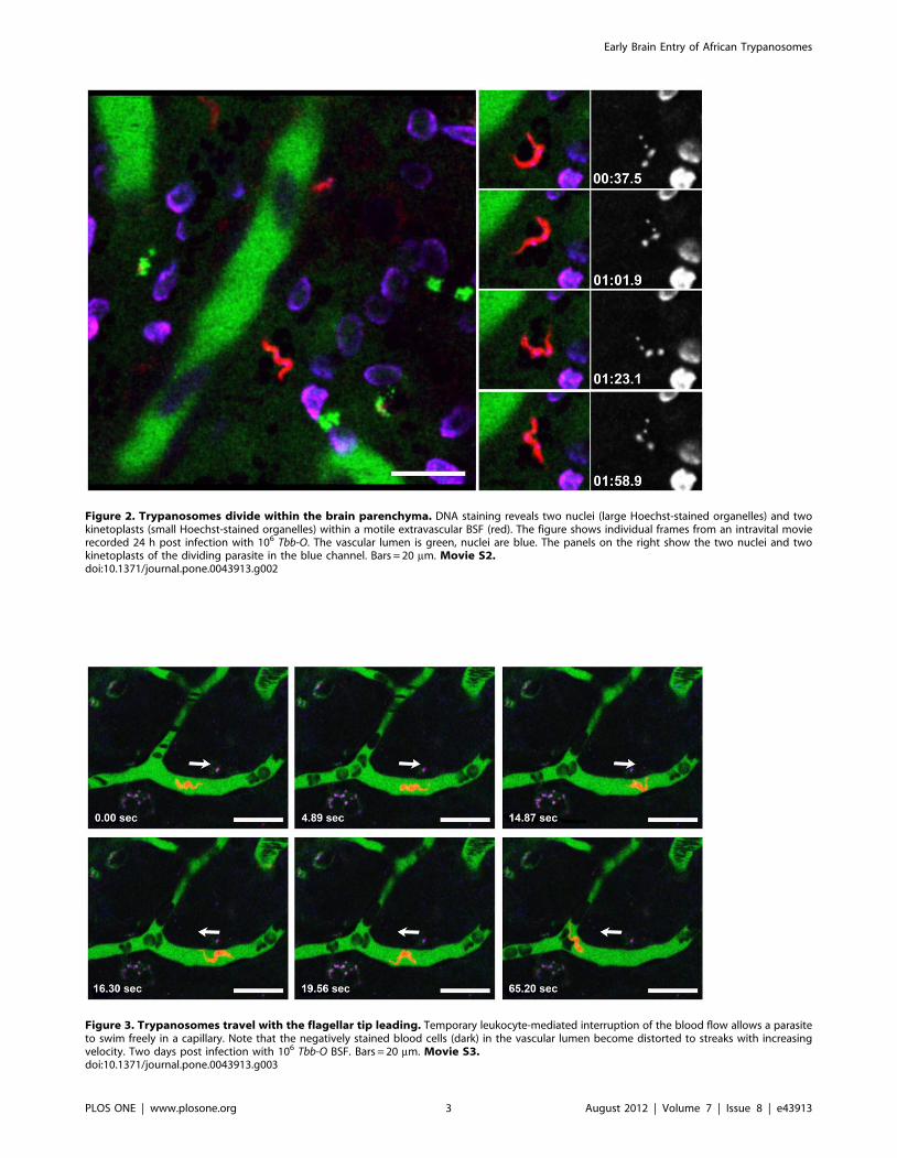
Figure 2. Trypanosomes divide within the brain parenchyma. DNA staining reveals two nuclei (large Hoechst-stained organelles) and twokinetoplasts (small Hoechst-stained organelles) within a motile extravascular BSF (red). The figure shows individual frames from an intravital movierecorded 24 h post infection with 106 Tbb-O. The vascular lumen is green, nuclei are blue. The panels on the right show the two nuclei and twokinetoplasts of the dividing parasite in the blue channel. Bars = 20 mm. Movie S2.doi:10.1371/journal.pone.0043913.g002
Figure 3. Trypanosomes travel with the flagellar tip leading. Temporary leukocyte-mediated interruption of the blood flow allows a parasiteto swim freely in a capillary. Note that the negatively stained blood cells (dark) in the vascular lumen become distorted to streaks with increasingvelocity. Two days post infection with 106 Tbb-O BSF. Bars = 20 mm. Movie S3.doi:10.1371/journal.pone.0043913.g003
Early Brain Entry of African Trypanosomes
PLOS ONE | www.plosone.org 3 August 2012 | Volume 7 | Issue 8 | e43913

Five Hours Post Infection: T. b. rhodesiense IL 1852 andT. b. brucei GVR/35 Enter the Brain Parenchyma
Finally, we tested the potential for BBB traversal of the human
CSF isolate T. b. rhodesiense IL1852 and the T. b. brucei-GVR/35
chronic mouse model trypanosome. Five hours after intravenous
inoculation of 106 parasites, motile Tbr-T BSF (Fig. 8, MovieS10) and Tbb GVR/35-PKH67 (Fig. 8, Movie S11) were found
in the cortical microvasculature and brain parenchyma of the
infected mice. Importantly, arrested leukocytes were rare and
vascular leakage was not evident at this early stage of the infection.
Location of Extravascular TrypanosomesDetermination of the location of all extravascular trypanosomes
detected in this study (N = 44) with respect to the cortical
microvascular tree revealed that the overwhelming majority of
the BSF was associated with PCVs (66%) or similar-sized vessels
(7%), few with arterioles (9%), and none with capillaries. The
remaining parasites (18%) had moved too far into the parenchyma
to be classified. The preferential localization of the parasites near
PCV validates previous reports concerning the site of trypanosome
entry into the brain [2] and confirms that parasite entry into the
brain parenchyma is not simply a surgery-induced artifact.
Discussion
IVM allowed us to visualize real time events at the level of the
neurovascular unit (NVU) that occur near the surface of the
cerebral cortex. Nonetheless, with its dense microvasculature,
study of this region of the brain provides a valid snapshot as to
what may occur in other brain areas. It is accepted that
homeostatic interactions between the cellular components of the
NVU (BBB endothelium, astroglia, neurons) are required for
maintaining normal brain function [31–34]. Furthermore, links
between NVU deregulation and CNS disease have been suggested
for multiple sclerosis, Alzheimer’s disease, stroke, certain depres-
sion disorders, and microbial infections [35–38]. With the cells of
the NVU being rarely located more than 20–40 mm from a
microvessel, a 15–20 mm trypanosome does not have to travel far
to invade the NVU and infiltrate the brain parenchyma.
Figure 4. Brain sections reveal extravascular trypanosomes after low-dose infection. Screening of large areas of brain tissue was requiredto detect extravascular (A, arrow) and intravascular (B, arrow) Tbb-O (red) 24 h after infection with 105 BSF. Infected mice were injected with BSA-Alexa 647 (green) and the nuclear stain Hoechst (blue) prior to removal of the brain. Immediately after vibratome sectioning, 100 mm slices of livebrain tissue were subjected to ex vivo imaging. Traces of the vascular marker remain visible after sectioning and outline the microvascular lumen.Bars = 20 mm.doi:10.1371/journal.pone.0043913.g004
Figure 5. Leukocyte recruitment and vascular leakage. Two days post infection with 106 Tbb-O BSF, the number of both intra- andextravascular trypanosomes has increased. A) Motile BSF are located in the PVS (arrowhead) surrounding a PCV and in the parenchyma (arrows). B) Acluster of extravascular BSF can be seen in a PVS (arrowhead), while numerous others have entered the parenchyma (arrows). The vascular markerBSA-Alexa 647 (shown in green) has leaked into the PVS. The PCVs contain arrested leukocytes (w). The narrow dark streaks in the PCV represent fast-moving blood cells and demonstrate unimpaired blood flow. Bars = 20 mm. Movie S4 and S5.doi:10.1371/journal.pone.0043913.g005
Early Brain Entry of African Trypanosomes
PLOS ONE | www.plosone.org 4 August 2012 | Volume 7 | Issue 8 | e43913

Nevertheless, although the mechanism of trypanosome passage
across neuroimmunological BBB is likely the same throughout the
brain, the morphological correlate of the NVU in the cortex likely
differs from that in other areas of the brain so that the response of
the parenchyma may be focally distinct. For example, selective
sensitivity of the sleep centers to trypanosome infection would
explain the characteristic clinical signs and pathology of HAT.
Further, iNOS activation was reported to increase throughout the
infection process in certain central compartments of the brain, in
particular the thalamus and hypothalamus where the sleep/wake
regulation is located [39,40]. Therefore, preferential death and
disintegration of trypanosomes in these areas with release of toxins
and inflammatory mediators together with the cytotoxic effect of
NO and its derivatives would cause neuronal injury and altered
signaling and thus contribute to the typical pathogenesis of HAT
[12]. Conversely, areas of low iNOS expression and NO
production such as cortex may support parasite survival.
The murine model supports our hypothesis that the parasites
are able to invade the CNS shortly after entering the bloodstream.
Using fluorescent BSA to define the luminal boundary of brain
microvascular endothelial cells that measure only 0.2–0.3 mm in
thickness [41,42], it was not difficult to define a 5620 mm BSF
trypanosome on the abluminal side of the BBB endothelium. Both
T. b. brucei and T. b. rhodesiense BSF gained access to the brain
parenchyma within hours after infection, before a significant level
of microvascular inflammation was detectable. Extravascular BSF
were viable as indicated by motility and cell division and
predominantly associated with PCVs suggesting that early brain
infection occurs by parasite passage across the neuroimmunolog-
ical BBB. Because we did not pre-label the endothelial cells or
their tight junctions, our studies are not able to determine parasites
in the active act of paracellular crossing.
From our studies we cannot with certainty decipher whether
trypanosomes act alone or require communication amongst a
quorum of parasites to enter the brain. If individual trypanosomes
are capable of early brain entry, our finding challenges the current
paradigm that trypanosomes cross the murine BBB only at the
onset of neurological signs, i.e. days after infection with T. b. brucei
or T. b. rhodesiense and several weeks after infection with T. b. gam-
biense [43], at a time when the choroid plexus is already parasitized
and meningoencephalitis is established [19,44–47]. On the other
hand, a higher parasitemia may be required as a prerequisite for
CNS entry to initiate the neurological signs associated with Stage-
2 HAT. Nonetheless, the finding that motile dividing extravascular
Tbb-O and Tbr-T BSF were detectable for at least 3 days post
infection (data not shown) suggests that the parasites can both
cross the BBB and possibly survive in the brain parenchyma. In
support of our finding, Stoppini and colleagues described the use
Figure 6. Leukocyte recruitment and vascular occlusion. Upper panel: Two days after infection with 105 Tbb-O, a motile BSF (red) is located inthe brain parenchyma near a PCV. While numerous other trypanosomes travel at bloodstream velocity in the PCV, the flow in a neighboring capillary(top) is impaired by arrested leukocytes. Lower panel: An arrested mononuclear leukocyte occludes a capillary (arrowhead) and diverts the blood flowinto a collateral microvessel (arrow). Some Tbb-O (red) appear distorted due to their high velocity. Bars = 20 mm. Movie S6 and S7.doi:10.1371/journal.pone.0043913.g006
Early Brain Entry of African Trypanosomes
PLOS ONE | www.plosone.org 5 August 2012 | Volume 7 | Issue 8 | e43913

of a murine organotypic brain culture to study the intracerebral
phase of T. b. brucei-mediated trypanosomiasis in vitro [48].
Remarkably, the parasites survived for several weeks around the
periphery and within the nervous parenchyma, although the
culture medium alone was toxic to the parasites. The same also
holds true for T. b. rhodesiense IL1852 in human organotypic
cerebral cortex culture (Grab, unpublished observation). Further-
more, dividing parasites after 3 weeks did not per se interfere with
neuronal activity of CNS based on extracellular electrophysiolog-
ical recordings [48].
Our finding of early brain invasion by Tbb GVR/35-PKH67 is
potentially profound. While Tbr-T and Tbb-O cause acute
infection identical to wild type parasites, the T. b. brucei GVR/
35 strain establishes a chronic CNS infection in mice with survival
rates of up to 35 days [49]. Based on the degree of neuropath-
ological damage, the level of clinical disability as quantified by
reliable grading scales [50,51], and magnetic resonance imaging
[52], the T. b. brucei GVR/35 induced chronic murine disease
reproduces features typically associated with CNS HAT [4].
Recent experiments conducted at the University of York have
shown T. b. brucei GVR/35 BSF in murine brain sections by day 2
post infection (Lorna MacLean, personal communication), and
studies conducted at the University of Glasgow show GVR/35
BSF in the murine brain on day 5 post infection by two-photon
microscopy and on day 8 by bioluminescence imaging with an
IVIS system (Elmarie Myburgh, personal communication). Most
importantly, some Tbr-infected patients in the Soroti region of
Uganda presented with an early onset of neurodysfunction during
Stage-1 HAT [53]. Together with these findings, our data
challenge the current paradigm that T. b. brucei GVR/35 BSF
invade the CNS only between 14–21 days, because mice can be
cured with the non-CNS drug Berenil if treated within 14–21 days,
i.e. before neurological involvement occurs, but not if treated later
Figure 7. Leukocyte recruitment to PCVs. The number of arrested mononuclear leukocytes increases between 24 h and 48 h post infection with105 Tbb-O. In contrast, arrested cells are absent from the cortical microvasculature of an uninfected control mouse. Whereas intravascular Tbb-O arerare at 24 h, multiple red streaks in the vascular lumen indicate the increase in parasitemia at 48 h post infection. Bars = 20 mm. Movie S8 and S9.doi:10.1371/journal.pone.0043913.g007
Figure 8. Trypanosomes enter the brain parenchyma within hours. Selected frames from intravital movies show motile BSF in the brainparenchyma 5 h after infection with 106 BSF. Note the absence of arrested leukocytes and leakage of the vascular marker into the surrounding tissue.Upper panel: A motile Tbr-T BSF (red) is located in the cerebral parenchyma adjacent to a PCV. Lower panel: A motile Tbb GVR/35-PKH67 BSF (green)can be seen next to a PCV. Intravascular parasites (arrow) are rare. The vascular lumen is red, nuclei are blue. Bars = 20 mm. Movie S10 and S11.doi:10.1371/journal.pone.0043913.g008
Early Brain Entry of African Trypanosomes
PLOS ONE | www.plosone.org 6 August 2012 | Volume 7 | Issue 8 | e43913

than 21 days after infection when Berenil is no longer curative
[4,5,52].
The cerebral microvascular system contains two functionally
distinct barriers, the physiological BBB, which is formed by
capillaries and serves as a tight diffusion barrier for solutes, and the
neuroimmunological BBB, which allows diapedesis of immune
cells and plays a unique role in neuroinflammation [28]. Our IVM
observations show that extravascular trypanosomes are predom-
inantly associated with PCVs rather than capillaries or arterioles,
thus validating previous reports [2]. While our current studies do
not reveal the exact route of the parasites to this location, our
hypothesis is that trypanosomes rapidly cross the neuroimmuno-
logical BBB and enter the PVS [54]. However, until this event is
documented in real time by further study, alternative possibilities
must be considered. For example, some parasites may conceivably
reach the PVS by following the flow of the CSF from the choroid
plexus. While often considered a hostile environment for
trypanosomes [11,14,19], CSF from Stage-1 and Stage-2 HAT
patients supports the survival of T. b. brucei for 20 and 10 hours,
respectively [55]. This window provides ample time for the
parasites to travel inside the PVS from the choroid plexus all the
way down to the level of PCVs [13,15]. IVM studies in
combination with static microscopic examination are currently
underway to distinguish between these different scenarios.
The cortical microvasculature of trypanosome-infected mice
exhibited arrested mononuclear leukocytes, whose number
appeared to increase with disease progression. Leukocyte arrest
was observed in PCVs and capillaries, but not in arterioles, and
occasionally resulted in microvascular occlusion and focal
interruption of the blood flow. Which leukocyte subpopulation is
associated with microvascular inflammation and/or entry into the
PVS and brain parenchyma remains to be determined. Leukocyte
accumulation was accompanied by vascular leakage into the
surrounding brain parenchyma, the extent of which appeared to
correlate with size of parasite inoculum and time post infection.
While there was no evidence for BBB impairment 5 h post
infection with 106 Tbr-T or 106 Tbb GVR/35-PKH67 BSF, 24 h
post infection with 105 or 106 Tbb-O BSF, or 48 h post infection
with 105 Tbb-O BSF, leakage of fluorescent BSA into the
parenchyma was seen 48 h after inoculation of 106 Tbb-O BSF,
i.e. at the time of leukocyte recruitment to PCV. Interestingly,
different neurotropic microorganisms possess unique mechanisms
for interaction with the BBB [25]. For example, while African
trypanosomes appear to traverse the BBB without inflicting
significant damage [21], the strictly intravascular malaria parasite
Plasmodium can induce severe microvascular inflammation and
coagulation resulting in neurological impairment, coma, and death
[25,56–58]. Using a murine model of cerebral malaria and IVM,
we found that neurological signs are closely correlated with the
recruitment of various immune cell populations to PCVs and
widespread disruption of the neuroimmunological BBB [27].
While the immune response to trypanosome infection awaits
further characterization, our data demonstrate that both T. b. bru-
cei and T. b. rhodesiense enter the brain parenchyma within hours,
long before disruption of the BBB is apparent. Interestingly,
trypanosomes were also found in the cerebrospinal fluid of
monkeys early after infection when Stage-1 drugs were still
effective [59].
Many questions still remain and further clarification of parasite
interactions with the BBB endothelium and underlying CNS
components is needed. Trypanosome passage across the murine
BBB is a complex process that depends on many biological
variables related to parasite subspecies and host genetics [60]. Is
the ability to transmigrate across the BBB early an inherent
property of a single parasite, or does the passage across the BBB
require the interaction from a quorum of parasites? It is also
possible that a large parasite load needs to invade the brain to
produce a CNS infection significant enough to produce a CSF
pleocytosis. Perhaps a few parasites manage to enter the brain
during the early stage of infection, but it may take a week or two
for a sufficiently large parasite load to produce CNS disease.
Further, in vitro models suggest that trypanosome traversal across
the BBB may occur bidirectionally [61], which is supported by the
relapses observed in animals after treatment with non-CNS drugs
such as Berenil [62–64]. For our data to be clinically important,
one would expect the parasites to persist in the brain parenchyma.
Considering the dense vascularization of the brain with a mean
intercapillary distance of only 40 mm [65], extravascular trypano-
somes can readily be tracked for long periods of time using a
chronic model for brain IVM [66] in combination with
trypanocidal drugs for elimination of intravascular parasites.
Parasites that are inaccessible to IVM analysis because they are
located in deeper layers of the cortex or the white matter, in the
cerebellum or the brain stem, can be monitored with conventional
techniques or ex vivo in live tissue sections.
While our findings have implications for both animal models
and human disease, the only current way to correlate data from
mouse models with those from HAT patients is to detect
trypanosomes and/or white blood cells in the CSF at Stage-2 of
the disease. Because there are presently no methods to directly
visualize trypanosomes in the human CNS, the complex
mechanisms associated with the pathogenesis of HAT are still
poorly understood. It is currently impossible to distinguish Stage-1
from Stage-2 disease on the basis of clinical findings alone and
CSF analysis remains the only accepted method to diagnose CNS
infection. Interestingly, neurological features were recently report-
ed in sleeping sickness patients with Stage-1 disease [53]. Although
the possibility cannot be excluded that systemic infection can
induce CNS symptoms, this study supports our hypothesis in that
trypanosomes may enter the brain during early stages of the
infection and cause mild early neurological symptoms, but that
protective IL-6 and IL-10 mediated immune responses of the host
prevent major damage to the CNS until later stages of the disease
[67]. Our knowledge of the early pathogenesis of HAT, which
includes the role of host genetics (i.e. rodent strain) in brain
susceptibility to early trypanosomes entry, is still in its infancy.
However, the presence of trypanosomes in the brain hours after
infection may be supportive of treating very early Stage-2 disease
with early stage drugs, a notion that has been suggested as
constituting an intermediate stage of infection [3]. As this is the
first IVM demonstration of trypanosomes in the brain parenchy-
ma as early as hours post infection, the significance of this finding
to human disease and the use of Stage-1 drugs must await the
results of future clinical trials including assessment of whether
current Stage-1 drugs have any brain penetration and/or allow/
bolster neuroimmune systems to protect the brain early in disease.
Nevertheless, the observations reported here suggest that some
case failures of Stage-1 drugs may be due to early access to brain
parenchyma by trypanosomes.
During these studies, several precautions were taken to ascertain
that pathophysiological findings could be distinguished from
methodological artifact. Data acquisition based on confocal Z-
stacks through the 10–15 mm thick meninges in astrocyte-rich
areas showed that microscopic imaging was possible to a depth of
approximately 50 mm. Thus, it was clear that confocal microscopy
would be a suitable methodology to examine in real time, live
extravascular trypanosomes in the cortical microvasculature.
Furthermore, intravital brain microscopy using over one hundred
Early Brain Entry of African Trypanosomes
PLOS ONE | www.plosone.org 7 August 2012 | Volume 7 | Issue 8 | e43913

mice infected with brain tropic-pathogens that include Trypanosoma
brucei (this study), murine-infective Plasmodium (Movila, Nacer and
Frevert, manuscript in preparation, and [27]) and Borrelia burgdorferi
(Movila and Frevert, unpublished) has enabled us to distinguish
between pathophysiological disease-associated vascular alterations
and surgery-induced artifacts. Mice with damage to the pial or
cortical vasculature were excluded from the study. Optimization of
excitation conditions under reduced laser power minimized
phototoxicity and bleaching. In addition, precautions implement-
ed to avoid surgery related-artifacts included i) imaging of
microvessels only at the center of the cranial window to avoid
possible artifacts along the perimeter of the craniotomy, ii)
exclusion of pial microvessels in close proximity to the cranial
coverslip, iii) stabilization of body temperature, and iv) minimi-
zation of respiration-induced movement of the brain. Using these
procedures, none of the Trypanosoma-infected mice exhibited
evidence for neurological impairment prior to surgery and imaging
[30]. Finally, perhaps the most important confirmation that
parasite entry into the brain parenchyma is not simply a surgery-
induced artifact in the preferential localization of the parasites
near PCVs, a finding validating previous reports concerning the
site of trypanosome entry into the brain [2].
By establishing a method for long-term IVM analysis of CNS
trypanosomiasis through a cranial window, we have set the stage
for future functional studies with molecular markers, knockout
mice, transgenic parasites, and RNAi. These studies will provide a
better understanding of how intracellular signaling events allow
trypanosome entry into the parenchyma and how endothelial
activation, induction of inflammatory processes, cytokine secre-
tion, leukocyte recruitment, and BBB alterations are involved in
disease progression. A better understanding of parasite entry into
the CNS, whether individually or as a group, may lead to novel
therapeutic approaches and targets for treatment of neurological
dysfunction.
Materials and Methods
Ethics StatementThis study was conducted in strict accordance with the
recommendations in the Guide for the Care and Use of
Laboratory Animals of the National Institutes of Health. The
protocol was approved by the Institutional Animal Care and Use
Committee of NYU School of Medicine (protocol number
100651). All surgery was performed under ketamine/xylazine/
acepromazine anesthesia and all efforts were made to minimize
suffering.
TrypanosomesFor our in vivo model of experimental CNS infection we chose 1)
the human CSF isolate T. b. rhodesiense IL 1852 [68], 2) T. b. brucei
90-13 bloodstream forms (BSF) [69], a strain previously used to
demonstrate the possible role for brucipain in facilitating
trypanosome entry into the brain [26], and 3) T. b. brucei GVR/
35, a strain that causes a chronic CNS disease in mice [4]. For
visualization by IVM, T. b. brucei GVR/35 BSF were fluorescently
tagged with the cell tracker dye PKH67 (Sigma, St. Louis, MO)
[21] and named Tbb GVR-PKH67 here. Parasites were main-
tained in mice and readily adapt to in vitro culture [70].
Construction of Plasmids Encoding Fluorescent ProteinsT. b. brucei 90-13 were engineered to express monomeric
mOrange (Tbb-O) and T. b. rhodesiense IL 1852 BSF to expressing
tandem dimeric tdTomato (Tbr-T). The phD1034 vector and the
plasmids pRSETOrange and tdTomato were gifts from Oliver
Balmer [71] and Roger Tsien [72], respectively. The open reading
frame for each fluorescent marker was amplified by PCR using the
following primers:
mOrange/eGFP fwd: 59-aagcttATGGTGAGCAAGGGC-
GAGGAG-39
mOrange/eGFP rev: 59-ggatccTTACTTGTA-
CAGCTCGTCCAT-39
tdTomato fwd: 59-cagatatctaagcttATGGTGAGCAAGGGC-
GAGGAG-39
tdTomato rev: 59-cagagactaggatccTTACTTGTA-
CAGCTCGTCCCA-39.
The amplified open reading frames for each fluorescent marker
were then sub-cloned into the pHD1034 vector at the HinDIII/
BamHI sites. Each recombinant plasmid was propagated and
amplified in E. coli.
Trypanosome TransfectionTen mg of pHD1034 vector containing cDNAs for each
fluorescent marker was linearized with NotI restriction endonucle-
ase and precipitated with ethanol. The DNA was re-suspended in
10 ml of H2O and mixed with a 100 mL suspension of T. brucei in
Nucleofector T Cell solution (Lonza Inc. Allendale NJ). For
electroporation, 107 parasites were pelleted by centrifugation. The
parasites were pulsed with the Nucleofector program6001. After
pulsing, the parasites were transferred to 24 mL of complete
medium and incubated overnight at 37uC with 5% CO2. To select
for parasites stably expressing the fluorescent proteins, puromycin
was added to the parasites at a concentration of 0.2 mg/ml and
the parasites were aliquoted in a 24 well plate for 7–12 days.
Stably transfected trypanosomes confirmed by fluorescent micros-
copy and flow cytometry for these studies were maintained in
normal HMI-9 medium. Both Tbr-T and Tbb-O were confirmed
to cause acute infection in mice similar to wild type parasites and
behave like their respective wild type parental strains in acute
mouse models [26]. For example, mice inoculated with 250
fluorescent Tbr show parasitemia within 6 days post infection and
succumb to the disease within 21 days and mice infected with 600
Tbb-O begin to die 13 days after infection (data not shown).
Mice and InfectionFour month-old female C57Bl/6 mice (Taconic Farms) were
inoculated into the retroorbital sinus or intraperitoneally with 105
or 106 culture-derived fluorescent Tbr-T, Tbb-O, or Tbb GVR/35-
PHK67 BSF as indicated for the individual experiments. Mice
were housed in a parasite-free environment on a 12 h light/dark
program and maintained and used in accordance with recom-
mendations in the guide for the Care and Use of Laboratory
Animals.
Anesthesia and CraniotomyAt selected times post infection, mice were anesthetized by
intraperitoneal injection of a cocktail of 50 mg/kg ketamine
(Ketaset, Fort Dodge Animal Health, Fort Dodge, IO), 10 mg/kg
xylazine (Rompun, Bayer, Shawnee Mission, KS), and 1.7 mg/kg
acepromazine (Boehringer Ingelheim Vetmedica, St. Joseph, MO)
(KXA mix) as described [73,74] and surgically prepared for
intravital imaging of the brain [75]. After immobilization of the
head in a stereotaxic apparatus (mouse and neonatal rat adaptor;
Stoelting, Wood Dale, IL), the skull was exposed by midline
incision of the skin and a cranial window 4–5 mm in diameter was
generated using a high-speed microdrill (Fine Science Tools,
Foster City, CA). After removal of the bone flap, the exposed Dura
mater was kept moist with gelatin foam (Gelfoam, Pfizer Inc., New
York) until the window was closed with a cover slip glued to the
Early Brain Entry of African Trypanosomes
PLOS ONE | www.plosone.org 8 August 2012 | Volume 7 | Issue 8 | e43913

skull [76]. Placement of the cranial window over the parietal
cortex allowed imaging of cortical branches of the anterior
cerebral artery, smaller arterioles down to capillary size, PCV, and
superficial parietal veins that drain into the superior sagittal sinus.
Intravital MicroscopyThe cortical microvasculature was imaged with an inverted
Leica TCS SP2 AOBS confocal system [73,74]. Microvessels at
the center of the cranial window were chosen for imaging to avoid
possible surgery-related artifacts along the perimeter of the
craniotomy. Meningeal vessels, which are readily identifiable by
their location on the very surface, i.e. in direct contact with the
coverslip, were excluded from the study. Respiration-induced
movement of the brain was minimized by immobilization of the
cover slip on a custom-made stainless steel adapter for the
microscope stage. The body temperature of the animals was
stabilized with a temperature-controlled Ludin chamber attached
to the microscope. Periodic subcutaneous reinjection of KXA mix
allowed intravital microscopic examination of the brain for 1–2
hours as in the past with Plasmodium-infected mice [27]. To
visualize nuclei and the vascular lumen, mice were injected into
the retroorbital sinus with 150 ml of a mixture of 1–2 mg/ml of the
membrane-permeable DNA stain Hoechst 33342 and 5 mg/ml
Alexa 647-conjugated bovine serum albumin (BSA) (Invitrogen,
Carlsbad, CA), respectively. Appropriate laser lines were used to
excite Hoechst (405 nm), tdTomato (543 nm), mOrange
(543 nm), and Alexa 647 (633 nm). Laser power was reduced to
a minimum to minimize phototoxicity and bleaching. Optimized
excitation conditions have allowed us to monitor live fluorescent
parasites for a period of up to 6 h without any apparent effect on
viability [73,74].
Intravital brain microscopy of more than one hundred mice,
conducted in the course of the studies presented here, IVM of
experimental cerebral malaria (Movila, Nacer and Frevert,
manuscript in preparation, and [27]) and neuroborreliosis (Movila
and Frevert, unpublished), has enabled us to distinguish between
disease-associated vascular alterations and surgery-induced arti-
facts. Mice with damage to the pial or cortical vasculature typically
exhibit extensive bleeding and profuse leakage of the fluorescent
vascular marker. Such animals do not produce useful data and
were excluded from the study.
Vibratome SectioningVibratome sections of unfixed brain tissue from infected mice
that had been injected with fluorescent markers were used to
facilitate parasite detection at early times post infection. Thick
sections (100–200 mm) were cut with a Leica VT1200 Semiauto-
matic Vibrating Blade Microtome (Leica Microsystems, Bannock-
burn, IL) and imaged immediately after transfer into medium.
Image ProcessingMultiple confocal time series of the cortical microvasculature
were acquired with Leica Confocal Software. Imaris 7.4 and
Imaris Track (Bitplane, Saint Paul, MN), Image-Pro Plus (Media
Cybernetics, Bethesda, MD), AutoDeBlur (Media Cybernetics,
Bethesda, MD), and NIH ImageJ were used for deconvolution and
analysis of time sequences.
Supporting Information
Movie S1 Trypanosomes enter the brain parenchyma.One day post infection with 106 Tbb-O, a motile Tbb-O BSF (red) is
located adjacent to a PCV. The vascular lumen is visualized with
Alexa 647-conjugated BSA (shown in green). RBCs in the lumen
of the PCV exclude the vascular maker and appear as negatively
stained (dark) streaks. Nuclei are blue.
(AVI)
Movie S2 Trypanosomes divide within the brain paren-chyma. DNA staining reveals two nuclei (large Hoechst-stained
organelles) and two kinetoplasts (small Hoechst-stained organelles)
within a motile extravascular BSF (red) 24 h post infection with
106 Tbb-O. The vascular lumen is green, nuclei are blue.
(AVI)
Movie S3 Trypanosomes travel with the flagellar tipleading. Temporary leukocyte-mediated interruption of the
blood flow allows a Tbb-O (red) to swim back and forth in a
capillary 48 h post infection with 106 BSF.
(AVI)
Movie S4 Extravascular trypanosomes and leukocyterecruitment to PCVs. Two days post infection with 106 Tbb-O
BSF, motile BSF are located in the brain parenchyma adjacent to
a PCV. Large numbers of red streaks in the vascular lumen
indicate high parasitemia. A leukocyte adheres to the wall of the
PCV.
(AVI)
Movie S5 Leukocyte recruitment and vascular leakage.Two days post infection with 106 Tbb-O BSF, a cluster of
extravascular BSF is located in the PVS (lower left), while many
other BSF have entered the parenchyma (upper right). The
vascular marker BSA-Alexa 647 (shown in green) has leaked into
the PVS. The PCV contains arrested leukocytes (dark cells). The
narrow dark streaks in the center of the PCV represent fast-
moving blood cells demonstrating unimpaired blood flow.
(AVI)
Movie S6 Leukocyte recruitment and vascular occlu-sion. Two days after inoculation of 105 Tbb-O, a motile BSF (red)
is located in the brain parenchyma near a PCV. While numerous
other trypanosomes travel at bloodstream velocity in the PCV, the
flow in a neighboring capillary (top) is impaired by arrested
leukocytes.
(AVI)
Movie S7 Leukocyte recruitment and capillary occlu-sion. An arrested mononuclear leukocyte occludes a capillary and
diverts the blood flow into a collateral microvessel. Tbb-O BSF
(red) become distorted when moving at high velocity. The vascular
lumen is green, nuclei are blue.
(AVI)
Movie S8 Leukocyte recruitment after low-dose infec-tion. Intravascular Tbb-O and arrested mononuclear leukocytes
are rare 24 h post infection with 105 BSF.
(AVI)
Movie S9 Leukocyte recruitment after prolonged low-dose infection. The number of arrested mononuclear leukocytes
has increased between 24 h and 48 h post infection with 105 Tbb-
O. Numerous red streaks in the lumen of the PCV indicate the
higher level of parasitemia.
(AVI)
Movie S10 Tbr-T BSF in the parenchyma 5 hours postinfection. A motile Tbr-T BSF (red) is located in the brain
parenchyma adjacent to a PCV after infection with 106 BSF. Note
the absence of arrested leukocytes and leakage of the vascular
marker (red) into the surrounding tissue. The vascular lumen is
green, nuclei are blue.
(AVI)
Early Brain Entry of African Trypanosomes
PLOS ONE | www.plosone.org 9 August 2012 | Volume 7 | Issue 8 | e43913

Movie S11 Tbb BSF in the parenchyma 5 hours postinfection. A motile Tbb GVR/35 BSF, labeled with PKH67
(green), can be seen next to a PCV. Intravascular parasites are
rare. Note the absence of arrested leukocytes and leakage of the
vascular marker (red) into the surrounding tissue. Nuclei are blue.
(AVI)
Acknowledgments
We are indebted to Dr. Oliver Balmer (FiBL, Switzerland) for providing us
with the phD1034 vector, Dr. Roger Tsien (University of California at San
Diego, USA) for the pRSETOrange and dtTomato plasmids, and Dr.
Lorna MacLean (University of York, UK) and Dr. Jean Rogers (University
of Glasgow, UK) for a gift of T. b. brucei GVR/35. We thank Drs. Rahul
Bakshi, Dr. J. Stephen Dumler, Dr. Paul Englund (Johns Hopkins
University, Baltimore, USA) for critically reading of the manuscript.
Author Contributions
Conceived and designed the experiments: UF DJG JM. Performed the
experiments: UF AM OVN JR ZBM MA DJG. Analyzed the data: UF
AM JM DJG. Contributed reagents/materials/analysis tools: UF DJG JM.
Wrote the paper: UF DJG ZBM JM.
References
1. Grab DJ, Kennedy PE (2008) Traversal of human and animal trypanosomes
across the blood-brain barrier. J Neurovirology 14: 344–351.
2. Kristensson K, Nygard M, Bertini G, Bentivoglio M (2010) African trypanosome
infections of the nervous system: parasite entry and effects on sleep and synaptic
functions. Prog Neurobiol 91: 152–171.
3. Kennedy PG (2004) Human African trypanosomiasis of the CNS: current issues
and challenges. J Clin Invest 113: 496–504.
4. Jennings FW, Gray GD (1983) Relapsed parasitaemia following chemotherapyof chronic T. brucei infections in mice and its relation to cerebral trypanosomes.
Contrib Microbiol Immunol 7: 147–154.
5. Jennings FW, Hunter CA, Kennedy PG, Murray M (1993) Chemotherapy ofTrypanosoma brucei infection of the central nervous system: the use of a rapid
chemotherapeutic regimen and the development of post-treatment encephalop-athies. Trans R Soc Trop Med Hyg 87: 224–226.
6. Fink E, Schmidt H (1979) Meningoencephalitis in chronic Trypanosoma brucei
rhodesiense infection of the white mouse. Tropenmed Parasitol 30: 206–211.
7. Stevens DR, Moulton JE (1977) Experimental meningoencephalitis inTrypanosoma brucei infection of deer mice (Peromyscus maniculatus). A light,
immunofluorescent, and electron microscopic study. Acta Neuropathol 38:173–180.
8. Poltera AA, Hochmann A, Rudin W, Lambert PH (1980) Trypanosoma brucei
brucei: a model for cerebral trypanosomiasis in mice–an immunological,histological and electronmicroscopic study. Clin Exp Immunol 40: 496–507.
9. Abolarin MO, Stamford SA, Ormerod WE (1986) Interaction between
Trypanosoma brucei and the ependymal cell of the choroid plexus. Trans R SocTrop Med Hyg 80: 618–625.
10. Schultzberg M, Ambatsis M, Samuelsson EB, Kristensson K, van Meirvenne N
(1988) Spread of Trypanosoma brucei to the nervous system: early attack oncircumventricular organs and sensory ganglia. J Neurosci Res 21: 56–61.
11. Philip KA, Dascombe MJ, Fraser PA, Pentreath VW (1994) Blood-brain barrier
damage in experimental African trypanosomiasis. Ann Trop Med Parasitol 88:607–616.
12. Van Marck EA, Le Ray D, Beckers A, Jacob W, Wery M, et al. (1981) Light andelectron microscope studies on extravascular Trypanosoma brucei gambiense in the
brain of chronically infected rodents. Ann Soc Belg Med Trop 61: 57–78.
13. Schmidt H (1983) The pathogenesis of trypanosomiasis of the CNS. Studies onparasitological and neurohistological findings in Trypanosoma rhodesiense infected
vervet monkeys. Virchows Arch A Pathol Anat Histopathol 399: 333–343.
14. Wolburg H, Mogk S, Acker S, Frey C, Meinert M, et al. (2012) Late stageinfection in sleeping sickness. PLoS ONE 7: e34304.
15. Pentreath VW (1995) Trypanosomiasis and the nervous system. Pathology and
immunology. Trans R Soc Trop Med Hyg 89: 9–15.
16. Hunter CA, Kennedy PG (1992) Immunopathology in central nervous systemhuman African trypanosomiasis. J Neuroimmunol 36: 91–95.
17. Poltera AA, Owor R, Cox JN (1977) Pathological aspects of human African
trypanosomiasis (HAT) in Uganda. A post-mortem survey of fourteen cases.Virchows Arch A Pathol Anat Histol 373: 249–265.
18. Manuelidis EE, Robertson DH, Amberson JM, Polak M, Haymaker W (1965)Trypanosoma rhodesiense encephalitis. Clinicopathological study of five cases of
encephalitis and one of mel B hemorrhagic encephalopathy. Acta Neuropathol
5: 176–204.
19. Mulenga C, Mhlanga JD, Kristensson K, Robertson B (2001) Trypanosoma
brucei brucei crosses the blood-brain barrier while tight junction proteins are
preserved in a rat chronic disease model. Neuropathol Appl Neurobiol 27: 77–85.
20. Girard M, Giraud S, Courtioux B, Jauberteau-Marchan MO, Bouteille B (2005)
Endothelial cell activation in the presence of African trypanosomes. MolBiochem Parasitol 139: 41–49.
21. Grab DJ, Nikolskaia O, Kim YV, Lonsdale-Eccles JD, Ito S, et al. (2004) Africantrypanosome interactions with an in vitro model of the human blood-brain
barrier. J Parasitol 90: 970–979.
22. Nikolskaia OV, de ALAP, Kim YV, Lonsdale-Eccles JD, Fukuma T, et al. (2006)Blood-brain barrier traversal by African trypanosomes requires calcium
signaling induced by parasite cysteine protease. J Clin Invest 116: 2739–2747.
23. Nikolskaia OV, de ALAP, Kim YV, Lonsdale-Eccles JD, Fukuma T, et al. (2008)Erratum. J Clin Invest 118: 1974.
24. Grab DJ, Garcia-Garcia JC, Nikolskaia OV, Kim YV, Brown A, et al. (2009)
Protease activated receptor signaling is required for African trypanosome
traversal of human brain microvascular endothelial cells. PLoS Negl Trop Dis 3:
e479.
25. Grab DJ, Chakravorty SJ, van der Heyde H, Stins MF (2011) How can
microbial interactions with the blood-brain barrier modulate astroglial and
neuronal function? Cell Microbiol 13: 1470–1478.
26. Abdulla MH, O’Brien T, Mackey ZB, Sajid M, Grab DJ, et al. (2008) RNA
Interference of Trypanosoma brucei cathepsin B and L affects disease progression in
a mouse model. PLoS Negl Trop Dis 2: e298.
27. Nacer A, Movila A, Baer K, Mikolajczak SA, Kappe S, et al. (2012)
Neuroimmunological blood brain barrier disruption in experimental cerebral
malaria. Manuscript submitted.
28. Owens T, Bechmann I, Engelhardt B (2008) Perivascular spaces and the two
steps to neuroinflammation. J Neuropathol Exp Neurol 67: 1113–1121.
29. Ross MH, Romrell LJ, Kaye GI (1995) Histology, a text and atlas. Baltimore
MD: Williams and Wilkins.
30. Carroll RW, Wainwright MS, Kim KY, Kidambi T, Gomez ND, et al. (2010) A
rapid murine coma and behavior scale for quantitative assessment of murine
cerebral malaria. PLoS ONE 5.
31. Abbott NJ, Ronnback L, Hansson E (2006) Astrocyte-endothelial interactions at
the blood-brain barrier. Nature reviews Neuroscience 7: 41–53.
32. Persidsky Y, Ramirez SH, Haorah J, Kanmogne GD (2006) Blood-brain barrier:
structural components and function under physiologic and pathologic condi-
tions. J Neuroimmune Pharmacol 1: 223–236.
33. Lok J, Gupta P, Guo S, Kim WJ, Whalen MJ, et al. (2007) Cell-cell signaling in
the neurovascular unit. Neurochem Res 32: 2032–2045.
34. Bonkowski D, Katyshev V, Balabanov RD, Borisov A, Dore-Duffy P (2011) The
CNS microvascular pericyte: pericyte-astrocyte crosstalk in the regulation of
tissue survival. Fluids Barriers CNS 8: 8.
35. Zlokovic BV (2008) The blood-brain barrier in health and chronic neurode-
generative disorders. Neuron 57: 178–201.
36. Holman DW, Klein RS, Ransohoff RM (2011) The blood-brain barrier,
chemokines and multiple sclerosis. Biochim Biophys Acta 1812: 220–230.
37. Reale M, Iarlori C, Thomas A, Gambi D, Perfetti B, et al. (2009) Peripheral
cytokines profile in Parkinson’s disease. Brain Behav Immun 23: 55–63.
38. Chung YC, Ko HW, Bok E, Park ES, Huh SH, et al. (2010) The role of
neuroinflammation on the pathogenesis of Parkinson’s disease. BMB Rep 43:
225–232.
39. Amrouni D, Gautier-Sauvigne S, Meiller A, Vincendeau P, Bouteille B, et al.
(2010) Cerebral and peripheral changes occurring in nitric oxide (NO) synthesis
in a rat model of sleeping sickness: identification of brain iNOS expressing cells.
PLoS ONE 5: e9211.
40. Amrouni D, Meiller A, Gautier-Sauvigne S, Piraud M, Bouteille B, et al. (2011)
Cerebral changes occurring in arginase and dimethylarginine dimethylamino-
hydrolase (DDAH) in a rat model of sleeping sickness. PLoS ONE 6: e16891.
41. Pardridge WM (2005) Molecular biology of the blood-brain barrier. Mol
Biotechnol 30: 57–70.
42. Pardridge WM (2005) The blood-brain barrier: bottleneck in brain drug
development. NeuroRx 2: 3–14.
43. Kennedy PG (2008) The continuing problem of human African trypanosomiasis
(sleeping sickness). Ann Neurol 64: 116–126.
44. Masocha W, Rottenberg ME, Kristensson K (2007) Migration of African
trypanosomes across the blood-brain barrier. Physiology & behavior 92: 110–
114.
45. Darsaud A, Chevrier C, Bourdon L, Dumas M, Buguet A, et al. (2004) Megazol
combined with suramin improves a new diagnosis index of the early meningo-
encephalitic phase of experimental African trypanosomiasis. Trop Med Int
Health 9: 83–91.
46. Chevrier C, Canini F, Darsaud A, Cespuglio R, Buguet A, et al. (2005) Clinical
assessment of the entry into neurological state in rat experimental African
trypanosomiasis. Acta Trop 95: 33–39.
47. Buguet A, Bourdon L, Bouteille B, Cespuglio R, Vincendeau P, et al. (2001) The
duality of sleeping sickness: focusing on sleep. Sleep Med Rev 5: 139–153.
Early Brain Entry of African Trypanosomes
PLOS ONE | www.plosone.org 10 August 2012 | Volume 7 | Issue 8 | e43913

48. Stoppini L, Buchs PA, Brun R, Muller D, Duport S, et al. (2000) Infection of
organotypic slice cultures from rat central nervous tissue with Trypanosoma brucei
brucei. Int J Med Microbiol 290: 105–113.
49. Alfonso MO, Almeida APG, Atouguia JM, Costa J (1996) Preliminary study of
infection of Glossinia morsitans morsitans (Dipteria, Glossinidae) with Trypanosoma
brucei GVR 35 (Kinetoplastidae, Trypanosomatidae) in mice CD-1. XX
International Congress of Entomology. Firenzi, Italy.50. Kennedy PG, Rodgers J, Jennings FW, Murray M, Leeman SE, et al. (1997) A
substance P antagonist, RP-67,580, ameliorates a mouse meningoencephalitic
response to Trypanosoma brucei brucei. Proc Natl Acad Sci U S A 94: 4167–4170.51. Kennedy PG, Rodgers J, Bradley B, Hunt SP, Gettinby G, et al. (2003) Clinical
and neuroinflammatory responses to meningoencephalitis in substance Preceptor knockout mice. Brain 126: 1683–1690.
52. Rodgers J, McCabe C, Gettinby G, Bradley B, Condon B, et al. (2011) Magneticresonance imaging to assess blood-brain barrier damage in murine trypanoso-
miasis. Am J Trop Med Hyg 84: 344–350.
53. MacLean LM, Odiit M, Chisi JE, Kennedy PG, Sternberg JM (2010) Focus-specific clinical profiles in human African trypanosomiasis caused by Trypanosoma
brucei rhodesiense. PLoS Negl Trop Dis 4: e906.54. Kristensson K (2011) Microbes’ roadmap to neurons. Nat Rev Neurosci 12:
345–357.
55. Pentreath VW, Owolabi AO, Doua F (1992) Survival of Trypanosoma brucei brucei
in cerebrospinal fluid. Ann Trop Med Parasitol 86: 29–34.
56. van der Heyde HC, Nolan J, Combes V, Gramaglia I, Grau GE (2006) A unifiedhypothesis for the genesis of cerebral malaria: sequestration, inflammation and
hemostasis leading to microcirculatory dysfunction. Trends Parasitol 22: 503–508.
57. Moxon CA, Heyderman RS, Wassmer SC (2009) Dysregulation of coagulation
in cerebral malaria. Mol Biochem Parasitol 166: 99–108.58. Haldar K, Murphy SC, Milner DA, Taylor TE (2007) Malaria: mechanisms of
erythrocytic infection and pathological correlates of severe disease. Annu RevPathol 2: 217–249.
59. Ngotho M, Kagira JM, Kariuki C, Maina N, Thuita JK, et al. (2011) Influence
of trypanocidal therapy on the haematology of vervet monkeys experimentallyinfected with Trypanosoma brucei rhodesiense. Acta Trop 119: 14–18.
60. Masocha W, Amin DN, Kristensson K, Rottenberg ME (2008) Differentialinvasion of Trypanosoma brucei brucei and lymphocytes into the brain of C57BL/6
and 129Sv/Ev mice. Scand J Immunol 68: 484–491.61. Untucht C, Rasch J, Fuchs E, Rohde M, Bergmann S, et al. (2011) An optimized
in vitro blood-brain barrier model reveals bidirectional transmigration of African
trypanosome strains. Microbiology 157: 2933–2941.62. Jennings FW, Gray GD, Whitelaw DD (1982) Chemotherapy of Trypanosoma
brucei in mice with diminazene aceturate. Variation in the aparasitaemic periodover 5 years. Z Parasitenkd 67: 337–340.
63. Jennings FW, Whitelaw DD, Holmes PH, Chizyuka HG, Urquhart GM (1979)
The brain as a source of relapsing Trypanosoma brucei infection in mice after
chemotherapy. Int J Parasitol 9: 381–384.
64. Whitelaw DD, Moulton JE, Morrison WI, Murray M (1985) Central nervous
system involvement in goats undergoing primary infections with Trypanosoma
brucei and relapse infections after chemotherapy. Parasitology 90: 255–268.
65. Lebowitz JH (2005) A breach in the blood-brain barrier. Proc Natl Acad Sci U S A
102: 14485–14486.
66. Holtmaat A, Bonhoeffer T, Chow DK, Chuckowree J, De Paola V, et al. (2009)
Long-term, high-resolution imaging in the mouse neocortex through a chronic
cranial window. Nat Protocols 4: 1128–1144.
67. Sternberg JM, Rodgers J, Bradley B, Maclean L, Murray M, et al. (2005)
Meningoencephalitic African trypanosomiasis: Brain IL-10 and IL-6 are
associated with protection from neuro-inflammatory pathology.
J Neuroimmunol 167: 81–89.
68. Grab DJ, Nikolskaia OV, Inoue N, Thekisoe OM, Morrison LJ, et al. (2011)
Using detergent to enhance detection sensitivity of African trypanosomes in
human CSF and blood by loop-mediated isothermal amplification (LAMP).
PLoS Negl Trop Dis 5: e1249.
69. Wirtz E, Leal S, Ochatt C, Cross GA (1999) A tightly regulated inducible
expression system for conditional gene knock-outs and dominant-negative
genetics in Trypanosoma brucei. Mol Biochem Parasitol 99: 89–101.
70. Hirumi H, Hirumi K (1989) Continuous cultivation of Trypanosoma brucei blood
stream forms in a medium containing a low concentration of serum protein
without feeder cell layers. J Parasitol 75: 985–989.
71. Shaner NC, Steinbach PA, Tsien RY (2005) A guide to choosing fluorescent
proteins. Nat Methods 2: 905–909.
72. Balmer O, Tostado C (2006) New fluorescence markers to distinguish co-
infecting Trypanosoma brucei strains in experimental multiple infections. Acta Trop
97: 94–101.
73. Frevert U, Engelmann S, Zougbede S, Stange J, Ng B, et al. (2005) Intravital
observation of Plasmodium berghei sporozoite infection of the liver. PLoS Biol 3:
e192.
74. Baer K, Klotz C, Kappe SHIK, Schnieder T, Frevert U (2007) Release of
hepatic Plasmodium yoelii merozoites into the pulmonary microvasculature. PLoS
Pathog 3: e171.
75. Belluscio L (2005) Two-photon imaging in live rodents. Current protocols in
neuroscience/editorial board, Jacqueline N Crawley [et al] Chapter 2: Unit 2 9.
76. Pouratian N, Toga AW (2002) Optical imaging based on intrinsic signals. In:
Toga AW, Mazziotta JC, editors. Brain Mapping The Methods. 2 ed. New
York: Academic Press. 97–140.
Early Brain Entry of African Trypanosomes
PLOS ONE | www.plosone.org 11 August 2012 | Volume 7 | Issue 8 | e43913
Copyright © 2022 FDOKUMEN