Trypanosomes and the solution to a 50-year mitochondrial calcium mystery
14
Trypanosomes and the solution of a fifty years-mitochondrial calcium mystery Roberto Docampo 1,* and Julius Lukeš 2 1 Center for Tropical and Emerging Global Diseases and Department of Cellular Biology, University of Georgia, Athens, GA 30620, USA 2 Biology Centre, Institute of Parasitology, and Faculty of Sciences, University of South Bohemia, 37005 České Budějovice (Budweis), Czech Republic Abstract The ability of mitochondria to take up Ca 2+ was discovered 50 years ago. This calcium uptake, through a mitochondrial calcium uniporter (MCU), is important not only for the regulation of cellular ATP concentration but also for more complex pathways such as shaping Ca 2+ signals and activation of programmed cell death. The molecular nature of the uniporter remained unknown for decades. By a comparative study of mitochondrial protein profiles of organisms lacking or possessing MCU, such as yeast in the former case and vertebrates and trypanosomes in the latter, two groups recently found the protein that possesses all the characteristics of the MCU. These results add another success story to the already substantial contributions of trypanosomes to mammalian biochemistry. Mitochondrial discovery Mitochondria have a central role in intracellular Ca 2+ homeostasis, and it is well established that intramitochondrial Ca 2+ concentration can reach micromolar values of tens to hundreds upon a few micromolar rise in cytosolic Ca 2+ [1,2]. This is because mitochondria are exposed to microdomains of high Ca 2+ concentration in proximity to sites of Ca 2+ release at the endoplasmic reticulum, or to Ca 2+ channels at the plasma membrane [1–6]. This Ca 2+ uptake is important for shaping the amplitude and spacio-temporal patterns of cytosolic Ca 2+ increases [7–9] and for regulating the activity of three mitochondrial dehydrogenases. Intramitochondrial Ca 2+ stimulates a pyruvate dehydrogenase phosphatase that activates the pyruvate dehydrogenase or allosterically activates 2-oxoglutarate- and isocitrate- dehydrogenases, resulting in increased ATP production [10–15]. Activation by Ca 2+ of metabolite carriers on the external face of the mitochondrial inner membrane also facilitates this stimulation of energy production [16,17]. Excessive Ca 2+ uptake, however, favors the formation of the ‘permeability transition pore’, leading to the release of pro-apoptotic factors in the cytosol and cell death (reviewed in [18]). Under physiological conditions, mitochondrial Ca 2+ uptake occurs by a uniport mechanism driven electrophoretically by the negative-inside membrane potential without direct coupling to ATP hydrolysis or transport of other ions [19]. The activity of this mitochondrial calcium © 2011 Elsevier Ltd. All rights reserved. * Corresponding author: Docampo, R. ([email protected]). Publisher's Disclaimer: This is a PDF file of an unedited manuscript that has been accepted for publication. As a service to our customers we are providing this early version of the manuscript. The manuscript will undergo copyediting, typesetting, and review of the resulting proof before it is published in its final citable form. Please note that during the production process errors may be discovered which could affect the content, and all legal disclaimers that apply to the journal pertain. NIH Public Access Author Manuscript Trends Parasitol. Author manuscript; available in PMC 2013 January 1. Published in final edited form as: Trends Parasitol. 2012 January ; 28(1): 31–37. doi:10.1016/j.pt.2011.10.007. NIH-PA Author Manuscript NIH-PA Author Manuscript NIH-PA Author Manuscript
Transcript of Trypanosomes and the solution to a 50-year mitochondrial calcium mystery

Trypanosomes and the solution of a fifty years-mitochondrialcalcium mystery
Roberto Docampo1,* and Julius Lukeš2
1Center for Tropical and Emerging Global Diseases and Department of Cellular Biology,University of Georgia, Athens, GA 30620, USA2Biology Centre, Institute of Parasitology, and Faculty of Sciences, University of South Bohemia,37005 České Budějovice (Budweis), Czech Republic
AbstractThe ability of mitochondria to take up Ca2+ was discovered 50 years ago. This calcium uptake,through a mitochondrial calcium uniporter (MCU), is important not only for the regulation ofcellular ATP concentration but also for more complex pathways such as shaping Ca2+ signals andactivation of programmed cell death. The molecular nature of the uniporter remained unknown fordecades. By a comparative study of mitochondrial protein profiles of organisms lacking orpossessing MCU, such as yeast in the former case and vertebrates and trypanosomes in the latter,two groups recently found the protein that possesses all the characteristics of the MCU. Theseresults add another success story to the already substantial contributions of trypanosomes tomammalian biochemistry.
Mitochondrial discoveryMitochondria have a central role in intracellular Ca2+ homeostasis, and it is well establishedthat intramitochondrial Ca2+ concentration can reach micromolar values of tens to hundredsupon a few micromolar rise in cytosolic Ca2+ [1,2]. This is because mitochondria areexposed to microdomains of high Ca2+ concentration in proximity to sites of Ca2+ release atthe endoplasmic reticulum, or to Ca2+ channels at the plasma membrane [1–6]. This Ca2+
uptake is important for shaping the amplitude and spacio-temporal patterns of cytosolic Ca2+
increases [7–9] and for regulating the activity of three mitochondrial dehydrogenases.Intramitochondrial Ca2+ stimulates a pyruvate dehydrogenase phosphatase that activates thepyruvate dehydrogenase or allosterically activates 2-oxoglutarate- and isocitrate-dehydrogenases, resulting in increased ATP production [10–15]. Activation by Ca2+ ofmetabolite carriers on the external face of the mitochondrial inner membrane also facilitatesthis stimulation of energy production [16,17]. Excessive Ca2+ uptake, however, favors theformation of the ‘permeability transition pore’, leading to the release of pro-apoptotic factorsin the cytosol and cell death (reviewed in [18]).
Under physiological conditions, mitochondrial Ca2+ uptake occurs by a uniport mechanismdriven electrophoretically by the negative-inside membrane potential without direct couplingto ATP hydrolysis or transport of other ions [19]. The activity of this mitochondrial calcium
© 2011 Elsevier Ltd. All rights reserved.*Corresponding author: Docampo, R. ([email protected]).Publisher's Disclaimer: This is a PDF file of an unedited manuscript that has been accepted for publication. As a service to ourcustomers we are providing this early version of the manuscript. The manuscript will undergo copyediting, typesetting, and review ofthe resulting proof before it is published in its final citable form. Please note that during the production process errors may bediscovered which could affect the content, and all legal disclaimers that apply to the journal pertain.
NIH Public AccessAuthor ManuscriptTrends Parasitol. Author manuscript; available in PMC 2013 January 1.
Published in final edited form as:Trends Parasitol. 2012 January ; 28(1): 31–37. doi:10.1016/j.pt.2011.10.007.
NIH
-PA Author Manuscript
NIH
-PA Author Manuscript
NIH
-PA Author Manuscript

uniporter (MCU) was found 50 years ago [20,21], and the biophysical properties of thisCa2+-selective channel were extensively characterized [19,22]. However, the molecularnature of the channel was only recently identified due to progress in genome sequencing andthe knowledge of the distribution of the uniporter in different eukaryotes [23,24].Trypanosomes had a fundamental role in this discovery.
Discovery of the mitochondrial calcium uniporter (MCU) of trypanosomesFor many years after discovery of the MCU in mammalian mitochondria [20,21] it wasthought that less complex life forms such as plants, insects and other invertebrates [25] orunicellular organisms, such as yeast [26], lacked a specific uptake pathway. This situationwas rectified in 1989 [27,28] when it was reported that epimastigotes of Trypanosoma cruzi,the etiologic agent of Chagas disease, possesses a MCU with characteristics similar to thosedescribed in mammalian mitochondria: electrogenic transport, sensitivity to ruthenium redand low affinity for the cation. As occurs with mammalian mitochondria, addition of Ca2+ todigitonin-permeabilized T. cruzi epimastigotes in the presence of mitochondrial substrates,like succinate, and absence of ATP, stimulates respiration (Figure 1a), and this isaccompanied by ruthenium red-sensitive Ca2+ uptake (Figure 1b) [28]. Successive Ca2+
addition reveals the high capacity of these mitochondria to accumulate Ca2+ (Figure 1b)[28]. Ca2+ uptake also results in a small decrease in membrane potential in agreement withits electrophoretic transfer into the mitochondria (Figure 1c) [29].
This MCU was later described in other trypanosomatids such as Leishmania brasiliensis[30], Leishmania mexicana, Leishmania agamae, Crithidia fasciculata [31], Leishmaniadonovani [32], in the infective stages of T. cruzi [33,34], and finally in Trypanosoma brucei[35–37]. The finding of a MCU uniporter in the bloodstream (BS) stage of T. brucei [38]was surprising because these stages lack a respiratory chain. However, Lehninger et al. haddescribed in 1963 [39] that Ca2+ uptake into rat liver mitochondria under favorableconditions could be energized by ATP in the absence of respiration, in which case it wasinhibited by oligomycin, and not by inhibitors of the respiratory chain. This is also whathappens in BS trypanosomes: the mitochondrial membrane potential is dependent onhydrolysis of ATP by the ATP synthase which acts as an ATPase [38,40–42], allowing forCa2+ to still be electrophoretically transported by the MCU [38]. Figure 1d shows that themembrane potential of BS trypanosomes is collapsed by oligomycin. Ca2+ uptake by BStrypanosomes has three characteristics: 1) It occurs until the ambient free Ca2+ concentrationis lowered to 0.6–0.7 μM, 2) It is inhibited by oligomycin, and 3) It is associated with thedepolarization of the inner membrane energized by ATP. These results indicate that Ca2+
uptake is mediated by the ATPase-dependent energization of the inner mitochondrialmembrane [38].
Discovery of the MCU ProteinThe evolutionary conservation of a MCU in vertebrates and kinetoplastids, and its absencein yeast, was utilized to identify proteins required for Ca2+ uptake [43]. From an inventoryof 1 098 mouse mitochondrial proteins from 14 tissues, 1 013 of which mapped to humangenes (MitoCarta, [44]), 18 fit the following criteria: (i) localization in the innermitochondrial membrane, (ii) expression in the majority of mammalian tissues, and (iii)having homologues in vertebrates and kinetoplastids but not in the yeast Saccharomycescerevisiae [43]. An RNAi screen of the top 13 candidates allowed identification of themitochondrial calcium uptake 1 (MICU1) protein, an MCU regulator. Use of a similarexclusion method and examining proteins with at least two transmembrane domains that arenot expressed in yeast but conserved in kinetoplastids, one protein (NP_001028431 in Musmusculus) was identified and named MCU [23]. Figure 2 shows that MCU has two highly
Docampo and Lukeš Page 2
Trends Parasitol. Author manuscript; available in PMC 2013 January 1.
NIH
-PA Author Manuscript
NIH
-PA Author Manuscript
NIH
-PA Author Manuscript

conserved transmembrane domains present in several eukaryotes including trypanosomatids.Real time PCR demonstrated a universal tissue expression of the MCU protein and co-expression with MICU1 in mice [23]. Working with HeLa cells, silencing MCU by RNAirevealed a role of this protein in mitochondrial Ca2+ uptake independent of changes in themitochondrial membrane potential. Overexpression of the gene increased the speed of Ca2+
uptake and mitochondrial Ca2+ concentration, and sensitized the cells to cell death followingH2O2 or ceramide treatment due to Ca2+ overload. The recombinant protein was purifiedand showed channel activity in lipid bilayers, whereas mutagenesis of charged amino acids(glutamines) in the presumed pore-forming region of MCU abolished its channel activity. Inparallel, another study performed complementary computational analyses to predict proteinsfunctionally related to MICU1 and essential for mitochondrial Ca2+ uptake and spotlightedthe same protein CCDC109A (NM_138357.1 in Homo sapiens) which was also namedMCU [24]. RNAi experiments were also performed in HeLa and HEK-293 cells, as well asin mice liver to investigate the role of MCU in mitochondrial Ca2+ uptake. In contrast to theresults of De Stefani et al. [23], overexpression of MCU by Baughman et al. [24] failed tostimulate Ca2+ uptake; their topology experiments suggested that the N- and C terminus ofMCU face the matrix rather than the intermembrane space, and a large complex was neededto induce Ca2+ transport rather than MCU alone. These discrepancies will need to be workedout in the future.
Roles of mitochondrial Ca2+ in trypanosomesThe roles of mitochondrial Ca2+ in trypanosomes are apparently more limited than inmammalian cells. None of the dehydrogenases stimulated by Ca2+ in vertebrates [45] hasbeen studied in detail in trypanosomatids. There is no evidence that the pyruvatedehydrogenase E1 subunit, whose gene was identified in T. cruzi [46], is activated bydephosphorylation, as is the mammalian orthologous enzyme, although it seems to possessphosphorylation sites with similarity to those of the mammalian enzyme [46]. Themitochondrial isocitrate dehydrogenase present in trypanosomatids is nicotinamide adeninedinucleotide phosphate (NADP)-dependent [47], in contrast to the Ca2+-regulatedmammalian nicotinamide adenine dinucleotide (NAD)-dependent isocitrate dehydrogenase.The flavin adenine dinucleotide (FAD)-glycerol phosphate dehydrogenase, which isactivated by Ca2+ in vertebrates and invertebrates but apparently not in yeast and plants [45]is, as in these latter organisms, devoid of the Ca2+-binding EF-hands domains andpresumably insensitive to Ca2+. In addition, BS T. brucei probably do not express thesedehydrogenases, although they possess a MCU [38]. Although there are sequences withhomology to the aspartate-glutamate carrier (AGC) and ATP-Mg-Pi carriers (SCaMCs),which in mammalian cells are known to be regulated by Ca2+ [17], the orthologs intrypanosomes lack EF-hand domains that are present even in the S. cerevisiae homologue[48], and are therefore presumably Ca2+ insensitive.
Experiments using aequorin targeted to the mitochondria of T. brucei revealed thatintramitochondrial Ca2+ concentrations in T. brucei can reach values much higher thancytosolic Ca2+ rises when Ca2+ influx through the plasma membrane or Ca2+ release fromacidic calcium stores (acidocalcisomes) are stimulated [37], just as in mammalian cells [1,2].In fact, membrane potential-dependent Ca2+ uptake into the mitochondrion of T. brucei canbe induced, as occurs in the human organelle, at both nano- and micromolar concentrations[49]. These results suggest a very close proximity of these organelles and the presence ofmicrodomains of high Ca2+ concentration in the vicinity of the plasma membrane oracidocalcisomes [37]. Because the sarcoplasmic-endoplasmic reticulum Ca2+-ATPase[SERCA] of T. brucei has low sensitivity to thapsigargin, a microdomain of high Ca2+
concentration between the endoplasmic reticulum (ER) and the mitochondria could not beestablished in these studies [37]. However, these results suggest that one of the main
Docampo and Lukeš Page 3
Trends Parasitol. Author manuscript; available in PMC 2013 January 1.
NIH
-PA Author Manuscript
NIH
-PA Author Manuscript
NIH
-PA Author Manuscript

functions of the MCU in trypanosomes would be, as in mammalian mitochondria [7,8,9], toshape the amplitude and spacio-temporal patterns of cytosolic Ca2+ increases. In mammaliancells, clustering of the outer mitochondrial membrane voltage-dependent anion channels(VDACs) at the ER/mitochondrial contact sites and in close contact with the inositol 1,4,5-trisphosphate receptor (IP3R) appear limiting for the Ca2+ uptake capacity of the organellewhen Ca2+ is released from the ER [50]. Trypanosomes possess a single VDAC orthologue,porin, which is required for mitochondrial metabolite transport and is essential under growthconditions that depend on oxidative phosphorylation [51,52], yet the localization of theirIP3R-like proteins is unknown [53].
Mitochondrial Ca2+ is a recognized contributor to programmed cell death (PCD), orapoptosis, in trypanosomatids. Morphological features that can be attributed to PCD, such asshrinking, membrane blebbing, mitochondrial alterations and chromatin condensation weredescribed in T. cruzi as early as 1977 [54]. Trypanosomatids, however, lack some of the keyregulatory or effector molecules involved in apoptosis in mammalian cells, such as thetumor necrosis factor (TNF)-related family of receptors, Bcl-2 family members and caspases[55,56]. Mitochondrial Ca2+ overload with changes in mitochondrial membrane potential,reactive oxygen species (ROS) generation and release of cytochrome c have been observedupon different triggers of cell death in trypanosomatids [57]. In T. brucei, the production ofROS impairs mitochondrial Ca2+ transport, leading to its accumulation in the nucleus,causing cell death [58]. In Leishmania, a mitochondrial endonuclease G is released andtranslocated to the nucleus [59] leading to stimulation of a caspase-independent, apoptosis-like cell death (reviewed in [57]). T. cruzi appears to be highly resistant to mitochondrialpermeability transition [27], and apoptosis-like death upon mitochondrial Ca2+ overload isdependent on superoxide anion generation [60].
In summary, mitochondrial Ca2+ uptake in trypanosomatids appears to have a role inshaping the amplitude of cytosolic Ca2+ increases after influx through the plasma membraneor release from acidocalcisomes, and in apoptosis-like death, but apparently not in theregulation of ATP production.
How mitochondrial Ca2+ is released in trypanosomesThe mitochondrial Ca2+ efflux pathway in mammalian cells appears to promote theexchange of matrix Ca2+ by external Na+ (in excitable cells) or H+ (in non-excitable cells)[61]. A gene encoding the Na+/Ca2+ exchanger (NCLX) was recently identified [62] and theencoded protein was shown to possess all of the characteristics of the Na+/Ca2+ exchangeactivity described years ago [61]. The exchanger is located in the inner mitochondrialmembrane and is inhibited by CGP-37157, which was originally discovered as an inhibitorof this activity in 1988 [63]; its overexpression enhances Na+/Ca2+ exchange activity, and itssilencing reduces it. However, there are no orthologs to this gene in trypanosomatids.Evidence for a Ca2+ efflux pathway in T. cruzi has been presented [27], and in agreementwith those results, trypanosomatids possess an ortholog to the Letm1 protein, which hasrecently been described as encoding a mitochondrial Ca2+/H+ exchanger [64]. Surprisingly,the mammalian exchanger is blocked by ruthenium 360, and partially inhibited byCGP-37157. This finding is puzzling because the insensitivity of mitochondrial Ca2+
exchangers to ruthenium red had been established before [61], and further work is necessaryto confirm, or exclude, the direct role of Letm1 in mitochondrial Ca2+ handling [50].
Uniqueness of the trypanosome mitochondrionTrypanosomes harbor peculiar mitochondria. As members of Excavata, recently viewed asthe most basal eukaryotic supergroup [65], they retain some putatively very primitivefeatures, in particular the unusual biogenesis of cytochrome c [66] and highly simplified
Docampo and Lukeš Page 4
Trends Parasitol. Author manuscript; available in PMC 2013 January 1.
NIH
-PA Author Manuscript
NIH
-PA Author Manuscript
NIH
-PA Author Manuscript

protein-import machinery [67]. This machinery likely evolved immediately subsequent toendosymbiosis, qualifying kinetoplastids as strong candidates for one of the earliest extanteukaryotic lineages [68].
The existence of a single mitochondrion per cell in either active or repressed form (seebelow), along with the availability of high quality mitoproteome of procyclic form (PF) T.brucei [69], and in combination with our rather advanced knowledge of the kinetoplastidorganelle qualify it as a very suitable model mitochondrion, already successfully explored inseveral ways.
The trypanosome mitochondrion as a model organelleSo far, we have presented an elegant use of trypanosomes in elucidating the molecular basisof mitochondrial Ca2+ influx. Similarly, dissection of the replication and maintenance of thekDNA network, the first extranuclear DNA ever observed, was very instrumental for studiesof less abundant organellar DNAs in other eukaryotes, and provided one of the key insightsinto the topology of circular DNA molecules (for recent reviews see [70,71]). Anotherlandmark, achieved by studying this organelle in T. brucei, Leishmania tarentolae andCrithidia fasciculata, was the discovery of RNA editing (for recent reviews see [72,73]).More recently, it was the conspicuous absence of several genes in the genomes oftrypanosomatids and a few other eukaryotes that was instrumental for the identification,through phylogenetic profiling, of novel subunits of human NADH dehydrogenase(respiratory complex I) [44].
T. brucei is particularly suitable for studies of processes that control the activity of its singlemitochondrion. While the organelle in the PC stage is metabolically and physiologicallysimilar to the conventional eukaryotic mitochondrion, it transforms into a highly suppressedform in the BS stage [74]. Proteins involved in kDNA replication, mitochondrial RNAediting and processing, tRNA import and translation are present and essential throughout thelife cycle [75–79], however, the morphology and metabolism of the organelle undergoextensive remodeling [74]. The ability to obtain fully functional PC mitochondria, as well asthe down-regulated vesicles from the BS stage, makes them very attractive for studies ofdifferential expression and/or import of mitochondrial proteins.
As mentioned above, another major difference between the PC and BS mitochondria is thatFoF1-ATP synthase produces ATP in the former, but consumes it in the latter organelle,being essential in both [41]. The dramatic switch between the antagonistic activities of FoF1-ATP synthase during the trypanosome life cycle strikingly resembles the frequently lethalswitch of orthologous synthase in the mitochondria of human heart during myocardialischemia. This is not the only peculiar and unexpected similarity between the human and T.brucei mitochondria. Despite its uniquely simple protein import machinery [67,68], the T.brucei organelle readily accepts complex human mitochondrial import signals, makingfunctional analyses of human proteins quite straightforward in this background [79,80].Moreover, it is worth noting that mitoribosomes in humans and trypanosomes are the mostprotein-rich and rRNA-poor ribosomes known [69,81], thus it is possible that they aresubject to similar, yet presently unknown, selective pressures.
Another interesting phenomenon observed in the African trypanosomes is that some lineagesare prone, in nature or in the laboratory, to lose parts of their kDNA, with somemitochondria being totally devoid of kDNA [82,83]. Their host strains, T. brucei evansi, arein fact ‘petite ’ mutants [83], which spread out of Africa due to their acquired independencefrom the tse-tse fly as a vector [84]. These trypanosomes are particularly suitable foranalyses of the interactions between the mitochondrion and cell nucleus, as organellartranscription and translation are absent without the requisite mitochondrial-encoded genes. It
Docampo and Lukeš Page 5
Trends Parasitol. Author manuscript; available in PMC 2013 January 1.
NIH
-PA Author Manuscript
NIH
-PA Author Manuscript
NIH
-PA Author Manuscript
is rather counterintuitive that proteins responsible for kDNA replication and RNAmetabolism continue to be imported [83,85], and the same was recently shown for import ofnuclear-encoded tRNAs into the mitochondrion [76, 77]. It will be exciting to furtherexamine the extent of this apparent lack of communication between the autonomousmitochondrion and the nucleus.
Concluding remarksThe inner mitochondrial membrane of trypanosomatids possesses a uniport carrier forcalcium (MCU). This carrier allows the electrogenic entry of the cation driven by theelectrochemical gradient generated by respiration in most trypanosomes, or by ATPhydrolysis in T. brucei BS forms (Figure 3). Calcium efflux, however, takes place by adifferent pathway, which appears to catalyze the electroneutral exchange of internal calciumby external protons, probably undertaken by an ortholog of Letm1. Biochemical evidencefor Ca2+ uptake and for Ca2+-release channels is available for several trypanosomatids. Thediscovery of a functional MCU in trypanosomes, as well as knowledge of its widedistribution in other eukaryotes and absence in yeast, not only led to finding the molecularnature of this channel in mammalian mitochondria, but also demonstrates the valuablecontribution of an organelle of a unicellular parasite in dissecting functions of mitochondrialproteins in general.
AcknowledgmentsWe thank Hassan Hashimi for comments on the manuscript, Ludék Korený for designing Figure 2, andSABioscience (QIAGEN) for a modified map version for Figure 3. R.D. is supported by the U.S. Public HealthService (NIH grants AI068647 and AI077538), and J.L. by the Grant Agency of the Czech Republic 204/09/1667,the Ministry of Education of the Czech Republic 6007665801 and the Praemium Academiae award.
Glossary
Acidocalcisomes acidic calcium stores rich in polyphosphate present in differentorganisms from bacteria to humans
Aequorin fluorescent protein from the jellyfish Aquora victoria used todetect calcium in vivo
Antimycin A potent inhibitor of the respiratory chain at the level ofcytochrome b-c1
Aspartate-glutamatecarrier
transporter that exchanges aspartate for glutamate located atthe mitochondrial outer membrane
ATP-Mg-Pi carrier transporter that exchanges ATP-Mg for Pi located at themitochondrial outer membrane
Bcl-2 (B cell lymphoma2) family
is a family of apoptosis regulator proteins
Caspases proteases involved in cell death
Excavata a supergroup of unicellular eukaryotes that include manyhuman parasites
Isocitratedehydrogenase
enzyme that catalyzes the conversion of isocitrate to succinatein the mitochondrial matrix
Mitochondria membrane-enclosed organelles found in most eukaryotic cells.Only one mitochondrion per cell is present in trypanosomes.
Docampo and Lukeš Page 6
Trends Parasitol. Author manuscript; available in PMC 2013 January 1.
NIH
-PA Author Manuscript
NIH
-PA Author Manuscript
NIH
-PA Author Manuscript
As occurs in other eukaryotes its compartments include theouter membrane, the intermembrane space, the innermembrane, and the matrix
Oligomycin inhibitor of the mitochondrial ATP synthase
Petite yeasts and trypanosomes that have lost most or all of theirmitochondrial DNA
Pyruvatedehydrogenase
enzyme that catalyzes the conversion of pyruvate into acetyl-CoA
Ruthenium red potent inhibitor of the mitochondrial calcium uniporter
Ruthenium 360 potent inhibitor of the mitochondrial calcium uniporter relatedto ruthenium red
Thapsigargin potent inhibitor of sarcoplasmic-endoplasmic reticulum(SERCA) calcium ATPase
References1. Rizzuto R, Brini M, Murgia M, Pozzan T. Microdomains with high Ca2+ close to IP3-sensitive
channels that are sensed by neighboring mitochondria. Science. 1993; 262:744–747. [PubMed:8235595]
2. Montero M, et al. Chromaffin-cell stimulation triggers fast millimolar mitochondrial Ca2+ transientsthat modulate secretion. Nat Cell Biol. 2000; 2:57–61. [PubMed: 10655583]
3. Rizzuto R, et al. Close contacts with the endoplasmic reticulum as determinants of mitochondrialCa2+ responses. Science. 1998; 280:1763–1766. [PubMed: 9624056]
4. Csordas G, et al. Quasi-synaptic calcium signal transmission between endoplasmic reticulum andmitochondria. EMBO J. 1999; 18:96–108. [PubMed: 9878054]
5. Csordas G, et al. Imaging interorganelle contacts and local calcium dynamics at the ER-mitochondrial interface. Mol Cell. 2010; 39:121–132. [PubMed: 20603080]
6. Giacomello M, et al. Ca2+ hot spots on the mitochondrial surface are generated by Ca2+
mobilization from stores, but not by activation of store-operated Ca2+ channels. Mol Cell. 2010;38:280–290. [PubMed: 20417605]
7. Hajnoczky G, et al. Mitochondria suppress local feedback activation of inositol 1,4, 5-trisphosphatereceptors by Ca2+ J Biol Chem. 1999; 274:14157–14162. [PubMed: 10318833]
8. Boitier E, et al. Mitochondria exert a negative feedback on the propagation of intracellular Ca2+
waves in rat cortical astrocytes. J Cell Biol. 1999; 145:795–808. [PubMed: 10330407]9. Tinel H, et al. Active mitochondria surrounding the pancreatic acinar granule region prevent
spreading of inositol trisphosphate-evoked local cytosolic Ca2+ signals. EMBO J. 1999; 18:4999–5008. [PubMed: 10487752]
10. Denton RM, McCormack JG. Ca2+ as a second messenger within mitochondria of the heart andother tissues. Annu Rev Physiol. 1990; 52:451–466. [PubMed: 2184763]
11. McCormack JG, et al. Role of calcium ions in regulation of mammalian intramitochondrialmetabolism. Physiol Rev. 1990; 70:391–425. [PubMed: 2157230]
12. Jouaville LS, et al. Regulation of mitochondrial ATP synthesis by calcium: evidence for a long-term metabolic priming. Proc Natl Acad Sci USA. 1999; 96:13807–13812. [PubMed: 10570154]
13. Hajnoczky G, et al. Decoding of cytosolic calcium oscillations in the mitochondria. Cell. 1995;82:415–424. [PubMed: 7634331]
14. Voronina SG, et al. Dynamic changes in cytosolic and mitochondrial ATP levels in pancreaticacinar cells. Gastroenterology. 2010; 138:1976–1987. [PubMed: 20102715]
15. Balaban RS. The role of Ca2+ signaling in the coordination of mitochondrial ATP production withcardiac work. Biochim Biophys Acta. 2009; 1787:1334–1341. [PubMed: 19481532]
Docampo and Lukeš Page 7
Trends Parasitol. Author manuscript; available in PMC 2013 January 1.
NIH
-PA Author Manuscript
NIH
-PA Author Manuscript
NIH
-PA Author Manuscript

16. Lasorsa FM, et al. Recombinant expression of the Ca2+-sensitive aspartate/glutamate carrierincreases mitochondrial ATP production in agonist-stimulated Chinese hamster ovary cells. J BiolChem. 2003; 278:38686–38692. [PubMed: 12851387]
17. Satrustegui J, et al. Mitochondrial transporters as novel targets for intracellular calcium signaling.Physiol Rev. 2007; 87:29–67. [PubMed: 17237342]
18. Kroemer G, et al. Mitochondrial membrane permeabilization in cell death. Physiol Rev. 2007;87:99–163. [PubMed: 17237344]
19. Gunter KK, Gunter TE. Transport of calcium by mitochondria. J Bioenerg Biom. 1994; 26:471–485.
20. De Luca HF, Engstrom GW. Ca2+ uptake by rat kidney mitochondria. Proc Natl Acad Sci USA.1961; 47:1744–1750. [PubMed: 13885269]
21. Vasington FD, Murphy JV. Ca2+ uptake by rat kidney mitochondria and its dependence onrespiration and phosphorylation. J Biol Chem. 1962; 237:2670–2677. [PubMed: 13925019]
22. Kirichok Y, et al. The mitochondrial calcium uniporter is a highly selective ion channel. Nature.2004; 427:360–364. [PubMed: 14737170]
23. De Stefani D, et al. A forty-kilodalton protein of the inner membrane is the mitochondrial calciumuniporter. Nature. 2011; 476:336–340. [PubMed: 21685888]
24. Baughman JM, et al. Integrative genomics identifies MCU as an essential component of themitochondrial calcium uniporter. Nature. 2011; 476:341–345. [PubMed: 21685886]
25. McCormack JG, Denton RM. Ca2+ as a second messenger within mitochondria. Trends BiochemSci. 1986; 11:258–262.
26. Carafoli E, Lehninger AL. A survey of the interaction of calcium ions with mitochondria fromdifferent tissues and species. Biochem J. 1971; 122:681–690. [PubMed: 5129264]
27. Docampo R, Vercesi AE. Characteristics of Ca2+ transport by Trypanosoma cruzi mitochondria insitu. Arch Biochem Biophys. 1989; 272:122–129. [PubMed: 2500059]
28. Docampo R, Vercesi AE. Ca2+ transport by coupled Trypanosoma cruzi mitochondria in situ. JBiol Chem. 1989; 264:108–111. [PubMed: 2491844]
29. Vercesi AE, et al. Digitonin permeabilization does not affect mitochondrial function and allows thedetermination of the mitochondrial membrane potential of Trypanosoma cruzi in situ. J BiolChem. 1991; 266:14431–14434. [PubMed: 1860850]
30. Benaim G, et al. Ca2+ transport in isolated mitochondrial vesicles from Leishmania braziliensispromastigotes. Mol Biochem Parasitol. 1990; 39:61–68. [PubMed: 2304488]
31. Vercesi AE, et al. Ca2+ transport in digitonin-permeabilized trypanosomatids. Mol BiochemParasitol. 1990; 42:119–124. [PubMed: 2233896]
32. Vercesi AE, Docampo R. Ca2+ transport by digitonin-permeabilized Leishmania donovani. Effectsof Ca2+, pentamidine and WR-6026 on mitochondrial membrane potential in situ. Biochem J.1992; 284:463–467. [PubMed: 1376113]
33. Moreno SNJ, et al. Calcium homeostasis in Trypanosoma cruzi amastigotes: presence of inositolphosphates and lack of an inositol 1,4,5-trisphosphate-sensitive calcium pool. Mol BiochemParasitol. 1992; 52:251–261. [PubMed: 1620163]
34. Docampo R, et al. Effect of thapsigargin on calcium homeostasis in Trypanosoma cruzitrypomastigotes and epimastigotes. Mol Biochem Parasitol. 1993; 59:305–313. [PubMed:8341327]
35. Moreno SNJ, et al. Calcium homeostasis in procyclic and bloodstream forms of Trypanosomabrucei. Lack of inositol 1,4,5-trisphosphate-sensitive Ca2+ release. J Biol Chem. 1992; 267:6020–6026. [PubMed: 1556113]
36. Vercesi AE, et al. Thapsigargin causes Ca2+ release and collapse of the membrane potential ofTrypanosoma brucei mitochondria in situ and of isolated rat liver mitochondria. J Biol Chem.1993; 268:8564–8568. [PubMed: 8473301]
37. Xiong ZH, et al. Selective transfer of calcium from an acidic compartment to the mitochondrion ofTrypanosoma brucei. Measurements with targeted aequorins. J Biol Chem. 1997; 272:31022–31028. [PubMed: 9388251]
Docampo and Lukeš Page 8
Trends Parasitol. Author manuscript; available in PMC 2013 January 1.
NIH
-PA Author Manuscript
NIH
-PA Author Manuscript
NIH
-PA Author Manuscript

38. Vercesi AE, et al. Energization-dependent Ca2+ accumulation in Trypanosoma brucei bloodstreamand procyclic trypomastigotes mitochondria. Mol Biochem Parasitol. 1992; 56:251–257.[PubMed: 1484549]
39. Lehninger AL, et al. Respiration-dependent accumulation of inorganic phosphate and Ca ions byrat liver mitochondria. Biochem Biophys Res Commun. 1963; 10:444–448. [PubMed: 13929376]
40. Nolan DP, Voorheis HP. The mitochondrion in bloodstream forms of Trypanosoma brucei isenergized by the electrogenic pumping of protons catalysed by the F1F0-ATPase. Eur J Biochem.1992; 209:207–216. [PubMed: 1327770]
41. Schnaufer A, et al. The F1-ATP synthase complex in bloodstream stage trypanosomes has anunusual and essential function. EMBO J. 2005; 24:4029–4040. [PubMed: 16270030]
42. Brown SV, et al. ATP synthase is responsible for maintaining mitochondrial membrane potential inbloodstream form Trypanosoma brucei. Eukaryot Cell. 2006; 5:45–53. [PubMed: 16400167]
43. Perocchi F, et al. MICU1 encodes a mitochondrial EF hand protein required for Ca2+ uptake.Nature. 2010; 467:291–296. [PubMed: 20693986]
44. Pagliarini DJ, et al. A mitochondrial protein compendium elucidates complex I disease biology.Cell. 2008; 134:112–123. [PubMed: 18614015]
45. Denton RM. Regulation of mitochondrial dehydrogenases by calcium ions. Biochim Biophys Acta.2009; 1787:1309–1316. [PubMed: 19413950]
46. Buscaglia CA, et al. A putative pyruvate dehydrogenase alpha subunit gene from Trypanosomacruzi. Biochim Biophys Acta. 1996; 1309:53–57. [PubMed: 8950176]
47. Leroux AE, et al. Functional characterization of NADP-dependent isocitrate dehydrogenaseisozymes from Trypanosoma cruzi. Mol Biochem Parasitol. 2011; 177:61–64. [PubMed:21291916]
48. Cavero S, et al. Identification and metabolic role of the mitochondrial aspartate-glutamatetransporter in Saccharomyces cerevisiae. Mol Microbiol. 2003; 50:1257–1269. [PubMed:14622413]
49. Xiong ZH, Ruben L. Trypanosoma brucei: the dynamics of calcium movement between thecytosol, nucleus, and mitochondrion of intact cells. Exp Parasitol. 1998; 88:231–239. [PubMed:9562427]
50. Mammucari C, et al. Molecules and roles of mitochondrial calcium signaling. BioFactors. 2011;37:219–227. [PubMed: 21674643]
51. Pusnik M, et al. The single mitochondrial porin of Trypanosoma brucei is the main metabolitetransporter in the outer mitochondrial membrane. Mol Biol Evol. 2009; 26:671–680. [PubMed:19091722]
52. Singha UK, et al. Downregulation of mitochondrial porin inhibits cell growth and alters respiratoryphenotype in Trypanosoma brucei. Eukaryot Cell. 2009; 8:1418–1428. [PubMed: 19617393]
53. Ulrich PN, et al. Identification of contractile vacuole proteins in Trypanosoma cruzi. PloS On.2011; e6:e18013.
54. Docampo R, et al. Trypanosoma cruzi: ultrastructural and metabolic alterations of epimastigotes bybeta-lapachone. Exp Parasitol. 1977; 42:142–149. [PubMed: 324785]
55. Smirlis D, et al. Targeting essential pathways in trypanosomatids gives insights into protozoanmechanisms of cell death. Parasit Vectors. 2010; 3:107. [PubMed: 21083891]
56. Kaczanowski S, et al. Evolution of apoptosis-like programmed cell death in unicellular protozoanparasites. Parasit Vectors. 2011; 4:44. [PubMed: 21439063]
57. Smirlis D, Soteriadou K. Trypanosomatid apoptosis: ‘Apoptosis’ without the canonical regulators.Virulence. 2011; 2:253–256. [PubMed: 21566464]
58. Ridgley EL, et al. Reactive oxygen species activate a Ca2+-dependent cell death pathway in theunicellular organism Trypanosoma brucei brucei. Biochem J. 1999; 340:33–40. [PubMed:10229656]
59. Gannavaram S, et al. Conservation of the pro-apoptotic nuclease activity of endonuclease G inunicellular trypanosomatid parasites. J Cell Sci. 2008; 121:99–109. [PubMed: 18073240]
Docampo and Lukeš Page 9
Trends Parasitol. Author manuscript; available in PMC 2013 January 1.
NIH
-PA Author Manuscript
NIH
-PA Author Manuscript
NIH
-PA Author Manuscript

60. Irigoin F, et al. Mitochondrial calcium overload triggers complement-dependent superoxide-mediated programmed cell death in Trypanosoma cruzi. Biochem J. 2009; 1418:595–604.[PubMed: 19053945]
61. Carafoli E. The fateful encounter of mitochondria with calcium: how did it happen? BiochimBiophys Acta. 2010; 1797:595–606. [PubMed: 20385096]
62. Palty R, et al. NCLX is an essential component of mitochondrial Na+/Ca2+ exchange. Proc NatlAcad Sci USA. 2010; 107:436–441. [PubMed: 20018762]
63. Chiesi M, et al. Structural dependency of the inhibitory action of benzodiazepines and relatedcompounds on the mitochondrial Na+-Ca2+ exchanger. Biochem Pharmacol. 1988; 37:4399–4403.[PubMed: 3196362]
64. Jiang D, et al. Genome-wide RNAi screen identifies Letm1 as a mitochondrial Ca2+/H+ antiporter.Science. 2009; 326:144–147. [PubMed: 19797662]
65. Hampl V, et al. Phylogenomic analyses support the monophyly of Excavata and resolverelationships among eukaryotic “supergroups”. Proc Natl Acad Sci USA. 2009; 106:3859–3864.[PubMed: 19237557]
66. Allen JW, et al. Order within a mosaic distribution of mitochondrial c-type cytochrome biogenesissystems? FEBS J. 2008; 275:2385–2402. [PubMed: 18393999]
67. Lithgow T, Schneider A. Evolution of macromolecular import pathways in mitochondria,hydrogenosomes and mitosomes. Phil Trans R Soc Lond B, Biol Sci. 2010; 365:799–817.[PubMed: 20124346]
68. Cavalier-Smith T. Kingdoms protozoa and chromista and the eozoan root of the eukaryotic tree.Biol Lett. 2010; 6:342–345. [PubMed: 20031978]
69. Panigrahi AK, et al. A comprehensive analysis of Trypanosoma brucei mitochondrial proteome.Proteomics. 2009; 9:434–450. [PubMed: 19105172]
70. Shlomai J. The structure and replication of kinetoplast DNA. Curr Mol Med. 2004; 4:623–647.[PubMed: 15357213]
71. Liu B, et al. Fellowship of the rings: the replication of kinetoplast DNA. Trends Parasitol. 2005;21:363–369. [PubMed: 15967722]
72. Stuart KD, et al. Complex management: RNA editing in trypanosomes. Trends Biochem Sci. 2005;30:97–105. [PubMed: 15691655]
73. Lukeš J, et al. Unexplained complexity of the mitochondrial genome and transcriptome inkinetoplastid flagellates. Curr Genet. 2005; 48:277–299. [PubMed: 16215758]
74. Hannaert V, et al. Evolution of energy metabolism and its compartmentation in Kinetoplastida.Kinetoplastid Biol Dis. 2003; 2:11. [PubMed: 14613499]
75. Hashimi H, et al. The assembly of F1F0-ATP synthase is disrupted upon interference of RNAediting in Trypanosoma brucei. Int J Parasitol. 2010; 40:45–54. [PubMed: 19654010]
76. Cristodero M, et al. Mitochondrial translation is essential in bloodstream form of Trypanosomabrucei. Mol Microbiol. 2010; 78:757–769. [PubMed: 20969649]
77. Paris Z, et al. Futile import of tRNAs and proteins into the mitochondrion of Trypanosoma bruceievansi. Mol Biochem Parasitol. 2011; 176:116–120. [PubMed: 21195112]
78. Niemann M, et al. Mitochondrial translation in trypanosomatids: a novel target for chemotherapy?Trends Parasitol. 2011; 27:429–433. [PubMed: 21531629]
79. Long S, et al. Stage-specific requirement for Isa1 and Isa2 proteins in the mitochondrion ofTrypanosoma brucei and heterologous rescue by human and Blastocystis orthologues. MolMicrobiol. 2011; 81:1403–1418. [PubMed: 21790804]
80. Long S, et al. Mitochondrial localization of human frataxin is necessary but processing is not forrescuing frataxin deficiency in Trypanosoma brucei. Proc Natl Acad Sci USA. 2008; 105:13468–13473. [PubMed: 18768799]
81. Zíková A, et al. Trypanosoma brucei mitochondrial ribosomes: affinity purification and componentidentification by mass spectrometry. Mol Cell Proteom. 2008; 7:1286–1296.
82. Schnaufer A, et al. Natural and induced dyskinetoplastic trypanosomatids: how to live withoutmitochondrial DNA. Int J Parasitol. 2002; 32:1071–1084. [PubMed: 12117490]
Docampo and Lukeš Page 10
Trends Parasitol. Author manuscript; available in PMC 2013 January 1.
NIH
-PA Author Manuscript
NIH
-PA Author Manuscript
NIH
-PA Author Manuscript

83. Lai DH, et al. Adaptations of Trypanosoma brucei to gradual loss of kinetoplast DNA:Trypanosoma equiperdum and Trypanosoma evansi are petite mutants of T. brucei. Proc NatlAcad Sci USA. 2008; 105:1999–2004. [PubMed: 18245376]
84. Lun ZR, et al. Trypanosoma brucei: two steps to spread out from Africa. Trends Parasitol. 2010;26:424–427. [PubMed: 20561822]
85. Domingo GJ, et al. Dyskinetoplastic Trypanosoma brucei contains functional editing complexes.Eukaryot Cell. 2003; 2:569–577. [PubMed: 12796302]
Docampo and Lukeš Page 11
Trends Parasitol. Author manuscript; available in PMC 2013 January 1.
NIH
-PA Author Manuscript
NIH
-PA Author Manuscript
NIH
-PA Author Manuscript

Figure 1.Evidence for a mitochondrial calcium uniporter (MCU) in Trypanosoma cruzi. (a) Trace ashows that oxygen uptake by digitonin-permeabilized epimastigotes (E) in the presence ofsuccinate increases after addition of ADP indicating oxidative phosphorylation. The rate ofnonphosphorylating respiration was obtained by the addition of oligomycin (OLIG) and themaximal rate of respiration was induced by addition of the uncoupler carbonyl cyanide p-trifluoromethoxylhydrazone (FCCP). Antimycin A (ANT) completely abolished respiration.Trace b show that addition of CaCl2 (Ca2+) to these preparations stimulated respirationindicating its electrophoretic transport into the mitochondria. (b) Succesive Ca2+ additionsto these mitochondria results in Ca2+ uptake until their capacity to take up Ca2+ isexhausted. This uptake is inhibited by ruthenium red (RR). (c) The mitochondrial membranepotential in digitonin-permeabilized epimastigotes in the presence of succinate can bemeasured with safranine (S). After safranine addition there is an increase in absorbance thatindicates stacking of the dye to the energized mitochondrial membrane. A membranepotential value of 140–150 mV was calculated using the Nernst equation. Addition of CaCl2to these preparations results in a decrease in membrane potential, compatible with theelectrophoretic influx of Ca2+ into the mitochondria. (d) Determination of the mitochondrialmembrane potential of BS trypanosomes in situ. The increase in absorbance after safranine(S) addition is reversed by the subsequent addition of oligomycin (OLIGO) or FCCP.Titration of ΔΨ was performed by the addition of known concentrations of KCl (arrows) inthe presence of valinomycin (V). A membrane potential value of 130 mV was calculated.Reproduced with permission from references [28] (a,b), [29] (c) and [38] (d).
Docampo and Lukeš Page 12
Trends Parasitol. Author manuscript; available in PMC 2013 January 1.
NIH
-PA Author Manuscript
NIH
-PA Author Manuscript
NIH
-PA Author Manuscript

Figure 2.The mitochondrial calcium uniporter includes two highly conserved transmembranedomains. The alignment is of the putative transmembrane domain and pore region of MCUproteins from 19 eukaryotes including several trypanosomatids. The graph indicates thesequence conservation.
Docampo and Lukeš Page 13
Trends Parasitol. Author manuscript; available in PMC 2013 January 1.
NIH
-PA Author Manuscript
NIH
-PA Author Manuscript
NIH
-PA Author Manuscript
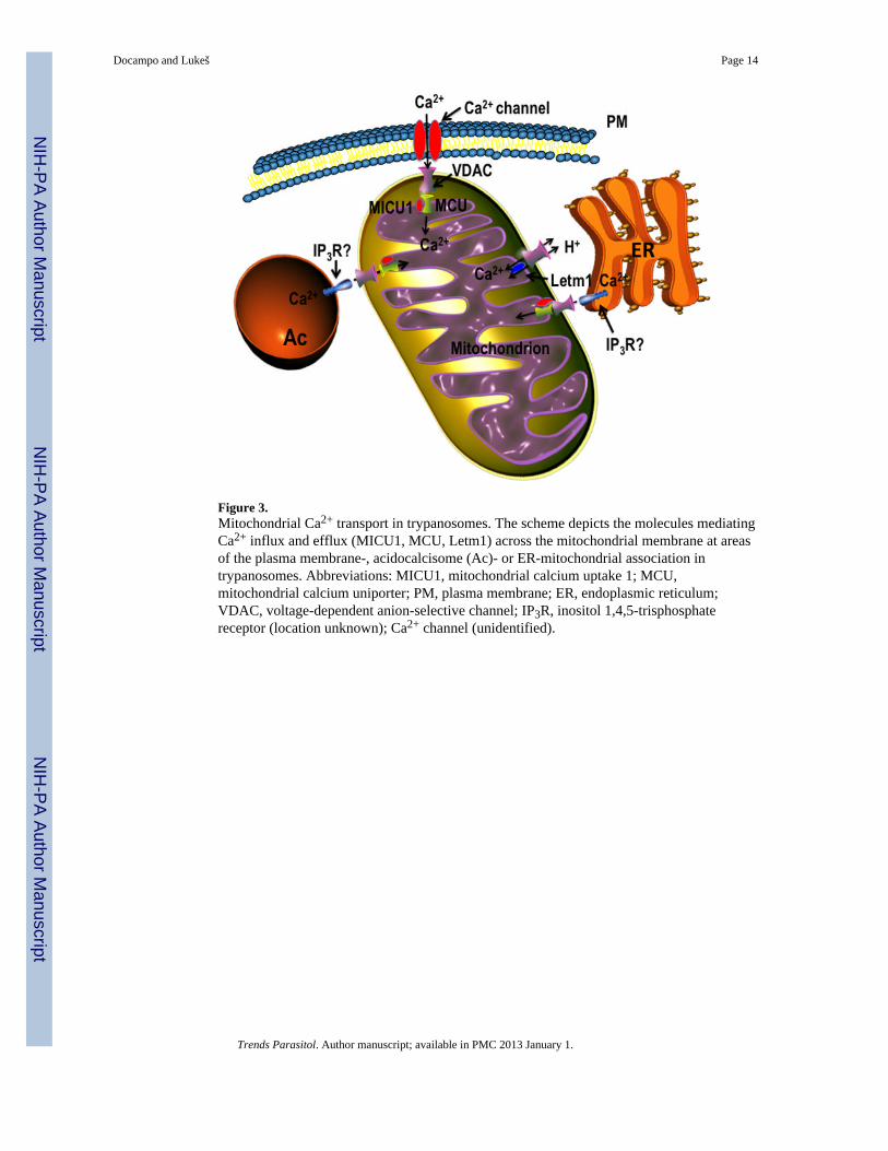
Figure 3.Mitochondrial Ca2+ transport in trypanosomes. The scheme depicts the molecules mediatingCa2+ influx and efflux (MICU1, MCU, Letm1) across the mitochondrial membrane at areasof the plasma membrane-, acidocalcisome (Ac)- or ER-mitochondrial association intrypanosomes. Abbreviations: MICU1, mitochondrial calcium uptake 1; MCU,mitochondrial calcium uniporter; PM, plasma membrane; ER, endoplasmic reticulum;VDAC, voltage-dependent anion-selective channel; IP3R, inositol 1,4,5-trisphosphatereceptor (location unknown); Ca2+ channel (unidentified).
Docampo and Lukeš Page 14
Trends Parasitol. Author manuscript; available in PMC 2013 January 1.
NIH
-PA Author Manuscript
NIH
-PA Author Manuscript
NIH
-PA Author Manuscript