Subcellular localization of a variable surface glycoprotein phosphatidylinositol-specific...
10
SubceUular Localization of a Variable Surface Glycoprotein Phosphatidylinositol-specific Phospholipase-C in African Trypanosomes D. J. Grab, P. Webster, S. Ito,* W. R. Fish, Y. Verjee, and J. D. Lonsdale-Eccles International Laboratory for Research on Animal Diseases, Nairobi, Kenya; and *Department of Anatomy, Harvard Medical School, Boston, Massachusetts 02175 Abstract. African trypanosomes contain a membrane- bound enzyme capable of removing dimyristylglycerol from the membrane-attached form of the variable surface glycoprotein (mfVSG; Ferguson, M. A. J., K. Halder, and G. A. M. Cross, 1985, J. Biol Chem., 260:4963-4968). Although mfVSG phospholipase-C has been implicated in the removal of the VSG from the trypanosome surface (Cardoso de Almeida, M. L., and M. J. Turner, 1983, Nature (Lond.)., 302:349- 352; Ferguson, M. A. J., K. Halder, and G. A. M. Cross, 1985, J. Biol Chem., 260:4963-4968), its pre- cise function and subcellular location have not been determined. We have developed a procedure for the separation of the cell fractions and organelles of Trypanosoma brucei brucei (and other trypanosome species) by differential sucrose and isopycnic Percoll R centrifugation. These fractions were tested for mfVSG phospholipase activity using Trypanosoma brucei mfVSG labeled with 3H-myristic acid as substrate. The highest enzyme-specific activity was associated with the flagella and evidence is presented to suggest that it is localized in the flagellar pocket. Some activ- ity was also associated with the Golgi complex. These results suggest that the mfVSG phospholipase is local- ized primarily in the membrane of the flagella pocket and possibly other membrane organelles derived from and associated with this structure, and may be part of the VSG-membrane recycling system in African trypanosomes. The activity of mfVSG phospholipase amongst various trypanosome species was determined. We show that, in contrast to the bloodstream forms of Trypanosoma brucei, cultured procyclic Trypanosoma brucei and bloodstream Trypanosoma vivax had little or no mfVSG phospholipase activity. The activity found in bloodstream forms of Trypanosoma con- golense was intermediate between Trypanosoma vivax and Trypanosoma brucei. RICAN trypanosomes, which cause sleeping sickness in man or nagana in livestock, have a 12-15-nm-thick coat of a variable surface glycoprotein (VSG) ~ cov- eting the entire surface of the organism (53). For Trypano- soma brucei and T. congolense, the generation of a new population of trypanosomes with a different surface coat has been shown to be the result of a clone-specific change in the expression of a particular VSG gene (14, 40). It appears clear that the remarkable ability of the trypanosome to alter its sur- face glycoprotein (14) may preclude the development of a conventional vaccine against trypanosomiasis. Since the in- This work was presented in part at the 6th International Congress of Parasi- tology (Brisbane, Australia) August 25-29, 1986. (Grab, D. J., S. Ito, J. D. Lonsdale-Eccles, P. Webster, and Y. Verjee, 1986, ICOPA FI Handbook, 140. [Abstr.]) 1. Abbreviations used in this paper: CRD, cross-reacting determinant; GPDH, glycerol-3-phosphate dehydrogenase; LG fraction, large granule fraction; mfVSG, membrane-attached form of the variable surface glyco- protein; SG fraction, small granule fraction; sVSG, soluble form of the vari- able surface glycoprotein; VSG, variable surface glycoprotein. tegrity of the surface coat is known to be essential for parasite survival in the host, the purpose of this study was to inves- tigate VSG synthesis, processing, and replacement on the cell surface. A clearer understanding of the cell biology of the VSG may suggest alternative approaches for the possible control of trypanosomiasis. The VSGs from T. brucei are proteins that contain com- plex carbohydrate moeities of mannose, galactose, glucos- amine, and myristilated phosphotidylinositol (17, 18, 39) conjugated to the protein through the alpha-carboxyl group of the carboxy-terminal amino acid via an ethanolamine resi- due (13, 28, 39). The biological importance to VSG of this carbohydrate side chain is evident since it is present on all variable antigens of T. brucei and T. congolense organisms so far studied and is probably responsible for cross-reactivity observed between soluble forms of VSGs (also known as the cross-reacting determinant [CRD]) (14). The carbohydrate side chain may be involved in anchoring the VSG to the plasma membrane (15, 17, 18, 39), and maintaining the in- tegrity of VSG-VSG interactions within the coat (24). © The Rockefeller University Press, 0021-9525/87108/737/10 $2.00 The Journal of Cell Biology, Volume 105, August 1987 737-746 737 on November 15, 2014 jcb.rupress.org Downloaded from Published August 1, 1987
-
Upload
independent -
Category
Documents
-
view
2 -
download
0
Transcript of Subcellular localization of a variable surface glycoprotein phosphatidylinositol-specific...

SubceUular Localization of a Variable Surface Glycoprotein Phosphatidylinositol-specific Phospholipase-C in African Trypanosomes D. J. Grab, P. Webster, S. Ito,* W. R. Fish, Y. Verjee, and J. D. Lonsdale-Eccles
International Laboratory for Research on Animal Diseases, Nairobi, Kenya; and * Department of Anatomy, Harvard Medical School, Boston, Massachusetts 02175
Abstract. African trypanosomes contain a membrane- bound enzyme capable of removing dimyristylglycerol from the membrane-attached form of the variable surface glycoprotein (mfVSG; Ferguson, M. A. J., K. Halder, and G. A. M. Cross, 1985, J. Biol Chem., 260:4963-4968). Although mfVSG phospholipase-C has been implicated in the removal of the VSG from the trypanosome surface (Cardoso de Almeida, M. L., and M. J. Turner, 1983, Nature (Lond.)., 302:349- 352; Ferguson, M. A. J., K. Halder, and G. A. M. Cross, 1985, J. Biol Chem., 260:4963-4968), its pre- cise function and subcellular location have not been determined. We have developed a procedure for the separation of the cell fractions and organelles of Trypanosoma brucei brucei (and other trypanosome species) by differential sucrose and isopycnic Percoll R centrifugation. These fractions were tested for mfVSG phospholipase activity using Trypanosoma brucei mfVSG labeled with 3H-myristic acid as substrate.
The highest enzyme-specific activity was associated with the flagella and evidence is presented to suggest that it is localized in the flagellar pocket. Some activ- ity was also associated with the Golgi complex. These results suggest that the mfVSG phospholipase is local- ized primarily in the membrane of the flagella pocket and possibly other membrane organelles derived from and associated with this structure, and may be part of the VSG-membrane recycling system in African trypanosomes.
The activity of mfVSG phospholipase amongst various trypanosome species was determined. We show that, in contrast to the bloodstream forms of Trypanosoma brucei, cultured procyclic Trypanosoma brucei and bloodstream Trypanosoma vivax had little or no mfVSG phospholipase activity. The activity found in bloodstream forms of Trypanosoma con- golense was intermediate between Trypanosoma vivax and Trypanosoma brucei.
RICAN trypanosomes, which cause sleeping sickness in man or nagana in livestock, have a 12-15-nm-thick coat of a variable surface glycoprotein (VSG) ~ cov-
eting the entire surface of the organism (53). For Trypano- soma brucei and T. congolense, the generation of a new population of trypanosomes with a different surface coat has been shown to be the result of a clone-specific change in the expression of a particular VSG gene (14, 40). It appears clear that the remarkable ability of the trypanosome to alter its sur- face glycoprotein (14) may preclude the development of a conventional vaccine against trypanosomiasis. Since the in-
This work was presented in part at the 6th International Congress of Parasi- tology (Brisbane, Australia) August 25-29, 1986. (Grab, D. J., S. Ito, J. D. Lonsdale-Eccles, P. Webster, and Y. Verjee, 1986, ICOPA FI Handbook, 140. [Abstr.])
1. Abbreviations used in this paper: CRD, cross-reacting determinant; GPDH, glycerol-3-phosphate dehydrogenase; LG fraction, large granule fraction; mfVSG, membrane-attached form of the variable surface glyco- protein; SG fraction, small granule fraction; sVSG, soluble form of the vari- able surface glycoprotein; VSG, variable surface glycoprotein.
tegrity of the surface coat is known to be essential for parasite survival in the host, the purpose of this study was to inves- tigate VSG synthesis, processing, and replacement on the cell surface. A clearer understanding of the cell biology of the VSG may suggest alternative approaches for the possible control of trypanosomiasis.
The VSGs from T. brucei are proteins that contain com- plex carbohydrate moeities of mannose, galactose, glucos- amine, and myristilated phosphotidylinositol (17, 18, 39) conjugated to the protein through the alpha-carboxyl group of the carboxy-terminal amino acid via an ethanolamine resi- due (13, 28, 39). The biological importance to VSG of this carbohydrate side chain is evident since it is present on all variable antigens of T. brucei and T. congolense organisms so far studied and is probably responsible for cross-reactivity observed between soluble forms of VSGs (also known as the cross-reacting determinant [CRD]) (14). The carbohydrate side chain may be involved in anchoring the VSG to the plasma membrane (15, 17, 18, 39), and maintaining the in- tegrity of VSG-VSG interactions within the coat (24).
© The Rockefeller University Press, 0021-9525/87108/737/10 $2.00 The Journal of Cell Biology, Volume 105, August 1987 737-746 737
on Novem
ber 15, 2014jcb.rupress.org
Dow
nloaded from
Published August 1, 1987
It has been proposed that VSG is anchored to the p lasma m e m b r a n e by a hydrophobic domain within the C R D (16-18, 39). Al though the precise structure of the C R D is not com- pletely known, the presence of a covalently l inked glycosyl- sn- l ,2 dimyristoyl phosphatidylinosi tol in the VSG of try- panosomes has been revealed (18). The possibil i ty that the VSG is anchored to the m e m b r a n e via the myris tyl residues is supported by the evidence that the membrane-a t tached form of the T. brucei VSG (mfVSG) contains glycerol and myrist ic acid whereas the soluble form of VSG (sVSG) lacks these residues (15, 17, 39).
The conversion of the mfVSG to sVSG may be mediated by the action of an endogenous phosphol ipase-C- l ike en- zyme (also designated Enzyme-X; 9, 10, 15, 17, 18, 30, 39). It has been postulated that mfVSG is selectively released in vivo from the plasma m e m b r a n e of t rypanosomes as a result o f the removal of the diacylglycerol domain from the phos- phatidylinosi tol (17, 18, 39), and that the VSG-releas ing en- zyme is possibly located in the p lasma membrane (10, 15, 17, 18, 39). However, the intraceUular localizat ion of the enzyme responsible for the conversion of mfVSG to sVSG is still un- certain. The present report is an at tempt to address this problem.
Materials and Methods
Biochemicals
All reagents were of analytical grade and were obtained from the following sources. Uridine diphospho-tr-[6-3H]galactose (640 GBq/mmol), [9,10(n)]- 3H-myristic acid (2.04 TBq/mmol), ~C-casein (1.1 MBq/mg protein), L-[3SS]methionin¢ (30 TBq/mmol), and Hyperfilm MP were obtained from Radiochemical Centre Limited (Amersham, England). Aquasol and EN3HANCE were obtained from New England Nuclear (Boston, MA). [N-(2-acetamido)]-2-Amino ethanesulfonic acid (ACES), 2-(N-morpho- lino)-ethanesulfonic acid (MES); Hepes; and [N,N-bis-(2-hydroxyethyl)] glycine (BICINE) were from Research Organics Inc. (Cleveland, OH). Aristar grade sucrose was from BDH Chemicals (Poole, England). RPMI- 1640 and Iscove's culture media were from Flow Laboratories, Inc. (Irvine, Scotland). Zwittergent TM 3-14 was from Calbiochem-Behring Corp. (La Jolla, CA). Poly/Sep 47 IEF buffer was from Folysciences, Inc. (Warring- ton, PA). Sephacryl S-200 and Percoll were from Pharmacia Fine Chemi- cals (Uppsala, Sweden). DE-53 was from Whatman Ltd., (Maldstone, Kent, England). All phospholipids, and 4-methylumbelliferyl phosphate were from Koch-Light Ltd. (Haverhill, Suffolk, England); dimyristin was from Serva (Heidelberg, Federal Republic of Germany), and the protease inhibitors leupeptin, chymostatin, antipain, and E-64 were from Cambridge Research Biochemicals (Cambridgeshire, England). All solutions were pre- pared in a purification system developed by Christ (Aesch, Switzerland); namely, deionized water which had been filtered through a 5-~tm coarse filter, an ultrafilter with an 80,000-D cutoff, and sterilized by ultraviolet radi- ation. The conductivity of the water was <15 megaohms.
Organisms
Trypanosoma brucei clones MITat 1.2, 1.52, IL'lht 1.1, T. congolense clone ILNat 2.1, and T. vivax ILDat 1.2 (subclone 1392) were grown from cryopreserved stabilates in lethally irradiated rats (600-900 lad). The or- ganisms were isolated from the infected blood using isopycnic Percoll gra- dients (21). Infected blood was mixed 1:2 with 90% Percoll containing 1% glucose, 0.73 % NaC1 (buffered to pH 7.4 with solid Hepes), and centrifuged at 15,000 rpm for 15 rain in a JA20 rotor. The trypanosomes were collected, the pH was adjusted to 8.0 with 1 M Tris-base, and the trypanosomes immediately passed through a DE-53 column equilibrated in phosphate- buffered saline glucose (PSG), pH 8.0 (33) supplemented with nucleesides (0.1 mM adenosine, 0.05 mM hypoxanthine, 0.05 mM thymidine) as well as Baltz's additions; namely, 0.2 mM 2-mercaptoethanol and 2 mM pyruvate (2). After elution the pH was adjusted back to pH 7.4 with Hepes. Platelets and other blood cells are effectively removed by these isolation conditions.
However, the ATP contents of the isolated parasites are several-fold higher than those isolated in PSG by previous workers (19, 35).
Subcellular b3"actionatio,
For each experiment '~10" trypanosornes were washed in SHK (250 mM sucrose, 50 mM Hepes, 25 mM KC1, pH 7.4, at 5°C) buffer containing known inhibitors of the major trypanosomal proteinases (36; 50-100 ~tg/ml of leupeptin, E-64, antipain, and chymostatin). The parasites were dis- rupted by passage through a French Pressure Cell under a chamber pressure of 2,500 psi.
Fractions containing lysosomes, glycosomes, and flagella were prepared at 0-5°C as follows: EDTA was added to the homogenate (to a final concen- tration of 1 raM; SHKE, pH 7.4), and unbroken cells, nuclei, and cell debris were sedimented at 705 g~ for 10 rain in a JA-20 rotor (Beckman Instru- ments Inc., Palo Alto, CA). The resulting pellet was designated the crude nuclear pellet. The resulting supernatant was centrifuged at 2,800 g~, for 10 rain to produce a large granule (LG) fraction; this was followed by a small granule (SG) fraction produced at 15,000 g~ for 10 min. Centrifugation of the (l~st-SG) supernatant at 123,000 g~ for 90 rain in a 50.2 "li rotor (Beckman Instruments Inc.) gave the crude microsomal pellet fraction and the soluble supernatant.
The LG and SG fractions were further fractionated by isopycnic centrifu- gation in Percoll. The LG and SG fractions were made 57.6% (vol/vol) with respect to Percoll in SHKE (pH 7.4) and layered under a discontinuous gra- dient consisting of 43.2, 28.8, and 20.3% PercoH in the same buffer. Cen- trifugation was for 30-35 min at 25,000 rpm in either an SW-41 or an SW-27 rotor. Fractions banding between the 28.8/43.3 % and 43.3/57.6 % Percoll in- terfaces were made 57.6% with regard to Percoll and centrifuged again in either the gradient described above or in continuous Percoll gradients in SHKE. Fractions on top of and within the 20.3 % Percoll layer (crude flagella) were adjusted to 28.8% Percoll and layered under a discontinuous gradient containing 20.3, 17.3, and 14.4% Percoll in SHKE buffer, pH 7.4, and centrifuged again as above to obtain the lighter membrane fractions. De- pending on yields, the fractions were washed with SHKE buffer and stored at -80"C, For some preparations, the entire fractionafion was done in the presence of 5 mM magnesium instead of the EDTA.
To obtain endoplasmic reticulum and Golgi fractions, 5 mM MgSO4 was added to the homogenate (in SHKM, pH 7.4) and processed using estab- lished procedures as described by Grab et al. (23) except that the separation
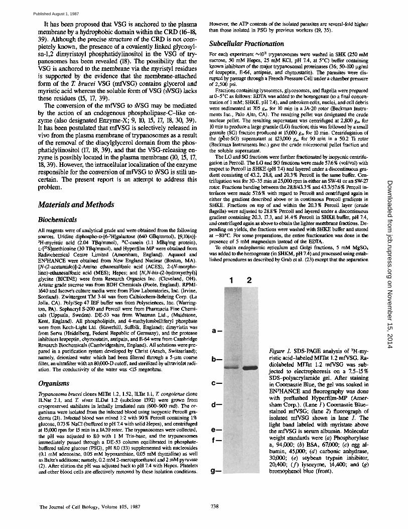
Figure 1. SDS-PAGE analysis of 3H-my- ristic acid-labeled MITat 1.2 mfVSG. Ra- diolabeled Mrrat 1.2 mfVSG was sub- jected to electrophoresis on a Z5-15% SDS-polyacrylamide gel. After staining in Coomassie Blue, the gel was soaked in EN3HANCE and fluorography was done with preflashed Hyperfilm-MP (Amer- sham Corp.). (Lane I ) Coomassie Blue- stained mfVSG; (lane 2) fluorograph of isolated mfVSG shown in lane 1. The light band labeled with myristate above the mfVSG is serum albumin. Molecular weight standards were (a) Phospborylase a, 94,000; (b) BSA, 67,000; (c) egg al- bumin, 45,000; (d) carbonic anhydrase, 30,000; (e) soybean trypsin inhibitor, 20,400; ( f ) lysozyme, 14,400; and (g) bromophenol blue (front).
The Journal of Cell Biology, Volume 105, 1987 738
on Novem
ber 15, 2014jcb.rupress.org
Dow
nloaded from
Published August 1, 1987

of smooth endoplasmic reticulum from rough endoplasmic reticulum com- ponents was done in a vertical VTi50 rotor (50,000 rpm for 1 or 2 h) instead of an SW-27 swinging bucket rotor. The latter requires longer centrifugation times. These fractions were washed with SHKE buffer before further analy- sis or storage by freezing at -80°C. These fractionation procedures, devel- oped for T. brucei bloodstream forms, may be used for T. congolense and Z v/vax bloodstream forms as well as for cultured procyclic Z brucei.
Morphology For electron microscopy, suspended fractions were fixed in a fixative con- raining picric acid, formaldehyde, and glutaraldehyde (29) in 100 mM so- dium phosphate buffer, pH 7.2. After fixation at room temperature for 1 h, the fractions were centrifuged in an Eppendorf microfuge for 1-15 rain to obtain a visible pallet. The samples were washed with 100 mM sodium cac- odylate buffer, treated with 1% OsO4 in sodium cacodylate buffer en bloc, stained with uranyl acetate, dehydrated, and embedded in Epon-Araidite. Thin sections were cut across the entire thickness of the pellet, stained with uranyl acetate and lead citrate, and examined in the electron microscope.
Biochemical Analysis Acid phosphatase was assayed using as substrate either beta-glycerophos- phate (52) or 4-methylumbelliferylphosphate (51). Acid proteinase was as- sayed using r~C-methylated casein or benzyloxycarbonyl-Phe-Arg-7-amido- 4-methylcoumarin as substrate (37). Glycerol-3-phosphate dehydrogenase was assayed according the method of Rovis and Baekkeskov (50). The trans- fer of galactose from UDP-galactose to endogenous protein substrates (galactosyltransferase) and adenyl cyclase were assayed as described by Grab et al. (23). Malate dehydrogenase was measured according to the method of Opperdoes et al. (47). Protein determination was performed ac- cording to the method of Vincent and Nadeau (54) using BSA as a protein standard. SDS-PAGE was performed according to the method of Neville (43) except that a slab gel (2 nun thick by 200 mm long) containing a linear 10-20% polyacrylamide gradient was used.
Preparation of 3H-Myristic-labeled MITat 1.2 mfVSG 3H-Myristic acid (185 MBq) was dried in a rotary evaporator, washed sev- eral times with absolute ethanol/benzene 1:1 (vol/vol) to remove the toluene, and once with absolute ethanol. The labeled fatty acid (in ethanol) was cou- pled to defatted BSA (55), lyophilized, and added (3.7 MBq) to either Iscove's medium with transferrin (1 lag/ml), albumin (400 Ixg/ml), soybean lipid (100 lsg/ml), or RPMI-1640. The media was also supplemented with 0.45 % glucose, and nucleotides, and Baltz additions as described above. MITat 1.2 trypanosomes (5 × 10⁷/ml) were incubated in the fatty acid con- taining medium for 2-3 h at 37°C. In some cases the trypanosomes were incubated with [35S]methionine (3.7 MBq) instead of labeled lipid. After washing the organisms several times in radiolabel-free medium, the mfVSG was extracted (31). Analysis by SDS-PAGE showed that only mfVSG labeled with 3H-myristic acid (Fig. 1).
mfVSG Phospholipase Assay The assay mixture for VSG phospholipase contained, in a volume of 30 ~tl,
2.26 ~tg labeled mfVSG (8,500 cpm/lxg VSG protein), and 7.5 rig membrane protein (in 15 Ixl SHKE) or Sephacryl S-200 enzyme protein (diluted in 15 Ixl SHKE), 50 mM Hepes, pH 7.4, 0.025% polyethylene glycol (PEG; Mr 6,000, wt/vol), 0.5% wt/vol Triton X-100, protease inhibitors (20 ~tg/ml each of E-64, leupeptin, chymostatin, and antipain). Under the conditions stated, the protein substrate-to-enzyme ratio was 300:1, while the ratios of substrate phosphatidylinositol to total lipid, glycolipid, total phospholipid, and membrane phosphatidylinositol were at least U:I, 107:1, 16:1, and 160:1, respectively, based on the data of Baekkeskov et al. (1). After a 15-30 min incubation at 37°C the reaction was terminated by boiling in 100 Ixl of 0.3 % (wt/vol) Zwittergent TM 3-14 after which 300 Ixl of 0.3 M NaCI/90% metha- nol (vol/vol) was added followed by 800-900 I.tl of n-hexane. The sample was vortexed until emulsification occurred and then centrifuged in an Ep- pendorf microfuge to separate the phases. The release of labeled myristic acid into the hexane phase was monitored by liquid scintillation spectrome- try. The above assay was done in duplicate or triplicate and was found to be reproducible. No proteolysis occurred during incubation with the various fractions when [35S]methionine-labeled mfVSG was the substrate.
TLC of the hexane-extractable product was done on heat-activated (100*C for 1 h) 0.25-mm-thick silica gel 60 TLC plates in a solvent system containing pentane, diethyl ether, glacial acetic acid (80:20:1; vol/vol/vol) (41). The plates were dried for 30 min at 60°C, sprayed with EN3HANCE, dried for 1 h at 65°C, and exposed to Hyperfilm MP film at -80"C (Amersham).
Sephacryl S-200 Column Chromatography of mfVSG Phospholipase Flagellar membrane fractions which banded on top of 14.4% Pereoll (in SHKE) were solubilized in 2% 3-[(3)-cholamidopropyl]dimethyl-ammo- nio-l-propanesulfonate (CHAPS) (wt/vol), containing the protease inhibi- tors E-64 and leupeptin (20 ~tg/ml each) and centrifuged at 100,000 g for 1 h to remove undissolved cytoskeletal components. After dialysis against 0.1% CHAPS in 10 mM Hepes, 0.05% PEG, pH 7.0 (CHP buffer), the sam- ple was applied on to a 1.6- x 95-cm Sephacryl S-200 column equilibrated in the same buffer. The active fractions were pooled. IEF was done on a Poly/Sep 47 column (6). Product analysis was conducted by TLC (41) as described above.
Results
Subcellular Fractionation: Morphological and Biochemical A nalysis Discontinuous Percoll gradients produced highly enriched preparations of lysosomes, glycosomes, and flagella. An im- portant feature of the isolation procedure appears to be the hypertonicity of the 250 mM SHKE buffer (,,o410 mosmols) as more isotonic buffers resulted in fractions with poor puri- ties (data not shown). The isolated lysosome-like organdies (Fig. 2 A) were spherical with a rim of high density substance and a clear center. These membrane-bounded structures
Figure 2. (A) Lysosome fraction f rom the 28.8/43.2% Percoll interface. (B) Glycosome fraction f rom the 43.2/57.6% Percoll interface. Bar, 2 ~tm.
Grab et al. Phospholipase in African Trypanosomes 739
on Novem
ber 15, 2014jcb.rupress.org
Dow
nloaded from
Published August 1, 1987
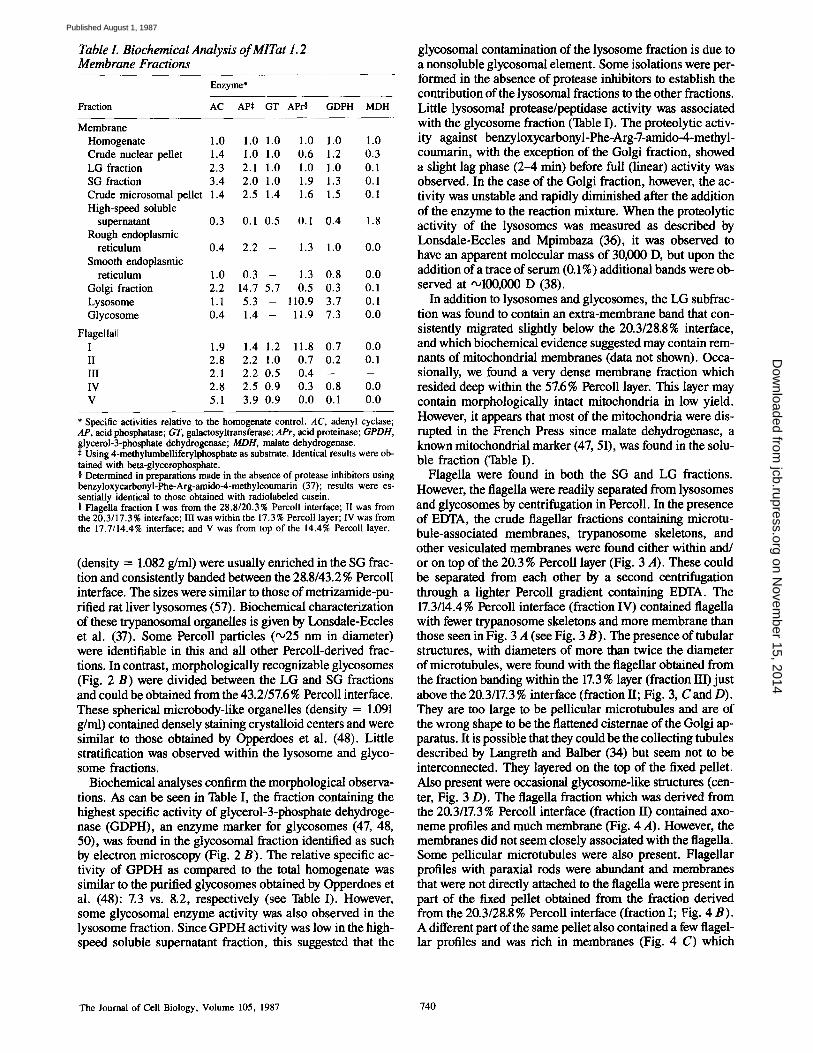
Table I. Biochemical Analysis of MITat 1.2 Membrane Fractions
Enzyme*
Fraction AC APe GT APr~ GDPH MDH
Membrane Homogenate 1.0 1.0 1.0 1.0 1.0 1.0 Crude nuclear pellet 1.4 1.0 1.0 0.6 1.2 0.3 LG fraction 2.3 2.1 1.0 1.0 1.0 0.1 SG fraction 3.4 2 .0 1.0 1.9 1.3 0.1 Crude microsomal pellet 1.4 2.5 1.4 1.6 1.5 0.1 High-speed soluble
supernatant 0.3 0.1 0.5 0.1 0.4 1.8 Rough endoplasmic
reticulum 0.4 2.2 - 1.3 1.0 0 .0 Smooth endoplasmic
reticulum 1.0 0.3 - 1.3 0.8 0.0 Golgi fraction 2.2 14.7 5.7 0.5 0.3 0.1 Lysosome 1.1 5.3 - 110.9 3.7 0.1 Glycosome 0.4 1.4 - 11.9 7.3 0 .0
Flagellall I 1.9 1.4 1.2 11.8 0.7 0.0 II 2.8 2.2 1.0 0.7 0.2 0.1 III 2.1 2.2 0.5 0.4 - - IV 2.8 2.5 0.9 0.3 0.8 0.0 V 5.1 3.9 0.9 0.0 0.1 0 .0
* Specific activities relative to the homogenate control. AC, adenyl cyclase; AP, acid phosphatase; GT, galaetosyltransferas¢; APr, acid proteinase; GPDH, glycerol-3-phosphate dehydrogenase; MDH, malate dehydrogenase.
Using 4-methylumbelliferylphosphate as substrate. Identical results were ob- tained with beta-glycerophosphate. § Determined in preparations made in the absence of protease inhibitors using benzyloxycarbonyl-Phe-Arg-amido-4-methylcoumarin (37); results were es- sentially identical to those obtained with radiolabeled casein. II Flagella fraction I was from the 28.8/20.3% Percoll interface; II was from the 20.3/17.3% interface; III was within the 17.3% Percoll layer; IV was from the 17.7/14.4% interface; and V was from top of the 14.4% Percoll layer.
(density = 1.082 g/ml) were usually enriched in the SG frac- tion and consistently banded between the 28.8/43.2 % Percoll interface. The sizes were similar to those of metrizamide-pu- rifled rat liver lysosomes (57). Biochemical characterization of these trypanosomal organelles is given by Lonsdale-Eccles et al. (37). Some Percoll particles (•25 nm in diameter) were identifiable in this and all other Percoll-derived frac- tions. In contrast, morphologically recognizable glycosomes (Fig. 2 B) were divided between the LG and SG fractions and could be obtained from the 43.2/57.6% Percoll interface. These spherical microbody-like organelles (density = 1.091 g/ml) contained densely staining crystalloid centers and were similar to those obtained by Opperdoes et al. (48). Little stratification was observed within the lysosome and glyco- some fractions.
Biochemical analyses confirm the morphological observa- tions. As can be seen in Table I, the fraction containing the highest specific activity of glycerol-3-phosphate dehydroge- nase (GDPH), an enzyme marker for glycosomes (47, 48, 50), was found in the glycosomal fraction identified as such by electron microscopy (Fig. 2 B). The relative specific ac- tivity of GPDH as compared to the total homogenate was similar to the purified glycosomes obtained by Opperdoes et al. (48): 7.3 vs. 8.2, respectively (see Table I). However, some glycosomal enzyme activity was also observed in the lysosome fraction. Since GPDH activity was low in the high- speed soluble supernatant fraction, this suggested that the
glycosomal contamination of the lysosome fraction is due to a nonsoluble glycosomal element. Some isolations were per- formed in the absence of protease inhibitors to establish the contribution of the lysosomal fractions to the other fractions. Little lysosomal protease/peptidase activity was associated with the glycosome fraction (Table I). The proteolytic activ- ity against benzyloxycarbonyl-Phe-Arg-7-amido-4-methyl- coumarin, with the exception of the Golgi fraction, showed a slight lag phase (2-4 rain) before full (linear) activity was observed. In the case of the Golgi fraction, however, the ac- tivity was unstable and rapidly diminished after the addition of the enzyme to the reaction mixture. When the proteolytic activity of the lysosomes was measured as described by Lonsdale-Eccles and Mpimbaza (36), it was observed to have an apparent molecular mass of 30000 D, but upon the addition of a trace of serum (0.1%) additional bands were ob- served at ,,o100,000 D (38).
In addition to lysosomes and glycosomes, the LG subfrac- tion was found to contain an extra-membrane band that con- sistenfly migrated slightly below the 20.3/28.8% interface, and which biochemical evidence suggested may contain rem- nants of mitochondrial membranes (data not shown). Occa- sionally, we found a very dense membrane fraction which resided deep within the 57.6% Percoll layer. This layer may contain morphologically intact mitochondria in low yield. However, it appears that most of the mitochondria were dis- rupted in the French Press since malate dehydrogenase, a known mitochondrial marker (47, 51), was found in the solu- ble fraction (Table I).
Flagella were found in both the SG and LG fractions. However, the flagella were readily separated from lysosomes and glycosomes by centrifugation in Percoll. In the presence of EDTA, the crude flagellar fractions containing microtu- bule-associated membranes, trypanosome skeletons, and other vesiculated membranes were found either within and/ or on top of the 20.3% Percoll layer (Fig. 3 A). These could be separated from each other by a second centrifugation through a lighter Percoll gradient containing EDTA. The 17.3/14.4 % Percoll interface (fraction IV) contained flagella with fewer trypanosome skeletons and more membrane than those seen in Fig. 3 A (see Fig. 3 B). The presence of tubular structures, with diameters of more than twice the diameter of microtubules, were found with the flagellar obtained from the fraction banding within the 17.3 % layer (fraction l/I)just above the 20.3/17.3 % interface (fraction II; Fig. 3, C and D). They are too large to be pellicular microtubules and are of the wrong shape to be the flattened cisternae of the Golgi ap- paratus. It is possible that they could be the collecting tubules described by Langreth and Balber (34) but seem not to be interconnected. They layered on the top of the fixed pellet. Also present were occasional glycosome-like structures (cen- ter, Fig. 3 D). The flagella fraction which was derived from the 20.3/17.3% Percoll interface (fraction II) contained axo- neme profiles and much membrane (Fig. 4 A). However, the membranes did not seem closely associated with the flagella. Some pellicular microtubules were also present. Flagellar profiles with paraxial rods were abundant and membranes that were not directly attached to the flagella were present in part of the fixed pellet obtained from the fraction derived from the 20.3/28.8% Percoll interface (fraction I; Fig. 4 B). A different part of the same pellet also contained a few flagel- lar profiles and was rich in membranes (Fig. 4 C) which
The Journal of Cell Biology, Volume 105, 1987 740
on Novem
ber 15, 2014jcb.rupress.org
Dow
nloaded from
Published August 1, 1987

Figure 3. (A) Crude flagella fraction from the first Percoll gradient (entire 20.2% layer). Contains flagella with associated membranes as well as without. This fraction also contains other flagella-free membranes as well as trypanosome skeletons. (B) Fraction from 14.4/17.3 % Percoll interface. Similar to Fig. 3 A but with fewer trypanosome skeletons and more membranes. Many flageUar profiles have membranes associated with them. (C) This fraction banded within the 17.3% Percoll layer above the 17.3/20.3% interface. It contains many flagellar profiles and, as shown in a higher magnification micrograph (D), tubular structures having more than twice the diameter of microtubules. Bars: (A-C) 4 )xm; (D) 2 )tm.
Figure 4. (A) Flagella fraction derived from the 17.3/20.2% Percoll interface. Contains axoneme profiles and membranes not associated with the flagella. Some pellicular microtubules are also present. (B) Flagella profiles with paraxial rods abundant are found in the fraction banding at the 20.2/28.8% Percoll interface. (C) Different part of the pellet seen in B. This fraction is rich in membranes many of which appear to be coated vesicles. A few flagellar profiles are present. (D) Higher magnification of C. Bars: (A-C) 4 )xm; (D) 1 lain.
Grab et al. Phospholipase in African Trypanosomes 741
on Novem
ber 15, 2014jcb.rupress.org
Dow
nloaded from
Published August 1, 1987
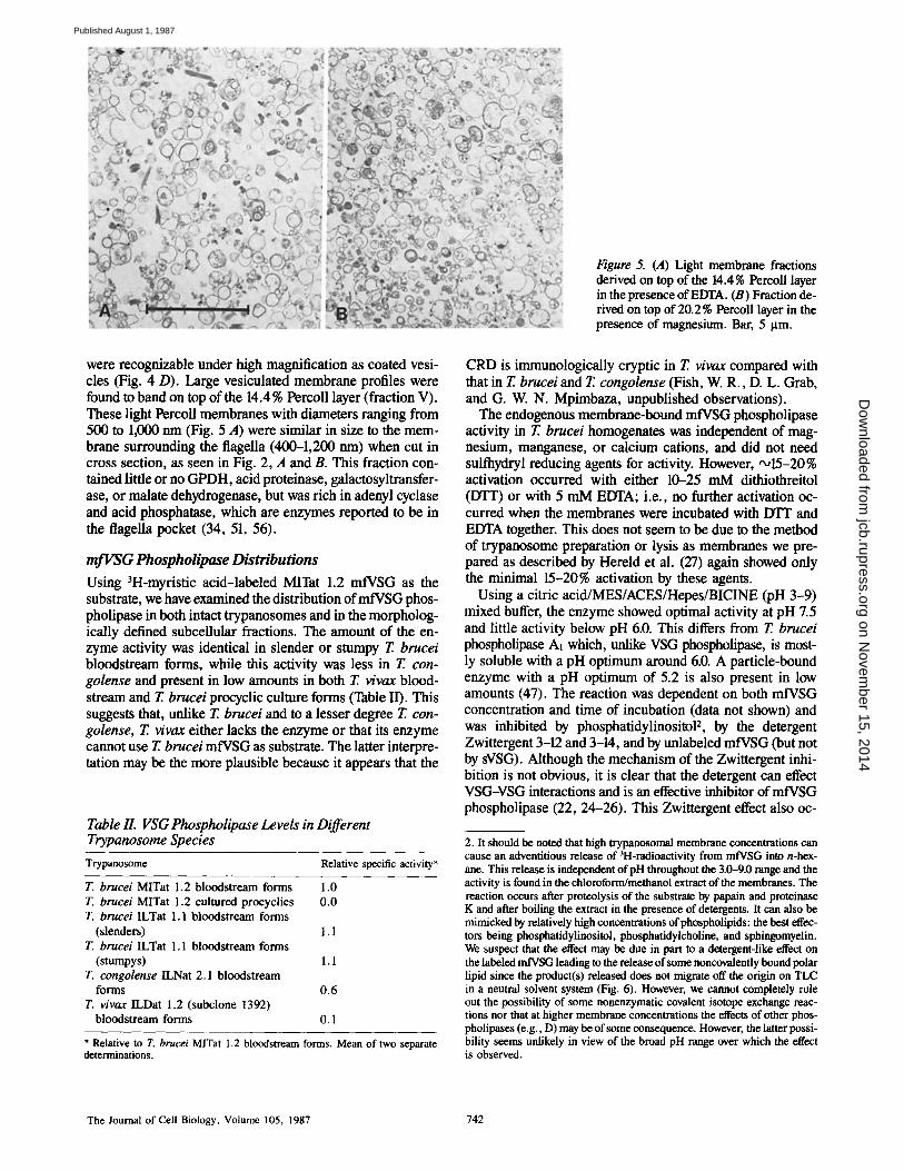
Figure 5. (A) Light membrane fractions derived on top of the 14.4% Percoll layer in the presence of EDTA. (B) Fraction de- rived on top of 20.2% Percoll layer in the presence of magnesium. Bar, 5 ttm.
were recognizable under high magnification as coated vesi- cles (Fig. 4 D). Large vesiculated membrane profiles were found to band on top of the 14.4% Percoll layer (fraction V), These light Percoll membranes with diameters ranging from 500 to 1,000 nm (Fig. 5 A) were similar in size to the mem- brane surrounding the flagella (400-1,200 nm) when cut in cross section, as seen in Fig. 2, A and B. This fraction con- tained little or no GPDH, acid proteinase, galactosyltransfer- ase, or malate dehydrogenase, but was rich in adenyl cyclase and acid phosphatase, which are enzymes reported to be in the flagella pocket (34, 51, 56).
mfVSG Phospholipase Distributions
Using 3H-myristic acid-labeled MITat 1.2 mfVSG as the substrate, we have examined the distribution of mfVSG phos- pholipase in both intact trypanosomes and in the morpholog- ically defined subcellular fractions. The amount of the en- zyme activity was identical in slender or stumpy T. brucei bloodstream forms, while this activity was less in T. con- golense and present in low amounts in both T. v/vax blood- stream and T. brucei procyclic culture forms (Table II). This suggests that, unlike T. brucei and to a lesser degree T. con- golense, T. vivax either lacks the enzyme or that its enzyme cannot use T. brucei mfVSG as substrate. The latter interpre- tation may be the more plausible because it appears that the
Table II. VSG Phospholipase Levels in Different Trypanosome Species
Trypanosome Relative specific activity*
T. brucei MITat 1.2 bloodstream forms 1.0 T. brucei MITat 1.2 cultured procyclics 0.0 T. brucei ILTat 1.1 bloodstream forms
(slenders) 1.1 T. brucei ILTat 1.1 bloodstream forms
(stumpys) l . 1 T. congolense ILNat 2.1 bloodstream
forms 0.6 T. vivax ILDat 1.2 (subclone 1392)
bloodstream forms 0.1
* Relative to T. brucei MITat 1.2 bloodstream forms. Mean of two separate determinations.
CRD is immunologically cryptic in T. v/vax compared with that in T. brucei and T. congolense (Fish, W. R., D. L. Grab, and G. W. N. Mpimbaza, unpublished observations).
The endogenous membrane-bound mfVSG phospholipase activity in T. brucei homogenates was independent of mag- nesium, manganese, or calcium cations, and did not need sulfhydryl reducing agents for activity. However, •15-20% activation occurred with either 10-25 mM dithiothreitol (DTT) or with 5 mM EDTA; i.e., no further activation oc- curred when the membranes were incubated with DTT and EDTA together. This does not seem to be due to the method of trypanosome preparation or lysis as membranes we pre- pared as described by Hereld et al. (27) again showed only the minimal 15-20% activation by these agents.
Using a citric acid/MES/ACES/Hepes/BICINE (pH 3-9) mixed buffer, the enzyme showed optimal activity at pH 7.5 and little activity below pH 6.0. This differs from T. brucei phospholipase A~ which, unlike VSG phospholipase, is most- ly soluble with a pH optimum around 6.0. A particle-bound enzyme with a pH optimum of 5.2 is also present in low amounts (47). The reaction was dependent on both mfVSG concentration and time of incubation (data not shown) and was inhibited by phosphatidylinositol 2, by the detergent Zwittergent 3-12 and 3-14, and by unlabeled mfVSG (but not by sVSG). Although the mechanism of the Zwittergent inhi- bition is not obvious, it is clear that the detergent can effect VSG-VSG interactions and is an effective inhibitor of mfVSG phospholipase (22, 24-26). This Zwittergent effect also oc-
2. It should be noted that high trypanosomal membrane concentrations can cause an adventitious release of 3H-radioactivity from mfVSG into n-hex- ane. This release is independent ofpH throughout the 3.0-9.0 range and the activity is found in the chloroform/methanol extract of the membranes. The reaction occurs after proteolysis of the substrate by papain and proteinase K and after boiling the extract in the presence of detergents. It can also be mimicked by relatively high concentrations of phospholipids: the best effec- tors being phosphatidylinositol, phosphatidyicholine, and sphingomyelin. We suspect that the effect may be due in part to a detergent-like effect on the labeled mfVSG leading to the release of some noncovalently bound polar lipid since the product(s) released does not migrate off the origin on TLC in a neutral solvent system (Fig. 6). However, we cannot completely rule out the possibility of some nonenzymatic covalent isotope exchange reac- tions nor that at higher membrane concentrations the effects of other phos- pholipases (e.g., D) may be of some consequence. However, the latter possi- bility seems unlikely in view of the broad pH range over which the effect is observed.
The Journal of Cell Biology, Volume 105, 1987 742
on Novem
ber 15, 2014jcb.rupress.org
Dow
nloaded from
Published August 1, 1987
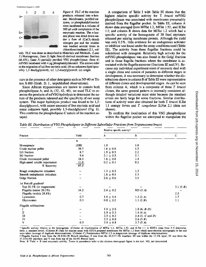
Figure 6. TLC of the reaction products released into n-hex- ane. Membranes, purified en- zyme, or phosphatidylinositol were incubated in a volume of 500 I~1 until completion of the enzymatic reaction. The n-hex- ane phase was dried down un- der a flow of (CaC1E-dried) nitrogen gas and the residue was washed several times in chloroform/methanol (2:1, vol/
vol). TLC was done as described in Materials and Methods. (Lane 1) Homogenate; (lane 2) light Percoll-derived membrane fraction (14.4%); (lane 3) partially purified VSG phospholipase; (lane 4) mfVSG incubated with 4 Ixg phosphatidylinositol. The arrows refer to the migration of (a) free myristic acid; (b) an unknown lipid (pos- sibly 1,3 diaclyglycerol); (c) 1,2-diacylglycerol; (o) origin.
curs in the presence of other detergents such as NP-40 or Tri- ton X-100 (Grab, D. J., unpublished observations).
Since African trypanosomes are known to contain both phospholipase Al and A2 (32, 42, 46), we used TLC to ex- amine the products of mfVSG hydrolysis to determine the na- ture of the products released and the specificity of our assay system. The major hydrolysis product was found to be 1,2- diacylglycerol, with minor amounts of free myristic acid and some unknown lipid, possibly 1,3-diacylglycerol (Fig. 6). This confirms the phospholipase-C nature of the reaction as- sayed.
A comparison of Table I with Table HI shows that the highest relative specific activity for T. brucei mfVSG phospholipase was associated with membranes presumably derived from the flagellar pocket. In Table III, column A shows data averaged from MITat 1.2, MITat 1.52, and ILTat 1.1; and column B shows data for MITat 1.2 which had a specific activity of the homogenate of 38 fmol myristate released per min/ng membrane protein. Although the yield was only 0.5%, little evidence for an endogenous activator or inhibitor was found under the assay conditions used (Table III). The activity from these flagellar fractions could be solubilized with detergent. Relatively high activity for the mfVSG phospholipase was also found in the Golgi fraction and in those flagella fractions where the membrane is as- sociated with the flagella axoneme (fractions HI and IV). Be- cause any individual experiment must of necessity deal with a single clone and consist of parasites in different stages in development, it was necessary to determine whether the dis- tributions shown in column B of Table III were representative of different clones and developmental stages. As can be seen from column A, which is a composite of three T. brucei clones, the same general pattern is internally consistent al- though detailed variations must exist because the standard errors are fairly large for some fractions. Similar distribu- tions of activity were also obtained for both T. brucei ILTat 1.1 stumpy forms and T. congolense ILNat 2.1 (data not shown).
To confirm the localization of this VSG phospholipase within the flagellar pocket we attempted to manipulate the
Table III. Distribution of VSG Phospholipase in Different Subcellular Fractions from Trypanosoma brucei
Relative specific activity*
Fraction Yield A B C
%
Homogenate (100) 1.0 1.0 Crude nuclear pellet 39.7 1.6 + 0.6 1.5 LG fraction 8.7 1.9 + 0.5 2.2 SG fraction 9.6 2.1 + 0.5 2.1 Crude microsomal pellet 18.5 1.6 + 0.6 1.4 High-speed soluble supernatant 11.0 0.2 + 0.1 0.2
% Recovery 87.5
Rough endoplasmic reticulum - 1.3 + 0.3 1.3 Smooth endoplasmic reticulum - 1.6 + 0.4 1.3 Golgi fraction - 2.6 + 0.6 3.5
1st Percoll gradientS; Top 20.3% (in magnesium) - - - Flagella (entire 20.3%) 14.2 2.4 + 0.2 ND (3 A) Flagella (within 28.8%) - - - Lysosomes 0.7 1.6 + 0.4 1.3 (2 A) Glycosomes 0.3 0.8 + 0.2 1.1 (2 B)
Flagella subfractions I - 1 .9 + 0.6 1.8 (4, B-D) II - 1.9 + 0.3 1.4 (4 A) III - 2.5 + 0.2 2.4 (3, C and D) IV - 2.5 + 0.6 2.6 (3 B) V 0.5 3.9 + 0.8 3.7 (5 A)
3.1 (5 B)
2.5 1.7 1.1
* Specific activity relative to the homogenate. (Column A) Fractionation of MITat 1.2, MITat 1.52, and ILTat 1.1 in EDTA (data from 3-5 determina- tions + standard error). (Column B) Data for enzyme assay with EDTA prepared membranes for MITat 1.2 from which most electron micrographs in the text were taken (average of duplicate determinations). (Column C) Fractionation MITat 1.2 in magnesium (average of duplicate determinations). ~t Flagella fraction I was from the 28.8/20.3% Percoll interface; II was from the 20.3/17.3% interface; III was within the 17.3% layer; IV was from the 17.3/14.4% interface; and V was from top of the 14.4% Percoll layer. Note: % Yield = % total enzymatic activity. Terms in parenthesis refer to the electron micrograph figure in the text. ND, not determined.
Grab et al. Phospholipase in African Trypanosomes 743
on Novem
ber 15, 2014jcb.rupress.org
Dow
nloaded from
Published August 1, 1987
densities of the various fractions and try to correlate these density changes with enzyme activity. Parallel changes in the enzyme activity and morphological structures were observed when the Percoll density centrifugation gradients were run in the presence of 5 mM magnesium instead of EDTA. The density of the fagellar fraction, morphologically similar to crude flagella (see Fig. 3 A), increased dramatically with the band migrating to within a few millimeters above the lyso- some band within the 28.8% Percoll layer. The mfVSG phos- pholipase activity also shifted with this fraction (column C in Table III). Again this activity was solubilized by deter- gents. Although we do not understand the mechanism in- volved in the magnesium-induced transition, it is known that the concentration of magnesium in the isolation medium is critical when isolating flagella from other trypanosomatids (49). In addition, after centrifugation in magnesium, a mem- brane fraction was consistently observed on top of the 20.3 % layer well away from the shifted flagella and other mem- branes, which had a high specific activity for mfVSG phos- pholipase (column C in Table III). Morphologically and bio- chemically this fraction resembles the EDTA-derived light Percoll flagella membranes (compare Fig. 5, A and B). This minor fraction banded on top of 14.4% Percoll. Collectively, the data suggest that the 14.4% Percoll layer (fraction V) and the 20.3% fraction obtained on Mg-Percoll gradients are identical.
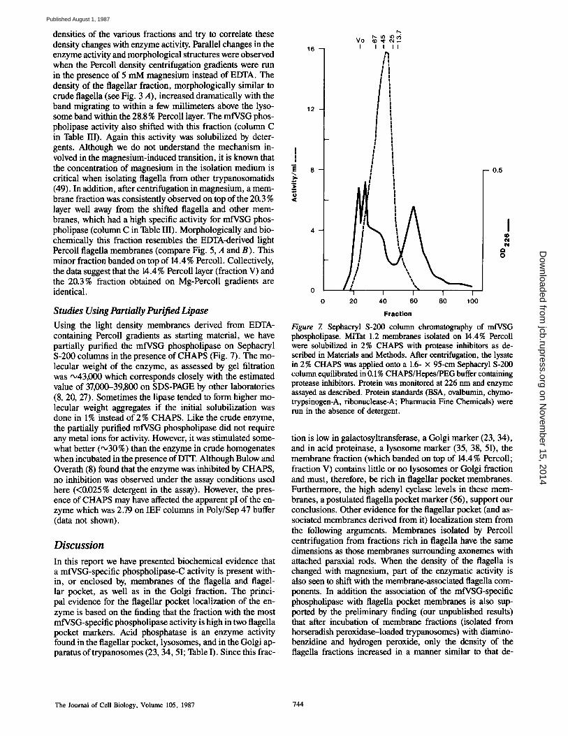
Studies Using Partially Purified Lipase Using the light density membranes derived from EDTA- containing Percoll gradients as starting material, we have partially purified the mfVSG phospholipase on Sephacryl S-200 columns in the presence of CHAPS (Fig. 7). The mo- lecular weight of the enzyme, as assessed by gel filtration was ~43,000 which corresponds closely with the estimated value of 37,000-39,800 on SDS-PAGE by other laboratories (8, 20, 27). Sometimes the lipase tended to form higher mo- lecular weight aggregates if the initial solubilization was done in 1% instead of 2 % CHAPS. Like the crude enzyme, the partially purified mfVSG phospholipase did not require any metal ions for activity. However, it was stimulated some- what better (~30%) than the enzyme in crude homogenates when incubated in the presence of DTT. Although Bulow and Overath (8) found that the enzyme was inhibited by CHAPS, no inhibition was observed under the assay conditions used here (<0.025 % detergent in the assay). However, the pres- ence of CHAPS may have affected the apparent pI of the en- zyme which was 2.79 on IEF columns in Poly/Sep 47 buffer (data not shown).
Discussion
In this report we have presented biochemical evidence that a mfVSG-specific phospholipase-C activity is present with- in, or enclosed by, membranes of the flagella and flagel- lar pocket, as well as in the Golgi fraction. The princi- pal evidence for the flagellar pocket localization of the en- zyme is based on the finding that the fraction with the most mfVSG-specific phospholipase activity is high in two flagella pocket markers. Acid phosphatase is an enzyme activity found in the flagellar pocket, lysosomes, and in the Golgi ap- paratus oftrypanosomes (23, 34, 51; Table I). Since this frac-
1 6 -
12 -
~ 8 -
4 -
f ~
vo ~ , ~ I I I I I
I I ! !
!
i
l
I #
!
I 2 0 4 0
I I I 6 0 8 0 1 0 0
- - 0 .5
I O
O O
F r a c t i o n
Figure 7. Sephacryl S-200 column chromatography of mfVSG phospholipase. MITat 1.2 membranes isolated on 14.4% Pereoll were solubil~xl in 2% CHAPS with protease inhibitors as de- scribed in Materials and Methods. After centrifugation, the lysate in 2% CHAPS was applied onto a 1.6- x 95-cm Sephacryl S-200 column equilibrated in 0.1% CHAPS/Hepes/PEG buffer containing protease inhibitors. Protein was monitored at 226 nm and enzyme assayed as described. Protein standards (BSA, ovalbumin, chymo- trypsinogen-A, ribonuclease-A; Pharmacia Fine Chemicals) were run in the absence of detergent.
tion is low in galactosyltransferase, a Golgi marker (23, 34), and in acid proteinase, a lysosome marker (35, 38, 51), the membrane fraction (which banded on top of 14.4% Percoll; fraction V) contains little or no lysosomes or Golgi fraction and must, therefore, be rich in flagellar pocket membranes. Furthermore, the high adenyl cyclase levels in these mem- branes, a postulated flagella pocket marker (56), support our conclusions. Other evidence for the flagellar pocket (and as- sociated membranes derived from it) localization stem from the following arguments. Membranes isolated by Percoll centrifugation from fractions rich in fagella have the same dimensions as those membranes surrounding axonemes with attached paraxial rods. When the density of the flagella is changed with magnesium, part of the enzymatic activity is also seen to shift with the membrane-associated flagella com- ponents. In addition the association of the mfVSG-specific phospholipase with flagella pocket membranes is also sup- ported by the preliminary finding (our unpublished results) that after incubation of membrane fractions (isolated from horseradish peroxidase-loaded trypanosomes) with diamino- benzidine and hydrogen peroxide, only the density of the flagella fractions increased in a manner similar to that de-
The Journal o f Cell Biology, Volume 105, 1987 744
on Novem
ber 15, 2014jcb.rupress.org
Dow
nloaded from
Published August 1, 1987

scribed for peroxidase-loaded endosomes (11). These shifts in band density were paralleled by shifts in the phospholipase ac- tivity which also shifted after incubation with hydrogen perox- ide. Collectively, these data indicate the presence of the en- zyme in the region of the flagella pocket and in the Golgi region. The possibility that the enzyme may also be found in coated vesicles, endosomes, or other prelysosomal compart- ments, membranes which are presumably derived from the flagellar pocket, cannot be excluded.
It has been reported that mfVSG phospholipase-C is a thiol-dependent enzyme. However, the role of thiols in mfVSG-lipase activation is not clear. Several laboratories report a 1.4-5.4-fold increase in enzyme activity with high concentrations of DTT (10-25 mM) (8, 20, 27). However, there have also been reports of enzyme activation by EDTA (8, 20, 27). Our data suggests that EDTA and DTT have simi- lar activating effects and that these are not additive. Thus the DTT may act by either chelating heavy metal ions as sug- gested by Fox et al. (20) or by changing the tertiary structure of the substrate (27). The variations reported by several groups in enzyme stimulation by thiol reagents and chelators may simply reflect the purity of the water or other reagents used in the isolation buffers.
It is not clear what role the mfVSG phospholipase plays in trypanosome's. The enzyme is neither more active nor dis- tributed differently between T. brucei slender or stumpy bloodstream forms of the parasites. Furthermore, the idea that the enzyme may be involved in the release of VSG during differentiation of bloodstream forms into uncoated procyclic forms is inconsistent with its disappearance before the re- lease of the surface glycoprotein (7, 10). Thus, this release of VSG from the parasite may simply be due to dying para- sites (5), or to some other complex interactions due to the use of inappropriate isolation/incubation conditions (35, 37). Consequently, if the VSG-specific phospholipase is involved in the release of VSG from the parasite surface as they are transformed to the supposedly VSG-free procyclic forms (14), then this release is not due to a simple increase in the total amount of enzyme. Our proposition is that the mfVSG phospholipase may be involved in surface coat recycling via an endocytotic mechanism. The involvement of phospholi- pase-C in VSG traffic within the parasite has also been postu- lated by Billow and Overath (8). If the enzyme is in the same lipid bilayer as the VSG (8), activation of the lipase within the flagella pocket or endosomes would release the VSG from the membrane and be taken up by the endocytotic system. In addition to removal of VSG from the membrane, the enzyme, with the help of an acid proteinase, could release the CRD for subsequent recycling into the VSG synthetic pathway (Webster, P., and D. J. Grab, manuscript submitted for publi- cation). The release of diacylglycerol from VSG could also regulate protein kinase C, if present, within the parasite (see references 4, 39). However, the release of the enzyme either actively or as the result of dying parasites could have some other far-reaching effects on the host. We have shown that un- der certain conditions, living trypanosomes can release a phospholipase-C which is capable of using mfVSG as sub- strate (35). Decay-accelerating factor, a membrane-bound glycoprotein that inhibits amplification of the complement system (44), as well as acetylcholinesterase, have been shown to contain a phosphatidylinositol membrane-binding
domain similar, if not identical, to the CRD (12, 39). Anemia is a hallmark of African trypanosomiasis. It is thus tempting to speculate that iftrypanosomes released mfVSG phospho- lipase, either actively, or from dying organisms, it could re- move the decay-accelerating factor from the surface of cell membranes, such as erythrocytes. Erythrocyte membranes altered as a result of decay-accelerating factor removal would then be susceptable to hemolysis and/or removal from the circulation by the reticuloendothelial system as occurs in pa- tients with paroxysmal nocturnal hemoglobinuria (45).
The cultured T. brucei procyclic forms were kindly provided by Mr. Robert Nelson, Dr. N. Borowy, and Dr. H. Hirumi of the International Laboratory for Research on Animal Diseases (ILRAD) and trypanosome stumpy forms were provided by Ms. Tamara Aboagye-Kwarteng (ILRAD). We would also like to thank Dr. Onesmo ole-MoiYoi for his interest and helpful discus- sions, and Steven Wasike for his technical assistance. We are grateful to Dr. Paul T. Englund and his colleagues for communication of manuscripts be- fore publication. Dr. D. J. Grab dedicates this work to his wife Jacqueline, to his newborn son Ryan Tsavo, and to the memory of Rene Raiole. This is ILRAD publication No. 493.
Received for publication 3 March 1987, and in revised form 23 April 1987.
References
1. Baekkeskov, S., L. Rovis, and Y. Verjee. 1979. The lipids of African trypanosomes. ILRAD Annual Report, 64-65.
2. Baltz, T., D. Baltz, C. Giroud, and J. Crockett. 1985. Cultivation in a semi-defined medium of animal infective forms of Trypanosoma brucei, 7". equiperdum, T. evansi, T. rhodesiense, and T. gambiense. EMBO (Eur. Mol. Biochem. J.) J. 4:1273-1277.
3. Bangs, J. D., D. Herald, J. L. Krakow, G. W. Hart, and P. T. Englund. 1985. Rapid processing of the carboxyl terminus of a trypanosome variant surface glycoprotein. Proc. Natl. Acad. Sci. USA. 82:3207-3211.
4. Bell, R. M. 1986. Protein kinase C activation by diacylglycerol second messengers. Cell. 45:631-632.
5. Black, S. J., R. S. Hewett, and C. N. Sendashonga. 1982. Trypanosoma brucei variable surface antigen is released by degenerating parasites but not by actively dividing parasites. Parasite Immunol. (Oxf.). 4:233-244.
6. Brown, W. C., and D. J. Grab. 1985. Biological and biochemical character- ization of bovine interleukin 2. Studies with cloned bovine T ceils. J. lm- munol. 133:3184-3190.
7. Billow, R., and P. Overath. 1985. Synthesis of a hydrolase for the mem- brane-form variant surface glycoprotein is repressed during transforma- tion of Trypanosoma brucei. FEBS (Fed. Eur. Biochem. Soc.) Lett. 187:105-110.
8. Biilow, R., and P. Overath. 1986. Purification and characterization of the membrane-form variant surface glycoprotein hydrolase of Trypanosoma brucei. J. Biol. Chem. 261:11918-11923.
9. Cardoso de Almeida, M. L., and M. J. Turner. 1983. The membrane form of variant surface glycoproteins of Trypanosoma brucei. Nature (Lond.). 302:349-352.
10. Cardoso de Almeida, M. L., L. M. Allan, and M. J. Turner. 1984. Purifica- tion and properties of the membrane form of variant surface glycoproteins (VSGs) from Trypanosoma brucei. J. Protozool. 31:53-60.
11. Courtoy, P. J., J. Quintart, and P. Baudhuin. 1984. Shift of equilibrium density induced by 3,Y-diaminobenzidine cytochemistry: a new proce- dure for the analysis and purification of peroxidase-containing organelles. J. Cell Biol. 98:870-876.
12. Davitz, M. A., M. G. Low, and V. Nussenzweig. 1986. Release of decay- accelerating factor (DAF) from the cell membrane by phosphotidylinosi- tol-specific phospholipase C (PIPLC). J. Exp. Med. 163:1150-1161.
13. Duvillier, G., A. Nouvelot, C. Richet, T. Baltz, and P. Degland. 1983. Presence of glycerol and fatty acids in the C-terminal end of a variant sur- face glycoprotein from Trypanosorna equiperdum. Biochem. Biophys. Res. Commun. 114:119-125.
14. Englund, P. T., S. L. Hajduk, and J. C. Marini. 1982. The molecular biol- ogy of trypanosomes. Annu. Rev. Biochem. 51:695-726.
15. Ferguson, M. A. J., and G. A. M. Cross. 1984. Myristylation of the mem- brane form of a Trypanosoma brucei variant surface glycoprotein. J. Biol. Chem. 259:3011-3015.
16. Ferguson, M. A. J., M. Duszenko, G. S. Lamont, P. Overath, and G. A. M. Cross. t986. Biosynthesis of Trypanosoma brucei variant surface glyco- proteins. N-glycosylation and addition of a phosphotidylinositol mem- brane anchor. J. Biol. Chem. 261:356-362.
17. Ferguson, M. A. J., K. Halder, and G. A. M. Cross. 1985. Trypanosoma
Grab et al. Phospholipase in African Trypanosomes 745
on Novem
ber 15, 2014jcb.rupress.org
Dow
nloaded from
Published August 1, 1987
brucei variant surface glycoprotein has a sn-l,2-dimyristyl glycerol membrane anchor at its COOH terminus. J. Biol. Chem. 260:4963-4968.
18. Ferguson, M. A. J., M. G. Low, andG. A. M. Cross. 1985. Glycosyl-sn- 1,2-dimyristylphosphotidylinositol is covalently linked to Trypanosoma brucei variant surface glycoprotein. J. Biol. Chem. 260:14547-14555.
19. Fish, W. R., D. L. Looker, J. J. Marr, and R. L. Berens. 1982. Purine metabolism in the bloodstream forms of Trypanosoma gambiense and Trypanosoma rhodesiense. Biochim. Biophys. Acta. 719:223-231.
20. Fox, J. A., M. Duszenko, M. A. J. Ferguson, M. G. Low, and G. A. M. Cross. 1986. Purification and characterization of a novel glycan-phospha- tidylinositol-specific phospholipase C from Trypanosoma brucei. J. Biol. Chem. 261:15767-15771.
21. Grab, D. J., and J. J. Bwayo. 1983. Isopycnic isolation of African trypano- somes on Percoll gradients formed in situ. Acta Trop. 39:363-366.
22. Grab, D. J., and Y. Verjee. 1983. Assembly of variable surface glycopro- tein (VSG) on the cell surface of African trypnosomes. 10th International Congress of the World Association of Animal Veterinary Parasitologists. 83. (Abstr.)
23. Grab, D. J., U. A. K. Kara, S. Ito, andL. Rovis. 1984. Glycosyltransferase activities in Golgi complex and endoplasmic reticulum fractions isolated from African trypanosomes. J. Cell Biol. 99:569-577.
24. Grab, D. J., P. Webster, and Y. Verjee. 1984. The intracellular pathway and assembly of newly formed variable surface glycoprotein of Trypano- soma brucei. Proc. Natl. Acad. Sci. USA. 81:7703-7707.
25. Gurnett, A. M., J. Raper, and M. J. Turner. 1986. Solution properties of the variant surface glycoprotein of Trypanosoma brucei. Mol. Biochem. Parasitol. 18:141-153.
26. Gurnett, A. M., J. Ward, J. Raper, and M. J. Turner. 1986. Purification and characterisation of membrane-form variant surface glycoproteins of Trypanosoma brucei. Mol. Biochem. Parasitol. 20:1-13.
27. Hereld, D., J. L. Krakow, J. D. Bangs, G. W. Hart, and P. T. Englund. 1986. A phospholipase C from Trypanosoma brucei which selectively cleaves the glycolipid on the variant surface glycoprotein. J. Biol. Chem. 261:13813-13819.
28. Holder, A. A. 1983. Carbohydrate is linked through ethanolamine to the C-terminal amino acid of Trypanosoma brucei variant surface glycopro- tein. Biochem. J. 209:261-262.
29. Ito, S., and M. J. Karnovsky. 1968. Formaldehyde-glutaraldehyde fixatives containing trinitro compounds. J. Cell Biol. 34:168a. (Abstr.)
30. Jackson, D. G., and H. P. Voorheis. 1985. Release of the variable surface coat glycoprotein from Trypanosoma brucei requires the cleavage of a phosphate ester. J. Biol. Chem. 260:5179-5183.
31. Jackson, D. G., M. J. Owen, and H. P. Voorheis. 1985. A new method for the rapid purification of both membrane-bound and released forms of the variant surface glycoprotein from Trypanosoma brucei. Biochem. J. 230:195-202.
32. Khaukha, G. W., O. ole-MoiYoi, and J. D. Lonsdale-Eccles. 1984. Try- panosome phospholipases and haemolytic activities. Fed. Proc. 43:1628. (Abstr.)
33. Lanhan, S. M., and D. G. Godfrey. 1970. Isolation of salivarian trypano- somes from man and other mammals using DEAE-cellulose. Exp. Parasitol. 28:521-534.
34. Langreth, S. G., and A. E. Balber. 1975. Protein uptake and digestion in bloodstream and in culture forms of Trypanosoma brucei. J. Protozool. 22:40-53.
35. Lonsdale-Eccles, J. D., and D. J. Grab. 1987. Purification of African trypanosomes can cause biochemical changes in the parasites. J. Pro- tozool. In press.
36. Lonsdale-Eccles, J. D., and G. W. N. Mpimbaza. 1986. Thiol-dependent proteases of African trypanosomes. Analysis by electrophoresis in so- dium dodecyl sulphate/polyacrylamide gels co-polymerized with fibrino- gen. Eur. J. Biochem. 155:469-473.
37. Lonsdale-Eccles, J. D., D. J. Grab, and S. Ito. 1985. Subcellular localiza- tion of African trypanosomal proteases. 13th International Congress of Biochemistry (Amsterdam). Abstract No. TU-167.
38. Lonsdale-Eccles, J. D., G. W. N. Mpimbaza, and D. J. Grab. 1986. An
activator of lysosomal thiol-proteinases is present in blood. J. Cell Biol. 103(5, Pt. 2):357a. (Abstr.)
39. Low, M. G., M. A. J. Ferguson, A. H. Futerman, and I. Silman. 1986. Covalently attached phosphotidylinositol as a hydrophobic anchor for membrane proteins. Trends Biochem. Sci. 11:212-215.
40. Majiwa, P. A. O., J. R. Young, R. Hamers, and G. Matthyssens. 1986. Minichromosomal variable surface glycoprotein genes and molecular karyotypes of Trypanosoma (Nannomonas) congolense. Gene. 41:183- 192.
41. Mangold, M. K. 1961. Thin-layer chromatography of lipids. J. Am. Oil Chem. Soc. 38:708-727.
42. Mellors, A. 1985. Phospholipases of Trypanosomes. In Immunology and Pathogenesis of Trypanosomiasis. I. Tizard, editor. CRC Press, Inc., Boca Raton, FL. 67-74.
43. Neville, D. M., Jr. 1971. Molecular weight determination of protein- dodecyl sulfate complexes by gel electrophoresis in a discontinuous buffer system. J. Biol. Chem. 246:6328-6334.
44. Nicholson-Weller, A., J. Burge, D. T. Fearon, P. F. Weller, and K. F. Austen. 1982. Isolation of a human erythrocyte membrane glycoprotein with decay accelerating activity from C3 convertases of the complement system. J. Immunol. 129:184-189.
45. Nicholson-WeUer, A., J. P. March, S. I. Rosenfeld, and K. F. Austen. 1983. Affected erythrocytes of patients with paroxysmal nocturnal hemo- globinuria are deficient in the complement regulatory protein, decay ac- celerating factor. Proc. Natl. Acad. Sci. USA. 80:5066-5070.
46. Opperdoes, F. R., and J. Van Roy. 1982. The phospholipases of Trypano- soma brucei bloodstream forms and cultured procyclics. Mol. Biochem. Parasitol. 5:309-319.
47. Opperdoes, F. R., P. Borst, S. Bakker, and W. Leene. 1977. Localization of glycerol-3-phosphate oxidase in the mitochondrion and particulate NAD+-linked glycerol-3-phosphate dehydrogenase in the microbodies of the bloodstream form of Trypanosoma brucei. Eur. J. Biochem. 76:29-39.
48. Opperdoes, F. R., P. Bauduin, I. Coppens, C. De Roe, S. W. Edwards, P. J. Weijers, and O. Misset. 1984. Purification, morphometric analysis, and characterization of the glycosomes (microbodies) of the protozoan hemo- flagellate Trypanosoma brucei. J. Cell Biol. 98:1178-1184.
49. Pereira, N. M., W. De Souza, R. D. Machado, and F. T. DeCastro. 1977. Isolation and properties of flagella of trypanosomatids. J. Protozool. 24:511-514.
50. Rovis, L., and S. Baekkeskov. 1980. Sub-cellular fractionation of Trypano- soma brucei. Isolation and characterization of plasma membranes. Para- sitology. 80:507-524.
51. Steiger, R. F., F. R. Opperdoes, and J. Bomemps. 1980. Subcellular frac- tionation of Trypanosoma brucei bloodstream forms with special refer- ence to hydrolases. Fur. J. Biochem. 105:163-175.
52. Tulkens, P., H. Beaufay, and A. Trouet. 1974. Analytical fractionation of homogenates from cultured rat embryo fibroblasts. Z Cell Biol. 63: 383--401.
53. Vickerman, K., and A. G. Luckins. 1969. Localisation of variable antigens in the surface coat of Trypanosoma brucei using ferritin conjugated anti- body. Nature (Lond.). 224:1125-1127.
54. Vincent, R., and N. Nadeau. 1983: A micromethod for the quantitation of cellular proteins in Percoll with the Coomassie Brilliant Blue dye-binding assay. Anal. Biochem. 135:355-362.
55. Voorheis, H. P. 1980. Fatty acid uptake by bloodstream forms of Trypano- soma brucei and other species of the kinetoplastida. Mol. Biochem. Parasitol. 1:177-186.
56. Walter, R. D., and F. R. Opperdoes. 1982. Subcellular distribution of ade- nylate cyclase, cyclic-AMP phosphodiesterase, protein kinases and phos- phoprotein phosphatase in Trypanosoma brucei. Mol. Biochem. Para- sitol. 6:287-295.
57. Wattiaux, R., S. Wattiaux-de Coninck, M.-F. Ronveaux-Dupal, and F. Du- bois. 1978. Isolation of rat liver lysosomes by isopycnic centrifugation in a metrizamide gradient. J. Cell Biol. 78:349-368.
The Journal of Cell Biology, Volume 105, 1987 746
on Novem
ber 15, 2014jcb.rupress.org
Dow
nloaded from
Published August 1, 1987