Phosphatidylinositol 4,5-Bisphosphate Anchors Cytosolic Group IVA Phospholipase A2 to Perinuclear...
36
Phosphatidylinositol 4,5-bisphosphate anchors cytosolic group IVA phospholipase A 2 to perinuclear membranes and decreases its calcium requirement for translocation in live cells Javier Casas, Miguel A. Gijón, Ana G. Vigo, Mariano Sánchez Crespo, Jesús Balsinde 1 and María A. Balboa 2 Institute of Molecular Biology and Genetics, Spanish Research Council. 47003 Valladolid, Spain. 1 To whom correspondence should be addressed. Instituto de Biología y Genética Molecular (IBGM-CSIC), Universidad de Valladolid, Facultad de Medicina. Calle Sanz y Forés s/n, 47003 Valladolid, Spain. Phone: 34-983-423-062; FAX: 34- 983-423-588; E-mail: [email protected] 2 Also corresponding author. Instituto de Biología y Genética Molecular (IBGM- CSIC), Universidad de Valladolid, Facultad de Medicina. Calle Sanz y Forés s/n, 47003 Valladolid, Spain. Phone: 34-983-184-833; FAX: 34-983-423-588; E-mail: [email protected] RUNNING TITLE: PIP 2 -induced translocation of cPLA 2 α.
-
Upload
independent -
Category
Documents
-
view
4 -
download
0
Transcript of Phosphatidylinositol 4,5-Bisphosphate Anchors Cytosolic Group IVA Phospholipase A2 to Perinuclear...

Phosphatidylinositol 4,5-bisphosphate anchors cytosolic group
IVA phospholipase A2 to perinuclear membranes and decreases
its calcium requirement for translocation in live cells
Javier Casas, Miguel A. Gijón, Ana G. Vigo, Mariano Sánchez Crespo, Jesús
Balsinde1 and María A. Balboa2
Institute of Molecular Biology and Genetics, Spanish Research Council. 47003
Valladolid, Spain.
1To whom correspondence should be addressed. Instituto de Biología y Genética
Molecular (IBGM-CSIC), Universidad de Valladolid, Facultad de Medicina. Calle
Sanz y Forés s/n, 47003 Valladolid, Spain. Phone: 34-983-423-062; FAX: 34-
983-423-588; E-mail: [email protected]
2Also corresponding author. Instituto de Biología y Genética Molecular (IBGM-
CSIC), Universidad de Valladolid, Facultad de Medicina. Calle Sanz y Forés s/n,
47003 Valladolid, Spain. Phone: 34-983-184-833; FAX: 34-983-423-588; E-mail:
RUNNING TITLE: PIP2-induced translocation of cPLA2α.

Abstract
The eicosanoids are centrally involved in the onset and resolution of inflammatory
processes. A key enzyme in eicosanoid biosynthesis during inflammation is Group
IVA phospholipase A2 (also known as cytosolic phospholipase A2α, cPLA2α). This
enzyme is responsible for generating free arachidonate (AA) from membrane
phospholipids. cPLA2α translocates to perinuclear membranes shortly after cell
activation, in a process that is governed by the increased availability of intracellular
Ca2+. However, cPLA2α also catalyzes membrane phospholipid hydrolysis in
response to agonists that do not mobilize intracellular Ca2+. How cPLA2α interacts
with membranes under these conditions is a major, still unresolved issue. Here we
report that phosphatidylinositol 4,5-bisphosphate (PtdIns(4,5)P2) promotes
translocation of cPLA2α to perinuclear membranes of intact cells in a manner that is
independent of rises in the intracellular Ca2+ concentration. PtdIns(4,5)P2 anchors the
enzyme to perinuclear membranes and allows for a proper interaction with its
phospholipid substrate to release arachidonate.
2
Introduction
It is well established that the hydrolytic attack of AA-containing phospholipids by
cPLA2α constitutes the first regulatory step of the eicosanoid biosynthetic cascade
(Bonventre, 2004; Hirabayashi et al., 2004). cPLA2α is unique among all the
phospholipase A2 family members (Six and Dennis, 2000) due to its substrate
selectivity for AA-containing phospholipids and its tight regulation by Ca2+ and
phosphorylation (Clark et al., 1991; Lin et al., 1993; Gijón et al., 2000a; Dessen et al.,
1999). Free AA generated by activated cPLA2α can be oxygenated into a variety of
compounds, called the eicosanoids, that not only are involved in acute and chronic
inflammatory diseases such as arthritis, asthma, and postischaemic tissue-injury, but
can also regulate physiological processes such as renal function, and female
reproductive events including parturition (Bonventre, 2004; Hirabayashi et al., 2004;
Uozumi et al., 1997; Bonventre et al., 1997).
cPLA2α is a cytosolic enzyme in resting cells and translocates to membranes
when cell activation takes place. The enzyme possesses a calcium-binding domain
(C2) in its N-terminal half that helps the enzyme interact with membrane phospholipid
when the intracellular Ca2+ concentration ([Ca2+]i) rises. A single mutation in this
domain, i.e. substitution of Asp-43 with Asn, D43N) completely abrogates the Ca2+-
dependent translocation of cPLA2α to cellular membranes and, as a result, AA
release is inhibited (Perisic et al., 1999; Gijón et al., 1999; Qiu et al., 1998).
However, stimuli that are known not to affect [Ca2+]i such as bacterial
lipopolysaccharide, phorbol esters or okadaic acid, are nonetheless able to induce
robust AA mobilization and eicosanoid production responses in a cPLA2α-dependent
manner (Shinohara et al., 1999; Gijón et al., 2000b). The mechanism by which
3

cPLA2α interacts with membranes under conditions that do not involve increase of
intracellular Ca2+ concentrations ([Ca2+]i) has remained unaddressed.
In vitro activity studies carried by Leslie and Channon (1990) in the early 90's
utilizing a partially purified cPLA2α preparation, had suggested that anionic
phospholipids such as PtdIns(4,5)P2 and phosphatidic acid, when incorporated into
the vesicle substrate, were capable of stimulating the activity of the enzyme and
decreasing its Ca2+ requirement from mM to nM levels. More recently, Mosior et al.
(1998) described the potent and specific increase in affinity of pure cPLA2α for
surfaces containing PtdIns(4,5)P2 at physiological concentrations, and this effect
paralleled an increase in substrate hydrolysis. Importantly, the enhancing effects of
PtdIns(4,5)P2 were observed even in the presence of EGTA ([free Ca2+] < 2 nM). Das
and Cho (2002) recently described a cluster of cationic residues in the catalytic
domain of cPLA2α (Lys488, Lys541, Lys543, and Lys544 in the human sequence) that
appears to play a role in the in vitro activation of the enzyme by PtdIns(4,5)P2, a
finding that was corroborated later on by Six and Dennis (2003).
With regard to intact cells, we observed that in UV light-treated macrophages,
increased PtdIns(4,5)P2 synthesis occurs at resting Ca2+ levels, and this parallels the
cPLA2α-dependent mobilization of AA (Balsinde et al., 2000). These data led us to
propose that PtdIns(4,5)P2 might be involved in cPLA2α activation at Ca2+ levels
equaling those pertaining to unstimulated cells (Balsinde et al., 2000). In this paper
we demonstrate that PtdIns(4,5)P2 anchors cPLA2α to perinuclear membranes of
intact cells in a manner that is independent of rises in [Ca2+]i, thus allowing for proper
interaction of the enzyme with its phospholipid substrate to optimally release AA.
4

Results and Discussion
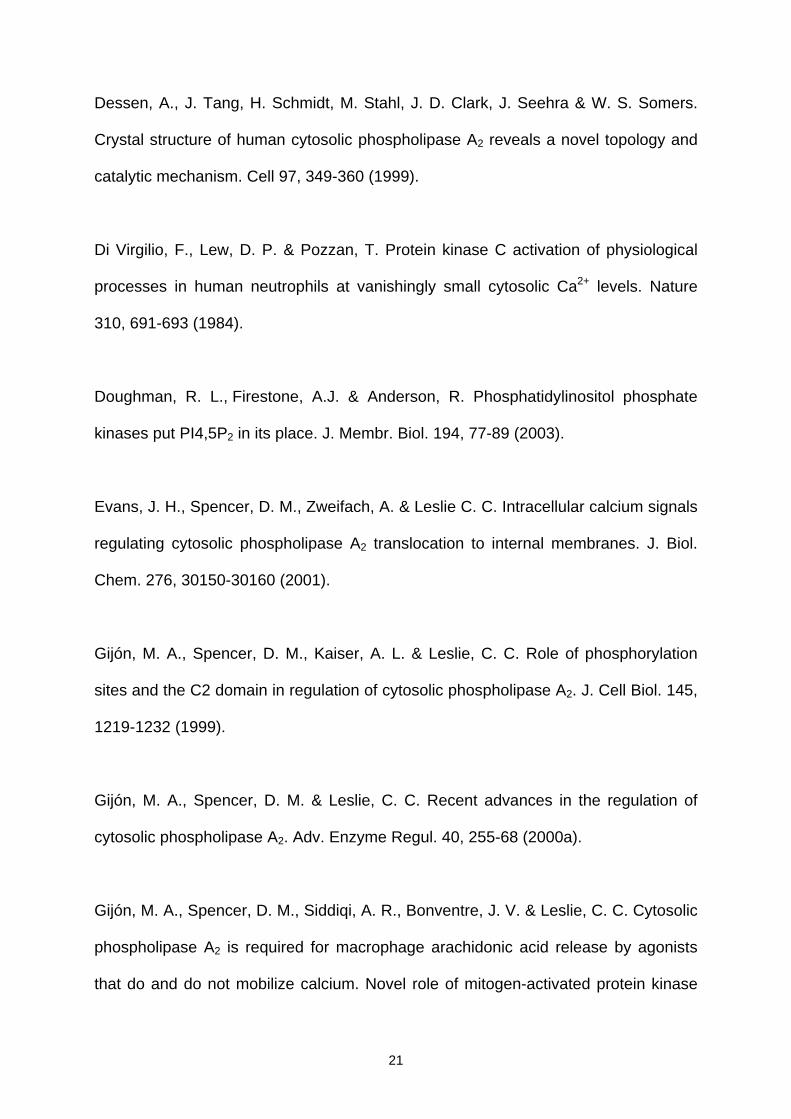
We were interested in examining whether cellular increases in PtdIns(4,5)P2 regulate
cPLA2α translocation to intracellular membranes in live cells. To this end, we
prepared HEK cells stably expressing a fusion protein of enhanced green fluorescent
protein (EGFP) and human cPLA2α. The cells were studied under the confocal
microscope during treatment with fluorescent PtdIns(4,5)P2 (BODIPYRTR-tagged
PtdIns(4,5)P2; abbreviated as TR-PI(4,5)P2). When complexed with histone or
neomycin carriers that counteract the negative charges of TR-PI(4,5)P2 and make it
membrane-permeable (Ozaki et al., 2000), it takes 5-10 min for the phospholipid to
penetrate into the cell (Fig. 1). Once inside, TR-PI(4,5)P2 localized primarily to
perinuclear membranes, in accord with previous observations (Ozaki et al., 2000).
Movement of TR-PI(4,5)P2 to perinuclear membranes was closely followed by the
complete translocation of cPLA2α to the same location (Fig. 1A). When EGFP-cPLA2
transfected cells were treated only with the carrier, EGFP-cPLA2 did not translocate
(Fig. 1B). Furthermore, when cells transfected with EGFP alone were exposed to TR-
PI(4,5)P2, the green fluorescence pattern did not change (Fig. 1C). AA mobilization
was studied under these conditions. Figure 1D shows that exposure of the cells to
TR-PI(4,5)P2 enhanced AA release, thus indicating that membrane phospholipid
hydrolysis follows from translocation of cPLA2 to perinuclear membranes.
A possible explanation for TR-PI(4,5)P2-induced cPLA2α membrane
translocation and subsequent activation could be that the phosphoinositide, once
inside the cell, is hydrolyzed by phospholipase C to generate inositol 1,4,5-
trisphosphate which results in elevated [Ca2+]i and hence cPLA2α translocation.
However, cPLA2α translocation to membranes was also observed when the cells
were treated with TR-PI(3,4)P2, whose hypothetical hydrolysis by phospholipase C
5

would not yield inositol 1,4,5-trisphosphate (Fig. 1E). That both PtdIns(4,5)P2 and
PtdIns(3,4)P2 isomers modify the subcellular localization of cPLA2α suggests a
charge effect rather than the result of a metabolic transformation via phospholipase
C. The PtdIns(3,4)P2–induced translocation of cPLA2α was accompanied by an
increased release of AA, similar in extent to that produced by PtdIns(4,5)P2 (data not
shown). This is consistent with the in vitro studies of Six and Dennis (2003) showing
increased cPLA2α activity in response to PtdIns(3,4)P2. Interestingly, Das and Cho
(2002) failed to observe an effect of PtdIns(3,4)P2 on the activity of cPLA2α. The
reasons for this discrepancy may likely arise from differences in the experimental
protocols followed by these authors.
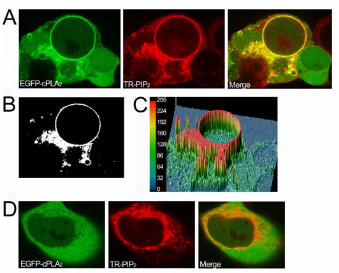
Experiments were also conducted with the macrophage cell line RAW 264.7.
Owing to their immunoinflammatory nature, RAW 264.7 macrophages possess a
robust machinery for eicosanoid biosynthesis and thus provide a more appropriate
cell context to validate cPLA2α translocation and AA release in response to
PtdIns(4,5)P2. Exposure of EGFP-cPLA2α-transfected macrophages to TR-PI(4,5)P2
resulted in membrane translocation of the enzyme (Fig. 2A) and enhanced AA
release (Fig. 2B).
We also performed measurements with pure PtdIns(4,5)P2, isolated from
bovine brain. Probably because of the presence of long chain fatty acids in pure
PtdIns(4,5)P2, it was not possible for us to deliver this phospholipid into the cell's
interior by using a shuttle. Thus, for these experiments we utilized digitonin-
permeabilized cells (Balsinde et al., 2000). Using this procedure, we confirmed that
native PtdIns(4,5)P2 induced translocation of EGFP-cPLA2α to perinuclear
membranes in a similar fashion to that previously observed with BODIPY-TR-
PI(4,5)P2 in Fig. 1 (data not shown).
6

Mosior et al. (1998) have shown that binding of cPLA2α to phospholipid
vesicles enriched in PtdInsP2 is essentially a Ca2+-independent process. To verify
whether this is also the case in live cells, we incubated the cells in a Ca2+-free EGTA-
containing buffer. The continued presence of cells in Ca2+-free buffer lowers the
intracellular Ca2+ level well below that normally occurring in resting cells incubated in
a regular Ca2+-containining buffer (Di Virgilio et al., 1984). Under these Ca2+-free
conditions, no translocation of cPLA2α was observed after addition of TR-PI(4,5)P2,
even though the phosphoinositide penetrated normally into the cell (Fig. 3).
Interestingly, if Ca2+ was restored to the incubation medium, a very rapid distribution
of cPLA2α to the perinuclear membranes of TR-PI(4,5)P2-treated cells was observed
(Fig. 3). These results suggest that, in cells, the PtdInsP2 effect is not Ca2+-
independent; rather, a threshold Ca2+ is necessary for cPLA2α to translocate to
membranes. This view is consistent with previous data by Das and Cho (2002).
To further characterize this Ca2+ effect, we conducted experiments with a
mutant construct where the cPLA2α has the mutation D43N (EGFP-D43N-cPLA2)
which severely limits the ability of the enzyme to bind calcium (Bittova et al., 1999;
Perisic et al., 1999; Gijón et al., 1999). As shown in Fig. 4, the mutant EGFP-D43N-
cPLA2 did not translocate in response to TR-PI(4,5)P2, even in the presence of 1.3
mM CaCl2 in the incubation buffer, demonstrating that a functional C2 domain is
necessary for cPLA2 translocation to perinuclear structures in response to PtdInsP2.
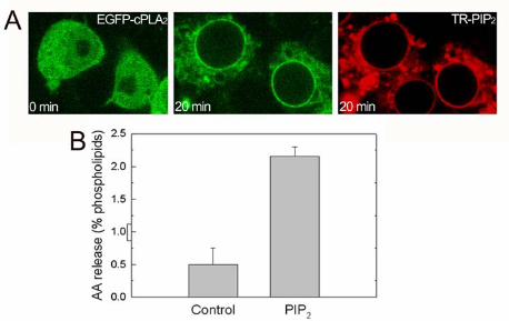
Since TR-PI(4,5)P2 and cPLA2α appear to move to similar intracellular
structures (Figs. 1 to 3), a co-localization analysis was performed. Fig. 5A shows the
merge of green (from the EGFP-cPLA2) and red (from the TR-PI(4,5)P2)
fluorescences in cells treated for 20 min with TR-PI(4,5)P2, suggesting co-localization
in the perinuclear region. Analysis of co-localization with the LaserPix confocal image
7

software (BioRad) provides a better definition of the pixels showing perfect co-
localization of both fluorescences (Fig. 5B, white mask). In Fig, 5C, a three-
dimensional representation of such co-localization is shown, and the color of each
pixel defines the level of co-localization. Nuclear membrane and perinuclear
structures (most likely Golgi and endoplasmic reticulum) show the most intense co-
localization. As a control for the co-localization analyses, note a cell in the bottom left
corner of Figs 5A-C that does not express EGFP-cPLA2α and thus stains red, and
another cell in the bottom right corner that expresses the EGFP-cPLA2α but did not
take TR-TR-PI(4,5)P2 in, and thus stains only green. Accordingly, these two cells do
not show in the co-localization mask (Fig. 5B) or in the three-dimensional
representation (Fig. 5C).
Taking advantage of the fact that in the absence of extracellular calcium, TR-
PI(4,5)P2 fails to induce EGFP-cPLA2α translocation to membranes (see above),
green and red fluorescences can be analyzed in the same cell at different
localizations (Fig. 5D). Under these conditions, the colocalization coefficients
obtained with the LaserPix software were: red to green, 0.37 ± 0.08, and green to
red, 0.33 ± 0.06. After restoring calcium to the extracellular medium, the co-
localization analysis was repeated on the same cells and the data were: red to green,
0.40 ± 0.05; and green to red, 0.88 ± 0.01.
Altogether, the results presented so far indicate that PtdIns(4,5)P2 functions to
anchor cPLA2 to the perinuclear and nuclear membranes in live cells, by a process
that requires the presence of a certain threshold level of Ca2+ as well as a functional
Ca2+ binding site in the enzyme (Fig. 3). Thus, PtdIns(4,5)P2 may either provide a
second binding site for cPLA2α in addition to the Ca2+-binding site, or act to lower the
Ca2+ requirement of cPLA2α for membrane binding. To evaluate these two
8

possibilities, changes in [Ca2+]i were monitored in Fluo-3-loaded cells exposed to the
phosphoinositide (Fig. 6). TR-PI(4,5)P2 induced changes in [Ca2+]i with a very
different pattern, potency and kinetics from the calcium ionophore ionomycin, which
is also known to potently induce cPLA2α membrane translocation (Gijón et al.,
2000a) (cf. Figs. 6A and 6B). Given these differences, we proceeded to determine
the minimal Ca2+ requirements for TR-PI(4,5)P2 to induce cPLA2α translocation. To
this end, cells were incubated in a Ca2+-free medium, exposed to either TR-PI(4,5)P2
or ionomycin. Thereafter different CaCl2 concentrations were added to the incubation
medium, and confocal microscopy images of the cPLA2α movement were taken.
[Ca2+]i was monitored in Fluo-3-loaded cells in parallel under exact experimental
conditions. Figs. 7A and 7B show that TR-PI(4,5)P2 promoted cPLA2α membrane
translocation at extracellular Ca2+ concentrations between 6-30 µM, which translate
to 50-220 nM in terms of [Ca2+]i. It is particularly striking that TR-PI(4,5)P2-induced
translocation of cPLA2α begins to be detected in all the cells analyzed already at an
extracellular Ca2+ concentration of 15 µM, which only induces minimal changes in
[Ca2+]i (~50 nM) (Figs. 7A and 7C). Note, on the other hand, that ionomycin promotes
visible translocation of cPLA2α only at higher extracellular Ca2+ concentrations,
approx. 60-150 µM, which raise [Ca2+]i up to 350-400 nM (Figs. 7B and 7D). These
results provide strong evidence that TR-PI(4,5)P2-induced cPLA2α translocation
occurs at substantially lower [Ca2+]i levels than that triggered by ionomycin.
In vitro mutagenesis studies have identified a cluster of four Lys as the
putative PtdInsP2-binding site (Das and Cho, 2002; Six and Dennis, 2003). In the
human enzyme, these are Lys488, Lys541, Lys543, and Lys544. We constructed a mutant
in which Lys488 was substituted by Glu and the three other Lys by Ala (EGFP-
K488E/K541A/K543A/K544A-cPLA2α; EGFP-4KE/A-cPLA2α) (Das and Cho, 2002;
9

Six and Dennis, 2003). When transfected into cells, the EGFP-4KE/A-cPLA2α mutant
actually translocated in response to TR-PI(4,5)P2 (Fig. 8A), although the degree of
co-localization between mutant enzyme and phospholipid was considerably lower
than that previously observed with the wild type enzyme (Fig. 8B). More importantly,
cells transfected with the EGFP-4KE/A-cPLA2α mutant did not mobilize AA in
response to TR-PI(4,5)P2, while cells transfected with the wild-type enzyme did it
readily (Fig. 9). These results demonstrate that the four-Lys cluster described by Das
and Cho (2002) is indeed crucial for cPLA2α to translocate to membrane in a
functionally-active form. That the EGFP-4KE/A-cPLA2α mutant also translocates to
membrane in response to PtdInsP2 may suggest the existence of additional binding
site(s) within the enzyme. However, since membrane binding of the EGFP-4KE/A-
cPLA2α mutant is not productive (i.e. no AA is released), it is probably of very limited
biological significance. The behavior of the mutant EGFP-4KE/A-cPLA2 clearly
suggests that translocation of the enzyme to membrane does not necessary lead to
enhanced AA release.
It has previously been demonstrated that PtdInsP2 does not increase the
specific activity of the K541A/K543A/K544A mutant, as measured by an in vitro assay
(Das and Cho, 2002; Six and Dennis, 2003). In our intact cell system, PtdInsP2
promotes translocation of the construct EGFP-4KE/A-cPLA2 to perinuclear
membranes, but this is not accompanied by an increased AA release, which is in
accordance with the aforementioned in vitro studies (Das and Cho, 2002; Six and
Dennis, 2003). In these studies an increase in the basal activity of the mutated
enzyme as compared to the wild type enzyme was also shown. Our studies in intact
cells failed to obtain reproducible evidence for such a phenomenon, as measured by
basal AA release. It appears likely that in intact cells, the basal state of the cPLA2α
10

may be governed by multiple factors, some of which may not be apparent in an in
vitro assay.
Our data, along with results from previous studies (Das and Cho, 2002; Six
and Dennis, 2003) demonstrate that the positive charge generated by Lys488, Lys541,
Lys543 and Lys544, allows the cPLA2 to properly bind to PtdInsP2. Mutation of these
Lys leads to changes in the specific activity of the enzyme compared to the wild type
enzyme, as measured in an in vitro assay (Das and Cho, 2002; Six and Dennis,
2003). We have extended these data by showing decreased AA release in the
mutants in response to PtdInsP2. The structure of those four Lys does not conform to
the structure of well known phosphoinositide-binding domains such as the plekstrin-
homology (PH), the FYVE, or the Phox-homology (PX) domains (Lemmon, 2003). It
is likely that this cationic cluster belongs in a novel, yet-undefined phosphoinositide-
binding domain, as suggested elsewhere (Das and Cho, 2002).
Based on all the above findings, it is intriguing to speculate with novel modes
of cPLA2α interaction with cellular membranes leading to enhanced phospholipid
hydrolysis. By using a methodology that preserves cellular integrity, i.e. shuttling
fluorescent PtdInsP2 into the cell by the use of lipophilic carriers, we have studied the
interaction between PtdInsP2 and cPLA2α under a physiologically relevant setting,
thus avoiding potential problems arising from the use of cellular fixation or
permeabilization techniques. Our live cell studies clearly indicate that variations in the
cellular PtdIns(4,5)P2 may help regulate the physical location of cPLA2α. Importantly
however, for this regulatory role of PtdIns(4,5)P2 to manifest, an intact C2 domain
within the enzyme and a certain threshold level of intracellular calcium are required.
The results suggest a sequential mode of cPLA2α interaction with the membrane,
where the enzyme first binds via the C2 domain in the presence of low Ca2+ levels.
11

This is followed by binding to PtdIns(4,5)P2 via the cationic cluster of four Lys, which
positions the enzyme in the proper manner to effect phospholipid hydrolysis and AA
release.
In this model of cPLA2α activation by PtdIns(4,5)P2, the threshold Ca2+ level
required is far below that found in stimulated cells, and resembles more that of
resting cells, i.e. ~50 nM. This is in contrast with studies in vitro, which reflected no
Ca2+ requirement for cPLA2α binding to and hydrolysis of phospholipids in mixed
micelles Six and Dennis, 2003). This difference highlights the very complex nature of
the interaction of cPLA2α with a biological membrane in a live cell context. cPLA2α
has been shown to physically interact with ceramide 1-phosphate (Subramanian et
al., 2005) and a number of proteins, including vimentin (Nakatani et al., 2000) and a
splice variant of Tip60 (Sheridan et al., 2001). Thus it is likely that in cells, numerous
entities in addition to PtdInsP2 may interact with cPLA2α and modulate its membrane-
binding properties.
It is important to note that the above scenario does not exclude the possibility
that PtdInsP2 binding may occur first, and this in turn augments the calcium-
dependent lipid binding of the C2 domain. Such a sequence of events has received
some experimental support by the recent work of Subramanian et al. (2005) in which
ceramide 1-phosphate was found to increase cPLA2α activity by interacting with the
C2 domain, thereby lowering the calcium requirement for translocation.
Our findings provide a mechanistic insight into the intracellular signaling
triggered by stimuli like bacterial lipopolysaccharide, UV light, phorbol esters or
okadaic acid, which do not mobilize Ca2+ from internal stores, yet they all activate the
cPLA2α−mediated release of AA (Shinohara et al., 1999; Gijón et al., 2000b). It is
likely that the increased synthesis of PtdInsP2 that occurs during cellular stimulation
12

by these agonists (Balsinde et al., 2000; Taylor et al., 1984), together with the low
nanomolar levels of [Ca2+]i, provides the signals to anchor cPLA2α to perinuclear
membranes. Two subfamilies of kinases, termed type I and type II, are involved in
the biosynthesis of PtdIns(4,5)P2 during cell activation. Type I consists of PtdIns(4)P
5-kinases, while type II consists of PtdIns(5)P 4-kinases. Members of both
subfamilies have been localized in the perinuclear region of cells (Doghman et al.,
2003). On the basis of our findings we propose that PtdIns(4,5)P2 formed by
phosphatidylinositol monophosphate kinases at the perinuclear envelope may help
recruit cPLA2α to this compartment at low [Ca2+]i levels. Future studies should shed
additional light on the mechanisms governing PtdIns(4,5)P2 accumulation in
perinuclear membranes and their overall role in cPLA2α-mediated AA signaling.
13

Materials and Methods
Reagents. Cell culture media and supplements were purchased from Gibco (Paisley,
Scotland, UK). BODIPYR-TR-X C6-PI(4,5)P2, BODIPYR-TR-X C6-PI(3,4)P2, carriers
(neomycin and histone), and Fluo-3-AM were purchased from Molecular Probes
(Carlsbad, CA, USA). Other reagents were from Sigma Ibérica (Madrid, Spain).
Lipid preparation. Lipids were prepared following the manufacturer's instructions.
Briefly, 2 μg phospholipid was mixed with 2 μl carrier (neomycin or histone, 0.5 mM),
resuspended in 200 μl HBSS containing 10 mM HEPES and, unless otherwise
indicated, 1.3 mM CaCl2, sonicated in a water bath for 2 min, and allowed to rest at
37°C for 10 min before use. Final concentration of PtdInsP2 in the solution is 5.7 μM.
Cells - HEK cells were cultured in DMEM supplemented with 2 mM glutamine, 10%
fetal calf serum, 100 U/ml penicillin and 100 μg/ml streptomycin at 37ºC in a 5% CO2
humidified incubator. Cells were passaged twice a week by trypsinization. Cells (40-
70% confluence) were transfected with 1 μg plasmid/ml using Lipofectamine PlusTM
(Invitrogen), following the manufacturer’s instructions. For stably transfected cells, 1
mg/ml G418 was used for selection and subsequent passages. RAW 264.7 cells
were cultured in RPMI medium supplemented with 10% fetal calf serum, 100 U/ml
penicillin, 100 μg/ml streptomycin and 2 mM glutamine at 37 ºC in a 5% CO2
humidified incubator.
Constructs. The DNA sequence of human cPLA2α was cloned into the pEGFP vector
(Clontech, Palo Alto, CA) using Hind-III and Pst-I cloning sites. This construct codes for the
14

expression of a fusion protein containing an N-terminal EGFP followed by the entire
sequence of the human cPLA2α. Wild type cPLA2α was mutagenized within the C2 domain
by replacing Asp43 with Asn (D43N), using the QuikChange XL Site-Directed Mutagenesis kit
from Stratagene and the oligonucleotides 5’-
CATGCTTGATACTCCAAATCCCTATGTGGAAC-3’ (forward) and 5’-
GTTCCACATAGGGATTTGGAGTATCAAGCATG-3’ (reverse). Mutagenesis was confirmed
by sequencing. Wild type EGFP-cPLA2α was also mutagenized to produce EGFP-
D488E/D541A/D543A/D544A-cPLA2 in two steps. First, the mutant EGFP-
D541A/D543A/D544A-cPLA2 was obtained by using the QuikChange XL Site-
Directed Mutagenesis kit from Stratagene and the oligonucleotides 5´-
GAGCCTCTGGATGTCGCAAGTGCAGCGATTCATGTAGTGGACAG-3´ (forward)
and 5´-CTGTCCACTACATGAATCGCTGCACTTGCGACATCCAGAGGCTC-3´.
Then, a second round of mutagenesis was performed to change Lys488 to Glu using
the primers: 5´-GGACGTGCTGGGGAGGTACACAACTTCATGC-3´ (forward) and 5´-
GCATGAAGTTGTGTACCTCCCCAGCACGTCC-3´(reverse).
Confocal microscopy - Cells were seeded on glass bottom culture dishes (MatTek
Corp., Ashland, MA, USA) coated with Poly-L-lysine (Sigma). After 24 hours, the
culture medium was replaced with HBSS containing 10 mM HEPES and 1.3 mM
CaCl2. Cells were monitored at 37°C by confocal microscopy using a Bio-Rad laser
scanning Radiance 2100 system coupled to a Nikon TE-2000U inverted microscope
equipped with a DH-35 tissue culture dish heater (Warner Instruments). Images were
obtained with a CFI Plan Apo 60X, oil immersion, 1.40 NA objective, which provided
a theoretical confocal layer thickness of approximately 0.4 μm at the wavelengths
used. Green fluorescence from the EGFP was monitored at 488 nm argon laser
excitation and the combination of a HQ500 long band pass and a HQ560 short band
15

pass blocking filters. Red fluorescence from BODYPI-TRx was monitored at 543 nm
HeNe laser excitation using a HQ590/570 long band pass blocking filter. In some
experiments cells were incubated with HBSS without calcium and then discrete
amounts of CaCl2 were added sequentially to obtain extracellular concentrations of 6,
15, 30, 60, 150, and 300 μM.
AA release - HEK cells (2.5 x 10⁵/well) were labeled with 0.5 μCi [3H]AA for 20 h.
Cells were then extensively washed and overlaid with 0.5 ml serum-free DMEM
supplemented with 0.1 mg/ml of albumin, and treated with 5 μM thimerosal for 15 min
to blunt fatty acid reacylation (Pérez et al., 2004). Cells were then treated with
PtdInsP2, and the supernatants were collected at different time points. After
extraction, lipids were separated by thin-layer chromatography using the system n-
hexane/diethyl ether/acetic acid (70:30:1 by volume). Spots corresponding to AA and
phospholipid were scraped and radioactivity was quantified by liquid scintillation
counting.
Intracellular Ca2+ measurements. HEK cells were loaded with 3 μM Fluo-3-AM for 20
min in medium with 10% serum at 37ºC in a 5% CO2 incubator. Cells were then
washed and incubated with HBSS containing 10 mM HEPES and 1.3 mM CaCl2.
Fluorescence was monitored under confocal microscope at 488 laser excitation and
the combination of a HQ500 long band pass and a HQ560 short band pass blocking
filters, having the iris totally open. At the end of each experiment calibration was done
as previously described by Kao et al. (1989), using MnSO4 at a final concentration of
2 mM, and lysis with 40 μM saponin to obtain the background signal. Where analysis
of intracellular calcium levels was studied in the presence of different concentration of
16
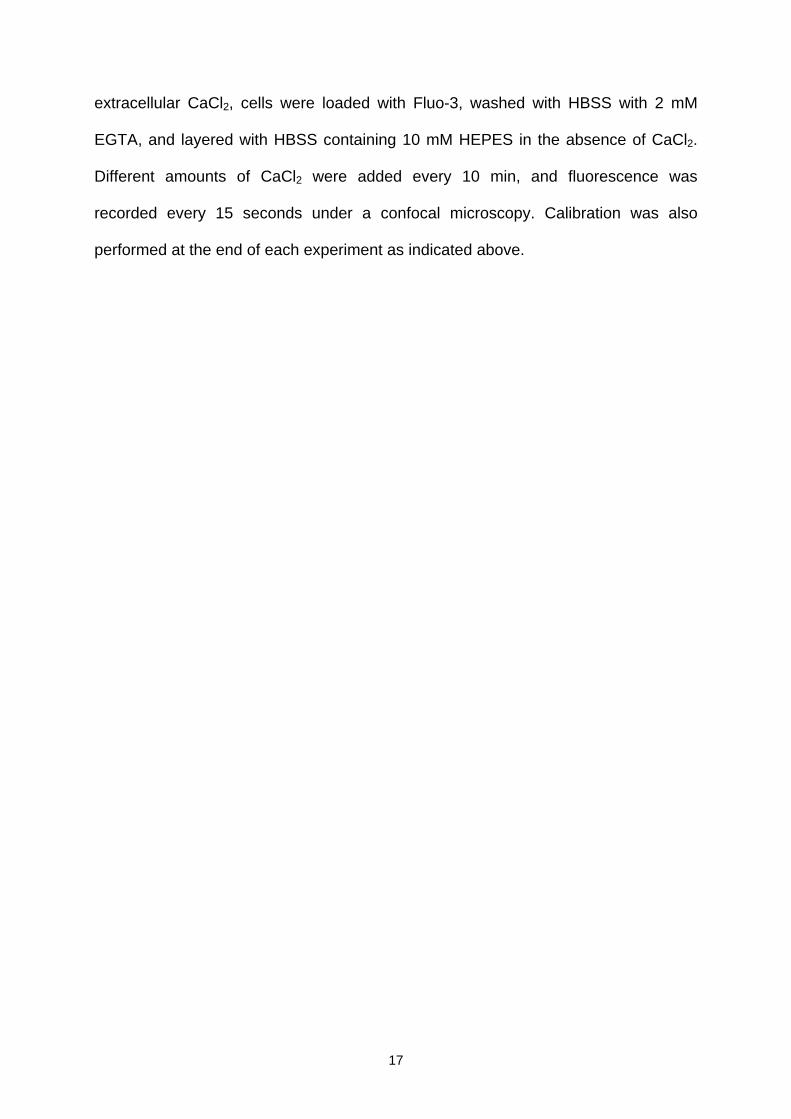
extracellular CaCl2, cells were loaded with Fluo-3, washed with HBSS with 2 mM
EGTA, and layered with HBSS containing 10 mM HEPES in the absence of CaCl2.
Different amounts of CaCl2 were added every 10 min, and fluorescence was
recorded every 15 seconds under a confocal microscopy. Calibration was also
performed at the end of each experiment as indicated above.
17

Acknowledgements
We thank Alberto Alvarez Barrientos (National Center for Cardiovascular Research,
Madrid) and Alberto Sánchez Guijo for expert technical help with the confocal
microscope. This work was supported by Grant BMC2001-2244 from the Spanish
Ministry of Science and Technology, Grants BFU2004-01886/BMC, SAF2004-04676,
and SAF2004-01232 from the Spanish Ministry of Education and Science, and Red
Brucella, Red Respira, and Red Temática de Investigación Cardiovascular, from
Instituto de Salud Carlos III. A.G.V. was supported by a predoctoral fellowship from
Instituto Carlos III (Becas de formación en investigación, BEFI 029034). M.A.G.'s
present address is: Department of Pharmacology, University of Colorado Health
Sciences Center, Mail Stop 8303, P.O. Box 6511, Aurora, CO 80045, USA. E-mail:
18

Abbreviations List
AA, arachidonic acid; cPLA2α, cytosolic Group IVA phospholipase A2; [Ca2+]I,
intracellular Ca2+ concentration; PtdIns, phosphatidylinositol bisphosphate.
19

References
Balsinde, J., Balboa, M. A., Li, W. H., Llopis, J. & Dennis, E. A. Cellular regulation of
cytosolic group IV phospholipase A2 by phosphatidylinositol bisphosphate levels. J.
Immunol. 164, 5398-5402 (2000).
Bittova, L., Sumandea, M. & Cho, W. A structure-function study of the C2 domain of
cytosolic phospholipase A2. Identification of essential calcium ligands and
hydrophobic membrane binding residues. J. Biol. Chem. 274, 9665-9672 (1999).
Bonventre J. V. Cytosolic phospholipase A2α reigns supreme in arthritis and bone
resorption. Tends Immunol. 25, 116-119 (2004).
Bonventre, J. V., et. al. Reduced fertility and postischaemic brain injury in mice
deficient in cytosolic phospholipase A2. Nature 390, 622-625 (1997).
Clark, J. D., L. L. Lin, R. W. Kriz, C. S. Ramesha, L. A. Sultzman, A. Y. Lin, N. Milona
& Knopf, J. D. A novel arachidonic acid-selective cytosolic PLA2 contains a Ca2+-
dependent translocation domain with homology to PKC and GAP. Cell 65, 1043-1051
(1991).
Das, S. & Cho W. Roles of catalytic domain residues in interfacial binding and
activation of group IV cytosolic phospholipase A2. J. Biol. Chem. 277, 23838-23846
(2002).
20
Dessen, A., J. Tang, H. Schmidt, M. Stahl, J. D. Clark, J. Seehra & W. S. Somers.
Crystal structure of human cytosolic phospholipase A2 reveals a novel topology and
catalytic mechanism. Cell 97, 349-360 (1999).
Di Virgilio, F., Lew, D. P. & Pozzan, T. Protein kinase C activation of physiological
processes in human neutrophils at vanishingly small cytosolic Ca2+ levels. Nature
310, 691-693 (1984).
Doughman, R. L., Firestone, A.J. & Anderson, R. Phosphatidylinositol phosphate
kinases put PI4,5P2 in its place. J. Membr. Biol. 194, 77-89 (2003).
Evans, J. H., Spencer, D. M., Zweifach, A. & Leslie C. C. Intracellular calcium signals
regulating cytosolic phospholipase A2 translocation to internal membranes. J. Biol.
Chem. 276, 30150-30160 (2001).
Gijón, M. A., Spencer, D. M., Kaiser, A. L. & Leslie, C. C. Role of phosphorylation
sites and the C2 domain in regulation of cytosolic phospholipase A2. J. Cell Biol. 145,
1219-1232 (1999).
Gijón, M. A., Spencer, D. M. & Leslie, C. C. Recent advances in the regulation of
cytosolic phospholipase A2. Adv. Enzyme Regul. 40, 255-68 (2000a).
Gijón, M. A., Spencer, D. M., Siddiqi, A. R., Bonventre, J. V. & Leslie, C. C. Cytosolic
phospholipase A2 is required for macrophage arachidonic acid release by agonists
that do and do not mobilize calcium. Novel role of mitogen-activated protein kinase
21

pathways in cytosolic phospholipase A2 regulation. J. Biol. Chem. 275, 20146-20156
(2000b).
Hirabayashi., T., Murayama, T. & Shimizu, T. Regulatory Mechanism and
Physiological Role of Cytosolic Phospholipase A2. Biol. Pharm. Bull. 27, 1168-1173
(2004).
Kao, J.P.Y. Harootunian A.T. & Tsien R.Y. Photochemically generated cytosolic
calcium pulses and their detection by Fluo-3. J. Biol. Chem. 264, 8179-8184 (1989).
Lemmon, M. A. Phosphoinositide recognition domains. Traffic 4, 201-213 (2003)
Leslie, C. C. & Channon, J. Y. Anionic phospholipids stimulate an arachidonyl-
hydrolyzing phospholipase A2 from macrophages and reduce the calcium
requirement for activity. Biochim. Biophys. Acta. 1045: 261-270 (1990).
Lin, L. L., M. Wartmann, A. Y. Lin, J. L. Knopf, A. Seth & R. Davis. cPLA2 is
phosphorylated and activated by MAP kinase. Cell 72, 269-278 (1993).
Mosior, M., Six, D. A. & Dennis, E. A. Group IV cytosolic phospholipase A2 binds with
high affinity and specificity to phosphatidylinositol 4,5-bisphosphate resulting in
dramatic increases in activity. J. Biol. Chem. 273, 2184-2191 (1998).
Nakatani, Y., Tanioka, T., Sunaga, S., Murakami, M. & Kudo, I. Identification of a
cellular protein that functionally interacts with the C2 domain of cytosolic
22

phospholipase A2α. J. Biol. Chem. 275,1161-8 (2000).
Ozaki, S., DeWald, D. B., Shope, J. C., Chen, J. & Prestwich, G. D. Intracellular
delivery of phosphoinositides and inositol phosphates using polyamine carriers. Proc.
Natl. Acad. Sci. U.S.A. 97, 11286-11291 (2000).
Pérez, R., Melero, R., Balboa, M. A. & Balsinde, J. Role of group VIA calcium-
independent phospholipase A2 in arachidonic acid release, phospholipid fatty acid
incorporation, and apoptosis in U937 cells responding to hydrogen peroxide. J. Biol.
Chem. 279, 40385-40391 (2004).
Perisic, O., Paterson, H. F., Mosedale, G., Lara-Gonzalez, S. & Williams, R. L.
Mapping the phospholipid-binding surface and translocation determinants of the C2
domain in regulation of cytosolic phospholipase A2. J. Biol. Chem. 274, 14979-14987
(1999).
Qiu, Z.-H., Gijón, M. A., de Carvalho, M. S., Spencer, D. M. & Leslie, C. C. The role
of calcium and phosphorylation of cytosolic phospholipase A2 in regulating
arachidonic acid release in macrophages. 273, 8203-8211 (1998).
Sheridan, A. M., Force, T., Yoon, H. J., O'Leary, E., Choukroun, G., Taheri, M. R., &
Bonventre, J. V. PLIP, a novel splice variant of Tip60, interacts with group IV
cytosolic phospholipase A2, induces apoptosis, and potentiates prostaglandin
production. Mol. Cell. Biol. 21, 4470-4481 (2001).
23

Shinohara, H., Balboa, M. A., Johnson, C. A., Balsinde, J. & Dennis, E. A. Regulation
of delayed prostaglandin production in activated P388D1 macrophages by group IV
cytosolic and group V secretory phospholipase A2s. J. Biol. Chem. 274, 12263-12268
(1999).
Six, D. A. & Dennis, E. A. The expanding superfamily of phospholipase A2 enzymes:
classification and characterization. Biochim. Biophys. Acta 1488, 1- 19 (2000).
Six, D. A. & Dennis, E. A. Essential Ca2+-independent Role of the Group IVA
Cytosolic Phospholipase A2 C2 Domain for Interfacial Activity. J. Biol. Chem. 278,
23842-23850 (2003).
Subramanian, P., Stahelin, R.V., Szulc, Z., Bielawska, A., Cho, W. & Chalfant, C.E.
Ceramide-1-phosphate acts as a positive allosteric activator of group IVA cytosolic
phospholipase A2α and enhances the interaction of the enzyme with
phosphatidylcholine. J. Biol. Chem. 280, 17601-17607 (2005).
Taylor, M. V., Metcalfe, J. C., Hesketh, T. R., Smith, G. A. & Moore, J. P. Mitogens
increase phosphorylation of phosphoinositides in thymocytes. Nature 312, 462-465
(1984).
Uozumi, N., et. al. Role of cytosolic phospholipase A2 in allergic response and
parturition. Nature 390, 618-622 (1997).
24

FIGURE LEGENDS
Figure 1. EGFP-cPLA2 translocation and AA release driven by TR-PIP2. EGFP-
cPLA2- (A, B, E) or EGFP- (C) transfected HEK cells were treated with TR-
PI(4,5)P2/histone (A, C), TR-PI(3,4)P2/histone (E), or carrier only (histone, B).
Pictures were taken at the indicated time points under the confocal microscope. D)
[3H]AA-labeled HEK cells were either untreated (open circles) or treated with TR-
PI(4,5)P2/carrier (closed circles), and [3H]AA release was measured at the indicated
time periods. Each experiment was performed at least three times with identical
results.
Figure 2. TR-PIP2 effects on RAW 264.7 cells. RAW 264.7 macrophages transfected
with EGFP-cPLA2 were treated with TR-PI(4,5)P2/histone for the indicated periods of
time and analyzed by confocal microscopy (A) or for [3H]AA release (B) as in Figure
1. Each experiment was performed at least three times with identical results.
Figure 3. Ca2+ is necessary for EGFP-cPLA2 translocation in response to TR-
PI(4,5)P2. EGFP-cPLA2-transfected HEK cells were treated with TR-PI(4,5)P2/histone
in HBSS medium without Ca2+, and pictures were taken under the confocal
microscope. After a 30-min treatment, 1.3 mM Ca2Cl was added to the incubation
medium (the two columns on the right hand side), and pictures were taken at the
indicated time points. Each experiment was performed at least three times with
identical results.
Figure 4. A intact Ca2+ binding domain is necessary for EGFP-cPLA2 translocation
in response to TR-PI(4,5)P2. HEK cells transfected with the mutant EGFP-D43N-
25

cPLA2 were treated with TR-PI(4,5)P2/histone in HBSS with 1.3 mM CaCl2 and
pictures were taken at the indicated time points by confocal microscopy. Each
experiment was performed at least three times with identical results.
Figure 5. EGFP-cPLA2 and TR-PI(4,5)P2 colocalization analysis. HEK cells
transfected with the construct EGFP-cPLA2 were treated with TR-PI(4,5)P2 for 20 min
in the presence (A) or absence (D) of extracellular calcium, and pictures of green and
red fluorescences were taken by confocal microscopy. The merge of both
fluorescences is also shown. LaserPix software analysis of co-localization
fluorescences in (A) is shown as a white mask (B), or as a colored three-dimensional
representation, where red means maximum level of colocalization (255) and dark
blue means the minimum (0) (C). Each experiment was performed at least three
times with identical results.
Figure 6. Intracellular Ca2+ levels in HEK cells. Intracellular measurements of Ca2+
were performed in Fluo-3-loaded HEK cells treated with TR-PI(4,5)P2/histone (A) or 5
μM ionomycin (B) in buffer containing 1.3 mM CaCl2. Arrows indicate the time point at
which the stimuli were added. Each data point represents the average of 20-40 cells.
Fluorescence of Fluo-3 was detected by microscopy and recorded every 15-30 s.
Each experiment was performed at least three times with identical results.
Figure 7. Intracellular Ca2+ requirements for TR-PI(4,5)P2-driven translocation of
EGFP-cPLA2. Fluo-3-loaded HEK cells were first treated with TR-PI(4,5)P2/histone
(A) or 5 μM ionomycin (B) in a buffer without Ca2+ and later, different CaCl2 amounts
were added as indicated. Fluorescence of Fluo-3 was detected by microscopy and
26

recorded every 15-30 s. Each data point represents the average of 20-40 cells.
Translocation of EGFP-cPLA2 was analyzed by confocal microscopy under the same
conditions indicated above for HEK cells (C, cells treated with TR-PI(4,5)P2/histone;
D cells treated with 5 mM ionomycin). Extracelular concentration of CaCl2 is
indicated. Each experiment was performed at least three times with identical results.
Figure 8. Analysis of the EGFP-4KE/A-cPLA2 mutant. (A) HEK cells transfected with
the mutant EGFP-4KE/A-cPLA2 were treated with TR-PI(4,5)P2/histone and pictures
were taken under the confocal microscope for the indicated time periods. (B) Co-
localization analyses (LaserPix software) of the fluorescence from EGFP-4KE/A-
cPLA2 mutant and TR-PI(4,5)P2 or EGFP-cPLA2 and TR-PI(4,5)P2 as a control, were
performed in the presence of absence of extracellular calcium, as indicated. Co-
localization index of red to green (black bars) or green to red (grey bars) is shown.
Each data barr represents the average of 10-20 cells. Experiments were performed
at least three times with identical results.
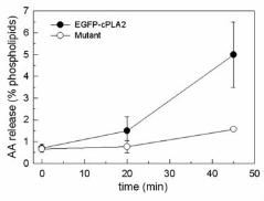
Figure 9. AA released by HEK cells transfected with the mutant EGFP-4KE/A-cPLA2.
[3H]AA-labeled HEK cells transfected with the EGFP-cPLA2 construct (closed circles)
or the mutant EGFP-4KE/A-cPLA2 (open circles) were treated with TR-
PI(4,5)P2/carrier and [3H]AA release was measured at the time indicated time points.
Each experiment was performed at least three times with identical results.
27


EGFP-cPLA2
a
TR-PIP2
0 mM 6 mM 15 mM 30 mM
0 mM 6 mM 15 mM 30 mM
c
d
0 mM 6 mM 15 mM 30 mM 60 mM 150 mM
b