Multiple roles of phosphoinositide-specific phospholipase C ...
20
BMB reports 415 http://bmbreports.org BMB reports *Corresponding author. Tel: 82-54-279-2293; Fax: 82-54-279-0645; E-mail: [email protected] Received 3 June 2008 Keywords: Alternative splicing variant, Phosphoinositide-specific phospholipase C, Signal transduction Multiple roles of phosphoinositide-specific phospholipase C isozymes Pann-Ghill Suh 1, * , Jae-Il Park 1 , Lucia Manzoli 2 , Lucio Cocco 2 , Joanna C. Peak 3 , Matilda Katan 3 , Kiyoko Fukami 4 , Tohru Kataoka 5 , Sanguk Yun 1 & Sung Ho Ryu 1 1 Division of Molecular and Life Sciences, Pohang University of Science and Technology, Pohang, Korea, 2 Cellular Signaling Laboratory, Department of Anatomical Sciences, University of Bologna, Via Irnerio, 48 I-40126, Bologna, Italy, 3 Cancer Research UK Centre for Cell and Molecular Biology, Chester Beatty Laboratories, The Institute of Cancer Research, Fulham Road, London SW3 6JB, UK, 4 Laboratory of Genome and Biosignal, Tokyo University of Pharmacy and Life Science, 1432-1 Horinouchi, Hachioji, 192-0392 Tokyo, Japan, 5 Division of Molecular Biology, Department of Biochemistry and Molecular Biology, Kobe University Graduate School of Medicine, Chuo-ku, Kobe, Japan Phosphoinositide-specific phospholipase C is an effector mole- cule in the signal transduction process. It generates two sec- ond messengers, inositol-1,4,5-trisphosphate and diacylglycer- ol from phosphatidylinositol 4,5-bisphosphate. Currently, thir- teen mammal PLC isozymes have been identified, and they are divided into six groups: PLC-β, -γ, -δ, -ε, -ζ and -η. Sequence analysis studies demonstrated that each isozyme has more than one alternative splicing variant. PLC isozymes contain the X and Y domains that are responsible for catalytic activity. Several other domains including the PH domain, the C2 do- main and EF hand motifs are involved in various biological functions of PLC isozymes as signaling proteins. The dis- tribution of PLC isozymes is tissue and organ specific. Recent studies on isolated cells and knockout mice depleted of PLC isozymes have revealed their distinct phenotypes. Given the specificity in distribution and cellular localization, it is clear that each PLC isozyme bears a unique function in the modu- lation of physiological responses. In this review, we discuss the structural organization, enzymatic properties and molec- ular diversity of PLC splicing variants and study functional and physiological roles of each isozyme. [BMB reports 2008; 41(6): 415-434] INTRODUCTION Phosphoinositide-specific phospholipase C (PLC) hydrolyzes phosphatidylinositol 4,5-bisphosphate (PIP2) to generate two second messengers: diacylglycerol (DAG) and inositol 1,4,5-tri- sphosphate (IP3). DAG and IP3 initiate further signal transduction pathways through activation of protein kinase C (PKC) and intra- cellular calcium release (1-3; Fig. 1a). The first evidence of PLC activity was suggested by Hokin et al. in 1953 who reported specific hydrolysis of phospholipids in pigeon’s pancreas slices after cholinergic stimulation (4). The authors showed that the enhanced turnover of phosphor- ylinositol groups of phosphatidylinositol occurred in cells as a response to a variety of stimuli. In 1983, Streb et al. demon- strated that IP3 generated from PIP2 hydrolysis is responsible for mobilization of intracellular calcium in the pancreatic aci- nar cells (5). Takenawa et al. purified the first PLC with a mo- lecular weight of 68 kDa in 1981 (6). Subsequently, a number of PLCs of different molecular masses, isoelectric points and calcium dependency have been identified in several tissues. In the late 80 s, three PLC isozymes, namely PLC-γ, -β, and -δ, were isolated and their cDNA sequences were obtained (7). Since then, multiple types of PLC were found from various tis- sues using either an RT-PCR method by specifically designed primers or a screening method using low stringency hybrid- ization with probes made from the conserved domain X or Y. So far, 13 PLC isozymes have been identified in different mammalian tissues and they belong to six different subtypes. Identification of PLC isozymes, PLC-ε and PLC-η, has changed our understanding of the relations between receptor tyrosine kinase (RTK)-PLC-γ and G protein-coupled receptor (GPCR)- PLC-β (Fig. 1b). Little is known about functions, rela- tions and hierarchy of PLC isozymes. In this review, we discuss the questions of structural organization, enzymatic properties and molecular diversity of splicing variants of PLC isozymes in relation to their function and physiological significance. Structure of PLCs Catalytic X and Y domains Two highly conserved amino acid regions in PLC isozymes are the X and Y domains, which are located between EF-hand mo- tifs and C2 domain. The domains are composed of alternating α-helices and β-strands and resemble incomplete triose phos- phate isomerase (TIM) α /β-barrel (8). Crystallographic analysis Mini Review
-
Upload
khangminh22 -
Category
Documents
-
view
4 -
download
0
Transcript of Multiple roles of phosphoinositide-specific phospholipase C ...

BMB reports
415http://bmbreports.org BMB reports
*Corresponding author. Tel: 82-54-279-2293; Fax: 82-54-279-0645; E-mail: [email protected]
Received 3 June 2008
Keywords: Alternative splicing variant, Phosphoinositide-specificphospholipase C, Signal transduction
Multiple roles of phosphoinositide-specific phospholipase C isozymesPann-Ghill Suh1,*, Jae-Il Park1, Lucia Manzoli2, Lucio Cocco2, Joanna C. Peak3, Matilda Katan3, Kiyoko Fukami4, Tohru Kataoka5, Sanguk Yun1 & Sung Ho Ryu1
1Division of Molecular and Life Sciences, Pohang University of Science and Technology, Pohang, Korea, 2Cellular Signaling Laboratory, Department of Anatomical Sciences, University of Bologna, Via Irnerio, 48 I-40126, Bologna, Italy, 3Cancer Research UK Centre for Cell and Molecular Biology, Chester Beatty Laboratories, The Institute of Cancer Research, Fulham Road, London SW3 6JB, UK, 4Laboratory of Genome and Biosignal, Tokyo University of Pharmacy and Life Science, 1432-1 Horinouchi, Hachioji, 192-0392 Tokyo, Japan, 5Division of Molecular Biology, Department of Biochemistry and Molecular Biology, Kobe University Graduate School of Medicine, Chuo-ku, Kobe, Japan
Phosphoinositide-specific phospholipase C is an effector mole-cule in the signal transduction process. It generates two sec-ond messengers, inositol-1,4,5-trisphosphate and diacylglycer-ol from phosphatidylinositol 4,5-bisphosphate. Currently, thir-teen mammal PLC isozymes have been identified, and they are divided into six groups: PLC-β, -γ, -δ, -ε, -ζ and -η. Sequence analysis studies demonstrated that each isozyme has more than one alternative splicing variant. PLC isozymes contain the X and Y domains that are responsible for catalytic activity. Several other domains including the PH domain, the C2 do-main and EF hand motifs are involved in various biological functions of PLC isozymes as signaling proteins. The dis-tribution of PLC isozymes is tissue and organ specific. Recent studies on isolated cells and knockout mice depleted of PLC isozymes have revealed their distinct phenotypes. Given the specificity in distribution and cellular localization, it is clear that each PLC isozyme bears a unique function in the modu-lation of physiological responses. In this review, we discuss the structural organization, enzymatic properties and molec-ular diversity of PLC splicing variants and study functional and physiological roles of each isozyme. [BMB reports 2008; 41(6): 415-434]
INTRODUCTION
Phosphoinositide-specific phospholipase C (PLC) hydrolyzes phosphatidylinositol 4,5-bisphosphate (PIP2) to generate two second messengers: diacylglycerol (DAG) and inositol 1,4,5-tri-sphosphate (IP3). DAG and IP3 initiate further signal transduction pathways through activation of protein kinase C (PKC) and intra-
cellular calcium release (1-3; Fig. 1a). The first evidence of PLC activity was suggested by Hokin et al. in 1953 who reported specific hydrolysis of phospholipids in pigeon’s pancreas slices after cholinergic stimulation (4). The authors showed that the enhanced turnover of phosphor-ylinositol groups of phosphatidylinositol occurred in cells as a response to a variety of stimuli. In 1983, Streb et al. demon-strated that IP3 generated from PIP2 hydrolysis is responsible for mobilization of intracellular calcium in the pancreatic aci-nar cells (5). Takenawa et al. purified the first PLC with a mo-lecular weight of 68 kDa in 1981 (6). Subsequently, a number of PLCs of different molecular masses, isoelectric points and calcium dependency have been identified in several tissues. In the late 80 s, three PLC isozymes, namely PLC-γ, -β, and -δ, were isolated and their cDNA sequences were obtained (7). Since then, multiple types of PLC were found from various tis-sues using either an RT-PCR method by specifically designed primers or a screening method using low stringency hybrid-ization with probes made from the conserved domain X or Y. So far, 13 PLC isozymes have been identified in different mammalian tissues and they belong to six different subtypes. Identification of PLC isozymes, PLC-ε and PLC-η, has changed our understanding of the relations between receptor tyrosine kinase (RTK)-PLC-γ and G protein-coupled receptor (GPCR)- PLC-β (Fig. 1b). Little is known about functions, rela-tions and hierarchy of PLC isozymes. In this review, we discuss the questions of structural organization, enzymatic properties and molecular diversity of splicing variants of PLC isozymes in relation to their function and physiological significance.
Structure of PLCs
Catalytic X and Y domainsTwo highly conserved amino acid regions in PLC isozymes are the X and Y domains, which are located between EF-hand mo-tifs and C2 domain. The domains are composed of alternating α-helices and β-strands and resemble incomplete triose phos-phate isomerase (TIM) α /β-barrel (8). Crystallographic analysis
Mini Review
PLC isozymes as a key molecule of cellular functionsPann-Ghill Suh, et al.
416 BMB reports http://bmbreports.org
Fig. 1. Activation and function of various PLC isozymes. A) Phos-pholipase-dependent PIP2 hydrolysis. PLC catalyzes the hydrolysis of PIP2, a member of membrane phospholipids. Two products, diac-ylglycerol (DAG) and IP3 induce the activation of PKC and intra-cellular Ca2+ release, respectively. B) PLC related signaling pathway. Various extracellular signals stimulate hydrolysis of phosphatidylino-sitol 4,5-bisphosphate (PIP2) by phosphoinositide-specific phospholi-pase C (PLC). PLC hydrolyzes PIP2 to two important second mes-sengers, diacylglycerol and inositol 1,4,5-trisphosphate (IP3). These two second messengers lead to the activation of several protein kin-ase C (PKC) isoforms and the release of calcium from intracellular stores, respectively. PLC-β subtypes are activated by GPCR through several mechanisms. Pertusis toxin (PTX)-insensitive heterotrimericGq family proteins (Gq, G11, G14, G15 and G16) activate PLC-β sub-types via GTP-loaded Gα subunits. PLC-β subtypes are also acti-vated by Gβγ subunits liberated from PTX-sensitive Gi family proteins.Various growth factors such as platelet-derived growth factor (PDGF), epidermal growth factor (EGF), and nerve growth factor (NGF) canactivate PLC-γ isozymes. Upon growth factor stimulation, PLC-γ is recruited to activated growth factor receptors via SH2 domain-phos-photyrosine interaction and then subjected to phosphorylation by re-ceptor tyrosine kinases. PLC-ε can be activated by both GPCR and RTK activation with distinct activation mechanism. The activation mechanism for PLC-δ, η, ζ remains to be revealed.
revealed that histidine residue functions as a general base lead-ing to formation of 1,2-cyclic inositol 4,5-bisphosphate (9). Ca2+ ions are required for lowering the pKa and promoting the nucleophilic attack on 1-phosphate. The following stabilization of the cyclic phosphate intermediate at the active site produces acyclic product (10). Catalytic residues are highly conserved in all eukaryotic PLC isozymes. Based on structural analysis of PLC-δ1, it has been suggested that Lys438, Lys440, Ser522 and Arg549 residues are present at the active site which are im-
plicated in interactions with 4- or 5-phosphate of the substrate headgroup (9, 11). Preferential hydrolysis of PIP2 has been at-tributed to a positive charge at 549 position of PLC-δ1, which is conserved in the eukaryotic PLC catalytic core, This finding is supported by the fact that replacement of a charged residue causes suppression of PIP2 hydrolysis but not of PI (12).
PH domainIt has been shown that the PH domain in PLC-δ1 binds PIP2 and advances the access of PLC-δ1 onto the membrane surface (13). The PH domain of PLC-β2 and PLC-β3 binds specifically to the heterotrimeric G protein subunit, Gβγ (14). The domain also mediates interactions with PIP3, which is required for PI3K-dependent PLC-γ1 translocation and activation (15). PLC-γ1 and γ2 contain an additional PH domain which is split by two tandem Src homology 2 (SH2) and a Src homology 3 (SH3) domains. The C-terminal half of the PLC-γ split PH do-main has been implicated to interact directly with the TRPC3 calcium channel, thereby providing a direct coupling mecha-nism between PLC-γ and agonist-induced calcium entry (16).
C2 domain and EF-hands The C2 domain of PLC-δ1 possesses three to four Ca2+ binding sites (8). Calcium ions bind to the C2 domain and enhance the enzymatic activity of PLC-δ1 by forming an enzyme-phosphati-dylserine-Ca2+ ternary complex. EF-hand motifs are helix-turn- helix structural domains which bind Ca2+ ions. It was shown that the deletion of EF-hand motifs of PLC-δ1 resulted in a de-crease in PLC activity in a Ca2+-independent manner (17, 18). Although the EF-hand motif of PLC isozyme may have an im-portant regulatory function, currently there is no strong evidence to support the notion that EF-hand motifs bind to metal ions.
Tissue distribution of PLCs
Thirteen mammalian PLC isozymes have been identified, so far in various tissues and cell types. Analysis of expressed se-quence tags (EST) allows a systematic search of gene expression. We used EST database in NCBI Unigene, (http://www.ncbi.nlm. nih.gov/sites/entrez?db=unigene) to study the tissue distri-bution of PLC isozymes and to analyze their mRNA expression. We found that PLC-β1 is highly expressed in the cerebral cor-tex and hippocampus (19) compare to limited expression of PLC-β2 in hematopoietic cells (20, 21); PLC-β3 is found in the brain, liver, and parotid gland (22); PLC-β4 is present at the highest level in the cerebellum and retina (23-25). Our results showed that the highest EST score for PLC-β isozymes was found in the brain (~ 20-44% of analyzed EST sequences) (Table 1). EST sequences of PLC-β2 have greater value in blood and the bone marrow compare to other tissues. Two mammalian subtypes of PLC-γ isozymes have been identified. PLC-γ1 mRNA is widely detected in various tissues. It is abundantly expressed in embryonal cortical structures, neu-rons, oligodendrocytes and astrocytes (26) Unlike PLC-γ1,

PLC isozymes as a key molecule of cellular functionsPann-Ghill Suh, et al.
417http://bmbreports.org BMB reports
Table 1. Tissue-specific expression of PLCs as deduced from human UniGene database
PLC-γ2 mRNA is expressed in the limited areas of anterior pitui-tary and cerebellar Purkinje and granule cells (27). The ex-pression of PLC-γ2 is primarily limited to cells of haemato-poietic lineage. Our data search results revealed that the PLC-γ1 EST number is high in the brain and embryonic tissues, while the highest PLC-γ2 EST number is observed in lymph nodes. The observed expression patterns can serve as an explanation to the predominant role of PLC-γ1 in embryonic cell development and the role of PLC-γ2 in immune response (28-30). PLC-δ1 is present in high abundancy in the brain, heart, lung, skeletal muscle and testis (31). EST sequences analysis showed that PLC-δ1 isozyme is found at high levels in the brain, lung, reproductive organs, thymus and connective tissue (Table 1). PLC-δ3 is detected abundantly in brain, skeletal muscle and heart (32). EST sequences for PLC-δ3 isozyme are mainly represented in brain, lung, pancreas, and reproductive organ (Table 1). PLC-δ4 mRNA is expressed in various tissues with the highest levels detected selectively in the brain, skel-etal muscle, testis and kidney (33).
PLC-ε mRNA expression has been detected in various tis-sues, including brain, lung, and colon, with the highest ex-pression detected in heart (34). Two splicing variants of PLC-ε have been reported, i.e., PLC-ε1a and PLC-ε1b. They have dif-ferent sequences at the N-terminus that precedes the CDC25 domain. PLC-ε1a transcripts are expressed in various tissues ex-cept in peripheral blood leukocytes. PLC-ε1b mRNA has a lim-ited expression in the placenta, lung and spleen. EST sequences of PLC-ε are dominant in connective tissues and brain. Northern blot analysis of mouse tissues and EST sequences analysis using human UniGene database showed that PLC-ζ is only found in the testis (35). However, PLC-ζ transcripts are detected in spermatid cDNA but not in testis cDNA in the ab-sence of spermatid, suggesting that the PLC-ζ expression with-in the testis is sperm-specific. Two PLC-η isozymes, PLC-η1 and PLC-η2, were identified in human and mouse. The highest level of PLC-η1 mRNA is observed in the brain and kidney and smaller levels are de-tected in the lung, spleen, intestine, thymus and pancreas (36).

PLC isozymes as a key molecule of cellular functionsPann-Ghill Suh, et al.
418 BMB reports http://bmbreports.org
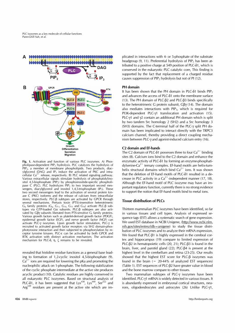
Fig. 2. Schematic diagram of alternative splicing variants for PLC isozymes in mouse. Schematic representing shows the PH domain, EF-hand motifs, catalytic X and Y domain, and C2 domain for the functional domains in all PLC isozymes. The total numbers of amino acid residues and GeneBank accession numbers for each splicing variant are written next to the diagram. The region of alternatively splic-ing variants for PLC isozyme, which differ in their amoino acid residues, is indicated by a square with yellow and organge color. Insertion of addtional exon is indicated by red color. The position of alternatively splicing residue and insertion of additional residue at each PLC isozyme are labelled with arabic numerals in blue color. All of PLC-β transcripts are found to have two alternative splicing var-iants, only differing in C-terminal sequences. Interestingly, in the mouse PLC-β2 gene, we found an alternative splicing variant form, con-taining the truncated C-terminal region. Like alternative splicing variants of PLC-β, two different transcripts in C-terminal regions of PLC-γ1 were identified in mouse. There are more than one additional transcripts found in PLC-δ isozyme in mouse. Surprisingly, mouse PLC-δ1b differs from PLC-δ1a by truncation of catalytic Y domain and C2 domain. Two and three alternative splicing variants of PLC-δ3 and -δ4 exist in mouse, respectively. Mouse PLC-ε isozyme has two novel splicing variants at different region. Mouse PLC-ε1c lacks Ras-GEF do-main and partial PH domain at N-terminus. In sperm-specific PLC-ζ, deduced amino acid sequences at C-terminal region differ from that of PLC-ζ in mouse. We found a novel splicing variant of mouse PLC-η1, excluding 20 amino acid residues in C-terminal region (we de-signed it as PLC-η1d). In addition, we found the three different splicing variants of PLC-η2 in mouse. In PLC-γ isozymes, two SH2, SH3 and split PH domain are indicated by green, pink and purple color, respectively. Protein GeneBank accession number of alternative splic-ing variant for PLC is inserted in a bracket (*1: 28aa missing from methionine start site; *2: Nucleotide GeneBank accession number; *3: Exon1 and Exon2 missing; *4: 21aa missing from methionine start site).
PLC-η2 mRNA is detected in the brain and intestine (37, 38).
Diverse splicing variants of PLCs
Most genes express a limited number of mRNA isoforms. But there are a number of genes that use alternative splicing to generate several numbers of isoforms. Alternative splicing var-iants have been previously reported for several of PLC iso-zymes including rat PLC-β1 (39), human PLC-β2 (40), rat PLC-β4 (41), rat PLC-δ4 (33), and human PLC-ε (42). Here, we analyzed and identified additional splicing variants for PLC isozyme using BLAST server in mouse species. Subsequently additional splicing variants for PLC isozyme were verified in exon-intron sequences from genomic sequences using UCSC Genome Bioinformatics web server (http://genome.ucsc.edu/
cgi-bin/hgBlat). Two splicing variants of the PLC-β1 isozyme derived from a single gene have been identified in human and rat that differ in their C-terminal sequences (39, 43). The mouse PLC-β1 gene possesses two alternative splicing variants. PLC-β1a contains putative NLS (nuclear localization sequences) and NES (nuclear export sequences) regions. Two variants of PLC-β isozyme dif-fer in their cellular localization, suggesting that this region reg-ulates the transit in and out of the nucleus (39). Two splicing variants of human PLC-β2 were reported; PLC-β2a and PLC-β2b. They differ in 15 amino acid residues at the C-termi-nal region as was shown in haematopoietic cells (21, 40). In the case of mouse PLC-β2 gene, we have found a splicing var-iant, which has truncated the entire C-terminal region. The al-ternative splicing variant of the mouse PLC-β3 gene showed

PLC isozymes as a key molecule of cellular functionsPann-Ghill Suh, et al.
419http://bmbreports.org BMB reports
that it contains the internal stop codon between 3039 and 3040 open reading frame in the wild PLC-β3 gene. Several al-terative splicing variants of PLC-β4 have been reported. We found and identified two alternative splicing variants from rat and bovine brain (24, 41, 44). The third splicing variant of rat PLC-β4 has an additional 37 nucleotide exon at the C-terminal region (23). All PLC-β genes have two alternative splicing var-iants, which differ in their C-terminal sequences (Fig. 2). Two alternative splicing variants of PLC-γ1 were found to dif-fer in their C-terminal sequences. The distinct function of the two alternative splicing variants still remains unknown. Only one transcript of the PLC-γ2 gene was identified in the mouse (Fig. 2). Alternative splicing variants of PLC-δ isozymes represent several different patterns of splicing variants unlike those of PLC-β isozymes. Mouse PLC-δ1b differs from PLC-δ1a by 274 amino acid residues that extend from the catalytic Y domain to the stop sequence, which are replaced with 21 distinct amino acid residues. Mouse PLC-δ1b has a truncated catalytic Y do-main, which implies that this variant may have no enzymatic activity. PRIP-1 (PLC-related inactive protein-1) has a high se-quence similarity to PLC-δ1 but has no PIP2-hydrolyzing activity. PRIP-1 regulates IP3-mediated Ca2+ signaling by bind-ing of IP3 (45). It would be interesting to investigate whether PLC-δ1b can also have such a regulatory function in PLC signaling. The second splicing variant of PLC-δ3 demonstrates the deletion of 51 nucleotides between the PH domain and EF-hand motifs. There are three alternative splicing variants of mouse PLC-δ4 (Fig. 2). PLC-δ4a contains additional 32 amino acid residues in the linker region between the catalytic X and Y domains. The earlier study reported that all of the above three splice variants have the catalytic X domain and/or the linker re-gion for X and Y domains (46). We found an additional splicing variant of PLC-δ4b that is different from the earlier reported PLC-δ4 isoforms at the C-terminal region. Two splicing variants of human PLC-ε with a different N-ter-minal region have been reported (42), but mouse PLC-ε iso-zyme was found to have two novel splicing variants in a differ-ent region. Surprisingly, mouse PLC-ε1c lacks the Ras-GEF do-main and partial PH domain at the N-terminus. Additionally, we searched the mouse EST database and identified one EST clone (EST clone; CJ197436) that included partial sequences for N-terminus of PLC-ε1c. s-PLC-ζ, an alternative splicing variant for PLC-ζ has been reported (47). It contains two internal stop codons at the N-ter-minus and lacks one and a half of EF-hand motifs. It was re-ported that this splicing variant of PLC-ζ does not affect Ca2+ oscillations. Moreover, we found an additional splicing variant of PLC-ζ, which has the C-terminal sequence different from that of s-PLC-ζ. Three splicing variants of PLC-η1 have been reported in hu-man and mouse (36). We found a novel splicing variant of mouse PLC-η1. It excludes 20 amino acid residues in the C-ter-minal region (designated as PLC-η1d; Fig. 2). Five alternative
splicing variants of PLC-η2 exist in human (37), and three splic-ing variants of PLC-η2 have been found in mouse. PLC-η2b and PLC-η2c contain additional 74 amino acid residues at N-terminal region. Moreover, splicing variant of PLC-η2c differ from the other PLC-η2 subtypes at the C-terminal region.
Molecular function and regulation of PLCs
PLC-β isozymes
Regulation of PLC-β subtype activity: PLC-β is a member of a large family of PLC enzymes and includes PLC-β1~4. PLC-β isozymes share many of the structural features present in other members of the PLC family, including conserved catalytic X and Y domains as well as two membrane-phospholipid bind-ing regions, the PH and C2 domains. PLC-β isozymes, how-ever, are distinguished by the presence of an elongated C-ter-minus consisting of about ~450 residues, which contains many of the determinants for interaction with Gq as well as for other functions such as membrane binding and nuclear local-ization (3, 48-50). Four PLC-β isotypes and additional splicing variants have been identified in mammals. These isoforms are regulated by heterotrimeric GTP-binding proteins, and have a high GTPase stimulating (GAP) activity. Mammalian PLC-β isozymes are dif-ferentially distributed in tissues, with the PLC-β1 being most widely expressed, especially in specific regions of the brain. PLC-β1 exists as alternative splicing variants PLC-β1a and -β1b, which differ in their C-terminal residues (39). The pri-mary transcript of PLC-β4 gene is also alternatively spliced in the region corresponding to the C-terminal region of the pro-tein downstream of the C2 domain (41). In the cytoplasm, PLC-β functions as effector enzymes for receptors belonging to the rhodopsin superfamily of transmembrane proteins that con-tain seven transmembrane spanning segments. They are acti-vated by a variety of stimuli and require special combinations of Gα and Gβ /γ subunits to couple to the effector (3). Activation could be also induced by phosphatidic acid (PA) in that recent studies suggest a novel role for PA in the regulation of GPCR signal transduction that is mediated through PLC β1 homodimerization (51). With the exception of PLC-β4, PLC-β isozymes are also activated by Gβγ dimmers (52-55). The rela-tive sensitivity of PLC-β isozymes to Gβγ subunits differs from that to Gqα subunits; PLC-β1 is the least sensitive to Gβγ (52, 53). Direct binding measurements indicate that, although the Gβγ dimer interacts with PLC-β1, -β2, and -β3, it exhibits a high affinity only for PLC-β2 (56). It has been shown that a variety of physiological stimuli that activate PLC-β isozymes in normal platelets were not able to activate these isozymes in platelets derived from αq knockout mice; αq is the only member of the Gqα subfamily expressed in normal platelets (57). Thus, Gαq appears essential for PLC-β activation and cannot be replaced by Gβγ in this regard. The region of PLC β that interacts with Gqα subunits differs from

PLC isozymes as a key molecule of cellular functionsPann-Ghill Suh, et al.
420 BMB reports http://bmbreports.org
that responsible for interaction with Gβγ. Thus, C-terminal truncation of PLC-β2 generated enzymes that were activated by Gβγ but not by Gαq (58). The PH domain of PLC-β2 exhibits high affinity for Gβγ subunits bound to membranes (59). Experi-ments with antisense oligonucleotides directed to mRNAs en-coding various G protein subunits suggested that the m1 mus-carinic acetylcholine receptor interacts only with G protein complexes composed of the subunits αq, α11, β1, β4, and γ 4 in order to activate PLC in RBL-2H3 cells, despite the fact that the subunits α14, β2, β3, γ2, γ3, γ5, and γ7 are also expressed in these cells (60). G protein–initiated signaling is turned off by hydrolysis of the GTP bound to the Gα subunit, a reaction cat-alyzed by the intrinsic GTPase activity of the α subunit itself, and the subsequent reassociation of the GDP-bound α subunit with the βγ subunits. This deactivation process was studied in detail by reconstituting the m1 muscarinic acetylcholine re-ceptor, G protein, and PLC-β1 in lipid vesicles (61).
PLC-β1 isozyme present the nucleus is located in nuclear speckles: PLC signaling occurs not only at the plasma mem-brane but also in the nucleus (62, 63). PLC-β1 is the major PLC isozyme in the nucleus of various cells (64, 65), and the C-terminal region of the protein is required for nuclear local-ization, even though also PLC-β2, -β3 and -β4 have been shown to be in this organelle (66). Where is located PLC-β1 located in the nucleus? PLC-β1 has been reported to localize to nuclear speckles, together with PIP kinases, PIP2, diacylglycerol kinase θ (DGK θ), PLC-δ4, PI 3-Kinase C2α, phosphatase and tension homologue deleted on chromosome 10 (PTEN) and SH2-domain containing inositol phosphatase 2 (SHIP2) (67-69). It has been demonstrated an as-sociation between PLC-β1 and both DGK ζ and PIP Kinase α, in immunoprecipitation experiments with a PLC-β1 specific antibody. With immuno-electron microscopy we also showed that DGK θ, PLC-β1 and PIP2 are associated to electron-dense particles within the nucleus, that correspond to nuclear speck-les as revealed by using the antibody against SC-35 (70).
PLC-β1 in hematological malignancies: The involvement of PLC-β1 in hematopoietic differentiation, i.e. affecting CD24 expression (71) prompted us to investigate the role of this sig-naling molecule in hematological malignancies, focusing par-ticularly on patients affected by myelodysplastic syndromes (MDS) at higher risk of evolution into acute myeloid leukemia (AML). By using fluorescence in situ hybridization (FISH) anal-ysis, our group demonstrated, in a small number of high-risk MDS patients (72), that subjects bearing a mono-allelic cryptic deletion of the PLC-β1 gene had a worse clinical outcome, as compared with patients having both alleles. In a subsequent study, it has also been shown that the expression profile of both PLC-β1a and PLC-β1b mRNAs, which are the two alter-native splicing subtype of PLC-β1, is altered in high-risk MDS, as compared to healthy donors. Interestingly, MDS cells al-ways expressed higher levels of PLC-β1b mRNA as compared
to PLC-β1a mRNA; this difference may reflect a specific role of PLC-β1 in MDS, given that the PLC-β1a splicing subtype dem-onstrates both nuclear and cytoplasmatic localization, while PLC-β1b splicing subtype is localized only in the nucleus. Furthermore, our recent studies demonstrated that not only is Akt specifically phosphorylated in high-risk MDS patients, but also mTOR and its downstream targets are activated (73), af-fecting cell survival and proliferation of MDS cells.
PLC-β isozyme expression and the epigenetic effect of DNA methyltransferase inhibition: Interestingly, it has been shown that a DNA methyltransferase inhibitor currently approved for the treatment of MDS and under experimental evaluation for other hematological malignancies, i.e. azacitidine, also affects PLC-β1 expression (74). Besides showing response rates of 50–80% in MDS, azacitidine has been reported to have a sig-nificant impact on the quality of life and progression into AML (75). Nevertheless, the molecular mechanisms underlying this drug are not completely understood. Low-dose regimens with azacitidine have been assumed to act by reversing the epi-genetic silencing of target genes involved in the control of cell growth and differentiation. However, although demethylation of a hypermethylated p15/ INK4B gene has been demonstrated in MDS patients treated with demethylating therapy (76), this observation does not necessarily mean that this is the main mechanism of action of the drug in vivo. Moreover, since p15 is unlikely to be the only hypermethylated and silenced gene in MDS, it was important to examine the presence of other tar-gets for demethylating treatment. We have shown that in pa-tients diagnosed with MDS who was treated with azacitidine a correlation of PLC-β1 expression with an almost complete re-mission (77). In particular a patient after reaching a partial hematologic remission, following the International Working Group (IWG) response criteria (78) and temporarily decreased hematological response, subsequently restored, finally reached a complete remission. During the therapy, the patient demon-strated an increase in PLC-β1 expression, as well as a down-regulation in the level of activated Akt, postulating an associa-tion between PLC β1 expression and azacitidine effectiveness. All in all these observations hint at the likelihood that nuclear PLC-β1 is a good candidate for both MDS prognosis and epi-genetic effect of antileukemic drugs. Indeed the fact that PLC-β1 could at the nucleus in a peculiar way, different from that at the plasma membrane, i.e. as a check point for the G1 phase of the cell cycle, is strengthened by the results showing that nuclear PLC-β1 activity is switched on and off by phos-phorylative events and targets cyclin D3 during both cell growth and differentiation (79-81).
Functions of PLC-β isozyme in mice: PLC-β1 null mice showed the epileptic seizures and this led to the sudden death of the mice. This result suggests that PLC-β1 is essential for the normal functioning of inhibitory neuronal circuitry (82). Chemoattrac-tant-mediated PLC activity was impaired in the neutrophills iso-

PLC isozymes as a key molecule of cellular functionsPann-Ghill Suh, et al.
421http://bmbreports.org BMB reports
lated from PLC-β2-deficient mice. This led to the decreased in-tracellular calcium release, superoxide production and cell sur-face expression of MAC-1 (83). PLC-β3-null mice demonstrated increased sensitivity to morphine compared with the wild type mice, which indicates that PLC-β3 is a significant negative mod-ulator for opioid response (84). The mice lacking PLC-β4 showed the ataxia and impaired visual processing, which are results of impaired PLC-linked signal transduction (82, 85).
PLC-γ isozymes
Regulation of PLC-γ isozyme activity: Two mammalian PLCγ forms (PLC-γ1 and PLC-γ2) have been identified. As described in previous sections, these enzymes are characterised by the in-sertion of a highly structured region (PLC-γ-specific array, γSA) comprising a split PH domain flanking two tandem SH2 do-mains and SH3 domain between the two halves of the TIM-bar-rel catalytic domain (86, 87). This region has been implicated in activation of PLC-γ isozymes downstream of receptors with intrinsic or associated tyrosine kinase activity. Although not fully understood, regulatory mechanisms are best characterised for PLC-γ1 activated in response to polypeptide growth factors that bind to receptor tyrosine kinases (RTKs) such as the epi-dermal growth factor receptor (EGFR) and platelet-derived growth factor receptor (PDGFR) (88, 89). The SH2 domains of PLC-γ1 mediate binding to autophosphorylated tyrosine resi-dues within the intracellular region of the receptor; this associa-tion is important for both membrane recruitment and tyrosine phosphorylation of PLC-γ1. Growth factor stimulation, such as stimulation by PDGF, induces PLC-γ1 phosphorylation at three tyrosine residues: Tyr771 and Tyr783 within the γ-specific region and Tyr1254 within the C-terminal tail; however, a recent study has shown that only phosphorylation at Tyr783 is essential for li-pase activation (90). This has been attributed to the intra-mo-lecular binding of phosphorylated Tyr783 to the C-terminal SH2 domain and further speculated that this interaction induces a conformational change required for activation (91). PLC-γ1 and PLC-γ2 can also be activated downstream of a va-riety of receptors that lack intrinsic tyrosine kinase activity. For PLC-γ1 this has been reported for receptors including GPCRs such as the angiotensin II and bradykinin receptors, cytokine re-ceptors and immunoreceptors such as the T cell receptor (92-95). PLC-γ2 is activated downstream of immunoglobulin and adhe-sion receptors on immune cells such as B cells, platelets and mast cells (96-98). In these instances, PLC-γ isozymes are phos-phorylated by non-receptor tyrosine kinases in the context of membrane-localised multimolecular signalling complexes. These assemblies have been well characterised in the context of anti-gen receptor signalling on B and T cells and include kinases of the Src, Syk and Tec families in addition to adaptor molecules such as BLNK and LAT that serve to recruit the different compo-nents to the complex (99). In tumour cells and some non-neo-plastic cells, PLC-γ1 interacts with another important adaptor protein GIT1 (GPCR kinase-interacting protein-1) (100-102).
This interaction facilitates Src kinase-mediated PLCγ1 activation downstream of GPCRs, RTKs and integrins and thereby could integrate signal transduction inputs emanating from these differ-ent classes of receptor (100-102). The multi-domain structure of PLC-γ1 permits interactions with a whole host of additional binding partners. The SH2 do-mains, SH3 domain and split PH domain have all been im-plicated in various protein/protein interactions. Some of these interactions, including EF-1α and the inositol 5’-phosphatase, SHIP1, enhance PLC-γ1’s activity whereas others such as Cbl and Grb2 serve as negative regulators of the enzyme (103-106). Localization and activation of PLC-γ isozymes at the cell pe-riphery are also governed by direct interactions with F-actin and are therefore subject to changes in the architecture and functioning of the cytoskeleton (107-110).
Function of PLC-γ isozyme in mice: The expression patterns of PLC-γ1 and PLC-γ2 show some degree of overlap; however, even in regions of co-expression, the two isoforms appear to perform independent, non-redundant functions (111). PLC-γ2 is most highly expressed in cells of the haematopoietic system and plays a key role in regulation of the immune response (19, 112). Consistent with this, PLC-γ2 null mice display defects in the functioning of B cells, platelets, mast cells and natural killer cells (29, 30). Loss of PLC-γ2 signalling in human B cells under-lies the immunodeficiency syndrome X-linked agammaglobuli-naemia (113). Furthermore, a point mutation in the murine PLC-γ2 gene has been linked to inflammatory and autoimmune responses through PLC-γ2 hyperactivation in cells of both the innate and acquired immune system (114). PLC-γ1, in contrast, is ubiquitously expressed and appears to regulate a multitude of cellular functions in many tissues. PLC-γ1 null mice die by embryonic day 9 highlighting the widespread importance of this enzyme (115). Although PLC-γ1 is classically activated in response to growth factor stimulation, its role in the regulation of cell proliferation remains controversial. Neutrali-sing antibodies and dominant negative PLC-γ1 fragments have been used to demonstrate a role for PLC-γ1 in mitogenic signal-ling (116, 117). However, observations made using signalling- restricted receptors and PLC-γ1 null fibroblasts support the idea that PLC-γ1 is not required for growth factor-stimulated pro-liferation (28, 116-118). More definitive roles for PLC-γ1 have been established in the regulation of cellular differentiation and survival programmes (119-121). Recently, several studies im-plicated PLC-γ1 as a determinant of the directionality of cell movement towards EGF and proposed that it can couple to the actin polymerisation machinery via multiple routes including regulation of coffilin and Rac/Cdc42 GTPases (122). Further-more, in vivo data where PLCγ activity is compromised support on important role for PLC-γ1 in cancer metastasis (123, 124).
Brain function in PLC-γ isozyme: PLC-γ1 also contributes to the functioning of specific cell types. Within the immune system, PLC-γ1 regulates the activation of T cells and mast cells there-

PLC isozymes as a key molecule of cellular functionsPann-Ghill Suh, et al.
422 BMB reports http://bmbreports.org
by complementing the role played by PLC-γ2 (125-127). In the brain, PLC-γ1 is activated in response to Nyk, fibroblast growth factor (FGF), and Trk family RTKs and has been implicated in the regulation of neuronal differentiation and neurite out-growth (128, 129). There is also evidence to suggest that PLC-γ1 forms an integral part of neural networks that underlie disparate features of brain function such as emotion, memory and motor activity (130-132). Consistent with the involvement of PLC-γ1 in the regulation of mood, polymorphisms in the hu-man PLC-γ1 gene have been linked to the pathogenesis of bi-polar disorder (133). Some of the roles of PLC-γ1 in the brain may be linked to regulation of ion channel function, as many ion channels are acutely sensitive to the levels of membrane PIP2 (134). PLC-mediated hydrolysis of PIP2 underlies the rapid sup-pression of multiple potassium channel currents (135-137). PLC-γ1 has also been shown to regulate agonist-induced cal-cium entry through plasma membrane channels of the canon-ical TRP (transient receptor potential) family (138, 139); this ef-fect is independent of PLC-γ1-mediated calcium release from intracellular stores. The second messenger DAG can directly activate several TRPC channels (140, 141); however, studies have shown that this function is independent of PLC-γ1’s li-pase activity and relies on the direct binding of PLC-γ enzymes and TRPC3 (139). A model has been proposed whereby the C-terminal half of PLC-γ1’s split PH domain associates with a region on TRPC3 to form a lipid-binding intermolecular PH domain although subsequent structural data suggest that the split PH domain of PLC-γ1 folds together to form an intact in-ternal PH domain (16, 142).
PLC-γ isozyme is involved in cellular proliferation: Several ad-ditional studies have reported on phospholipase-independent functions of PLC-γ1. Early studies focusing on the role of PLC-γ1 in proliferation demonstrated that lipase activity was dispensable for induction of DNA synthesis in quiescent fibro-blasts and attributed the mitogenic properties of the catalytic mutant to the SH3 domain (143, 144). Binding sites for many PLC-γ1 interaction partners have been mapped to the SH3 do-main (145, 146); in particular, SH3 domain binding of the Ras exchange factor, SOS1, enhances Ras activation providing a potential link to cell cycle regulation (147). More recently, it has been reported that the SH3 domain of PLC-γ1 can act as a guanine nucleotide exchange factor (GEF) for the brain-specific nuclear GTPase PIKE (PI3K enhancer) and dynamin-1 (148, 149). This novel activity of PLCγ1 may underlie several fea-tures of PLC-γ1 cellular function including regulation of pro-liferation and survival (150, 151). One aspect of study of PLC-γ isozymes recently consolidated and further extended their regulatory and biological roles. In addition to growth factors and immune receptors, integrins have been shown to activate PLC-γ isozymes in specific cell types including osteoclasts, fibroblasts and T cells (152-155). Further, PLC-γ2 activation downstream of integrins in platelets
has been implicated in the regulation of platelet adhesion and spreading (156-158) and several studies show that PLC-γ1 may also regulate cell attachment and morphology (152, 155). In keeping with the involvement of PLC-γ isozymes in influencing the dynamics of cell shape, PLC-γ1 plays a role in the regu-lation of cell migration not only in response to growth factors but also downstream of integrin engagement, as observed in several cell types including tumour cells, endothelial cells and fibroblasts (101, 159).
PLC-δ isozymes
Regulation of PLC-δ isozyme activity: PLC-δ is considered the most basic isozyme because its structure is very simple, com-prising a PH domain, EF hand motif, X and Y domains, and C2 domain. In addition, comparison of DNA sequences suggests an evolutionary relation in which PLC-δ appeared in primitive eukaryotes. PLC-δ isozyme is composed of three isozyme, PLC-δ1, -δ3, and -δ4 (160), and similarities in amino acid of each domain between these isozymes are very high. PLC is a basically soluble protein that is localized mainly in the cytosol, and it is translocated to the plasma membrane, where it functions to hydrolyze PIP2, in response to cell acti-vation. Thus, targeting of PLC to the plasma membrane is a crit-ical event for generating signals. Lemmon et al. proposed the “tether and fix” theory, by which binding to plasma membrane and activation of PLC-δ1 were described (161). First, PLC-δ1 is tethered to the plasma membrane through interactions of PH domain and PIP2. This is still inactive form. Additional other ac-tions with the membrane mediated by the C2 domain and cata-lytic domains expose the active site. This fixed (active) form hy-drolyzes PIP2 efficiently. This process may be triggered by a small increase in intracellular calcium concentration ([Ca2+]i). Although all PLC isozymes require calcium for activity, PLCδ-isozyme is one of the most sensitive to calcium, suggest-ing activity of PLCδ-isozyme is regulated by [Ca2+]i (162, 163). It is also worth noting that Gα (h). (tissue transglutaminase II (TGII)) associates directly and activates PLC-δ1 in vitro (164). TGII has transglutaminase activity as well as GTPase activity. More recent report indicated that expression of TGII mutants lacking the interaction with α-adrenoreceptor (αAR) or PLC-δ1 reduced the increase in [Ca2+]i (165), suggesting the involve-ment of PLC-δ1 in αAR signaling. Very interestingly, Rho GTPase-activating protein (RhoGAP), p122, was also reported to bind and activate PLC-δ 1 directly (166). Microinjection of the GTPase-activating domain of p122 suppressed the for-mation of stress fibers and focal adhesions induced by lysophos-phatidic acid, suggesting a specific GTPase-activating activity of p122 for Rho (167). These results demonstrate that p122 synerg-istically functions as a RhoGAP and an activator of PLC-δ1 in vivo and induces cytoskeletal reorganization.
Functions of PLC-δ isozyme in mice: Loss of function in PLC-δ isotype gene-deficient mice has revealed the critical function
PLC isozymes as a key molecule of cellular functionsPann-Ghill Suh, et al.
423http://bmbreports.org BMB reports
of each PLC isozyme in vivo. PLC-δ1 null mice showed marked hair loss associated with abnormal hair follicle structures and epidermal hyperplasia in interfollicle epidermis (168). Hyper- thickened epidermis and increased dermal cellularity are often observed in mice with skin inflammation (169). In fact, ex-pression of IL-1β and IL-6, pro-inflammatory cytokines, was in-creased significantly and infiltration of leukocytes such as mac-rophages, granulocytes, and T lymphocytes was observed in skin of PLC-δ1 null mice (170). These findings suggest that lack of PLC-δ1 results in induction of skin inflammation. Since these defects were clearly canceled by treatment of PLC-δ1 null mice with potent anti-inflammatory reagents, epidermal hyperplasia of PLC-δ1 null mice may be caused by skin inflammation. The appearance of a PLC-δ1 null mouse is very similar to that of a nude mouse (171). Hematoxylin and eosin (HE)-staining of skin sections from PLC-δ1 null and nude mice revealed that the hair shafts of both mice are bent and fail to penetrate the epidermis. In nude mice, the gene encoding the transcription factor Foxn1 is spontaneously mutated (172), and this mutation leads to insufficient hair keratin mHa3 expression and abnor-mal hair shaft structures (173). Expression of mHa3 was de-creased remarkably in the skin of PLC-δ1 null mice. In addition, exogenously expressed Foxn1 induced the expression of PLC-δ1, indicating that Foxn1 functions as an upstream molecule of PLC-δ1. Furthermore, in situ hybridization analysis revealed that PLC-δ1was abundantly expressed in hair follicles of control mice, whereas only faint expression of PLC-δ1 was observed in hair follicles of nude mice. These results indicate that Foxn1is the upstream regulator of PLC-δ1 expression and that PLC-δ1 may be involved in the pathway from Foxn1 to hair keratin mHa3 expression. Although PLC-δ3 null mice show no abnormality so far, PLC-δ1/-δ3 double null mice resulted in embryonic lethality at E11.5-E13.5 caused by differential defect of placental develop-ment (174). PLC-δ1/-δ3 double null mice exhibited severe dis-ruption of the normal labyrinth architecture in the placenta. The labyrinth layer of placenta contains a large number of ma-ternal and embryonic vessels and is the exchange site of oxy-gen, nutrient, and waste between mother and embryo. Remarkably, the numbers of these vessels in the labyrinth layer of PLC-δ1/-δ3 double null mice placentas were severely reduced. Furthermore, PLC-δ1/-δ3 double null mice embryos supplied with a normal placenta by the tetraploid aggregation method (175) survived beyond E14.5 and the placenta of res-cued PLC-δ1/-δ3 double null mice embryos contained many maternal and embryonic vessels, clearly showing that the em-bryonic lethality is caused by a defect in trophoblasts. These results indicate that PLC-δ1 and PLC-δ3 are essential in tropho-blast for placental development. PLC-δ4 null mice appeared healthy, however, PLC-δ4 null male mice either produced a few smaller litters or are sterile (176). In vitro fertilization studies showed that insemination with PLC-δ4 null mice sperm resulted in significantly fewer eggs becoming activated and that the calcium transients asso-
ciated with fertilization are absent or delayed. These results suggest that PLC-δ4 in sperm plays an essential role in an early step of fertilization. Histochemical analysis of testes revealed that PLC-δ4 is concentrated in the anterior acrosomal region of sperm. Furthermore, PLC-δ4 null mice sperm were unable to initiate the acrosome reaction, an exocytotic event required for fertilization and induced by interaction with the egg coat, the zona pellucida (ZP). These data demonstrate that PLC-δ4 func-tions in the ZP-induced acrosome reaction during mammalian fertilization. [Ca2+]i has a primary role in the execution of the acrosome reaction (177, 178). Wild-type sperm treated with ZP exhibited a continuous [Ca2+]i increase. In contrast, the ad-dition of ZP induced a minor [Ca2+]i increase in PLC-δ4 null mice sperm (179). These data indicate that the abnormal acro-some reaction induced by ZP in PLC-δ4 null mice’s sperm is due to impaired intracellular [Ca2+]i mobilization in these sperm and that PLC-δ4 plays a crucial role in the acrosome re-action during natural fertilization.
PLC-δ1 is identified as anti-oncogene: Recently PLC-δ1 was identified as anti-oncogene protein in human. Since chromo-some 3p22 was frequently deleted in esophageal squamous cell carcinoma (ESCC), Fu et al. searched genes that is located in this region and found that PLC-δ1 is a strong candidate for tu-mor-suppressor gene (180). Absent expression of PLC-δ1 was frequently detected in both primary ESCCs and ESCC cell lines, which was significantly associated with the promoter hypermethylation. They also reported introduction of PLC-δ1 into ESCC cells suppressed their tumorigenic ability assayed by colony formation in soft agar and tumor formation in nude mice. In addition, down-regulation of PLC-δ1 protein was sig-nificantly correlated with ESCC metastasis. Taken together with the observations of hyperprasia and enhanced proliferation in the skin of PLC-δ1 null mice, PLC-δ1 may play an important suppressive role in the development and progression of ESCC. On the other hand, Yuan et al. isolated a new gene fre-quently deleted in liver cancer, DLC-1. Human DLC-1 shares high homology with rat p122 RhoGAP (181), a PLC-δ1-binding protein as described before. Since DLC-1 inhibited human cancer cell growth and the in vivo tumorigenicity in nude mice, it may possible that p122 functions as an anti-oncogene by synergic interaction of PLC-δ1 and bying modulating the Rho-mediated actin cytoskeleton.
Nuclear function of PLC-δ1: Yagisawa’s group first reported that PLC-δ1 has both nuclear export and import sequences that contribute to its shuttling between the cytoplasm and nucleus (182). Although PLC-δ1 is generally found in the cytoplasm of quiescent cells, it localizes in nuclear structures in the G1/S boundary of the cell cycle (183). Recently Rebecchi’s group demonstrated that suppression of PLC-δ1 by siRNA increases the level of cyclin E, a key regulator of the G1/S boundary, al-ters S-phase progression, and inhibits cell proliferation (184). In addition, transient expression of PLC-δ1 suppressed the ex-

PLC isozymes as a key molecule of cellular functionsPann-Ghill Suh, et al.
424 BMB reports http://bmbreports.org
pression of cyclin E at the G1/S boundary. These results suggest that PLC-δ1 plays a role in cell cycle. Taken together with the report that nuclear PIP2 level was increased to several folds at the G1/S boundary and at least doubled at G0, respectively, the change in the PIP2 level may regulate nuclear functions such as a chromatin remodeling mediated by mammalian SWI/SNF-like BAF complex (185).
PLC-δ1 is involved in osmotic response: To understand trans-ductions of mechanical and osmotic stimuli, Ferrer-Montiel’s group used an expression cloning approach to screen a human dorsal root ganglion cDNA library to look for proteins that re-spond to hypotonicity by raising the [Ca2+]i (186). They identi-fied GAP43, a membrane-anchored neuronal protein impli-cated in axonal growth and synaptic plasticity, as an osmo-sensory protein that induces [Ca2+]i in response to hypotonicity. Interestingly, hypotonicity promoted the selective association of GAP43 with PLC-δ1, and concomitant increase in IP3 formation. These findings indicate that PLC-δ1 is involved in the pathway from hypo-osmotic activation of GAP43 to Ca2+ increase. Specific extracellular stimuli or receptors that couple to PLC-δ1 have not been well clarified yet, though, these results suggest that the most primitive and evolutionary conserved type of PLC-δ plays a role in the most fundamental situation.
PLC-ε isozyme
Regulation of GPCR-mediated PLC-ε activity: PLC-ε was first identified in C. elegans as a novel effector molecule of LET-60, nematode Ras (187). Three independent research groups have identified the mammalian PLC-ε in 2001 (34, 188, 189). It is the largest PLC isozyme identified up to date that has two do-mains which are not found in other PLC isozymes. RA do-mains are located at the C-terminus of PLC-ε and mediates GTP-dependent interaction with Ras family small G-proteins such as Ras or Rap (189, 190). CDC25 homology domain is lo-cated at the N-terminus of PLC-ε and functions as a guanine nucleotide exchange factor (GFF) for Rap1A, one of Ras family small G-proteins (191). These structural features suggest that PLC-ε has a role in interplay between the Ras-mediated and PLC- dependent signaling pathways. Various ligands can lead to activation of PLC-ε. Several li-gands of Gαs-coupled GPCRs such as adrenaline and PGE1 acti-vate PLC-ε (192, 193). Epac, a cyclic adenosine-3’5-mono-phosphate (cAMP)-dependent GEF for Rap GTPases, activates Rap2B upon GPCR stimulation. Activated Rap2B then associates with the RA2 domain of PLC-ε and induces PIP2 hydrolysis. GPCR ligands coupled with Gα12 and Gα13 such as LPA, S1P and thrombin, can activate PLC-ε (194-196). Various RhoGEFs are activated by Gα12 and Gα13 and they induce the formation of GTP-loaded RhoA. Activated RhoA stimulates PLC-ε activity by direct binding to the specific region of the PLC-ε Y domain. Several GPCR ligands such as LPA, thrombin and endothelin can activate PLC-ε as well as PLC-β. The isozyme-specific function
of PLC-β or PLC-ε in GPCR signaling remains to be revealed. However, Kelly et al. suggested that activation of these isoforms occurs in a temporally distinct manner whereby PLC-β3 is acti-vated acutely and PLC-ε in a sustained manner (195).
Regulation of growth factor-mediated PLC-ε activity: PLC-ε is activated by stimulation with growth factors such as EGF and PDGF. Upon growth factor stimulation, activated Ras recruits PLC-ε into the plasma membrane through direct association with the RA2 domain (189). PLC-ε also translocates to the peri-nuclear area to associate with activated Rap1A. The CDC25 domain of PLC-ε functions as a GEF for Rap1A and helps persis-tent Rap1A-dependent PLC-ε activation (191). RA2 domain also mediates the interaction with ubiquitin E3 ligase Siah, which leads to growth factor-dependent degradation of PLC-ε (197). The relationship between PLC-γ1 and PLC-ε in growth fac-tor-dependent signaling remains to be revealed. Kataoka et al demonstrated that a PDGF receptor mutant deficient in PLC-γ1 docking site retains its ability to activate PLC-ε (198). However, Schmidt et al. suggested that PLC-γ1 can be an upstream regu-lator of PLC-ε and may exert the effect via activation of RasGRP3 and Rap2B (199).
Function of PLC-ε isozyme in mice: Null mice studies revealed that PLC-ε plays a role in heart development and functions. Kataoka et al. showed that the ablation of PLC-ε leads to the defects in the development of the aortic and pulmonary valves (200). On the other hand, Smrcka et al. reported that mice lacking PLC-ε showed decreased contractile responses to acute isoproterenol administration, which indicates that the loss of PLC-ε can sensitize the heart to the development of hyper-trophy in response to chronic cardiac stress (201). PLC-ε is also important for the development of several other organs. Hinkes et al. reported that mutations in the catalytic region of PLC-ε resulted in the human renal pathology such as diffuse me-sangial sclerosis or focal semental glomerulosclerosis (202). It is suggested that the absence of PLC-ε may block kidney devel-opment at the capillary loop stage leading to the renal failure. Epidermal morphogenesis of C. elegans requires PLC-ε and its knock-down or mutation caused embryonic lethality (203).
PLC-ε is involved in cell proliferation: Several reports showed that PLC-ε is involved in cell proliferation. Mice lacking PLC-ε activity are less susceptible to carcinogen induced tumor for-mation (204). It was suggested that PLC-ε-dependent potentia-tion of skin inflammation is responsible for the promoting tu-mor formation (205). In addition, it was evidenced that PLC-ε plays a role in agonist-dependent proliferation. PLC-ε induced PDGF-dependent cell growth in BaF3 cells overexpressing PDGF receptor (198) or EGF-dependent cell growth in HEK293 cells or MEF cells (197, 206). A recent report indicates that thrombin-dependent astrocyte proliferation is mediated by PLC-ε (207). In that report, GEF activity of PLC-ε was required for the Erk activation and cell astrocyte proliferation. In addi-

PLC isozymes as a key molecule of cellular functionsPann-Ghill Suh, et al.
425http://bmbreports.org BMB reports
tion, PLC-ε promotes EGF-dependent cell growth by inhibition of EGF receptor downregulation (206). Taken together, PLC-ε seems to promote cell growth contributing to the normal devel-opmental processes or tumor formation.
PLC-ζ and PLC-η isozymes
Regulation of PLC-ζ isozyme activity: PLC-ζ is identified as sperm-specific PLC that is first identified from EST sequences of mouse and human testis. The molecular weight of PLC-ζ is about 70 kDa and is the smallest in size among other mamma-lian PLC isozymes (35). Interestingly, injection of the recombi-nant PLC-ζ protein generated Ca2+ oscillation in the egg, caused by IP3 production in fertilizated egg. Primary sequences of PLC-ζ showed that it consists of the EF-hand domain, cata-lytic X and Y domains, and the C2 domain. Comparative se-quence analysis revealed that the catalytic X and Y domains bear about 64% of sequential amino acid similarity between PLC-ζ and PLC-δ1 (208). Also, five catalytic residues - His311, Glu341, Asp343, His356, and Glu390 at the catalytic X domain, are highly conserved between PLC-ζ and PLC-δ1, indicating that catalytic activation of PLC-ζ is similar to that of PLC-δ1. The de-letion of the EF-hand domain of PLC-ζ abolished catalytic activ-ity in vitro, suggesting that the EF-hand motif plays an im-portant role in maintaining PLC activity (209). Unlike other PLC isozymes, PLC-ζ lacks the PH domain, so it is not clear how PLC-ζ can target its membrane-bound sub-strate, such as PIP2. It may be possible through the C2 domain. Deletion of the C2 domain from PLC-ζ reduced Ca2+ sensi-tivity only slightly, whereas PLC-ζ without the C2 domain does not cause Ca2+ oscillation in mouse egg. This indicates that the C2 domain of PLC-ζ is essential for its function but dis-pensable for Ca2+ ion binding (210).
Nuclear function of sperm-specific PLC-ζ isozyme: Originally, PLC-ζ protein has been discovered in soluble sperm extracts from mouse, hamsters and pigs. An earlier study showed that PLC-ζ protein was detected in both the soluble cytosolic frac-tions of sperm extracts, as well as in the pellet of extracts that contained the sperm heads (35). When PLC-ζ is injected into an embryo at a later stage of development, it continues to un-dergo nuclear sequestration. It suggests that in the case of PLC-ζ nuclear localization is required for egg activation and development. Indeed, PLC-ζ contains a nuclear localization signal, which promotes its accumulation in the pronuclei that form at the completion of meiosis II (208).
Regulation of PLC-η isozyme activity: Recently, we and other groups have independently identified two PLC-η isozymes called PLC-η1 and PLC-η2, in human and mice (36-38). Like other PLC isozymes, primary sequences of PLC-η isozymes showed that the PH domain, four EF-hand motifs, catalytic X and Y domains, and the C2 domain. Primary sequences of PLC-η isozymes contain a long C-terminal region. However,
BLAST analysis of the C-terminal extended region of PLC-η1 and PLC-η2 revealed no homology with any other protein. Most PLC isozymes exist in the cytosol and translocate to the plasma membrane upon agonist-induced stimulation (128, 211, 212). Surprisingly, extrinsic expression of PLC-η2 led to its predominant localization in the plasma membrane without extracelluar stimuli (38). When PH domain was deleted from PLC-η2, PLC- η2 was localized mainly in cytosol, but it was not detected in plasma membrane. This may suggest that the PH domain of PLC-η isozymes is essential for membrane localization. Both PLC-η1 and PLC-η2 isozymes exhibited Ca2+-dependent PIP2 hydrolysis in vitro (36, 38). Co-ex-pression of PLC-η2 with Gβγ resulted in enhanced PLC activity. We investigated whether PLC-η isozyme is activated by GPCR stimulation. We demonstrated that LPA- or PACAP can induce intracellular Ca2+ release in a dose-dependent man-ner in Neuro2A cells. However, LPA- or PACAP-induced Ca2+release was dramatically reduced in PLC-η1 knock-down cell line (unpublished data), suggesting that PLC-η isozymes are activated through GPCR stimulation. Both PLC-η isozymes are highly expressed in neuron-en-riched regions in the brain, suggesting that PLC-η isozymes are particularly involved in the regulation of neural or neuroendo-crine systems. However, physiological roles and cellular me-chanisms of PLC-η isozymes remain to be studied.
Conclusion
Recent identification of novel PLC isozymes and various splic-ing variants can lead to a better understanding of complex sig-naling networks. Isozyme-specific coupling to receptors is mediated by intracellular regulators such as G-proteins and adaptor molecules including NHERF2, and Shank 2. Multiple splicing variants of PLC isozymes seem to contrib-ute to functional diversities. In fact, PLC-β1 splicing led to the differential subcellular localization and functions of each splic-ing variant. We found a new splicing variant of PLC-ε devoid of CDC25 domain and PLC-δ1 variant devoid of Y domain and C2 domain. PLC-ε CDC25 domain functions as a GEF for Rap1A and helps persistent activation of PLC-ε during growth factor-de-pendent activation process. The lack of this domain may lead to more transient activation of this splicing variant, which provides another functional diversity. The lack of catalytic Y domain of mouse PLC-δ1b suggests the possibility that this splicing variant may suppress PH domain-dependent activation of PLC-δ1a. Otherwise, PLC-δ1b may have a novel function similar to that of PRIP which regulates IP3-dependent Ca2+release. The splicing variant specific functions of PLC isozymes need to be inves-tigated to fully understand PLC-dependent signaling pathway. The functional diversity of PLC is provided by their emerg-ing novel molecular events. The lipase-independent functions of PLC-isozymes have been intensively revealed. PLC-γ1 has GEF activity in its SH3 domain and regulates PIKE or dynamin activity. Also, growth hormone-induced signaling is attenuated

PLC isozymes as a key molecule of cellular functionsPann-Ghill Suh, et al.
426 BMB reports http://bmbreports.org
by the complex formation mediated by PLC-γ1 SH2 and SH3 domains. PLC-ε is a so-called dual enzyme since it has GEF ac-tivity along with its PIP2 hydrolyzing activity. Knock-out ani-mal studies revealed the various physiological functions of PLC isozymes. It is necessary to define more precisely the do-main functions in the animal model in the future. Knock-in mice for PLC-γ1 tyrosine phosphorylation site mutants are cur-rently being generated and this would unveil the function of PLC-γ1 tyrosine phosphorylation. Much remains to be done in the investigation of the specific roles of newly identified members of PLC isozyme and their hierarchy in signal transduction networks. In addition, it is in-dispensable to consider splicing variant specific functions of PLC isozymes. Advanced genetic and proteomic technology would provide us with answers to unsolved questions in this exciting field.
AcknowledgementsWe thank Dr. Marie Kim for editorial assistance and Dr. Roustem Narimanovich Miftahof (School of Interdisciplinary Bioscience and Bioengineering) and Dr. H. R. Lee (Division of Molecular Life Science) in Pohang University of Science and Technology for comments on the manuscript. This work was supported in part by the Korea Research Foun-dation Grant funded by the Korean Government (MOEHRD, Basic Research Promotion Fund) (KRF-2007-341-C00027) and National Research Laboratory of the Korea Science and Enginee-ring Foundation Grant M10600000281-06J0000-28110.
REFERENCES
1. Majerus, P. W., Connolly, T. M., Deckmyn, H., Ross, T. S., Bross, T. E., Ishii, H., Bansal, V. S. and Wilson, D. B. (1986) The metabolism of phosphoinositide-derived messenger molecules. Science 234, 1519-1526.
2. Singer, W. D., Brown, H. A. and Sternweis, P. C. (1997) Regulation of eukaryotic phosphatidylinositol-specific phospholipase C and phospholipase D. Annu. Rev. Biochem. 66, 475-509.
3. Rhee, S. G. (2001) Regulation of phosphoinositide-specific phospholipase C. Annu. Rev. Biochem. 70, 281-312.
4. Hokin, M. R. and Hokin, L. E. (1953) Enzyme secretion and the incorporation of P32 into phospholipides of pan-creas slices. J Biol. Chem. 203, 967-977.
5. Streb, H., Irvine, R. F., Berridge, M. J. and Schulz, I. (1983) Release of Ca2+ from a nonmitochondrial intracellular store in pancreatic acinar cells by inositol-1,4,5-trispho-sphate. Nature 306, 67-69.
6. Takenawa, T. and Nagai, Y. (1981) Purification of phos-phatidylinositol-specific phospholipase C from rat liver. J. Biol. Chem. 256, 6769-6775.
7. Suh, P. G., Ryu, S. H., Moon, K. H., Suh, H. W. and Rhee, S. G. (1988) Cloning and sequence of multiple forms of phospholipase C. Cell 54, 161-169.
8. Essen, L. O., Perisic, O., Cheung, R., Katan, M. and Williams, R. L. (1996) Crystal structure of a mammalian phosphoinositide-specific phospholipase C delta. Nature
380, 595-602.9. Ellis, M. V., U, S. and Katan, M. (1995) Mutations within a
highly conserved sequence present in the X region of phos-phoinositide-specific phospholipase C-delta 1. Biochem. J. 307, 69-75.
10. Williams, R. L. (1999) Mammalian phosphoinositide-spe-cific phospholipase C. Biochim. Biophys. Acta. 1441, 255-267.
11. Ellis, M. V., James, S. R., Perisic, O., Downes, C. P., Williams, R. L. and Katan, M. (1998) Catalytic domain of phosphoinositide-specific phospholipase C (PLC). Mutatio-nal analysis of residues within the active site and hydro-phobic ridge of plcdelta1. J. Biol. Chem. 273, 11650-11659.
12. Wang, L. P., Lim, C., Kuan, Y., Chen, C. L., Chen, H. F. and King, K. (1996) Positive charge at position 549 is es-sential for phosphatidylinositol 4,5-bisphosphate-hydro-lyzing but not phosphatidylinositol-hydrolyzing activities of human phospholipase C delta1. J. Biol. Chem. 271, 24505-24516.
13. Paterson, H. F., Savopoulos, J. W., Perisic, O., Cheung, R., Ellis, M. V., Williams, R. L. and Katan, M. (1995) Phospholipase C delta 1 requires a pleckstrin homology domain for interaction with the plasma membrane. Biochem. J. 312, 661-666.
14. Wang, T., Dowal, L., El-Maghrabi, M. R., Rebecchi, M. and Scarlata, S. (2000) The pleckstrin homology domain of phospholipase C-beta(2) links the binding of gbeta-gamma to activation of the catalytic core. J. Biol. Chem. 275, 7466-7469.
15. Falasca, M., Logan, S. K., Lehto, V. P., Baccante, G., Lemmon, M. A. and Schlessinger, J. (1998) Activation of phospholipase C gamma by PI 3-kinase-induced PH do-main-mediated membrane targeting. EMBO J. 17, 414-422.
16. Wen, W., Yan, J. and Zhang, M. (2006) Structural charac-terization of the split pleckstrin homology domain in phospholipase C-gamma1 and its interaction with TRPC3. J. Biol. Chem. 281, 12060-12068.
17. Nakashima, S., Banno, Y., Watanabe, T., Nakamura, Y., Mizutani, T., Sakai, H., Zhao, Y., Sugimoto, Y. and Nozawa, Y. (1995) Deletion and site-directed mutagenesis of EF-hand domain of phospholipase C-delta 1: effects on its activity. Biochem. Biophys. Res. Commun. 211, 365-369.
18. Otterhag, L., Sommarin, M. and Pical, C. (2001) N-termi-nal EF-hand-like domain is required for phosphoinosi-tide-specific phospholipase C activity in Arabidopsis thaliana. FEBS Lett. 497, 165-170.
19. Homma, Y., Takenawa, T., Emori, Y., Sorimachi, H. and Suzuki, K. (1989) Tissue- and cell type-specific expression of mRNAs for four types of inositol phospholipid-specific phospholipase C. Biochem. Biophys. Res. Commun. 164, 406-412.
20. Park, D., Jhon, D. Y., Kriz, R., Knopf, J. and Rhee, S. G. (1992) Cloning, sequencing, expression, and Gq-in-dependent activation of phospholipase C-beta 2. J. Biol. Chem. 267, 16048-16055.
21. Sun, L., Mao, G., Kunapuli, S. P., Dhanasekaran, D. N. and Rao, A. K. (2007) Alternative splice variants of phos-pholipase C-beta2 are expressed in platelets: effect on

PLC isozymes as a key molecule of cellular functionsPann-Ghill Suh, et al.
427http://bmbreports.org BMB reports
Galphaq-dependent activation and localization. Platelets. 18, 217-223.
22. Jhon, D. Y., Lee, H. H., Park, D., Lee, C. W., Lee, K. H., Yoo, O. J. and Rhee, S. G. (1993) Cloning, sequencing, purification, and Gq-dependent activation of phospholi-pase C-beta 3. J. Biol. Chem. 268, 6654-6661.
23. Adamski, F. M., Timms, K. M. and Shieh, B. H. (1999) A unique isoform of phospholipase Cbeta4 highly expressed in the cerebellum and eye. Biochim. Biophys. Acta. 1444, 55-60.
24. Min, D. S., Kim, D. M., Lee, Y. H., Seo, J., Suh, P. G. and Ryu, S. H. (1993) Purification of a novel phospholipase C isozyme from bovine cerebellum. J. Biol. Chem. 268, 12207-12212.
25. Alvarez, R. A., Ghalayini, A. J., Xu, P., Hardcastle, A., Bhattacharya, S., Rao, P. N., Pettenati, M. J., Anderson, R. E. and Baehr, W. (1995) cDNA sequence and gene locus of the human retinal phosphoinositide-specific phospholi-pase-C beta 4 (PLCB4). Genomics 29, 53-61.
26. Mizuguchi, M., Yamada, M., Kim, S. U. and Rhee, S. G. (1991) Phospholipase C isozymes in neurons and glial cells in culture: an immunocytochemical and im-munochemical study. Brain Res. 548, 35-40.
27. Tanaka, O. and Kondo, H. (1994) Localization of mRNAs for three novel members (beta 3, beta 4 and gamma 2) of phospholipase C family in mature rat brain. Neurosci. Lett. 182, 17-20.
28. Ji, Q. S., Ermini, S., Baulida, J., Sun, F. L. and Carpenter, G. (1998) Epidermal growth factor signaling and mito-genesis in Plcg1 null mouse embryonic fibroblasts. Mol. Biol. Cell. 9, 749-757.
29. Wang, D., Feng, J., Wen, R., Marine, J. C., Sangster, M. Y., Parganas, E., Hoffmeyer, A., Jackson, C. W., Cleveland, J. L., Murray, P. J. and Ihle, J. N. (2000) Phospholipase Cgamma2 is essential in the functions of B cell and several Fc receptors. Immunity 13, 25-35.
30. Hashimoto, A., Takeda, K., Inaba, M., Sekimata, M., Kaisho, T., Ikehara, S., Homma, Y., Akira, S. and Kurosaki, T. (2000) Cutting edge: essential role of phospholipase C-gamma 2 in B cell development and function. J. Immunol. 165, 1738-1742.
31. Lee, W. K., Kim, J. K., Seo, M. S., Cha, J. H., Lee, K. J., Rha, H. K., Min, D. S., Jo, Y. H. and Lee, K. H. (1999) Molecular cloning and expression analysis of a mouse phospholipase C-delta1. Biochem. Biophys. Res. Commun. 261, 393-399.
32. Lin, F. G., Cheng, H. F., Lee, I. F., Kao, H. J., Loh, S. H. and Lee, W. H. (2001) Downregulation of phospholipase C delta3 by cAMP and calcium. Biochem. Biophys. Res. Commun. 286, 274-280.
33. Lee, S. B. and Rhee, S. G. (1996) Molecular cloning, splice variants, expression, and purification of phospholi-pase C-delta 4. J. Biol. Chem. 271, 25-31.
34. Lopez, I., Mak, E. C., Ding, J., Hamm, H. E. and Lomasney, J. W. (2001) A novel bifunctional phospholipase c that is regulated by Galpha 12 and stimulates the Ras/mitogen- activated protein kinase pathway. J. Biol. Chem. 276, 2758-2765.
35. Saunders, C. M., Larman, M. G., Parrington, J., Cox, L. J., Royse, J., Blayney, L. M., Swann, K. and Lai, F. A. (2002) PLC
zeta: a sperm-specific trigger of Ca(2+) oscillations in eggs and embryo development. Development 129, 3533-3544.
36. Hwang, J. I., Oh, Y. S., Shin, K. J., Kim, H., Ryu, S. H. and Suh, P. G. (2005) Molecular cloning and characterization of a novel phospholipase C, PLC-eta. Biochem. J. 389, 181-186.
37. Zhou, Y., Wing, M. R., Sondek, J. and Harden, T. K. (2005) Molecular cloning and characterization of PLC-eta2. Biochem. J. 391, 667-676.
38. Nakahara, M., Shimozawa, M., Nakamura, Y., Irino, Y., Morita, M., Kudo, Y. and Fukami, K. (2005) A novel phos-pholipase C, PLC(eta)2, is a neuron-specific isozyme. J. Biol. Chem. 280, 29128-29134.
39. Bahk, Y. Y., Song, H., Baek, S. H., Park, B. Y., Kim, H., Ryu, S. H. and Suh, P. G. (1998) Localization of two forms of phospholipase C-beta1, a and b, in C6Bu-1 cells. Biochim. Biophys. Acta. 1389, 76-80.
40. Mao, G. F., Kunapuli, S. P. and Koneti Rao, A. (2000) Evidence for two alternatively spliced forms of phospholi-pase C-beta2 in haematopoietic cells. Br. J. Haematol. 110, 402-408.
41. Kim, M. J., Min, D. S., Ryu, S. H. and Suh, P. G. (1998) A cytosolic, galphaq- and betagamma-insensitive splice var-iant of phospholipase C-beta4. J. Biol. Chem. 273, 3618-3624.
42. Sorli, S. C., Bunney, T. D., Sugden, P. H., Paterson, H. F. and Katan, M. (2005) Signaling properties and expression in normal and tumor tissues of two phospholipase C epsi-lon splice variants. Oncogene 24, 90-100.
43. Peruzzi, D., Aluigi, M., Manzoli, L., Billi, A. M., Di Giorgio, F. P., Morleo, M., Martelli, A. M. and Cocco, L. (2002) Molecular characterization of the human PLC be-ta1 gene. Biochim. Biophys. Acta. 1584, 46-54.
44. Min, D. S., Kim, Y., Lee, Y. H., Suh, P. G. and Ryu, S. H. (1993) A G-protein-coupled 130 kDa phospholipase C iso-zyme, PLC-beta 4, from the particulate fraction of bovine cerebellum. FEBS Lett. 331, 38-42.
45. Harada, K., Takeuchi, H., Oike, M., Matsuda, M., Kanematsu, T., Yagisawa, H., Nakayama, K. I., Maeda, K., Erneux, C. and Hirata, M. (2005) Role of PRIP-1, a novel Ins(1,4,5)P3 binding protein, in Ins(1,4,5)P3-mediated Ca2+ signaling. J. Cell. Physiol. 202, 422-433.
46. Nagano, K., Fukami, K., Minagawa, T., Watanabe, Y., Ozaki, C. and Takenawa, T. (1999) A novel phospholi-pase C delta4 (PLCdelta4) splice variant as a negative reg-ulator of PLC. J. Biol. Chem. 274, 2872-2879.
47. Kouchi, Z., Fukami, K., Shikano, T., Oda, S., Nakamura, Y., Takenawa, T. and Miyazaki, S. (2004) Recombinant phospholipase Czeta has high Ca2+ sensitivity and induces Ca2+ oscillations in mouse eggs. J. Biol. Chem. 279, 10408-10412.
48. Rebecchi, M. J. and Pentyala, S. N. (2000) Structure, func-tion, and control of phosphoinositide-specific phospholi-pase C. Physiol. Rev. 80, 1291-1335.
49. Faenza, I., Bregoli, L., Ramazzotti, G., Gaboardi, G., Follo, M. Y., Mongiorgi, S., Billi, A. M., Manzoli, L., Martelli, A. M. and Cocco, L. (2008) Nuclear phospholi-pase C beta1 and cellular differentiation. Front. Biosci. 13, 2452-2463.
50. Drin, G. and Scarlata, S. (2007) Stimulation of phospholi-
PLC isozymes as a key molecule of cellular functionsPann-Ghill Suh, et al.
428 BMB reports http://bmbreports.org
pase Cbeta by membrane interactions, interdomain move-ment, and G protein binding--how many ways can you ac-tivate an enzyme? Cell. Signal. 19, 1383-1392.
51. Ross, E. M., Mateu, D., Gomes, A. V., Arana, C., Tran, T. and Litosch, I. (2006) Structural determinants for phospha-tidic acid regulation of phospholipase C-beta1. J. Biol. Chem. 281, 33087-33094.
52. Park, D., Jhon, D. Y., Lee, C. W., Ryu, S. H. and Rhee, S. G. (1993) Removal of the carboxyl-terminal region of phospholipase C-beta 1 by calpain abolishes activation by G alpha q. J. Biol. Chem. 268, 3710-3714.
53. Smrcka, A. V. and Sternweis, P. C. (1993) Regulation of purified subtypes of phosphatidylinositol-specific phos-pholipase C beta by G protein alpha and beta gamma subunits. J. Biol. Chem. 268, 9667-9674.
54. Lee, C. W., Lee, K. H., Lee, S. B., Park, D. and Rhee, S. G. (1994) Regulation of phospholipase C-beta 4 by ribonu-cleotides and the alpha subunit of Gq. J. Biol. Chem. 269, 25335-25338.
55. Camps, M., Carozzi, A., Schnabel, P., Scheer, A., Parker, P. J. and Gierschik, P. (1992) Isozyme-selective stim-ulation of phospholipase C-beta 2 by G protein beta gam-ma-subunits. Nature 360, 684-686.
56. Runnels, L. W. and Scarlata, S. F. (1999) Determination of the affinities between heterotrimeric G protein subunits and their phospholipase C-beta effectors. Biochemistry 38, 1488-1496.
57. Offermanns, S., Toombs, C. F., Hu, Y. H. and Simon, M. I. (1997) Defective platelet activation in G alpha(q)-deficient mice. Nature 389, 183-186.
58. Lee, S. B., Shin, S. H., Hepler, J. R., Gilman, A. G. and Rhee, S. G. (1993) Activation of phospholipase C-beta 2 mutants by G protein alpha q and beta gamma subunits. J. Biol. Chem. 268, 25952-25957.
59. Wang, T., Pentyala, S., Rebecchi, M. J. and Scarlata, S. (1999) Differential association of the pleckstrin homology domains of phospholipases C-beta 1, C-beta 2, and C-del-ta 1 with lipid bilayers and the beta gamma subunits of heterotrimeric G proteins. Biochemistry 38, 1517-1524.
60. Dippel, E., Kalkbrenner, F., Wittig, B. and Schultz, G. (1996) A heterotrimeric G protein complex couples the muscarinic m1 receptor to phospholipase C-beta. Proc. Natl. Acad. Sci. U. S. A. 93, 1391-1396.
61. Biddlecome, G. H., Berstein, G. and Ross, E. M. (1996) Regulation of phospholipase C-beta1 by Gq and m1 mus-carinic cholinergic receptor. Steady-state balance of re-ceptor-mediated activation and GTPase-activating pro-tein-promoted deactivation. J. Biol. Chem. 271, 7999- 8007.
62. Divecha, N. and Irvine, R. F. (1995) Phospholipid signaling. Cell 80, 269-278.
63. Cocco, L., Capitani, S., Maraldi, N. M., Mazzotti, G., Barnabei, O., Rizzoli, R., Gilmour, R. S., Wirtz, K. W., Rhee, S. G. and Manzoli, F. A. (1998) Inositides in the nu-cleus: taking stock of PLC beta 1. Adv. Enzyme Regul. 38, 351-363.
64. Martelli, A. M., Gilmour, R. S., Bertagnolo, V., Neri, L. M., Manzoli, L. and Cocco, L. (1992) Nuclear localization and signalling activity of phosphoinositidase C beta in Swiss 3T3 cells. Nature 358, 242-245.
65. Divecha, N., Letcher, A. J., Banfic, H. H., Rhee, S. G. and Irvine, R. F. (1995) Changes in the components of a nu-clear inositide cycle during differentiation in murine eryth-roleukaemia cells. Biochem. J. 312(Pt 1), 63-67.
66. Kim, C. G., Park, D. and Rhee, S. G. (1996) The role of carboxyl-terminal basic amino acids in Gqalpha-depend-ent activation, particulate association, and nuclear local-ization of phospholipase C-beta1. J. Biol. Chem. 271, 21187-21192.
67. Payrastre, B., Nievers, M., Boonstra, J., Breton, M., Verkleij, A. J. and Van Bergen en Henegouwen, P. M. (1992) A differential location of phosphoinositide kinases, diacylglycerol kinase, and phospholipase C in the nuclear matrix. J. Biol. Chem. 267, 5078-5084.
68. Deleris, P., Bacqueville, D., Gayral, S., Carrez, L., Salles, J. P., Perret, B. and Breton-Douillon, M. (2003) SHIP-2 and PTEN are expressed and active in vascular smooth muscle cell nuclei, but only SHIP-2 is associated with nu-clear speckles. J. Biol. Chem. 278, 38884-38891.
69. Didichenko, S. A. and Thelen, M. (2001) Phosphatidylino-sitol 3-kinase c2alpha contains a nuclear localization se-quence and associates with nuclear speckles. J. Biol. Chem. 276, 48135-48142.
70. Tabellini, G., Bortul, R., Santi, S., Riccio, M., Baldini, G., Cappellini, A., Billi, A. M., Berezney, R., Ruggeri, A., Cocco, L. and Martelli, A. M. (2003) Diacylglycerol kin-ase-theta is localized in the speckle domains of the nucleus. Exp. Cell Res. 287, 143-154.
71. Fiume, R., Faenza, I., Matteucci, A., Astolfi, A., Vitale, M., Martelli, A. M. and Cocco, L. (2005) Nuclear phospholipase C beta1 (PLCbeta1) affects CD24 expression in murine er-ythroleukemia cells. J. Biol. Chem. 280, 24221-24226.
72. Lo Vasco, V. R., Calabrese, G., Manzoli, L., Palka, G., Spadano, A., Morizio, E., Guanciali-Franchi, P., Fantasia, D. and Cocco, L. (2004) Inositide-specific phospholipase c beta1 gene deletion in the progression of myelodysplastic syndrome to acute myeloid leukemia. Leukemia 18, 1122-1126.
73. Follo, M. Y., Mongiorgi, S., Bosi, C., Cappellini, A., Finelli, C., Chiarini, F., Papa, V., Libra, M., Martinelli, G., Cocco, L. and Martelli, A. M. (2007) The Akt/mammalian target of rapamycin signal transduction pathway is acti-vated in high-risk myelodysplastic syndromes and influen-ces cell survival and proliferation. Cancer Res. 67, 4287-4294.
74. Kaminskas, E., Farrell, A., Abraham, S., Baird, A., Hsieh, L. S., Lee, S. L., Leighton, J. K., Patel, H., Rahman, A., Sridhara, R., Wang, Y. C. and Pazdur, R. (2005) Approval summary: azacitidine for treatment of myelodysplastic syndrome subtypes. Clin. Cancer Res. 11, 3604-3608.
75. Silverman, L. R., Demakos, E. P., Peterson, B. L., Kornblith, A. B., Holland, J. C., Odchimar-Reissig, R., Stone, R. M., Nelson, D., Powell, B. L., DeCastro, C. M., Ellerton, J., Larson, R. A., Schiffer, C. A. and Holland, J. F. (2002) Randomized controlled trial of azacitidine in patients with the myelodysplastic syndrome: a study of the cancer and leukemia group B. J. Clin. Oncol. 20, 2429-2440.
76. Daskalakis, M., Nguyen, T. T., Nguyen, C., Guldberg, P., Kohler, G., Wijermans, P., Jones, P. A. and Lubbert, M. (2002) Demethylation of a hypermethylated P15/INK4B

PLC isozymes as a key molecule of cellular functionsPann-Ghill Suh, et al.
429http://bmbreports.org BMB reports
gene in patients with myelodysplastic syndrome by 5-Aza-2'-deoxycytidine (decitabine) treatment. Blood 100, 2957-2964.
77. Follo, M. Y., Finelli, C., Bosi, C., Martinelli, G., Mongiorgi, S., Baccarani, M., Manzoli, L., Blalock, W. L., Martelli, A. M. and Cocco, L. (2008) PI-PLCbeta-1 and activated Akt levels are linked to azacitidine responsiveness in high-risk myelodysplastic syndromes. Leukemia 22, 198-200.
78. Cheson, B. D., Greenberg, P. L., Bennett, J. M., Lowenberg, B., Wijermans, P. W., Nimer, S. D., Pinto, A., Beran, M., de Witte, T. M., Stone, R. M., Mittelman, M., Sanz, G. F., Gore, S. D., Schiffer, C. A. and Kantarjian, H. (2006) Clinical application and proposal for modification of the International Working Group (IWG) response cri-teria in myelodysplasia. Blood 108, 419-425.
79. Cocco, L., Martelli, A. M., Vitale, M., Falconi, M., Barnabei, O., Stewart Gilmour, R. and Manzoli, F. A. (2002) Inositides in the nucleus: regulation of nuclear PI-PLCbeta1. Adv. Enzyme Regul. 42, 181-193.
80. Faenza, I., Matteucci, A., Manzoli, L., Billi, A. M., Aluigi, M., Peruzzi, D., Vitale, M., Castorina, S., Suh, P. G. and Cocco, L. (2000) A role for nuclear phospholipase Cbeta 1 in cell cycle control. J. Biol. Chem. 275, 30520-30524.
81. Faenza, I., Ramazzotti, G., Bavelloni, A., Fiume, R., Gaboardi, G. C., Follo, M. Y., Gilmour, R. S., Martelli, A. M., Ravid, K. and Cocco, L. (2007) Inositide-dependent phospholipase C signaling mimics insulin in skeletal mus-cle differentiation by affecting specific regions of the cy-clin D3 promoter. Endocrinology 148, 1108-1117.
82. Kim, D., Jun, K. S., Lee, S. B., Kang, N. G., Min, D. S., Kim, Y. H., Ryu, S. H., Suh, P. G. and Shin, H. S. (1997) Phospholipase C isozymes selectively couple to specific neurotransmitter receptors. Nature 389, 290-293.
83. Jiang, H., Kuang, Y., Wu, Y., Xie, W., Simon, M. I. and Wu, D. (1997) Roles of phospholipase C beta2 in chemo-attractant-elicited responses. Proc. Natl. Acad. Sci. U. S. A. 94, 7971-7975.
84. Xie, W., Samoriski, G. M., McLaughlin, J. P., Romoser, V. A., Smrcka, A., Hinkle, P. M., Bidlack, J. M., Gross, R. A., Jiang, H. and Wu, D. (1999) Genetic alteration of phos-pholipase C beta3 expression modulates behavioral and cellular responses to mu opioids. Proc. Natl. Acad. Sci. U. S. A. 96, 10385-10390.
85. Jiang, H., Lyubarsky, A., Dodd, R., Vardi, N., Pugh, E., Baylor, D., Simon, M. I. and Wu, D. (1996) Phospholipase C beta 4 is involved in modulating the vis-ual response in mice. Proc. Natl. Acad. Sci. U. S. A. 93, 14598-14601.
86. Katan, M. (1998) Families of phosphoinositide-specific phospholipase C: structure and function. Biochim. Biophys. Acta. 1436, 5-17.
87. Katan, M. and Williams, R. L. (1997) Phosphoinositide- specific phospholipase C: structural basis for catalysis and regulatory interactions. Semin. Cell Dev. Biol. 8, 287-296.
88. Carpenter, G. and Ji, Q. (1999) Phospholipase C-gamma as a signal-transducing element. Exp. Cell Res. 253, 15-24.
89. Kamat, A. and Carpenter, G. (1997) Phospholipase C-gam-ma1: regulation of enzyme function and role in growth factor-dependent signal transduction. Cytokine Growth Factor Rev. 8, 109-117.
90. Sekiya, F., Poulin, B., Kim, Y. J. and Rhee, S. G. (2004) Mechanism of tyrosine phosphorylation and activation of phospholipase C-gamma 1. Tyrosine 783 phosphor-ylation is not sufficient for lipase activation. J. Biol. Chem. 279, 32181-32190.
91. Poulin, B., Sekiya, F. and Rhee, S. G. (2005) Intramolecular interaction between phosphorylated tyrosine-783 and the C-terminal Src homology 2 domain activates phospholi-pase C-gamma1. Proc. Natl. Acad. Sci. U. S. A. 102, 4276-4281.
92. Espagnolle, N., Depoil, D., Zaru, R., Demeur, C., Champagne, E., Guiraud, M. and Valitutti, S. (2007) CD2 and TCR synergize for the activation of phospholipase Cgamma1/calcium pathway at the immunological synap se. Int. Immunol. 19, 239-248.
93. Marrero, M. B., Paxton, W. G., Schieffer, B., Ling, B. N. and Bernstein, K. E. (1996) Angiotensin II signalling events mediated by tyrosine phosphorylation. Cell. Signal. 8, 21-26.
94. Venema, V. J., Ju, H., Sun, J., Eaton, D. C., Marrero, M. B. and Venema, R. C. (1998) Bradykinin stimulates the ty-rosine phosphorylation and bradykinin B2 receptor asso-ciation of phospholipase C gamma 1 in vascular endothe-lial cells. Biochem. Biophys. Res. Commun. 246, 70-75.
95. Sozzani, P., Hasan, L., Seguelas, M. H., Caput, D., Ferrara, P., Pipy, B. and Cambon, C. (1998) IL-13 induces tyrosine phosphorylation of phospholipase C gamma-1 following IRS-2 association in human monocytes: relationship with the inhibitory effect of IL-13 on ROI production. Biochem. Biophys. Res. Commun. 244, 665-670.
96. Kurosaki, T., Maeda, A., Ishiai, M., Hashimoto, A., Inabe, K. and Takata, M. (2000) Regulation of the phos-pholipase C-gamma2 pathway in B cells. Immunol. Rev. 176, 19-29.
97. Watson, S. P., Auger, J. M., McCarty, O. J. and Pearce, A. C. (2005) GPVI and integrin alphaIIb beta3 signaling in platelets. J. Thromb. Haemost. 3, 1752-1762.
98. Wen, R., Jou, S. T., Chen, Y., Hoffmeyer, A. and Wang, D. (2002) Phospholipase C gamma 2 is essential for spe-cific functions of Fc epsilon R and Fc gamma R. J. Immunol. 169, 6743-6752.
99. Wilde, J. I., and Watson, S. P. (2001) Regulation of phos-pholipase C gamma isoforms in haematopoietic cells: why one, not the other?. Cell. Signal. 13, 691-701.
100. Haendeler, J., Yin, G., Hojo, Y., Saito, Y., Melaragno, M., Yan, C., Sharma, V. K., Heller, M., Aebersold, R. and Berk, B. C. (2003) GIT1 mediates Src-dependent activa-tion of phospholipase Cgamma by angiotensin II and epi-dermal growth factor. J. Biol. Chem. 278, 49936-49944.
101. Jones, N. P., Peak, J., Brader, S., Eccles, S. A. and Katan, M. (2005) PLCgamma1 is essential for early events in in-tegrin signalling required for cell motility. J. Cell Sci. 118, 2695-2706.
102. Jones, N. P. and Katan, M. (2007) Role of Phospholipase Cγ1 in Cell Spreading Requires Association with a β-Pix/ GIT1-Containing Complex, Leading to Activation of Cdc42 and Rac1. Mol. Cell. Biol. 27, 5790-5805.
103. Chang, J. S., Seok, H., Kwon, T. K., Min, D. S., Ahn, B. H., Lee, Y. H., Suh, J. W., Kim, J. W., Iwashita, S., Omori, A., Ichinose, S., Numata, O., Seo, J. K., Oh, Y. S.

PLC isozymes as a key molecule of cellular functionsPann-Ghill Suh, et al.
430 BMB reports http://bmbreports.org
and Suh, P. G. (2002) Interaction of elongation fac-tor-1alpha and pleckstrin homology domain of phospho-lipase C-gamma 1 with activating its activity. J. Biol. Chem. 277, 19697-19702.
104. Choi, J. H., Bae, S. S., Park, J. B., Ha, S. H., Song, H., Kim, J. H., Cocco, L., Ryu, S. H. and Suh, P. G. (2003) Cbl competitively inhibits epidermal growth factor-in-duced activation of phospholipase C-gamma1. Mol. Cells 15, 245-255.
105. Choi, J. H., Hong, W. P., Yun, S., Kim, H. S., Lee, J. R., Park, J. B., Bae, Y. S., Ryu, S. H. and Suh, P. G. (2005) Grb2 negatively regulates epidermal growth factor-in-duced phospholipase C-gamma1 activity through the di-rect interaction with tyrosine-phosphorylated phospholi-pase C-gamma1. Cell. Signal. 17, 1289-1299.
106. Song, M., Kim, M. J., Ha, S., Park, J. B., Ryu, S. H. and Suh, P. G. (2005) Inositol 5'-phosphatase, SHIP1 inter-acts with phospholipase C-gamma1 and modulates EGF-induced PLC activity. Exp. Mol. Med. 37, 161-168.
107. Bar-Sagi, D., Rotin, D., Batzer, A., Mandiyan, V. and Schlessinger, J. (1993) SH3 domains direct cellular local-ization of signaling molecules. Cell 74, 83-91.
108. Dearden-Badet, M. T. and Mouchiroud, G. (2005) Re-dis-tribution of phospholipase C gamma 2 in macrophage precursors is mediated by the actin cytoskeleton under the control of the Src kinases. Cell. Signal. 17, 1560-1571.
109. Nojiri, S. and Hoek, J. B. (2000) Suppression of epi-dermal growth factor-induced phospholipase C activa-tion associated with actin rearrangement in rat hep-atocytes in primary culture. Hepatology 32, 947-957.
110. Suzuki, K. and Takahashi, K. (2001) Actin filament as-sembly and actin-myosin contractility are necessary for anchorage- and EGF-dependent activation of phospholi-pase Cgamma. J. Cell. Physiol. 189, 64-71.
111. Regunathan, J., Chen, Y., Kutlesa, S., Dai, X., Bai, L., Wen, R., Wang, D. and Malarkannan, S. (2006) Differential and nonredundant roles of phospholipase Cgamma2 and phos-pholipase Cgamma1 in the terminal maturation of NK cells. J. Immunol. 177, 5365-5376.
112. Marshall, A. J., Niiro, H., Yun, T. J. and Clark, E. A. (2000) Regulation of B-cell activation and differentiation by the phosphatidylinositol 3-kinase and phospholipase Cgamma pathway. Immunol. Rev. 176, 30-46.
113. Satterthwaite, A. B., Li, Z. and Witte, O. N. (1998) Btk function in B cell development and response. Semin. Immunol. 10, 309-316.
114. Yu, P., Constien, R., Dear, N., Katan, M., Hanke, P., Bunney, T. D., Kunder, S., Quintanilla-Martinez, L., Huffstadt, U., Schroder, A., Jones, N. P., Peters, T., Fuchs, H., de Angelis, M. H., Nehls, M., Grosse, J., Wabnitz, P., Meyer, T. P., Yasuda, K., Schiemann, M., Schneider-Fre-senius, C., Jagla, W., Russ, A., Popp, A., Josephs, M., Marquardt, A., Laufs, J., Schmittwolf, C., Wagner, H., Pfeffer, K. and Mudde, G. C. (2005) Autoimmunity and in-flammation due to a gain-of-function mutation in phos-pholipase C gamma 2 that specifically increases external Ca2+ entry. Immunity 22, 451-465.
115. Ji, Q. S., Winnier, G. E., Niswender, K. D., Horstman, D., Wisdom, R., Magnuson, M. A. and Carpenter, G. (1997) Essential role of the tyrosine kinase substrate phospholi-
pase C-gamma1 in mammalian growth and development. Proc. Natl. Acad. Sci. U. S. A. 94, 2999-3003.
116. Smith, M. R., Liu, Y. L., Kim, H., Rhee, S. G. and Kung, H. F. (1990) Inhibition of serum- and ras-stimulated DNA synthesis by antibodies to phospholipase C. Science 247, 1074-1077.
117. Wang, Z., Gluck, S., Zhang, L. and Moran, M. F. (1998) Requirement for phospholipase C-gamma1 enzymatic activity in growth factor-induced mitogenesis. Mol. Cell. Biol. 18, 590-597.
118. Mohammadi, M., Dionne, C. A., Li, W., Li, N., Spivak, T., Honegger, A. M., Jaye, M. and Schlessinger, J. (1992) Point mutation in FGF receptor eliminates phosphatidyli-nositol hydrolysis without affecting mitogenesis. Nature 358, 681-684.
119. Bai, X. C., Deng, F., Liu, A. L., Zou, Z. P., Wang, Y., Ke, Z. Y., Ji, Q. S. and Luo, S. Q. (2002) Phospholipase C-gamma1 is required for cell survival in oxidative stress by protein kinase C. Biochem. J. 363, 395-401.
120. Mangat, R., Singal, T., Dhalla, N. S. and Tappia, P. S. (2006) Inhibition of phospholipase C-gamma 1 augments the decrease in cardiomyocyte viability by H2O2. Am. J. Physiol. Heart Circ. Physiol. 291, H854-860.
121. Oh, J. E., Kook, J. K., Park, K. H., Lee, G., Seo, B. M. and Min, B. M. (2003) Phospholipase C-gamma1 is required for subculture-induced terminal differentiation of normal human oral keratinocytes. Int. J. Mol. Med. 11, 491-498.
122. Kolsch, V., Charest, P. G. and Firtel, R. A. (2008) The regulation of cell motility and chemotaxis by phospholi-pid signaling. J. Cell Sci. 121, 551-559.
123. Kassis, J., Moellinger, J., Lo, H., Greenberg, N. M., Kim, H. G. and Wells, A. (1999) A role for phospholipase C-gamma-mediated signaling in tumor cell invasion. Clin. Cancer Res. 5, 2251-2260.
124. Shepard, C. R., Kassis, J., Whaley, D. L., Kim, H. G. and Wells, A. (2007) PLC gamma contributes to metastasis of in situ-occurring mammary and prostate tumors. Oncogene 26, 3020-3026.
125. Irvin, B. J., Williams, B. L., Nilson, A. E., Maynor, H. O. and Abraham, R. T. (2000) Pleiotropic contributions of phospholipase C-gamma1 (PLC-gamma1) to T-cell anti-gen receptor-mediated signaling: reconstitution studies of a PLC-gamma1-deficient Jurkat T-cell line. Mol. Cell. Biol. 20, 9149-9161.
126. Tkaczyk, C., Beaven, M. A., Brachman, S. M., Metcalfe, D. D. and Gilfillan, A. M. (2003) The phospholipase C gam-ma 1-dependent pathway of Fc epsilon RI-mediated mast cell activation is regulated independently of phosphatidy-linositol 3-kinase. J. Biol. Chem. 278, 48474-48484.
127. Wells, A. D., Liu, Q. H., Hondowicz, B., Zhang, J., Turka, L. A. and Freedman, B. D. (2003) Regulation of T cell activation and tolerance by phospholipase C gam-ma-1-dependent integrin avidity modulation. J. Immunol. 170, 4127-4133.
128. Bae, S. S., Lee, Y. H., Chang, J. S., Galadari, S. H., Kim, Y. S., Ryu, S. H. and Suh, P. G. (1998) Src homology domains of phospholipase C gamma1 inhibit nerve growth fac-tor-induced differentiation of PC12 cells. J. Neurochem. 71, 178-185.
129. Lin, H. Y., Xu, J., Ischenko, I., Ornitz, D. M., Halegoua,

PLC isozymes as a key molecule of cellular functionsPann-Ghill Suh, et al.
431http://bmbreports.org BMB reports
S. and Hayman, M. J. (1998) Identification of the cyto-plasmic regions of fibroblast growth factor (FGF) receptor 1 which play important roles in induction of neurite out-growth in PC12 cells by FGF-1. Mol. Cell. Biol. 18, 3762-3770.
130. Blum, S. and Dash, P. K. (2004) A cell-permeable phos-pholipase Cgamma1-binding peptide transduces neurons and impairs long-term spatial memory. Learn Mem. 11, 239-243.
131. Bolanos, C. A., Neve, R. L. and Nestler, E. J. (2005) Phospholipase C gamma in distinct regions of the ventral tegmental area differentially regulates morphine-induced locomotor activity. Synapse 56, 166-169.
132. Bolanos, C. A., Perrotti, L. I., Edwards, S., Eisch, A. J., Barrot, M., Olson, V. G., Russell, D. S., Neve, R. L. and Nestler, E. J. (2003) Phospholipase Cgamma in distinct re-gions of the ventral tegmental area differentially modu-lates mood-related behaviors. J. Neurosci. 23, 7569-7576.
133. Turecki, G., Grof, P., Cavazzoni, P., Duffy, A., Grof, E., Ahrens, B., Berghofer, A., Muller-Oerlinghausen, B., Dvorakova, M., Libigerova, E., Vojtechovsky, M., Zvolsky, P., Joober, R., Nilsson, A., Prochazka, H., Licht, R. W., Rasmussen, N. A., Schou, M., Vestergaard, P., Holzinger, A., Schumann, C., Thau, K., Rouleau, G. A. and Alda, M. (1998) Evidence for a role of phospholi-pase C-gamma1 in the pathogenesis of bipolar disorder. Mol. Psychiatry 3, 534-538.
134. Suh, B. C. and Hille, B. (2005) Regulation of ion channels by phosphatidylinositol 4,5-bisphosphate. Curr. Opin. Neurobiol. 15, 370-378.
135. Horowitz, L. F., Hirdes, W., Suh, B. C., Hilgemann, D. W., Mackie, K. and Hille, B. (2005) Phospholipase C in living cells: activation, inhibition, Ca2+ requirement, and regulation of M current. J. Gen. Physiol. 126, 243-262.
136. Kobrinsky, E., Mirshahi, T., Zhang, H., Jin, T. and Logothetis, D. E. (2000) Receptor-mediated hydrolysis of plasma membrane messenger PIP2 leads to K+-current desensitization. Nat. Cell Biol. 2, 507-514.
137. Suh, B. C., Inoue, T., Meyer, T. and Hille, B. (2006) Rapid chemically induced changes of PtdIns(4,5)P2 gate KCNQ ion channels. Science 314, 1454-1457.
138. Clapham, D. E. (2003) TRP channels as cellular sensors. Nature 426, 517-524.
139. Patterson, R. L., van Rossum, D. B., Ford, D. L., Hurt, K. J., Bae, S. S., Suh, P. G., Kurosaki, T., Snyder, S. H. and Gill, D. L. (2002) Phospholipase C-gamma is required for agonist-induced Ca2+ entry. Cell 111, 529-541.
140. Hofmann, T., Obukhov, A. G., Schaefer, M., Harteneck, C., Gudermann, T. and Schultz, G. (1999) Direct activation of human TRPC6 and TRPC3 channels by diacylglycerol. Nature 397, 259-263.
141. Venkatachalam, K., Ma, H. T., Ford, D. L. and Gill, D. L. (2001) Expression of functional receptor-coupled TRPC3 channels in DT40 triple receptor InsP3 knockout cells. J. Biol. Chem. 276, 33980-33985.
142. van Rossum, D. B., Patterson, R. L., Sharma, S., Barrow, R. K., Kornberg, M., Gill, D. L. and Snyder, S. H. (2005) Phospholipase Cγ1 controls surface expression of TRPC3 through an intermolecular PH domain. Nature 434, 99-104.
143. Huang, P. S., Davis, L., Huber, H., Goodhart, P. J., Wegrzyn, R. E., Oliff, A. and Heimbrook, D. C. (1995) An SH3 domain is required for the mitogenic activity of microinjected phospholipase C-gamma 1. FEBS Lett. 358, 287-292.
144. Smith, M. R., Liu, Y. L., Matthews, N. T., Rhee, S. G., Sung, W. K. and Kung, H. F. (1994) Phospholipase C-gamma 1 can induce DNA synthesis by a mechanism independent of its lipase activity. Proc. Natl. Acad. Sci. U. S. A. 91, 6554-6558.
145. Paronetto, M. P., Venables, J. P., Elliott, D. J., Geremia, R., Rossi, P. and Sette, C. (2003) Tr-kit promotes the for-mation of a multimolecular complex composed by Fyn, PLCgamma1 and Sam68. Oncogene 22, 8707-8715.
146. Tvorogov, D. and Carpenter, G. (2002) EGF-dependent association of phospholipase C-gamma1 with c-Cbl. Exp. Cell Res. 277, 86-94.
147. Kim, M. J., Chang, J. S., Park, S. K., Hwang, J. I., Ryu, S. H. and Suh, P. G. (2000) Direct interaction of SOS1 Ras exchange protein with the SH3 domain of phospholipase C-gamma1. Biochemistry 39, 8674-8682.
148. Choi, J. H., Park, J. B., Bae, S. S., Yun, S., Kim, H. S., Hong, W. P., Kim, I. S., Kim, J. H., Han, M. Y., Ryu, S. H., Patterson, R. L., Snyder, S. H. and Suh, P. G. (2004) Phospholipase C-gamma1 is a guanine nucleotide ex-change factor for dynamin-1 and enhances dynamin-1-de-pendent epidermal growth factor receptor endocytosis. J. Cell Sci. 117, 3785-3795.
149. Ye, K., Aghdasi, B., Luo, H. R., Moriarity, J. L., Wu, F. Y., Hong, J. J., Hurt, K. J., Bae, S. S., Suh, P. G. and Snyder, S. H. (2002) Phospholipase C gamma 1 is a physio-logical guanine nucleotide exchange factor for the nu-clear GTPase PIKE. Nature 415, 541-544.
150. Ye, K. (2005) PIKE/nuclear PI 3-kinase signaling in pre-venting programmed cell death. J. Cell Biochem. 96, 463-472.
151. Ye, K. and Snyder, S. H. (2004) PIKE GTPase: a novel mediator of phosphoinositide signaling. J. Cell Sci. 117, 155-161.
152. Choi, J. H., Yang, Y. R., Lee, S. K., Kim, I. S., Ha, S. H., Kim, E. K., Bae, Y. S., Ryu, S. H. and Suh, P. G. (2007) Phospholipase C-gamma1 potentiates integrin-dependent cell spreading and migration through Pyk2/paxillin activation. Cell. Signal. 19, 1784-1796.
153. Kanner, S. B., Grosmaire, L. S., Ledbetter, J. A. and Damle, N. K. (1993) Beta 2-integrin LFA-1 signaling through phospholipase C-gamma 1 activation. Proc. Natl. Acad. Sci. U. S. A. 90, 7099-7103.
154. Nakamura, I., Lipfert, L., Rodan, G. A. and Le, T. D. (2001) Convergence of alpha(v)beta(3) integrin- and mac-rophage colony stimulating factor-mediated signals on phospholipase Cgamma in prefusion osteoclasts. J. Cell Biol. 152, 361-373.
155. Tvorogov, D., Wang, X. J., Zent, R. and Carpenter, G. (2005) Integrin-dependent PLC-gamma1 phosphorylation mediates fibronectin-dependent adhesion. J. Cell Sci. 118, 601-610.
156. Inoue, O., Suzuki-Inoue, K., Dean, W. L., Frampton, J. and Watson, S. P. (2003) Integrin alpha2beta1 mediates outside-in regulation of platelet spreading on collagen

PLC isozymes as a key molecule of cellular functionsPann-Ghill Suh, et al.
432 BMB reports http://bmbreports.org
through activation of Src kinases and PLCgamma2. J. Cell Biol. 160, 769-780.
157. Ohmori, T., Yatomi, Y., Wu, Y., Osada, M., Satoh, K. and Ozaki, Y. (2001) Wheat germ agglutinin-induced platelet activation via platelet endothelial cell adhesion molecule-1: involvement of rapid phospholipase C gam-ma 2 activation by Src family kinases. Biochemistry 40, 12992-13001.
158. Wonerow, P., Pearce, A. C., Vaux, D. J. and Watson, S. P. (2003) A critical role for phospholipase Cgamma2 in alphaIIbbeta3-mediated platelet spreading. J. Biol. Chem. 278, 37520-37529.
159. Peak, J. C., Jones, N. P., Hobbs, S., Katan, M. and Eccles, S. A. (2008) Phospholipase Cgamma1 regulates the Rap GEF1-Rap1 signalling axis in the control of human pros-tate carcinoma cell adhesion. Oncogene 27, 2823–2832.
160. Irino, Y., Cho, H., Nakamura, Y., Nakahara, M., Furutani, M., Suh, P. G., Takenawa, T. and Fukami, K. (2004) Phospholipase C delta-type consists of three isozymes: bovine PLCdelta2 is a homologue of human/mouse PLCdelta4. Biochem. Biophys. Res. Commun. 320, 537- 543.
161. Lemmon, M. A., Falasca, M., Ferguson, K. M. and Schlessinger, J. (1997) Regulatory requirement of signal-ing melecules to the cell membrane by pleckstrin-homol-ogy domains. Trends Cell Biol. 7, 237-242.
162. Kim, Y. H., Park, T. J., Lee, Y. H., Baek, K. J., Suh, P. G., Ryu, S. H. and Kim, K. T. (1999) Phospholipase C-delta1 is activated by capacitative calcium entry that follows phospholipase C-beta activation upon bradykinin stimulation. J. Biol. Chem. 274, 26127-26134.
163. Allen, V., Swigart, P., Cheung, R., Cockcroft, S. and Katan, M. (1997) Regulation of inositol lipid-specific phospholipase cdelta by changes in Ca2+ ion concentrations. Biochem. J. 327, 545-552.
164. Feng, J. F., Rhee, S. G. and Im, M. J. (1996) Evidence that phospholipase delta1 is the effector in the Gh (transgluta-minase II)-mediated signaling. J. Biol. Chem. 271, 16451- 16454.
165. Kang, S. K., Kim, D. K., Damron, D. S., Baek, K. J. and Im, M. J. (2002) Modulation of intracellular Ca(2+) via alpha(1B)-adrenoreceptor signaling molecules, G al-pha(h) (transglutaminase II) and phospholipase C-delta 1. Biochem. Biophys. Res. Commun. 293, 383-390.
166. Homma, Y. and Emori, Y. (1995) A dual functional sig-nal mediator showing RhoGAP and phospholipase C-del-ta stimulating activities. EMBO J. 14, 286-291.
167. Sekimata, M., Kabuyama, Y., Emori, Y. and Homma, Y. (1999) Morphological changes and detachment of adher-ent cells induced by p122, a GTPase-activating protein for Rho. J. Biol. Chem. 274, 17757-17762.
168. Nakamura, Y., Fukami, K., Yu, H., Takenaka, K., Kataoka, Y., Shirakata, Y., Nishikawa, S., Hashimoto, K., Yoshida, N. and Takenawa, T. (2003) Phospholipase Cdelta1 is re-quired for skin stem cell lineage commitment. EMBO J. 22, 2981-2991.
169. Li, M., Chiba, H., Warot, X., Messaddeq, N., Gerard, C., Chambon, P. and Metzger, D. (2001) RXR-alpha ablation in skin keratinocytes results in alopecia and epidermal alterations. Development 128, 675-688.
170. Ichinohe, M., Nakamura, Y., Sai, K., Nakahara, M., Yamaguchi, H. and Fukami, K. (2007) Lack of phospholipase C-delta1 induces skin inflammation. Biochem. Biophys. Res. Commun. 356, 912-918.
171. Nakamura, Y., Ichinohe, M., Hirata, M., Matsuura, H., Fujiwara, T., Igarashi, T., Nakahara, M., Yamaguchi, H., Yasugi, S., Takenawa, T. and Fukami, K. (2008) Phospholi-pase C-delta1 is an essential molecule downstream of Foxn1, the gene responsible for the nude mutation, in nor-mal hair development. FASEB J. 22, 841-849.
172. Flanagan, S. P. (1966) 'Nude', a new hairless gene with pleiotropic effects in the mouse. Genet. Res. 8, 295-309.
173. Meier, N., Dear, T. N. and Boehm, T. (1999) Whn and mHa3 are components of the genetic hierarchy control-ling hair follicle differentiation. Mech. Dev. 89, 215-221.
174. Nakamura, Y., Hamada, Y., Fujiwara, T., Enomoto, H., Hiroe, T., Tanaka, S., Nose, M., Nakahara, M., Yoshida, N., Takenawa, T. and Fukami, K. (2005) Phospholipase C-delta1 and -delta3 are essential in the trophoblast for placental development. Mol. Cell Biol. 25, 10979-10988.
175. James, R. M., Klerkx, A. H., Keighren, M., Flockhart, J. H. and West, J. D. (1995) Restricted distribution of tetraploid cells in mouse tetraploid<==>diploid chimaeras. Dev. Biol. 167, 213-226.
176. Fukami, K., Nakao, K., Inoue, T., Kataoka, Y., Kurokawa, M., Fissore, R. A., Nakamura, K., Katsuki, M., Mikoshiba, K., Yoshida, N. and Takenawa, T. (2001) Requirement of phospholipase Cdelta4 for the zona pellucida-induced acrosome reaction. Science 292, 920-923.
177. Darszon, A., Beltran, C., Felix, R., Nishigaki, T. and Trevino, C. L. (2001) Ion transport in sperm signaling. Dev. Biol. 240, 1-14.
178. Breitbart, H. (2002) Intracellular calcium regulation in sperm capacitation and acrosomal reaction. Mol. Cell Endocrinol. 187, 139-144.
179. Fukami, K., Yoshida, M., Inoue, T., Kurokawa, M., Fissore, R. A., Yoshida, N., Mikoshiba, K. and Takenawa, T. (2003) Phospholipase Cdelta4 is required for Ca2+ mobilization essential for acrosome reaction in sperm. J. Cell Biol. 161, 79-88.
180. Fu, L., Qin, Y. R., Xie, D., Hu, L., Kwong, D. L., Srivastava, G., Tsao, S. W. and Guan, X. Y. (2007) Characterization of a novel tumor-suppressor gene PLC delta 1 at 3p22 in esophageal squamous cell carcinoma. Cancer Res. 67, 10720-10726.
181. Yuan, B. Z., Miller, M. J., Keck, C. L., Zimonjic, D. B., Thorgeirsson, S. S. and Popescu, N. C. (1998) Cloning, characterization, and chromosomal localization of a gene frequently deleted in human liver cancer (DLC-1) homologous to rat RhoGAP. Cancer Res. 58, 2196-2199.
182. Yamaga, M., Fujii, M., Kamata, H., Hirata, H. and Yagisawa, H. (1999) Phospholipase C-delta1 contains a functional nuclear export signal sequence. J. Biol. Chem. 274, 28537-28541.
183. Stallings, J. D., Tall, E. G., Pentyala, S. and Rebecchi, M. J. (2005) Nuclear translocation of phospholipase C-delta1 is linked to the cell cycle and nuclear phosphatidylinositol 4,5-bisphosphate. J. Biol. Chem. 280, 22060-22069.
184. Stallings, J. D., Zeng, Y. X., Narvaez, F. and Rebecchi, M. J. (2008) Phospholipase C-delta 1is linked to pro-

PLC isozymes as a key molecule of cellular functionsPann-Ghill Suh, et al.
433http://bmbreports.org BMB reports
liferation, DNA synthesis and cyclin E levels. J. Biol. Chem. 283, 13992-14001.
185. Zhao, K., Wang, W., Rando, O. J., Xue, Y., Swiderek, K., Kuo, A. and Crabtree, G. R. (1998) Rapid and phosphoi-nositol-dependent binding of the SWI/SNF-like BAF com-plex to chromatin after T lymphocyte receptor signaling. Cell 95, 625-636.
186. Caprini, M., Gomis, A., Cabedo, H., Planells-Cases, R., Belmonte, C., Viana, F. and Ferrer-Montiel, A. (2003) GAP43 stimulates inositol trisphosphate-mediated cal-cium release in response to hypotonicity. EMBO J. 22, 3004-3014.
187. Shibatohge, M., Kariya, K., Liao, Y., Hu, C. D., Watari, Y., Goshima, M., Shima, F. and Kataoka, T. (1998) Identification of PLC210, a Caenorhabditis elegans phos-pholipase C, as a putative effector of Ras. J. Biol. Chem. 273, 6218-6222.
188. Kelley, G. G., Reks, S. E., Ondrako, J. M. and Smrcka, A. V. (2001) Phospholipase C(epsilon): a novel Ras effector. EMBO J. 20, 743-754.
189. Song, C., Hu, C. D., Masago, M., Kariyai, K., Yamawaki- Kataoka, Y., Shibatohge, M., Wu, D., Satoh, T. and Kataoka, T. (2001) Regulation of a novel human phos-pholipase C, PLCepsilon, through membrane targeting by Ras. J. Biol. Chem. 276, 2752-2757.
190. Bunney, T. D., Harris, R., Gandarillas, N. L., Josephs, M. B., Roe, S. M., Sorli, S. C., Paterson, H. F., Rodrigues-Lima, F., Esposito, D., Ponting, C. P., Gierschik, P., Pearl, L. H., Driscoll, P. C. and Katan, M. (2006) Structural and mecha-nistic insights into ras association domains of phospholi-pase C epsilon. Mol. Cell 21, 495-507.
191. Jin, T. G., Satoh, T., Liao, Y., Song, C., Gao, X., Kariya, K., Hu, C. D. and Kataoka, T. (2001) Role of the CDC25 homology domain of phospholipase Cepsilon in amplifi-cation of Rap1-dependent signaling. J. Biol. Chem. 276, 30301-30307.
192. Schmidt, M., Evellin, S., Weernink, P. A., von Dorp, F., Rehmann, H., Lomasney, J. W. and Jakobs, K. H. (2001) A new phospholipase-C-calcium signalling pathway mediated by cyclic AMP and a Rap GTPase. Nat. Cell Biol. 3, 1020-1024.
193. Evellin, S., Nolte, J., Tysack, K., vom Dorp, F., Thiel, M., Weernink, P. A., Jakobs, K. H., Webb, E. J., Lomasney, J. W. and Schmidt, M. (2002) Stimulation of phospholipase C-epsilon by the M3 muscarinic acetylcholine receptor mediated by cyclic AMP and the GTPase Rap2B. J. Biol. Chem. 277, 16805-16813.
194. Hains, M. D., Wing, M. R., Maddileti, S., Siderovski, D. P. and Harden, T. K. (2006) Galpha12/13- and rho-depend-ent activation of phospholipase C-epsilon by lysophos-phatidic acid and thrombin receptors. Mol. Pharmacol. 69, 2068-2075.
195. Kelley, G. G., Kaproth-Joslin, K. A., Reks, S. E., Smrcka, A. V. and Wojcikiewicz, R. J. (2006) G-protein-coupled receptor agonists activate endogenous phospholipase Cepsilon and phospholipase Cbeta3 in a temporally dis-tinct manner. J. Biol. Chem. 281, 2639-2648.
196. Seifert, J. P., Wing, M. R., Snyder, J. T., Gershburg, S., Sondek, J. and Harden, T. K. (2004) RhoA activates purified phospholipase C-epsilon by a guanine nucleotide-depend-
ent mechanism. J. Biol. Chem. 279, 47992-47997.197. Yun, S., Moller, A., Chae, S. K., Hong, W. P., Bae, Y. J.,
Bowtell, D. D., Ryu, S. H. and Suh, P. G. (2008) Siah proteins induce the epidermal growth factor-dependent degradation of phospholipase Cepsilon. J. Biol. Chem. 283, 1034-1042.
198. Song, C., Satoh, T., Edamatsu, H., Wu, D., Tadano, M., Gao, X. and Kataoka, T. (2002) Differential roles of Ras and Rap1 in growth factor-dependent activation of phos-pholipase C epsilon. Oncogene 21, 8105-8113.
199. Stope, M. B., Vom Dorp, F., Szatkowski, D., Bohm, A., Keiper, M., Nolte, J., Oude Weernink, P. A., Rosskopf, D., Evellin, S., Jakobs, K. H. and Schmidt, M. (2004) Rap2B-dependent stimulation of phospholipase C-epsi-lon by epidermal growth factor receptor mediated by c-Src phosphorylation of RasGRP3. Mol. Cell Biol. 24, 4664-4676.
200. Tadano, M., Edamatsu, H., Minamisawa, S., Yokoyama, U., Ishikawa, Y., Suzuki, N., Saito, H., Wu, D., Masago- Toda, M., Yamawaki-Kataoka, Y., Setsu, T., Terashima, T., Maeda, S., Satoh, T. and Kataoka, T. (2005) Congenital semilunar valvulogenesis defect in mice deficient in phos-pholipase C epsilon. Mol.Cell Biol. 25, 2191-2199.
201. Wang, H., Oestreich, E. A., Maekawa, N., Bullard, T. A., Vikstrom, K. L., Dirksen, R. T., Kelley, G. G., Blaxall, B. C. and Smrcka, A. V. (2005) Phospholipase C epsilon modulates beta-adrenergic receptor-dependent cardiac contraction and inhibits cardiac hypertrophy. Circ. Res. 97, 1305-1313.
202. Hinkes, B., Wiggins, R. C., Gbadegesin, R., Vlangos, C. N., Seelow, D., Nurnberg, G., Garg, P., Verma, R., Chaib, H., Hoskins, B. E., Ashraf, S., Becker, C., Hennies, H. C., Goyal, M., Wharram, B. L., Schachter, A. D., Mudumana, S., Drummond, I., Kerjaschki, D., Waldherr, R., Dietrich, A., Ozaltin, F., Bakkaloglu, A., Cleper, R., Basel-Vanagaite, L., Pohl, M., Griebel, M., Tsygin, A. N., Soylu, A., Muller, D., Sorli, C. S., Bunney, T. D., Katan, M., Liu, J., Attanasio, M., O'Toole J, F., Hasselbacher, K., Mucha, B., Otto, E. A., Airik, R., Kispert, A., Kelley, G. G., Smrcka, A. V., Gudermann, T., Holzman, L. B., Nurnberg, P. and Hildebrandt, F. (2006) Positional cloning uncovers mutations in PLCE1 respon-sible for a nephrotic syndrome variant that may be reversible. Nat. Genet. 38, 1397-1405.
203. Vazquez-Manrique, R. P., Nagy, A. I., Legg, J. C., Bales, O. A., Ly, S. and Baylis, H. A. (2008) Phospholipase C-epsilon regulates epidermal morphogenesis in Caenorhabditis elegans. PLoS Genet. 4, e1000043.
204. Bai, Y., Edamatsu, H., Maeda, S., Saito, H., Suzuki, N., Satoh, T. and Kataoka, T. (2004) Crucial role of phospho-lipase Cepsilon in chemical carcinogen-induced skin tu-mor development. Cancer Res. 64, 8808-8810.
205. Ikuta, S., Edamatsu, H., Li, M., Hu, L. and Kataoka, T. (2008) Crucial role of phospholipase C epsilon in skin in-flammation induced by tumor-promoting phorbol ester. Cancer Res. 68, 64-72.
206. Yun, S., Hong, W. P., Choi, J. H., Yi, K. S., Chae, S. K., Ryu, S. H. and Suh, P. G. (2008) Phospholipase C-epsi-lon augments epidermal growth factor-dependent cell growth by inhibiting epidermal growth factor receptor

PLC isozymes as a key molecule of cellular functionsPann-Ghill Suh, et al.
434 BMB reports http://bmbreports.org
down-regulation. J. Biol. Chem. 283, 341-349.207. Citro, S., Malik, S., Oestreich, E. A., Radeff-Huang, J.,
Kelley, G. G., Smrcka, A. V. and Brown, J. H. (2007) Phospholipase Cepsilon is a nexus for Rho and Rap-mediated G protein-coupled receptor-induced as-trocyte proliferation. Proc. Natl. Acad. Sci. U. S. A. 104, 15543-15548.
208. Swann, K., Saunders, C. M., Rogers, N. T. and Lai, F. A. (2006) PLCzeta(zeta): a sperm protein that triggers Ca2+ oscillations and egg activation in mammals. Semin. Cell Dev. Biol. 17, 264-273.
209. Kouchi, Z., Shikano, T., Nakamura, Y., Shirakawa, H., Fukami, K. and Miyazaki, S. (2005) The role of EF-hand domains and C2 domain in regulation of enzymatic ac-tivity of phospholipase Czeta. J. Biol. Chem. 280,
21015-21021.210. Nomikos, M., Blayney, L. M., Larman, M. G., Campbell,
K., Rossbach, A., Saunders, C. M., Swann, K. and Lai, F. A. (2005) Role of phospholipase C-zeta domains in Ca2+-dependent phosphatidylinositol 4,5-bisphosphate hydrolysis and cytoplasmic Ca2+ oscillations. J. Biol. Chem. 280, 31011-31018.
211. Flesch, F. M., Yu, J. W., Lemmon, M. A. and Burger, K. N. (2005) Membrane activity of the phospholipase C-del-ta1 pleckstrin homology (PH) domain. Biochem. J. 389, 435-441.
212. Razzini, G., Brancaccio, A., Lemmon, M. A., Guarnieri, S. and Falasca, M. (2000) The role of the pleckstrin homol-ogy domain in membrane targeting and activation of phos-pholipase Cbeta(1). J. Biol. Chem. 275, 14873-14881.