Distribution, dynamics and functional roles of ...
8
REVIEW Open Access Distribution, dynamics and functional roles of phosphatidylserine within the cell Jason G. Kay 1* and Gregory D. Fairn 2,3,4* Abstract Phosphatidylserine (PtdSer), an essential constituent of eukaryotic membranes, is the most abundant anionic phospholipid in the eukaryotic cell accounting for up to 10% of the total cellular lipid. Much of what is known about PtdSer is the role exofacial PtdSer plays in apoptosis and blood clotting. However, PtdSer is generally not externally exposed in healthy cells and plays a vital role in several intracellular signaling pathways, though relatively little is known about the precise subcellular localization, transmembrane topology and intracellular dynamics of PtdSer within the cell. The recent development of new, genetically-encoded probes able to detect phosphatidylserine is leading to a more in-depth understanding of the biology of this phospholipid. This review aims to give an overview of recent developments in our understanding of the role of PtdSer in intracellular signaling events derived from the use of these recently developed methods of phosphatidylserine detection. Keywords: Phosphatidylserine, Plasma membrane, Flippase, Scramblase Background The ability to produce phosphatidylserine (PtdSer) is essen- tial for mammalian survival [1], while the lack of PtdSer pro- duction in yeast leads to growth defects and an increase in other negatively charged lipids in an attempt at compensa- tion [2, 3]. In addition, over production of PtdSer leads to the congenital disease Lenz-Majewski syndrome, character- ized by the combination of sclerosing bone dysplasia, intel- lectual disability and distinct craniofacial, dental, cutaneous and distal-limb anomalies [4]. PtdSer has important roles in apoptosis and blood clot- ting, and most of what is known about PtdSer applies to these roles. However, in homeostasis PtdSer is not gener- ally externally exposed, yet it clearly plays a vital role in healthy cells. The function of PtdSer, as with all lipids, is determined by both its concentration and sidedness in in- dividual organellar membranes. Mitochondria associated membranes (MAMs) of the endoplasmic reticulum (ER) have high rates of PtdSer synthesis and serve as a conduit for the transfer of lipids between the ER and adjacent mitochondria [5, 6]. The bulk subcellular distribution of PtdSer results from the coordinated actions of metabolic enzymes in conjunction with vesicular and nonvesicular transport pathways, while the topology of PtdSer results from the actions of transmembrane enzymes capable of moving PtdSer between lipid bilayers; PtdSer flippases, floppases, and scramblases [7, 8]. Until relatively recently, PtdSer distribution and topology studies depended solely on the fractionation and subsequent chemical analysis of cellular organelles. These early studies highlighted PtdSer distribution throughout the cell is unbalanced (Fig. 1a), being more concentrated in the plasma membrane (PM) (~ 10–15% total lipid) with lower levels in the ER (~ 4%) and mitochondria (~ 1%), the latter of which uses PtdSer as a source of phosphatidylethanolamine (PtdEtn) (reviewed in [7, 9, 10]). The PtdSer content of less abun- dant organelles, including the endosomal system, has gen- erally been less well defined because of the difficulty inherent in purifying them to homogeneity. In addition to difference of PtdSer content amongst organelles, the unequal bilayer distribution of PtdSer at the PM has long been appreciated [11], as has the im- portance of movement of PtdSer from the cytoplasmic to exofacial face of the PM being involved in critical sig- naling events including blood clotting [12] and apoptotic cell recognition and removal by macrophages [13]. Fur- thermore, the PM has a net-negative charge on its © The Author(s). 2019 Open Access This article is distributed under the terms of the Creative Commons Attribution 4.0 International License (http://creativecommons.org/licenses/by/4.0/), which permits unrestricted use, distribution, and reproduction in any medium, provided you give appropriate credit to the original author(s) and the source, provide a link to the Creative Commons license, and indicate if changes were made. The Creative Commons Public Domain Dedication waiver (http://creativecommons.org/publicdomain/zero/1.0/) applies to the data made available in this article, unless otherwise stated. * Correspondence: [email protected]; [email protected] 1 Department of Oral Biology, School of Dental Medicine, University at Buffalo, Buffalo, NY 14214, USA 2 Keenan Research Centre for Biomedical Science, St. Michael’s Hospital, Toronto, ON M5B 1W8, Canada Full list of author information is available at the end of the article Kay and Fairn Cell Communication and Signaling (2019) 17:126 https://doi.org/10.1186/s12964-019-0438-z
-
Upload
khangminh22 -
Category
Documents
-
view
1 -
download
0
Transcript of Distribution, dynamics and functional roles of ...
REVIEW Open Access
Distribution, dynamics and functional rolesof phosphatidylserine within the cellJason G. Kay1* and Gregory D. Fairn2,3,4*
Abstract
Phosphatidylserine (PtdSer), an essential constituent of eukaryotic membranes, is the most abundant anionicphospholipid in the eukaryotic cell accounting for up to 10% of the total cellular lipid. Much of what is knownabout PtdSer is the role exofacial PtdSer plays in apoptosis and blood clotting. However, PtdSer is generally notexternally exposed in healthy cells and plays a vital role in several intracellular signaling pathways, though relativelylittle is known about the precise subcellular localization, transmembrane topology and intracellular dynamics ofPtdSer within the cell. The recent development of new, genetically-encoded probes able to detectphosphatidylserine is leading to a more in-depth understanding of the biology of this phospholipid. This reviewaims to give an overview of recent developments in our understanding of the role of PtdSer in intracellularsignaling events derived from the use of these recently developed methods of phosphatidylserine detection.
Keywords: Phosphatidylserine, Plasma membrane, Flippase, Scramblase
BackgroundThe ability to produce phosphatidylserine (PtdSer) is essen-tial for mammalian survival [1], while the lack of PtdSer pro-duction in yeast leads to growth defects and an increase inother negatively charged lipids in an attempt at compensa-tion [2, 3]. In addition, over production of PtdSer leads tothe congenital disease Lenz-Majewski syndrome, character-ized by the combination of sclerosing bone dysplasia, intel-lectual disability and distinct craniofacial, dental, cutaneousand distal-limb anomalies [4].PtdSer has important roles in apoptosis and blood clot-
ting, and most of what is known about PtdSer applies tothese roles. However, in homeostasis PtdSer is not gener-ally externally exposed, yet it clearly plays a vital role inhealthy cells. The function of PtdSer, as with all lipids, isdetermined by both its concentration and sidedness in in-dividual organellar membranes. Mitochondria associatedmembranes (MAMs) of the endoplasmic reticulum (ER)have high rates of PtdSer synthesis and serve as a conduitfor the transfer of lipids between the ER and adjacentmitochondria [5, 6]. The bulk subcellular distribution of
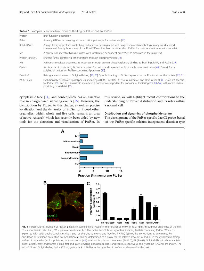
PtdSer results from the coordinated actions of metabolicenzymes in conjunction with vesicular and nonvesiculartransport pathways, while the topology of PtdSer resultsfrom the actions of transmembrane enzymes capable ofmoving PtdSer between lipid bilayers; PtdSer flippases,floppases, and scramblases [7, 8]. Until relatively recently,PtdSer distribution and topology studies depended solelyon the fractionation and subsequent chemical analysis ofcellular organelles. These early studies highlighted PtdSerdistribution throughout the cell is unbalanced (Fig. 1a),being more concentrated in the plasma membrane (PM)(~ 10–15% total lipid) with lower levels in the ER (~ 4%)and mitochondria (~ 1%), the latter of which uses PtdSeras a source of phosphatidylethanolamine (PtdEtn)(reviewed in [7, 9, 10]). The PtdSer content of less abun-dant organelles, including the endosomal system, has gen-erally been less well defined because of the difficultyinherent in purifying them to homogeneity.In addition to difference of PtdSer content amongst
organelles, the unequal bilayer distribution of PtdSer atthe PM has long been appreciated [11], as has the im-portance of movement of PtdSer from the cytoplasmicto exofacial face of the PM being involved in critical sig-naling events including blood clotting [12] and apoptoticcell recognition and removal by macrophages [13]. Fur-thermore, the PM has a net-negative charge on its
© The Author(s). 2019 Open Access This article is distributed under the terms of the Creative Commons Attribution 4.0International License (http://creativecommons.org/licenses/by/4.0/), which permits unrestricted use, distribution, andreproduction in any medium, provided you give appropriate credit to the original author(s) and the source, provide a link tothe Creative Commons license, and indicate if changes were made. The Creative Commons Public Domain Dedication waiver(http://creativecommons.org/publicdomain/zero/1.0/) applies to the data made available in this article, unless otherwise stated.
* Correspondence: [email protected]; [email protected] of Oral Biology, School of Dental Medicine, University atBuffalo, Buffalo, NY 14214, USA2Keenan Research Centre for Biomedical Science, St. Michael’s Hospital,Toronto, ON M5B 1W8, CanadaFull list of author information is available at the end of the article
Kay and Fairn Cell Communication and Signaling (2019) 17:126 https://doi.org/10.1186/s12964-019-0438-z
cytoplasmic face [14], and consequently has an essentialrole in charge-based signaling events [15]. However, thecontribution by PtdSer to this charge, as well as preciselocalization and the dynamics of PtdSer, or indeed otherorganelles, within whole and live cells, remains an areaof active research which has recently been aided by newtools for the detection and visualization of PtdSer. In
this review, we will highlight recent contributions to theunderstanding of PtdSer distribution and its roles withina normal cell.
Distribution and dynamics of phosphatidylserineThe development of the PtdSer-specific LactC2 probe, basedon the PtdSer-specific calcium independent discoidin-type
Fig. 1 Intracellular distribution of PtdSer. a Relative abundance of PtdSer in membranes as mol% of total lipids throughout organelles of the cell.ER – endoplasmic reticulum, PM – plasma membrane. b, c The probe LactC2 labels cytoplasmic-facing leaflets containing PtdSer. When co-expressed with additional organellar markers (such as the plasma membrane labelling PH-PLC (b)) relative correlations as determined bycalculation of Pearson’s correlative co-localization (c) can be determined as a proxy for the relative amounts of PtdSer in the cytoplasmic-facingleaflets of organelles (as first published in Hirama et al. [48]). Markers for plasma membrane (PH-PLC), ER (Sec61), Golgi (GalT), mitochondria (Mito(MitoTracker)), early endosomes (Rab5), fast and slow recycling endosomes (Rab4 and Rab11, respectively) and lysosome (LAMP1) are shown. Thelack of ER and Golgi labeling by LactC2 suggests a lack of PtdSer in the cytoplasmic leaflets as discussed in the text
Table 1 Examples of Intracellular Proteins Binding or Influenced by PtdSer
Protein Brief function description
K-Ras An early GTPase in many signal transduction pathways, for review see [77].
Rab-GTPases A large family of proteins controlling endocytosis, cell migration, cell progression and morphology: many are discussedin main text. Exactly how many of the Rho GTPases that bind or depend on PtdSer for their localization remains uncertain.
Src A central non-receptor tyrosine kinase with localization dependent on PtdSer, as discussed in the main text.
Protein kinase C Enzyme family controlling other proteins through phosphorylation [78].
Akt Activation mediates downstream responses through protein phosphorylation, binding to both PI(3,4,5)P3 and PtdSer [79].
Cavin1 As discussed in main text, PtdSer is required for cavin1 and caveolin1 to form stable caveolae in vivo [68]. Cavin1 formspolyhedral lattices on PtdSer- containing liposomes [80].
Evectin-2 Retrograde endosome to Golgi trafficking [72, 73]. Specific binding to PtdSer depends on the PH-domain of the protein [72, 81].
P4-ATPases Evolutionarily conserved lipid flippases (including ATP8A1, ATP8A2, ATP9A in mammals and Drs2 in yeast) [8]. Some are specificfor PtdSer [82] and as discussed in main text, a number are important for endosomal trafficking [76, 83–86], with recent reviewsproviding more detail [33].
Kay and Fairn Cell Communication and Signaling (2019) 17:126 Page 2 of 8
C2 binding domain of lactadherin (also known as Milk fatglobule-EGF factor 8 (MFGE8)) [16] has enabled thevisualization of PtdSer in live cells (Fig. 1b-c). Indeed, theinitial study using this probe showed for the first time thecytoplasmic-facing distribution of PtdSer in live cells. Thisinitial LactC2 study underscored the importance of PtdSerin providing the negative charge of the PM, finding thatcationic probes track the presence of LactC2-identifiedPtdSer, including in the absence of polyphosphoinositides[16]. The study also highlighted the presence of PtdSer in,and its ability to recruit charge-based protein probes to,endosomal compartments, while not being detectable inthe cytoplasmic-facing cis-Golgi, ER or mitochondria.While it is possible the LactC2 probe does not have highenough sensitivity to detect the relatively low levels of
PtdSer present in these organelles [9, 10], it is also possiblethat, like in the PM, PtdSer leaflet distribution in intracel-lular organelle membranes is asymmetrical [17]. Indeed,there existed significant evidence prior to the developmentof the LactC2 probe suggesting this is the case, at least inthe ER [18–21]. This evidence has since been strength-ened with additional data that does not require the bio-chemical isolation, and potential disruption of, thisintricate tubular organelle. Using a combined light micros-copy and on-section staining electron microscopy (EM)approach, the LactC2 probe was able to detect PtdSer onthe luminal but not cytoplasmic facing ER membrane[22]. A modified ER-targeted LactC2 probe has also beenused to successfully detect PtdSer in the ER lumen of livecells [23].
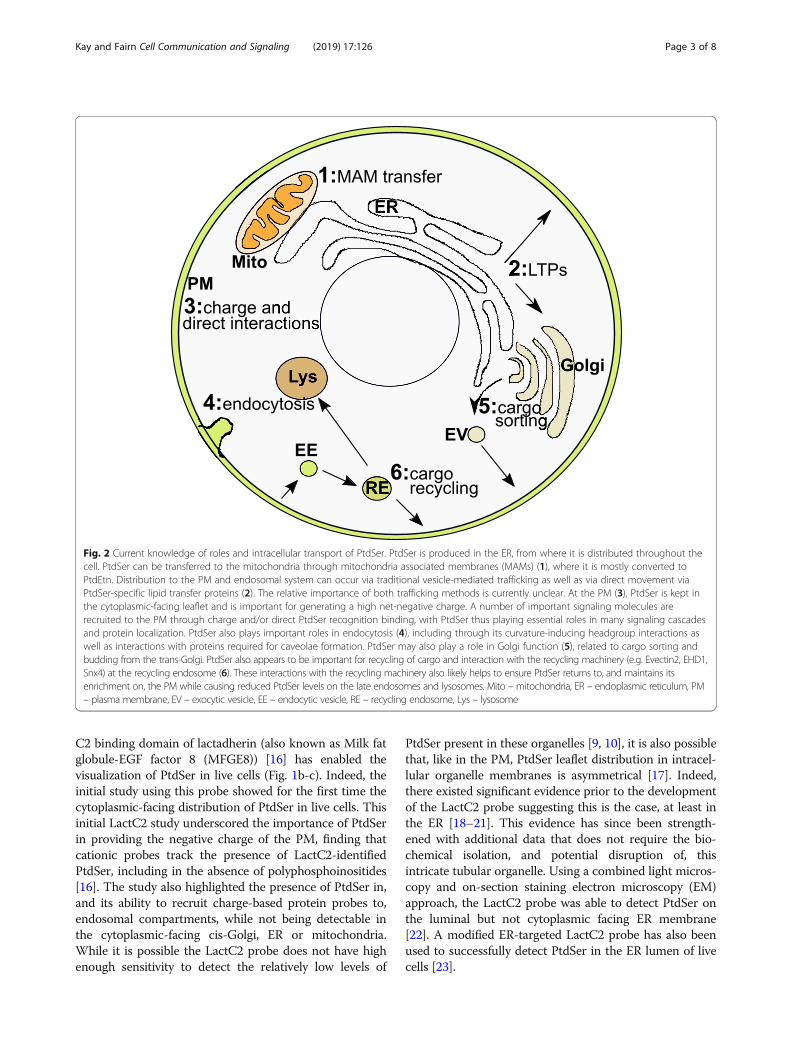
Fig. 2 Current knowledge of roles and intracellular transport of PtdSer. PtdSer is produced in the ER, from where it is distributed throughout thecell. PtdSer can be transferred to the mitochondria through mitochondria associated membranes (MAMs) (1), where it is mostly converted toPtdEtn. Distribution to the PM and endosomal system can occur via traditional vesicle-mediated trafficking as well as via direct movement viaPtdSer-specific lipid transfer proteins (2). The relative importance of both trafficking methods is currently unclear. At the PM (3), PtdSer is kept inthe cytoplasmic-facing leaflet and is important for generating a high net-negative charge. A number of important signaling molecules arerecruited to the PM through charge and/or direct PtdSer recognition binding, with PtdSer thus playing essential roles in many signaling cascadesand protein localization. PtdSer also plays important roles in endocytosis (4), including through its curvature-inducing headgroup interactions aswell as interactions with proteins required for caveolae formation. PtdSer may also play a role in Golgi function (5), related to cargo sorting andbudding from the trans-Golgi. PtdSer also appears to be important for recycling of cargo and interaction with the recycling machinery (e.g. Evectin2, EHD1,Snx4) at the recycling endosome (6). These interactions with the recycling machinery also likely helps to ensure PtdSer returns to, and maintains itsenrichment on, the PM while causing reduced PtdSer levels on the late endosomes and lysosomes. Mito – mitochondria, ER – endoplasmic reticulum, PM– plasma membrane, EV – exocytic vesicle, EE – endocytic vesicle, RE – recycling endosome, Lys – lysosome
Kay and Fairn Cell Communication and Signaling (2019) 17:126 Page 3 of 8

The ability of PtdSer to change membrane leafletsfaces a high energy barrier, with spontaneous transloca-tion estimated to only occur in the order of hours persingle molecular translocation event [24, 25]. Three cat-egories of proteins have been characterized that enablethe trans-leaflet movement of lipids: flippases that transferlipids to the cytosolic leaflet from the PM extracellular ororganellar luminal leaflet, floppases that transfer in the op-posite direction (out of the cytosolic facing leaflet), andscramblases that are bidirectional [26–28]. As the cyto-plasmic leaflet of the ER is where the active site of glycero-phospholipid enzymes reside [29], it has generally beenthought that most glycerophospholipids in the ER arescrambled equally between leaflets to allow for proper ERmembrane expansion and leaflet coupling [30, 31]. Howthis can be compliant with PtdSer having a polarized dis-tribution in the lumen of the ER is unclear. However, ex-pression of gain-of-function PtdSer synthase 1 identifiedfrom Lenz-Majewski syndrome patients does result in theappearance of cytosolic PtdSer in the ER, demonstratingthat the normal mechanism(s) that restrict PtdSer to theluminal leaflet are saturable [32]. One possibility is thatPtdSer, once in the luminal leaflet, is kept there throughinteractions with luminal proteins and/or Ca2+ [33]. Othernon-mutually exclusive possibilities are that movementPtdSer from the cytoplasmic-facing leaflet occurs at theMAM into the mitochondria where it is used for the pro-duction of PtdEth [34], or PtdSer is removed from thecytoplasmic leaflet through non-vesicular transport bylipid transfer proteins (LTPs).LTPs, along with vesicular trafficking, are how lipids
move between cellular membranes [9, 33, 35]. Recentstudies have highlighted the ability of specific LTPs,oxysterol-binding homology (Osh) proteins 6 and 7 inyeast [36, 37] and oxysterol-binding protein (OSBP)-re-lated proteins (ORPs) 5 and 8 in mammalian cells [36,38], to move PtdSer between membranes. The existenceof these PtdSer-specific LTPs thus provide a potentialmechanism for the generation and/or maintenance ofthe PtdSer cellular membrane gradient present in cells.Indeed, recent studies have shown that LTP-mediatedtransfer of PtdSer against its concentration gradient ispossible through exchange with phosphatidylinositol 4-phosphate (PtdIns4P) down its concentration gradientfrom the PM to the ER, where the phosphatase Sac1converts PtdIns4P to PtdIns [38, 39]. However, recentevidence suggests this exchange may be principally usedto fine tune the PM levels of PtdIns4P and PtdIns(4,5)P2rather than be responsible for bulk movement of PtdSerinto the PM [39, 40]. There is also compelling evidencefor the importance of vesicular trafficking in being themajor route for PtdSer trafficking and concentrationwithin the PM. For example, in yeast with temperature-sensitive mutations in secretory proteins Sec6 and Sec1,
the polarization of PtdSer in the PM normally seen at aforming bud is inhibited and PtdSer instead accumulateson the vesicle that are prevented from fusing with thePM [2]. Additionally, endosomal recycling is importantin the maintenance of high PtdSer levels, with inhibitioncausing a redistribution of PtdSer throughout the endo-somal system in yeast [41]. Similarly, disrupting LTPfunction in mammalian cells has been found to result inslightly altered, but not disrupted, cellular membranePtdSer distribution [38, 39]. Furthermore, Snx4, a mem-ber of the sorting nexin family of proteins involved inendosomal cargo sorting and recycling [42] that is spe-cifically involved in recycling of Snc1 in yeast [43] andtransferrin receptor in mammalian cells [44] has recentlybeen implicated in leading to the modification of endo-somal PtdSer levels [41].Thus, while nonvesicular lipid transport, mediated by
LTPs, play an important role, vesicular trafficking appearsto be a significant contributor for maintaining the inter-membrane PtdSer gradient within the cell. Though the fullmolecular mechanisms of how PtdSer is segregated fromother lipids remains to be fully elucidated, biochemicalstudies indicate a significant fraction of PtdSer in mamma-lian cells is enriched in PM-derived detergent-resistant,cholesterol-enriched “lipid-rafts” [45]. This biochemicaldata is supported by both electron microscopy analysisshowing PtdSer is not homogenously distributed through-out the PM [22] and the finding that cholesterol and PtdSerco-segregate throughout subcellular compartments, beingmost concentrated in the PM and early endosomal com-partments and relatively absent from the ER [22, 46, 47].Further, acute changes in either affect the distribution ofthe other; cholesterol is required for the normal distributionof PtdSer [2, 48] and acute changes in PM levels of PtdSeralter the distribution of cholesterol [46]. Evidence is alsobuilding for the likelihood that plasma membrane outerleaflet rafts, dependent on glycersphingolipids and choles-terol [49], are coupled to inner leaflet rafts [50, 51]. The im-portance of PtdSer in this coupling, in both the PM andendosomal membranes, is the subject of a recent excellentreview [52] so will not be further covered here.
Roles of intracellular phosphatidylserineAs described in Background, PtdSer is essential in mam-malian cells [1], while yeast lacking PtdSer are viable buthave greatly reduced growth kinetics [2, 3]. As well, asPtdSer-mediated extracellular signaling, such as duringblood clotting and apoptosis, has recently been reviewed[53–55], we will focus here on information regarding theroles of PtdSer within healthy non-apoptotic cells (Fig. 2).As described, at steady state in a healthy cell PtdSer
makes up to ~ 15 mol% of the total lipid in the PM. Fur-thermore, as it is nearly exclusively in the inner (cyto-plasmic-facing) leaflet it can therefore make up to ~ 30
Kay and Fairn Cell Communication and Signaling (2019) 17:126 Page 4 of 8
mol% of the lipid on this leaflet. As the major lipid witha net-negative charge, PtdSer is therefore responsible forproviding much of the inner leaflet’s charge density. Asignificant role of PtdSer then is interacting with pro-teins in a non-specific charge-based manner to permittheir appropriate localization within the cell (Table 1).For example, the protein kinase Src and Ras GTPasefamily members Rac1 and K-Ras are proteins whosemembrane targeting requires a polycationic stretch inaddition to lipid modifications [56, 57]. The polycationicstretch of K-Ras4B has a net charge of + 8, resulting in itslocalization almost exclusively at the PM. If PtdSer is re-moved [58], or if the net charge of this stretch is varied theresulting mutants are directed additionally to other mem-branes; constructs of intermediate charge (e.g., + 5) localizeto endosomal membranes [16]. Similarly, Src has a polyca-tionic stretch next to its myristoylated residue at the N-terminus with a net charge of + 5, and the kinase was foundto associate not only with the PM but also extensively withPtdSer-enriched endosomal membranes [16].Further evidence of the importance for PtdSer in
charge-based protein distributions has been observedwith the phagocytic process. When pathogens cause adepletion of PtdSer from phagosomes, Src is also lost[59]. In other instances, such charged motifs are not suf-ficient to direct proteins to a membrane but nonethelessinfluence their targeting, likely playing a complementaryrole [56, 60]. Evidence that this is the case comes fromstudies in yeast where polarized PtdSer is required forthe recruitment of the signaling and polarity-regulatingmolecule Cdc42 to the forming bud neck; withoutPtdSer Cdc42 remains Golgi-associated and buds arevery inefficiently formed, leading to poor growth [2].Similarly, Cdc42 and Rho1 are dependent on PtdSerpolarization for their proper localization and function inSchizosaccharomyces pombe [61]. In yet another ex-ample, the plant GTPase Rho of Plants (ROP) familymember ROP6 doesn’t appear to require PtdSer for itsPM association, but does require PtdSer to be stabilizedinto nanodomains within the membrane upon activationthat allows proper signal transduction [62]. WhetherPtdSer is required for, or can modulate, signaling ofother ROP family members, all of which contain a poly-basic stretch of amino acids at their C-terminus [62], re-mains to be seen.Traditionally, the interactions between polycationic
stretches in proteins and anionic phospholipid head-groups have been thought to be strictly charge basedwith little specificity. However, recent evidence chal-lenges this assumption. For instance, K-Ras4B whichcontains six lysine residues adjacent to a farnesylatedcysteine residue, has recently been shown to interactwith PtdSer preferentially [63]. The tail region of K-Ras4B adopts a series on conformations, disordered,
ordered and intermediate, with the disordered being thepreferred conformation. This conformation is also ableto H-bond PtdSer more effectively than the other twoconfirmations [63]. Conversely, other proteins such asK-RasG12V and Rac1 show no preference for PtdSer[63–65]. While these are only initial studies, the resultssuggest that some polybasic proteins may have a prefer-ence for PtdSer or other anionic lipids beyond simpleelectrostatically driven interactions.There are also multiple lines of evidence indicating the
charge of PtdSer contributes to PM curvature and is im-portant for the formation of some forms of endocyticvesicles. For example, caveolae are bulb-shaped nanodo-mains (50–100 nm) of the PM that have been linkedwith many physiological functions, including mechano-sensing and endocytic transport [66]. While caveolaehave been known to be enriched for cholesterol and spe-cific glycerosphingolipids, including GM3 [67], PtdSerhas recently been identified as being required for theirformation and maintenance [68]. This is likely at leastpartly due to the charge-based PtdSer binding of thecavin1 protein [69] which, along with caveolin1, is re-quired for in vivo caveola formation [70]. PtdSer is alsocapable of causing membrane curvature and induceendocytosis upon the acute removal of cholesterol, againa consequence of the charged headgroup of PtdSer [48].It is likely that cholesterol, which makes up ~ 40 mol%of PM lipids [10], helps to keep the PtdSer headgroupcharge density on the inner leaflet low enough to not in-duce spontaneous curvature. However, once cholesterolis removed the distance between phospholipid head-groups is decreased, resulting in high spontaneouscurvature capable of forming endocytic tubules [48, 71].Indeed, increasing PtdSer levels on the inner leaflet ofthe PM above homeostatic levels (and therefore chargedensity) without concomitant cholesterol removal is alsosufficient to increase formation of endocytic vesicles[48]. It is tempting to speculate that the cavin and cave-olin proteins are taking advantage of this curvature-inducing property of PtdSer to induce caveolae. Thus,while cholesterol appears important for PtdSer cellularlocalization, it also appears to be important for modula-tion of PtdSer spacing and membrane curvature induc-tion. This intimate relationship with cholesterol likelyplays important roles in other PtdSer function as well, assuggested by PtdSer dynamics and interactions with ca-veolae [68] and signaling proteins [2, 59, 62].The understanding of the role of PtdSer in internal
membranes remains even less clear than the roles at thePM. Similar to the plasma membrane, recycling endo-somes are rich in PtdSer [72] and recent work has dem-onstrated that PtdSer supports a variety of functions inthese endosomes. The endosomal protein Evectin-2 con-tains a pleckstrin homology domain that binds to PtdSer
Kay and Fairn Cell Communication and Signaling (2019) 17:126 Page 5 of 8

rather than phosphoinositides [72]. Depletion ofEvectin-2 or decreasing the availability of PtdSer pre-vents the movement of cholera toxin from the recyclingendosome to the Golgi. Similarly, depletion of Evectin-2and a reduction of PtdSer levels results in an inability ofGolgi proteins (e.g. TGN38) to be retrieved from endo-somes [72, 73]. In addition to the presence of PtdSer onthe cytosolic leaflet of recycling endosomes, PtdSer flip-pases (e.g., ATP8A1, ATP8A2) are also required to sup-port trafficking events. One critical effector downstreamof flipped PtdSer is the Eps15 homology domain-containing protein-1 (EHD1), an ATPase with dynamin-like activity and a role in membrane remodeling re-quired for the retrograde transport of Shiga toxin to theGolgi [74, 75]. Curiously, PtdSer, Evectin-2 and ATP8A1have all recently been implicated as regulators of Yes-associated protein (YAP) signaling and cell proliferation[76]. ATP8A1 knockdown results in the activation ofLats, which in turn phosphorylates YAP and prevents itstranslocation into the nucleus. Silencing of Evectin-2 re-sults in a decrease of Nedd4-mediated ubiquitination ofLats1, resulting in increased levels that also result in in-creased phosphorylation and inactivation of YAP. Thesestudies raise several questions regarding how PtdSer andits flipping in recycling endosomes are controlling theseeffectors. Additionally, since recycling endosomes re-ceive a lot of incoming membrane from the asymmetricplasma membrane, it is unclear where the luminal leafletPtdSer is coming from to serve as a substrate for theflippases. Much is still to be learned regarding the cellphysiology of PtdSer and we anticipate that the samebiophysical properties PtdSer imposes on the plasmamembrane will hold in endosomes and the trans-Golgi.
ConclusionsIt is becoming clear through recent studies that the es-sential phospholipid PtdSer is important for many intra-cellular processes in addition to its well-characterizedroles in apoptosis and blood clotting. This advancementof our understanding of the intracellular roles for PtdSerhas been fueled in part by the recent development ofnew probes to detect PtdSer. However, as described, ourknowledge of the normal roles for PtdSer in both signal-ing and cellular trafficking within the normal cell is stilldeveloping and many details remain to be discovered.
AbbreviationsEHD1: Eps15 homology domain-containing protein-1; ER: endoplasmicreticulum; LTP: lipid transfer protein; ORPs: oxysterol-binding protein-relatedproteins; Osh: oxysterol-binding homology; PM: plasma membrane;PtdSer: phosphatidylserine; ROP: Rho of Plants; YAP: Yes-associated protein
AcknowledgementsNot applicable.
Authors’ contributionsJK and GF equally contributed to the writing and editing of the manuscript.Both authors read and approved the final manuscript.
FundingJGK is supported by NIH/NIDCR grant R03DE025062, GDF is supported bythe Canadian Institutes of Health Research (MOP-133656) and the NaturalSciences and Engineering Research Council of Canada (RGPIN-2019-04425).
Availability of data and materialsNot applicable.
Ethics approval and consent to participateNot applicable.
Consent for publicationNot applicable.
Competing interestsThe authors declare that they have no competing interests.
Author details1Department of Oral Biology, School of Dental Medicine, University atBuffalo, Buffalo, NY 14214, USA. 2Keenan Research Centre for BiomedicalScience, St. Michael’s Hospital, Toronto, ON M5B 1W8, Canada. 3Departmentof Surgery, University of Toronto, Toronto, ON M5S 1A8, Canada.4Department of Biochemistry, University of Toronto, Toronto, ON M5S 1A8,Canada.
Received: 15 July 2019 Accepted: 10 September 2019
References1. Arikketh D, Nelson R, Vance JE. Defining the importance of
phosphatidylserine synthase-1 (PSS1): unexpected viability of PSS1-deficientmice. J Biol Chem. 2008;283(19):12888–97.
2. Fairn GD, Hermansson M, Somerharju P, Grinstein S. Phosphatidylserine ispolarized and required for proper Cdc42 localization and for developmentof cell polarity. Nat Cell Biol. 2011;13(12):1424–30.
3. Atkinson K, Fogel S, Henry SA. Yeast mutant defective in phosphatidylserinesynthesis. J Biol Chem. 1980;255(14):6653–61.
4. Sousa SB, Jenkins D, Chanudet E, Tasseva G, Ishida M, Anderson G, et al.Gain-of-function mutations in the phosphatidylserine synthase 1 (PTDSS1)gene cause Lenz-Majewski syndrome. Nat Genet. 2014;46(1):70–6.
5. Rusinol AE, Cui Z, Chen MH, Vance JE. A unique mitochondria-associatedmembrane fraction from rat liver has a high capacity for lipid synthesis andcontains pre-Golgi secretory proteins including nascent lipoproteins. J BiolChem. 1994;269(44):27494–502.
6. Ardail D, Lerme F, Louisot P. Involvement of contact sites inphosphatidylserine import into liver mitochondria. J Biol Chem. 1991;266(13):7978–81.
7. Leventis PA, Grinstein S. The distribution and function of phosphatidylserinein cellular membranes. Annu Rev Biophys. 2010;39:407–27.
8. Sebastian TT, Baldridge RD, Xu P, Graham TR. Phospholipid flippases:building asymmetric membranes and transport vesicles. Biochim BiophysActa. 2012;1821(8):1068–77.
9. Vance JE. Phospholipid synthesis and transport in mammalian cells. Traffic.2015;16(1):1–18.
10. van Meer G, Voelker DR, Feigenson GW. Membrane lipids: where they areand how they behave. Nat Rev Mol Cell Biol. 2008;9(2):112–24.
11. Gordesky SE, Marinetti GV. The asymetric arrangement of phospholipids inthe human erythrocyte membrane. Biochem Biophys Res Commun. 1973;50(4):1027–31.
12. Zwaal RF, Comfurius P, van Deenen LL. Membrane asymmetry and bloodcoagulation. Nature. 1977;268(5618):358–60.
13. Fadok VA, Voelker DR, Campbell PA, Cohen JJ, Bratton DL, Henson PM.Exposure of phosphatidylserine on the surface of apoptotic lymphocytestriggers specific recognition and removal by macrophages. J Immunol.1992;148(7):2207–16.
Kay and Fairn Cell Communication and Signaling (2019) 17:126 Page 6 of 8
14. Olivotto M, Arcangeli A, Carla M, Wanke E. Electric fields at the plasmamembrane level: a neglected element in the mechanisms of cell signalling.Bioessays. 1996;18(6):495–504.
15. McLaughlin S, Aderem A. The myristoyl-electrostatic switch: a modulator ofreversible protein-membrane interactions. Trends Biochem Sci. 1995;20(7):272–6.
16. Yeung T, Gilbert GE, Shi J, Silvius J, Kapus A, Grinstein S. Membranephosphatidylserine regulates surface charge and protein localization.Science. 2008;319(5860):210–3.
17. Bretscher MS. Membrane structure: some general principles. Science. 1973;181(4100):622–9.
18. Bollen IC, Higgins JA. Phospholipid asymmetry in rough- and smooth-endoplasmic-reticulum membranes of untreated and phenobarbital-treatedrat liver. Biochem J. 1980;189(3):475–80.
19. Higgins JA, Dawson RM. Asymmetry of the phospholipid bilayer of rat liverendoplasmic reticulum. Biochim Biophys Acta. 1977;470(3):342–56.
20. Vale MG. Localization of the amino phospholipids in sarcoplasmic reticulummembranes revealed by trinitrobenzenesulfonate and fluorodinitrobenzene.Biochim Biophys Acta. 1977;471(1):39–48.
21. Calderon F, Kim HY. Detection of intracellular phosphatidylserine in livingcells. J Neurochem. 2008;104(5):1271–9.
22. Fairn GD, Schieber NL, Ariotti N, Murphy S, Kuerschner L, Webb RI, et al.High-resolution mapping reveals topologically distinct cellular pools ofphosphatidylserine. J Cell Biol. 2011;194(2):257–75.
23. Kay JG, Koivusalo M, Ma X, Wohland T, Grinstein S. Phosphatidylserinedynamics in cellular membranes. Mol Biol Cell. 2012;23(11):2198–212.
24. Kornberg RD, McConnell HM. Inside-outside transitions of phospholipids invesicle membranes. Biochemistry. 1971;10(7):1111–20.
25. Bai J, Pagano RE. Measurement of spontaneous transfer and transbilayermovement of BODIPY-labeled lipids in lipid vesicles. Biochemistry. 1997;36(29):8840–8.
26. Sahu SK, Gummadi SN, Manoj N, Aradhyam GK. Phospholipid scramblases:an overview. Arch Biochem Biophys. 2007;462(1):103–14.
27. Graham TR. Flippases and vesicle-mediated protein transport. Trends CellBiol. 2004;14(12):670–7.
28. Daleke DL. Regulation of transbilayer plasma membrane phospholipidasymmetry. J Lipid Res. 2003;44(2):233–42.
29. Bell RM, Ballas LM, Coleman RA. Lipid topogenesis. J Lipid Res. 1981;22(3):391–403.30. Buton X, Morrot G, Fellmann P, Seigneuret M. Ultrafast glycerophospholipid-
selective transbilayer motion mediated by a protein in the endoplasmicreticulum membrane. J Biol Chem. 1996;271(12):6651–7.
31. Pomorski T, Holthuis JC, Herrmann A, van Meer G. Tracking down lipidflippases and their biological functions. J Cell Sci. 2004;117(Pt 6:805–13.
32. Sohn M, Ivanova P, Brown HA, Toth DJ, Varnai P, Kim YJ, et al. Lenz-Majewski mutations in PTDSS1 affect phosphatidylinositol 4-phosphatemetabolism at ER-PM and ER-Golgi junctions. Proc Natl Acad Sci U S A.2016;113(16):4314–9.
33. Hankins HM, Baldridge RD, Xu P, Graham TR. Role of flippases, scramblasesand transfer proteins in phosphatidylserine subcellular distribution. Traffic.2015;16(1):35–47.
34. Vance JE, Tasseva G. Formation and function of phosphatidylserine andphosphatidylethanolamine in mammalian cells. Biochim Biophys Acta. 2013;1831(3):543–54.
35. Lev S. Non-vesicular lipid transport by lipid-transfer proteins and beyond.Nat Rev Mol Cell Biol. 2010;11(10):739–50.
36. Maeda K, Anand K, Chiapparino A, Kumar A, Poletto M, Kaksonen M, et al.Interactome map uncovers phosphatidylserine transport by oxysterol-binding proteins. Nature. 2013;501(7466):257–61.
37. Moser von Filseck J, Copic A, Delfosse V, Vanni S, Jackson CL, Bourguet W,et al. INTRACELLULAR TRANSPORT. Phosphatidylserine transport by ORP/Osh proteins is driven by phosphatidylinositol 4-phosphate. Science. 2015;349(6246):432–6.
38. Chung J, Torta F, Masai K, Lucast L, Czapla H, Tanner LB, et al. INTRACELLULARTRANSPORT. PI4P/phosphatidylserine countertransport at ORP5- and ORP8-mediated ER-plasma membrane contacts. Science. 2015;349(6246):428–32.
39. Sohn M, Korzeniowski M, Zewe JP, Wills RC, Hammond GRV, HumpolickovaJ, et al. PI(4,5)P2 controls plasma membrane PI4P and PS levels via ORP5/8recruitment to ER-PM contact sites. J Cell Biol. 2018;217(5):1797–813.
40. Ghai R, Du X, Wang H, Dong J, Ferguson C, Brown AJ, et al. ORP5 and ORP8bind phosphatidylinositol-4, 5-biphosphate (PtdIns(4,5)P 2) and regulate itslevel at the plasma membrane. Nat Commun. 2017;8(1):757.
41. Ma M, Kumar S, Purushothaman L, Babst M, Ungermann C, Chi RJ, et al. Lipidtrafficking by yeast Snx4 family SNX-BAR proteins promotes autophagy andvacuole membrane fusion. Mol Biol Cell. 2018;29(18):2190–200.
42. Cullen PJ. Endosomal sorting and signalling: an emerging role for sortingnexins. Nat Rev Mol Cell Biol. 2008;9(7):574–82.
43. Hettema EH, Lewis MJ, Black MW, Pelham HR. Retromer and the sortingnexins Snx4/41/42 mediate distinct retrieval pathways from yeastendosomes. EMBO J. 2003;22(3):548–57.
44. Traer CJ, Rutherford AC, Palmer KJ, Wassmer T, Oakley J, Attar N, et al. SNX4coordinates endosomal sorting of TfnR with dynein-mediated transport intothe endocytic recycling compartment. Nat Cell Biol. 2007;9(12):1370–80.
45. Pike LJ, Han X, Gross RW. Epidermal growth factor receptors are localized tolipid rafts that contain a balance of inner and outer leaflet lipids: a shotgunlipidomics study. J Biol Chem. 2005;280(29):26796–804.
46. Maekawa M, Fairn GD. Complementary probes reveal thatphosphatidylserine is required for the proper transbilayer distribution ofcholesterol. J Cell Sci. 2015;128(7):1422–33.
47. Gagescu R, Demaurex N, Parton RG, Hunziker W, Huber LA, Gruenberg J. Therecycling endosome of Madin-Darby canine kidney cells is a mildly acidiccompartment rich in raft components. Mol Biol Cell. 2000;11(8):2775–91.
48. Hirama T, Lu SM, Kay JG, Maekawa M, Kozlov MM, Grinstein S, et al.Membrane curvature induced by proximity of anionic phospholipids caninitiate endocytosis. Nat Commun. 2017;8(1):1393.
49. Ahmed SN, Brown DA, London E. On the origin of sphingolipid/cholesterol-rich detergent-insoluble cell membranes: physiological concentrations ofcholesterol and sphingolipid induce formation of a detergent-insoluble,liquid-ordered lipid phase in model membranes. Biochemistry. 1997;36(36):10944–53.
50. Simons K, Toomre D. Lipid rafts and signal transduction. Nat Rev Mol CellBiol. 2000;1(1):31–9.
51. Sezgin E, Levental I, Mayor S, Eggeling C. The mystery of membraneorganization: composition, regulation and roles of lipid rafts. Nat Rev MolCell Biol. 2017;18(6):361–74.
52. Skotland T, Sandvig K. The role of PS 18:0/18:1 in membrane function. NatCommun. 2019;10(1):2752.
53. Kay JG, Grinstein S. Phosphatidylserine-mediated cellular signaling. Adv ExpMed Biol. 2013;991:177–93.
54. Heemskerk JW, Bevers EM, Lindhout T. Platelet activation and bloodcoagulation. Thromb Haemost. 2002;88(2):186–93.
55. Nagata S, Suzuki J, Segawa K, Fujii T. Exposure of phosphatidylserine on thecell surface. Cell Death Differ. 2016;23(6):952–61.
56. Michaelson D, Silletti J, Murphy G, D'Eustachio P, Rush M, Philips MR.Differential localization of rho GTPases in live cells: regulation byhypervariable regions and RhoGDI binding. J Cell Biol. 2001;152(1):111–26.
57. Sigal CT, Zhou WJ, Buser CA, Mclaughlin S, Resh MD. Amino-terminal basicresidues of Src mediate membrane-binding through electrostatic interactionwith acidic phospholipids. Proc Natl Acad Sci U S A. 1994;91(25):12253–7.
58. Cho KJ, van der Hoeven D, Zhou Y, Maekawa M, Ma X, Chen W, et al.Inhibition of acid sphingomyelinase depletes cellular phosphatidylserineand Mislocalizes K-Ras from the plasma membrane. Mol Cell Biol. 2016;36(2):363–74.
59. Yeung T, Heit B, Dubuisson JF, Fairn GD, Chiu B, Inman R, et al. Contributionof phosphatidylserine to membrane surface charge and protein targetingduring phagosome maturation. J Cell Biol. 2009;185(5):917–28.
60. Laude AJ, Prior IA. Palmitoylation and localisation of RAS isoforms are modulatedby the hypervariable linker domain. J Cell Sci. 2008;121(Pt 4:421–7.
61. Haupt A, Minc N. Gradients of phosphatidylserine contribute to plasmamembrane charge localization and cell polarity in fission yeast. Mol Biol Cell.2017;28(1):210–20.
62. Platre MP, Bayle V, Armengot L, Bareille J, Marques-Bueno MDM, Creff A,et al. Developmental control of plant rho GTPase nano-organization by thelipid phosphatidylserine. Science. 2019;364(6435):57–62.
63. Zhou Y, Prakash P, Liang H, Cho KJ, Gorfe AA, Hancock JF. Lipid-sortingspecificity encoded in K-Ras membrane anchor regulates signal output. Cell.2017;168(1–2):239–51 e16.
64. Zhou Y, Hancock JF. A novel prenyl-polybasic domain code determineslipid-binding specificity of the K-Ras membrane anchor. Small GTPases.2018:1–5.
65. Maxwell KN, Zhou Y, Hancock JF. Rac1 nanoscale organization on theplasma membrane is driven by lipid binding specificity encoded in themembrane anchor. Mol Cell Biol. 2018;38(18).
Kay and Fairn Cell Communication and Signaling (2019) 17:126 Page 7 of 8

66. Parton RG. Caveolae: structure, function, and relationship to disease. AnnuRev Cell Dev Biol. 2018;34:111–36.
67. Ortegren U, Karlsson M, Blazic N, Blomqvist M, Nystrom FH, Gustavsson J,et al. Lipids and glycosphingolipids in caveolae and surrounding plasmamembrane of primary rat adipocytes. Eur J Biochem. 2004;271(10):2028–36.
68. Hirama T, Das R, Yang Y, Ferguson C, Won A, Yip CM, et al.Phosphatidylserine dictates the assembly and dynamics of caveolae in theplasma membrane. J Biol Chem. 2017;292(34):14292–307.
69. Kovtun O, Tillu VA, Jung W, Leneva N, Ariotti N, Chaudhary N, et al.Structural insights into the organization of the cavin membrane coatcomplex. Dev Cell. 2014;31(4):405–19.
70. Hill MM, Bastiani M, Luetterforst R, Kirkham M, Kirkham A, Nixon SJ, et al.PTRF-Cavin, a conserved cytoplasmic protein required for caveola formationand function. Cell. 2008;132(1):113–24.
71. Fuller N, Benatti CR, Rand RP. Curvature and bending constants forphosphatidylserine-containing membranes. Biophys J. 2003;85(3):1667–74.
72. Uchida Y, Hasegawa J, Chinnapen D, Inoue T, Okazaki S, Kato R, et al.Intracellular phosphatidylserine is essential for retrograde membrane trafficthrough endosomes. Proc Natl Acad Sci U S A. 2011;108(38):15846–51.
73. Lee S, Uchida Y, Emoto K, Umeda M, Kuge O, Taguchi T, et al. Impairedretrograde membrane traffic through endosomes in a mutant CHO celldefective in phosphatidylserine synthesis. Genes Cells. 2012;17(8):728–36.
74. Daumke O, Lundmark R, Vallis Y, Martens S, Butler PJ, McMahon HT.Architectural and mechanistic insights into an EHD ATPase involved inmembrane remodelling. Nature. 2007;449(7164):923–7.
75. Melo AA, Hegde BG, Shah C, Larsson E, Isas JM, Kunz S, et al. Structuralinsights into the activation mechanism of dynamin-like EHD ATPases. ProcNatl Acad Sci U S A. 2017;114(22):5629–34.
76. Matsudaira T, Mukai K, Noguchi T, Hasegawa J, Hatta T, Iemura SI, et al.Endosomal phosphatidylserine is critical for the YAP signalling pathway inproliferating cells. Nat Commun. 2017;8(1):1246.
77. Zhou Y, Hancock JF. Deciphering lipid codes: K-Ras as a paradigm. Traffic.2018;19(3):157–65.
78. Newton AC. Protein kinase C: structure, function, and regulation. J BiolChem. 1995;270(48):28495–8.
79. Huang BX, Akbar M, Kevala K, Kim HY. Phosphatidylserine is a criticalmodulator for Akt activation. J Cell Biol. 2011;192(6):979–92.
80. Stoeber M, Schellenberger P, Siebert CA, Leyrat C, Helenius A, Grunewald K.Model for the architecture of caveolae based on a flexible, net-like assembly ofCavin1 and Caveolin discs. Proc Natl Acad Sci U S A. 2016;113(50):E8069–E78.
81. Okazaki S, Kato R, Uchida Y, Taguchi T, Arai H, Wakatsuki S. Structural basisof the strict phospholipid binding specificity of the pleckstrin homologydomain of human evectin-2. Acta Crystallogr D Biol Crystallogr. 2012;68(Pt 2:117–23.
82. Baldridge RD, Graham TR. Two-gate mechanism for phospholipid selectionand transport by type IV P-type ATPases. Proc Natl Acad Sci U S A. 2013;110(5):E358–67.
83. Tanaka Y, Ono N, Shima T, Tanaka G, Katoh Y, Nakayama K, et al. Thephospholipid flippase ATP9A is required for the recycling pathway from theendosomes to the plasma membrane. Mol Biol Cell. 2016;27(24):3883–93.
84. Muthusamy BP, Natarajan P, Zhou X, Graham TR. Linking phospholipidflippases to vesicle-mediated protein transport. Biochim Biophys Acta. 2009;1791(7):612–9.
85. Hankins HM, Sere YY, Diab NS, Menon AK, Graham TR. Phosphatidylserinetranslocation at the yeast trans-Golgi network regulates protein sorting intoexocytic vesicles. Mol Biol Cell. 2015;26(25):4674–85.
86. Lee S, Uchida Y, Wang J, Matsudaira T, Nakagawa T, Kishimoto T, et al.Transport through recycling endosomes requires EHD1 recruitment by aphosphatidylserine translocase. EMBO J. 2015;34(5):669–88.
Publisher’s NoteSpringer Nature remains neutral with regard to jurisdictional claims inpublished maps and institutional affiliations.
Kay and Fairn Cell Communication and Signaling (2019) 17:126 Page 8 of 8