Structural and Functional Analysis of the Spt16p N-terminal Domain Reveals Overlapping Roles of...
25
Structural and Functional Analysis of the Spt16p N-terminal Domain Reveals Overlapping Roles of yFACT Subunits * □ S Received for publication, October 19, 2007, and in revised form, December 3, 2007 Published, JBC Papers in Press, December 18, 2007, DOI 10.1074/jbc.M708682200 Andrew P. VanDemark ‡1 , Hua Xin ‡ , Laura McCullough ‡ , Robert Rawlins ‡ , Shayla Bentley § , Annie Heroux ¶ , David J. Stillman § , Christopher P. Hill ‡2 , and Tim Formosa ‡3 From the Departments of ‡ Biochemistry and § Pathology, University of Utah School of Medicine, Salt Lake City, Utah 84112 and ¶ Biology Department, Brookhaven National Laboratory, Upton, New York 11973 yFACT (heterodimers of Saccharomyces cerevisiae Spt16- Pob3 combined with Nhp6) binds to and alters the properties of nucleosomes. The essential function of yFACT is not disrupted by deletion of the N-terminal domain (NTD) of Spt16 or by mutation of the middle domain of Pob3, but either alteration makes yeast cells sensitive to DNA replication stress. We have determined the structure of the Spt16 NTD and find evidence for a conserved potential peptide-binding site. Pob3-M also contains a putative binding site, and we show that these two sites perform an overlapping essential function. We find that yFACT can bind the N-terminal tails of some histones and that this interaction is important for yFACT-nucleosome binding. How- ever, neither the Spt16 NTD nor a key residue in the putative Pob3-M-binding site was required for interactions with histone N termini or for yFACT-mediated nucleosome reorganization in vitro. Instead, both potential binding sites interact function- ally with the C-terminal docking domain of the histone H2A. yFACT therefore appears to make multiple contacts with differ- ent sites within nucleosomes, and these interactions are par- tially redundant with one another. The docking domain of H2A is identified as an important participant in maintaining stability during yFACT-mediated nucleosome reorganization, suggest- ing new models for the mechanism of this activity. yFACT (yeast facilitator of chromatin transcription or trans- actions) is a heterodimer of the Saccharomyces cerevisiae Spt16 and Pob3 proteins that is assisted in vivo and in vitro by the high mobility group type B domain DNA-binding protein Nhp6 (1, 2). In vitro, yFACT binds to histones (3, 4) and can alter the accessibility of DNA within nucleosomes without hydrolyzing ATP and without repositioning the histone octamer core rela- tive to the DNA (5–7). This activity is different from ATP-de- pendent chromatin remodeling and has been called nucleo- some reorganization (6). yFACT and related FACT complexes from other eukaryotes are needed for both normal regulation of transcription (5, 8 –11) and for DNA replication (12–20). Reor- ganization activity therefore appears to be important in a range of chromatin-based processes, including initiation and elonga- tion of transcription, establishment and maintenance of normal chromatin, and survival during DNA replication stress. Con- sistent with this broad functional importance, FACT family members have been found in all eukaryotes examined, and at least one of the subunits is essential for viability in all cases reported (9, 21, 19, 22). FACT complexes contain several distinct structural domains (16, 23), but little is known about how these domains contribute to FACT function. The middle domain of Pob3 (Pob3-M) forms two pleckstrin homology (PH) 4 folds that are closely juxtaposed (23), with highly conserved surface residues forming a patch in a region often associated with binding sites in PH domain pro- teins (23). Altering this patch caused increased sensitivity to hydroxyurea (HU) (23), a toxin that blocks dNTP synthesis and therefore causes replication stress. This suggests that the Pob3-M domain contributes to a binding interaction that is of increased importance when yeast cells encounter replication stress. Consistent with a role as a protein-binding module, Pob3-M was shown to interact physically and genetically with Rfa1 (23), a subunit of the eukaryotic single-stranded DNA binding factor RPA. yFACT and RPA appear to have overlap- ping functions in a process that affects nucleosome deposition during DNA replication (23). However, mutations in the con- served putative interaction surface on Pob3-M did not disrupt the yFACT-RPA interaction in vitro (23). Pob3-M may there- fore have multiple binding partners, with each interaction con- tributing to different functions of yFACT in different contexts. The N-terminal domain (NTD) of Spt16 forms an independ- ent structural unit (16, 23). Surprisingly, although this domain is conserved among all known Spt16 homologs, it is not essen- tial for viability in yeast cells, although it is required for normal growth in the presence of high levels of HU (16). The Spt16 NTD shares limited sequence similarity with a class of amin- opeptidases, but it does not have peptidase active site residues (see Ref. 24 and this study). These observations suggest that the * This work was supported by National Institutes of Health grants (to T. F., D. S., and C. P. H.) and an American Cancer Society grant (to A. P. V.). The costs of publication of this article were defrayed in part by the payment of page charges. This article must therefore be hereby marked “advertise- ment” in accordance with 18 U.S.C. Section 1734 solely to indicate this fact. The atomic coordinates and structure factors (code 3BIP, 3BIQ, 3BIT) have been deposited in the Protein Data Bank, Research Collaboratory for Structural Bioinformatics, Rutgers University, New Brunswick, NJ (http://www.rcsb.org/). □ S The on-line version of this article (available at http://www.jbc.org) contains supplemental Methods, Figs. S1–S5, and Table S1. 1 Present address: Dept. of Biological Sciences, University of Pittsburgh, Pitts- burgh, PA 15260. 2 To whom correspondence may be addressed. Tel.: 801-585-5536; Fax: 801- 581-7959; E-mail: [email protected]. 3 To whom correspondence may be addressed. Tel.: 801-581-5435; Fax: 801- 581-7959; E-mail: [email protected]. 4 The abbreviations used are: PH, pleckstrin homology; WT, wild type; HU, hydroxyurea; NTD, N-terminal domain; TEV, tobacco etch virus; PDB, Pro- tein Data Bank; 5-FOA, 5-fluoroorotic acid. THE JOURNAL OF BIOLOGICAL CHEMISTRY VOL. 283, NO. 8, pp. 5058 –5068, February 22, 2008 Printed in the U.S.A. 5058 JOURNAL OF BIOLOGICAL CHEMISTRY VOLUME 283 • NUMBER 8 • FEBRUARY 22, 2008 by guest on May 15, 2016 http://www.jbc.org/ Downloaded from by guest on May 15, 2016 http://www.jbc.org/ Downloaded from by guest on May 15, 2016 http://www.jbc.org/ Downloaded from by guest on May 15, 2016 http://www.jbc.org/ Downloaded from by guest on May 15, 2016 http://www.jbc.org/ Downloaded from by guest on May 15, 2016 http://www.jbc.org/ Downloaded from by guest on May 15, 2016 http://www.jbc.org/ Downloaded from by guest on May 15, 2016 http://www.jbc.org/ Downloaded from by guest on May 15, 2016 http://www.jbc.org/ Downloaded from by guest on May 15, 2016 http://www.jbc.org/ Downloaded from by guest on May 15, 2016 http://www.jbc.org/ Downloaded from by guest on May 15, 2016 http://www.jbc.org/ Downloaded from by guest on May 15, 2016 http://www.jbc.org/ Downloaded from by guest on May 15, 2016 http://www.jbc.org/ Downloaded from by guest on May 15, 2016 http://www.jbc.org/ Downloaded from
-
Upload
independent -
Category
Documents
-
view
0 -
download
0
Transcript of Structural and Functional Analysis of the Spt16p N-terminal Domain Reveals Overlapping Roles of...

Structural and Functional Analysis of the Spt16p N-terminalDomain Reveals Overlapping Roles of yFACT Subunits*□S
Received for publication, October 19, 2007, and in revised form, December 3, 2007 Published, JBC Papers in Press, December 18, 2007, DOI 10.1074/jbc.M708682200
Andrew P. VanDemark‡1, Hua Xin‡, Laura McCullough‡, Robert Rawlins‡, Shayla Bentley§, Annie Heroux¶,David J. Stillman§, Christopher P. Hill‡2, and Tim Formosa‡3
From the Departments of ‡Biochemistry and §Pathology, University of Utah School of Medicine, Salt Lake City, Utah 84112and ¶Biology Department, Brookhaven National Laboratory, Upton, New York 11973
yFACT (heterodimers of Saccharomyces cerevisiae Spt16-Pob3 combined with Nhp6) binds to and alters the properties ofnucleosomes. The essential function of yFACT is not disruptedby deletion of the N-terminal domain (NTD) of Spt16 or bymutation of the middle domain of Pob3, but either alterationmakes yeast cells sensitive to DNA replication stress. We havedetermined the structure of the Spt16 NTD and find evidencefor a conserved potential peptide-binding site. Pob3-M alsocontains a putative binding site, andwe show that these two sitesperform an overlapping essential function.We find that yFACTcan bind the N-terminal tails of some histones and that thisinteraction is important for yFACT-nucleosome binding. How-ever, neither the Spt16 NTD nor a key residue in the putativePob3-M-binding site was required for interactions with histoneN termini or for yFACT-mediated nucleosome reorganizationin vitro. Instead, both potential binding sites interact function-ally with the C-terminal docking domain of the histone H2A.yFACT therefore appears tomakemultiple contacts with differ-ent sites within nucleosomes, and these interactions are par-tially redundant with one another. The docking domain of H2Ais identified as an important participant inmaintaining stabilityduring yFACT-mediated nucleosome reorganization, suggest-ing new models for the mechanism of this activity.
yFACT (yeast facilitator of chromatin transcription or trans-actions) is a heterodimer of the Saccharomyces cerevisiae Spt16and Pob3 proteins that is assisted in vivo and in vitro by the highmobility group type B domain DNA-binding protein Nhp6 (1,2). In vitro, yFACT binds to histones (3, 4) and can alter theaccessibility of DNA within nucleosomes without hydrolyzingATP and without repositioning the histone octamer core rela-
tive to the DNA (5–7). This activity is different from ATP-de-pendent chromatin remodeling and has been called nucleo-some reorganization (6). yFACT and related FACT complexesfromother eukaryotes are needed for both normal regulation oftranscription (5, 8–11) and for DNA replication (12–20). Reor-ganization activity therefore appears to be important in a rangeof chromatin-based processes, including initiation and elonga-tion of transcription, establishment andmaintenance of normalchromatin, and survival during DNA replication stress. Con-sistent with this broad functional importance, FACT familymembers have been found in all eukaryotes examined, and atleast one of the subunits is essential for viability in all casesreported (9, 21, 19, 22).FACT complexes contain several distinct structural domains
(16, 23), but little is known about how these domains contributeto FACT function. Themiddle domain of Pob3 (Pob3-M) formstwo pleckstrin homology (PH)4 folds that are closely juxtaposed(23), with highly conserved surface residues forming a patch ina region often associated with binding sites in PH domain pro-teins (23). Altering this patch caused increased sensitivity tohydroxyurea (HU) (23), a toxin that blocks dNTP synthesis andtherefore causes replication stress. This suggests that thePob3-M domain contributes to a binding interaction that is ofincreased importance when yeast cells encounter replicationstress. Consistent with a role as a protein-binding module,Pob3-M was shown to interact physically and genetically withRfa1 (23), a subunit of the eukaryotic single-stranded DNAbinding factor RPA. yFACT and RPA appear to have overlap-ping functions in a process that affects nucleosome depositionduring DNA replication (23). However, mutations in the con-served putative interaction surface on Pob3-M did not disruptthe yFACT-RPA interaction in vitro (23). Pob3-M may there-fore have multiple binding partners, with each interaction con-tributing to different functions of yFACT in different contexts.The N-terminal domain (NTD) of Spt16 forms an independ-
ent structural unit (16, 23). Surprisingly, although this domainis conserved among all known Spt16 homologs, it is not essen-tial for viability in yeast cells, although it is required for normalgrowth in the presence of high levels of HU (16). The Spt16NTD shares limited sequence similarity with a class of amin-opeptidases, but it does not have peptidase active site residues(see Ref. 24 and this study). These observations suggest that the
* This work was supported by National Institutes of Health grants (to T. F.,D. S., and C. P. H.) and an American Cancer Society grant (to A. P. V.). Thecosts of publication of this article were defrayed in part by the payment ofpage charges. This article must therefore be hereby marked “advertise-ment” in accordance with 18 U.S.C. Section 1734 solely to indicate this fact.
The atomic coordinates and structure factors (code 3BIP, 3BIQ, 3BIT) have beendeposited in the Protein Data Bank, Research Collaboratory for StructuralBioinformatics, Rutgers University, New Brunswick, NJ (http://www.rcsb.org/).
□S The on-line version of this article (available at http://www.jbc.org) containssupplemental Methods, Figs. S1–S5, and Table S1.
1 Present address: Dept. of Biological Sciences, University of Pittsburgh, Pitts-burgh, PA 15260.
2 To whom correspondence may be addressed. Tel.: 801-585-5536; Fax: 801-581-7959; E-mail: [email protected].
3 To whom correspondence may be addressed. Tel.: 801-581-5435; Fax: 801-581-7959; E-mail: [email protected].
4 The abbreviations used are: PH, pleckstrin homology; WT, wild type; HU,hydroxyurea; NTD, N-terminal domain; TEV, tobacco etch virus; PDB, Pro-tein Data Bank; 5-FOA, 5-fluoroorotic acid.
THE JOURNAL OF BIOLOGICAL CHEMISTRY VOL. 283, NO. 8, pp. 5058 –5068, February 22, 2008Printed in the U.S.A.
5058 JOURNAL OF BIOLOGICAL CHEMISTRY VOLUME 283 • NUMBER 8 • FEBRUARY 22, 2008
by guest on May 15, 2016
http://ww
w.jbc.org/
Dow
nloaded from
by guest on May 15, 2016
http://ww
w.jbc.org/
Dow
nloaded from
by guest on May 15, 2016
http://ww
w.jbc.org/
Dow
nloaded from
by guest on May 15, 2016
http://ww
w.jbc.org/
Dow
nloaded from
by guest on May 15, 2016
http://ww
w.jbc.org/
Dow
nloaded from
by guest on May 15, 2016
http://ww
w.jbc.org/
Dow
nloaded from
by guest on May 15, 2016
http://ww
w.jbc.org/
Dow
nloaded from
by guest on May 15, 2016
http://ww
w.jbc.org/
Dow
nloaded from
by guest on May 15, 2016
http://ww
w.jbc.org/
Dow
nloaded from
by guest on May 15, 2016
http://ww
w.jbc.org/
Dow
nloaded from
by guest on May 15, 2016
http://ww
w.jbc.org/
Dow
nloaded from
by guest on May 15, 2016
http://ww
w.jbc.org/
Dow
nloaded from
by guest on May 15, 2016
http://ww
w.jbc.org/
Dow
nloaded from
by guest on May 15, 2016
http://ww
w.jbc.org/
Dow
nloaded from
by guest on May 15, 2016
http://ww
w.jbc.org/
Dow
nloaded from

Spt16 NTDmay have peptide binding activity but that this roleis dispensable for the core activity of yFACT.Here we report the structure of the Spt16 NTD and the
results of studies examining the role of this domain in yFACTfunction. The Spt16 NTD is structurally similar to aminopep-tidases, and the most highly conserved surface residues line acleft equivalent to the aminopeptidase substrate-binding site.The presence of potential peptide-binding sites in each subunitof yFACT led us to examine candidate substrates, initiallyfocusing on the attractive possibility that these sites bind to theN-terminal tails of histones that extend beyond the structuredcore of the nucleosome and are known to influence yFACTfunctions in vivo (6, 25). The N-terminal tails of histones werefound to have an important role in yFACT function and to bebound by yFACT with high affinity. However, this activity andother measurable functions of yFACT in vitro remained intactafter mutating Pob3-M or deleting the Spt16 NTD. Instead, theSpt16 NTD and Pob3-M domains were found to have overlap-ping roles in a process that involves the C-terminal extension ofH2A.This stirrup-like “docking domain” of histoneH2Aacts tostabilize the binding of H2A-H2B dimers to (H3-H4)2 tetram-ers within histone octamers (26–28). These results suggest thatyFACT makes multiple additive contacts with nucleosomesduring reorganization, and that some of these contacts areimportant after yFACT has induced the reorganization of thenucleosome. The docking domain of H2A is identified as animportant contributor to this process, perhaps tetheringnucleosomal components together or controlling the insertionof H2A-H2B dimers during nucleosome formation.
EXPERIMENTAL PROCEDURES
Protein Expression and Purification—DNA fragmentsencoding Spt16 residues 1–451 or 1–465 were amplified byPCR and inserted into amodified pET bacterial expression vec-tor that fused eight histidines and a TEV protease site to the Ntermini. TEV cleavage leaves the sequence “GHM . . . ” at the Nterminus in place of the native methionine. Each protein wasexpressed in Codon�(RIL) cells (Stratagene) and purified bynickel chelation chromatography (Qiagen). After TEV proteasedigestion and nickel chelation chromatography to removetaggedN termini, proteinswere further purified by gel filtrationon Superdex-200 (GEHealthcare) in 10mMHEPES, pH7.5, 150mM NaCl, 2% glycerol, 1 mM 2-mercaptoethanol. The Spt16NTD fragments eluted as apparent monomers and were thenconcentrated in gel filtration buffer.Spt16-Pob3 complexes with 12 histidines and a TEV site
fused to the N terminus of Pob3 were purified from yeast cellsoverexpressing each protein from the Gal1 promoter aftergrowth in galactose medium, as described (19). Complexeswere purified from extracts using nickel chelation and gel fil-tration as above, except using Sephacryl S300 (GE Healthcare)and omitting the TEV cleavage.Nucleosomes were prepared by dialysis from high ionic
strength solutions using derivatives of a sea urchin rDNAnucleosome positioning sequence and chicken histones or bac-terially expressed yeast histones, as described previously (7; seethe supplement material for details).
Crystallography—Single plate crystals of Spt16-(1–451)were grown at 4 °C over 2–3 weeks by sitting drop vapor diffu-sion against a reservoir solution of 200 mM NaCl, 100 mMsodium acetate, pH 4.5, and 35% polyethylene glycol 300. Thedrop consisted of 1�l of protein concentrated to 10–15mg/ml,1 �l of reservoir solution, and 0.4 �l of 100 mM cysteine. Sel-enomethionine-substituted Spt16-(1–451) was expressed asdescribed (29) and then purified and crystallized using condi-tions similar to those described above. Crystals were cryopro-tected by emersion in reservoir solution made up with 25%glycerol and then flash-frozen in liquid nitrogen. SAD datawere collected at National Synchrotron Light Source beamlineX26-C and processed with HKL2000 and SCALEPACK (30).Nine of the 10 possible selenium positions in the twomoleculesin the asymmetric unit were located by SOLVE (31), and aninitial model was built into the experimental electron densitymaps using RESOLVE (32).Two visually indistinguishable but distinct crystal forms
(space groups P1 and P21) of native Spt16-(1–465) were grownovernight at 22 °C from a single drop with 1.2 �l of 14 mg/mlprotein solution, 1.2 �l of reservoir solution, and 0.4 �l pen-tanediol by vapor diffusion against a reservoir solution of 25%pentaerythritol ethoxylate (15/4 EO/OH) and 100 mM sodiumacetate, pH 4.5. Data were collected and processed as above.The refined model of Spt16-(1–451) was used as a startingmodel for molecular replacement (PHASER; 33) into both nativedatasets. Model building for all structures reported here was per-formed using COOT (34). Refinement used REFMAC imple-mented within CCP4i (35), and the TLSMD server to generateTLS parameters (36). The structures have been deposited intothe Protein Data Bank under the codes 3BIP, 3BIQ, and 3BIT.Genetic Methods—Strains used are described in supplemen-
tal Table S1. Standard yeast methods and media were asdescribed (37).
RESULTS
Structure of the Spt16 NTD—The boundaries of the Spt16NTD were determined as described previously (16, 23), andsoluble fragments, including residues 1–451 and 1–465, wereexpressed and crystallized. Experimental phases were deter-mined by the SADmethod using selenomethionine-substitutedprotein, and the model was refined to Rfactor/Rfree values of18.0/22.3% against data to 1.9 Å resolution (crystallographicstatistics are given in Table 1). This model was used in molec-ular replacement calculations to determine two different crys-tal forms of native Spt16 NTD that were refined to Rfactor/Rfreevalues of 17.6/24.4% and 15.1/21.0% against data to 1.94 and1.75 Å resolution, respectively. The five independent Spt16NTD molecules in these three crystal forms displayed onlyminor differences, with a maximum root mean square devia-tion of 0.7 Å for overlap on 440 pairs of C-� atoms in pairwisecomparisons.Comparison with Other Structures—The Spt16 NTD
includes a smaller N-terminal lobe (residues 1–175) and alarger C-terminal lobe (residues 176–447; Fig. 1). The exten-sive packing between the lobes and the similarity of crystallo-graphically independent molecules suggest that the two lobesmaintain a fixed relative orientation in solution. The N-termi-
Spt16 NTD Structure and Function
FEBRUARY 22, 2008 • VOLUME 283 • NUMBER 8 JOURNAL OF BIOLOGICAL CHEMISTRY 5059
by guest on May 15, 2016
http://ww
w.jbc.org/
Dow
nloaded from

nal lobe structure is similar to the RuvC/RNaseH family, with aroot mean square deviation of 3.3 Å for overlap with RuvC (38;PDB 1HJR) over 89 pairs of C-� atoms (supplemental Fig. S1).Structures in this family include a 3-layer �/�/� sandwich inwhich the central �-sheet is composed of five strands (order32145) that, with the exception of strand 2, are parallel to eachother. The RuvC active site is formed by a cluster of four acidicresidues that lie in a deep cleft formed by both the �-sheet andthe helical regions on one side of the sheet. The Spt16 NTDlacks these catalytic residues and contains an additional helix(residues 36–62) that fills this cleft. Furthermore, the chargednucleic acid binding surface of RuvC and RNase H is not con-served in the Spt16 NTD. The limited structural similaritytherefore does not indicate a shared biochemical activity ofSpt16 with RuvC/RNase H, consistent with the earlier findingthat yFACT does not bind DNA in affinity purification or elec-trophoretic mobility shift experiments (6, 19).The Spt16NTDC-terminal lobe adopts a “pita bread” fold, in
which two ����� motifs associate with approximate 2-foldsymmetry to form a half-barrel structure (Fig. 1). This architec-ture is also found in the aminopeptidases that share limitedsequence similarity with the Spt16 NTD, including methionineaminopeptidase, prolidase, and aminopeptidase P. Other struc-turally similar proteins include creatinase (39, 40) and theErbB-3 receptor-binding protein (Ebp1; 41, 42). The C-termi-nal lobe shows closest structural similarity to the prolidaseenzyme from Pyrococcus furiosus (PDB 1PV9; 43), which over-laps with a rootmean square deviation of 2.0 Šover 215 pairs ofC-� atoms that share 23% sequence identity (Fig. 2).
The cleft formed at the open side of the half �-barrel inenzymeswith a pita bread fold is often found to house the activesite. The Spt16 NTD displays a cleft at this position that meas-ures �22 � 20 Å and is �10 Å deep. A portion of this cleft isloosely covered by a loop (residues 267–276), forming a shorttunnel or covered region within the cleft (orange in Figs. 1 and2). Surface residues that are evolutionarily conserved amongSpt16 homologs cluster within the tunnel region and the adja-cent section of the cleft (see Fig. 5 below), suggesting that thisregion is functionally important in Spt16.Prolidase, methionine aminopeptidase, and aminopeptidase
P are peptidases that cleave Xaa-Pro dipeptides, Met-Xaa, andXaa-Pro peptides, respectively. Consistent with the relatedchemistry, their active sites are similar structures that feature adinuclearmetal ion cluster. Creatinases are also hydrolases thatcleave a C–N bond, although in this case creatine is cleaved tosarcosine and urea. Notably, although ligand complex struc-tures demonstrate that the creatinase and peptidase active sitesoverlap spatially (Fig. 2), creatinase uses a histidine side chainrather than coordinated metal ions to drive catalysis. Spt16does not contain either the metal ion coordinating residues ofthe peptidases or the histidine of creatinase. Additionally,yFACTpurified from yeast cells was not found to containmetalatoms,5 and neither yFACT nor the Spt16 NTD displayed pep-tidase activity with a variety of substrates (see supplementalmaterial).6 We therefore disfavor the model that Spt16 NTD is
5 D. Winge, University of Utah, personal communication.6 M. Rechsteiner, University of Utah, personal communication.
TABLE 1Data collection and refinement statisticsValues in parentheses correspond to those in the outer resolution shell.
Native form I Native form II Selenomethionine form IIIData collectionSpace group P1 P21 P21Cell dimensions (Å) a � 51.1, b � 60.1, c � 85.9 a � 60.3, b � 50.8, c � 79.2 a � 40.7, b � 144.6, c � 88.5Unit cell angles (°) � � 72.6, � � 77.1, � � 89.9 � � 107.1 � � 101.4Resolution (Å) 50-1.94 50-1.75 40-1.90Detector edge shell (Å)a 2.44-2.30 2.38-2.20Resolution outer shell (Å) 2.01-1.95 1.81-1.75 1.97-1.90No. of observations 361,089 522,903 3,070,941No. of unique reflections 51,635 33,504 73,990Rsym (%)b 7.2 (40.7) 10.2 (32.6) 9.6 (45.2)I/�(I) 22.5 (3.1) 15.7 (2.4) 20.3 (2.4)Completeness to detector edge (%) 93.2 98.4Completeness to outer shell (%) 73.1 (24.9) 71.8 (12.4) 94.0 (69.6)
RefinementRwork/Rfree (%)c,d 17.6/24.4 15.1/21.0 18.0/22.3No. of atomsProtein 7,321 3,643 7,396Solvent 732 422 1,045
Average isotropic B-factor(Å)2 26.3 29.4 40.7Ramachandran plot (non-Gly)Most favorable region (%) 91.6 91.3 91.6Allowed region (%) 8.3 8.7 8.4Generous allowed region (%) 0.1 0.0 0.0Disallowed region (%) 0.0 0.0 0.0
Root mean square deviationsBond lengths (Å) 0.011 0.012 0.016Bond angles (°) 1.346 1.314 1.465
a Detector edge outer shell represents the bin of data that extended to the edge of the detector. Upon reexamination of the data, it was found that quality data could be obtainedin the corner of the diffraction images. The added data were of high quality but limited in completeness; therefore, we report completeness statistics both to the detector edgeand including the higher resolution data. The higher resolution data were included throughout structure refinement, and refinement statistics (including R values) werecalculated using all the data.
b Rsym � (�(�I � �I��)/(�I), where �I� is the average intensity of multiple measurements.c Rwork � ���Fobs� � �Fcalc��/��Fobs�d Rfree � the cross-validation R factor for 5% of reflections against which the model was not refined.
Spt16 NTD Structure and Function
5060 JOURNAL OF BIOLOGICAL CHEMISTRY VOLUME 283 • NUMBER 8 • FEBRUARY 22, 2008
by guest on May 15, 2016
http://ww
w.jbc.org/
Dow
nloaded from

an enzyme, although the remarkable divergence of the pepti-dase and creatinase active sites cautions that this possibilitycannot be completely discounted. Our preferred hypothesis isthat the Spt16 NTD groove is a binding site, perhaps for a pep-tide ligand from a nucleosome or other associated protein. Likethe Spt16 NTD, the pita bread fold domain of Ebp1 appears to
lack enzymatic activity but retains adistinctive cleft that might functionas a binding site (41, 42).The structural similarity with
prolidase and aminopeptidase P isnot limited to the Spt16NTDC-ter-minal lobe but also extends over theN-terminal lobe. In these pepti-dases, the N-terminal lobe mediatesoligomerization (forming tetramersof aminopeptidase P and dimers ofprolidase; see 43). Despite the struc-tural similarity, it seems unlikelythat the Spt16 NTD promotes anal-ogous self-association because rele-vant interactions are not seen in thecrystal structures; analytical ultra-centrifugation shows that yFACT isa heterodimer of 1:1 stoichiometry(19); the Spt16 NTD is not requiredfor the interaction between Spt16andPob3 (16, 23 and see below); andboth analytical ultracentrifugationand gel filtration indicate that theisolated Spt16 NTD fragment is amonomer even at very high proteinconcentrations (not shown). Nota-bly, although monomeric amino-peptidases exist that are missingthe N-terminal lobe (methionineaminopeptidase, for example; PDB1MAT, see Ref. 44), no Spt16homologs lacking the N-terminallobe have been identified. TheN-terminal lobe of Spt16 maytherefore act as a protein-proteininteraction domain but presum-ably with partners outside of theyFACT complex.Functional Overlap between the
Spt16NTDandPob3-M—Althoughthe NTD represents 43% of theSPT16 gene, and deletion of theentire gene is lethal (9), yeast cellssurvive deletion of residues 2–484(16). We confirmed this result indifferent genetic backgrounds byconstructing a genomic deletionallele that removes residues 2–468but causes no other alteration of thegenome. Strains with this allele ofSPT16 (spt16-NTD) grew nor-
mally under a variety of conditions, although they displayed amoderate retardation of growth when exposed to high concen-trations of HU, a toxin that causes DNA replication stress byinhibiting deoxynucleotide synthesis (supplemental Fig. S2).Screens for additional phenotypes caused by deletion of theNTD revealed only weak effects (supplementalmaterial). Nota-
FIGURE 1. Structure of the Spt16 NTD. Top, the residues in each structural domain of Spt16-Pob3 are indicated(16, 23). 31 Spt16 homologs chosen to include the full spectrum of eukaryotes were aligned and found to beabout 40% identical to the yeast sequence overall. The percent identity to the S. cerevisiae sequence varied foreach domain as indicated. All homologs included the NTD. The C-terminal domain is broken into two regionsfor this calculation, a less conserved but 50% acidic region (960 –1008) and a more highly conserved andneutral region (1009 –1029). D indicates the dimerization interface. Middle, ribbon diagrams of the Spt16 NTDstructure. The N-terminal lobe is shown in turquoise; the C-terminal lobe containing the putative binding cleftin blue, and the loop that encloses the cleft in orange. Labels indicate the secondary structure features. Bottom,secondary structures within the Spt16 NTD are aligned with the sequence. Residues that are conserved amongover 70% of the 31 Spt16 homologs compared are highlighted in red. Bars below the text indicate regionstargeted for site-directed mutagenesis, coded according to the severity of the synthetic defect with pob3-Q308K (Table 2).
Spt16 NTD Structure and Function
FEBRUARY 22, 2008 • VOLUME 283 • NUMBER 8 JOURNAL OF BIOLOGICAL CHEMISTRY 5061
by guest on May 15, 2016
http://ww
w.jbc.org/
Dow
nloaded from

bly, the strains were able to grow normally at elevated temper-atures and displayed only a very weak Spt� phenotype (Fig. 3;the Spt� phenotype is assayed here as growth onmedia lackinglysine because of use of aberrant transcription initiation siteswithin the lys2-128∂ allele; seeRef. 45). This shows that deletionof the NTD does not destabilize the protein significantly andleaves yFACT able to perform its role in regulating transcrip-tion initiation site selection normally. The stability of theSpt16-NTD protein was also confirmed by Western blotting(supplemental Fig. S4). The Spt16NTD is therefore dispensableunder normal laboratory growth conditions but provides a sig-nificant selective advantage in some contexts.Pob3-M forms a double-PH foldwith invariant residues clus-
tered at a potential binding surface (23). A Q308K substitutionwithin this surface leads to a stably folded Pob3 protein, butcauses sensitivity to high levels of HU and a strong Spt� phe-notype (23) (Fig. 3). These phenotypes show that the normalfeatures of this putative binding pocket on the surface of thePob3-M domain are needed during replication stress and dur-ing transcription. Because spt16-NTD and pob3-Q308Kmutations each affected potential peptide-binding sites, weexamined double mutants to see if they affected redundantfunctions. Double mutants in the W303 genetic backgroundrequired a plasmid with the WT version of SPT16 for survival(Fig. 3), indicating that the combination of mutations is lethal.The Spt16 NTD and Pob3-M domains are therefore at leastpartially redundant for providing an essential function.
Functional overlap between theSpt16 NTD and Pob3-M was alsoobserved in strains from the A364agenetic background, although inthis case the double mutant wasweakly viable, allowing the defect tobe examined more carefully (Fig.3B). Double mutants in A364a dis-played a growth defect at all temper-atures, inviability at elevated tem-peratures, and the inability totolerate even very low levels of HU(Fig. 3B). The Spt� phenotypecaused by pob3-Q308K was notaltered by the loss of the Spt16NTD, consistent with previous datashowing that transcriptional regula-tion is not significantly affected bydeletion of the Spt16 NTD (16).Althoughwe favor the interpreta-
tion that the synthetic defect causedby combining spt16-NTD andpob3-Q308K mutations is becauseof loss of redundant functions, analternative explanation is that theSpt16 NTD and Pob3-M domainsinteract with one another in a waythat stabilizes the proteins. The iso-lated Spt16 NTD and Pob3-M didnot copurify when expressedtogether (16, 23), and no interaction
was detected between the fragments by equilibrium sedimen-tation (not shown). Furthermore, we found that the Spt16-NTD fragment forms a stable heterodimer with Pob3-Q308Kprotein even in yeast cells shifted to 37 °C for several hours (seebelow and supplemental Fig. S4). We therefore conclude thatthe double mutant yFACT complex Spt16-NTD-Pob3-Q308K lacks an important activity but is structurally intact andstable.PointMutations Reveal Functional Overlap of the Spt16NTD
Cleft with Pob3-M—We used site-directed mutagenesis toexamine the importance of specific residues in the function ofthe Spt16 NTD, focusing on surface residues to avoid destabi-lization of the structure (Table 2). A strain with the entireSPT16 locus deleted from the genome was constructed andkept alive with a plasmid carrying SPT16 and URA3 genes.Mutations in SPT16 were introduced into a low copy plasmidmarked with LEU2; transformants with bothWT and mutatedplasmids were obtained, and strains with only the mutatedSPT16 gene were derived by selection on medium containing5-FOA, which is toxic to cells with theURA3 gene (46).Most ofthe mutations had no effect on growth under the conditionstested, although some caused a mild Spt� phenotype (Table 2and supplemental Fig. S3). In particular, the substitutions ofsurface residues did not cause temperature sensitivity and didnot significantly destabilize the Spt16 protein as assayed byWestern blotting (supplemental Fig. S4; consistent with previ-ous reports,mutations that perturb the hydrophobic core of the
FIGURE 2. The Spt16 NTD aligns with other pita bread fold proteins. A, Spt16 NTD (turquoise and blue) issuperimposed with prolidase (yellow). The loop of Spt16 that encloses the putative binding cleft is coloredorange. B and C, a peptide substrate in aminopeptidase P (AminoP) (green), an inhibitor of methionine amino-peptidase (MetAP) (pink), and the substrate creatine in creatinase (blue) are superimposed on the Spt16 NTDstructure. The surface of the loop is omitted in B and included in C to view different aspects of the tunnel andcleft.
Spt16 NTD Structure and Function
5062 JOURNAL OF BIOLOGICAL CHEMISTRY VOLUME 283 • NUMBER 8 • FEBRUARY 22, 2008
by guest on May 15, 2016
http://ww
w.jbc.org/
Dow
nloaded from

Spt16NTD did cause both temperature sensitivity and destabi-lization of Spt16 protein). Furthermore, none of the pointmutations that alter surface residues resulted in sensitivity toHU.We next examined the point mutants to determine which
features of the NTD are important for the functional overlapwith the Pob3-Mdomain. A strain with a deletion of SPT16 andalso carrying the pob3-Q308K allele was constructed and trans-formed with the same series of SPT16 plasmids describedabove. In this case, a subset of themutated plasmids was unableto support robust growth on medium containing 5-FOA (Fig.4A), indicating thatmutations such as Spt16-IIQ260DIR signif-icantly blocked the function of the Spt16 NTD that overlapswith Pob3-M (alleleswithmultiple changes are given as theWTsequence, the number of the first affected residue, and themutated sequence with changes underlined). Most of theSPT16 alleles tested supported normal growth on rich mediumwhen combined with pob3-Q308K, but some of the resultingstrains displayed extreme sensitivity to even mildly elevatedtemperatures or low levels of HU. For example, a pob3-Q308Kstrain with the Spt16-YS257DD plasmid failed to grow at 36 °Cor in the presence of 30 mM HU (Fig. 4B, 4th row), conditionsthat did not affect either single mutant. Tyr-257 and Ser-258protrude into the Spt16 NTD cleft near the tunnel (Fig. 5).Othermutations such as the triple changeQLYGN279DLDGRhad little or no additive effectwith pob3-Q308K (Fig. 4B). Theseresidues map to the upper portion of the C-terminal lobe of theSpt16 NTD away from the cleft (Fig. 5). Key results obtainedfrom this plasmid-based screen were confirmed by integratingmutations into the genome, and some complexmutations were
FIGURE 3. Simultaneous deletion of the Spt16 NTD and mutation ofPob3-M causes a severe synthetic defect. A, strains DY10890, DY11923, andDY12431 (W303 background) with the genotypes indicated and carrying alow copy plasmid with the POB3 and URA3 genes were grown to saturation inrich medium. Aliquots of 10-fold dilutions were spotted to complete syn-thetic medium or medium containing 5-FOA and incubated at 25 °C. Onlystrains that lose the URA3 plasmid during nonselective growth are able togrow on plates containing 5-FOA, so lack of growth in the 3rd row indicatesthat double mutants are inviable. B, strains 8277-26-1, 8137-10-3, 8151-1-2,and 8289-2-4 (A364a background) with the genotypes noted were dilutedand tested as indicated. YPAD is rich medium, HU (30) is YPAD with 30 mM
hydroxyurea, �lys is synthetic medium lacking lysine (reporting the Spt�
phenotype). Slow growth of the double mutant in the bottom row on �lysreflects the slow growth of this strain even on rich medium, not an alteredSpt� phenotype (compare with the same strain on YPAD at 30 °C).
TABLE 2Phenotypes caused by site-directed mutations in the Spt16 NTDThe sequences indicated in the 1st column were mutated to those in the 3rd column by site-directed mutagenesis within a full-length low copy SPT16 plasmid with thenative promoter. The derivatives were shuffled into 7784-1-1 pTF125 (spt16-) with the SPT16 locus deleted from the genome, and then the resulting strains were testedfor the ability to growwhen challengedwith 200mMHU, incubation at 37 °C, or onmedia lacking histidine or lysine (the Spt� phenotype). Some isolates had amoderate Spt�phenotype, but none displayed sensitivity to HU or elevated temperatures. The screen was repeated in 8319-2-4 (spt16- pob3-Q308K) using 6, 15, 30, and 60mMHU, 30, 33, 36,and 37 °C, andmedia lacking lysine. Dilutions were tested as shown in Fig. 4 and then rated from 0 (no additive defect with pob3-Q308K) to 6 (severe additive defect or lethal). Nochanges were noted for the Spt� phenotype, which is already severe for pob3-Q308Kmutants. The combined synthetic defect (SD Severity) was calculated by summing the scoresfor the Ts� and HU phenotypes. These scores were used to assign eachmutation into mild, moderate, or severe defect classes for use in Figs. 1 and 5.
WT sequence 1st residue Mutant Phenotype, Spt�Phenotype with pob3-Q308K
HUs Ts� Spt� SD severity Severity group0 0 0 6 0
SNAEN 39 KNDKK 1 0 1 6 1 MildYQK 45 DDD 0 2 3 6 5 ModerateQRNNK 104 DDDDD 2 3 2 6 5 ModerateIDIS 163 DRID 0 0 1 6 1 MildLKIT 210 DDID 0 1 2 6 3 MildNYKFN 245 KDDFD 0 4 3 6 7 ModerateNYKFN 245 KADFD 0 4 3 6 7 ModerateYS 257 DD 2 5 5 6 10 SevereIIQ 260 DIR 3 6 6 6 12 SevereI 260 D 4 5 6 9 SevereQ 262 R 1 2 6 3 MildDLR 268 KDD 2 5 4 6 9 SevereL 269 D 1 4 5 6 9 SevereVSARS 271 DDDDD 2 5 5 6 10 SevereV 271 D 3 3 6 6 ModerateQLYGN 279 DLDGR 0 1 1 6 2 MildS 289 D 1 3 6 4 ModerateKPGR 333 DPGD 4 3 3 6 6 ModerateKN 362 DR 0 0 2 6 2 MildEFR 371 AAA 0 1 3 6 4 ModerateQ 415 R 1 3 6 4 ModerateQ 415 A 0 1 6 1 MildDETE 425 KKKK 0 3 3 6 6 ModerateAKSQ 439 DDDA 0 4 3 6 7 Moderate
Spt16 NTD Structure and Function
FEBRUARY 22, 2008 • VOLUME 283 • NUMBER 8 JOURNAL OF BIOLOGICAL CHEMISTRY 5063
by guest on May 15, 2016
http://ww
w.jbc.org/
Dow
nloaded from

retested to determine the importance of individual residues.For example, IIQ 260DIRwas essentially lethal when combinedwith pob3-Q308K, and the I260D mutation was found to con-tribute more to this defect than Q262R (Table 2 and Fig. 4C).Western blotting was also used to show that these point muta-tions in Spt16 did not significantly destabilize Spt16 or Pob3proteins in either POB3 or pob3-Q308K strains (supplementalFig. S4).Combining mutations in the Spt16 NTD with pob3-Q308K
therefore caused a range of effects from no added defect to nearlethality. The severity of the synthetic defect is rated in Table 2and mapped to the structure in Fig. 5. Importantly, the strong-est synthetic defects are caused bymutations in residues withinthe putative peptide binding cleft and tunnel.We conclude thatthis region is important for performing the role of the Spt16NTD that functionally overlaps with Pob3-M.Spt16NTDDoesNot BindN-terminal Tails of Histones—The
four histone proteins all have N-terminal tails that extendbeyond the structured core of the nucleosome (47). The simi-larity of the Spt16 NTD to peptide-binding proteins suggestedthe obvious possibility that these tails are the substrate for bind-ing by the Spt16 NTD. We tested this idea genetically by look-ing for interactions between spt16-NTD andmutations in thehistone tails. Weak interactions consistent with a role ofyFACT in nucleosome deposition were observed, but overall
the pattern of interactions was not consistent with a direct rolefor the Spt16 NTD as the bindingmodule for histone tails (sup-plemental Materials and supplemental Fig. S5). We also askedwhether the Spt16 NTD binds to histone tails in vitro usingseveral different strategies. In one approach, we found that theN-terminal tails of histones are an important component of theinteraction between yFACT and nucleosomes, because nucleo-somes treated with trypsin to remove the tails were no longerable to bind to yFACT (Fig. 6A). However, yFACT complexeslacking the Spt16 NTD, with a Pob3-Q308K mutation or withboth defects, were able to bind to nucleosomes without adecrease in affinity (Fig. 6B). These mutated proteins were alsoable to produce the same increase in accessibility of nucleoso-mal DNA to nucleases observed with WT proteins (Fig. 6C).Finally, yFACTwas found to bind to synthetic histone peptideswith high affinity using a surface plasmon resonance assay, butthe purified Spt16NTDdid not have this activity, and an Spt16-NTD-Pob3 complex lacking the Spt16NTD retained the abil-ity to bind to peptides (Fig. 6D). We were therefore unable toobtain evidence to support the hypothesis that histone N-ter-minal tails are bound by the Spt16 NTD, although the tails of atleast H3 andH4 are bound by some component of yFACT in aninteraction that is important for yFACT function.The H2A C-terminal Extension Is Important for yFACT
Function—The specific structural changes that occur duringyFACT-mediated nucleosome reorganization are not known,but it has been suggested that H2A-H2B dimers might be par-tially or fully displaced (8). Nuclease sensitivity results are notconsistent with a simple displacement model (1, 7); however, itremains likely that altered contacts among histone proteinswithin the octamer core contribute to the changes produced byyFACT. Because the N-terminal histone tails do not appear tobe the substrate for binding by the Spt16 NTD or the region ofPob3-M disturbed by the Pob3-Q308Kmutation, we examinedthe nucleosome structure for other candidate regions whosestructure might be altered if H2A-H2B dimer contacts with(H3-H4)2 tetramers were broken. Aside from the unstructuredN-terminal tails, most of H2B, H3, and H4 exhibit independenttertiary structural organization. In contrast, the C-terminalregion of H2A (roughly residues 107–132) extends beyond theglobular histone fold region to traverse the surface of the (H3-H4)2 tetramer (Fig. 7). All but the last 6 residues are ordered inone monomer of the published crystal structure of the yeastnucleosome (27), but because this extended “docking” region ofH2A makes extensive contacts with tetramers, it is likely to beunstructured in free H2A-H2B dimers. This stirrup-likedomain contributes a large fraction of the buried surface areabetween H2A-H2B dimers and (H3-H4)2 tetramers (27), and ithas been shown to be important in preventing dissociation ofdimers and sliding of nucleosomes (26, 28). This region of H2Ais therefore a strong candidate for a domain that would beimportant for factors like yFACT that modulate nucleosomestability.We therefore tested for genetic interactions between yFACT
mutations and mutations in the H2A docking domain. Wecompared four strains, each with a single URA3-marked plas-mid as the only source of histone genes. The strains were oth-erwise either WT or had the single additional genomic muta-
FIGURE 4. Spt16 NTD point mutations cause synthetic defects with pob3-Q308K. A, strain 8319-2-4 (A364a background, spt16- pob3-Q308K) carryinga low copy plasmid with the SPT16 and URA3 genes was transformed with lowcopy plasmids carrying the alleles of SPT16 indicated and the LEU2 gene.Transformants were grown in medium lacking leucine and then tested onplates containing 5-FOA as in Fig. 3. Poor growth on 5-FOA indicates that thespt16-IIQ260DIR pob3-Q308K combination is viable but incapacitated. Thisallele of SPT16 supports normal growth in a WT POB3 strain (Table 2 andsupplemental material). B, as in A, except strains were recovered after treat-ment with 5-FOA, grown to saturation in rich medium, and then tested onYPAD or YPAD with 30 mM HU at the temperatures indicated. 2nd and 5th rowsare examples of mild synthetic defects with pob3-Q308K (Table 2); 3rd row isand example of a moderate defect, and the 4th row is an example of a severedefect. C, selected mutations were integrated into the genome (A364a back-ground) and then crossed to a strain with the pob3-Q308K mutation. Singleand double mutants (8127-5-2, 8324-1-4, 8364-I260D-2d, 8364-I260D-1c,8364-371-8b, and 8364-371-7c) were diluted and tested as in B.
Spt16 NTD Structure and Function
5064 JOURNAL OF BIOLOGICAL CHEMISTRY VOLUME 283 • NUMBER 8 • FEBRUARY 22, 2008
by guest on May 15, 2016
http://ww
w.jbc.org/
Dow
nloaded from

tions pob3-Q308K, spt16-NTD, or spt16-11. The latter allelecarries two mutations in the Spt16-M domain (6) and causesstrong HU sensitivity, temperature sensitivity, and the Spt�phenotype, much stronger defects than those caused by spt16-NTD. This strain serves as a control to ask whether effects arespecific to mutations that alter the putative binding motifsfound in Pob3-M and the Spt16 NTD, or are instead generalphenotypes associated with yFACT defects. The four strainswere transformed with plasmids expressing the normal H2B,H3, andH4proteins, but expressing amutantH2Aprotein. The
ability to lose the fully WT histone plasmid was determined byplating on medium containing 5-FOA to select for loss of theURA3-marked WT plasmid. Both WT and spt16-11 strainswere able to tolerate G107S, L109S, and H113R mutations inH2A (Fig. 7). In contrast, H2A-G107S could not support viabil-ity in either a pob3-Q308K strain or an spt16-NTD strain (Fig.7). H2A-H113R also could not support viability in a pob3-Q308K strain, but this mutation was not detrimental for thegrowth of an spt16-NTD strain. These tests show that muta-tions that disturb potential binding domains of yFACT require
FIGURE 5. Conservation correlates with the strength of synthetic defects with pob3-Q308K. Top, residues that are identical among at least 70% of the 31Spt16 homologs aligned (see Fig. 1) are indicated in red, revealing clustering near the canonical binding/active site cleft for prolidase, methionine aminopep-tidase, aminopeptidase P, and creatinase (asterisk). The full surface is shown on the left, and the loop residues 266 –274 are removed in the middle panel to revealthe enclosed tunnel region. The right panel shows a view rotated 180° about the vertical axis. Bottom, severity of the synthetic defect when combined withpob3-Q308K is indicated (Table 2); severe defects are shown in red, moderate defects in orange, and mild defects in yellow. If residues were tested individuallyand in multiple mutations, only the score from the single mutation is used here. Otherwise, the score for the complex mutation is assigned to all residuesaltered. WT sequences are indicated along with the number of the first residue. The orientations are the same as in the top panels.
Spt16 NTD Structure and Function
FEBRUARY 22, 2008 • VOLUME 283 • NUMBER 8 JOURNAL OF BIOLOGICAL CHEMISTRY 5065
by guest on May 15, 2016
http://ww
w.jbc.org/
Dow
nloaded from

nucleosomes to have normal features in the H2A dockingdomain. The H2A-G107S mutation was detrimental to anspt16-11 strain, but this combinationwas viable, indicating thatthe requirement for a normal H2A docking domain is at leastsomewhat specific for yFACT mutations that alter potentialbindingmotifs. Furthermore, Pob3-Mand Spt16NTDmutantsrequire overlapping but distinct features to remain unper-turbed within the H2A docking domain, consistent with theiroverlapping but distinct roles in promoting some essential
function, as revealed by their dis-tinct phenotypes when tested alonebut synthetic lethality when com-bined (Fig. 3). These results suggestthat the docking domain of H2Aplays an important role in yFACT-mediated nucleosome reorganiza-tion and that the potential bindingsites in the Pob3-M and Spt16N-terminal domains functionallyoverlap to perform this role. Severalpossible functions for these domainsare discussed below.
DISCUSSION
FACThomologs alter the proper-ties of nucleosomes in a way that isimportant for transcription andreplication, and this change is likelyto include altered contacts betweenH2A-H2B dimers and (H3-H4)2 tet-ramers (1, 7, 8, 13). Genetic interac-tions between yFACT and the Hir-Hpc complex suggest that a normalcycle of reorganization usuallyincludes restoration of normalnucleosome structure after provid-ing increased accessibility to thenucleosomal DNA (13). In this view,yFACT alters a nucleosome, tethersthe components together, and thenreturns them to their initial state.We have shown that the Spt16NTDand the Pob3-M domain each formpotential binding surfaces, and thatthese sites are functionally redun-dant for some essential role ofyFACT. Our analysis shows thatyFACT complexes lacking normalversions of these sites cannot sup-port viability but can still effectnucleosome reorganization in vitro.It is therefore likely that the poten-tial binding sites act during the teth-ering or restoration phases of thereorganization cycle, rather thanduring initiation of reorganization.The N-terminal tails of the his-
tones appear to be important duringan early stage of reorganization, because nucleosomes lackingthese peptides are not bound efficiently by yFACT.Our data donot support a role for the potential binding sites in Pob3-M orthe Spt16 NTD in binding these N-terminal tails, so the inter-action with the N-terminal histone tails appears to bemediatedby other regions of yFACT yet to be defined. However, we findthat the stirrup-likeC-terminal extension or docking domain ofH2Ahas a role in reorganization that functionally overlaps bothPob3-M and the Spt16 NTD, as any combination of double
FIGURE 6. Tests for features important to the direct interaction between yFACT and nucleosomes.A, histone tails contribute to yFACT binding to nucleosomes (Nuc). Nucleosomes were reconstituted in vitro(supplemental Methods) and then treated with increasing amounts of trypsin, followed by addition of a pro-tease inhibitor. Samples were mixed with Nhp6 or yFACT proteins as indicated then separated by native PAGE.Nhp6 bound to the nucleosomes normally even at the highest level of trypsin digestion, but even the lowestlevel of trypsin used blocked formation of yFACT-nucleosome complexes. B, mutation of putative peptidebinding domains does not inhibit yFACT interaction with nucleosomes. Binding of yFACT to nucleosomes wasmeasured using the electrophoretic mobility shift in native polyacrylamide gels, as in A. Spt16-Pob3 complexeswere titrated using saturating levels of Nhp6, and the percentage of the nucleosomes shifted to the slowermigrating form characteristic of complex formation was determined by phosphorimaging. WT (Spt16-Pob3),NTD (Spt16-NTD-Pob3), Q308K (Spt16-Pob3-Q308K), and the double mutant were compared in the sameexperiment. The concentration of each complex was determined by comparing the amount of intact Pob3 byCoomassie Blue staining after SDS-PAGE and by absorbance at 280 nm; the variability in determining theconcentration of intact Spt16-Pob3 was in the range of the small differences in affinity observed here. Theaffinity was reproducibly slightly higher for complexes lacking the Spt16 NTD. It is therefore clear that deletionof the Spt16 NTD did not decrease the affinity of yFACT for nucleosomes, but we cannot conclude that this doesnot cause a slight increase in the affinity. C, mutation of yFACT does not alter the ability to increase accessibilityto restriction endonucleases. The same four protein complexes used in B were tested for the ability to enhancedigestion by DraI. In each case, the initial rate of digestion was determined by plotting multiple time points andthen converted to a rate corrected for the amount of enzyme added as described previously (7). All fourcomplexes displayed equivalent accessibility, indicating that neither the pob3-Q308K mutation nor the dele-tion of the NTD of Spt16 prevent yFACT from promoting this change in nucleosomes. Similar results wereobtained with a different positioning sequence and the enzyme PstI (48; data not shown). D, N-terminallybiotinylated synthetic peptides representing residues 1–24 of H2A or 1–29 of H4 were immobilized on astreptavidin chip, and binding of WT or Spt16-NTD-Pob3 complexes was detected using surface plasmonresonance in two sequential tests. Binding to H4-(1–29) was complex and could not be fit to simple bindingkinetics, but both complexes were able to bind an H4 tail peptide but not an H2A tail peptide. The NTDtherefore may influence H4 tail peptide binding by yFACT, but is not required for it. Binding was also detectedusing an H3 tail peptide (data not shown).
Spt16 NTD Structure and Function
5066 JOURNAL OF BIOLOGICAL CHEMISTRY VOLUME 283 • NUMBER 8 • FEBRUARY 22, 2008
by guest on May 15, 2016
http://ww
w.jbc.org/
Dow
nloaded from

mutants within this set is lethal. Interaction between the H2Adocking domain and (H3-H4)2 tetramers is important for pre-venting H2A-H2B dimer dissociation and nucleosome sliding(28), making this an interface that is likely to be important foryFACT functions. We therefore consider several models forhow the H2A docking domain could be involved in reorganiza-tion that are consistent with our results.First, theH2Adocking domain could act during an early step.
For example, reorganization by yFACT could be initiated bybreaking contacts between the H2A docking domain and the(H3-H4)2 tetramer surface as this would facilitate rearrange-ment of the histone core and/or displacement of H2A-H2Bdimers. Our results do not address a role in this step becauseSpt16-NTD-Pob3-Q308K complexes bind to and reorganizenucleosomes normally. However, this model makes specificpredictions regarding the mechanism of reorganization thatcan now be tested.Second, the H2A docking domain could act to tether nucleo-
somal components together during reorganization. In onemodel, contact between Pob3-M and some part of the histonecore, between the Spt16 NTD and another part of the histonecore, and between the H2A docking domain and (H3-H4)2 tet-ramers all contribute independently to maintaining contactamong the components of the nucleosome during reorganiza-tion. The synthetic lethality observed between any pair ofmuta-
tions would then be explained if anytwo points of contact are sufficientto prevent intolerable amounts ofnucleosome damage from occur-ring, but one point of contact is not.Third, the H2A docking domain
could pose a barrier to restoration ofnucleosome structure, requiring achaperone to make insertion ofH2A-H2B dimers more efficient. Inthis model, the H2A dockingdomain becomes disordered duringreorganization because it is no lon-ger in contact with the (H3-H4)2tetramer. Restoration of the normalnucleosome therefore requirespositioning of this region in a con-formation compatible with thedocking interaction. The potentialbinding sites in Pob3-M and theSpt16 NTD could either act asredundant chaperones that caneach promote this conformationor each could contribute inde-pendently to this outcome. Ineither case, The H2A-G107Smutation could make it more dif-ficult to achieve the appropriateshape, making loss of either bind-ing site/chaperone lethal.Many other relatedmodels can be
imagined. The identification of spe-cific potential roles for domains of
yFACT and for features of nucleosomes allows us tomakemorerigorous, experimentally testable predictions about the mech-anism of yFACT-mediated nucleosome reorganization. Theinsight provided by the structural, genetic, and biochemicalresults reported here therefore allows us to begin a moredetailed analysis of this important component of chromatin-mediated processes.
Acknowledgments—We thank Mary Blanksma, Aileen Olsen, SusanRuone, and Elliot Ferris for technical assistance; Brad Cairns for his-tone peptides; Greg Pratt, Xiaolin Gao, and Martin Rechsteiner forprotease assays; Dennis Winge for metal analysis; and DavidMyszkafor SPRanalysis. Operations of theNational Synchrotron Light Sourceare supported by the United States Department of Energy, Office ofBasic Energy Sciences, and by the National Institutes of Health. Datacollection at theNational Synchrotron Light Source was funded by theNational Center for Research Resources.
REFERENCES1. Ruone, S., Rhoades, A. R., and Formosa, T. (2003) J. Biol. Chem. 278,
45288–452952. Singer, R. A., and Johnston, G. C. (2004) Biochem. Cell Biol. 82, 419–4273. Orphanides, G., Wu, W. H., Lane, W. S., Hampsey, M., and Reinberg, D.
(1999) Nature 400, 284–2884. De Koning, L., Corpet, A., Haber, J. E., and Almouzni, G. (2007) Nat.
Struct. Mol. Biol. 14, 997–1007
FIGURE 7. The H2A docking domain is important during yFACT function. A, strains DY9999, DY10003,8264-17-3, and 8407-10-2 with the genotypes indicated and lacking genomic histone genes but carrying a lowcopy URA3 plasmid with all four histone genes were transformed with low copy plasmids with the H2A muta-tions indicated. Cultures were tested on synthetic complete medium or medium containing 5-FOA as in Fig. 3.B, locations of H2A residues mutated in A are shown on the yeast nucleosome (PDB 1ID3; 27) Gly-107 is shownin red, Leu-109 in green, and His-113 in magenta. The remainder of H2A is orange; H2B is purple; H3 is blue; andH4 is green. The nucleosome is intact in the left panel and H3 and H4 are removed in the right panel to showGly-107, which is buried in the docking interface, and to emphasize the stirrup-like extension of the dockingdomains away from the globular domains of H2A-H2B dimers.
Spt16 NTD Structure and Function
FEBRUARY 22, 2008 • VOLUME 283 • NUMBER 8 JOURNAL OF BIOLOGICAL CHEMISTRY 5067
by guest on May 15, 2016
http://ww
w.jbc.org/
Dow
nloaded from

5. Biswas, D., Yu, Y., Prall, M., Formosa, T., and Stillman, D. J. (2005) Mol.Cell. Biol. 25, 5812–5822
6. Formosa, T., Eriksson, P.,Wittmeyer, J., Ginn, J., Yu, Y., and Stillman, D. J.(2001) EMBO J. 20, 3506–3517
7. Rhoades, A. R., Ruone, S., and Formosa, T. (2004) Mol. Cell. Biol. 24,3907–3917
8. Reinberg, D., and Sims, R. J., III (2006) J. Biol. Chem. 281, 23297–233019. Malone, E. A., Clark, C. D., Chiang, A., and Winston, F. (1991)Mol. Cell.
Biol. 11, 5710–571710. Rowley, A., Singer, R. A., and Johnston, G. (1991) Mol. Cell. Biol. 11,
5718–572611. Kaplan, C. D., Laprade, L., andWinston, F. (2003) Science 301, 1096–109912. Budd, M. E., Tong, A. H., Polaczek, P., Peng, X., Boone, C., and Campbell,
J. L. (2005) Plos Genet. 1, e6113. Formosa, T., Ruone, S., Adams, M. D., Olsen, A. E., Eriksson, P., Yu, Y.,
Rhoades, A. R., Kaufman, P. D., and Stillman, D. J. (2002) Genetics 162,1557–1571
14. Gambus, A., Jones, R. C., Sanchez-Diaz, A., Kanemaki, M., van Deursen,F., Edmondson, R. D., and Labib, K. (2006) Nat. Cell Biol. 8, 358–366
15. Hertel, L., De Andrea, M., Bellomo, G., Santoro, P., Landolfo, S., and Gari-glio, M. (1999) Exp. Cell Res. 250, 313–328
16. O’Donnell, A. F., Brewster, N. K., Kurniawan, J., Minard, L. V., Johnston,G. C., and Singer, R. A. (2004) Nucleic Acids Res. 32, 5894–5906
17. Okuhara, K., Ohta, K., Seo, H., Shioda, M., Yamada, T., Tanaka, Y.,Dohmae, N., Seyama, Y., Shibata, T., andMurofushi, H. (1999) Curr. Biol.9, 341–350
18. Schlesinger, M. B., and Formosa, T. (2000) Genetics 155, 1593–160619. Wittmeyer, J., Joss, L., and Formosa, T. (1999) Biochemistry 38,
8961–897120. Zhou, Y., and Wang, T. S. (2004)Mol. Cell. Biol. 24, 9568–957921. Lejeune, E., Bortfeld, M., White, S. A., Pidoux, A. L., Ekwall, K., Allshire,
R. C., and Ladurner, A. G. (2007) Curr. Biol. 17, 1219–122422. Zipperlen, P., Fraser, A. G., Kamath, R. S., Martinez-Campos, M., and
Ahringer, J. (2001) EMBO J. 20, 3984–399223. VanDemark, A. P., Blanksma, M., Ferris, E., Heroux, A., Hill, C. P., and
Formosa, T. (2006)Mol. Cell 22, 363–374; Correction (2007)Mol. Cell 27,171–172
24. Aravind, L., and Koonin, E. V. (1998) Curr. Biol. 8, R111–R11325. Biswas, D., Dutta-Biswas, R., Mitra, D., Shibata, Y., Strahl, B. D., Formosa,
T., and Stillman, D. J. (2006) EMBO J. 25, 4479–448926. Park, Y. J., Dyer, P. N., Tremethick, D. J., and Luger, K. (2004) J. Biol. Chem.
279, 24274–24282
27. White, C. L., Suto, R. K., and Luger, K. (2001) EMBO J. 20, 5207–521828. Ferreira, H., Somers, J., Webster, R., Flaus, A., and Owen-Hughes, T.
(2007)Mol. Cell. Biol. 27, 4037–404829. Van Duyne, G. D., Standaert, R. F., Karplus, P. A., Schreiber, S. L., and
Clardy, J. (1993) J. Mol. Biol. 229, 105–12430. Otwinowski, Z., and Minor, W. (1996)Methods Enzymol. 276, 307–32631. Terwilliger, T. C., and Berendzen, J. (1999) Acta Crystallogr. Sect. D Biol.
Crystallogr. 55, 849–86132. Terwilliger, T. C. (2003) Acta Crystallogr. Sect. D Biol. Crystallogr. 59,
38–4433. McCoy, A. J., Grosse-Kunstleve, R.W., Storoni, L. C., andRead, R. J. (2005)
Acta Crystallogr. Sect. D Biol. Crystallogr. 61, 458–46434. Emsley, P., and Cowtan, K. (2004) Acta Crystallogr. Sect. D Biol. Crystal-
logr. 60, 2126–213235. Collaborative Computational Project, No. 4 (1994) Acta Crystallogr. Sect.
D Biol. Crystallogr. 50, 760–76336. Painter, J., and Merritt, E. A. (2006) J. Appl. Crystallogr. 39, 109–11137. Burke, D., Dawson, D., and Stearns, T. (2000)Methods in Yeast Genetics: A
Cold Spring Harbor Laboratory CourseManual, Cold Spring Harbor Lab-oratory Press, Cold Spring Harbor, NY
38. Ariyoshi, M., Vassylyev, D. G., Iwasaki, H., Nakamura, H., Shinagawa, H.,and Morikawa, K. (1994) Cell 78, 1063–1072
39. Coll, M., Knof, S. H., Ohga, Y., Messerschmidt, A., Huber, R., Moellering,H., Russmann, L., and Schumacher, G. (1990) J. Mol. Biol. 214, 597–610
40. Padmanabhan, B., Paehler, A., and Horikoshi, M. (2002) Acta Crystallogr.Sect. D Biol. Crystallogr. 58, 1322–1328
41. Kowalinski, E., Bange, G., Bradatsch, B., Hurt, E., Wild, K., and Sinning, I.(2007) FEBS Lett. 581, 4450–4454
42. Monie, T. P., Perrin, A. J., Birtley, J. R., Sweeney, T. R., Karakasiliotis, I.,Chaudhry, Y., Roberts, L. O.,Matthews, S., Goodfellow, I. G., andCurry, S.(2007) EMBO J. 26, 3936–3944
43. Maher, M. J., Ghosh, M., Grunden, A. M., Menon, A. L., Adams, M. W.,Freeman, H. C., and Guss, J. M. (2004) Biochemistry 43, 2771–2783
44. Roderick, S. L., and Matthews, B. W. (1993) Biochemistry 32, 3907–391245. Simchen, G., Winston, F., Styles, C. A., and Fink, G. R. (1984) Proc. Natl.
Acad. Sci. U. S. A. 81, 2431–243446. Boeke, J. D., Trueheart, J., Natsoulis, G., and Fink, G. R. (1987) Methods
Enzymol. 154, 164–17547. Luger, K., Mader, A. W., Richmond, R. K., Sargent, D. F., and Richmond,
T. J. (1997) Nature 389, 251–26048. Fan, H. Y., He, X., Kingston, R. E., and Narlikar, G. J. (2003)Mol. Cell 11,
1311–1322
Spt16 NTD Structure and Function
5068 JOURNAL OF BIOLOGICAL CHEMISTRY VOLUME 283 • NUMBER 8 • FEBRUARY 22, 2008
by guest on May 15, 2016
http://ww
w.jbc.org/
Dow
nloaded from

1
VanDemark et al, Supplemental Materials Additional methods for analysis of yFACT in vitro. For Fig 6A, nucleosomes were prepared with a duplex DNA derived by PCR from the sea urchin 5S rDNA sequence (1). The product was digested with EcoRI and ScaI, yielding a 143 bp duplex with a 4 nucleotide single-stranded extension at the left end. The sequence before endonuclease digestion was: ...g·AATTCCAACGAATAACTTCCAGGGATTTATAAGCCGATGACGTCATAACATCCCTGACCCTTTAAATAGCTTAACTTTCATCAAGCAAGAGCCTACGACCATACCATGCTGAATATACCGGTTCTCGTCCGATCACCGAAGTCAAGT·act... Nucleosomes were reconstituted by gradual dialysis from a high salt solution using chicken histone octamers (2). Native polyacrylamide gels for the electrophoretic mobility shift assay (EMSA) were as described previously (3). For Figs 6B and 6C, the same 5S rDNA sequence was used, but it was amplified with primers that produce the following sequence after EcoRI digestion: AATTCCAACGAATAACTTCCAGGGATTTATAAGCCGATGACGTCATAACATCCCTGACCCTTTAAATAGCTTAACTTTCATCAAGCAAGAGCCTACGACCATACCATGCTGAATATACCGGTTCTCGTCCGATCACCGAAGTCAAGCagatatcggctcggttagt The 162 bp duplex with a 4 nucleotide single-stranded extension was assembled into nucleosomes using recombinant yeast histone octamers. The unique DraI site used to probe accessibility is underlined and is centered about 10 bp from the center of the 147 bp nucleosome positioning sequence (capitalized). The Spt16 NTD does not appear to be a binding module for N-terminal histone tails. Genetic analysis reveals weak interactions between the Spt16 NTD and N-terminal histone tails. We previously showed that the function of yFACT is influenced, both positively and negatively, by modifications of the H3 and H4 tails (5, 3). For example, methylation of H3-K4 by Set1 and acetylation at various sites by different HATs support the role of yFACT, whereas methylation of H3-K36 by Set2 opposes yFACT function (5). These interactions could be mediated through other proteins that recognize modified histones or they could be due to direct binding of the N-terminal tails by yFACT. None of the subunits of yFACT have known sequence motifs associated with recognition of modified residues such as bromodomains or chromodomains, but other domains could have this property. To test this idea, we initially asked whether the Spt16 NTD contributes to recognition of histone N-terminal tail modifications genetically. If the Spt16 NTD is responsible for recognizing a modified histone tail, and if this interaction is the only role of both the binding domain and the modified tail, then deleting the Spt16 NTD or the histone tail completely should each have the same effect because each will disrupt the interaction completely. Further, a strain with both mutations should have the same phenotype as a strain with either single mutation, because no further disruption of the interaction should be possible. In contrast, mutations that partially inactivate either the ability to bind or the ability to be recognized can have additive effects because either single mutation only partly disrupts the binding, and combining mutations can therefore cause an additive decrease in the interaction. Deleting the Spt16 NTD causes weak sensitivity to 120 mM HU at 30°, and deleting the N-terminal tail of histone H3 causes somewhat more severe sensitivity (Fig S5A). Combining these deletions causes an additive defect, as the double mutant does not grow under these conditions. This result suggests that Spt16 NTD is not simply a binding module for the H3 N-terminal tail. We were unable to do a similar test with the H4 N-terminal tail because deleting this tail is lethal in the strains we tested (6). We showed previously that some point mutations in yFACT cause strong synthetic defects when combined with point mutations in H4 that block the acetylation pattern that is associated with nucleosome deposition (6). For example, combining pob3-Q308K with H4-K5R, K12R caused decreased viability. Each of these mutations only partially disrupts the ability to deposit nucleosomes, as each single mutant is viable and healthy under normal conditions, so in this case additive sensitivity was interpreted

2
as an indication that yFACT has a function in a pathway that overlaps nucleosome deposition (6). Combining a deletion of the Spt16 NTD with H4-K5R, K12R also caused an additive defect for HU sensitivity (Fig S5A). This pattern of interactions suggests that the Spt16 NTD is not responsible for recognizing the modified H4 N-terminal tail directly. Instead, it appears to have an indirect role in a process that involves nucleosome deposition, and this role becomes more important when cells are subjected to a replication stress. However, it is also possible that the H3 tail, the H4 tail, or the Spt16 NTD contribute to HU resistance in multiple independent and non-overlapping ways. The Spt16 NTD is not required for nucleosome binding or reorganization by yFACT (additional description of Fig 6) If the Spt16 NTD is involved in recognizing the N-terminal tails of histones then yFACT complexes lacking this domain should have lower binding affinity for nucleosomes. To test this, we constructed expression plasmids lacking residues 2-468 of the Spt16 NTD, coexpressed this Spt16(469-1035) (Spt16-∆NTD) in yeast cells along with a His12-tagged Pob3 protein, then purified the Pob3 protein by nickel affinity and size exclusion chromatography. Spt16-∆NTD copurified as a 1:1 complex with Pob3, consistent with previous results showing that the Spt16 NTD is not required for heterodimer formation (6,7). We titrated this complex and WT complexes with nucleosomes in an EMSA experiment to determine the relative binding affinities. As shown in Fig 6B, the two complexes were able to bind nucleosomes at similar concentrations, indicating that deletion of the Spt16 NTD did not cause decreased affinity for intact nucleosomes. We also asked whether mutation of the potential peptide binding surface on Pob3-M contributes to the interaction between yFACT and nucleosomes using the EMSA assay. Complexes with the Pob3-Q308K mutation and intact Spt16 or Spt16-∆NTD were readily purified, and both were found to bind nucleosomes with the same affinity as WT complexes in an EMSA test (Fig S4B). This mutation of Pob3 therefore also does not significantly alter the affinity of yFACT for nucleosomes. yFACT-mediated reorganization of nucleosomes is detected in vitro as increased accessibility of nucleosomal DNA to nucleases (4,8). We therefore asked whether the normal binding to nucleosomes observed above with mutant yFACT complexes resulted in normal reorganization. yFACT complexes lacking the Spt16 NTD, containing the Pob3-Q308K mutation, or with both alterations were tested for their ability to alter accessibility of nucleosomal DNA to DNAse I or to the restriction endonuclease DraI. All four versions of yFACT were essentially identical in the DNase I assay (not shown) and in a DraI accessibility assay (Fig 6C). yFACT can therefore bind to and alter the properties of nucleosomes without the Spt16 NTD, with a mutation in Pob3-M, or with both changes. The defects caused by these mutations individually and when combined in vivo are therefore not related to a gross inability to effect nucleosome reorganization as detected in vitro. yFACT displayed reduced affinity for nucleosomes whose N-terminal histone tails had been removed with trypsin (Fig 6A), suggesting that yFACT can bind to histone tails. We tested this directly using a surface plasmon resonance assay (SPR). Various biotinylated peptides representing the N-terminal tails of histones were synthesized and immobilized on a streptavidin surface, then different fragments of Spt16-Pob3 were tested for binding to this surface. WT Spt16-Pob3 bound to histone H3 and H4 peptides with high affinity, displaying a Kd of about 2-6 nM. However, this number is a rough approximation because the kinetics of binding and dissociation were complex and did not fit a simple 2-component binding model (Fig 6D and not shown). The interaction appeared to be at least somewhat specific, as no binding was detected with the similarly charged H2A tail peptide assayed in parallel. Spt16-Pob3 therefore is able to bind to isolated histone tail peptides with high affinity but this interaction is complex. The same assay was performed with Spt16-∆NTD-Pob3 and with the purified Spt16 NTD. Spt16-∆NTD-Pob3 still bound to histone tail peptides, but the kinetics of binding were somewhat altered compared with WT Spt16-Pob3, suggesting that the Spt16 NTD contributed in some way to the complex interaction with peptides but was not responsible for the interaction itself (Fig S5D). Consistent with this, the isolated Spt16 NTD did not display robust binding to any histone peptides in this assay (not shown;

3
David Myszka, personal communication). These experiments show that yFACT can interact with the N-terminal tails of histones, but that the Spt16 NTD has no more than a minor role in this binding. Genetic effects caused by mutation of the Spt16 NTD are not due to instability of yFACT proteins. The structure of the Spt16 NTD explains the behavior of some previously reported mutant proteins. Partial deletion of the Spt16 NTD and some point mutations in this region cause a Ts- phenotype (failure to grow at elevated temperatures) even though deletion of the entire domain does not (7). This suggested that improper folding of the NTD could destabilize the entire Spt16 protein (7). Consistent with this, mutations such as G132D that cause the Ts- phenotype are found to disturb residues in the hydrophobic core of the structure of the Spt16 NTD, which is expected to make the resulting protein more difficult to fold. In contrast, substitution of surface residues did not cause the Ts- phenotype or instability of either Spt16 or Pob3 proteins (Fig S4). It was therefore a surprise that combining some of these spt16 mutations with the pob3-Q308K allele caused temperature sensitivity. This result either indicates that the combination of mutations caused yFACT proteins to become unstable or that yeast cells require higher levels of yFACT activity at elevated temperatures. We therefore used quantitative western blots to determine the level of Spt16 and Pob3 proteins in WT, single mutant, and double mutant cells both in cultures grown under permissive conditions (25°) and after shifting cultures to 37° for several hours. Fig S4 shows that Spt16 and Pob3 proteins were stable even in mutant combinations that fail to support growth at elevated temperatures. Levels of yFACT did drop about 2-fold in some cases, but mutations that cause temperature sensitivity on their own, such as pob3-L78R and spt16-T434I, displayed reductions of 5-10 fold even in cells grown under permissive conditions. We conclude that the 2-fold changes observed with some double mutants may be real but cannot account for the phenotypes observed, because even lower levels of yFACT can be tolerated. Instead, we propose that the Ts- phenotype results from the loss of a function whose role increases in importance as the temperature increases. For example, coordination of events during replication or reassembly of nucleosomes may become more difficult at elevated temperatures. Consistent with the idea that elevated temperatures alone cause replication stress, we note that the toxicity of HU increases at elevated temperatures even with WT strains (Fig S2). Misfolding of the Spt16 NTD therefore can cause defects in yFACT, but the point mutations in surface residues described here appear to disturb a function that overlaps with Pob3-M without causing destabilization of the yFACT complex. The Spt16 NTD does not appear to have peptidase activity Due to the low level of sequence similarity between the Spt16 NTD and several types of peptidases, we considered the possibility that yFACT and the Spt16 NTD have protease activity using a standard assay in vitro. 100 µl samples containing 110 nM Spt16-Pob3 or 380 nM Spt16 NTD and 100 µM peptide were incubated at RT for 2 hours. Peptidase activity was measured as fluorescence due to liberation of the MCA moiety by hydrolysis of the peptide bond. Consistent with the lack of active site residues in the Spt16 NTD known to participate in proteolysis (the metal-coordinating residues D209, D220, H284, E313 and E327 in the prolidase structure 1PV9 align with S289, N300, S366, S397, and A417 in Spt16, and the conserved active site histidine in creatinase aligns with V271 in Spt16), no significant activity was detected in any reaction, including substrates listed below that should reveal dipeptidase or prolidase activity. Any of the substrates with an unblocked N-terminus should detect peptidase activity. We therefore conclude that yFACT does not have peptidase activity. We thank Greg Pratt and Marty Rechsteiner for performing these assays. Substrates tested: N-terminus unblocked: Gly-Pro-MCA Lys-Ala-MCA Leu-MCA Pro-Phe-Arg-MCA

4
Arg-MCA Ala-Ala-Phe-MCA Phe-MCA N-terminus blocked: LLE-MCA LLVY-MCA LRR-MCA

5
Supplemental Figures
Fig S1 Comparison of RuvC with the Spt16 NTD The N-terminal lobe of the Spt16 NTD in cyan is aligned with RuvC in grey (9; PDB 1HJR). The structural alignment is stronger in the left portion (Spt16 NTD residues 30-38, 62-77, 105-164), than in the right portion, where the RuvC active site residues (purple) are found.

6
Fig S2 Combined effects of HU and elevated temperature Strains 8127-7-4 (A364a WT), 8289-12-2 (A364a spt16-∆NTD), DY150 (W303 WT), and 8368-3-2 (W303 spt16-∆NTD) were grown to saturation in rich medium, then aliquots of 10-fold serial dilutions were placed on plates with no HU or the amount of HU indicated (mM) and incubated at the temperature shown. Deleting the Spt16 NTD consistently caused a growth defect on media containing HU, but the concentration of HU and the temperature at which this defect was maximal varied with time and with genetic background. For example, a clear defect was noted on HU60 in both strain backgrounds after 2 days at 30°, but was no longer evident after 3 days. This shows that the defect is largely a retardation of growth, not a loss of viability. Increasing the temperature or increasing the concentration of HU enhanced the growth defect, but the WT strain was also more severely affected. The W303 strains appear to be inherently less able to withstand these simultaneous stresses, as even the WT failed to grow on HU200 at 33°, a condition tolerated by the A364a strain even with the spt16-∆NTD mutation. We conclude that elevated temperatures and the presence of HU are independent, additive stresses even for WT yeast strains and the effect is enhanced by deletion of the Spt16 NTD. This pattern is consistent with intact stress checkpoints but a less effective response to the stress.

7
Fig S3 Some point mutations in the Spt16 NTD cause mild phenotypes. 7784-1-1 (spt16-∆, S288c background) carrying spt16 mutant alleles as indicated on a low copy (YCp) plasmid) was grown to saturation and aliquots of 10-fold serial dilutions were placed on various plates and incubated at 30°. Temperatures including 16° and 38° were also tested but no variation in growth rate was observed (not shown). Other mutants listed in Table 3 were also tested, but no phenotypes were observed (not shown). Other phenotypes tested include the ability to grow on media containing 10 µg/ml camptothecin, 600 mM NaCl, 6 µg/ml phleomycin, 75 µg/ml 6-azauracil, 0.03% MMS, 3% formamide, or 10 mM caffeine. Further, cells were tested for growth on rich medium after exposure to ultraviolet light. No effects of the mutations relative to WT were noted. A strain with the full deletion of the Spt16 NTD was tested for maintenance of repression at telomeres and was found to be normal (no telomere position effect disruption). Kinetics of induction of the GAL1 promoter were also found to be normal in an spt16-∆NTD strain (Debu Biswas, personal communication), consistent with previously published results indicating normal regulation of transcription in spt16-∆NTD strains (7).

8
Fig S4 Stability of Spt16-Pob3 proteins determined by western blotting. Strains were grown to logarithmic phase at 24° or 25° C as indicated, then shifted to 37° for 2-4 hours. Cells were harvested and lysed by vortexing with glass beads and SDS sample buffer. Proteins were separated by SDS-polyacrylamide gel electrophoresis, then transferred to nitrocellulose. Spt16 and Pob3 were detected with antisera generated against the purified proteins, then infrared-labeled secondary antibodies were used to detect the primary rabbit antibodies. Blots were scanned using a Li-Cor infrared scanner and quantitated using Odyssey software. This method of detection provides a large window of linear detection, enhancing the reliability of quantitation. In each experiment, the level of intact Spt16 or Pob3 detected in each lane was normalized to the total protein in the lane detected by scanning a parallel gel stained with Coomassie Blue dye, then each signal was normalized to the result for the WT strain at 24°, which was defined to be 100%. This allowed experiments performed on different days to be compared, as the absolute intensities of signals from different blots varied. Comparison with purified standards indicated that WT cells contain about 25,000 copies of Pob3 per cell. Spt16 was more difficult to quantitate as it was more prone to proteolysis during extraction, giving roughly 10,000 copies per cell for the intact form. Given the instability of Spt16 and the heterodimeric nature of yFACT, we assume the number for Pob3 is a more reliable estimate of the number of heterodimers in a living cell. These estimates are consistent with the estimates of 10,000-50,000 copies per cell that have been reported previously (10,11, 12).
Fig S4A) Several strains with the relevant genotype indicated were tested. Data from A364a, W303, and S288c strain backgrounds were combined; the number of independent experiments for each condition is given (N). Pob3 is stable in a WT strain after a shift to 37°, but the level of Spt16 reproducibly drops about 2-fold. The level of Pob3 detected in a pob3-L78R strain growing at the permissive temperature of 24° is decreased about 10-fold relative to a WT strain, and does not decrease dramatically after a shift to 37°. This indicates that cells can grow, although slowly, with about 10% of the normal level of Pob3, and

9
suggests that the need for Pob3 increases as the temperature increases. Consistent with previous results that showed that Spt16 can be overexpressed independent of Pob3 in yeast cells (11), the level of Spt16 in a pob3-L78R strain is close to normal when the cells are growing at 24°, but the excess Spt16 is lost after a shift to 37°. No pools of free Spt16 or Pob3 were detected in normal yeast cells (11), so these results suggest that free Spt16 is unstable at elevated temperatures. Similarly, the level of Pob3 is normal in strains with mutations in SPT16 that cause temperature sensitivity, and drop after a shift to 37°. Different alleles show somewhat different levels of Spt16 protein under permissive conditions, with Spt16-24 (T434I) consistently displaying the lowest levels at about 20% of the WT. The mutant Spt16 proteins all show some instability at the elevated temperature, but this is difficult to interpret as the magnitude of the decrease is similar to the change observed with WT protein. The instability therefore may contribute to the temperature sensitivity of growth in these strains, but an increased requirement for yFACT may also be responsible.
Fig S4B) Pob3 and Spt16 levels were measured in strains lacking the Spt16 NTD, with the pob3-Q308K allele, lacking HPC2, or with both spt16-∆NTD and the other mutations. The levels of Pob3 and Spt16 decreased 2-3 fold in the spt16-∆NTD pob3-Q308K strain at 37°, but the level of yFACT in these cells remained well above the 10% level shown to be sufficient for growth in panel A. As these cells grew very poorly even at 30° (Fig 3), the 2-3 fold decrease in yFACT level is not likely to be the principle cause of the growth defect. Rather, the cause is likely to be a defect in activity in the doubly mutant complex. Combining a deletion of HPC2 with spt16-∆NTD also caused a slight drop in yFACT levels at 37°, but comparison with panel A suggests that this is not the cause of the significant growth defect observed with this strain (Fig S5).

10
Fig S4C) Pob3 and Spt16 levels in strains with point mutations in the Spt16 NTD integrated into the genome were tested alone or in combination with pob3-Q308K. Columns with error bars indicate comparison of 3-5 independent experiments, those without are the average of 2 independent experiments. 260 indicates the IIQ260DIR mutation, 268 is DLR268KDD, and 371 is EFR371AAA. 399 is a G399V mutation isolated in a screen for Ts- mutants in SPT16 (Huyen Bui, personal communication), and affects a buried residue in the Spt16 NTD. The point mutations in surface residues do not cause the Ts- phenotype and both Spt16 and Pob3 levels are stable, even though these three mutations all cause temperature sensitivity at 37° when combined with pob3-Q308K (Table 2). In contrast, the G399V mutation causes Ts- by itself, and leads to gross instability of the Spt16 protein at elevated temperatures in a pob3-Q308K strain. This analysis shows that the point mutants in surface residues cause a defect in a function that overlaps with the middle domain of Pob3, rather than causing instability of yFACT.

11
Fig S5 Histone mutations and Hir/Hpc mutations interact with spt16-∆NTD A) Strains 8252-1-1 (WT) and 8310-4-3 (spt16-∆NTD) with both copies of the genes encoding H3 and H4 deleted from the genome and carrying a low-copy plasmid encoding the alleles of H3 and H4 indicated were constructed. Aliquots of 10-fold dilutions were spotted to rich medium without (YPAD) or with 120 mM HU and incubated at 30°. Deleting the N-terminal tail of H3 causes a severe growth defect, and this is enhanced by simultaneous deletion of the Spt16 NTD. Similar additive effects were observed with point mutations in the H4 tail, especially for the K5R, K12R combination, which affects residues that are modified during nucleosome deposition. B) Strains 8277-26-1 (WT), 8137-10-3 (spt16-∆NTD), 8281-2-3 (hpc2-∆), 8281-8-4 (hpc2-∆ spt16-∆NTD) 8151-1-2 (pob3-Q308K), and 8289-2-4 (pob3-Q308K spt16-∆NTD), isogenic with the A364a genetic background, were constructed and aliquots of 10-fold serial dilutions were tested as indicated. Rows 1, 2, 5, and 6 here duplicate the data shown in Figure 3B of the main text. YPAD is rich medium, HU (30) indicates 30 mM hydroxyurea added to rich medium, -lys is synthetic medium lacking lysine. Growth on -lys medium indicates the Spt- phenotype. Slow growth of the double mutant in the bottom row on -lys reflects the slow growth of this strain even on rich medium, not an altered Spt- phenotype (compare with the same strain on YPAD at 30°).
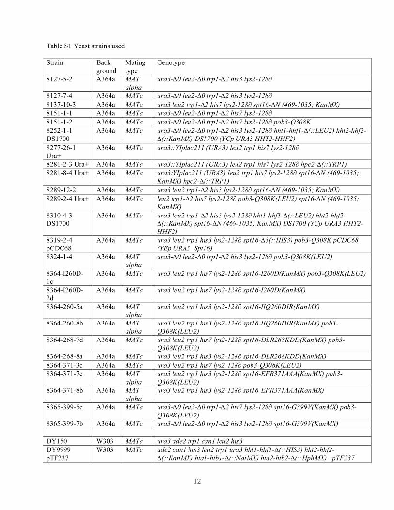
12
Table S1 Yeast strains used Strain Back
ground Mating type
Genotype
8127-5-2 A364a MAT alpha
ura3-∆0 leu2-∆0 trp1-∆2 his3 lys2-128∂
8127-7-4 A364a MATa ura3-∆0 leu2-∆0 trp1-∆2 his3 lys2-128∂ 8137-10-3 A364a MATa ura3 leu2 trp1-∆2 his7 lys2-128∂ spt16-∆N (469-1035; KanMX) 8151-1-1 A364a MATa ura3-∆0 leu2-∆0 trp1-∆2 his7 lys2-128∂ 8151-1-2 A364a MATa ura3-∆0 leu2-∆0 trp1-∆2 his7 lys2-128∂ pob3-Q308K 8252-1-1 DS1700
A364a MATa ura3-∆0 leu2-∆0 trp1-∆2 his3 lys2-128∂ hht1-hhf1-∆(::LEU2) hht2-hhf2-∆(::KanMX) DS1700 (YCp URA3 HHT2-HHF2)
8277-26-1 Ura+
A364a MATa ura3::YIplac211 (URA3) leu2 trp1 his7 lys2-128∂
8281-2-3 Ura+ A364a MATa ura3::YIplac211 (URA3) leu2 trp1 his7 lys2-128∂ hpc2-∆(::TRP1) 8281-8-4 Ura+ A364a MATa ura3:YIplac211 (URA3) leu2 trp1 his7 lys2-128∂ spt16-∆N (469-1035;
KanMX) hpc2-∆(::TRP1) 8289-12-2 A364a MATa ura3 leu2 trp1-∆2 his3 lys2-128∂ spt16-∆N (469-1035; KanMX) 8289-2-4 Ura+ A364a MATa leu2 trp1-∆2 his7 lys2-128∂ pob3-Q308K(LEU2) spt16-∆N (469-1035;
KanMX) 8310-4-3 DS1700
A364a MATa ura3 leu2 trp1-∆2 his3 lys2-128∂ hht1-hhf1-∆(::LEU2) hht2-hhf2-∆(::KanMX) spt16-∆N (469-1035; KanMX) DS1700 (YCp URA3 HHT2-HHF2)
8319-2-4 pCDC68
A364a MATa ura3 leu2 trp1 his3 lys2-128∂ spt16-∆3(::HIS3) pob3-Q308K pCDC68 (YEp URA3 Spt16)
8324-1-4 A364a MAT alpha
ura3-∆0 leu2-∆0 trp1-∆2 his3 lys2-128∂ pob3-Q308K(LEU2)
8364-I260D-1c
A364a MATa ura3 leu2 trp1 his7 lys2-128∂ spt16-I260D(KanMX) pob3-Q308K(LEU2)
8364-I260D-2d
A364a MATa ura3 leu2 trp1 his7 lys2-128∂ spt16-I260D(KanMX)
8364-260-5a A364a MAT alpha
ura3 leu2 trp1 his3 lys2-128∂ spt16-IIQ260DIR(KanMX)
8364-260-8b A364a MAT alpha
ura3 leu2 trp1 his3 lys2-128∂ spt16-IIQ260DIR(KanMX) pob3-Q308K(LEU2)
8364-268-7d A364a MATa ura3 leu2 trp1 his7 lys2-128∂ spt16-DLR268KDD(KanMX) pob3-Q308K(LEU2)
8364-268-8a A364a MATa ura3 leu2 trp1 his3 lys2-128∂ spt16-DLR268KDD(KanMX) 8364-371-3c A364a MATa ura3 leu2 trp1 his7 lys2-128∂ pob3-Q308K(LEU2) 8364-371-7c A364a MAT
alpha ura3 leu2 trp1 his3 lys2-128∂ spt16-EFR371AAA(KanMX) pob3-Q308K(LEU2)
8364-371-8b A364a MAT alpha
ura3 leu2 trp1 his3 lys2-128∂ spt16-EFR371AAA(KanMX)
8365-399-5c A364a MATa ura3-∆0 leu2-∆0 trp1-∆2 his7 lys2-128∂ spt16-G399V(KanMX) pob3-Q308K(LEU2)
8365-399-7b A364a MATa ura3-∆0 leu2-∆0 trp1-∆2 his3 lys2-128∂ spt16-G399V(KanMX) DY150 W303 MATa ura3 ade2 trp1 can1 leu2 his3 DY9999 pTF237
W303 MATa ade2 can1 his3 leu2 trp1 ura3 hht1-hhf1-∆(::HIS3) hht2-hhf2-∆(::KanMX) hta1-htb1-∆(::NatMX) hta2-htb2-∆(::HphMX) pTF237

13
(YCp URA3 HHT2-HHF2, HTA1-HTB1)
DY10003 pTF237
W303 MATa ade2 can1 his3 leu2 met15 trp1 ura3 spt16-11 hht1-hhf1-∆(::HIS3) hht2-hhf2-∆(::KanMX) hta1-htb1-∆(::NatMX) hta2-htb2-∆(::HphMX) pTF237 (YCp URA3 HHT2-HHF2, HTA1-HTB1)
DY10890 W303 MATa ade2 can1 his3 leu2 lys2 trp1 ura3 pob3-Q308K DY11923 W303 MAT
alpha ade2 can1 his3 leu2 trp1 ura3 spt16-∆N(469-1035; KanMX)
DY12431 pJW4
W303 MATa ade2 can1 his3 leu2 trp1 ura3 pob3-Q308K spt16-∆N(469-1035; KanMX) pJW4 (YCp URA3 POB3)
8264-17-3 pTF237
W303 MATa ade2 can1 his3 leu2 trp1 ura3 hht1-hhf1-∆(::HIS3) hht2-hhf2-∆(::KanMX3) pob3-Q308K hta1-htb1-∆(::NatMX) hta2-htb2-∆(::HphMX) pTF237 (YCp URA3 HHT2-HHF2, HTA1-HTB1)
8368-3-2 W303 MATa ade2 can1 his3 leu2 trp1 ura3 spt16-∆N(469-1035; KanMX) 8407-10-2 W303 MAT
alpha ade2 can1 his3 leu2 trp1 ura3 spt16-∆N(469-1035) hht1-hhf1-∆(::HIS3) hht2-hhf2-∆(::KanMX) hta1-htb1-∆(::NatMX) hta2-htb2-∆(::HphMX) pTF237 (YCp URA3 HHT2-HHF2, HTA1-HTB1)
7784-1-1 pTF125
S288c MATa leu2-∆1 trp1-∆63 ura3-52 his4-912∂ lys2-128∂ spt16-∆(::TRP1) pTF125 (YEp URA3 SPT16)
Supplemental References 1. Simpson, R. T., and Stafford, D. W. (1983) Proc Natl Acad Sci U S A 80(1), 51-55. 2. Graziano, V., Gerchman, S. E., and Ramakrishnan, V. (1988) J Mol Biol 203(4), 997-1007 3. Formosa, T., Eriksson, P., Wittmeyer, J., Ginn, J., Yu, Y., and Stillman, D. J. (2001) EMBO J
20(13), 3506-3517. 4. Rhoades, A. R., Ruone, S., and Formosa, T. (2004) Mol Cell Biol 24(9), 3907-3917 5. Biswas, D., Dutta-Biswas, R., Mitra, D., Shibata, Y., Strahl, B. D., Formosa, T., and Stillman, D.
J. (2006) EMBO J 25(19), 4479-4489 6. VanDemark, A. P., Blanksma, M., Ferris, E., Heroux, A., Hill, C. P., and Formosa, T. (2006) Mol
Cell 22(3), 363-374 7. O'Donnell, A. F., Brewster, N. K., Kurniawan, J., Minard, L. V., Johnston, G. C., and Singer, R.
A. (2004) Nucleic Acids Res 32(19), 5894-5906 8. Ruone, S., Rhoades, A. R., and Formosa, T. (2003) J Biol Chem 278(November 14), 45288-
45295 9. Ariyoshi, M., Vassylyev, D. G., Iwasaki, H., Nakamura, H., Shinagawa, H., and Morikawa, K.
(1994) Cell 78(6), 1063-1072 10. Brewster, N. K., Johnston, G. C., and Singer, R. A. (1998) J Biol Chem 273(34), 21972-21979 11. Wittmeyer, J., Joss, L., and Formosa, T. (1999) Biochemistry 38(28), 8961-8971. 12. Ghaemmaghami, S., Huh, W. K., Bower, K., Howson, R. W., Belle, A., Dephoure, N., O'Shea, E.
K., and Weissman, J. S. (2003) Nature 425(6959), 737-741

Annie Heroux, David J. Stillman, Christopher P. Hill and Tim FormosaAndrew P. VanDemark, Hua Xin, Laura McCullough, Robert Rawlins, Shayla Bentley,
Overlapping Roles of yFACT SubunitsStructural and Functional Analysis of the Spt16p N-terminal Domain Reveals
doi: 10.1074/jbc.M708682200 originally published online December 18, 20072008, 283:5058-5068.J. Biol. Chem.
10.1074/jbc.M708682200Access the most updated version of this article at doi:
Alerts:
When a correction for this article is posted•
When this article is cited•
to choose from all of JBC's e-mail alertsClick here
Supplemental material:
http://www.jbc.org/content/suppl/2007/12/19/M708682200.DC1.html
http://www.jbc.org/content/283/8/5058.full.html#ref-list-1
This article cites 48 references, 17 of which can be accessed free at
by guest on May 15, 2016
http://ww
w.jbc.org/
Dow
nloaded from