Innovative and Beneficial Use of Dredged Material Advisory ...
Upload
independentCategory
view
0download
0
Current Enzyme Inhibition, 2007, 3, ???-??? 1
1573-4080/07 $50.00+.00 © 2007 Bentham Science Publishers Ltd.
Phospholipase D Inhibition: Beneficial and Harmful Consequences for a
Double-Dealer Enzyme
G. Auricchio*,1, F. D’Aquilio
2, V. Chiurchiù
2, G. Mancino
1 and P.M. Baldini
2
1A.Fa.R. Research Centre, S. Pietro Hospital, Rome,
2Dept. of Biology, University of Rome “Tor Vergata”, Italy
Abstract: One of the most promising strategies for drug design and development is the identification of new molecules
able to selectively inhibit those enzymes involved in pathological processes, without affecting other enzymes associated
with physiological functions. Nevertheless, some enzymes can show a double-edge aspect of their own inhibition, which
can lead to positive as well as negative consequences according to the pathological state. Phospholipase D (PLD), an
ubiquitous enzyme nowadays considered as a critical regulator of several aspects of cell biology and signal transduction
pathways, is a clear example of those double-dealer enzymes. While a great deal has been learned about PLD structure,
biological functions and activation/regulation mechanisms, little yet is known about the derivable effects of its potential
negative regulation, also due to the lack of specific inhibitors. Multiple evidences on PLD involvement in many patho-
logical states development and progression, including inflammation, carcinogenesis and metastases, have been supplied,
so that a deregulation of its activity could contribute to attenuate or slow down the inflammatory and tumour forma-
tion/progression processes. On the other hand, in agreement with other previous observations, we have recently demon-
strated the direct contribution of PLD activation in promoting intracellular mycobacterial killing. In this case, PLD inhibi-
tion resulted in a significant reduction of antimicrobial innate immune response and, hence, in a possible harmful effect.
In the light of the above reported considerations, besides the recent advances in characterising new compounds able to se-
lectively inhibit PLD activity and/or signalling, this review aimed at elucidating the potential dual beneficial/harmful con-
sequences of PLD activity modulation.
Keywords: Phospholipase D, inhibition, tumour, oxidative burst, inflammation, phagocytosis, tuberculosis.
PHOSPHOLIPASE D
Phospholipase D (PLD) is an enzyme which catalyses the hydrolysis of the terminal diester bond of membrane glyc-erophosholipids, resulting in the formation of phosphatidic acid (PA) and the corresponding water-soluble head group as shown in Fig. (1) [1, 2]. PLD was first identified in carrot extracts in 1947 [3] but its activity was not demonstrated in mammalian cells until 1975 [4]. Within mammals, PLD ac-tivity has been mainly described in the brain and the lung but even in hepatocytes, endothelial cells, platelets and sper-matozoa [5]. Its activity varies according to cell types even if it acquires a remarkable importance in phagocytic cells of the immune system [6]. Concerning its subcellular localisa-tion, PLD is enriched in plasma membrane, but also in intra-cellular compartments including cytosol, Golgi, endoplasmic reticulum, endosomes, lysosomes and nuclei but not in mito-chondria [7, 8]. Following the initial PLD cloning by Hammond et al. in 1995 [9], several genes and splice vari-ants have been identified and cloned from different sources including plants, mouse and human and they were thus in-cluded in a superfamily which exerts phosphodiesterase and/or phosphatidyltransferase activities. In mammalian cells, two PLDs isoforms, PLD1 and PLD2, have been cloned and expressed from human and mouse species [9, 10]. Both these isoforms are widely expressed, although their relative levels vary among different cell types [11]. Mam-malian PLD1 cDNA was cloned from a HeLa cell library, it encodes for a 1074 amino acids protein, and is mainly local-ized perinuclearly, exhibiting a broad distribution to several
*Address correspondence to this author at the A.Fa.R Researh Centre, S.
Pietro Hospital, Rome, Italy; E-mail: [email protected]
organelles, including endosomes, lysosomes, secretory gran-ules, plasma membrane, Golgi apparatus as well as diffuse cytoplasmic staining. This isoform of PLD is, in turn, char-acterised by two alternately spliced forms, named PLD1a and PLD1b [10]. Even though PLD1b is 38 amino acid shorter than PLD1a, they both retain a similar activity and regulation. The second mammalian PLD, PLD2, was cloned from mouse cDNA and is 50% homologous to PLD1. PLD2 gene was actually cloned due to the identification of related sequences in expressed sequence tags databases. It is local-ised in lipid raft fractions on the plasma membrane in quies-cent cells but becomes associated with endocytic vesicles following serum activation, suggesting a putative role of PLD2 in changing cell morphology [11]. Furthermore, im-munoelectron microscopy demonstrated that PLD2 was pre-sent also on Golgi cisternal rims. Both PLDs share a phox homology (PX) domain followed by a pleckstrin homology (PH) domain, allowing these enzymes to bind to phosphoi-nositides [12]. Moreover, four motifs, named I-IV, can be identified within all the members of PLD superfamily, point-ing out their essential role in catalysis. In particular, motif II and IV are also known as HKD or PLDc (Fig. (2)) [13]. Mammalian PLDs exert their activity both as membrane-bound and cytosolic forms, reflecting also a different sub-strate-specificity [14]. Even if measurements of biochemical activity indicate that PLD is expressed in most cells, the dif-ferential expression of the two isoforms has been only partial due to the lack of high-affinity antibodies coupled with the low levels of expression of these proteins [15]. Endogenous PLD isoforms expression levels can also be detected through measurements of mRNA levels. This analysis clearly dem-onstrates a differential level of PLD1 and PLD2 expression with respect to primary tissues and cell types, even though

2 Current Enzyme Inhibition, 2007, Vol. 3, No. 4 Auricchio et al.
most cells co-express both isoforms. PLD activity seems to be particularly evident in the brain. In particular, astrocytes and oligodendrocytes are the main cell types enriched in PLD2 and PLD1, respectively. This appears to be particu-larly interesting, since neurons are not the major sites of PLD enzymes [16].
PLD Enzymology, Regulation and Functions
PLD activation is receptor-linked and it occurs rapidly in a variety of cells in response to diverse agents. This receptor-mediated hydrolysis of cellular phospholipids is a ubiquitous event of central importance in cell signal transduction.
Fig. (1). Regulation of PLD. PLD’s hydrolysis of phosphatidylcholine (alternatively phosphaditylethanolamine or phoshatidylinositol) is
mainly regulated by G-proteins, either trimeric or small GTP-binding proteins (Rho and ARF), and Kinases such as PKC and tyrosine
kinases. Another level of regulation is exerted by Ca2+
ions and PLD’s cofactor PIP2.
Fig. (2). Schematic representation of mammalian PLD enzymes. CR I, II, III and IV represent the four conserved regions. HKD domain
flanked by two conserved regions that are postulated to be important for substrate recognition. HKD motifs are in pink. PLD1 possess a 116-
residue loop, located after the first HKD motif (absent in PLD2) and termed the ‘activation loop’, which might be involved in the regulation
of enzyme activity. Abbreviations: PX, phox domain; PH, pleckstrin-homology domain; HKD, H(X)K(X)(4)D conserved hydrolysis domain;
CT, carboxyl-terminal region essential for enzyme activity.
Phospholipase D Inhibition Current Enzyme Inhibition, 2007, Vol. 3, No. 4 3
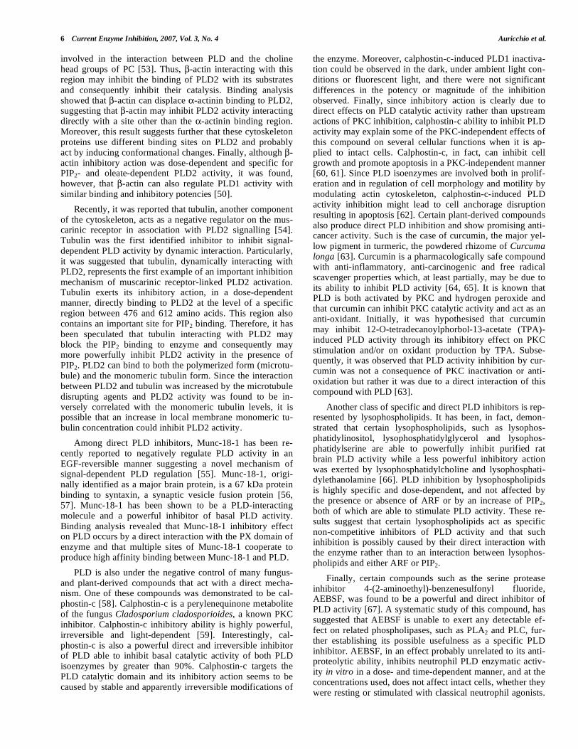
Available evidence indicates that phosphatidylcholine (PC), the major membrane phospholipid, is the preferred substrate for PLD, thus yielding PA and choline
[17]. However, in
some systems such as NIH 3T3 cells fibroblasts, PLD cata-lyzes the hydrolysis of both PC and phosphatidylethanola-mine (PE) upon stimulation of different growth factors [18]. Exclusive degradation of PE occurs in cultured glial cells and in rat mesangial cells
[19, 20]. In addition, a PLD-
catalyzed hydrolysis of phoshatidylinositol (PI) in Madin-Darby canine kidney (MDCK) cells has been demonstrated [14]. Thus, several phospholipids other than PC, can be hy-drolysed. Moreover, PLD catalyzes the transphosphatidyla-tion reaction whereby the phosphatidyl group of the phos-pholipid is transferred to glycerol or a primary alcohol such as ethanol, propanol, or butanol. This reaction is, in fact, regarded as the unequivocal evidence of PLD activity be-cause it yields a phosphatidylalcohol at the expense of PA, the putative second messenger [21]. PLD catalytic reaction proceeds by a ping-pong mechanism in which a phosphoen-zyme intermediate is formed and then hydrolysed in the sec-ond step of the reaction. Thus, at first PLD attacks the phos-pholipid substrate to form a transient phosphatidyl-PLD in-termediate by covalent linkage of PA to a histidine, then either water or other nucleophilic acceptors, such as primary alcohols, are transferred in the second stage of the reaction, as shown in Fig. (3). This explains the particular propensity of PLD enzymes to function as phosphatidyltransferases. PA is the PLD-derived second messenger akin to phospholipase C (PLC)-derived diacilglycerol (DAG). However, PA can also be metabolized to other biologically important lipid me-tabolites including lyso-PA (LPA) and DAG itself. LPA is produced by the action of a specific phospholipase A2 (PLA2) whilst DAG is produced by the action of phosphati-date phosphohydrolase (PAP). Although there is accepted evidence that PA per se is a signalling molecule, its immedi-ate products, DAG and LPA, are also known to have very important physiological functions [22].
Although the various PLD isoforms share the common
catalytic domains and their activity is phosphoinositides-
dependent, their regulation is particularly complex and di-
versified since the dynamics of PLDs intracellular signalling
vary depending on the stimulus, on subcellular localisation and on substrates- and regulators-susceptibilities. Thus, the
regulatory mechanism of PLD activation is still under in-
tense investigation and the precise sequence of upstream PLD activation events remains to be clarified. Overall, three
major upstream activators can be considered: G proteins,
protein kinase C (PKC) and tyrosine kinases. G protein-dependent PLD regulation has been demonstrated through
the observation of an increased PLD activity by several me-
diators, such as the platelet-activating factor, the chemotactic peptide N-formyl-methionyl-leucyl-phenylalanine (fMLP),
the leukotriene B4, the C5a complement component, all of
which function via G proteins activation [23]. Moreover, it was later demonstrated that membrane-bound PLD isoform
activation requires small GTP-binding proteins of the ADP
ribosylation factor (ARF) and the Rho families. There are six ARF proteins in mammalian cells [24]. They do not show a
significant difference in activating PLDs, although they ap-
parently differ in their subcellular localisation. These differ-ences may thus result in a selective involvement of different
ARFs in controlling PLD activity in different regions of the
cell. Rho GTP-binding proteins family was first reported to stimulate PLD activity in several cell types, including neu-
trophils and HL-60 cells. In particular, RhoA was the most
effective Rho family member for PLD activation in both HL-60 and liver cells. Furthermore, Rho is known to act syner-
gistically with ARF to activate a PLD isoform from porcine
brain. In addition, the binding of different agonists, ie fMLP or IL-8, to its seven-transmembrane receptor induces ARF
and RhoA translocation, leading to a subsequent PLD activa-
tion [25]. Besides the small GTP-binding proteins-mediated PLD regulation, the main regulator of PLD activity is repre-
sented by PKC. In fact, it has been clearly demonstrated how
PKC activators such as phorbol 12-myristate 13-acetate (PMA) greatly stimulate PLD in a large variety of tissues
and cells [26, 27]. PKC-mediated PLD1 regulation has been
specifically investigated in some detail: in vitro, either PKC-
Fig. (3). Catalytic mechanism of PLD. PLD catalytic reaction proceeds by a ping-pong mechanism in which a phospatidyl-PLD intermediate
is formed in the first step of the reaction and then either a water molecule or any other nucleophilic acceptor replace the phospatidyl group.

4 Current Enzyme Inhibition, 2007, Vol. 3, No. 4 Auricchio et al.
, PKC- I or PKC- II all stimulate PLD1 activity. This
mode of regulation has been shown to be independent of, but
synergistic with, the other PLD activators [28]. Moreover, the general mechanisms by which this kinase is able to regu-
late PLD activity can be abridged into two different types of
regulation: through a direct activation or downregulation of PKC and an indirect activation by agonist cell stimulation
including several PKC inhibitors. However, the role of PKC
in PLD regulation seems to be rather contradictory since some agonists-induced PLD stimulation results not to be
inhibited by PKC inhibitors, thus once again suggesting the
existence of various PLD isoforms. Finally the role of tyro-sine phosphorylation in controlling PLD activity is also of
great interest since several evidences are accounting for a
tyrosin kinases-dependent PLD regulation even if the precise mechanism is yet to be elucidated. In fact, tyrosin kinase
receptors, including those for epidermal growth factor (EGF)
and platelet-derived growth factor (PDGF), are able to stimulate PLD activity in different cell types [29]. This has
been suggested either by means of tyrosine kinase inhibitors
or through tyrosine phosphatases inhibitors [30]. In tyrosine kinase-mediated PLD activation, the regulation could be
exerted through a direct or indirect tyrosine phosphorylation
mechanism of PLD isoforms. Indirect tyrosine phosphoryla-tion-mediated PLD activation is brought about by several
downstream intermediates such as Src, Syk and Ras [31, 32].
Among other factors that are involved in PLD regulation, Ca
2+ ions and phosphatidylinositol bisphosphate (PIP2) have
to be taken into account. With respect to Ca2+
, a rise in cyto-
solic Ca2+
determines PLD stimulation even if there’s still no evidence whether this is a Ca
2+-dependent PKC isozymes
process or it accounts for an actual Ca2+
involvement in PLD
activation [22]. Furthermore, the role of PIP2 in PLD activa-tion has been first discovered in 1993 [33] and later con-
firmed by several other studies; this has led to the identifica-
tion of PIP2 as a cofactor for PLD activity. Finally, it is not surprising that, given their differential localisation, PLD1
and PLD2 modes of regulation show several differences. The
stimulation of PLD1 and PLD2 activities by PKC and PIP2 has been well documented. Additionally, members of the
Rac- and Rho-GTPase family regulate both PLD1 and PLD2
activities. However, the best characterised regulator of PLD1 is represented by the small GTP-binding protein ARF. Al-
though PLD1 and PLD2 are activated by ARFs, the two iso-
forms vary in their sensitivities to individual ARFs. The GTP-bound form of ARF1 stimulates PLD1 catalytic activity
more than 13-fold, whereas PLD2 is only stimulated 1.5-
fold. Interestingly, deletion of the N-terminal 308 amino acid residues renders PLD2 as ARF responsive as PLD1.
According to its ubiquitous distribution and its complex regulation, the physiological role of PLD is almost heteroge-neous since this enzyme is involved in different biological processes, including neutrophil activation, steroidogenesis, synthesis of acetylcholine, insulin release [34]. The differen-tial distribution of PLD1 and PLD2 implies that these en-zymes have separate functions in the Golgi apparatus. Be-cause PLD2 has a relatively high basal activity, it might function as a “housekeeping” enzyme and maintain a local-ised pool of PA, whereas ARF1-activated PLD1 might play a role in modulation PA levels. In addition, the rim localisa-
tion of PLD2 implies a regulatory role in mediating vescicu-lar trafficking, analogous to the recent observations that pro-tein components of the retrograde transport machinery are localized to rims of the Golgi apparatus [35]. Thus the most evident and studied biological process regulated by PLD is vescicular trafficking. PLD involvement in vescicular trans-port was, in fact, first suggested by observations that many secretion stimuli were able to increase PLD activity in HL-60 cells [36]. Subsequent evidences confirmed the phosphol-ipase involvement in several vescicular transport processes, such as the transport from the endoplamic reticulum to Golgi and fusion of secretory vescicles with plasma membrane. Concerning this function, it has been clearly demonstrated that PLD participation in vescicular trafficking is actually associated with ARF and Rho [5]. In addition, several other physiological processes are exerted by PLD due to the sec-ond messenger role played by its product PA. This metabo-lite is, in fact, involved in a numerous functions, including cell signalling, mitogenesis, inflammation, cell proliferation, actin assembly and respiratory burst [37, 27]. PLD activity has also been shown to be significantly elevated in human cancers including breast, renal, gastric and colon cancer sug-gesting that PLD might be implicated in tumourigenesis. Indeed, Uchida et al. [38, 39] both showed that PLD activity is increased in human breast cancer tissue and in human gas-tric carcinoma. In addition, PLD activity is significantly higher in tumour tissue than in adjacent normal tissue of hu-mans with renal cancer and that the abundance of PLD2 in the nucleus was increased in the carcinoma cells [40]. Fur-thermore, Yoshida et al. [41] detected increased levels of oleate-dependent and ARF-dependent PLD isoforms in ex-perimental colon cancer. Finally, mouse fibroblasts overex-pressing either PLD1 or PLD2 were shown to undergo an-chorage-independent growth in soft agar and to form undif-ferentiated sarcomas in nude mice [42]. All this observations are also supported by the suggestion that PLD2 gene is a susceptibility locus for colo-rectal cancer in Japanese indi-viduals, and that this polymorphism can be used as a genetic marker for predisposition to this condition [43].
THE ROUTES OF PLD INHIBITION
Although the importance of PLD as a critical regulator of several aspects of cell biology and signal transduction path-ways is well documented, little is currently known concern-ing PLD negative regulation due mainly to the lack of spe-cific inhibitors of this phospholipase. To this regard, how-ever, remarkable advances have been recently made and a series of compounds of varied origin with evident anti-PLD activity, schematized in Fig. (4), are now beginning to emerge. These include phospholipid- and sphingolipid-derived products, cytosolic proteins purified and character-ised in many mammalian tissues, principally rat and bovine brain, G-protein related factors, steroidal and non-steroidal drugs, plant- and fungus-derived products and other chemi-cal compounds. The results available to date suggest that the diverse and function related PLD inhibitors may regulate PLD activity by different mechanisms in different cells even though in some cases there is not enough evidence to distin-guish whether the inhibitory effect of these molecules is due to a direct or an indirect mechanism.

Phospholipase D Inhibition Current Enzyme Inhibition, 2007, Vol. 3, No. 4 5
Direct Inhibitors
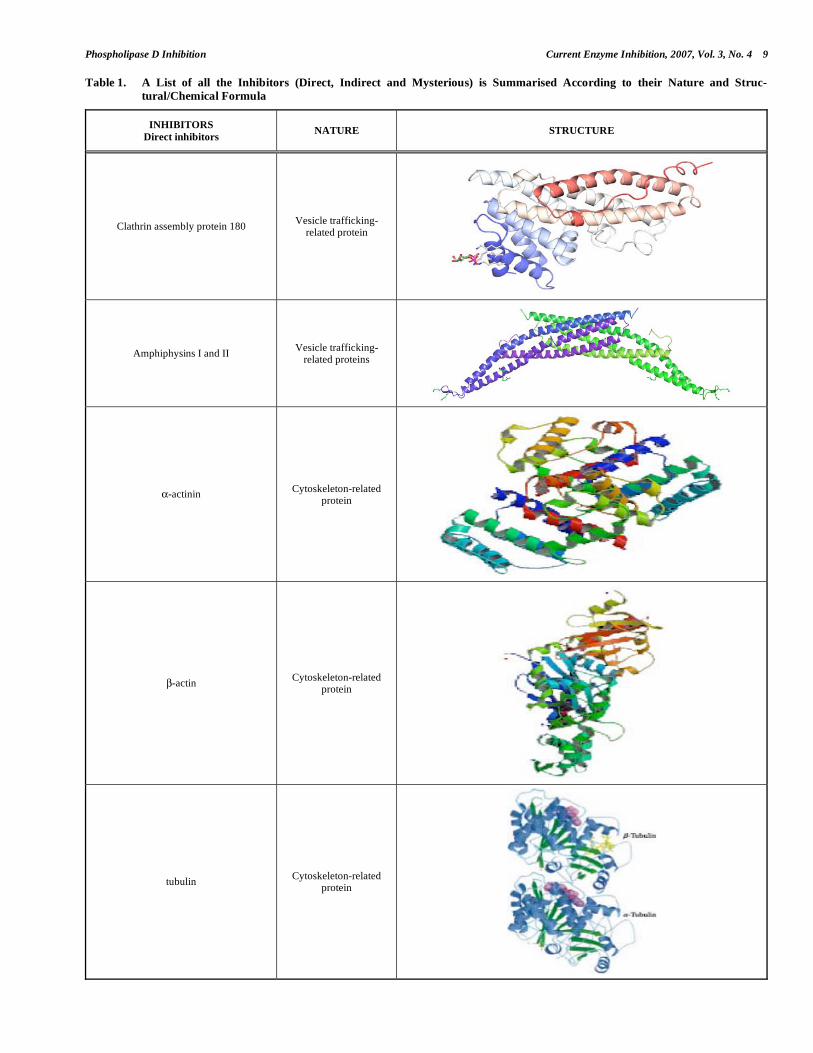
PLD negative regulation has been reported to be under the control of several compounds acting with direct mecha-nisms. Among direct PLD inhibitors that have been identi-fied, the several distinct proteins purified from rat brain cy-tosol and classified as vesicle trafficking-related proteins are the best known. Particularly, clathrin assembly protein 180 (AP180, previously known as clathrin assembly protein 3 or AP3) [44], one of several assembly proteins involved in clathrin-coated vesicles formation, and amphiphysins I and II [45], are able to inhibit PLD activity directly interacting with PLD molecule. AP180 dose-dependently inhibits human PLD1 activity and such an inhibitory effect appears to be independent by PIP2 binding because the potency and effi-cacy of AP180 as a PLD inhibitor were similar in the ab-sence and presence of this essential cofactor. AP180 NH2-terminal 33 kDa contains the region required for PLD inhibi-tion and its C-terminal 3 kDa region might be critical for interaction with PLD1. Moreover, the fact that AP180 is a synapse-specific protein suggests that AP180-induced PLD inhibition might play a regulatory role that is restricted to the rapid cycling of synaptic vesicles [44]. Amphiphysins I and II are highly concentrated in nerve-terminals and are both involved in clathrin-mediated endocytosis [46, 47]. These proteins, which form a heterodimer, exert their inhibitory effect on PLD1 and PLD2 through a direct interaction with
both enzymes. Particularly, it has been observed that the NH2 terminus of amphiphysins I was critical for both inhibition of and binding to PLD [45].
It has been reported that PLD activity can be also nega-tively modulated by various inhibitors classified as cy-toskeleton-related proteins. Since it is widely confirmed that PLD is crucially involved in cell actin-based cytoskeleton rearrangement [48], cytoskeleton proteins regulation of PLD activity may play an important role in controlling the cy-toskeleton-related PLD functions. Among the several cy-toskeletal proteins with PLD-inhibitory activity, -actinin and -actin have been found to act as specific and direct PLD inhibitors [49, 50]. -actinin, an F-actin cross-linking protein known as a key regulator of actin-based structures such as stress fibres and focal adhesions [51], was found to powerfully inhibit PLD2 basal activity interacting directly with the N-terminal 185 amino acids, which contain PX do-main of enzyme [49]. Moreover, PLD was inhibited by -actinin in an ARF-reversible manner. Particularly, ARF1 overcomes -actinin-attenuated PLD2 activity by eliminat-ing its interaction with enzyme. -actin, extracted from rat brain, negatively regulates PLD by a direct binding to the region between amino acids 613 and 723 of the enzyme [50]. This -actin-binding region of PLD2 contains the conserved region III that seems critically important for PLD function [52]. It has been, in fact, suggested that this region might be
Fig. (4). Inhibition of PLD. The figure shows the schematic structure of mammalian PLD2 whose activity is inhibited by direct inhibitors
which directly interact with phospholipase itself, indirect inhibitors whose action is worked out at the upstream regulators of PLD, and mys-
terious inhibitors whose exact mechanism of inhibition is still equivocal. The same inhibitors exert similar action on mammalian PLD1 as
well. Abbreviations: PX: phox domain; PH: pleckstrin-homology domain; CR: conserved region; CT: carboxyl-terminal region essential for
enzyme activity.
6 Current Enzyme Inhibition, 2007, Vol. 3, No. 4 Auricchio et al.
involved in the interaction between PLD and the choline head groups of PC [53]. Thus, -actin interacting with this region may inhibit the binding of PLD2 with its substrates and consequently inhibit their catalysis. Binding analysis showed that -actin can displace -actinin binding to PLD2, suggesting that -actin may inhibit PLD2 activity interacting directly with a site other than the -actinin binding region. Moreover, this result suggests further that these cytoskeleton proteins use different binding sites on PLD2 and probably act by inducing conformational changes. Finally, although -actin inhibitory action was dose-dependent and specific for PIP2- and oleate-dependent PLD2 activity, it was found, however, that -actin can also regulate PLD1 activity with similar binding and inhibitory potencies [50].
Recently, it was reported that tubulin, another component of the cytoskeleton, acts as a negative regulator on the mus-carinic receptor in association with PLD2 signalling [54]. Tubulin was the first identified inhibitor to inhibit signal-dependent PLD activity by dynamic interaction. Particularly, it was suggested that tubulin, dynamically interacting with PLD2, represents the first example of an important inhibition mechanism of muscarinic receptor-linked PLD2 activation. Tubulin exerts its inhibitory action, in a dose-dependent manner, directly binding to PLD2 at the level of a specific region between 476 and 612 amino acids. This region also contains an important site for PIP2 binding. Therefore, it has been speculated that tubulin interacting with PLD2 may block the PIP2 binding to enzyme and consequently may more powerfully inhibit PLD2 activity in the presence of PIP2. PLD2 can bind to both the polymerized form (microtu-bule) and the monomeric tubulin form. Since the interaction between PLD2 and tubulin was increased by the microtubule disrupting agents and PLD2 activity was found to be in-versely correlated with the monomeric tubulin levels, it is possible that an increase in local membrane monomeric tu-bulin concentration could inhibit PLD2 activity.
Among direct PLD inhibitors, Munc-18-1 has been re-cently reported to negatively regulate PLD activity in an EGF-reversible manner suggesting a novel mechanism of signal-dependent PLD regulation [55]. Munc-18-1, origi-nally identified as a major brain protein, is a 67 kDa protein binding to syntaxin, a synaptic vesicle fusion protein [56, 57]. Munc-18-1 has been shown to be a PLD-interacting molecule and a powerful inhibitor of basal PLD activity. Binding analysis revealed that Munc-18-1 inhibitory effect on PLD occurs by a direct interaction with the PX domain of enzyme and that multiple sites of Munc-18-1 cooperate to produce high affinity binding between Munc-18-1 and PLD.
PLD is also under the negative control of many fungus- and plant-derived compounds that act with a direct mecha-nism. One of these compounds was demonstrated to be cal-phostin-c [58]. Calphostin-c is a perylenequinone metabolite of the fungus Cladosporium cladosporioides, a known PKC inhibitor. Calphostin-c inhibitory ability is highly powerful, irreversible and light-dependent [59]. Interestingly, cal-phostin-c is also a powerful direct and irreversible inhibitor of PLD able to inhibit basal catalytic activity of both PLD isoenzymes by greater than 90%. Calphostin-c targets the PLD catalytic domain and its inhibitory action seems to be caused by stable and apparently irreversible modifications of
the enzyme. Moreover, calphostin-c-induced PLD1 inactiva-tion could be observed in the dark, under ambient light con-ditions or fluorescent light, and there were not significant differences in the potency or magnitude of the inhibition observed. Finally, since inhibitory action is clearly due to direct effects on PLD catalytic activity rather than upstream actions of PKC inhibition, calphostin-c ability to inhibit PLD activity may explain some of the PKC-independent effects of this compound on several cellular functions when it is ap-plied to intact cells. Calphostin-c, in fact, can inhibit cell growth and promote apoptosis in a PKC-independent manner [60, 61]. Since PLD isoenzymes are involved both in prolif-eration and in regulation of cell morphology and motility by modulating actin cytoskeleton, calphostin-c-induced PLD activity inhibition might lead to cell anchorage disruption resulting in apoptosis [62]. Certain plant-derived compounds also produce direct PLD inhibition and show promising anti-cancer activity. Such is the case of curcumin, the major yel-low pigment in turmeric, the powdered rhizome of Curcuma longa [63]. Curcumin is a pharmacologically safe compound with anti-inflammatory, anti-carcinogenic and free radical scavenger properties which, at least partially, may be due to its ability to inhibit PLD activity [64, 65]. It is known that PLD is both activated by PKC and hydrogen peroxide and that curcumin can inhibit PKC catalytic activity and act as an anti-oxidant. Initially, it was hypothesised that curcumin may inhibit 12-O-tetradecanoylphorbol-13-acetate (TPA)-induced PLD activity through its inhibitory effect on PKC stimulation and/or on oxidant production by TPA. Subse-quently, it was observed that PLD activity inhibition by cur-cumin was not a consequence of PKC inactivation or anti-oxidation but rather it was due to a direct interaction of this compound with PLD [63].
Another class of specific and direct PLD inhibitors is rep-resented by lysophospholipids. It has been, in fact, demon-strated that certain lysophospholipids, such as lysophos-phatidylinositol, lysophosphatidylglycerol and lysophos-phatidylserine are able to powerfully inhibit purified rat brain PLD activity while a less powerful inhibitory action was exerted by lysophosphatidylcholine and lysophosphati-dylethanolamine [66]. PLD inhibition by lysophospholipids is highly specific and dose-dependent, and not affected by the presence or absence of ARF or by an increase of PIP2, both of which are able to stimulate PLD activity. These re-sults suggest that certain lysophospholipids act as specific non-competitive inhibitors of PLD activity and that such inhibition is possibly caused by their direct interaction with the enzyme rather than to an interaction between lysophos-pholipids and either ARF or PIP2.
Finally, certain compounds such as the serine protease inhibitor 4-(2-aminoethyl)-benzenesulfonyl fluoride, AEBSF, was found to be a powerful and direct inhibitor of PLD activity [67]. A systematic study of this compound, has suggested that AEBSF is unable to exert any detectable ef-fect on related phospholipases, such as PLA2 and PLC, fur-ther establishing its possible usefulness as a specific PLD inhibitor. AEBSF, in an effect probably unrelated to its anti-proteolytic ability, inhibits neutrophil PLD enzymatic activ-ity in vitro in a dose- and time-dependent manner, and at the concentrations used, does not affect intact cells, whether they were resting or stimulated with classical neutrophil agonists.
Phospholipase D Inhibition Current Enzyme Inhibition, 2007, Vol. 3, No. 4 7
In addition, its inhibitory effect was exerted directly on the enzyme as demonstrated in three cell-free systems: cell soni-cates, purified plant enzyme, and anti-PLD immunoprecipi-tates. The fluoride atom is necessary for its PLD inhibitory effect since the compound AEBSNH2, which has the fluoride replaced by an –NH2 group, failed to affect PLD activity as did other compounds structurally related to AEBSF with known protease inhibitory capabilities. The p-aminoethyl group was also important to confer an effect [67].
Indirect Inhibitors
Among indirect PLD inhibitors, recently, 5-[4-acridin-9-ylamino]-5-methyl-3-methylendihydrofuran-2-one (CYL-26z) was found to negatively regulate the fMLP-stimulated PLD activity emerging as an attractive potential anti-inflammatory compound [68]. CYL-26z seems, in fact, to interrupt the receptor-coupled signalling pathways leading to PLD activation upon fMLP stimulation. Particularly, it has been hypothesised that CYL-26z may exert its inhibitory action on PLD activity mainly blocking of RhoA activation and probably also inhibiting membrane recruitment of PKC .
Another possible indirect way of inhibiting PLD activity is represented by the enzymes and/or compounds regulating PIP2 levels, since PIP2 is an essential cofactor for PLD activ-ity [22]. To this regard, synaptojanin, a specific inositol polyphosphate phosphatase purified from rat brain cytosol, has been identified as a PLD-inhibitory protein that acts by decreasing PIP2 levels through its PIP2 5-phosphatase activ-ity [69]. Synaptojanin inhibitory action of ARF and PIP2-stimulated PLD, in fact, was not due to a competition with the activators or substrate of PLD but it was rather attributed to its ability to dephosphorylate PIP2. Since the degree of PIP2 hydrolysis is correlated with the extent of PLD1 inhibi-tion, it was concluded that synaptojanin inhibited PLD1 by hydrolysing this necessary co-factor. In addition, the treat-ment of intact cells with clostridial toxins, which decrease PIP2 by inhibition phosphatidylinositol-4-phosphate 5-kinase (PI-4-P 5-kinase), causes inhibition of agonist-induced PLD stimulation [70]. Particularly, Clostridium difficile toxin B, an agent which inactivates Rho-family low-molecular mass G proteins [71, 72], powerfully and selectively interferes with receptor coupling to PLD as well as with GTP (S)-induced PLD stimulation [73]. Toxin B does not modify PLD enzyme per se but may inhibit receptor signalling to PLD decreasing the cellular levels of PIP2, which depend on the activity state of Rho proteins. Toxin B inhibitory effect of PLD activity, in fact, seems to be due to Rho inactivation following the subsequent inhibition of PI-4-P 5-kinase acti-vation and reduced supply of PIP2 cofactor limiting, thus, the stimulation of PLD activity. In vitro studies indicate that PLD activity is also regulated by PIP3 [2]. Therefore, another interesting possibility to inhibit PLD would be to prevent phosphatidylinositol trisphosphate (PIP3) generation from PIP2 by phosphatidylinositol-3-phosphate kinase (PI3K). Several studies, in fact, have shown that wortmannin and LY294002, inhibitors of PI3K, inhibit agonist-induced PLD activation [74, 75]. The first indirect PLD inhibitors to be extensively evaluated, in fact, were wortmannin and its de-rivates [76].
A natural compound which acts through an indirect in-hibitory mechanism is farnesol. It is a catabolite of the cho-lesterol/isoprenoid biosynthetic pathway whose exogenous administration has been observed to preferentially cause apoptosis in neoplastic vs. normal cells [77, 78]. PLD signal-ling is a key mediator of farnesol-induced apoptosis and far-nesol inhibition of PLD signal transduction enhanced apop-tosis [79]. Consistent with the importance of DAG signalling is the requirement for PLD conversion of PC to DAG for effective inhibition of farnesol-induced apoptosis. The non-metabolisable DAG mimetic -TPA, a pharmacological agent widely used to active DAG-responsive PKC enzymes and other proteins containing DAG-binding C1 domains [80, 83], also attenuated farnesol-induced apoptosis. As DAG and
-TPA inhibited farnesol-induced apoptosis this implies that farnesol prevented direct inhibition of an antiapoptotic DAG binding target. It is, therefore, proposed that the PLD signal transduction pathway substantially contributes to the genera-tion of an antiapoptotic/pro-proliferative DAG pool, and signalling by this DAG pool is inhibited by farnesol. The precise farnesol target remains to be determined but the evi-dences support a mechanism by which farnesol inhibits DAG activation of an antiapoptotic C1-domain containing protein [79].
Among substances recognised to have indirect inhibitory properties on PLD activity, tamoxifen (TAM), a non-steroidal anti-estrogen, is of great interest. This drug, in fact, has been used widely and effectively to treat hormone-responsive breast cancer [84, 85]. Long term (24 h) but not short term (30 min) treatments with clinically relevant (2-5 μM) TAM concentrations, inhibited PMA-induced PLD ac-tivity by 50-80%, both in MCF-7MDR cells, an estrogen receptor-negative multidrug-resistant subline of human breast carcinoma, and NIH 3T3 fibroblasts [86]. Although the negative regulation of TAM on PLD activity was not caused by membrane translocation or down-regulation of PKC- , the major mediator of phorbol meristate acetate ef-fects on PLD activation [87], PKC- function as a regulator of PLD activity in MCF-7MDR cells was blocked by ex-tended treatments with TAM. These results, therefore, raise the possibility that prolonged inhibition of PKC- regulated PLD system, an important component of the signal transduc-tion pathways in breast cancer cells, may contribute to the cytotoxic effects of TAM and to be clinically relevant in treating breast cancer patients. Finally, with respect to sphin-golipids, ceramide, a product of sphingomyelinase activity, has been found to indirectly inhibit membrane-bound PLD activity previously stimulated by ARF and PKC [88]. This finding, in fact, suggested that PLD stimulation by PKC was the target for this sphingolipid inhibitor rather than a direct action on PLD.
Mysterious Inhibitors
As above reported, PLD activity can be also inhibited by several compounds whose exact mechanism is poorly known. Such is the case of cytoskeleton protein, non-erythroid spectrin fodrin, the first naturally described PLD inhibitor [89]. This protein, purified from bovine brain cyto-sol, fully and quickly inhibited PLD activity, whatever the stimuli used. At the light of the results obtained, it seemed

8 Current Enzyme Inhibition, 2007, Vol. 3, No. 4 Auricchio et al.
unlikely that fodrin could exert its inhibitory action on PLD preventing the access of enzyme to either its substrate (PC) or its cofactor (PIP2). Fodrin, instead, may act preventing PLD translocation to the membrane, thus maintaining the enzyme under an inactive state. To restore an active enzyme, post-translational modifications of the cytoskeleton protein and/or PLD itself, including proteolysis and/or phosphoryla-tions, are probably necessary. It has been proposed that the enzyme regulation is due to complex relationships between PLD itself, fodrin, and cytosolic proteins such as small G-proteins. Thus, PLD would be normally maintained inactive in resting cells by fodrin. Following cell activation, changes at the level of several cytoskeletal proteins may modify fodrin inhibitory effect on PLD by removing either a direct effect of fodrin on the enzyme or its access to the plasma membrane. It is also possible that the fodrin PH domain in-hibits PLD activity by PIP2 binding.
- and -synucleins, components of synaptic vesicles, add to the group of PLD inhibitory proteins extracted and purified from bovine brain [90]. Although a detailed mecha-nism of action by which synucleins inhibit PLD activity has not been established yet, the inhibitory effect is clearly inde-pendent of the activating effect of PIP2. Moreover, although the basic catalytic properties of both PLD isoenzymes are very similar, synucleins display a stronger selectivity for PLD2 inhibition rather than PLD1. Even though it is logical to postulate that this effect may be explained only through a direct interaction between PLD2 and synuclein probably occurring at the membrane surface, further investigations will be required to test this possibility.
Among inhibitory compounds of PLD activation acting with a mechanism which is not completely clear, the multi-ple phospholipid- and sphingolipid-derived products are the best characterised [91]. To this regard, the ether-linked phospholipid analogues include several families of com-pounds with antiproliferative, cytotoxic and antitumoural properties whose mechanism of action is poorly understood [92]. Particularly, the alkylphosphocholines (APCh), phos-pholipid-derived compounds, retain a remarkable importance by means of their promising anti-cancer activity. Chemi-cally, the APCh are ether lipids without the glycerol back-bone and, according to their structure, they can be classified into three different groups: 1) APCh with a typical choline polar head and a saturated alkyl chain; 2) APCh with a cyclic polar head and a saturated alkyl chain; 3) APCh with a typi-cal or slightly modified choline polar head and an unsatu-rated alkyl chain [93]. Among them, one the most attractive and effective compounds with antitumoural effects has turned out to be hexadecyl-phosphorylcholine (HePC), also known as miltefosine, an APCh used for the topical treat-ment of cutaneous metastases and local recurrences in breast cancer patients [94]. Initially, some reports have related HePC antitumoural activity to its ability to interfere with phospholipid metabolism. Interestingly, at the light of the current knowledge, it appears that the potential anti-cancer properties of HePC may be related to its strong inhibitory action on PLD [92], in agreement with the anti-proliferative effects of APC compounds. Although HePC ability to inhibit PLD activation was common to both PLD1 and PLD2 iso-forms, the exact mechanism by which this compound exerts its inhibitory action has yet to be determined. HePC, in fact,
may act either through direct action on PLD (non-specific proteolysis) or through a direct effect on PKC.
Some substances such as aluminium fluoride (AlF4), be-ryllium fluoride, and orthovanadate can be added to the group of inhibitory compounds of PLD acting as competitive inhibitors. More specifically, AlF4 has been found to inhibit GTP- -S-induced PLD activation in rat submandibular gland acinar cells. Such AlF4 inhibitory effect was confirmed in permeabilized cells in a concentration-dependent manner [95]. It is known that PLD stimulation with ARF/GTP- -S involves ARF translocation from cytosol to membranes [96]. In submandibular membranes, however, AlF4 inhibited rARF-stimulated PLD without blocking ARF translocation to membranes. This effect suggests that the site of action of AlF4 in PLD inhibition is located at a post-ARF-translocation locus. In this case, since AlF4 inhibited PLD in the membrane fraction alone, a non GTP-binding membrane-associated target is proposed. This target could be a PLD regulatory protein, or the enzyme itself [95]. Since fatty acid (oleate)-sensitive PLD, which is not guanine nucleotide-dependent, was also powerfully inhibited by AlF4 in a con-centration-dependent way, it is excluded a G-proteins in-volvement in the mechanism through which AlF4 inhibits PLD activity. Additional finding of AlF4 ability to inhibit PLD, which is highly expressed on Golgi-enriched mem-branes from submandibular cells, led to speculation that this substance may act blocking vesicle transport in the Golgi following PLD inhibition. Although the exact mechanism of action is unknown, it is proposed that AlF4 inhibits different form of PLD by a G-protein-independent mechanism and that it acts via a membrane-associated target which seems to be PLD itself. Moreover, inhibitory effect was not caused by either fluoride or aluminum alone and was reversed by alu-minum chelation. Moreover, AlF4-induced PLD inhibition was not mediated by cAMP, phosphatases 1, 2A or 2B, or PAP.
With respect to antibiotics and other drugs, some antima-larial compounds such as chloroquine and amodiaquine have been recognized to have PLD inhibitory properties although the precise inhibitory mechanism of these molecules is not clearly elucidated [97]. Chloroquine, a drug known to inter-fere with lysosomal function, was found to inhibit intestinal Ca
2+ absorption [98]. Amodiaquine is a 4-aminoquinoline
derivative used in the malaria prophylaxis and treatment with schizonticidal activity [99]. It accumulates in the lysosomes and brings about loss of function. Such agents are cationic amphiphilic compounds and they contain a hydrophobic re-gion and primary or substituted amino group providing a net positive charge. They interact with hydrophobic regions and with the anionic groups of phospholipids [100, 101]. Chloro-quine and amodiaquine dose-dependently inhibited Ca
2+
stimulated rat enterocyte mitochondrial PLD activity whereas these drugs had no significant effect on oxidant- or polyamines-stimulated PLD. Moreover, increasing the Ca
2+
concentration partially reversed the PLD inhibition by these compounds. These observations suggest that inhibitory effect of these drugs could be produced either by competing for the same binding site as of calcium on the enzyme or possibly through their binding with calcium making it unavailable for PLD activation. Concerning antibiotics, aminoglycosides such as neomycin, kanamycin and several others have been
Phospholipase D Inhibition Current Enzyme Inhibition, 2007, Vol. 3, No. 4 9
Table 1. A List of all the Inhibitors (Direct, Indirect and Mysterious) is Summarised According to their Nature and Struc-
tural/Chemical Formula
INHIBITORS
Direct inhibitors NATURE STRUCTURE
Clathrin assembly protein 180 Vesicle trafficking-
related protein
Amphiphysins I and II Vesicle trafficking-
related proteins
-actinin Cytoskeleton-related
protein
-actin Cytoskeleton-related
protein
tubulin Cytoskeleton-related
protein

10 Current Enzyme Inhibition, 2007, Vol. 3, No. 4 Auricchio et al.
(Table 1). Contd…..
Munc 18-1 A major brain protien enriched in neurons
?
Calphostin-c
Perylenequinone me-tabolite of the fungus
Cladosporium cal-dosporoide
CH3
O
O
OH
O
CH3OOH
O
H3C
O
H3C
OH O
O
CH3
O
O
OH
Curcumin
Yellow pigment in turmeric, the powdered
rhizome of Curcuma longa
CH CH
CH3O
OH OH
OCH3
CH CH
C CO OCH2
lysophospholipids lipids
O
O
O
P
O
O
O-
R
HO H
inositol (LPI)
glycerol (LPG)
R = serine (LPS)
choline (LPC)
ethanolamine (LPE)
AEBSF Serine protease inhibitor
H2N
S
F
O O
Indirect Inhibitors
CYL-26z A synthetic -methylene- -
butyrolactone derivative
N
HN
OO
H3C
synaptojanin Inositol polyphosphate
phosphatase

Phospholipase D Inhibition Current Enzyme Inhibition, 2007, Vol. 3, No. 4 11
(Table 1). Contd…..
Clostridium difficile toxin B protein
Wortmannin A metabolite of the
fungus Penicillium
funiculosum O
O
O O
O
H3C O
H3CO
OCH3
CH3
LY294002 A synthetic PI3K inhibi-
tor O N
O
O
Farnesol
Cholesterol/isoprenoid
biosynthetic pathway catabolite
CH2OH
Tamoxifen Non-steroidal anti-
estrogen
O
N
Ceramide Sphingolipid
OH
CH2O
NH
O
H
Fatty acid
Sphingesine
Mysterious Inhibitors
Fodrin Cytoskeleton protein ?
- -synucleins Synaptic vesicles pro-
teins
-synuclein

12 Current Enzyme Inhibition, 2007, Vol. 3, No. 4 Auricchio et al.
(Table 1). Contd…..
Hesadecyl-phosphocholine (HePC) or
miltefosine
Phospholipid-derived
compound C16H33 O P
O-
O
O
CH2 CH2 N+
CH3
CH3
CH3
Aluminium fluoride (AlF4) Chemical compound Al
F
F
F
F
Chloroquine Antimalarial drug
N
HN
NEt2
Cl
Amodiaquine
4-aminoquinoline de-
rivative, antimalarial compound
NCl
NH
N
OH
2'
3'
4'
5'
6'
Neomycin Aminoglycosides anti-
biotic O
O
OHOCH2
O
OH
OH
H2NCH2
NH2OH
O
OH
NH2
NH2
OH2NCH2
OH
OH
NH2
Kanamycin Aminoglycosides anti-
biotic
O
O
O
O
HO
CH2X
HO
YNH2
NH2HO
HO
CH2OH
OH
A
Saponins 1 and 2 Triterpenoids
OO
O
O
O
OO
OHO
HO
R
R''
R'
HO
HO
HOH2CO
OH
HO
HOH2C
HO
HO OH
28
16
3
Glu
Ara
Saponin 1
R = H, R’ = H, R” = OH (Xyl)
Saponin 2
R = CH–, R’ = OH, R” = H (Rha)
Phospholipase D Inhibition Current Enzyme Inhibition, 2007, Vol. 3, No. 4 13
reported to negatively modulate PLD activity [102]. Differ-ent mechanisms have been hypothesised to explain their in-hibitory effect. Aminoglycosides, in fact, may bind to vicinal (annular) acidic phospholipids and thus inhibit PLD activity, either simply by steric hindrance or because these acidic lipids are essential cofactors for PLD activity. Alternatively, aminoglycosides might stabilise the phosphatidyl-enzyme intermediate by binding to it and preventing its release from the catalytic site. Although further studies will be necessary to elucidate the exact mechanism of PLD inhibition, the pos-sibility that aminoglycosides antibiotics directly interact with PLD molecule or with its phospholipid substrate PC to in-hibit enzyme activity is excluded.
Finally, the PLD-inhibitory activity of a methanol extract from the leaves of a New Zealand plant, Myrsine australis, was attributed to two new triterpene called saponins 1 and 2 [103]. These compounds showed inhibitory activity dose-dependent vs. PMA-stimulated PLD in human promyelocytic leukemia (HL-60) cells. Saponins 1 and 2 also inhibited fMLP-stimulated PLD but their inhibition mechanism is still completely unknown.
A list of all the inhibitors (direct, indirect and mysteri-ous) is summarised in Table 1.
WHY INHIBIT PLD ACTIVITY: PATHOLOGICAL IMPLICATIONS
As discussed in the previous sections, PLD is a ubiqui-tous enzyme with a very complex regulation. Consequently, its physiological roles are quite heterogeneous. The contribu-tion of PLD in several biological processes has been ascer-tained [34], and so the involvement of its product PA [27, 37]. For these reasons, we can consider PLD as one of the most multifaceted enzymes in human biology, showing a very wide range of physiological functions. All these in-volvements are essential for the normal cellular metabolism. Nevertheless, for each of them it is reasonable to consider a negative aspect of PLD activity that can contribute to several pathological states, particularly in case of dysregulation. Thus, we can think about PLD also as a therapeutic target for drug design and development. Below are listed the most im-portant pathological aspects in which PLD has been in-volved, focusing most of the attention upon tumour devel-opment and progression, and providing evidences for the need of enzyme inhibition.
The Major Topic: Tumour
Deregulation of PLD activity and consequent impairment of PLD-dependent biological processes have been evidenced in the development of a variety of human tumours, as re-viewed by Foster and co-workers [104]. Up-regulated PLD activity has been reported in a range of human tumours af-fecting breast, stomach, kidneys, colorectal tract and glia [105, 40, 39, 43, 106], and products of PLD-dependent me-tabolism have been suggested as biomarkers for monitoring cancer development or tumour response to therapies [107]. The involvement of the enzyme has been proven in different aspect of cancer, including cell proliferation, anti-apoptotic signalling and metastasis.
Cell Proliferation
PLD has been shown to transduce mitogenic signals de-riving from different upstream modulators. These signals can come from members of the small GTPase family, such as ARF- and Rho-family GTPases [108-110], leading respec-tively to mitogenic stimulation [111-115] and cellular migra-tion [116-118]. Also Ral-family GTPases has been demon-strated to interact with PLD for the transduction of mitogenic signalling [119], although they are not sufficient to directly activate the enzyme [32, 120]. Anyway, Ral GTPases are required for the activation of PLD activity by several onco-genic factors, such as Ras [32, 121], Raf [122] and Src [32] and mitogenic modulators, such as EGF, PDGF and insulin [123]. Another mitogenic stimulus involving PLD activation comes from PKC- [124, 125]. The mechanism through which PKC- activates PLD is not clear, since there are con-trasting evidences about phosphorylation [125, 127] or pro-tein-protein interaction [128] requirement. Indeed, PKC- is implicated in both PDGF and EGF mitogenic stimulation [120, 129]. Metabolites deriving from PLD activation, par-ticularly PA, have been postulated to be responsible for the above-mentioned mitogenic signals [130, 22]. PA is required for the activation of phosphatidylinositol-4-phosphate kinase (PI4K) [131, 132], an enzyme essential for the formation of PIP2, an important co-factor for PLD, suggesting the exis-tence of a positive feedback for the activation of PLD. Moreover, PA is implicated in the activation of mitogen-activated protein kinases (MAPKs) in response to mitogenic stimuli, and this involvement is due to different activities exerted by the metabolite. Firstly, PA directly binds Raf [133], thus leading to its recruitment to the plasma mem-brane where it can contribute to the activation of MAPKs pathway [134]. Secondly, PA can modulate membrane vesi-cles formation for the endocytosis of mitogenic receptors [134], a crucial event for the transduction of the proliferative message. In fact, it is known that internalization of different mitogen-activated receptors, such as nerve growth factor (NGF) and EGF, is necessary for the fulfilment of MAPKs pathway and the subsequent realization of mitotic program [135, 136]. The contribution of PLD in this kind of vesicular trafficking has been extensively demonstrated and exhaus-tively reviewed by Roth and collaborators [137, 138]. Never-theless, the mechanism through which PA is responsible for these processes is not clear, and it has been hypothesised by Rizzo and co-workers to be a Ca
2+-dependent aggregation of
PA molecules leading to a curvature in the membrane that could initiate the invagination of vesicles [134].
Cell Survival
The contribution of PLD in the development of tumour pathological states is also evident within the circle of cell survival. Activation of PLD has been demonstrated to effec-tively hinder apoptotic process induced through hydrogen peroxide (H2O2), glutamate or tumour necrosis factor-
(TNF- ) administration [139-141]. In human astroglioma cells, the increased proliferation and survival of tumour cells is mediated by PLD phosphorylation by casein kinase II in serine/threonine sites, and these evidences have been ob-served both in vitro and in vivo [106]. If properly activated, both PLD1 and PLD2 are able to provide survival signals in cells with c-Src over expression and deprived of serum, a
14 Current Enzyme Inhibition, 2007, Vol. 3, No. 4 Auricchio et al.
condition that normally leads to apoptosis [142]. On the other hand, PLD inhibition causes apoptosis in v-Src-transformed/serum-deprived cells, which are normally resis-tant to death [142]. In a similar way, both enzymes have been shown to counteract Raf-induced apoptotic signals [143]. The ability of PLD to inhibit Raf-mediated cell death may derive from the emerging capacity o f the enzyme to down-modulate p53 expression and consequently suppress p53-dependent response pathway [144] that usually exerts its pro-apoptotic effect in response to excessive damage within the cell. In fact, Hui and collaborators have recently demon-strated that elevated expression of PLD is able to suppress DNA damage-induced/p53-mediated apoptosis [144, 145]. Particularly, PLD decreases p53 stabilisation and increases the expression of the p53 E3 ubiquitin ligase MDM2, thus increasing p53 turnover. Such effect seems to be mediated by mammalian target of rapamycin (mTOR) [144, 146, 147], which is directly activated by PA, and MAPKs pathways [144], whereas it is hampered by PKC- activation leading to tumour-suppressing effects [148]. Since both mTOR and PLD are able to enhance Myc expression [149, 146], it is reasonable to hypothesise that survival signals mediated by PLD are at least partially dependent on Myc activity, that is known to enhance transformation of primary cells [150, 151]. It has been demonstrated that Myc expression is re-sponsible for survival signals generated by PLD as well as estrogens stimulation [152, 153]. Particularly, both PLD and estrogens suppress phosphorylation of Myc at Thr58, a site that target Myc for degradation in the proteasome, thus re-ducing the turnover of the protein [153]. Furthermore, it has been recently investigated the dependence of mTOR activa-tion on PLD1 and PLD2 expression. Ha et al. [154] reported that PLD2 forms a functional complex with mTOR and its binding partner Raptor through a TOR signalling (TOS) mo-tif in PLD2. This interaction seemed to be essential for mito-gen stimulation of mTOR. Thus, apart the ability of PA to activate mTOR, there are several studies that have shown a PLD requirement for the activation of mTOR, although it is not clear how both PLD1 and PLD2 contribute to its activa-tion. It has been proposed that elevated PLD1 leads to the activation of PLD2 by increasing the levels of PIP2 required for the activity of PLD2. This could explain the apparent involvement of both PLD1 and PLD2 in the activation of the mTOR/Raptor complex. The ability of PLD to activate mTOR pathway, leading to anti-apoptotic effects, underlines the contribution of the enzyme for the drug resistance dis-played by some tumours [155]. Rapamycin is a highly spe-cific inhibitor of mTOR, often employed in clinical trials for the treatment of breast cancer. It has been demonstrated that resistance to rapamycin-induced cell death is strictly corre-lated with PLD expression in MCF-7 human breast cancer cells, whereas inhibition of PLD activity increases rapamy-cin sensitivity [146]. In fact, the significant percentage of cancers with elevated PLD activity strongly suggest that this pathway for activating mTOR be carefully considered espe-cially because targeting mTOR with rapamycin in cancer would be strongly influenced by the level of PLD activity in the cancer cells. Thus, the elevated PLD activity would not only generate rapamycin resistance, but it could also make cells more malignant because elevated PLD activity also stimulates the metastatic phenotypes of increased cell migra-tion and invasion [156]. Hence, the potential for PLD to af-
fect the targeting of mTOR argues strongly that serious at-tention be given to the role of PLD and PA in the regulation of mTOR, especially with regard to anticancer therapies. Future studies on the interaction of PA with mTOR could provide insights into the generation of rapamycin derivatives that are not as sensitive to PA levels. This would be particu-larly important for the development of strategies for target-ing the large percentage of human cancers where mTOR provides survival signals. Etoposide is a chemotherapeutic agent which is often used for the treatment of a wide range of solid tumours. Kim and co-workers recently demonstrated that elevated expression of PLD attenuates the expression of the tumour suppressor early growth response-1 and the phosphatase and tensin homologue deleted on chromosome 10 tumour suppressor and apoptosis during etoposide treat-ment [157]. Both these observations focus the attention upon the contribution of PLD to the failure of chemotherapeutic treatment of some specific tumour [155], besides enforcing the role of the enzyme in anti-apoptotic mechanisms.
Tumour Invasion
The importance of tumour invasion and metastasis in the pathology of tumour progression is a crucial point for the development of therapeutic approaches. The direct contribu-tion of PLD to tumour invasion have been firstly suggested by Imamura and collaborators, which found the induction of in vitro tumour cell invasion by LPA or PLD administration [158], whereas Pai and co-workers demonstrated that the inhibition of PLD blocks tumour invasion [159]. A pivotal role in cell motility correlated to tumour invasion is played by cytoskeleton rearrangement, a physiologically important phenomenon that can be dysregulated provoking the activa-tion and spreading of metastasis [160]. In this regard, PLD has been shown to induce reorganization of actin cytoskele-ton [161, 162] and cell motility [161]. Also Rho family GTPases, which directly activate PLD1 [163], have been demonstrated to modulate cell motility, thus suggesting a role for GTPases and PLD1 itself in the spread of metastasis [117]. However, the major contribution given by PLD to cancer cells invasion is correlated with the secretion and activation of matrix metalloproteases (MMPs) [164-166]. MMPs are a family of zinc- and calcium-dependent extracel-lular endopeptidases that, upon activation, are able to de-grade components of the extracellular matrix (ECM) such as collagens, proteoglycans and glycoproteins [167]. Although degradation of ECM is a crucial event in several physiologi-cal processes, including tissue remodelling, wound healing and embryogenesis, many pathological states have been cor-related with MMPs over-expression/activation [168]. The studies by Reich and collaborators demonstrated that the activation of MMP-2 (gelatinase) by laminin (one of the ma-jor constituents of ECM) is mediated by a PLD-derived PA-dependent mechanism, and that tumour cells are engaged in such PLD-dependent pathway to accomplish metastatic inva-sion [164]. Successively, a paper by Willinger et al. con-firmed the role of PLD in the regulation of MMPs produc-tion, particularly evidencing the induction of MMP-9 secre-tion from HT1080 fibrosarcoma cells depending on PLD activation [166]. The mechanism through which MMP-9 facilitates tumour migration can be correlated with the cell surface glycoprotein CD44 metabolism. CD44 expression has been shown to contribute to tumour formation and me-

Phospholipase D Inhibition Current Enzyme Inhibition, 2007, Vol. 3, No. 4 15
tastasis [169]. Interestingly, MMP-9 has been shown to lo-calize and co-immunoprecipitate with CD44, and the disrup-tion of this association has been demonstrated to inhibit tu-mour invasion in vivo [170]. Moreover, treatment of metas-tatic F3II murine carcinoma cells with PA up-regulated the expression of CD44, whereas inhibition of PKC down-regulated the expression [171], suggesting the direct contri-bution of PLD in the modulation of CD44 levels and conse-quently in the regulation of CD44/MMP-9-mediated cancer cell invasiveness. Recently, Kato and co-workers confirmed the role of PLD in the activation of MMP-9 in mouse metas-tatic melanoma, showing that the previously demonstrated activation of the protease by extracellular acidic pH follows the path of PLD recruitment, MAPKs cascade engaging and nuclear factor B (NF- B) migration to the nucleus for MMP-9 synthesis [172].
According to the whole above-mentioned observations, it appears that PLD can be considered as a crucial and promis-ing therapeutic target to address for the treatment of several aspects of tumour development and progression.
The Oxidative Burst
Phagocytic cells, such as neutrophils and mono-cytes/macrophages, relies their ability to kill ingested micro-organisms upon the fulfilment of respiratory burst process. Such mechanism is activated by stimulated phagocytes and lead to the transformation of molecular oxygen into reactive species (ROS) which are highly toxic for invading pathogens [173]. These reactive species include superoxide anion (O2
.-),
hydroxylyic radical (OH.), H2O2 and hypochlorous acid
(HClO). It is important to underline that all these compounds are extremely useful in response to bacterial infection, being the most powerful weapons employed by the components of our innate immune system. Nevertheless, due to their strong reactivity, it is comprehensible that an excess in the release of these molecules can lead to tissue damage. Therefore, the production of ROS by immune cells, as well as the activation of scavenger molecules that can neutralize their oxidant ac-tivity, should be finely balanced. In this context, it has been extensively documented the involvement of PLD in the con-trol of phagocytes oxidative burst, as reviewed by Steed et al. [76]. Nicotinamide adenine dinucleotide hydrogen phos-phate (NADPH) oxidase is the most important enzyme in-volved in ROS production within phagocytes. During the resting phase of cells, the enzyme appears separated in two proteic fractions, one residing within the cytosol, one an-chored to the cell membrane [173]. The first is formed by the phosphoproteins p47
phox, p40
phox and p67
phox. The second
comprehends p22 and gp91, together to form the flavocyto-chrome b556 which also contains two hemes and a flavin ade-nine dinucleotide (FAD) [173]. Upon activation of the phagocyte, the cytosolic fraction of NADPH oxidase moves to the membrane, and the subsequent interaction with the membrane portion lead to phosphorylation of p47
phox. Suc-
cessively, the intervention of Rac and Rap, small Rho-family related GTP binding proteins associated with the membrane fraction [174], together with a series of conformational changes within the flavocytochrome, cause the achievement of the correct electrostatic state to carry out the electron transfer, which is responsible for the formation of ROS [175, 176].
The involvement of Rho-family members in the activa-
tion of NADPH oxidative response, suggest the participation
of PLD in such phenomenon. However, further evidences have confirmed this hypothesis. PLD has been first demon-
strated to directly modulate the activation of NADPH oxi-
dase through the investigations of Rossi and co-workers and Bauldry and co-workers [177, 178]. More recently, several
aspects of PLD activity have been coupled to the oxidative
burst. For instance, it has been shown that the oxidative pathway mediated by PLD and induced by the chemotactic
peptide fMLP [37, 179] is dependent on the modulation by
the MAPK extracellular-signal-regulated kinase 1/2 (ERK1/2), and this process is based upon PLD2 and not
PLD1 activity [180]. Besides fMLP, further receptor-
activated oxidative bursts have been showed to rely on PLD activity. In particular, Melendez and collaborators recently
observed that the signal transduction through Fc RI, leading
to immune complex trafficking for internalization and deg-radation and oxidative burst, requires the activation of PLD
[181]. Interestingly, PLD1 is the interested isozyme for such
activity, suggesting the existence of different pathways con-cerning different receptors and PLD isozymes. The latter
observations have been confirmed by Loegering and collabo-
rators, which coupled the ROS-inducing signalling pathways through Fc R with PLD, PLC, PKC and ERK1/2 in RAW
264.7 cells [182].
Although PLD has been widely accepted to play a role in
the actuation of the respiratory burst, more controversial is
the role of PA in that process. In fact, it has been reported that administration of PA alone within electropermeabilised
neutrophils is not sufficient to induce NADPH oxidase acti-
vation, suggesting the presence of PLD to be compulsory for the formation of ROS [178]. However, an increasing number
of studies are being performed to confirm the direct in-
volvement of the metabolite in ROS production. In has been demonstrated that PA, alone or in association with DAG, is
able to stimulate the respiratory burst, through activation of
NADPH oxidase, in phagocytic cells [183]. More recently, other studies performed in cell-free systems have demon-
strated the direct involvement of PA in activating NADPH
oxidase [184] in a fashion dependent on protein kinase activ-ity and other phosphorylation-independent mechanisms
[185]. Protein kinase-dependent mechanisms have been sug-
gested to involve phosphorylation of the p47phox
and p22phox
components of NADPH oxidase by an unidentified PA-
activated kinase [186, 187], as well as the conversion of
DAG into PA by DAG kinase [184]. Conversely, phosphory-lation-independent mechanisms have been suggested to be
due to the direct interaction of PA and DAG with NADPH
components [188].
Apart from the specific mechanism through which PLD can cooperate to the production of ROS, it appears to have a crucial role in the process. NADPH oxidase activation and the subsequent formation of oxygen intermediates are fun-damental for the establishment of effective immunological defences against microorganisms. Nevertheless, a dysregula-tion of ROS production has been coupled with the develop-ment of a large number of diseases, such as atherosclerosis, rheumatoid arthritis, emphysema, malignancy and more [189]. Thus, inhibition of PLD activity, within some specific

16 Current Enzyme Inhibition, 2007, Vol. 3, No. 4 Auricchio et al.
conditions, could be an appealing target for the treatment of those pathological conditions.
The Surroundings of Inflammation
Inflammation is the primary reaction exerted by immune system in response to infection or irritation. It is character-ised by the movement of leukocytes and blood plasma from blood vessels to the inflamed tissues due the increased capil-lary permeability and to the expression of adhesion mole-cules and chemo-attractant factors from endothelial and im-mune cells. Importantly, inflammation is not per se healthy or unhealthy, because it helps the immune system to coun-teract pathological states such as infection, but it can also lead to progressive tissue damaging if unbalanced or pro-longed. In the latter occurrence, it turns out that inhibition of any passage in the inflammation pathway can be useful for the attenuation of its harmful consequences. A number of evidences have been provided to demonstrate a role for PLD within at least two aspects of the inflammation features, namely the stimulation of leukocytes granules secretion and the synthesis of inflammatory mediators. Both these func-tions of PLD are discussed below.
Leukocyte Degranulation
During the immune response against a number of patho-logical conditions, the most important of which is bacterial infection, leukocytes, such as mast cells, neutrophils and natural killer (NK) cells are induced to secrete the content of their granules. Degranulation is an effective weapon to raise against microorganisms, since granules include a series of immuno-active or bactericidal compounds. Nevertheless, granules also contain proteases and other enzymes that are capable of tissue damaging, as well as many inflammatory mediators. Thus the uncontrolled activation of degranulation pathway can be responsible of extensive tissue destruction within the inflamed tissue. For this reason, the contribution of PLD to the encompassment of granule secretion can lead to the exacerbation of the pathological implications of in-flammation. The first evidence connecting PLD activity with granule secretions were reported by Dubyak and collabora-tors, which found such activity to be regulated by both G-proteins engagement and tyrosine-phosphorylating kinase activity [190]. Successively, Gewirtz and co-workers dem-onstrated that neutrophil degranulation is not only dependent on PLD activation, but also that the whole process is en-hanced through Na
+/H
+ antiport inhibition and subsequent
intracellular hyperacidification [191]. The role of PLD within such mechanism, lying beneath granule secretion triggered by CD16 engagement, has been confirmed through ethanol administration, leading to inhibition of both PLD activity and degranulation. PLA2 and PLC were shown to be not involved in such process [192]. Giving a further insight within the mechanism of PLD-mediated granule secretion, it has been shown that the ARF-6, a protein belonging to the Ras superfamily of regulatory GTP-binding proteins, is in-volved in the vesicular trafficking leading to PLD-mediated exocytosis in chromaffin cells [193]. Activation of ARFs is facilitated by specific guanine nucleotide exchange factors (GEFs). On this subject, it has been shown that inhibition of ARNO, a member of the Brefeldin A-insensitive ARF-GEF family, induces a reduction of both PLD activation and
catecholamine secretion in calcium-stimulated cells or growth hormone secretion in PC12 cells [194]. These evi-dences, due to the ARF-mediated PLD regulation, suggest PLD1 to be the isoform involved in these processes. Re-cently, it has been demonstrated that exogenous LPA, which is known to stimulate PLD activity, when administered to human neutrophils induces PA formation, PLD activity and degranulation in a time and concentration dependent manner [195]. As regards these considerations, inhibition of PLD activity could be a way to prevent tissue damaging resulting from the release of granule contents by immune cells during inflammatory pathologies.
Prostaglandin Synthesis
The features of inflammation are mediated by a series of molecules which belongs to the arachidonic acid (AA) me-tabolism pathway. Such mediators, usually known as eicosa-noids, include prostaglandins (PGs), leukotrienes and lipox-ins, and derive from the transformation of AA by the action of specific enzymes, such as prostaglandin H synthases (Cy-clooxygenase (COX)1-2, peroxidase), or lipooxygenases (FLAP, Alox5) [196]. In particular, the production of AA has been suggested as the very limiting step of PG synthesis [197]. The role of AA metabolites has been extensively ad-dressed by numerous researchers, and it appeared a complex picture in which these molecules demonstrated to influence several cell tasks. Being their involvement within inflamma-tory processes openly confirmed [198], several functions of COX-dependent prostaglandins E2 and F2 (PGE2 and PGF2) have been demonstrated in many physio-pathological proc-esses in the whole organism. Within the cardiovascular sys-tem, COX2-dependent PGEs act as potent vasodilators, with a consequent decrease of peripheral resistance and blood pressure. Such effects dramatically complicate the outcome of inflammatory pathologies such as atherosclerosis, there-fore the selective inhibition of COX2, which is the main source of PGEs in endothelial cells, has been suggested for the treatment of the disease. In the renal system, the vasodi-lating effect of PGE2, which is the mainly synthesized AA metabolite [199], regulates several processes as glomerular filtration rate, vascular resistance, renin secretion and salt/water excretion. Evidently, during the progress of an inflammation, the kidney can be strongly affected by the unbalanced expression of COX-derived prostaglandins, evi-dencing their putative pathological implications. In a similar way, other systems have been demonstrated to be under the influence of AA metabolites activity, namely musculoskele-tal and respiratory systems [198], being affected in terms of pathological states such as rheumatoid arthritis [200] or acute respiratory distress syndrome [201] respectively.
According to the above reported evidences, it appears that prostaglandins metabolism could be addressed for the
prevention or treatment of several inflammatory diseases.
We previously referred that prostaglandins derives from the transformation of AA. In its turn, AA results from PLD-
derived PA through two consecutive reactions, the first
dephosphorylating PA and giving rise to DAG, a reaction mediated by PAP, the second converting DAG into AA, a
reaction mediated by DAG lipase [202]. Thus, the direct
involvement of PLD in the production of such AA metabo-lites can be easily inferred. In addition, several studies have
Phospholipase D Inhibition Current Enzyme Inhibition, 2007, Vol. 3, No. 4 17
confirmed PLD commitment in the field. Using some inhibi-
tors of the PLD pathway, it has been demonstrated that PGF2
synthesis in osteoblastic cells occurs via a PLD-derived DAG dependent pathway [203], and that release of PGE2 by
osteoblasts following ATP stimulation is specifically inhib-
ited though the addition of PAP inhibitor as well as PLD and DAG lipase inhibitors [204]. This PLD-mediated prosta-
glandin release has been found in various cell types, such as
human amniotic WISH cells and rat aortic smooth muscle cells [205, 206]. In synovial cells, the production of PGE2
after stimulation with bradykinin has been shown to be am-
plified by the pro-inflammatory cytokine interleukin (IL)-1 [207], and such effect is inhibited by ethanol, thus sug-
gesting the existence of a PLD-dependent passage in the IL-
1 -mediated boosting effect. Since IL-1 is implicated in various inflammatory diseases, such as rheumatoid arthritis
[208], the latter evidences enforce the hypothesis of a strong
contribution of PLD in the development of inflammatory pathologies. Thus, it could be reasonable to consider PLD as
a good therapeutic target also for the treatment of such kinds
of inflammation-based diseases.
WHY ENHANCE PLD ACTIVITY: THE REVERSE OF THE COIN
In the previous section we have reported a series of evi-dences demonstrating the importance of PLD inhibition in a great number of pathological conditions. Tumour expansion and metastases, inflammation and tissue damaging due to ROS overproduction are clear and well-documented exam-ples of what we mean. Nevertheless, we want to focus the attention upon the other important aspect of PLD activity, concerning its beneficial effects. Actually, in normal condi-tions the importance of this multifunctional enzyme is out of discussion. Vesicular formation and trafficking, cell signal-ling, mitogenesis, DNA synthesis and actin assembly are only some of the physiological processes in which PLD has been involved [25, 27, 37, 108]. Evidently, due to these con-siderations, an effective PLD activity is desirable for an ideal cellular metabolism. In particular conditions, there could be even the necessity to improve the development of PLD-mediated pathway, so depicting this enzyme as a double-dealer actor in physio-pathology. Those conditions fall mainly within the sphere of microbial infections. The follow-ing sections will discuss the current knowledge about the helpful effects of PLD in innate response against microbes, and report evidences for the need of enhancing its activity for the treatment of pathological states.
The Dynamics of Phagocytosis
The first line of defence against pathogens is represented by innate immunity. Besides the resistance made by physical barrier and other mechanical component of aspecific re-sponses, the cellular compartment of immune system can face the advancing of microbial agents by recruiting phago-cytic cells such as neutrophils and monocytes/macrophages. These are professional phagocytes able to engulf microbes and directly destroy them through the use of conventional weapons such as ROS, defensins and proteolytic enzymes. Thus, a correct fulfilment of phagocytic program is essential
for the elimination of bacteria. In the last two decades, a lot of researchers focused their attention upon the mechanism lying beneath this process, and the contribution of PLD has been extensively characterised. In a brilliant study conducted upon neutrophils, Fallman and co-workers demonstrated that phagocytosis triggered by complement receptor (CR) lead to the formation of PA within phagocytes, thus suggesting the activation of PLD [209]. CR-mediated phagocytosis was also associated with phosphorylation of MARCKS, a cellular protein kinase C substrate. Interestingly, in electropermeabi-lised cells, which were incapable of ingesting particles, the engagement of CR was still able to activate PLD and induce the formation of downstream metabolites. The latter infor-mation suggested that the activation of PLD is not a conse-quence of phagocytosis, but an upstream event probably triggered by bindings of particles to the cell surface. These observations were expanded by other interesting papers from the same research group, which clarified the mechanism of PLD participation in CR3- and CR1-mediated phagocytosis in human neutrophils [210, 211]. In particular, they con-firmed the activation of PLD after the cross-linking of mem-brane-bound anti-CR3/CR1 antibodies, and observed a rapid and transient mobilisation of intracellular Ca
2+ that is a PLD-
correlated metabolite involved in phagosome-lysosome fu-sion [212]. Moreover, the direct inhibition of PLD activity was found to reduce ingestion of complement-opsonised yeast particles, and further experiment confirmed the activa-tion of PLD as a primary event after CR engagement, thus suggesting an important and early role of the enzyme in CR-mediated phagocytosis [211].
The routes of phagocytic ingestions are diverse, and one of the most effective and characterised is IgG-driven Fc R-mediated one. Also for this way of phagocytosis many evi-dences were produced about PLD involvement. In 1997, Suchard and collaborators observed that treatment of poly-morfonuclear leukocytes (PMNs) with short chain ceramides inhibited the phagocytosis of IgG-opsonised erythrocytes (EIgG) [212]. Interestingly, they observed a spontaneous increase of ceramides production within PMNs during phagocytosis that strictly paralleled the termination of in-gesting process, suggesting these metabolites to act as nega-tive regulators of Fc R-mediated phagocytosis. The observa-tion that ceramides directly block the formation of PLD products led them to postulate the enzyme as the intracellular target of ceramides action and consequently the mediator of IgG-driven ingestion. These observations prompted other researchers to deeply investigate the role of PLD in this topic. Consequently, the study was extended to macro-phages. In such a system, the activation of PLD during phagocytosis was firstly confirmed [213]. In particular, PLD activity was coupled with the engagement of Fc RI, Fc RII and Fc RIII, and direct inhibition of the enzyme through the addition of 2,3-diphosphoglycerate or 1-butanol strongly reduced EIgG ingestion by macrophages. Moreover, addition of purified PLD in cells in which endogenous enzyme was inhibited, restored control levels of phagocytosis. The Fc RI/PLD axis in phagocytosis was also reported by Bod-man-Smith and collaborators. In a study based on BIAcore technology, they observed that C-reactive protein, the proto-typic acute-phase protein, mediated ingestion of opsonised particles by directly binding the extracellular portion of

18 Current Enzyme Inhibition, 2007, Vol. 3, No. 4 Auricchio et al.
Fc RI with a higher affinity than IgG [214]. Such process was found to be dependent upon PLD activation, thus con-firming its contribution in different features of phagocytosis mechanism. Recently, it was demonstrated that PLD, RhoA, and ARF1 are constitutively present in lipid rafts, whereas other signalling protein for phagocytosis, such as Raf-1 and PKC , are recruited to rafts during ingestion signals, thus activating one another [215].
Phagocytosis is not a peculiar characteristic of microbe-killer cells, such as neutrophils or macrophages. The inges-tion of microbes, as well as of any other non-self particle, can also be intended to antigen presentation by professional antigen-presenting cells (APCs). Dendritic cells (DCs) play a key role in activating the immune response against invading pathogens, and they exert this activity by engulfing mi-crobes, processing ingested material and presenting peptides to lymphocytes within lymphonodes. The molecular mecha-nism involved in DCs phagocytosis has recently been eluci-dated. In a mouse model, it has been shown that the initiating passage of DCs phagocytosis, as stimulated by treatment with synthetic peptides, is strictly dependent upon the activa-tion of PLD via Ca
2+ increase and PKC activation [216].
This process can be inhibited through the addition of buta-nol, and the administration of PA in PLD-inhibited cells can restore phagocytic activity. The contribution of PLD in DCs physiological activity is also confirmed by the evidences by Kang and co-workers. They found that the enzymatic activ-ity and the protein level of both PLD1a and PLD2 were sig-nificantly up-regulated during the differentiation of human monocytes into DCs [217]. This event was accompanied by a parallel amplification of PLC . Moreover, they demonstrated also in human system that phagocytosis was inhibited by the presence of specific PLD inhibitors, whereas enhanced through the addition of PA.
Another important aspect of PLD involvement in phago-cytosis concerns the isoform implicated in the process. It was suggested by Locati and co-workers that PLD1, normally expressed in resting circulating human monocytes, is down-modulated during cell culture, and can be strongly induced through the stimulation with lipopolysaccaride and other pathogen-derived agonists [218]. Conversely, none of the tested agonists induced a significant increment of the basal expression of PLD2, which resulted to be a constitutive en-zyme in circulating monocytes. Thus, they supposed PLD1 to be the most involved isoform in immune response against pathogens [218]. However, the contribution of the enzymes to phagocytosis was still unclear. Iyer and collaborators in-vestigated this matter and demonstrated a cooperation of both PLD1 and PLD2 in the regulation of macrophage phagocytosis [219]. They found the activation of both iso-forms in human macrophages challenged with complement-opsonised zymosan particles. Importantly, transfection of dominant-negative PLD1 or PLD2 each inhibited the extent of phagocytosis by approximately 50%, and the combined effects of both transfections were additive, leading to 90% of reduction. However, the two implicated isoforms were found to reside in different cellular compartments within resting cells and activated phagocytising-macrophages, and only PLD1 has been observed within phagosome membrane [219]. These observations were confirmed and extended by Corrotte and co-workers, which explored the features of
phagocytosis in a mouse model by means of RNA interfer-ence technology. They first confirmed the importance of both PLD1 and PLD2 in phagocytosis within murine macro-phages, and assessed the formation of PA at site of phagosome formation [220]. Successively, they observed PLD1 to be associated with cytoplasmic vesicles, such as late endosomal/lysosomal compartments, and to be recruited to nascent and internalized phagosomes in phagocytising cells. Conversely, PLD2 is initially localized at the plasma membrane, and can be observed only on nascent phagosomes [220]. Anyway, it has emerged from these con-siderations that both PLD isoforms are important for the ful-filment of phagocytosis, and they could be particularly im-portant for the fusion of phagosomes with lysosomes, a cru-cial step for a correct immune response against pathogens. Since the ingestion and destruction of pathogenic microor-ganisms, as well as the presentation of microbial antigens, are crucial events in innate and acquired immune responses, it could be addressed the possibility to enhance PLD activity in order to facilitate the resolution of pathogen-based pa-thologies.
The Tuberculosis (TB) Paradigm
In the previous paragraph we discussed the participation of PLD in a very important defence mechanism exerted by immune cells, namely phagocytosis. Naturally, we are talk-ing about pathological conditions in which the enzyme can be absolutely useful for the preservation of our healthy state, so that we could hypothesise to enhance its activity enforc-ing our natural weapons. There are a lot of pathologies whose outcome is based upon the result of patho-gen/phagocyte interaction, at least in the first stage of the disease. One of the most important of such diseases and in-disputably the most diffused worldwide is tuberculosis (TB). TB is primarily an infectious disease affecting the respiratory system, and Mycobacterium tuberculosis (MTB) is the aeti-ological agent causing the infection when inhaled. One third of the world population (or 2 billion people) is nowadays infected with MTB, leading to two million deaths each year. Thus, we can state that TB incidence is absolutely far from being eradicated.
With respect to immuno-pathogenesis of TB, a funda-mental step for the outcome of infection is the encounter between inhaled bacteria and alveolar residing macrophages. In fact, the balance between MTB virulence and macrophage activation is responsible for the progress or the eradication of the infection. Once MTB has been phagocytised by alveolar macrophages, its intracellular fate depends on its ability to overcome macrophages microbicidal mechanisms. Initial evidences from Armstrong and co-workers showed that pre-vention of phagosome-lysosome fusion is a mechanism by which MTB survives inside macrophages [221]. Several other studies have been performed to confirm this hypothe-sis, and diverse mycobacterial components and metabolic products have been indicated as responsible for the inhibition of phagolysosome maturation [222, 223]. We have reported in the previous section that PLD has been extensively stud-ied for its implication in phagocytosis and phagolysosome maturation. Hence, it is not surprising that the enzyme could be also involved in the correct immune response against MTB. The first strong evidence about the participation of

Phospholipase D Inhibition Current Enzyme Inhibition, 2007, Vol. 3, No. 4 19
PLD in human macrophage defence mechanisms against MTB were from a study by Kusner and collaborators. They found that phagocytosis of virulent as a well as attenuated strain of mycobacteria by macrophages was paralleled by PLD activation and consequent PA formation [224]. Their observation was supported by the observation that PLD inhi-bition led to comparable inhibition of MTB ingestion, and that addition of purified PLD to inhibitor-treated cells re-stored phagocytosis of MTB to control levels [224]. One year later, Lammas and co-workers started a series of three papers through which they demonstrated the role of PLD in ATP-mediated killing of mycobacteria [225-227]. They first showed that treatment of Mycobacterium bovis Bacillus Calmette-Guerin (BCG)-infected macrophages with ATP induced both macrophage death and killing of intracellular bacteria [225]. Such effect was dependent on purinergic P2X7 receptors, but independent of reactive nitrogen and oxygen intermediates. The last observation suggested the presence of different actors in ATP-driven mycobactericidal effect. Successively, the same workgroup expanded such observations demonstrating that ATP-mediated killing was dependent upon intracellular Ca
2+ mobilisation [226]. Inter-
estingly, they also found that killing was strictly coupled with acidification of mycobcteria-containing phagosomes [226], suggesting the occurrence of phagosome-lysosome fusion. Such evidence was particularly important, because prompted those researchers to investigate the contribution of PLD in that system, since the enzyme was known to partici-pate to phagolysosome maturation. In fact, in a subsequent publication based upon confocal and electron microscopy studies, they found that, following ATP stimulation, BCG-containing phagosomes rapidly fused with lysosomes [227]. Moreover, what is more important, inhibition of PLD activ-ity by means of 1-butanol administration blocked BCG kill-ing, but not macrophage death [227]. Two kinds of conclu-sions were inferred by authors from the latter evidences. Firstly, PLD is directly responsible for BCG killing in ATP-driven mycobactericidal activity. Secondly, macrophage death is not coupled with bacterial killing, and the point of divergence can be identified just in proximity of PLD activa-tion.
A very closely related path among the routes of myco-bacterial killing was covered by the group of Kusner and collaborators, which reached the same conclusions in a sys-tem concerning virulent MTB. In fact, with two publications with one year interval between them, they first showed that treatment of MTB infected macrophages with extracellular ATP led to mycobacterial killing in a fashion dependent on PLD activation [228], than coupled this phenomenon with the increase in intracellular Ca
2+ mobilisation and the fusion
of phagosomes with lysosomes [229]. Anyway, both these series of publications strongly confirmed the importance of PLD contribution in the mechanisms of ATP-related myco-bactericidal effects.
The above reported data opened an important avenue concerning the relationship between PLD and microbicidal pathways. Once established that the enzyme was crucial for the realization of antimicrobial strategies, the attention was moved to those molecules that were able to enhance PLD activity to the effects those compounds have upon the pro-gression of tuberculosis. The initial suggestions in that sense
came from a paper by Malik and collaborators, which ad-dressed the mechanisms of phagolysosome escaping exerted by MTB [230]. They first confirmed that MTB blocks the mobilisation of intracellular Ca
2+, thus leading to the inhibi-
tion of calcium-dependent phagolysosome maturation. Then, they demonstrated that such activity is mediated by MTB inhibition of macrophage sphingosine kinase (SK) [230]. Successively, these concepts were confirmed through the observation that MTB inhibits the translocation of SK-1 from the cytosol to the phagosome membrane, an event that usually occurs in case of phagocytosis [231]. SKs have been implicated in a wide range of cellular processes, including cell proliferation, apoptosis, calcium homeostasis, angio-genesis and vascular maturation [232]. Since SKs act through the formation of the bioactive phospholipid sphin-gosine-1-phosphate (S1P), it was reasonable to hypothesise a role for S1P in the execution of the phagocytic program, and consequently in the mechanisms of microbicidal activity. Indeed, one year later Garg and co-workers investigated the contribution of S1P in the restriction of intracellular myco-bacterial viability. They demonstrated that S1P stimulation of macrophages infected with non-pathogenic Mycobacte-rium smegmatis (Msm) or pathogenic MTB led to the killing of intracellular bacteria, and, importantly, that such activity was dependent on PLD activation [233]. They also analyzed the outcome of mice MTB infection after intravenous injec-tion of S1P, observing a strong reduction of mycobacterial growth and tissue damaging in the lung [233]. More re-cently, the same group extended these observations to the human model of pulmonary TB, observing that TB patients show less S1P levels in airway surface fluid than those ob-served in non-TB control patients [234]. Furthermore, the in vitro stimulation of bronchoalveolar lavage cells from TB patients with S1P significantly reduced intracellular growth of endogenous mycobacterial isolates [234]. These evidences enforced the idea of a protective role exerted by S1P during TB, and confirmed the fundamental contribution of PLD in antimycobacterial processes.
Other molecules were studied for their capacity to en-hance PLD activity and, consequently, reduce mycobacterial
viability both in vitro and in vivo. Synthetic oligodeoxynu-
cleotides (ODNs) that contain CpG motifs are similar to those found in bacterial DNA, and stimulate a strong im-
mune response in leukocytes by triggering Toll Like Recep-
tor 9 [235]. Auricchio and collaborators observed, in an in vitro human model, that the different ability of infected
macrophages to eliminate Msm or MTB was dependent on
the activation of PLD that was high in Msm infection and low in MTB infection [236]. So, they thought to inhibit PLD
activity in course of avirulent Msm infection and to activate
it in presence of virulent MTB. They found that inhibition of PLD activity enhanced Msm replication, whereas its activa-
tion, through the stimulation of infected macrophages with
CpG ODN, resulted in a strong reduction of MTB viability, as summarised in Fig. (5) [236]. Thus, CpG ODN was
shown to induce PLD activation, and the central role of the
enzyme in controlling mycobacterial replication was con-firmed. Successively, the same workgroup elucidated the
mechanism through which the axis CpG/PLD leads to myco-
bacterial killing, demonstrating the CpG ODN induced cyto-solic Ca
2+ influx of extracellular origin which, in turn, acti-
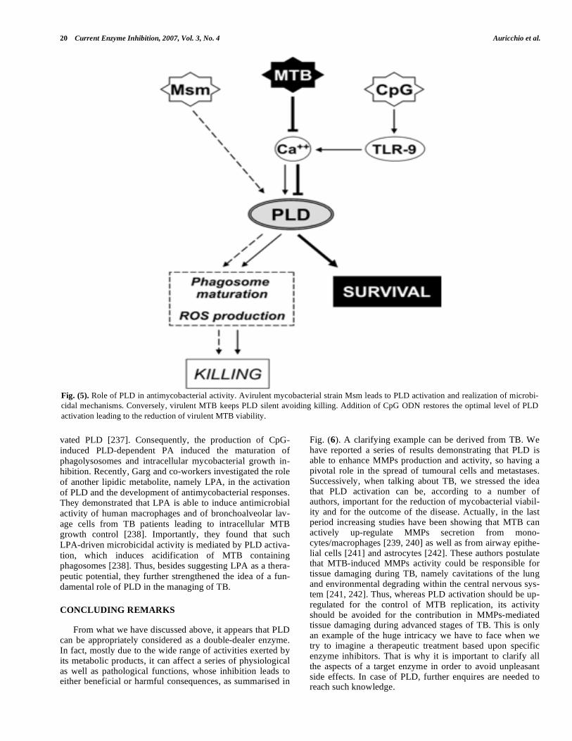
20 Current Enzyme Inhibition, 2007, Vol. 3, No. 4 Auricchio et al.
vated PLD [237]. Consequently, the production of CpG-
induced PLD-dependent PA induced the maturation of
phagolysosomes and intracellular mycobacterial growth in-hibition. Recently, Garg and co-workers investigated the role
of another lipidic metabolite, namely LPA, in the activation
of PLD and the development of antimycobacterial responses. They demonstrated that LPA is able to induce antimicrobial
activity of human macrophages and of bronchoalveolar lav-
age cells from TB patients leading to intracellular MTB growth control [238]. Importantly, they found that such
LPA-driven microbicidal activity is mediated by PLD activa-
tion, which induces acidification of MTB containing phagosomes [238]. Thus, besides suggesting LPA as a thera-
peutic potential, they further strengthened the idea of a fun-
damental role of PLD in the managing of TB.
CONCLUDING REMARKS
From what we have discussed above, it appears that PLD can be appropriately considered as a double-dealer enzyme. In fact, mostly due to the wide range of activities exerted by its metabolic products, it can affect a series of physiological as well as pathological functions, whose inhibition leads to either beneficial or harmful consequences, as summarised in
Fig. (6). A clarifying example can be derived from TB. We have reported a series of results demonstrating that PLD is able to enhance MMPs production and activity, so having a pivotal role in the spread of tumoural cells and metastases. Successively, when talking about TB, we stressed the idea that PLD activation can be, according to a number of authors, important for the reduction of mycobacterial viabil-ity and for the outcome of the disease. Actually, in the last period increasing studies have been showing that MTB can actively up-regulate MMPs secretion from mono-cytes/macrophages [239, 240] as well as from airway epithe-lial cells [241] and astrocytes [242]. These authors postulate that MTB-induced MMPs activity could be responsible for tissue damaging during TB, namely cavitations of the lung and environmental degrading within the central nervous sys-tem [241, 242]. Thus, whereas PLD activation should be up-regulated for the control of MTB replication, its activity should be avoided for the contribution in MMPs-mediated tissue damaging during advanced stages of TB. This is only an example of the huge intricacy we have to face when we try to imagine a therapeutic treatment based upon specific enzyme inhibitors. That is why it is important to clarify all the aspects of a target enzyme in order to avoid unpleasant side effects. In case of PLD, further enquires are needed to reach such knowledge.
Fig. (5). Role of PLD in antimycobacterial activity. Avirulent mycobacterial strain Msm leads to PLD activation and realization of microbi-
cidal mechanisms. Conversely, virulent MTB keeps PLD silent avoiding killing. Addition of CpG ODN restores the optimal level of PLD
activation leading to the reduction of virulent MTB viability.
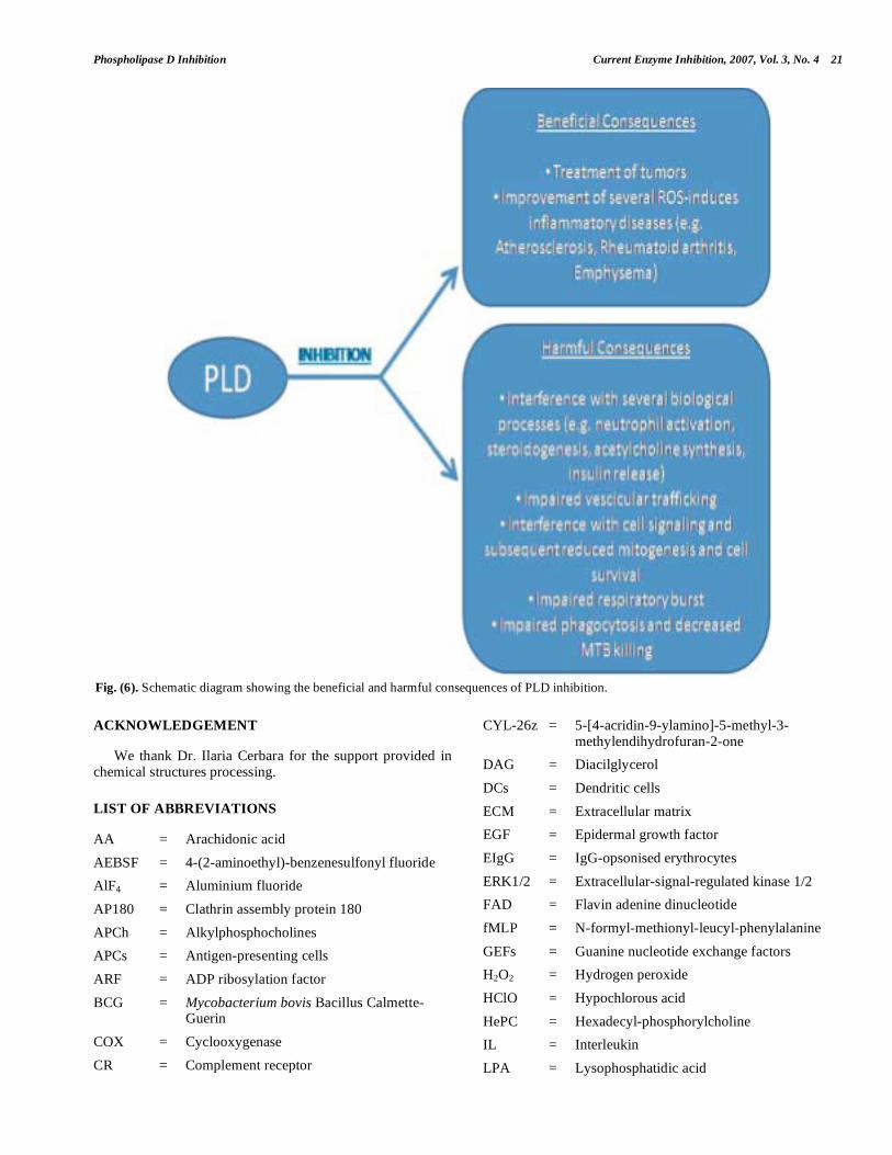
Phospholipase D Inhibition Current Enzyme Inhibition, 2007, Vol. 3, No. 4 21
ACKNOWLEDGEMENT
We thank Dr. Ilaria Cerbara for the support provided in chemical structures processing.
LIST OF ABBREVIATIONS
AA = Arachidonic acid
AEBSF = 4-(2-aminoethyl)-benzenesulfonyl fluoride
AlF4 = Aluminium fluoride
AP180 = Clathrin assembly protein 180
APCh = Alkylphosphocholines
APCs = Antigen-presenting cells
ARF = ADP ribosylation factor
BCG = Mycobacterium bovis Bacillus Calmette-Guerin
COX = Cyclooxygenase
CR = Complement receptor
CYL-26z = 5-[4-acridin-9-ylamino]-5-methyl-3-methylendihydrofuran-2-one
DAG = Diacilglycerol
DCs = Dendritic cells
ECM = Extracellular matrix
EGF = Epidermal growth factor
EIgG = IgG-opsonised erythrocytes
ERK1/2 = Extracellular-signal-regulated kinase 1/2
FAD = Flavin adenine dinucleotide
fMLP = N-formyl-methionyl-leucyl-phenylalanine
GEFs = Guanine nucleotide exchange factors
H2O2 = Hydrogen peroxide
HClO = Hypochlorous acid
HePC = Hexadecyl-phosphorylcholine
IL = Interleukin
LPA = Lysophosphatidic acid
Fig. (6). Schematic diagram showing the beneficial and harmful consequences of PLD inhibition.

22 Current Enzyme Inhibition, 2007, Vol. 3, No. 4 Auricchio et al.
MAPKs = Mitogen-activated protein kinases
MDCK = Madin-Darby canine kidney
MMPs = Matrix metalloproteases
Msm = Mycobacterium smegmatis
MTB = Mycobacterium tuberculosis
mTOR = Mammalian target of rapamycin
NADPH = Nicotinamide adenine dinucleotide hydrogen phosphate
NF B = Nuclear factor B
NGF = Nerve growth factor
NK = Natural killer
O2.- = Superoxide anion
ODN = Oligodeoxynucleotides
OH. = Hydroxylic radical
PA = Phosphatidic acid
PAP = Phosphatidate phosphohydrolase
PC = Phosphatidylcholine
PDGF = Platelet-derived growth factor
PE = Phosphatidylethanolamine
PG = Prostaglandin
PH = Pleckstrin homology
PI = Phoshatidylinositol
PI3K = Phosphatidylinositol-3-phosphate kinase
PI4K = Phosphatidylinositol-4-phosphate kinase
PI-4-P 5- = Phosphatidylinositol-4-phosphate 5-kinase
kinase
PIP2 = Phosphatidylinositol bisphosphate
PIP3 = Phosphatidylinositol trisphosphate
PKC = Protein kinase C
PLA2 = Phospholipase A2
PLC = Phospholipase C
PLD = Phospholipase D
PMNs = Polymorfonuclear leukocytes
PX = Phox homology
ROS = Reactive oxygen species
S1P = Sphingosine-1-phosphate
SK = Sphingosine kinase
TAM = Tamoxifen
TB = Tuberculosis
TNF- = Tumour necrosis factor-
S = TOR signalling
TPA = 12-O-tetradecanoylphorbol-13-acetate
REFERENCES
[1] Morris, A.J.; Engebrecht, J.; Frohman, M.A. Trends Pharmacol.
Sci., 1996, 17, 182-185.
[2] Exton, J.H. Biochim. Biophys. Acta, 1999, 1439, 121-133.
[3] Hanahan, D. J.; Chaikoff, I. L. J. Biol. Chem., 1948, 172, 191–198.
[4] Saito, M.; Kanfer, J. Arch. Biochem. Biophys, 1975, 169, 318-323.
[5] Gomez-Cambronero, J.; Keire, P. Cell. Signal., 1998, 10, 387-397.
[6] Agwu, D.E.; McCall, C.E.; McPhail, L.C. J. Immunol., 1991, 146,
3895-3903.
[7] Provost, J.J.; Fudge, J.; Israelit, S.; Siddiqi, A.R.; Exton, J.H. Bio-
chem. J., 1996, 319, 285-291.
[8] Balboa, M.A.; Insel, P.A. J. Biol. Chem., 1995, 270, 29843-29847.
[9] Hammond, S.M.; Altshuller, Y.M.; Sung, T.C.; Rudge, S.A., Rose,
K.; Engebrecht, J.; Morris, A.J.; Frohman, M.A. J. Biol. Chem.,
1995, 270, 29640-29643.
[10] Hammond, S.M.; Jenco, J.M.; Nakashima, S.; Cadwallader, K.; Gu,
Q.; Cook, S.; Nozawa, Y.; Prestwich, G.D.; Frohman, M.A.; Mor-
ris, A.J. J. Biol. Chem., 1997, 272, 3860-3860.
[11] Colley, W.C.; Altshuller, Y.M.; Sue-Ling, C.K.; Copeland, N.G.;
Gilbert, D.G.; Jenkins, N.A.; Branch, K.D.; Tsirka, S.E.; Bollag,
R.J.; Bollag, W.B.; Frohman, M.A. Biochem. J., 1997, 326, 745-
753.
[12] Stahelin, R.V.; Ananthanarayanan, B.; Blatner, N.R.; Singh, S.;
Bruzik, K.S.; Murray, D.; Cho, W. J. Biol. Chem., 2004, 279,
54918-54926.
[13] Sung, T.C.; Roper, R.L.; Zhang, Y.; Rudge, S.A.; Temel, R.;
Hammond, S.M.; Morris, A.J.; Moss, B.; Engebrecht, J.; Frohman,
M.A. EMBO J., 1997, 16, 4519-4530.
[14] Huang, C.; Wykle, R.L.; Daniel, L.W., Cabot, M.C. J. Biol. Chem.,
1992, 26, 16859-16865.
[15] Meier, K.E.; Gibbs, T.C.; Knoepp, S.M.; Ella, K.M. Biochim. Bio-
phys. Acta, 1999, 1439, 199-213.
[16] Saito, S.; Sakagami, H.; Kondo, H. Brain Res. Dev. Brain Res.,
2000, 120, 41-47.
[17] Billah, M.M.; Anthes, J.C. Biochem. J., 1990, 269, 281-291.
[18] Kiss, Z. Biochem. J., 1992, 285, 229-233.
[19] McNulty, S.; Lloyd, G.S.; Rumsby, R.M.; Sayner, R.M.; Rumsby,
M.G.; Neurosci. Lett., 1992, 139, 183-187.
[20] Harris, W.E.; Burstin, S.L. Biochem. J., 1992, 281, 675-682.
[21] Ponting, C.P.; Kerry, I.D. Prot. Sci., 1996, 5, 914-922.
[22] Exton, J.H. Physiol. Rev., 1997, 77, 303-320.
[23] Klinker, J.F.; Wenzel-Seifert, K.; Seifert, R. Gen. Pharmacol.,
1996, 27, 33-54.
[24] Boman, A.L.; Kahn, R.A. Trends Biochem. Sci., 1995, 20, 147-150.
[25] Houle, M.G.; Bourgoin, S. Biochem. Cell Biol., 1996, 74, 459-467.
[26] Bocckino, S.B.; Wilson, P.B.; Exton, J.H. Proc. Natl. Acad. Sci.
U.S.A., 1991, 88, 6210-6213.
[27] Exton, J.H. Biochim. Biophys. Acta, 1994, 1212, 26-42.
[28] Singer, W.D.; Brown, H.A.; Jiang, X.; Sternweis, P.C. J. Biol.
Chem., 1996, 271, 4504-4510.
[29] Yeo, E.J.; Exton, J.H. J. Biol. Chem., 1995, 270, 3980-3988.
[30] Natarajan, V.; Taher, M.M.; Roehm, B.; Parinandi, N.L.; Schmid,
H.H.; Kiss, Z.; Garcia, J.G. J. Biol. Chem., 1993, 268, 930-937.
[31] Jiang, H.; Lu, Z.; Luo, J.Q.; Wolfman, A.; Foster, D.A. J. Biol.
Chem., 1995, 270, 6006-6009.
[32] Jiang, H.; Luo, J.Q.; Urano, T.; Frankel, P.; Lu, Z.; Foster, D.A.;
Feig, L.A. Nature, 1995, 378, 409-412.
[33] Brown, H.A.; Gutowski, S.; Moomaw, C.R.; Slaughter, C.; Stern-
wies, P.C. Cell, 1993, 75, 1137-1144.
[34] Wu, H.; Turner, J.T.; Halenda, S.P. J. Pharmacol. Exp. Ther.,
1991, 258, 607-612.
[35] Martinez-Menarguez, J.A.; Prekeris, R.; Oorschot, V.M.; Scheller,
R.; Slot, J.W.; Geuze, H.J.; Klumperman, J. J. Cell. Biol., 2001,
155, 1213-1224.
[36] Cockcroft, S. Chem. Phys. Lipids, 1996, 80, 59-80.
[37] Cockcroft, S. Biochim. Biophys. Acta, 1992, 1113, 135-160.
[38] Uchida, N.; Okamura, S.; Nagamachi, Y.; Yamashita, S. J. Cancer
Res. Clin. Oncol., 1997, 123, 280–285.
[39] Uchida, N.; Okamura, S.; Kuwano, H. Anticancer Res., 1999, 19,
671–675.
[40] Zhao, Y.; Ehara, H.; Akao, Y.; Shamoto, M.; Nakagawa, Y.;
Banno, Y.; Deguchi, T.; Ohishi, N.; Yagi, K.; Nozawa, Y. Bio-
chem. Biophys. Res. Commun., 2000, 278, 140–143.
[41] Yoshida, M.; Okamura, S.; Kodaki, T.; Mori, M.; Yamashita, S.
Oncol. Res., 1998, 10, 399–406.
[42] Min, D.S.; Kwon, T.K.; Park, W.S.; Chang, J.S.; Park, S.K.; Ahn,
B.H.; Ryoo, Z.Y.; Lee, Y.H.; Lee, Y.S.; Rhie, D.J.; Yoon, S.H.;

Phospholipase D Inhibition Current Enzyme Inhibition, 2007, Vol. 3, No. 4 23
Hahn, S.J.; Kim, M.S.; Jo, Y.H. Carcinogenesis, 2001, 22, 1641-
1647.
[43] Yamada, Y.; Hamajima, N.; Kato, T.; Iwata, H.; Yamamura, Y.;
Shinoda, M.; Suyama, M.; Mitsudomi, T.; Tajima, K.; Kusakabe,
S.; Yoshida, H.; Banno, Y.; Akao, Y.; Tanaka, M.; Nozawa, Y. J.
Mol. Med., 2003, 81, 126-131.
[44] Lee, C.; Kang, H.S.; Chung, J.K.; Sekiya, F.; Kim J.R.; Han J.S.;
Kim S.R.; Bae Y.S.; Morris A.J.; Rhee S.G. J. Biol. Chem., 1997,
272, 15986-15992.
[45] Lee, C.; Kim, S.R.; Chung, J.K., Frohman, M.A., Kilimann, M.W.;
Rhee, S.G. J. Biol. Chem., 2000, 275, 18751-18758.
[46] Marsh, M.; McMahon, H.T. Sciene., 1999, 285, 215-220.
[47] Wigge, P.; McMahon, H.T. Trends Neurosci., 1998, 21, 339-344.
[48] Morris A.J. Biochem. Soc. Symp., 2007, 74, 247-257.
[49] Park, J.B.; Kim, J.H.; Kim, J.; Ha, S.H.; Yoo, J.S.; Du, G.;
Frohman, M.A.; Su, P.G.; Ryu, S.H. J. Biol. Chem., 2000, 275,
21295-21301.
[50] Lee, S.; Park, J.B.; Kim, J.H.; Kim, Y.; Kim, J.H.; Shin, K.J.; Lee,
J.S.; Ha, S.H.; Suh, P.G.; Ryu, S.H. J. Biol. Chem., 2001, 276,
28252-28260.
[51] Yamada, K.M.; Geiger, B. Curr. Opin. Cell Biol., 1997, 9, 76-85.
[52] Frohman, M.A.; Sung, T.C.; Morris, A.J. Biochim. Biophys. Acta,
1999, 1439, 175-186.
[53] Harel, M.; Schalk, I.; Ehret-Sabatier, L.; Bouet, F.; Goeldner, M.;
Hirth, C.; Axelsen, P.H.; Silman, I.; Sussman, J.L. Proc. Natl.
Acad. Sci. USA, 1993, 90, 9031-9035.
[54] Chae, Y.C.; Lee, S.; Lee, H.J.; Heo, K.; Kim, J.H., Kim, J.H.; Suh,
P.G.; Ryu, S.H. J. Biol. Chem., 2005, 280, 3723-3730.
[55] Lee, H.J.; Park, J.B.; Jang, I.H.; Chae, Y.C.; Kim, J.H.; Kim, I.S.;
Suh, P.G.; Ryu, S.H. J. Biol. Chem., 2004, 279, 16339-16348.
[56] Hata, Y.; Slaughter, C.A.; Südhof, T.C. Nature., 1993, 366, 347-
351.
[57] Garcia, E.P.; Gatti, E.; Butler, M.; Burton, J.; De Camilli, P. Proc.
Natl. Acad. Sci. U. S. A., 1994, 91, 2003-2007.
[58] Sciorra, V.A.; Hammond, S.M.; Morris, A.J. Biochemistry, 2001,
40, 2640-2646.
[59] Diwu, Z.; Zimmermann, J.; Meyer, T.; Lown, J.W. Biochem.
Pharmacol., 1994, 373-385.
[60] Ikemoto, M.; Tani, E.; Ozaki, I.; Kitagawa, H.; Arita, N. Cell
Death Differ., 2000, 7, 511-520.
[61] Murata, M.; Nagai, M.; Fujita, M.; Ohmori, M.; Takahara, J. Cell.
Mol. Life Sci., 1997, 53, 737-743.
[62] Honda, A.; Nogami, M.; Yokozeki, T.; Yamazaki, M.; Nakamura,
H.; Watanabe, H.; Kawamoto, K.; Nakayama, K.; Morris, A.J.;
Frohman, M.A.; Kanaho, Y. Cell., 1999, 99, 521-532.
[63] Yamamoto, H.; Hanada, K.; Kawasaki, K.; Nishijima, M. FEBS
Lett., 1997, 417, 196-198.
[64] Ammon, H.P.; Wahl, M.A. Planta Med., 1991, 57, 1-7.
[65] Huang, M.T.; Smart, R.C.; Wong, C.Q.; Conney, A.H. Cancer
Res., 1988, 48, 5941-5946.
[66] Ryu, S.B.; Palta, J.P. J. Lipid Res., 2000, 41, 940-944.
[67] Andrews, B.; Bond, K.; Lehman J.A.; Horn, J.M.; Dugan, A.; Go-
mez-Cambronero, J. Biochem. Biophys. Res. Commun., 2000, 273,
302-311.
[68] Kuan, Y.H.; Lin, R.H.; Tsao, L.T.; Chen, Y.L.; Tzeng, C.C.; Wang,
J.P. Biochem. Pharmacol., 2005, 70, 901-910.
[69] Chung, J.K.; Sekiya, F.; Kang, H.S.; Lee, C.; Han, J.S.; Kim, S.R.;
Bae, Y.S.; Morris, A.J.; Rhee, S.G. J. Biol. Chem., 1997, 272,
15980-15985.
[70] El-Hadj, N.B.; Popoff, M.R.; Marvaud, J.C.; Payrastre, B.; Boquet,
P.; Geny, B. J. Biol. Chem., 1999, 274, 14021-14031.
[71] Just, I.; Fritz, G.; Aktories, K.; Giry, M.; Popoff, M.; Boquet, P.;
Hegenbarth, S.; von Eichel-Streiber, C. J. Biol. Chem., 1994, 269,
10706-107112.
[72] Just, I.; Selzer, J.; Wilm, M.; von Eichel-Streiber, C.; Mann, M.;
Aktories, K. Nature., 1995, 375, 500-503.
[73] Schmidt, M.; Rumenapp, U.; Nehls, C.; Ott, S.; Keller, J.; von
Eichel-Streiber, C.; Jacobs, K.H. Eur. J. Biochem., 1996, 240, 707-
712.
[74] Reinhold, S.L.; Prescott, S.M.; Zimmerman, G.A.; McIntyre, T.M.
FASEB J., 1990, 4, 208-214.
[75] Kozawa, O.; Blume-Jensen, P.; Heldin, C.H.; Ronnstrand, L. Eur.
J. Biochem., 1997, 248, 149-155.
[76] Steed, P.M.; Chow, A.H.M. Curr. Pharm. Biotechnol., 2001, 2,
241-256.
[77] Adany, I.; Yazlovitskaya, E.M.; Haug J.S.; Voziyan P.A.; Melnyk-
ovych G. Cancer Lett., 1994, 79, 175-179.
[78] Rioja, A.; Pizzey, A.R.; Marson, C.M.; Thomas, N.S. FEBS Lett.,
2000, 467, 291-295.
[79] Taylor, M.M.; MacDonald, K.; Morris, A.J.; McMaster C.R. FEBS
J., 2005, 272, 5056-5063.
[80] Newton, A.C. Trends Pharmacol., 2004, 25, 175-177.
[81] Mellor, H.; Parker, P.J. Biochem. J., 1998, 332, 281-292.
[82] Voziyan, P.A.; Haug, J.S.; Melnykovych, G. Biochem. Biophys.
Res. Commun., 1995, 212, 479-486.
[83] Quest, A.F.; Ghosh, S.; Xie W.Q.; Bell, R.M. Adv. Exp. Medical.
Biol., 1997, 400A, 297-303.
[84] Mouridsen, H.; Palshof, T.; Patterion, J.; Pattersby, I. Cancer Treat.
Rev., 1978, 5, 131-141.
[85] Jordan, V.C. Breast Cancer Res. Treat., 1990, 15, 125-136.
[86] Kiss, Z.; Anderson, W.H. FEBS Lett., 1997, 400, 145-148.
[87] Kiss, Z. Chem. Phys. Lipids, 1996, 80, 81-102.
[88] Venable, M.E.; Bielawska, A.; Obeid, L.M. J. Biol. Chem., 1996,
271, 24800-24805.
[89] Lukowski, S.; Lecomte, M.C.; Mira, J.P.; Marin, P.; Gautero, H.;
Russo-Marie, F.; Geny, B. J. Biol. Chem., 1996, 271, 32482-32490.
[90] Jenco, J.M.; Rawlingson, A.; Daniels, B.; Morris, A.J. Biochemis-
try., 1998, 37, 4901-4909.
[91] Rodrìguez-Gonzàlez, A., Ramirez de Molina, A., Benitez-Rajal, J.,
Lacal, J.C. Prog. Cell. Cycle Res., 2003, 5, 191-201.
[92] Lucas, L.; Hernandez-Alcoceba, R.; Penalva, V.; Lacal, J.C. Onco-
gene., 2001, 20, 1110-1117.
[93] Konstantinov, S.M.; Eibl, H.; Berger, M.R. Cancer Chemother.
Pharmacol., 1998, 41, 210-216.
[94] Unger, C.; Peukert, M.; Sindermann, H.; Hilgard, P.; Nagel, G.;
Eibl, H. Cancer Treat. Rev., 1990, 17, 243-246.
[95] Li, L.; Fleming, N. FEBS Lett., 1999, 458, 419-423.
[96] Li, L.; Mellow, L.; Bhullar, R.P.; Fleming, N. Arch. Oral Biol.,
1998, 43, 211-219.
[97] Madesh, M.; Balasubramanian, K.A. Life Sci., 1998, 62, 177-184.
[98] Nemere, I.; Leathers, V.; Norman, A.W. J. Biol. Chem., 1986, 261,
16106-16114.
[99] Neftel, K.A.; Woodtly, W.; Schmid, M.; Frick, P.G.; Fehr, J. Br.
Med. J. (Clin. Res. Ed.), 1986, 292, 721-723.
[100] Seydel, J.K.; Wassermann O. Biochem. Pharmacol., 1976, 25,
2357-2364.
[101] Lullmann, H.; Wehling, M. Biochem. Pharmacol., 1979, 28, 3409-
3415.
[102] Liscovitch, M.; Chalifa, V.; Danin, M.; Eli, Y. Biochem. J., 1991,
279, 319-321.
[103] Hedge, V.R.; Silver, J.; Patel, M.G.; Bryant, R.; Pai, J.; Das, P.R.;
Puar, M.S.; Cox, P.A. J. Nat. Prod., 1995, 58, 1492-1497.
[104] Foster, D.A.; Xu, L. Mol. Cancer Res., 2003, 1, 789-800.
[105] Noh, D.Y.; Ahn, S.J.; Lee, R.A.; Park, I.A.; Kim, J.H.; Suh, P.G.;
Ryu, S.H.; Lee, K.H.; Han, J.S. Cancer Lett., 2000, 161, 207-214.
[106] Ahn, B.H.; Min, G.; Bae, Y.S.; Min, D.S. Exp. Mol. Med., 2006,
38, 55-62.
[107] Glunde, K.; Serkova, N.J. Pharmacogenomics, 2006, 7, 1109-1123.
[108] Cockcroft, S.; Thomas, G.M.; Fensome, A.; Geny, B.; Cunning-
ham, E.; Gout, I.; Hiles, I.; Totty, N.F.; Truong, O.; Hsuan, J.J. Sci-
ence, 1994, 263, 523-526.
[109] Lopez, I.; Arnold, R.S.; Lambeth, J.D. J. Biol. Chem., 1998, 273,
12846-12852.
[110] Kim, S.W.; Hayashi, M.; Lo, J.F.; Yang, Y.; Yoo, J.S.; Lee, J.D. J.
Biol. Chem., 2003, 78, 2661-2668.
[111] Massenburg, D.; Han, J.S.; Liyanage, M.; Patton, W.A.; Rhee,
S.G.; Moss, J.; Vaughan, M. Proc. Natl. Acad. Sci. U S A., 1994,
91, 11718-11722.
[112] Shome, K.; Vasudevan, C.; Romero, G. Curr. Biol., 1997, 7, 387-
396.
[113] Luo, J.Q.; Liu, X.; Frankel, P.; Rotunda, T.; Ramos, M.; Flom, J.;
Jiang, H.; Feig, L.A.; Morris, A.J.; Kahn, R.A.; Foster, D.A. Proc.
Natl. Acad. Sci. U S A., 1998, 95, 3632-3637.
[114] Shome, K.; Rizzo, M.A.; Vasudevan, C.; Andresen, B.; Romero, G.
Endocrinology, 2000, 141, 2200-2208.
[115] Kotter, K.; Ji, A.S.; von Eichel-Streiber, C.; Park, J.B.; Ryu, S.H.;
Klein, J. Biochim. Biophys. Acta., 2000, 1485, 153-162.
24 Current Enzyme Inhibition, 2007, Vol. 3, No. 4 Auricchio et al.
[116] Cross, M.J.; Roberts, S.; Ridley, A.J.; Hodgkin, M.N.; Stewart, A.;
Claesson-Welsh, L.; Wakelam, M.J. Curr. Biol., 1996, 6, 588-597.
[117] Schmitz, A.A.; Govek, E.E.; Bottner, B.; Van Aelst, L. Exp. Cell.
Res., 2000, 261, 1-12.
[118] Kam, Y.; Exton, J.H. Mol. Cell. Biol., 2001, 21, 4055-4066.
[119] Feig, L.A.; Urano, T.; Cantor, S. Trends Biochem. Sci., 1996, 21,
438-441.
[120] Luo, J.Q.; Liu, X.; Hammond, S.M.; Colley, W.C.; Feig, L.A.;
Frohman, M.A.; Morris, A.J.; Foster, D.A. Biochem. Biophys. Res.
Commun., 1997, 235, 854-859.
[121] Urano, T.; Emkey, R.; Feig, L.A. EMBO J., 1996, 15, 810-816.
[122] Frankel, P.; Ramos, M.; Flom, J.; Bychenok, S.; Joseph, T.; Kerk-
hoff, E.; Rapp, U.R.; Feig, L.A.; Foster, D.A. Biochem. Biophys.
Res. Commun., 1999, 255, 502-507.
[123] Voss, M.; Weernink, P.A.; Haupenthal, S.; Moller, U.; Cool, R.H.;
Bauer, B.; Camonis, J.H.; Jakobs, K.H.; Schmidt, M. J. Biol.
Chem., 1999, 274, 34691-34698.
[124] del Peso, L.; Lucas, L.; Esteve, P.; Lacal, J.C. Biochem. J., 1997,
322, 519-528.
[125] Hornia, A.; Lu, Z.; Sukezane, T.; Zhong, M.; Joseph, T.; Frankel,
P.; Foster, D.A. Mol. Cell. Biol., 1999, 19, 7672-7680.
[126] Conricode, K.M.; Brewer, K.A.; Exton, J.H. J. Biol. Chem., 1992,
267, 7199-7202.
[127] Kim, Y.; Han, J.M.; Han, B.R.; Lee, K.A.; Kim, J.H.; Lee, B.D.;
Jang, I.H.; Suh, P.G.; Ryu, S.H. J. Biol. Chem., 2000, 275, 13621-
13627.
[128] Oka, M.; Hitomi, T.; Okada, T.; Nakamura Si, S.; Nagai, H.; Ohba,
M.; Kuroki, T.; Kikkawa, U.; Ichihashi, M. Biochem. Biophys. Res.
Commun., 2002, 294, 1109-1113.
[129] Yeo, E.J.; Kazlauskas, A.; Exton, J.H. J. Biol. Chem., 1994, 269,
27823-27826.
[130] Durieux, M.E.; Lynch, K.R. Trends Pharmacol. Sci., 1993, 14,
249-254.
[131] Moritz, A.; De Graan, P.N.; Gispen, W.H.; Wirtz, K.W. J. Biol.
Chem., 1992, 267, 7207-7210.
[132] Jenkins, G.H.; Fisett,e P.L.; Anderson, R.A. J. Biol. Chem., 1994,
269, 11547-11554.
[133] Ghosh, S.; Strum, J.C.; Scorra, V.A.; Daniel, L.; Bell, R.M. J. Biol.
Chem., 1996, 271, 8472-8480.
[134] Rizzo, M.; Romero, G. Pharmacol. Ther., 2002, 94, 35-50.
[135] Riccio, A.; Pierchala, B.A.; Ciarallo, C.L.; Ginty, D.D. Science,
1997, 277, 1097-1100.
[136] Kranenburg, O.; Verlaan, I.; Moolenaar, W.H. J. Biol. Chem.,
1999, 274, 35301-35304.
[137] Roth, M.G.; Sternweis, P.C. Curr. Opin. Cell. Biol., 1997, 9, 519-
526.
[138] Roth, M.G.; Bi, K.; Ktistakis, N.T.; Yu, S. Chem. Phys. Lipids.,
1999, 98, 141-152.
[139] Nozawa, Y. Biochim. Biophys. Acta, 2002, 1585, 77-86.
[140] Kim, K.O.; Lee, K.H.; Kim, Y.H.; Park, S.K.; Han, J.S. Exp. Mol.
Med., 2003, 35, 38-45.
[141] Birbes, H.; Zeiller, C.; Komati, H.; Nemoz, G.; Lagarde, M.; Pri-
gent, A.F. FEBS Lett., 2006, 580, 6224-6232.
[142] Zhong, M.; Shen, Y.; Zheng, Y.; Joseph, T.; Jackson, D.; Foster,
D.A. Biochem. Biophys. Res. Commun., 2003, 302, 615-619.
[143] Joseph, T.; Bryant, A.; Frankel, P.; Wooden, R.; Kerkhoff, E.;
Rapp, U.R.; Foster, D.A. Oncogene, 2002, 21, 3651-3658.
[144] Hui, L.; Abbas, T.; Pielak, R.M.; Joseph, T.; Bargonetti, J.; Foster,
D.A. Mol. Cell. Biol., 2004, 24, 5677-5686.
[145] Hui, L.; Zheng, Y.; Yan, Y.; Bargonetti, J.; Foster, D.A. Oncogene,
2006, 25, 7305-7310.
[146] Chen, Y.; Zheng, Y.; Foster, D.A. Oncogene, 2003, 22, 3937-3942.
[147] Chen, Y.; Rodrik, V.; Foster, D.A. Oncogene, 2005, 24, 672-679.
[148] Abbas, T.; White, D.; Hui, L.; Yoshida, K.; Foster, D.A.; Bargon-
etti, J. J. Biol. Chem., 2004, 279, 9970-9977.
[149] West, M.J.; Stoneley, M.; Willis, A.E. Oncogene, 1998, 17, 769-
780.
[150] Lu, Z.; Hornia, A.; Joseph, T.; Sukezane, T.; Frankel, P.; Zhong,
M.; Bychenok, S.; Xu, L.; Feig, L.A.; Foster, D.A. Mol. Cell. Biol.,
2000, 20, 462-467.
[151] Joseph, T.; Wooden, R.; Bryant, A.; Zhong, M.; Lu, Z.; Foster,
D.A. Biochem. Biophys. Res. Commun., 2001, 289, 1019-1024.
[152] Rodrik, V.; Zheng, Y.; Harrow, F.; Chen, Y.; Foster, D.A. Mol.
Cell. Biol., 2005, 25, 7917-7925.
[153] Rodrik, V.; Gomes, E.; Hui, L.; Rockwell, P.; Foster, D.A. FEBS
Lett., 2006, 580, 5647-5652.
[154] HA, S.H.; Kim, D.H.; Kim, I.S.; Kim, J.H.; Lee, M.N.; Lee, H.J.;
Kim, J.H.; Jang, S.K.; Suh, P.G.; Ryu, S.H. Cell Signal., 2006, 18,
2283-2291.
[155] Fiucci, G.; Czarny, M.; Lavie, Y.; Zhao, D.; Berse, B.; Blusztajn,
J.K.; Liscovitch, M. Int. J. Cancer, 2000, 85, 882-888.
[156] Zheng, Y.; Rodrik, V.; Toschi, A.; Shi, M.; Hui, L.; Shen, Y.;
Foster, D.A. J. Biol. Chem., 2006, 281, 15862-15868.
[157] Kim, J.; Lee, Y.H.; Kwon, T.K.; Chang, J.S.; Chung, K.C.; Min,
D.S. Cancer Res., 2006, 66, 784-793.
[158] Imamura, F.; Horai, T.; Mukai, M.; Shinkai, K.; Sawada, M.;
Akedo, H. Biochem. Biophys. Res. Commun., 1993, 193, 497-503.
[159] Pai, J.K.; Frank, E.A.; Blood, C.; Chu, M. Anticancer Drug Des.,
1994, 9, 363-372.
[160] Kassis, J.; Lauffenburger, D.A.; Turner, T.; Wells, A. Semin. Can-
cer Biol., 2001, 11, 105-117.
[161] Aguirre Ghiso, J.A.; Farias, E.F.; Alonso, D.F.; Arregui, C.; Bal de
Kier Joffe, E. Int. J. Cancer., 1997, 71, 881-890.
[162] Colley, W.C.; Sung, T.C.; Roll, R.; Jenco, J.; Hammond, S.M.;
Altshuller, Y.; Bar-Sagi, D.; Morris, A.J.; Frohman, M.A. Curr.
Biol., 1997, 7, 191-201.
[163] Exton, J.H. FEBS Lett., 2002, 531, 58-61.
[164] Reich, R.; Blumenthal, M.; Liscovitch, M. Clin. Exp. Metastasis.,
1995, 13, 134-140.
[165] Williger, B.T.; Reich, R.; Neeman, M.; Bercovici, T.; Liscovitch,
M.J. Biol. Chem., 1995, 270, 29656-29659.
[166] Williger, B.T.; Ho, W.T.; Exton, J.H. J. Biol. Chem., 1999, 274,
735-738.
[167] Curran, S.; Murray, G.I. Eur. J. Cancer., 2000, 36, 1621-1630.
[168] McCawley, L.J.; Matrisian, L.M. Mol. Med. Today., 2000, 6, 149-
156.
[169] Gunthert, U.; Hofmann, M.; Rudy, W.; Reber, S.; Zoller, M.;
Haussmann, I.; Matzku, S.; Wenzel, A.; Ponta, H.; Herrlich, P.
Cell, 1991, 65, 13-24.
[170] Yu, Q.; Stamenkovic, I. Genes Dev., 1999, 13, 35-48.
[171] Ladeda, V.; Aguirre Ghiso, J.A.; Bal de Kier Joffe, E. Exp. Cell.
Res., 1998, 242, 515-527.
[172] Kato, Y.; Lambert, C.A.; Colige, A.C.; Mineur, P.; Noel, A.;
Frankenne, F.; Foidart, J.M.; Baba, M.; Hata, R.; Miyazaki, K.;
Tsukuda, M. J. Biol. Chem., 2005, 280, 10938-10944.
[173] Clark, R.A. J. Infect. Dis., 1990, 161, 1140-1147.
[174] Quinn, M.T.; Parkos, C.A.; Walker, L.; Orkin, S.H.; Dinauer,
M.C.; Jesaitis, A.J. Nature, 1989, 342, 198-200.
[175] Rotrosen, D.; Yeung, C.L.; Leto, T.L.; Malech, H.L.; Kwong, C.H.
Science, 1992, 256, 1459-1462.
[176] Koshkin, V.; Pick, E. FEBS Lett., 1994, 338, 285-289.
[177] Rossi, F.; Grzeskowiak, M.; Della Bianca, V.; Calzetti, F.; Gandini,
G. Biochem. Biophys. Res. Commun., 1990, 168, 320-327.
[178] Bauldry, S.A.; Elsey, K.L.; Bass, D.A. J. Biol. Chem., 1992, 267,
25141-25152.
[179] Wang, J.P.; Chang, L.C.; Hsu, M.F.; Lin, C.N. Naunyn Schmiede-
bergs Arch. Pharmacol., 2003, 368, 166-174.
[180] Paruch, S.; El-Benna, J.; Djerdjouri, B.; Marullo, S.; Perianin, A.
FASEB J., 2006, 20, 142-144.
[181] Melendez, A.J.; Bruetschy, L.; Floto, R.A.; Harnett, M.M.; Allen,
J.M. Blood, 2001, 98, 3421-3428.
[182] Loegering, D.J.; Lennartz, M.R. Inflammation, 2004, 28, 23-31.
[183] Siddiqui, R.A.; English, D.; Harvey, K.; Cui, Y.; Martin, M.I.;
Wentland, J.; Akard, L.; Jansen, J.; Thompson, J.; Garcia, J.G. J.
Leukoc. Biol., 1995, 58, 189-195.
[184] Erickson, R.W.; Langel-Peveri, P.; Traynor-Kaplan, A.E.; Hey-
worth, P.G.; Curnutte, J.T. J. Biol. Chem., 1999, 274, 22243-22250.
[185] Regier, D.S.; Greene, D.G.; Sergeant, S.; Jesaitis, A.J.; McPhail,
L.C. J. Biol. Chem., 2000, 275, 28406-28412.
[186] Regier, D.S.; Waite, K.A.; Wallin, R.; McPhail, L.C. J. Biol.
Chem., 1999, 274, 36601-36608.
[187] Waite, K.A.; Wallin, R.; Qualliotine-Mann, D.; McPhail, L.C. J.
Biol. Chem., 1997, 272, 15569-15578.
[188] Palicz, A.; Foubert, T.R.; Jesaitis, A.J.; Marodi, L.; McPhail, L.C.
J. Biol. Chem., 2001, 276, 3090-3097.
[189] Babior, B.M. Am. J. Med., 2000, 109, 33-44.
[190] Dubyak, G.R.; Schomisch, S.J.; Kusner, D.J.; Xie, M. Biochem. J.,
1993, 292, 121-128.

Phospholipase D Inhibition Current Enzyme Inhibition, 2007, Vol. 3, No. 4 25
[191] Gewirtz, A.T.; Seetoo, K.F.; Simons, E.R. J. Leukoc. Biol., 1998,
64, 98-103.
[192] Micella, M.; Gismondi, A.; Roncatoli, P.; Calmieri, G.; Morrone,
S.; Piccoli, M.; Frati, L.; Cifone, M.G.; Santoni, A. J. Immunol.,
1999, 162, 2064-2072.
[193] Caumont, A.S.; Galas, M.C.; Vitale, N.; Aunis, D.; Bader, M.F. J.
Biol. Chem., 1998, 273, 1373-1379.
[194] Caumont, A.S.; Vitale, N.; Gensse, M.; Galas, M.C.; Casanova,
J.E.; Bader, M.F. J. Biol. Chem., 2000, 275, 15637-15644.
[195] Tou, J.S.; Gill, J.S. Cell Signal., 2005, 17, 77-82.
[196] Austin, S.C.; Funk, C.D. Prostaglandins Other Lipid Mediat.,
1999, 58, 231-252.
[197] Smith, W.L. Biochem. J., 1989, 259, 315-324.
[198] Miller, S.B. Semin. Arthritis Rheum., 2006, 36, 37-49.
[199] Ferreri, N.R.; Schwartzman, M.; Ibraham, N.G.; Chander, P.N.;
McGiff, J.C. J. Pharmacol. Exp. Ther., 1984, 231, 441-448.
[200] Goodwin, J. Am. J. Med., 1984, 77, 7-15.
[201] Mathe, A.A.; Hedqvist, P. Am. Rev. Respir. Dis., 1975, 111, 313-
320.
[202] Khan, W.A.; Blobe, G.C.; Hannun, Y.A. Cell Signal., 1995, 7, 171-
184.
[203] Kozawa, O.; Suzuki, A.; Kotoyori, J.; Tokuda, H.; Watanabe, Y.;
Ito, Y.; Oiso, Y. J. Cell. Biochem., 1994, 55, 373-379.
[204] Watanabe-Tomita, Y.; Suzuki, A.; Shinoda, J.; Oiso, Y.; Kozawa,
O. Prostaglandins Leukot. Essent. Fatty Acids., 1997, 57, 335-339.
[205] Balboa, M.A.; Balsinde, J.; Dennis, E.A. J. Biol. Chem., 1998, 273,
7684-7690.
[206] Shinoda, J.; Kozawa, O.; Suzuki, A.; Watanabe-Tomita, Y.; Oiso,
Y.; Uematsu, T. Eur. J. Endocrinol., 1997, 136, 207-212.
[207] Angel, J.; Audubert, F.; Bismuth, G.; Fournier, C. J. Immunol.,
1994, 152, 5032-5040.
[208] Arend, W.P.; Dayer, J.M. Arthritis Rheum., 1990, 33, 305-315.
[209] Fallman, M.; Gullberg, M.; Hellberg, C.; Andersson, T. J. Biol.
Chem., 1992, 267, 2656-2663.
[210] Fallman, M.; Andersson, R.; Andersson, T. J. Immunol., 1993, 151,
330-338.
[211] Serrander, L.; Fallman, M.; Stendahl, O. Inflammation, 1996, 20,
439-450.
[212] Suchard, S.J.; Hinkovska-Galcheva, V.; Mansfield, P.J.; Boxer,
L.A.; Shayman, J.A. Blood, 1997, 89, 2139-2147.
[213] Kusner, D.J.; Hall, C.F.; Jackson, S. J. Immunol., 1999, 162, 2266-
2274.
[214] Bodman-Smith, K.B.; Melendez, A.J.; Campbell, I.; Harrison, P.T.;
Allen, J.M.; Raynes, J.G. Immunology, 2002, 107, 252-260.
[215] Mansfield, P.J.; Hinkovska-Galcheva, V.; Borofsky, M.S.; Shay-
man, J.A.; Boxer, L.A. Biochem. Biophys. Res. Commun., 2005,
331, 132-138.
[216] Lee, H.Y.; Kang, H.K.; Jo, E.J.; Kim, J.I.; Lee, Y.N.; Lee, S.H.;
Park, Y.M.; Ryu, S.H.; Kwak, J.Y.; Bae, Y.S. Exp. Mol. Med.,
2004, 36, 135-144.
[217] Kang, H.K.; Lee, H.Y.; Lee, Y.N.; Jo, E.J.; Kim, J.I.; Kim, G.Y.;
Park, Y.M.; Min, D.S.; Yano, A.; Kwak, J.Y.; Bae, Y.S. Int. Im-
munopharmacol., 2004, 4, 911-920.
[218] Locati, M.; Ribaldi, E.; Bonecchi, R.; Transidico, P.; Bernasconi,
S.; Haribabu, B.; Morris, A.J.; Mantovani, A.; Sozzoni, S. Bio-
chem. J., 2001, 358, 119-125.
[219] Iyer, S.S.; Barton, J.A.; Bourgoin, S.; Kusner, D.J. J. Immunol.,
2004, 173, 2615-2623.
[220] Corrotte, M.; Chasserot-Golaz, S.; Huang, P.; Du, G.; Ktistakis,
N.T.; Frohman, M.A.; Vitale, N.; Bader, M.F.; Grant, N.J. Traffic.,
2006, 7, 365-377.
[221] Armstrong, J.A.; Hart, P.D. J. Exp. Med., 1971, 134, 713-740.
[222] Goren, M.B.; D'Arcy Hart, P.; Young, M.R.; Armstrong, J.A. Proc.
Natl. Acad. Sci. U S A., 1976, 73, 2510-2514.
[223] Gordon, A.H.; Hart, P.D.; Young, M.R. Nature, 1980, 286, 79-80.
[224] Kusner, D.J.; Hall, C.F.; Schlesinger, L.S. J. Exp. Med., 1996, 184,
585-595.
[225] Lammas, D.A.; Stober, C.; Harvey, C.J.; Kendrick, N.; Panchal-
ingam, S.; Kumararatne, D.S. Immunity, 1997, 7, 433-444.
[226] Stober, C.B.; Lammas, D.A.; Li, C.M.; Kumararatne, D.S.; Light-
man, S.L.; McArdle, C.A. J. Immunol., 2001, 166, 6276-6286.
[227] Fairbairn, I.P.; Stober, C.B.; Kumararatne, D.S.; Lammas, D.A. J.
Immunol., 2001, 167, 3300-3307.
[228] Kusner, D.J.; Adams, J. J. Immunol., 2000, 164, 379-388.
[229] Kusner, D.J.; Barton, J.A. J. Immunol., 2001, 167, 3308-3315.
[230] Malik, Z.A.; Thompson, C.R.; Hashimi, S.; Porter, B.; Iyer, S.S.;
Kusner, D.J. J. Immunol., 2003, 170, 2811-2815.
[231] Thompson, C.R.; Iyer, S.S.; Melrose, N.; VanOosten, R.; Johnson,
K.; Pitson, S.M.; Obeid, L.M.; Kusner, D.J. J. Immunol., 2005,
174, 3551-3561.
[232] Leclercq, T.M.; Pitson, S.M. IUBMB Life, 2006, 58, 467-472.
[233] Garg, S.K.; Volpe, E.; Calmieri, G.; Mattei, M.; Galati, D.; Mar-
tino, A.; Piccioni, M.S.; Valente, E.; Bonanno, E.; De Vito, P.;
Baldini, P.M.; Spagnoli, L.G.; Coalizzi, V.; Fraziano, M. J. Infect.
Dis., 2004, 189, 2129-2138.
[234] Garg, S.K.; Cantucci, M.B.; Panetti, M.; Pupillo, L.; Bocchino, M.;
Okajima, F.; Bisen, P.S.; Saltini, C.; Fraziano, M. Clin. Immunol.,
2006, 121, 260-264.
[235] Hemmi, H.; Takeuchi, O.; Kawai, T.; Kaisho, T.; Sato, S.; Sanjo,
H.; Matsumoto, M.; Hoshino, K.; Wagner, H.; Takeda, K.; Akira,
S. Nature, 2000, 408, 740-745.
[236] Auricchio, G.; Garg, S.K.; Martino, A.; Volpe, E.; Ciaramella, A.;
De Vito, P.; Baldini, P.M.; Coalizzi, V.; Fraziano, M. Cell. Micro-
biol., 2003, 5, 913-920.
[237] Greco, E.; De Spirito, M.; Papi, M.; Fossati, M.; Auricchio, G.;
Fraziano, M. Biochem. Biophys. Res. Commun., 2006, 347, 963-
969.
[238] Garg, S.K.; Valente, E.; Greco, E.; Cantucci, M.B.; De Spirito, M.,
Papi, M.; Bocchino, M.; Saltini, C.; Fraziano, M. Clin. Immunol.,
2006, 121, 23-28.
[239] Chang, J.C.; Wysocki, A.; Tchou-Wong, K.M.; Moskowitz, N.;
Zhang, Y.; Rom, W.N. Thorax, 1996, 51, 306-311.
[240] Friedland, J.S.; Shaw, T.C.; Price, N.M.; Dayer, J.M. Matrix Biol.,
2002, 21, 103-110.
[241] Elkington, P.T.; Emerson, J.E.; Lopez-Pascua, L.D.; O'Kane, C.M.;
Horncastle, D.E.; Boyle, J.J.; Friedland, J.S. J. Immunol., 2005,
175, 5333-5340.
[242] Harris, J.E.; Nuttall, R.K.; Elkington, P.T.; Green, J.A.; Horncastle,
D.E.; Graeber, M.B.; Edwards, D.R.; Friedland, J.S. J. Immunol.,
2007, 178, 1199-1207.
Received: March 26, 2007 Revised: June 11, 2007 Accepted: June 13, 2007
Copyright © 2022 FDOKUMEN