SEISMIC SOIL-STRUCTURE INTERACTION: BENEFICIAL OR DETRIMENTAL
Upload
khangminh22Category
view
3download
0
Citation: Zhao, L.; Wang, S.;
Zhang, N.; Zhou, J.; Mehmood, A.;
Raka, R.N.; Zhou, F.; Zhao, L. The
Beneficial Effects of Natural Extracts
and Bioactive Compounds on the
Gut-Liver Axis: A Promising
Intervention for Alcoholic Liver
Disease. Antioxidants 2022, 11, 1211.
https://doi.org/10.3390/
antiox11061211
Academic Editors: Thea Magrone
and Luigi Santacroce
Received: 7 May 2022
Accepted: 19 June 2022
Published: 20 June 2022
Publisher’s Note: MDPI stays neutral
with regard to jurisdictional claims in
published maps and institutional affil-
iations.
Copyright: © 2022 by the authors.
Licensee MDPI, Basel, Switzerland.
This article is an open access article
distributed under the terms and
conditions of the Creative Commons
Attribution (CC BY) license (https://
creativecommons.org/licenses/by/
4.0/).
antioxidants
Review
The Beneficial Effects of Natural Extracts and BioactiveCompounds on the Gut-Liver Axis: A Promising Interventionfor Alcoholic Liver DiseaseLiang Zhao 1 , Shaoxuan Wang 1, Nanhai Zhang 2 , Jingxuan Zhou 2, Arshad Mehmood 1,3,Rifat Nowshin Raka 1, Feng Zhou 2,* and Lei Zhao 1,*
1 Beijing Advanced Innovation Center for Food Nutrition and Human Health, Beijing Engineering andTechnology Research Center of Food Additives, School of Food and Health, Beijing Technology and BusinessUniversity, Beijing 100048, China; [email protected] (L.Z.); [email protected] (S.W.);[email protected] (A.M.); [email protected] (R.N.R.)
2 Beijing Key Laboratory of Functional Food from Plant Resources, College of Food Science and NutritionalEngineering, China Agricultural University, Beijing 100083, China; [email protected] (N.Z.);[email protected] (J.Z.)
3 Department of Food Science and Technology, University of Haripur, Haripur 22620, Pakistan* Correspondence: [email protected] (F.Z.); [email protected] (L.Z.)
Abstract: Alcoholic liver disease (ALD) is a major cause of morbidity and mortality worldwide. It cancause fatty liver (steatosis), steatohepatitis, fibrosis, cirrhosis, and liver cancer. Alcohol consumptioncan also disturb the composition of gut microbiota, increasing the composition of harmful microbesand decreasing beneficial ones. Restoring eubiosis or preventing dysbiosis after alcohol consumptionis an important strategy in treating ALD. Plant natural products and polyphenolic compounds exertbeneficial effects on several metabolic disorders associated with ALD. Natural products and relatedphytochemicals act through multiple pathways, such as modulating gut microbiota, improving redoxstress, and anti-inflammation. In the present review article, we gather information on natural extractand bioactive compounds on the gut-liver axis for the possible treatment of ALD. Supplementationwith natural extracts and bioactive compounds promoted the intestinal tight junction, protectedagainst the alcohol-induced gut leakiness and inflammation, and reduced endotoxemia in alcohol-exposed animals. Taken together, natural extracts and bioactive compounds have strong potentialagainst ALD; however, further clinical studies are still needed.
Keywords: bioactive compounds; gut-liver axis; alcoholic liver disease; gut microbiota
1. ALD: Epidemiology, Progression, Pathogenesis, and Treatment
Alcohol abuse is the fifth leading cause of disease and death worldwide. Around2.4 billion people drink alcohol globally, including 1.5 billion (1.4–1.6) men and 0.9 bil-lion (0.8–1.0) women [1]. However, drinking condition differs from country to country.For instance, the total per capita intake of alcohol in France is 12–13 L/adult, followedby the United Kingdom, Eastern Europe, United States of America, Italy, and NorthAfrica/Middle East of 11–12 L/adult, 11–13 L/adult, 10 L/adult, 7 L/adult, and only0–2 L/adult, respectively [2].
Alcohol is a major hazard of alcoholic liver disease (ALD), which begins with fatty liverdisease (steatosis) and later progresses to alcoholic steatohepatitis (ASH) and fibrosis [3,4].Distinguishing between acute and chronic alcohol intake and its impact on ALD prognosisis critical [5]. Although alcohol intake is the main cause of ALD, environmental variablesand genetics may contribute to its progression [6]. Steatosis is one of several initial man-ifestations of ALD, essentially defined by an oversized liver. ALD can lead to seriousconsequences, such as cirrhosis and hepatocellular carcinoma, and there are currently noFDA (Food and Drug Administration)-approved treatments [4].
Antioxidants 2022, 11, 1211. https://doi.org/10.3390/antiox11061211 https://www.mdpi.com/journal/antioxidants

Antioxidants 2022, 11, 1211 2 of 18
Despite steatosis being prevalent in ALD, the development of cirrhosis is observedin only 10% to 15%, whereas the incidence of ASH was reported in up to 20–40% ofindividuals [7,8]. Each year, around 2 million individuals die from liver failure worldwide,and alcohol consumption is responsible for up to 50% of cirrhosis mortality.
In 2010, the global rate of death from alcohol-related cirrhosis was 7.2 per 100,000 people,which includes 9.7 in 100,000 males and 4.6 in 100,000 females [9]. So far, our expertiseregarding the underlying pathogenesis of ALD is quite limited [10]. Liquor is indeed aprimary hepatotoxin, and its consumption initiates a cascade of metabolic reactions thatcontribute to the final hepatotoxic consequence [11].
The preliminary clarification of malnutrition as the primary pathogenic pathway hasbeen superseded by the current concept that alcohol is metabolized by the hepatocyte,initiating pathogenic processes that require the yield of peptides and cytokine production,immunology action, and oxidative damage [12]. In certain situations, the duration of liverdamage is proportional to the amount of alcohol consumed [13]. With prolonged alcoholintake, alcohol-induced liver diseases can develop into infections, such as steatohepatitis,fibrosis, cirrhosis, and possibly hepatocellular carcinoma (HCC). Alcoholic-induced fattyliver is an alcohol-related disease in which alcohol promotes fat storage in liver cells [6].If patients do not receive any treatment, hepatic fibrosis/cirrhosis or liver failure mayoccur [14]. Excessive alcohol intake can also cause steatohepatitis, which causes varyingdegrees of liver damage, such as steatosis, blistering, alcohol foam degeneration, lobu-lar/fibrous inflammation, and acute cholestasis [15]. Alcoholic hepatitis (AH) is an acuteinflammatory liver disease with high morbidity and mortality. Notably, AH was not as-sociated with alcohol dose. AH is directly related to liver dysfunction and hepatic ductformation [16]. HCC is the third leading cause of cancer-related death in the world. Alcoholconsumption can lead to oral, bowel, and liver cancers. Alcohol plays an important role incausing cancer by increasing the expression of many oncogenes, leading to cancer-causingmutations [6]. Addressing alcohol-induced liver injury requires an understanding of thecomplicated interaction of numerous distinct hepatic cell types [17,18].
The liver-gut microbiota axis involved reciprocal processes, including genetic, nu-tritional, and environmental variables. Modulation of the intestinal barrier explains therelationship between the gut and liver, occasionally with adverse consequences for theliver. Alcohol directly acts on liver parenchymal cells during liver pathogenesis, causingchanges in intestinal barrier function, alteration of the microbiota, and enhancement oftoll-like receptors (TLRs) activation in hepatic cells. Particular attention should be paid tomodifying the gut flora, which contributes to the pathogenesis of liver disorders [19].
Modern therapeutics for ALD involve abstinence from alcohol, use of corticosteroids, s-adenosylmethionine, pentoxifylline, specific anti-TNF-α therapy, and type of diet. However,these therapies have no significant effect on fighting ALD. Therefore, lifestyle interventionon diet and exercise becomes the primary recommendation for ALD subjects [3,20,21].Liver transplantation is a last resort, although invasive and expensive, and remains thetherapeutic option when all other techniques fail to ameliorate the disease, usually onlywhen patients abstain from alcohol [22,23]. Earlier, several researchers reported that naturalextracts and bioactive compounds might be an ideal option for the prevention and treatmentof many diseases, including ALD [24–32]. The preliminary purpose of the current reviewis to provide in-depth information regarding the effects of natural extracts and bioactivecompounds against ALD via the gut-liver axis. This review will help ALD investigatorsunderstand nutritional therapy in regard to ALD.
2. Effect of Alcohol on Gut Microbiota
Alcohol abuse significantly affected many microbes in various parts of the gastroin-testinal tract (Figure 1). The extent of small intestine bacterial overgrowth was reported toincrease after alcohol exposure [33,34]. Moreover, many key phyla, such as Actinobacteria,Bacteroidetes, Firmicutes, and Proteobacteria, are affected by alcohol. The increment in theabundance of Proteobacteria phylum in response to alcohol was also documented [35,36].

Antioxidants 2022, 11, 1211 3 of 18
The abundance of Enterobacteriaceae increased, while the abundance of Bacteroidetes de-creased [37,38].
Antioxidants 2022, 11, 1211 3 of 18
Bacteroidetes, Firmicutes, and Proteobacteria, are affected by alcohol. The increment in the abundance of Proteobacteria phylum in response to alcohol was also documented [35,36]. The abundance of Enterobacteriaceae increased, while the abundance of Bacteroidetes de-creased [37,38].
The relative abundance of Corynebacterium and Actinobacteria phylum was reported to increase after alcohol exposure, whereas phylum Firmicutes decreased [37,39]. The phy-lum Firmicutes contains many genera, namely Lactobacillus, Ruminococcus, Subdoligranu-lum, Faecalibacterium, and Roseburia that were decreased, whereas Clostridium, Streptococ-cus, Holdemania, and Coprobacillus increased after alcohol exposure [35,36,38,40,41]. The phylum Verrucomicrobia, which contains the Akkermansia genus, has decreased in the stool of people exposed to alcohol [36,42].
Collectively, chronic alcohol administration or intake results in dysbiosis, which is related to a decrease in beneficial bacteria. Restoring eubiosis or preventing dysbiosis after alcohol intake is an important strategy against ALD.
Figure 1. Alcohol abuse significantly affects many microbes in the gut. ↑, the abundance was upreg-ulated; ↓, the abundance was down-regulated.
3. Mechanisms of Dysbiosis Driving Alcohol-Related Liver Diseases 3.1. Dysregulation of Bile Acid Metabolism
One of the significant communicators between the intestine and liver is bile acids. The hepatic biliary system secretes conjugated bile acids, which are converted as needed by intestinal bacteria in the duodenum [43]. Then the modified bile acids enter the enter-ohepatic circulation and reach the liver again. As previously mentioned, liver cirrhosis lowers normal bile flow in patients [44]. Bile acids stimulate the farnesoid X receptor in intestinal epithelial cells, leading to the induction of antimicrobial molecules [45]. On the contrary, the overgrowth of intestinal bacteria results from decreased bile flow. The ex-perimental model of alcohol feeding in rodents showed a correlation between bile acid metabolism and the intestinal microbiome. Due to ethanol intake, taurine-conjugated bile acids were reduced in rats’ intestines and livers [46]. Nevertheless, the level of glycine-conjugated and unconjugated bile acids increased [47]. The partial reason behind this could be the overgrowth of gastrointestinal bacteria as cirrhosis patients exhibit escalated bile acids deconjugation [48]. Chronic abuse of alcohol in patients raises the total amount of bile acids, secondary bile acids, lithocholic acid, deoxycholic acid, and secondary-to-
Figure 1. Alcohol abuse significantly affects many microbes in the gut. ↑, the abundance wasupregulated; ↓, the abundance was down-regulated.
The relative abundance of Corynebacterium and Actinobacteria phylum was reported toincrease after alcohol exposure, whereas phylum Firmicutes decreased [37,39]. The phylumFirmicutes contains many genera, namely Lactobacillus, Ruminococcus, Subdoligranulum,Faecalibacterium, and Roseburia that were decreased, whereas Clostridium, Streptococcus,Holdemania, and Coprobacillus increased after alcohol exposure [35,36,38,40,41]. The phylumVerrucomicrobia, which contains the Akkermansia genus, has decreased in the stool of peopleexposed to alcohol [36,42].
Collectively, chronic alcohol administration or intake results in dysbiosis, which isrelated to a decrease in beneficial bacteria. Restoring eubiosis or preventing dysbiosis afteralcohol intake is an important strategy against ALD.
3. Mechanisms of Dysbiosis Driving Alcohol-Related Liver Diseases3.1. Dysregulation of Bile Acid Metabolism
One of the significant communicators between the intestine and liver is bile acids. Thehepatic biliary system secretes conjugated bile acids, which are converted as needed byintestinal bacteria in the duodenum [43]. Then the modified bile acids enter the enterohep-atic circulation and reach the liver again. As previously mentioned, liver cirrhosis lowersnormal bile flow in patients [44]. Bile acids stimulate the farnesoid X receptor in intestinalepithelial cells, leading to the induction of antimicrobial molecules [45]. On the contrary,the overgrowth of intestinal bacteria results from decreased bile flow. The experimentalmodel of alcohol feeding in rodents showed a correlation between bile acid metabolismand the intestinal microbiome. Due to ethanol intake, taurine-conjugated bile acids werereduced in rats’ intestines and livers [46]. Nevertheless, the level of glycine-conjugatedand unconjugated bile acids increased [47]. The partial reason behind this could be theovergrowth of gastrointestinal bacteria as cirrhosis patients exhibit escalated bile acidsdeconjugation [48]. Chronic abuse of alcohol in patients raises the total amount of bileacids, secondary bile acids, lithocholic acid, deoxycholic acid, and secondary-to-primarybile acid ratio in the stool [49]. If any patient develops advanced cirrhosis, they show anincrement in serum level of conjugated bile acids and a reduction in the amount of totalbile acids [49,50]. The secretion dimension of bile acid in the intestine of cirrhotic patientsmay be the underlying cause of both phenomena [44]. For a better understanding of

Antioxidants 2022, 11, 1211 4 of 18
pathogenesis related to chronic alcohol abuse and to develop potential therapeutic agents,extensive research is needed to further explore the interactions between bile acids and gutmicrobiota. This bidirectional crosstalk can better define the communications between theliver and intestine.
3.2. Microbial Products Contribute to Liver Inflammation and Disease
The liver readily absorbs toxins from the portal vein circulation as the unadulter-ated intestinal products reach the liver first. The promoters of hepatocellular injury aremicrobial toxins; including microbial pathogen-associated molecular patterns (PAMPs),fungal exotoxins (such as candidalysin), bacterial exotoxins (such as cytolysin secreted byEnterococcus), bacterial endotoxins (such as lipopolysaccharide [LPS] from gram-negativebacteria), hepatic toll-like receptors activated by endotoxins, and PAMPs that directlyinteract with pattern-recognition receptors present on hepatic stellate and Kupffer cells.The microbial products can advance cytokine stimulation, fibrotic changes, and oxidativestress (OS) of the inflammatory cascade [51].
Specific exotoxins exert pathogenicity in ALD patients. Compared to heavy drinkingcontrol, patients with AH show an increased abundance of Enterococcus faecalis, a cytolysin-producing bacteria. The quantity of cytolysin is associated with both the mortality andextremity of disease, the same as the fungal exotoxin candidalysin, which is also found inhigher concentrations in AH patients [52]. Ethanol-comprising diet worsened liver injuryin candidalysin-producing Candida colonized mice [53].
Studies have shown that dysbiosis may be related to the amount of endotoxins thatcirculate freely. Dysbiosis in ALD patients, along with AH and alcohol-related cirrhosispatients, revealed a correlated upsurge in flowing LPS [54–56]. Alcohol-related cirrhosisseemed to show a greater degree of endotoxemia than non-alcohol-induced cirrhosis,despite the end-stage liver disease scores being irrelevant [54]. Intestinal permeability isprobably a vital implementer of endotoxemia. Markedly, half of the patients with ALD(about half of alcohol use disorders patients) showed increased permeability in the intestinalbarrier, revealing a close association with microbiome changes. Therefore, increasedintestinal permeability caused by microbial dysregulation is an important prerequisite forthe progression of ALD [57,58].
3.3. Short-Chain Fatty Acids (SCFAs)
There are numerous processes involved in the regulation of intestinal permeabil-ity. Consumption of alcohol can affect many of those processes. Chronic alcohol intakedeteriorates dysbiosis and subsequently disturbs the integrity of the tight junctions ofthe enterocyte, as SCFA-producing commensals are involved in maintaining barrier in-tegrity [51]. Additionally, hepatic inflammation and adiposity can be mitigated by SC-FAs [59]. ALD patients exhibited a consistent decrease in the microbiomes of Lachnospiraceaeand Ruminococcaceae families [42,50,54,60–62]. Conversely, Veillonella is also known to pro-duce SCFAs and is often expanded in patients with ALD [50,60,62,63]. There was a signifi-cant reduction in SCFAs in the feces of AH patients compared to heavy drinkers, despitechanges in specific microbial patterns [60]. In short, intestinal permeability increases withthe fading production of SCFAs, ultimately leading to hepatic inflammation.
3.4. Endotoxin
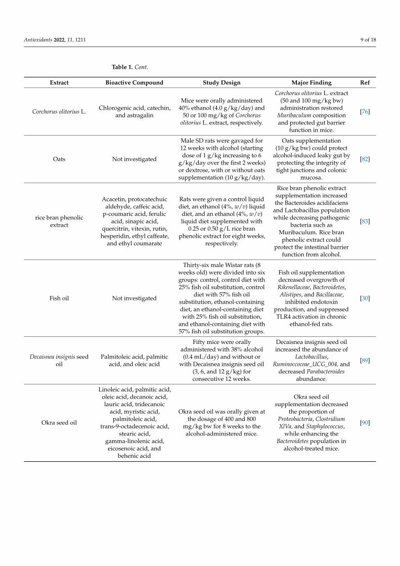
Endotoxin is one of the main components of the outer membrane of the gram-negativebacteria cell wall. Compared to non-alcoholic subjects, plasma of alcohol abused patientscontained a 5-fold higher concentration of endotoxin [64]. Intake of alcohol disturbsthe intestinal barrier functions and magnifies intestinal permeability. In the rat model,alcohol administration enabled systemic translocation and absorption of endotoxin [65].The intensity of ethanol-induced liver injury in rats was significantly interrelated withendotoxin levels in plasma [66]. Endotoxin can cross the intestinal barrier and activate the

Antioxidants 2022, 11, 1211 5 of 18
Kupffer cells that generate TNF-α and superoxide in the liver, resulting in severe hepaticdamage [67].
4. Protective Effect of Natural Products and Their Bioactive Compoundsagainst Alcoholic Induced Gut Microbiota Dysbiosis4.1. Bioactive Compounds against Alcohol-Induced Gut Microbiota Dysbiosis
Phenolic compounds are a large group of chemicals, such as phenolic acids, flavonoids,stilbenes, lignans, and other chemicals, commonly present in various edible plants. Phenoliccompounds possess countless health benefits, including the hepatoprotective effect [26,27].Earlier, Yuan et al. reported that epigallocatechin-3-gallate (EGCG) protected ALD via in-hibiting alcohol-induced gut leakiness and inflammatory factors expressions, and reducingendotoxemia in rats [26]. Later, another study documented that EGCG acted as a prebioticfor L. plantarum, developing microbead synbox, and was promising as a therapeutic optionfor the ALD [28]. Similarly, other polyphenolic compounds, puerarin and kaempferol,alleviated ALD in mice via regulating intestinal tight junctions and inhibiting endotoxinleakage [29,30].
Earlier, tributyrin supplementation was found to protect mice from alcohol via ex-pression and co-localization of tight junction (TJ) proteins (ZO-1, occludin), as well as bu-tyrate receptor (GPR109A) and transporter (SLC5A8) in the ileum and proximal colon [68].Aplysin, a brominated sesquiterpene compound purified from red alga Laurencia tristichawas studied against ALD. The daily treatment of aplysin (150 mg/kg bw) for 12 weeksmarkedly modulated the composition of Escherichia coli, Bacteroides fragilis, Lactobacillus,Bifidobacterium, and other key biomarkers, thus protecting ALD [69].
Astaxanthin was also reported to protect ALD via modulating mouse gut microbiota,such as decreasing the Bacteroidetes, Proteobacteria, Parabacteroides, Butyricimonas, Bilophilaand increasing Akkermansia and Verrucomicrobia in mice [31].
Berberine is a natural compound present in many plant extracts and possesses multiplehealth effects. Recently, Li et al. conducted a study to explore the protective effect of berber-ine against alcohol-mediated gut microbiota dysbiosis. Berberine (10, 50, and 100 mg/kgbw) was orally administrated to the mice for 33 days. Results revealed that berberinetreatments markedly improve gut microbiota dysbiosis by increasing the abundance ofAkkermansia muciniphila [32]. More recently, Han et al. investigated the protective effectof cornel iridoid glycoside isolated from Cornus officinalis Sieb. et Zucc against ALD. Thecornel iridoid glycoside was supplemented at the dosage of 50, 100, and 200 mg/kg bw for16 days in the mice. The results revealed that cornel iridoid glycoside supplementation sig-nificantly attenuated ALD via enhancing antioxidant activities, reducing inflammation, andaltering intestinal microbial diversity. Cornel iridoid glycoside supplementation increasedthe abundance of Lactobacillus and decreased the proportion of norank_f_Muribaculaceaeand norank_f_Desulfovibrionaceae in mice [70]. Furthermore, ursolic acid and antrodin Ahave recently been reported in two different studies to protect against alcohol-induced liverinjury via the gut-liver axis [71,72].
Polysaccharides are polymeric carbohydrate molecules abundantly present in variousplants, algae, microorganisms, and animals, exerting a wide array of biological activities,including hepatoprotective activities [73]. Many authors reported that polysaccharidescould protect the liver from alcohol damage via multiple pathways, including restoring gutdysbiosis [32,74–76]. In detail, Wang et al. isolated polysaccharides from garlic (molecularweight: 10 Kda, acid heteropolysaccharide), which was further studied against ALD inmice. Results showed that daily garlic polysaccharide administration (150 and 250 mg/kgbw for 30 days) could alleviate various biochemical indicators, increasing the abundance ofLachnospiraceae and Lactobacillus, and decreasing the abundance of Facklamia and Firmicutesin ethanol-induced mice [74]. Yang et al. found that inulin administration could ameliorateALD via inhibiting the LPS-TLR4-Mψ axis, and rectified gut dysbiosis mainly by increasingthe abundance of Lactobacillus, Lactococcus, and Allobaculum and reducing the abundanceof Parasutterella [75]. In another study, Coprinus comatus polysaccharides could regulate
Antioxidants 2022, 11, 1211 6 of 18
gut microbiota in ALD mice by increasing the proportion of Lachnospiraceae, Firmicutes,Muribaculaceae, and Bacteroidetes and decreasing the Rikenellaceae proportion, which showedprebiotic-like effects on the intestinal flora in ALD mice [32]. Similarly, Wolfiporia cocospolysaccharides were also reported to modulate gut microbiota in ALD mice, mainly byincreasing the Firmicutes to Proteobacteria ratio and the abundance of Lachnospiraceae [77].Oral administration of oyster (Crassostrea gigas) polysaccharides (282 mg/kg bw) could alsoincrease the proportion of Roseburia spp. and Lactobacillus reuteri, and decrease Escherichiaproportion in ALD mice [78].
4.2. Natural Product Extracts against Alcohol-Induced Gut Microbiota Dysbiosis4.2.1. Fruits and Vegetables
Lychee (Litchi chinensis Sonn.) pulp extract rich in polyphenolic compounds (pro-cyanidin B2, (-)-epicatechin, quercetin-3-O-rutinoside-7-O-α-L-rhamnosidase, rutin, andisorhamnetin-3-O-rutinoside) was orally given (0.2 and 0.4 g/L bw) to the ethanol-exposed(4%, v/v) mice for 8 weeks. Results revealed that compared with the ethanol group, lycheepulp extract supplementation increased the relative abundance of the Lactobacillus genus,Bacteroides acidifaciens species, Actinobacteria phylum, and Coriobacteriaceae family, whereasit decreased the abundance of Dehalobacteriaceae family and Odoribacter genus. Furthermore,it was also observed that lychee pulp extract supplementation could upregulate the expres-sion of intestinal tight junction proteins, antimicrobial proteins, and mucus proteins whiledeclining the serum endotoxin level. They concluded that lychee pulp extract has strongpotential against alcoholic abuse [79]. In another study, pomegranate extract could alsoprevent intestinal apoptosis, endotoxemia, alcohol-induced intestinal leakage, and inflam-mation by regulating TJ/ adherent junction proteins [80]. Corchorus olitorius L., also knownas molokhia, is a pantropical plant consumed as a vegetable in Africa and Eastern Asia thatexerts a protective effect against several diseases [76,81]. Recently, Do et al. documentedthat administration of molokhia extract (50 and 100 mg/kg bw) restored the composition ofMuribaculum and enhanced the intestinal barrier function in mice [76].
4.2.2. Cereals
Tang et al. documented that oats supplementation (10 g/kg bw) for 12 weeks to therats could prevent alcohol-induced intestinal leakage by protecting the integrity of tightjunctions and colonic mucosa [82]. Similarly, supplementation with rice bran phenolicextract was also reported to combat alcohol-induced liver injury via alleviating intestinalmicrobiota dysbiosis. Briefly, rice bran phenolic extract supplementation increased theabundance of Bacteroides acidifaciens and Lactobacillus, whereas it decreased pathogenicbacteria such as Muribaculum. Furthermore, it was also observed that rice bran phenolicextract could protect the intestinal barrier from alcohol [83]. Recently, Yang et al. reportedthat wheat embryo globulin could maintain the composition of gut microbiota [84].
4.2.3. Oils
Fish oil contains a significant amount of n-3 polyunsaturated fatty acids (PUFAs),which have been reported to alter gut microbiota. It has also been documented that fish oilsupplementation can increase Bifidobacterium and decrease Escherichia coli in the feces of ratsfed alcohol [85–87]. More recently, Chen et al. also reported that fish oil supplementationreduced the overgrowth of Rikenellaceae, Bacteroidetes, Alistipes, and Bacillaceae, inhibitedendotoxin production, and suppressed TLR4 activation in chronic ethanol-fed rats [30].
Flaxseed oil was also reported to protect against the adverse effect of alcohol bymodulating gut microbiota in alcohol-induced liver injury mice [88]. According to thereport, Decaisnea insignis seed oil (containing palmitoleic acid, palmitic acid, and oleic acid)could protect against alcohol-associated liver damage in mice via increasing the abundanceof Lactobacillus, Ruminoccoceae_UCG_004 and decreasing Parabacteroides abundance [89].
Okra seed oil supplementation has been reported to improve ALD via regulatingintestinal microbiota. Briefly, Okra seed oil supplementation at the dosage of 400 and

Antioxidants 2022, 11, 1211 7 of 18
800 mg/kg bw for 8 weeks decreased the proportion of Proteobacteria, Clostridium XlVa, andStaphylococcus, while enhancing the abundance of Bacteroidetes in alcohol-treated mice [90].
4.2.4. Tea
It has been reported that Pu-erh tea extract (PTE) played a protective role againstALD mainly through improving OS, lipid accumulation, inflammation, and microbiotadysbiosis. PTE treatment increased the relative abundance of potentially beneficial bac-teria (Bifidobacterium and Allobaculum) and decreased the relative abundance of harmfulbacteria (Helicobacter and Bacteroides) [91]. More recently, Li et al. studied the effects ofsix tea samples: two black teas (Dianhong tea and Yingde Black tea), two oolong teas(Tieguanyin Tea and Fenghuang Danzong Tea), and two dark teas (Fuzhuan Brick tea andSelenium-Enriched Dark tea) against ALD in mice. Results revealed that all tea samplesupplementation markedly protected from the adverse effect of alcohol. However, moreprofound results were observed in oolong tea and dark tea. Moreover, their findingssuggested that Akkermansia is the target microorganism for Tieguanyin Tea and Fu BrickTea [92].
4.2.5. Fermented Liquids
Vinegar is fermented acidic food rich in various bioactive compounds such as polyphe-nols, flavonoids, and melanoidins. It was previously reported that Shanxi aged vinegarextract (SAVE) contains chlorogenic acid, p-hydroxybenzoic acid, ferulic acid, rutin, sy-ringic acid, gallic acid, and other polyphenols, which exerts high antioxidant activity andprotects liver cells from oxidative damage [83]. In another study, the same research groupdocumented that polyphenol-rich SAVE attenuated ALD via regulating gut microbiota.Briefly, they found that various microbes (Lactobacillus, Bacteroidetes, Akkermansia, Verru-comicrobia) showed a significant positive correlation with OS and inflammatory indictors(occludin, Reg3b, Reg3g, and ZO-1), whereas Proteobacteria, Parabacteroides, Firmicutes,Bilophila, and Butyricimonas exhibited the opposite effect [93]. In addition, this researchgroup also investigated the protective effect of another vinegar (Zhenjiang aromatic vine-gar; ZAV) against ethanol-induced liver injury. Results showed that ZAV could regulatethe composition of gut microbiota and immune factors in ALD mice. Additionally, Lach-nospiraceae_NK4A136_group, Bacteroidetes, and Akkermansia were positively correlated withantimicrobial peptides and intestinal immune factors, but negatively correlated with in-flammatory and OS parameters [94].
Fermented rice liquors (called Makgeolli in Korea) were also documented to restore fecalmicrobiota compositions in mice induced by alcohol. The abundance of Bacteroidetes andFirmicutes phyla was observed to return to the control group level. Moreover, treatment withfermented rice liquors also increased the content of fecal SCFA and reduced inflammatoryresponses in mice induced by alcohol [95]. Baijiu, a Chinese traditional fermented liquorcontaining volatile compounds such as esters, acids, and phenols, was also reported toincrease the relative abundance (11%) of Lactobacillus compared to the ethanol-treated group(1.80%) [96].
Ran et al. studied the protective effect of sea buckthorn-fermented liquid againstALD. The sea buckthorn was fermented with a Lactobacillus plantarum BNCC194165 strain,exhibiting a significant increment in the total flavonoids, total triterpenes, and SCFAscompared with the unfermented sea buckthorn. Furthermore, fermented sea buckthornliquid was sterilized and orally given (1.75, 2.675, and 5.35 g/kg bw) to the mice for 15 days.Results showed that fermented sea buckthorn liquid could protect the liver from alcohol byimproving OS, decreasing inflammation, and regulating gut microbiota. The high dosageof fermented sea buckthorn liquid (5.35 g/kg bw) significantly enhanced the abundance ofLactobacillus and decreased the abundance of Ruminiclostridium, Akkermansia, Alistipes, andTuricibacter in mice [97].

Antioxidants 2022, 11, 1211 8 of 18
4.2.6. Herbs and Miscellaneous Extracts
Rhubarb (Rheum palmatum and Rheum officinale), a natural edible herb, contains avariety of bioactive compounds, including anthraquinone derivatives with hepatoprotectiveeffects [98]. Neyrinck et al. reported that rhubarb extract (0.3%) could change the microbialcomposition of Akkermansia muciniphila and Parabacteroides goldsteinii, improving hepaticinjury and decreasing inflammatory and OS biomarkers in alcohol-induced mice [99].
The mixture of Ginkgo biloba and Rosa roxburghii juice, rich in bioactive compounds(rutin, quercetin, kaempferol, isorhamnetin, ginkgolide C, bilobalide, ginkgolide A, andginkgolide B), was reported to protect alcoholic intestinal barrier dysfunction via restoringtight junctions [100]. In another study, Lactobacillus fermentum KP-3-fermented ginseng(Panax ginseng) was orally administrated (390 mg/kg bw) to alcohol-exposed mice for14 days. Results revealed that fermented ginseng supplementation could improve gutmicrobiota dysbiosis via restoring the abundance of Lactobacillus and Bifidobacteria, Bac-teroidetes phylum, and the Proteobacteria genus of the Sutterella phylum, Verrucomicrobiaphylum, Allobaculum genus, Ruminococcus genus, Adlercreutzia genus, and Actinobacteriaphylum [101].
Choi et al. conducted a study to explore the protective effect of defatted Tenebriomolitor larva fermented extract against chronic alcohol-fed rats. Results showed thatdefatted Tenebrio molitor larva fermented with Saccharomyces cerevisiae strain (KCTC 17299)extract at the dosage of 200 mg/kg/day could attenuate ALD via modulating intestinalmicroflora, steatosis, and inflammation. It was observed that defatted Tenebrio molitor larvaextract restored the Lactobacillus johnsonii abundance [102]. Similarly, many other naturalextracts such as Pogostemon cablin, edible insect Gryllus bimaculatus, Semen Hoveniae, andDendropanax morbifera leaf extract were reported to protect against alcohol-mediated gutmicrobiota dysbiosis [103–106].
5. Discussion
A large amount of evidence suggests that intestinal microbiome dysregulation is a keyrisk factor for the progression/development of ALD. The graphical summary of the effectsof chronic alcohol consumption on the gut-liver axis is shown in Figure 2. Prevention ofalcohol-induced dysbiosis is an important strategy in the treatment of ALD. In this regard,natural products and bioactive compounds play a significant role. As mentioned above,edible plants and their bioactive compounds could restore gut microbiota in animal models(Table 1).
Table 1. Summary of the protective effect of various natural products against ALD.
Extract Bioactive Compound Study Design Major Finding Ref
Litchi chinensis Sonn
Procyanidin B2,(-)-epicatechin,
quercetin-3-O-rutinoside-7-O-α-L-rhamnosidase, rutin,
and isorhamnetin-3-O-rutinoside
Lychee pulp extract was orallygiven (0.2 and 0.4 g/L bw) to theethanol-exposed (4%, v/v) mice
for eight weeks.
Lychee pulp extractsupplementation
upregulated the expressionof intestinal tight junction
proteins, antimicrobialproteins, and mucus
protecting proteins whiledecreasing the serum
endotoxin level.
[79]
Pomegranate Not investigated
Age-matched 7-week-old femaleFischer 344 wild-type rats wereorally administered a daily dose
of 600 mg pomegranateextracts/kg, based on the safety
and effective dosages ofpomegranate extract binge
alcohol (5 g/kg/dose).
Pomegranate extract couldprotect ALD via modulatingTJ/AJ proteins, preventing
elevated apoptosis ofenterocytes, endotoxemia,
alcohol-induced gutleakiness, and inflammation.
[80]
Antioxidants 2022, 11, 1211 9 of 18
Table 1. Cont.
Extract Bioactive Compound Study Design Major Finding Ref
Corchorus olitorius L. Chlorogenic acid, catechin,and astragalin
Mice were orally administered40% ethanol (4.0 g/kg/day) and
50 or 100 mg/kg of Corchorusolitorius L. extract, respectively.
Corchorus olitorius L. extract(50 and 100 mg/kg bw)administration restored
Muribaculum compositionand protected gut barrier
function in mice.
[76]
Oats Not investigated
Male SD rats were gavaged for12 weeks with alcohol (startingdose of 1 g/kg increasing to 6
g/kg/day over the first 2 weeks)or dextrose, with or without oatssupplementation (10 g/kg/day).
Oats supplementation(10 g/kg bw) could protect
alcohol-induced leaky gut byprotecting the integrity oftight junctions and colonic
mucosa.
[82]
rice bran phenolicextract
Acacetin, protocatechuicaldehyde, caffeic acid,
p-coumaric acid, ferulicacid, sinapic acid,
quercitrin, vitexin, rutin,hesperidin, ethyl caffeate,
and ethyl coumarate
Rats were given a control liquiddiet, an ethanol (4%, w/v) liquid
diet, and an ethanol (4%, w/v)liquid diet supplemented with
0.25 or 0.50 g/L rice branphenolic extract for eight weeks,
respectively.
Rice bran phenolic extractsupplementation increasedthe Bacteroides acidifaciens
and Lactobacillus populationwhile decreasing pathogenic
bacteria such asMuribaculum. Rice bran
phenolic extract couldprotect the intestinal barrier
function from alcohol.
[83]
Fish oil Not investigated
Thirty-six male Wistar rats (8weeks old) were divided into sixgroups: control, control diet with25% fish oil substitution, control
diet with 57% fish oilsubstitution, ethanol-containingdiet, an ethanol-containing dietwith 25% fish oil substitution,
and ethanol-containing diet with57% fish oil substitution groups.
Fish oil supplementationdecreased overgrowth ofRikenellaceae, Bacteroidetes,Alistipes, and Bacillaceae,
inhibited endotoxinproduction, and suppressedTLR4 activation in chronic
ethanol-fed rats.
[30]
Decaisnea insignis seedoil
Palmitoleic acid, palmiticacid, and oleic acid
Fifty mice were orallyadministered with 38% alcohol(0.4 mL/day) and without or
with Decaisnea insignis seed oil(3, 6, and 12 g/kg) forconsecutive 12 weeks.
Decaisnea insignis seed oilincreased the abundance of
Lactobacillus,Ruminoccoceae_UCG_004, and
decreased Parabacteroidesabundance.
[89]
Okra seed oil
Linoleic acid, palmitic acid,oleic acid, decanoic acid,lauric acid, tridecanoic
acid, myristic acid,palmitoleic acid,
trans-9-octadecenoic acid,stearic acid,
gamma-linolenic acid,eicosenoic acid, and
behenic acid
Okra seed oil was orally given atthe dosage of 400 and 800
mg/kg bw for 8 weeks to thealcohol-administered mice.
Okra seed oilsupplementation decreased
the proportion ofProteobacteria, ClostridiumXlVa, and Staphylococcus,
while enhancing theBacteroidetes population in
alcohol-treated mice.
[90]

Antioxidants 2022, 11, 1211 10 of 18
Table 1. Cont.
Extract Bioactive Compound Study Design Major Finding Ref
Pu-erh tea extract
(−)-allocatechin,(−)-gallocatechin gallate,
(−)-epicatechin,(−)-epicatechin gallate,
(−)-epigallocatechin,(−)-epigallocatechingallate, (−)-catechin,(−)-catechin gallate,γ-aminobutyric acid
Mice were orally given Pu-erhtea extract at the dosage of 0.1 or
0.4% (1 or 4 g/L, w/v) for 4weeks.
PTE treatment increased therelative abundance of
potentially beneficial bacteria(Bifidobacterium and
Allobaculum) and decreasedthe relative abundance of
harmful bacteria (Helicobacterand Bacteroides).
[91]
Green tea
Gallic acid, gallocatechin,epigallocatechin, catechin,chlorogenic acid, caffeine,epigallocatechin gallate,epicatechin, ellagic acid,
myricetin, quercitrin,astragalin, theaflavin, and
kaempferol
Green tea samples were given tothe mice at a dosage of
200 mg/kg bw for 4 weeks.
Akkermansia is the targetmicrobe for the protective
effects of Tieguanyin Tea andFu Brick Tea toward ALD.
[92]
Tenebrio molitor larva Not investigated
The alcohol-fed rats wereadministered defatted Tenebrio
molitor larva (50, 100, or200 mg/kg/day) orally for eight
weeks
Defatted Tenebrio molitorlarva fermented with
Saccharomyces cerevisiae strain(KCTC 17299) extract at thedosage of 200 mg/kg/day
attenuated ALD viamodulating intestinal
microflora (restoring theLactobacillus johnsonii
abundance), steatosis, andinflammation.
[102]
Garlic polysaccharide Acid heteropolysaccharide
The purified garlicpolysaccharide was orally
administrated at a dosage of 150and 250 mg/kg bw for 30 days.
Daily garlic polysaccharideadministration (150 and
250 mg/kg bw for 30 days)could alleviate variousbiochemical indicators,
increasing the abundance ofLachnospiraceae and
Lactobacillus, and decreasingthe abundance of Facklamia
and Firmicutes inethanol-induced mice.
[74]
Coprinus comatuspolysaccharides Not investigated
Coprinus comatuspolysaccharides (200 mg per kgbw) were orally administered for
30 days.
Coprinus comatuspolysaccharides could
regulate gut microbiota inALD mice by increasing theproportion of Lachnospiraceae,
Firmicutes, Muribaculaceae,and Bacteroidetes, and by
decreasing the Rikenellaceaeproportion, which showedprebiotic-like effects on the
intestinal flora in ALD mice.
[32]

Antioxidants 2022, 11, 1211 11 of 18
Table 1. Cont.
Extract Bioactive Compound Study Design Major Finding Ref
Oyster (Crassostreagigas) Not investigated
Oyster polysaccharides(282 mg/kg bw) were orally
given to mice.
Oral administration of oyster(Crassostrea gigas)
polysaccharides (282 mg/kgbw) could also increase theproportion of Roseburia spp.and Lactobacillus reuteri, and
decrease Escherichiaproportion in ALD mice.
[78]
Antioxidants 2022, 11, 1211 9 of 18
Figure 2. Summary of the effects of chronic alcohol consumption on the gut-liver axis.
Table 1. Summary of the protective effect of various natural products against ALD.
Extract Bioactive Compound Study Design Major Finding Ref
Litchi chinensis Sonn
Procyanidin B2, (-)-epi-catechin, quercetin-3-O-rutinoside-7-O-α-L-rhamnosidase, rutin,
and isorhamnetin-3-O-rutinoside
Lychee pulp extract was orally given (0.2 and 0.4 g/L bw) to the ethanol-exposed
(4%, v/v) mice for eight weeks.
Lychee pulp extract supplementation upregulated the expression of intesti-nal tight junction proteins, antimicro-bial proteins, and mucus protecting proteins while decreasing the serum
endotoxin level.
[79]
Pomegranate Not investigated
Age-matched 7-week-old female Fischer 344 wild-type rats were orally ad-
ministered a daily dose of 600 mg pomegranate ex-tracts/kg, based on the
safety and effective dosages of pomegranate extract
binge alcohol (5 g/kg/dose).
Pomegranate extract could protect ALD via modulating TJ/AJ proteins, preventing elevated apoptosis of en-terocytes, endotoxemia, alcohol-in-duced gut leakiness, and inflamma-
tion.
[80]
Corchorus olitorius L. Chlorogenic acid, cate-chin, and astragalin
Mice were orally adminis-tered 40% ethanol (4.0
g/kg/day) and 50 or 100 mg/kg of Corchorus olitorius
L. extract, respectively.
Corchorus olitorius L. extract (50 and 100 mg/kg bw) administration re-
stored Muribaculum composition and protected gut barrier function in mice.
[76]
Figure 2. Summary of the effects of chronic alcohol consumption on the gut-liver axis.
Briefly, Lactobacillus, a therapeutically relevant bacterial genus, decreased after alcoholintake, which can be further restored through various bioactive compounds and naturalproducts [70,74,78,79,96,97]. Lactobacillus is a beneficial bacteria that produces bacteriocinssuch as antibiotics, which further inhibit harmful microbes of the Enterobacteriaceae family,such as Salmonella or Shigella [107]. Lactobacillus can protect against pathogenic andinvasive bacteria by adhering to intestinal epithelial cells [108,109]. In addition, theyproduce SCFAs (lactic acid, propionic acid, or butyric acid), which provide nutrition toepithelial cells [110].
Allobaculum and Bifidobacterium are beneficial intestinal bacteria that produce SC-FAs (butyric and lactic acids) and a small amount of ethanol from glucose. In addition,Bifidobacterium is a potential acetaldehyde accumulator [101]. Natural products and bioac-tive compounds increased the abundance of Allobaculum and Bifidobacterium in alcohol-exposed mice [91].
The Bacteroidetes phylum is composed of three major classes of gram-negative bacteriapresent in the intestines, upper respiratory tract, mouth, and genital tracts of animalsand humans, exerting both beneficial and harmful functions. Bacteroidetes may lead to

Antioxidants 2022, 11, 1211 12 of 18
endogenous infections due to microecological imbalance. It has been documented thatBacteroides fragilis can produce polysaccharide A to relieve colitis in animals [111]. Con-versely, it also produces toxins, which facilitate pro-carcinogenic effects and mediate colontumorigenesis [112]. Moreover, the growth of Proteobacteri (pro-inflammatory intestinal mi-crobes) increased due to imbalanced microbial composition, linked with the occurrence anddevelopment of disease [113]. Natural products and bioactive compounds were reported todecrease the abundance of Bacteroidetes and Proteobacteria in alcohol-exposed animals [31].
Several studies have documented the beneficial effects of Akkermansia on host metabo-lites. In the phylum Verrucomicrobia, Akkermansia is a dominant genus that interferes withintestinal mucin, enhances gut barrier function, increases mucus thickness, and inverselycorrelates with metabolic syndrome and inflammation [114–116]. Akkermansia deficiencyis an early sign of alcoholic gut dysbiosis [117]. Furthermore, alcohol exposure reducedthe population of Akkermansia in both mice and humans, and alcoholic fatty liver disease(AFLD) can be improved by supplementation of the genus, indicating the protective roleof this bacterium against AFLD [118]. Due to the nature of probiotics, the abundance ofAkkermansia may be affected by dietary ingredient supplementation [119]. On the otherhand, the abundance of Staphylococcus was directly related to the expression of TNF-αin the liver of ALD, and the overabundance of Staphylococcus in the gut may be linkedwith the aggravation of hepatic inflammation [120]. Bioactive compounds could decreasethe Helicobacter abundance, a key marker in patients with gastric disease multiplying andgrowing in the intestine, slowing its production of a huge amount of endotoxin in thegut [121,122].
Although many studies support that natural products and bioactive compoundscould modulate gut microbiota in animal experiments, there are still some limitations incurrent studies, such as that (a) some plant-based functional food extracts are reportedto exert a protective effect against ALD dysbiosis. However, their bioactive componentshave not yet been characterized. (b) the dosage needs to be optimized to avoid adverseeffects and contribute to the beneficial effects of plant-based functional foods, (c) effects ofplant-based functional foods and their bioactive components are mostly investigated basedon animal models, which lacks the in-depth systematic analyses, (d) large-scale clinicaltrials investigating the role of plant-based functional foods and their bioactive componentsagainst ALD have not been conducted as results based on animal models and humansmay differ. Despite limitations and gaps, plant-based functional foods and their bioactivecomponents are a viable approach for treating ALD dysbiosis.
6. Conclusions
ALD is a disease caused by excessive consumption of alcohol with high morbidityand mortality worldwide. Gut microbiota plays a key role in many metabolic processesbeneficial to the host, such as the production of SCFAs and vitamins. However, exces-sive intake of alcohol is also associated with gut dysbiosis in the pathogenesis of ALD.Natural products and phytochemicals are important sources of novel therapeutic agentsagainst chronic diseases, including ALD. Natural products and related phytochemicalsact through multiple pathways, such as modulating gut microbiota, improving redoxstress, and anti-inflammation. Natural products and phytochemicals can increase the rela-tive abundance of beneficial microbes (Lactobacillus, Bacteroides acidifaciens, Actinobacteria,Coriobacteriaceae, Akkermansia, Verrucomicrobia, etc.) and decrease the relative abundanceof harmful microbes (Bacteroidetes, Proteobacteria, Parabacteroides, Butyricimonas, Bilophila,etc.), indicating the protective effects against ALD. Natural products could also preventintestinal apoptosis, endotoxemia, alcohol-induced intestinal leakage, and inflammation byregulating TJ/adherent junction proteins, LPS-TLR4 pathway, and OS biomarkers, therebyprotecting against ALD. Based on animal studies, natural products and related phytochem-icals have proved ideal candidates for combating ALD and its complications. Notably,the transferability of these findings might not yet be possible because clinical trials arestill warranted.

Antioxidants 2022, 11, 1211 13 of 18
Author Contributions: Conceptualization, L.Z. (Liang Zhao) and L.Z. (Lei Zhao); formal analysis,N.Z.; investigation, J.Z. and S.W.; writing—original draft preparation, L.Z. (Liang Zhao); writing—review and editing, L.Z. (Liang Zhao); visualization, R.N.R. and A.M.; supervision, F.Z.; projectadministration, L.Z. (Lei Zhao) and F.Z.; funding acquisition, L.Z. (Liang Zhao) All authors have readand agreed to the published version of the manuscript.
Funding: This research was funded by the Research Foundation for Youth Scholars of BeijingTechnology and Business University and the project of Discipline Construction-Food Science andEngineering (SPKX-202204).
Acknowledgments: This research was funded by the Research Foundation for Youth Scholars ofBeijing Technology and Business University and the project of Discipline Construction-Food Scienceand Engineering (SPKX-202204).
Conflicts of Interest: The authors declare no conflict of interest.
References1. Griswold, M.G.; Fullman, N.; Hawley, C.; Arian, N.; Zimsen, S.R.M.; Tymeson, H.D.; Venkateswaran, V.; Tapp, A.D.; Forouzanfar,
M.H.; Salama, J.S.; et al. Alcohol Use and Burden for 195 Countries and Territories, 1990–2016: A Systematic Analysis for theGlobal Burden of Disease Study 2016. Lancet 2018, 392, 1015–1035. [CrossRef]
2. World Health Organization. Global Status Report on Alcohol and Health 2014; WHO: Geneva, Switzerland, 2014.3. Orman, E.S.; Odena, G.; Bataller, R. Alcoholic Liver Disease: Pathogenesis, Management, and Novel Targets for Therapy. J.
Gastroenterol. Hepatol. 2013, 28, 77–84. [CrossRef] [PubMed]4. Dastidar, S.G.; Warner, J.B.; Warner, D.R.; McClain, C.J.; Kirpich, I.A. Rodent Models of Alcoholic Liver Disease: Role of Binge
Ethanol Administration. Biomolecules 2018, 8, 3. [CrossRef] [PubMed]5. Mann, R.E.; Smart, R.G.; Govoni, R. The Epidemiology of Alcoholic Liver Disease. Alcohol Res. Health 2003, 27, 209–219. [PubMed]6. Gao, B.; Bataller, R. Alcoholic Liver Disease: Pathogenesis and New Therapeutic Targets. Gastroenterology 2011, 141, 1572–1585.
[CrossRef]7. Grant, B.F.; Goldstein, R.B.; Saha, T.D.; Chou, S.P.; Jung, J.; Zhang, H.; Pickering, R.P.; Ruan, W.J.; Smith, S.M.; Huang, B.;
et al. Epidemiology of DSM-5 Alcohol Use Disorder Results From the National Epidemiologic Survey on Alcohol and RelatedConditions III. JAMA Psychiatry 2015, 72, 757–766. [CrossRef]
8. Lucey, M.R.; Mathurin, P.; Morgan, T.R. MEDICAL PROGRESS Alcoholic Hepatitis. N. Engl. J. Med. 2009, 360, 2758–2769.[CrossRef]
9. Rehm, J.; Samokhvalov, A.V.; Shield, K.D. Global Burden of Alcoholic Liver Diseases. J. Hepatol. 2013, 59, 160–168. [CrossRef]10. Dunn, W.; Shah, V.H. Pathogenesis of Alcoholic Liver Disease. Clin. Liver Dis. 2016, 20, 445–456. [CrossRef]11. Russmann, S.; Kullak-Ublick, G.A.; Grattagliano, I. Current Concepts of Mechanisms in Drug-Induced Hepatotoxicity. Curr. Med.
Chem. 2009, 16, 3041–3053. [CrossRef]12. Jiang, Y.; Zhang, T.; Kusumanchi, P.; Han, S.; Yang, Z.; Liangpunsakul, S. Alcohol Metabolizing Enzymes, Microsomal Ethanol Ox-
idizing System, Cytochrome P450 2E1, Catalase, and Aldehyde Dehydrogenase in Alcohol-Associated Liver Disease. Biomedicines2020, 8, 50. [CrossRef] [PubMed]
13. Guy, J.; Peters, M.G. Liver Disease in Women: The Influence of Gender on Epidemiology, Natural History, and Patient Outcomes.Gastroenterol. Hepatol. 2013, 9, 633–639.
14. Lackner, C.; Tiniakos, D. Fibrosis and Alcohol-Related Liver Disease. J. Hepatol. 2019, 70, 294–304. [CrossRef] [PubMed]15. Sancho-Bru, P.; Altamirano, J.; Rodrigo-Torres, D.; Coll, M.; Millan, C.; Jose Lozano, J.; Miquel, R.; Arroyo, V.; Caballeria, J.; Gines,
P.; et al. Liver Progenitor Cell Markers Correlate with Liver Damage and Predict Short-Term Mortality in Patients with AlcoholicHepatitis. Hepatology 2012, 55, 1931–1941. [CrossRef]
16. Testino, G. Alcoholic Hepatitis. J. Med. Life 2013, 6, 161–167.17. Cordero-Espinoza, L.; Huch, M. The Balancing Act of the Liver: Tissue Regeneration versus Fibrosis. J. Clin. Investig. 2018, 128,
85–96. [CrossRef]18. Nagy, L.E.; Ding, W.-X.; Cresci, G.; Saikia, P.; Shah, V.H. Linking Pathogenic Mechanisms of Alcoholic Liver Disease with Clinical
Phenotypes. Gastroenterology 2016, 150, 1756–1768. [CrossRef]19. Fairfield, B.; Schnabl, B. Gut Dysbiosis as a Driver in Alcohol-Induced Liver Injury. JHEP Rep. 2021, 3, 100220. [CrossRef]20. Frazier, T.H.; Stocker, A.M.; Kershner, N.A.; Marsano, L.S.; McClain, C.J. Treatment of Alcoholic Liver Disease. Therap. Adv.
Gastroenterol. 2011, 4, 63–81. [CrossRef]21. Beier, J.I.; Arteel, G.E.; McClain, C.J. Advances in Alcoholic Liver Disease. Curr. Gastroenterol. Rep. 2011, 13, 56–64. [CrossRef]22. Cassard, A.-M.; Ciocan, D. Microbiota, a Key Player in Alcoholic Liver Disease. Clin. Mol. Hepatol. 2018, 24, 100–107. [CrossRef]
[PubMed]23. Mathurin, P.; Moreno, C.; Samuel, D.; Dumortier, J.; Salleron, J.; Durand, F.; Castel, H.; Duhamel, A.; Pageaux, G.-P.; Leroy, V.;
et al. Early Liver Transplantation for Severe Alcoholic Hepatitis. N. Engl. J. Med. 2011, 365, 1790–1800. [CrossRef] [PubMed]
Antioxidants 2022, 11, 1211 14 of 18
24. Bae, J.-Y.; Park, W.-S.; Kim, H.-J.; Kim, H.-S.; Kang, K.-K.; Kwak, S.-S.; Ahn, M.-J. Protective Effect of Carotenoid Extract fromOrange-Fleshed Sweet Potato on Gastric Ulcer in Mice by Inhibition of NO, IL-6 and PGE2 Production. Pharmaceuticals 2021,14, 1320. [CrossRef]
25. Gugliandolo, E.; Cordaro, M.; Fusco, R.; Peritore, A.F.; Siracusa, R.; Genovese, T.; D’Amico, R.; Impellizzeri, D.; Di Paola, R.;Cuzzocrea, S.; et al. Protective Effect of Snail Secretion Filtrate against Ethanol-Induced Gastric Ulcer in Mice. Sci. Rep. 2021, 11,3638. [CrossRef]
26. Yuan, G.; Gong, Z.; Zhou, X.; Zhang, P.; Sun, X.; Li, X. Epigallocatechin-3-Gallate Ameliorates Alcohol-Induced Liver Injury inRats. Int. J. Mol. Sci. 2006, 7, 204–219. [CrossRef]
27. Hossen, I.; Hua, W.; Ting, L.; Mehmood, A.; Jingyi, S.; Duoxia, X.; Yanping, C.; Hongqing, W.; Zhipeng, G.; Kaiqi, Z.; et al.Phytochemicals and Inflammatory Bowel Disease: A Review. Crit. Rev. Food Sci. Nutr. 2020, 60, 1321–1345. [CrossRef]
28. Rishi, P.; Arora, S.; Kaur, U.J.; Chopra, K.; Kaur, I.P. Better Management of Alcohol Liver Disease Using a “MicrostructuredSynbox” System Comprising, L. Plantarum and EGCG. PLoS ONE 2017, 12, e0168459. [CrossRef]
29. Peng, J.-H.; Cui, T.; Huang, F.; Chen, L.; Zhao, Y.; Xu, L.; Xu, L.-L.; Feng, Q.; Hu, Y.-Y. Puerarin Ameliorates ExperimentalAlcoholic Liver Injury by Inhibition of Endotoxin Gut Leakage, Kupffer Cell Activation, and Endotoxin Receptors Expression. J.Pharmacol. Exp. Ther. 2013, 344, 646–654. [CrossRef]
30. Chen, Y.-L.; Shirakawa, H.; Lu, N.-S.; Peng, H.-C.; Xiao, Q.; Yang, S.-C. Impacts of Fish Oil on the Gut Microbiota of Rats withAlcoholic Liver Damage. J. Nutr. Biochem. 2020, 86, 108491. [CrossRef]
31. Liu, H.; Liu, M.; Fu, X.; Zhang, Z.; Zhu, L.; Zheng, X.; Liu, J. Astaxanthin Prevents Alcoholic Fatty Liver Disease by ModulatingMouse Gut Microbiota. Nutrients 2018, 10, 1298. [CrossRef]
32. Li, W.; Wang, Y.; Sun, M.; Liang, Y.; Cai, X.; Qi, D.; Zhang, Y.; Han, C. The Prebiotic-Like Effects of Coprinus ComatusPolysaccharides on Gut Microbiota in Normal Mice and Those with Acute Alcoholic Liver Injury: A Comparative Study. Evid.Based Complement Altern. Med. 2020, 2020, 2027570. [CrossRef]
33. Bode, J.C.; Bode, C.; Heidelbach, R.; Dürr, H.K.; Martini, G.A. Jejunal Microflora in Patients with Chronic Alcohol Abuse.Hepatogastroenterology 1984, 31, 30–34. [PubMed]
34. Baraona, E.; Julkunen, R.; Tannenbaum, L.; Lieber, C.S. Role of Intestinal Bacterial Overgrowth in Ethanol Production andMetabolism in Rats. Gastroenterology 1986, 90, 103–110. [CrossRef]
35. Bjorkhaug, S.T.; Neupane, S.P.; Bramness, J.G.; Aanes, H.; Skar, V.; Medhus, A.W.; Valeur, J. Plasma Cytokine Levels in Patientswith Chronic Alcohol Overconsumption: Relations to Gut Microbiota Markers and Clinical Correlates. Alcohol 2020, 85, 35–40.[CrossRef] [PubMed]
36. Gurwara, S.; Dai, A.; Ajami, N.J.; Graham, D.Y.; White, D.L.; Chen, L.; Jang, A.; Chen, E.; El-Serag, H.B.; Petrosino, J.F.; et al.Alcohol Use Alters the Colonicmucosa-Associated Gut Microbiota in Humans. Nutr. Res. 2020, 83, 119–128. [CrossRef] [PubMed]
37. Bull-Otterson, L.; Feng, W.; Kirpich, I.; Wang, Y.; Qin, X.; Liu, Y.; Gobejishvili, L.; Joshi-Barve, S.; Ayvaz, T.; Petrosino, J.; et al.Metagenomic Analyses of Alcohol Induced Pathogenic Alterations in the Intestinal Microbiome and the Effect of LactobacillusRhamnosus GG Treatment. PLoS ONE 2013, 8, e53028. [CrossRef]
38. Litwinowicz, K.; Choroszy, M.; Waszczuk, E. Changes in the Composition of the Human Intestinal Microbiome in Alcohol UseDisorder: A Systematic Review. Am. J. Drug Alcohol Abuse 2020, 46, 4–12. [CrossRef]
39. Harnisch, J.P.; Tronca, E.; Nolan, C.M.; Turck, M.; Holmes, K.K. Diphtheria among Alcoholic Urban Adults. A Decade ofExperience in Seattle. Ann. Int. Med. 1989, 111, 71–82. [CrossRef]
40. Yang, F.; Zhao, Y.-E.; Wei, J.D.; Lu, Y.F.; Zhang, Y.; Sun, Y.L.; Ma, M.Y.; Zhang, R.L. Comparison of Microbial Diversity andComposition in the Jejunum and Colon of Alcohol-Dependent Rats. J. Microbiol. Biotechnol. 2018, 28, 1883–1895.
41. Tsuruya, A.; Kuwahara, A.; Saito, Y.; Yamaguchi, H.; Tsubo, T.; Suga, S.; Inai, M.; Aoki, Y.; Takahashi, S.; Tsutsumi, E.; et al.Ecophysiological Consequences of Alcoholism on Human Gut Microbiota: Implications for Ethanol-Related Pathogenesis ofColon Cancer. Sci. Rep. 2016, 6, 27923. [CrossRef]
42. Addolorato, G.; Ponziani, F.R.; Dionisi, T.; Mosoni, C.; Vassallo, G.A.; Sestito, L.; Petito, V.; Picca, A.; Marzetti, E.; Tarli, C.; et al.Gut Microbiota Compositional and Functional Fingerprint in Patients with Alcohol Use Disorder and Alcohol-Associated LiverDisease. Liver Int. 2020, 40, 878–888. [CrossRef] [PubMed]
43. Dawson, P.A.; Karpen, S.J. Thematic Review Series: Intestinal Lipid Metabolism: New Developments and Current InsightsIntestinal Transport and Metabolism of Bile Acids. J. Lipid Res. 2015, 56, 1085–1099. [CrossRef]
44. Raedsch, R.; Stiehl, A.; Gundert-Remy, U.; Walker, S.; Sieg, A.; Czygan, P.; Kommerell, B. Hepatic Secretion of Bilirubin and BiliaryLipids in Patients with Alcoholic Cirrhosis of the Liver. Digestion 1983, 26, 80–88. [CrossRef]
45. Inagaki, T.; Moschetta, A.; Lee, Y.K.; Peng, L.; Zhao, G.X.; Downes, M.; Yu, R.T.; Shelton, J.M.; Richardson, J.A.; Repa, J.J.; et al.Regulation of Antibacterial Defense in the Small Intestine by the Nuclear Bile Acid Receptor. Proc. Natl. Acad. Sci. USA 2006, 103,3920–3925. [CrossRef] [PubMed]
46. Braulio, V.B.; Cortes, M.D.; Borges-Neto, A.A.; Bastos, M.A.; Cruz, M.S.; Fracalanza, S.E. Inhibition of Bacterial Translocation byChronic Ethanol Consumption in the Rat. APMIS 2001, 109, 809–815. [CrossRef] [PubMed]
47. Xie, G.; Zhong, W.; Li, H.; Li, Q.; Qiu, Y.; Zheng, X.; Chen, H.; Zhao, X.; Zhang, S.; Zhou, Z.; et al. Alteration of Bile AcidMetabolism in the Rat Induced by Chronic Ethanol Consumption. Faseb J. 2013, 27, 3583–3593. [CrossRef] [PubMed]

Antioxidants 2022, 11, 1211 15 of 18
48. Theisen, J.; Nehra, D.; Citron, D.; Johansson, J.; Hagen, J.A.; Crookes, P.F.; DeMeester, S.R.; Bremmer, C.G.; DeMeester, T.R.;Peters, J.H. Suppression of Gastric Acid Secretion in Patients with Gastroesophageal Reflux Disease Results in Gastric BacterialOvergrowth and Deconjugation of Bile Acids. J. Gastrointest. Surg. 2000, 4, 50–54. [CrossRef]
49. Kakiyama, G.; Hylemon, P.B.; Zhou, H.; Pandak, W.M.; Heuman, D.M.; Kang, D.J.; Takei, H.; Nittono, H.; Ridlon, J.M.; Fuchs, M.;et al. Colonic Inflammation and Secondary Bile Acids in Alcoholic Cirrhosis. Am. J. Physiol. Gastroint. Liver Physiol. 2014, 306,G929–G937. [CrossRef]
50. Kakiyama, G.; Pandak, W.M.; Gillevet, P.M.; Hylemon, P.B.; Heuman, D.M.; Daita, K.; Takei, H.; Muto, A.; Nittono, H.; Ridlon,J.M.; et al. Modulation of the Fecal Bile Acid Profile by Gut Microbiota in Cirrhosis. J. Hepatol. 2013, 58, 949–955. [CrossRef]
51. Tripathi, A.; Debelius, J.; Brenner, D.A.; Karin, M.; Loomba, R.; Schnabl, B.; Knight, R. The Gut-Liver Axis and the Intersectionwith the Microbiome. Nat. Rev. Gastroenterol. Hepatol. 2018, 15, 397–411. [CrossRef]
52. Duan, Y.; Llorente, C.; Lang, S.; Brandl, K.; Chu, H.; Jiang, L.; White, R.C.; Clarke, T.H.; Nguyen, K.; Torralba, M.; et al.Bacteriophage Targeting of Gut Bacterium Attenuates Alcoholic Liver Disease. Nature 2019, 575, 505–511. [CrossRef]
53. Chu, H.; Duan, Y.; Lang, S.; Jiang, L.; Wang, Y.; Llorente, C.; Liu, J.; Mogavero, S.; Bosques-Padilla, F.; Abraldes, J.G.; et al. TheCandida Albicans Exotoxin Candidalysin Promotes Alcohol-Associated Liver Disease. J. Hepatol. 2020, 72, 391–400. [CrossRef]
54. Bajaj, J.S.; Heuman, D.M.; Hylemon, P.B.; Sanyal, A.J.; White, M.B.; Monteith, P.; Noble, N.A.; Unser, A.B.; Daita, K.; Fisher, A.R.;et al. Altered Profile of Human Gut Microbiome Is Associated with Cirrhosis and Its Complications. J. Hepatol. 2014, 60, 940–947.[CrossRef] [PubMed]
55. Mutlu, E.A.; Gillevet, P.M.; Rangwala, H.; Sikaroodi, M.; Naqvi, A.; Engen, P.A.; Kwasny, M.; Lau, C.K.; Keshavarzian, A. ColonicMicrobiome Is Altered in Alcoholism. Am. J. Physiol. Gastroint. Liver Physiol. 2012, 302, G966–G978. [CrossRef]
56. Puri, P.; Liangpunsakul, S.; Christensen, J.E.; Shah, V.H.; Kamath, P.S.; Gores, G.J.; Walker, S.; Comerford, M.; Katz, B.; Borst, A.;et al. The Circulating Microbiome Signature and Inferred Functional Metagenomics in Alcoholic Hepatitis. Hepatology 2018, 67,1284–1302. [CrossRef] [PubMed]
57. Maccioni, L.; Gao, B.; Leclercq, S.; Pirlot, B.; Horsmans, Y.; De Timary, P.; Leclercq, I.; Fouts, D.; Schnabl, B.; Starkel, P. IntestinalPermeability, Microbial Translocation, Changes in Duodenal and Fecal Microbiota, and Their Associations with Alcoholic LiverDisease Progression in Humans. Gut Microbes 2020, 12, 1782157. [CrossRef]
58. Leclercq, S.; Matamoros, S.; Cani, P.D.; Neyrinck, A.M.; Jamar, F.; Staerkel, P.; Windey, K.; Tremaroli, V.; Backhed, F.; Verbeke, K.;et al. Intestinal Permeability, Gut-Bacterial Dysbiosis, and Behavioral Markers of Alcohol-Dependence Severity. Proc. Natl. Acad.Sci. USA 2014, 111, E4485–E4493. [CrossRef] [PubMed]
59. Sahuri-Arisoylu, M.; Brody, L.P.; Parkinson, J.R.; Parkes, H.; Navaratnam, N.; Miller, A.D.; Thomas, E.L.; Frost, G.; Bell, J.D.Reprogramming of Hepatic Fat Accumulation and “browning” of Adipose Tissue by the Short-Chain Fatty Acid Acetate. Int. J.Obes. 2016, 40, 955–963. [CrossRef] [PubMed]
60. Smirnova, E.; Puri, P.; Muthiah, M.D.; Daitya, K.; Brown, R.; Chalasani, N.; Liangpunsakul, S.; Shah, V.H.; Gelow, K.; Siddiqui,M.S.; et al. Fecal Microbiome Distinguishes Alcohol Consumption From Alcoholic Hepatitis But Does Not Discriminate DiseaseSeverity. Hepatology 2020, 72, 271–286. [CrossRef]
61. Ciocan, D.; Rebours, V.; Voican, C.S.; Wrzosek, L.; Puchois, V.; Cassard, A.-M.; Perlemuter, G. Characterization of IntestinalMicrobiota in Alcoholic Patients with and without Alcoholic Hepatitis or Chronic Alcoholic Pancreatitis. Sci. Rep. 2018, 8, 4822.[CrossRef]
62. Dubinkina, V.B.; Tyakht, A.V.; Odintsova, V.Y.; Yarygin, K.S.; Kovarsky, B.A.; Pavlenko, A.V.; Ischenko, D.S.; Popenko, A.S.;Alexeev, D.G.; Taraskina, A.Y.; et al. Links of Gut Microbiota Composition with Alcohol Dependence Syndrome and AlcoholicLiver Disease. Microbiome 2017, 5, 141. [CrossRef] [PubMed]
63. Lang, S.; Fairfied, B.; Gao, B.; Duan, Y.; Zhang, X.; Fouts, D.E.; Schnabl, B. Changes in the Fecal Bacterial Microbiota Associatedwith Disease Severity in Alcoholic Hepatitis Patients. Gut Microbes 2020, 12, 1785251. [CrossRef]
64. Parlesak, A.; Schafer, C.; Schutz, T.; Bode, J.C.; Bode, C. Increased Intestinal Permeability to Macromolecules and Endotoxemiain Patients with Chronic Alcohol Abuse in Different Stages of Alcohol-Induced Liver Disease. J. Hepatol. 2000, 32, 742–747.[CrossRef]
65. Tamai, H.; Kato, S.; Horie, Y.; Ohki, E.; Yokoyama, H.; Ishii, H. Effect of Acute Ethanol Administration on the Intestinal Absorptionof Endotoxin in Rats. Alcoholism 2000, 24, 390–394. [CrossRef]
66. Nanji, A.A.; Khettry, U.; Sadrzadeh, S.M.; Yamanaka, T. Severity of Liver Injury in Experimental Alcoholic Liver Disease.Correlation with Plasma Endotoxin, Prostaglandin E2, Leukotriene B4, and Thromboxane B2. Am J Pathol 1993, 142, 367–373.
67. Wheeler, M.D. Endotoxin and Kupffer Cell Activation in Alcoholic Liver Disease. Alcohol Res. Health 2003, 27, 300–306.68. Cresci, G.A.; Bush, K.; Nagy, L.E. Tributyrin Supplementation Protects Mice from Acute Ethanol-Induced Gut Injury. Alcoholism
2014, 38, 1489–1501. [CrossRef] [PubMed]69. Ma, Y.; Li, R.; Liu, Y.; Liu, M.; Liang, H. Protective Effect of Aplysin Supplementation on Intestinal Permeability and Microbiota in
Rats Treated with Ethanol and Iron. Nutrients 2018, 10, 681. [CrossRef]70. Han, X.; Liu, J.; Bai, Y.; Hang, A.; Lu, T.; Mao, C. An Iridoid Glycoside from Cornus Officinalis Balances Intestinal Microbiome
Disorder and Alleviates Alcohol-Induced Liver Injury. J. Funct. Food 2021, 82, 104488. [CrossRef]71. Yan, X.; Ren, X.; Liu, X.; Wang, Y.; Ma, J.; Song, R.; Wang, X.; Dong, Y.; Fan, Q.; Wei, J.; et al. Dietary Ursolic Acid Prevents
Alcohol-Induced Liver Injury via Gut-Liver Axis Homeostasis Modulation: The Key Role of Microbiome Manipulation. J. Agric.Food Chem. 2021, 69, 7074–7083. [CrossRef]

Antioxidants 2022, 11, 1211 16 of 18
72. Yi, Z.; Liu, X.; Liang, L.; Wang, G.; Xiong, Z.; Zhang, H.; Song, X.; Ai, L.; Xia, Y. Antrodin A from Antrodia Camphorata Modulatesthe Gut Microbiome and Liver Metabolome in Mice Exposed to Acute Alcohol Intake. Food Funct. 2021, 12, 2925–2937. [CrossRef]
73. Yuan, Y.; Che, L.; Qi, C.; Meng, Z. Protective Effects of Polysaccharides on Hepatic Injury: A Review. Int. J. Biol. Macromol. 2019,141, 822–830. [CrossRef] [PubMed]
74. Wang, Y.; Guan, M.; Zhao, X.; Li, X. Effects of Garlic Polysaccharide on Alcoholic Liver Fibrosis and Intestinal Microflora in Mice.Pharm. Biol. 2018, 56, 325–332. [CrossRef] [PubMed]
75. Yang, X.; He, F.; Zhang, Y.; Xue, J.; Li, K.; Zhang, X.; Zhu, L.; Wang, Z.; Wang, H.; Yang, S. Inulin Ameliorates Alcoholic LiverDisease via Suppressing LPS-TLR4-M Axis and Modulating Gut Microbiota in Mice. Alcoholism 2019, 43, 411–424. [CrossRef][PubMed]
76. Do, M.H.; Lee, H.H.L.; Kim, Y.; Lee, H.-B.; Lee, E.; Park, J.H.; Park, H.-Y. Corchorus Olitorius L. Ameliorates Alcoholic LiverDisease by Regulating Gut-Liver Axis. J. Funct. Food. 2021, 85, 104648. [CrossRef]
77. Sun, S.; Wang, K.; Sun, L.; Cheng, B.; Qiao, S.; Dai, H.; Shi, W.; Ma, J.; Liu, H. Therapeutic Manipulation of Gut Microbiota byPolysaccharides of Wolfiporia Cocos Reveals the Contribution of the Gut Fungi-Induced PGE(2) to Alcoholic Hepatic Steatosis.Gut Microbes 2020, 12, 1830693. [CrossRef]
78. Jiang, S.; Ma, Y.; Li, Y.; Liu, R.; Zeng, M. Mediation of the Microbiome-Gut Axis by Oyster (Crassostrea Gigas) Polysaccharides: APossible Protective Role in Alcoholic Liver Injury. Int. J. Biol. Macromol. 2021, 182, 968–976. [CrossRef] [PubMed]
79. Xiao, J.; Zhang, R.; Zhou, Q.; Liu, L.; Huang, F.; Deng, Y.; Ma, Y.; Wei, Z.; Tang, X.; Zhang, M. Lychee (Litchi Chinensis Sonn.)Pulp Phenolic Extract Provides Protection against Alcoholic Liver Injury in Mice by Alleviating Intestinal Microbiota Dysbiosis,Intestinal Barrier Dysfunction, and Liver Inflammation. J. Agric. Food Chem. 2017, 65, 9675–9684. [CrossRef] [PubMed]
80. Cho, Y.-E.; Song, B.-J. Pomegranate Prevents Binge Alcohol-Induced Gut Leakiness and Hepatic Inflammation by SuppressingOxidative and Nitrative Stress. Redox Biol. 2018, 18, 266–278. [CrossRef] [PubMed]
81. Ben Yakoub, A.R.; Abdehedi, O.; Jridi, M.; Elfalleh, W.; Nasri, M.; Ferchichi, A. Flavonoids, Phenols, Antioxidant, and An-timicrobial Activities in Various Extracts from Tossa Jute Leave (Corchorus Olitorus L.). Ind. Crop. Prod. 2018, 118, 206–213.[CrossRef]
82. Tang, Y.; Forsyth, C.B.; Banan, A.; Fields, J.Z.; Keshavarzian, A. Oats Supplementation Prevents Alcohol-Induced Gut Leakiness inRats by Preventing Alcohol-Induced Oxidative Tissue Damage. J. Pharmacol. Exp. Ther. 2009, 329, 952–958. [CrossRef] [PubMed]
83. Xiao, J.; Zhang, R.; Wu, Y.; Wu, C.; Jia, X.; Dong, L.; Liu, L.; Chen, Y.; Bai, Y.; Zhang, M. Rice Bran Phenolic Extract Protects againstAlcoholic Liver Injury in Mice by Alleviating Intestinal Microbiota Dysbiosis, Barrier Dysfunction, and Liver InflammationMediated by the Endotoxin–TLR4–NF-KB Pathway. J. Agric. Food Chem. 2020, 68, 1237–1247. [CrossRef]
84. Yang, C.; Liao, A.-M.; Cui, Y.; Yu, G.; Hou, Y.; Pan, L.; Chen, W.; Zheng, S.; Li, X.; Ma, J.; et al. Wheat Embryo Globulin Protectsagainst Acute Alcohol-Induced Liver Injury in Mice. Food Chem. Toxicol. 2021, 153, 112240. [CrossRef] [PubMed]
85. Chen, J.-R.; Chen, Y.-L.; Peng, H.-C.; Lu, Y.-A.; Chuang, H.-L.; Chang, H.-Y.; Wang, H.-Y.; Su, Y.-J.; Yang, S.-C. Fish Oil ReducesHepatic Injury by Maintaining Normal Intestinal Permeability and Microbiota in Chronic Ethanol-Fed Rats. Gastroenterol. Res.Pract. 2016, 2016, 4694726. [CrossRef]
86. Chen, P.; Miyamoto, Y.; Mazagova, M.; Lee, K.-C.; Eckmann, L.; Schnabl, B. Microbiota Protects Mice Against Acute Alcohol-Induced Liver Injury. Alcoholism 2015, 39, 2313–2323. [CrossRef]
87. Chen, Y.-L.; Peng, H.-C.; Hsieh, Y.-C.; Yang, S.-C. Epidermal Growth Factor Improved Alcohol-Induced Inflammation in Rats.Alcohol 2014, 48, 701–706. [CrossRef]
88. Zhang, X.; Wang, H.; Yin, P.; Fan, H.; Sun, L.; Liu, Y. Flaxseed Oil Ameliorates Alcoholic Liver Disease via Anti-Inflammation andModulating Gut Microbiota in Mice. Lipids Health Dis. 2017, 16, 44. [CrossRef]
89. Liu, X.; Zhao, K.; Yang, X.; Zhao, Y. Gut Microbiota and Metabolome Response of Decaisnea Insignis Seed Oil on MetabolismDisorder Induced by Excess Alcohol Consumption. J. Agric. Food Chem. 2019, 67, 10667–10677. [CrossRef]
90. Zhang, J.; Lu, Y.; Yang, X.; Zhao, Y. Supplementation of Okra Seed Oil Ameliorates Ethanol-Induced Liver Injury and ModulatesGut Microbiota Dysbiosis in Mice. Food Funct. 2019, 10, 6385–6398. [CrossRef] [PubMed]
91. Liu, Y.; Luo, Y.; Wang, X.; Luo, L.; Sun, K.; Zeng, L. Gut Microbiome and Metabolome Response of Pu-Erh Tea on MetabolismDisorder Induced by Chronic Alcohol Consumption. J. Agric. Food Chem. 2020, 68, 6615–6627. [CrossRef]
92. Li, B.; Mao, Q.; Zhou, D.; Luo, M.; Gan, R.; Li, H.; Huang, S.; Saimaiti, A.; Shang, A.; Li, H. Effects of Tea against Alcoholic FattyLiver Disease by Modulating Gut Microbiota in Chronic Alcohol-Exposed Mice. Foods 2021, 10, 1232. [CrossRef] [PubMed]
93. Xia, T.; Zhang, B.; Li, S.; Fang, B.; Duan, W.; Zhang, J.; Song, J.; Wang, M. Vinegar Extract Ameliorates Alcohol-Induced LiverDamage Associated with the Modulation of Gut Microbiota in Mice. Food Funct. 2020, 11, 2898–2909. [CrossRef]
94. Xia, T.; Duan, W.; Zhang, Z.; Li, S.; Zhao, Y.; Geng, B.; Zheng, Y.; Yu, J.; Wang, M. Polyphenol-Rich Vinegar Extract RegulatesIntestinal Microbiota and Immunity and Prevents Alcohol-Induced Inflammation in Mice. Food Res. Int. 2021, 140, 110064.[CrossRef]
95. Lee, J.-E.; Ha, J.S.; Park, H.-Y.; Lee, E. Alteration of Gut Microbiota Composition by Short-Term Low-Dose Alcohol Intake IsRestored by Fermented Rice Liquor in Mice. Food Res. Int. 2020, 128, 108800. [CrossRef] [PubMed]
96. Ji, M.; Fang, C.; Jia, W.; Du, H.; Xu, Y. Regulatory Effect of Volatile Compounds in Fermented Alcoholic Beverages on GutMicrobiota and Serum Metabolism in a Mouse Model. Food Funct. 2021, 12, 5576–5590. [CrossRef] [PubMed]

Antioxidants 2022, 11, 1211 17 of 18
97. Ran, B.; Guo, C.-E.; Li, W.; Li, W.; Wang, Q.; Qian, J.; Li, H. Sea Buckthorn (Hippophae Rhamnoides L.) Fermentation Liquid Protectsagainst Alcoholic Liver Disease Linked to Regulation of Liver Metabolome and the Abundance of Gut Microbiota. J. Sci. FoodAgric. 2021, 101, 2846–2854. [CrossRef]
98. Zhao, Y.-L.; Wang, J.-B.; Zhou, G.-D.; Shan, L.-M.; Xiao, X.-H. Investigations of Free Anthraquinones from Rhubarb AgainstAlpha-Naphthylisothiocyanate-Induced Cholestatic Liver Injury in Rats. Basic Clin. Pharmacol. Toxicol. 2009, 104, 463–469.[CrossRef]
99. Neyrinck, A.M.; Etxeberria, U.; Taminiau, B.; Daube, G.; Van Hul, M.; Everard, A.; Cani, P.D.; Bindels, L.B.; Delzenne, N.M.Rhubarb Extract Prevents Hepatic Inflammation Induced by Acute Alcohol Intake, an Effect Related to the Modulation of the GutMicrobiota. Mol. Nutr. Food Res. 2017, 61, 1500899. [CrossRef]
100. Li, H.; Qiu, P.; Wang, J.; Niu, C.; Pan, S. Effects of Compound Ginkgo Biloba on Intestinal Permeability in Rats with Alcohol-Induced Liver Injury. Food Funct. 2015, 6, 470–478. [CrossRef]
101. Fan, J.; Wang, Y.; You, Y.; Ai, Z.; Dai, W.; Piao, C.; Liu, J.; Wang, Y. Fermented Ginseng Improved Alcohol Liver Injury inAssociation with Changes in the Gut Microbiota of Mice. Food Funct. 2019, 10, 5566–5573. [CrossRef]
102. Choi, R.-Y.; Ham, J.R.; Ryu, H.-S.; Lee, S.S.; Miguel, M.A.; Paik, M.-J.; Ji, M.; Park, K.-W.; Kang, K.-Y.; Lee, H.-I.; et al. DefattedTene-brio MolitorLarva Fermentation Extract Modifies Steatosis, Inflammation and Intestinal Microflora in Chronic Alcohol-Fed Rats.Nutrients 2020, 12, 1426. [CrossRef]
103. Qiu, P.; Dong, Y.; Zhu, T.; Luo, Y.; Kang, X.; Pang, M.; Li, H.; Xu, H.; Gu, C.; Pan, S.; et al. Semen Hoveniae Extract AmelioratesAlcohol-Induced Chronic Liver Damage in Rats via Modulation of the Abnormalities of Gut-Liver Axis. Phytomedicine 2019, 52,40–50. [CrossRef]
104. Hwang, B.B.; Chang, M.H.; Lee, J.H.; Heo, W.; Kim, J.K.; Pan, J.H.; Kim, Y.J.; Kim, J.H. The Edible Insect Gryllus BimaculatusProtects against Gut-Derived Inflammatory Responses and Liver Damage in Mice after Acute Alcohol Exposure. Nutrients 2019,11, 857. [CrossRef] [PubMed]
105. Eom, T.; Ko, G.; Kim, K.C.; Kim, J.-S.; Unno, T. Dendropanax Morbifera Leaf Extracts Improved Alcohol Liver Injury in Associationwith Changes in the Gut Microbiota of Rats. Antioxidants 2020, 9, 911. [CrossRef] [PubMed]
106. Xu, L.; Huang, Q.; Tan, X.; Zhao, Q.; Wu, J.; Liao, H.; Ai, W.; Liu, Y.; Lai, Z.; Fu, L. Patchouli Alcohol Ameliorates Acute LiverInjury via Inhibiting Oxidative Stress and Gut-Origin LPS Leakage in Rats. Int. Immunopharmacol. 2021, 98, 107897. [CrossRef][PubMed]
107. Turroni, F.; Ventura, M.; Butto, L.F.; Duranti, S.; O’Toole, P.W.; Motherway, M.O.; van Sinderen, D. Molecular Dialogue betweenthe Human Gut Microbiota and the Host: A Lactobacillus and Bifidobacterium Perspective. Cell. Mol. Life Sci. 2014, 71, 183–203.[CrossRef]
108. Candela, M.; Perna, F.; Carnevali, P.; Vitali, B.; Ciati, R.; Gionchetti, P.; Rizzello, F.; Campieri, M.; Brigidi, P. Interaction of ProbioticLactobacillus and Bifidobacterium Strains with Human Intestinal Epithelial Cells: Adhesion Properties, Competition againstEnteropathogens and Modulation of IL-8 Production. Int. J. Food Microbiol. 2008, 125, 286–292. [CrossRef]
109. Bernet, M.F.; Brassart, D.; Neeser, J.R.; Servin, A.L. Lactobacillus Acidophilus LA 1 Binds to Cultured Human Intestinal Cell Linesand Inhibits Cell Attachment and Cell Invasion by Enterovirulent Bacteria. Gut 1994, 35, 483–489. [CrossRef]
110. Parvez, S.; Malik, K.A.; Kang, S.A.; Kim, H.-Y. Probiotics and Their Fermented Food Products Are Beneficial for Health. J. Appl.Microbiol. 2006, 100, 1171–1185. [CrossRef]
111. Surana, N.K.; Kasper, D.L. The Yin Yang of Bacterial Polysaccharides: Lessons Learned from B. Fragilis PSA. Immunol. Rev. 2012,245, 13–26. [CrossRef]
112. Chung, L.; Orberg, E.T.; Geis, A.L.; Chan, J.L.; Fu, K.; Shields, C.E.D.; Dejea, C.M.; Fathi, P.; Chen, J.; Finard, B.B.; et al. BacteroidesFragilis Toxin Coordinates a Pro-Carcinogenic Inflammatory Cascade via Targeting of Colonic Epithelial Cells. Cell Host Microbe2018, 23, 203–214. [CrossRef] [PubMed]
113. Shin, N.-R.; Whon, T.W.; Bae, J.-W. Proteobacteria: Microbial Signature of Dysbiosis in Gut Microbiota. Trends Biotechnol. 2015, 33,496–503. [CrossRef]
114. Derrien, M.; Vaughan, E.E.; Plugge, C.M.; de Vos, W.M. Akkermansia Muciniphila Gen. Nov., Sp Nov., a Human IntestinalMucin-Degrading Bacterium. Int. J. Syst. Evol. Microbiol. 2004, 54, 1469–1476. [CrossRef]
115. Li, J.; Lin, S.; Vanhoutte, P.M.; Woo, C.W.; Xu, A. Akkermansia Muciniphila Protects Against Atherosclerosis by Preventing MetabolicEndotoxemia-Induced Inflammation in Apoe −/− Mice. Circulation 2016, 133, 2434–2446. [CrossRef] [PubMed]
116. Dao, M.C.; Everard, A.; Aron-Wisnewsky, J.; Sokolovska, N.; Prifti, E.; Verger, E.O.; Kayser, B.D.; Levenez, F.; Chilloux, J.; Hoyles,L.; et al. Akkermansia Muciniphila and Improved Metabolic Health during a Dietary Intervention in Obesity: Relationship withGut Microbiome Richness and Ecology. Gut 2016, 65, 426–436. [CrossRef] [PubMed]
117. Lowe, P.P.; Gyongyosi, B.; Satishchandran, A.; Iracheta-Vellve, A.; Ambade, A.; Kodys, K.; Catalano, D.; Ward, D.V.; Szabo, G.Alcohol-Related Changes in the Intestinal Microbiome Influence Neutrophil Infiltration, Inflammation and Steatosis in EarlyAlcoholic Hepatitis in Mice. PLoS ONE 2017, 12, e0174544.
118. Grander, C.; Adolph, T.E.; Wieser, V.; Lowe, P.; Wrzosek, L.; Gyongyosi, B.; Ward, D.V.; Grabherr, F.; Gerner, R.R.; Pfister, A.; et al.Recovery of Ethanol-Induced Akkermansia Muciniphila Depletion Ameliorates Alcoholic Liver Disease. Gut 2018, 67, 891–901.[CrossRef]
119. Derrien, M.; Belzer, C.; de Vos, W.M. Akkermansia Muciniphila and Its Role in Regulating Host Functions. Microb. Pathog. 2017,106, 171–181. [CrossRef]

Antioxidants 2022, 11, 1211 18 of 18
120. Wen, S.; Lin, L.; Wu, H.; Yu, L.; Lin, L.; Zhu, L.; Li, H.; Zhang, H.; Li, C. TNF-α Increases Staphylococcus Aureus-Induced Deathof Human Alveolar Epithelial Cell Line A549 Associated with RIP3-Mediated Necroptosis. Life Sci. 2018, 195, 81–86. [CrossRef]
121. Qi, Y.; Sun, J.; Ren, L.; Cao, X.; Dong, J.; Tao, K.; Guan, X.; Cui, Y.; Su, W. Intestinal Microbiota Is Altered in Patients with GastricCancer from Shanxi Province, China. Dig. Dis. Sci. 2019, 64, 1193–1203. [CrossRef]
122. Fu, L.; Xie, C. A Lucid Review of Helicobacter Pylori-Induced DNA Damage in Gastric Cancer. Helicobacter 2019, 24, e12631.[CrossRef] [PubMed]
Copyright © 2022 FDOKUMEN