Bioactive and Therapeutic Dental Materials - UILIS:Unsyiah
216
Bioactive and Therapeutic Dental Materials Salvatore Sauro www.mdpi.com/journal/materials Edited by Printed Edition of the Special Issue Published in Materials
-
Upload
khangminh22 -
Category
Documents
-
view
2 -
download
0
Transcript of Bioactive and Therapeutic Dental Materials - UILIS:Unsyiah

Bioactive and Therapeutic Dental Materials
Salvatore Sauro
www.mdpi.com/journal/materials
Edited by
Printed Edition of the Special Issue Published in Materials

Bioactive and Therapeutic Dental Materials


Bioactive and Therapeutic Dental Materials
Special Issue Editor
Salvatore Sauro
MDPI • Basel • Beijing • Wuhan • Barcelona • Belgrade

Special Issue EditorSalvatore Sauro
CEU Cardenal Herrera University,
SpainKing’s College London Dental Institute,
UK
Editorial Office
MDPISt. Alban-Anlage 66
4052 Basel, Switzerland
This is a reprint of articles from the Special Issue published online in the open access journal Materials (ISSN 1996-1944) from 2018 to 2019 (available at: http://www.mdpi.com/journal/materials/special issues/bioactive and therapeutic dental materials)
For citation purposes, cite each article independently as indicated on the article page online and as indicated below:
LastName, A.A.; LastName, B.B.; LastName, C.C. Article Title. Journal Name Year, Article Number,
Page Range.
ISBN 978-3-03921-419-8 (PDF)ISBN 978-3-03921-420-4 (Pbk)
Cover image courtesy of Mary Anne Melo
c© 2019 by the authors. Articles in this book are Open Access and distributed under the Creative
Commons Attribution (CC BY) license, which allows users to download, copy and build upon
published articles, as long as the author and publisher are properly credited, which ensures maximum
dissemination and a wider impact of our publications.
The book as a whole is distributed by MDPI under the terms and conditions of the Creative Commons
license CC BY-NC-ND.

Contents
About the Special Issue Editor . . . . . . . . . . . . . . . . . . . . . . . . . . . . . . . . . . . . . . vii
Preface to ”Bioactive and Therapeutic Dental Materials” . . . . . . . . . . . . . . . . . . . . . . ix
Salvatore Sauro, Irina Makeeva, Vicente Faus-Matoses, Federico Foschi, Massimo Giovarruscio, Paula Maciel Pires, Maria Elisa Martins Moura, Aline Almeida Neves and Vicente Faus-Llacer
Effects of Ions-Releasing Restorative Materials on the Dentine Bonding Longevity of Modern Universal Adhesives after Load-Cycle and Prolonged Artificial Saliva AgingReprinted from: Materials 2019, 12, 722, doi:10.3390/ma12050722 . . . . . . . . . . . . . . . . . . . 1
Minas Leventis, Peter Fairbairn, Chas Mangham, Antonios Galanos, Orestis Vasiliadis,
Danai Papavasileiou and Robert Horowitz
Bone Healing in Rabbit Calvaria Defects Using a Synthetic Bone Substitute: A Histological andMicro-CT Comparative StudyReprinted from: Materials 2018, 11, 2004, doi:10.3390/ma11102004 . . . . . . . . . . . . . . . . . . 15
Salvatore Sauro, Vicente Faus-Matoses, Irina Makeeva, Juan Manuel Nunez Martı, Raquel Gonzalez Martınez, Jose Antonio Garcıa Bautista and Vicente Faus-Llacer
Effects of Polyacrylic Acid Pre-Treatment on Bonded-Dentine Interfaces Created with a Modern Bioactive Resin-Modified Glass Ionomer Cement and Subjected to Cycling Mechanical StressReprinted from: Materials 2018, 11, 1884, doi:10.3390/ma11101884 . . . . . . . . . . . . . . . . . . 28
Luiz Filipe Barbosa-Martins, Jossaria Pereira de Sousa, Lıvia Ara ujo Alves, Robert Philip Wynn Davies and Regina Maria Puppin-Rontanti
Biomimetic Mineralizing Agents Recover the Micro Tensile Bond Strength of Demineralized DentinReprinted from: Materials 2018, 11, 1733, doi:10.3390/ma11091733 . . . . . . . . . . . . . . . . . . 42
Maria Salem Ibrahim, Faisal D. AlQarni, Yousif A. Al-Dulaijan, Michael D. Weir, Thomas W. Oates, Hockin H. K. Xu and Mary Anne S. Melo
Tuning Nano-Amorphous Calcium Phosphate Content in Novel Rechargeable Antibacterial Dental SealantReprinted from: Materials 2018, 11, 1544, doi:10.3390/ma11091544 . . . . . . . . . . . . . . . . . . 56
Mar Gonzalvez-Garcıa, Carlos M. Martinez, Victor Villanueva, Ana Garcıa-Hernandez,
Miguel Blanquer, Luis Meseguer-Olmo, Ricardo E. Onate Sanchez, Jose M. Moraleda and
Francisco Javier Rodrıguez-Lozano
Preclinical Studies of the Biosafety and Efficacy of Human Bone Marrow Mesenchymal StemCells Pre-Seeded into β-TCP Scaffolds after TransplantationReprinted from: Materials 2018, 11, 1349, doi:10.3390/ma11081349 . . . . . . . . . . . . . . . . . . 69
Koichi Nakamura, Shigeaki Abe, Hajime Minamikawa and Yasutaka Yawaka
Calcium Charge and Release of Conventional Glass-Ionomer Cement ContainingNanoporous SilicaReprinted from: Materials 2018, 11, 1295, doi:10.3390/ma11081295 . . . . . . . . . . . . . . . . . . 83
v

Carmen Llena, Mar Collado-Gonzalez, Christopher Joseph Tomas-Catala, David Garcıa-Bernal, Ricardo Elıas Onate-Sanchez, Francisco Javier Rodrıguez-Lozano and Leopoldo Forner
Human Dental Pulp Stem Cells Exhibit Different Biological Behaviours in Response to Commercial Bleaching ProductsReprinted from: Materials 2018, 11, 1098, doi:10.3390/ma11071098 . . . . . . . . . . . . . . . . . . 91
Diana A. Cunha, Nara S. Rodrigues, Lidiane C. Souza, Diego Lomonaco, Flavia P. Rodrigues,
Felipe W. Degrazia, Fabrıcio M. Collares, Salvatore Sauro and Vicente P. A. Saboia
Physicochemical and Microbiological Assessment of an Experimental Composite Doped withTriclosan-Loaded Halloysite NanotubesReprinted from: Materials 2018, 11, 1080, doi:10.3390/ma11071080 . . . . . . . . . . . . . . . . . . 106
Robert Stencel, Jacek Kasperski, Wojciech Pakieła, Anna Mertas, Elzbieta Bobela, Izabela Barszczewska-Rybarek and Grzegorz Chladek
Properties of Experimental Dental Composites Containing Antibacterial Silver-Releasing FillerReprinted from: Materials 2018, 11, 1031, doi:10.3390/ma11061031 . . . . . . . . . . . . . . . . . . 119
Manuel Toledano-Osorio, Jegdish P. Babu, Raquel Osorio, Antonio L. Medina-Castillo,
Franklin Garcıa-Godoy and Manuel Toledano
Modified Polymeric Nanoparticles Exert In Vitro Antimicrobial Activity Against Oral BacteriaReprinted from: Materials 2018, 11, 1013, doi:10.3390/ma11061013 . . . . . . . . . . . . . . . . . . 146
Grzegorz Sokolowski, Agata Szczesio, Kinga Bociong, Karolina Kaluzinska, Barbara Lapinska, Jerzy Sokolowski, Monika Domarecka and Monika Lukomska-Szymanska Dental Resin Cements—The Influence of Water Sorption on Contraction Stress Changes and Hydroscopic ExpansionReprinted from: Materials 2018, 11, 973, doi:10.3390/ma11060973 . . . . . . . . . . . . . . . . . . . 158
Chung-Min Kang, Jiwon Hwang, Je Seon Song, Jae-Ho Lee, Hyung-Jun Choi and Yooseok Shin
Effects of Three Calcium Silicate Cements on Inflammatory Response and Mineralization-Inducing Potentials in a Dog Pulpotomy ModelReprinted from: Materials 2018, 11, 899, doi:10.3390/ma11060899 . . . . . . . . . . . . . . . . . . . 173
Akira Kuroiwa, Yoshiaki Nomura, Tsuyoshi Ochiai, Tomomi Sudo, Rie Nomoto, Tohru Hayakawa, Hiroyuki Kanzaki, Yoshiki Nakamura and Nobuhiro Hanada
Antibacterial, Hydrophilic Effect and Mechanical Properties of Orthodontic Resin Coated with UV-Responsive PhotocatalystReprinted from: Materials 2018, 11, 889, doi:10.3390/ma11060889 . . . . . . . . . . . . . . . . . . . 186
Robert Stencel, Jacek Kasperski, Wojciech Pakieła, Anna Mertas, Elzbieta Bobela, Izabela Barszczewska-Rybarek and Grzegorz Chladek
Correction: Stencel, R., et al. Properties of Experimental Dental Composites Containing Antibacterial Silver-Releasing Filler. Materials 2018, 11, 1031Reprinted from: Materials 2018, 11, 2173, doi:10.3390/ma11112173 . . . . . . . . . . . . . . . . . . 201
vi

About the Special Issue Editor
Sauro Salvatore (Orcid number: 0000-0002-2527-8776) obtained his Ph.D. in Dental Biomaterials
Research Pre-clinical Dentistry, and postdoctorate in Dental Biomaterials/Pre-clinical Dentistry at
King’s College London Dental Institute, London.
Currently full professor in dental biomaterials and minimally invasive dentistry at the
Departamento de Odontologıa, Facultad de Ciencias de la Salud, Universidad CEU-Cardenal
Herrera, coordinator of the Dental Research, and principal investigator of the research group In Situ
Dental Tissues Engineering and Minimally Invasive Therapeutic Adhesive Rehabilitation.
Professor Sauro is honorary senior lecturer, at the Faculty of Dentistry, Oral & Craniofacial
Sciences, King’s College London Dental Institute (KCLDI), and Visiting Professor at the School of
Dentistry, Sechenov University of Moscow, Russia.
Professor Sauro has been working in dental research for more 15 years (JCR—h-index: 28) and
in collaboration with internationally renowned researchers, has published more than 100 articles in
international peer-review journals with high impact in the dental field.
vii


Preface to ”Bioactive and Therapeutic Dental Materials”A new generation of dental materials has been developed during the last ten years. These are
identified as bioactive dental materials, which are able to release calcium, phosphate and other specific ions to help rebuild demineralized dentin and enamel. Such a phenomenon is known as biomineralization, which refers to the exchange of therapeutic ions with the dental substrates forming new apatite or, in many cases, repairing existing demineralized apatite. Moreover, smart materials can react to pH changes in the oral environment, as well as elicit reparative processes within the bonding interface in the presence of body fluids such as saliva, crevicular fluid and blood.
This book focus principally on ion-releasing and other smart dental materials for application in preventive and restorative dentistry, as well as in endodontics in the form of adhesives, resin-based composites, pastes, varnishes, liners, and dental cements. Special attention has been given to bioactive materials developed to induce cells differentiation/stimulation, hard tissue formation, and exert antimicrobial actions. New innovations are necessary to continue to help reinforce existing technologies and to introduce new paradigms for treating dental disease and restoring teeth seriously compromised by caries lesions via biomimetic and more biological operative approaches. Dental bioactive materials is arguably the latest research area in dentistry and, thus, the amount of new research is overwhelming. However, in this day and age of evidence-based practice, it important for this new information to be distilled into a practical and understandable format. I would like to thank all the authors who took the time to contribute to this volume. Moreover, special thanks go to Professor Irina Makeeva (Sechenov University Russia, Moscow, Russia) and Dr. Massimo Giovarruscio (Sechenov University Russia, Moscow, Russia) for their scientific collaboration and contribution towards organizing and managing this Special Issue. I am sure you will find the material presented in this book enlightening and informative.
Salvatore Sauro
Special Issue Editor
ix


materials
Article
Effects of Ions-Releasing Restorative Materials on theDentine Bonding Longevity of Modern UniversalAdhesives after Load-Cycle and Prolonged ArtificialSaliva Aging
Salvatore Sauro 1,2,*, Irina Makeeva 2, Vicente Faus-Matoses 3, Federico Foschi 2,4,
Massimo Giovarruscio 2,4, Paula Maciel Pires 1,5, Maria Elisa Martins Moura 1,6,
Aline Almeida Neves 5 and Vicente Faus-Llácer 3
1 Departamento de Odontologia, Facultad de Ciencias de la Salud, Universidad CEU Cardenal Herrera,46115 Valencia, SPAIN
2 Institute of Dentistry, I. M. Sechenov First Moscow State Medical University, 119146 Moscow, Russia;[email protected]
3 Departamento de Estomatología, Facultad de Medicina y Odontología, Universitat de Valencia,46010 Valencia, SPAIN; [email protected] (V.F.-M.); [email protected] (V.F.-L.)
4 Department of Restorative Dentistry, Faculty of Dentistry, Oral & Craniofacial Sciences at King’s CollegeLondon, Tower Wing, Guy’s Hospital, Great Maze Pond, London SE1 9RT, UK;[email protected] (F.F.); [email protected] (M.G.)
5 Department of Pediatric Dentistry, Federal University of Rio de Janeiro, 21941-617 Rio de Janeiro, Brazil;[email protected] (P.M.P.); [email protected] (A.A.N.)
6 Materiais Dentários, Universidade Federal do Ceará, Fortaleza, 60430-355 Ceará, Brazil;[email protected]
* Correspondence: [email protected]
Received: 28 January 2019; Accepted: 25 February 2019; Published: 1 March 2019
Abstract: This study aimed at evaluating the microtensile bond strength (MTBS) and fractographicfeatures of dentine-bonded specimens created using universal adhesives applied in etch-and-rinse (ER)or self-etching (SE) mode in combination with modern ion-releasing resin-modified glass-ionomercement (RMGIC)-based materials after load cycling and artificial saliva aging. Two universal adhesives(FTB: Futurabond M+, VOCO, Germany; SCU: Scotchbond Universal, 3M Oral Care, USA) were used.Composite build-ups were made with conventional nano-filled composite (AURA, SDI, Australia),conventional resin-modified glass ionomer cement (Ionolux VOCO, Germany), or a (RMGIC)-basedcomposite (ACTIVA, Pulpdent, USA). The specimens were divided in three groups and immersedin deionized water for 24 h, load-cycled (350,000 cycles; 3 Hz; 70 N), or load-cycled and cut intomatchsticks and finally immersed for 8 months in artificial saliva (AS). The specimens were cutinto matchsticks and tested for microtensile bond strength. The results were analyzed statisticallyusing three-way ANOVA and Fisher’s LSD post hoc test (p < 0.05). Fractographic analysis wasperformed through stereomicroscope and FE-SEM. FTB showed no significant drop in bond strengthafter aging. Unlike the conventional composite, the two RMGIC-based materials caused no bondstrength reduction in SCU after load-cycle aging and after prolonged aging (8 months). The SEMfractographic analysis showed severe degradation, especially with composite applied on dentinebonded with SCU in ER mode; such degradation was less evident with the two GIC-based materials.The dentine-bond longevity may be influenced by the composition rather than the mode of application(ER vs. SE) of the universal adhesives. Moreover, the choice of the restorative material may playan important role on the longevity of the finalrestoration. Indeed, bioactive GIC-based materialsmay contribute to maintain the bonding performance of simplified universal adhesives over time,especially when these bonding systems are applied in ER mode.
Materials 2019, 12, 722; doi:10.3390/ma12050722 www.mdpi.com/journal/materials1

Materials 2019, 12, 722
Keywords: adhesion; cycling mechanical stress; dentine; longevity; glass-ionomer cements;universal adhesives
1. Introduction
Direct restorations in modern operative dentistry are frequently accomplished using conventionalresin composites due to their excellent mechanical and aesthetic properties [1,2]. Nevertheless, suchrestorative materials are still characterized by important downsides associated to polymerizationshrinkage; a phenomenon that may induce stress at resin–dentine interfaces during the light-curingprocedures and jeopardize their longevity [3–5]. Indeed, it has been widely demonstrated that thevolumetric contraction of conventional resin composites can transfer polymerization stress directly tothe adhesive-bonded interface, causing its innermost deformation due to a lack of proper bondingperformance of some adhesive systems [3,6,7]. Consequently, the sealing between composite anddental hard tissues (i.e., dentine and enamel) can be seriously compromised. This will result in gapsand marginal leakage formation, which are pathways for microleakage of oral fluids, bacteria, andenzymes penetration [3,8–10]. Such issues may translate into important clinical problems such aspost-operative sensitivity, marginal discoloration, recurrent caries, and advanced pulp pathology in allthose cases that are seriously compromised by the caries process [11,12].
The recently introduced universal adhesive systems are currently very popular in general dentalpractices, as well as in dental hospitals, due to the fact that they can be applied both in self-etching(SE) and etch-and-rinse (ER) modes. Considering their compositions, universal adhesives can be alsoclassified as simplified systems because all ingredients, including acidic functional monomers andsolvents, are incorporated into one bottle. They are similar to one-step self-etching systems, so thatthey might still present issues related to bonding performance, degradation, and longevity [9,13].However, application in self-etching mode minimizes recontamination of the dentine by blood andsaliva during etch washing and drying. This makes SE a less technique-sensitive procedure comparedto ER application mode. Moreover, SE systems present further benefits such as less post-operativesensitivity due to residual smear plugs, which are usually only partially removed from inside thedentinal tubules because of the mild acidic nature of SE systems. Indeed, the tubules remain occludedand the dentinal fluid movement is less evident compared to that usually experienced with ERsystems [9,11].
On the other hand, great attention has been given to improve the effectiveness and longevityof resin–dentine bonds through several clinical strategies that may abate stress concentration at theresin–dentine interface during polymerization [14]. For instance, the use of flowable compositesor resin-modified glass-ionomer cements (RMGIC) as liners or as dentine substitute materialsmay represent a suitable method to provide a sort of “stress-absorption” effect at the bondinginterface [15,16]. This has been advocated to prevent stress development at the dentine-bondedinterface and reduce gap formation, microleakage, and degradation over time [14,17,18]. AlthoughRMGIC are self-adhesive materials, they are also often applied in dentine after etching andadhesive application, especially in those situations where the structure of the dental crown is highlycompromised and a lack of mechanical retention is encountered [19–21].
It is also important to consider that occlusal stress during mastication, swallowing, as wellas in cases of parafunctional habits, can affect the integrity of the bonding interface, making sucha structure more susceptible to “quicker” degradation in the oral environment [22]. This seemsto be of particular interest in modern, minimally invasive therapeutic restorative dentistry sinceit has been demonstrated that cyclic mechanical stress can promote gap formation at the marginsalong the composite restorations; bacteria penetration into narrow marginal gaps might ultimatelypromote secondary caries formation [23]. Recently, it has been advocated that ion-releasing resin-based
2

Materials 2019, 12, 722
restorative materials can reduce such biofilm penetration into marginal gaps of simulated toothrestorations; the risk for development and propagation of secondary caries is also reduced [24].
It is widely accepted that glass-ionomer cement (GIC)-based materials have a bioactive ability torelease therapeutic ions such as fluoride. The presence of such ions has been associated with long-termcaries inhibition when GIC-based materials are applied as a dentine substitute [25–27]. Moreover,GIC-based materials are an ideal dentine substitute as their physical properties, such as the coefficientof thermal expansion, dimensional stability, optical properties (i.e., opacity), and microhardness, arevery close to that of dentine [28]. ACTIVA BioActive Restorative is a new type of restorative, bioactive,flowable, resin-based composite comparable to RMGICs. It contains fluoro-aluminum silicate particlesand polyacid components of glass ionomer that undergo the acid-base setting reaction. Moreover,a bioactive ionic resin matrix is also contained in ACTIVA, which confers both light and chemicalpolymerization. According to the manufacturer, ACTIVA release calcium, phosphate, and fluoridewhen in contact with saliva. It has been advocated that restorative materials able to release specific“therapeutic” ions (e.g., calcium, phosphates, fluoride, strontium, and other minerals) into the dentalhard tissues may buffer the constant assault of day-to-day ingestion of acidic food and beveragesand encourage remineralization along the margins of the restoration with the tooth [29]. However,it is of great relevance that the use of ion-releasing materials in restorative dentistry may contributeto the reduced activity of proteases such as metalloproteinases (MMPs) and cathepsins involved incollagen degradation. Such enzymes are considered one of the main causes for reduction of bondinglongevity when simplified bonding systems are applied in dentine with self-etching or etch-and-rinseprotocols [30,31]. Moreover, there is a lack of knowledge about the effects of modern ion-releasingmaterials based on glass ionomer cements on resin–dentine interfaces created using current universaladhesives after mechanical load cycling and prolonged storage in artificial saliva.
Thus, the aim of this study was to evaluate, after short-term load-cycle aging or after load-cyclestress followed by prolonged aging (8 months) in artificial saliva (AS), the microtensile bondstrength (MTBS) of resin–dentine bonded specimens created using universal adhesives applied in anetch-and-rinse or self-etching mode in combination with modern ion-releasing RMGIC-based materials.Fractographic analysis was also performed using field-emission scanning electron microscopy(FE-SEM).
The hypothesis tested was that compared to conventional resin composite, the use of modernion-releasing materials would preserve the bonding performance of modern universal adhesives,applied in etch-and-rinse or self-etching, after mechanical load cycling and/or prolonged storage inartificial saliva (8 months).
2. Materials and Methods
2.1. Preparation of Dentine Specimens and Experimental Design
Sound human molars were extracted for periodontal or orthodontic reasons (ethical approvalnumber: LEC No 11.18, 05/12/2018) and stored in distilled water at 5 ◦C for no longer than 3 months.The roots were removed 1 mm beneath the cemento–enamel junction using a diamond blade (XL 12205;Benetec, London, UK) mounted on a low-speed microtome (Remet evolution, REMET, Bologna, Italy).A second parallel cut was made to remove the occlusal enamel and expose mid-coronal dentine.
Three main groups (n = 72 specimens/group) were created based on the restorative materialsused in this study: (i) RC: Resin composite (Aura SDI, Bayswater Victoria, Australia), applied in2 mm increment layers up to 6 mm, and light-cured as per manufacturer’s instructions; (ii) RMGIC:Resin-modified glass-ionomer cement (Ionolux; VOCO GmbH, Cuxhaven, Germany) mixed for 10 sin a trituration unit and applied in bulk. Two capsules of RMGIC were used and each one waslight-cured as per manufacturer’s instructions to obtain 6 mm build-ups; (iii) ACTIVA (ACTIVABioActive Restorative, PULPDENT, Watertown, MA, USA) applied in 2 mm increment layers upto 6 mm and light-cured as per manufacturer’s instructions. Light-curing was performed using an
3

Materials 2019, 12, 722
light-emitting diode (LED) light source ( >1000 mW/cm2) (Radii plus, SDI Ltd., Bayswater Victoria,Australia). The experimental design of this study required that the specimens in each main groupwere subsequently subdivided into four sub-groups (n = 18 specimens/group) based on the protocolemployed for bonding procedures. Two modern universal adhesives were employed in this study:SCU (Scotchbond Universal, 3M Oral Care, St. Paul, MN, USA); FTB: (Futurabond M+, VOCO,Cuxhaven, Germany). These adhesives were applied as per manufacturer’s instructions in self-etching(SE) or in etch-and-rinse (ER) mode (Table 1). In groups SCU–ER and FTB–ER, dentine was etchedwith 37% orthophosphoric acid for 15 s and subsequently rinsed with distilled water (15 s) andblotted, leaving the substrate moist. Adhesives were light-cured for 10 s. In groups SCU–SE andFTB–SE, the adhesives were applied with a microbrush for 20 s and air dried for 5 s to evaporatethe solvent. These were finally light-cured for 10 s using am LED light source ( >1000 mW/cm2)(Radii plus, SID Ltd., Bayswater VIC, Australia). The specimens were finally restored with theselected restorative materials as aforementioned in the main groups. At this point, the specimensin each sub-group were furtherly divided into three groups (n = 6 specimens/group) based on theaging protocol: CTR: no aging (control, 24 h in deionized water); LC: Load cycling (350,000 cyclesin artificial saliva); LC–AS: Load cycling (350,000 cycles in artificial saliva), followed by prolongedwater storage (8 months in artificial saliva). A detailed description of the test groups can be found inTable 2 (Experimental design). The composition of the artificial saliva was AS: 0.103 g L−1 of CaCl2,0.019 g L−1 of MgCl2·6H2O, 0.544 g L−1 of KH2PO4, 30 g L−1 of KCl, and 4.77 g L−1 HEPES (acid)buffer, pH 7.4] [32]. The specimens in the subgroup LC and LC–AS were mounted in plastic rings withacrylic resin for load cycle testing. A compressive load was applied to the flat surface (3 Hz; 70 N)using a 5 mm diameter spherical stainless-steel plunger attached to a cyclic-load machine (modelS-MMT-250NB; Shimadzu, Tokyo, Japan) while immersed in AS [18,33].
Table 1. Adhesive system, composition, and application procedures.
Name Composition Application
Scotchbond Universal,3M Oral Care, USA
(lot: 627524)
10-MDP, HEMA, silane,dimethacrylate resins, Vitrebond™
copolymer, filler, ethanol, water,initiators, and catalysts (pH 2.7)
1. Apply the adhesive on the surfaceand rub it for 20 s.
2. Gently air-dry the adhesive forapproximately 5 s for the solvent to
evaporate.3. Light cure for 10 s
(>500 mW/cm2).
FuturaBond M+,VOCO, Germany
(lot: 1742551)
HEMA, BIS-GMA, ethanol, Acidicadhesive monomer (10-MDP), UDMA,catalyst ethanol, water, initiators, and
catalysts (pH 2.8)
1. Apply the adhesive homogenously tothe surface.
2. Rub for 20 s.3. Dry off the adhesive layer with dry,
oil-free air for at least 5 s.4. Light cure for 10 s
(>500 mW/cm2).
Abbreviations: 10-MDP 10-methacryloxydecyl dihydrogen phosphate, Bis-GMA bisphenol A diglycidylmethacrylate, HEMA 2-hydroxyethyl methacrylate, UDMA urethane dimethacrylate.
2.2. Micro-Tensile Bond Strength and Failure/Fractographic Analysis
The specimens were cut after the aging period using a hard-tissue microtome (Remetevolution, REMET, Bologna, Italy) across the resin–dentine interface, obtaining approximately 15–18matchstick-shaped specimens from each tooth (Ø 0.9 mm2). These were submitted to microtensile bondstrength tests using a device with a stroke length of 50 mm, peak force of 500 N, and a displacementresolution of 0.5 mm. Modes of failure were evaluated at 50× magnification using stereoscopicmicroscopy and conveyed in a percentage of adhesive (A), mixed (M), or cohesive (C) bonding fracture.Five representative fractured specimens from each sub-group were mounted on aluminum stubswith carbon glue after the critical-point drying process. The specimens were gold-sputter-coated and
4

Materials 2019, 12, 722
analyzed using field-emission scanning electron microscopy (FE-SEM S-4100; Hitachi, Wokingham,UK) at 10 kV and a working distance of 15 mm.
Bond strength values in MPa were initially assessed for normality distribution and varianceshomogeneity using Kolmogorov–Smirnov and Levene’s tests, respectively. Data were then analyzedusing a three-way Analysis of Variance (ANOVA Factors: restorative material, adhesive, and agingprotocol) and Newman–Keuls multiple-comparison test (α = 0.05). SPSS V16 for Windows (SPSS Inc.,Chicago, IL, USA) was used.
Table 2. Experimental design. Distribution of specimens in groups and sub-groups for evaluation viamicrotensile bond strength (MTBS), interface confocal microscopy, and SEM fractographic analysis.CTR = control, no aging; LC = load-cycling; AS = artificial saliva.
Total Number ofSpecimens in Main
Groups
RESIN COMPOSITE(72 Specimens)
RMGIC(72 Specimens)
ACTIVA(72 Specimens)
Number of specimensin sub-groups
(18/group)Number of specimens in aging sub-groups (6/ group)
SCU–ER: ScotchbondEtch and rinse
CTR6 spec
LC6 spec
LC+AS6 spec
CTR6 spec
LC6 spec
LC+AS6 spec
CTR6 spec
LC6 spec
LC+AS6 spec
FTB–ER Futurabond M+Etch and rinse
CTR6 spec
LC6 spec
LC+AS6 spec
CTR6 spec
LC6 spec
LC+AS6 spec
CTR6 spec
LC6 spec
LC+AS6 spec
SCU–SE: ScotchbondSelf-etch
CTR6 spec
LC6 spec
LC+AS6 spec
CTR6 spec
LC6 spec
LC+AS6 spec
CTR6 spec
LC6 spec
LC+AS6 spec
FTB–SE: Futurabond M+Self-etch
CTR6 spec
LC6 spec
LC+AS6 spec
CTR6 spec
LC6 spec
LC+AS6 spec
CTR6 spec
LC6 spec
LC+AS6 spec
3. Results
Micro-Tensile Bond Strength (MTBS) and Failure Mode Analysis
There were no pre-test failures before the microtensile bond strength assessment. Three-wayANOVA revealed a significant effect of adhesive (F = 28.75, p < 0.001) and restorative material (F = 6.68,p < 0.001) on the bond strength, whereas the aging protocol was not statistically significant (F = 8.17;p = 0.125). The interactions between the three variables were significant (p < 0.001).
The results of the microtensile bond strength test (mean and ± SD) are depicted in Table 3. It wasobserved that there was no significant difference (p > 0.05) at 24 h testing between the two adhesiveswhen applied in etch-and-rinse (ER) or self-etching (SE) mode and then restored using the conventionalRC or the two RMGIC-based materials (IONOLUX and ACTIVA). Conversely, the specimens createdwith the conventional RMGIC presented no significant differences (p > 0.05) when bonded using thetwo adhesives applied in ER or SE mode. However, all the specimens created with the conventionalRMGIC showed a significant lower bond strength compared to those created with RC or ACTIVA.The failure mode showed that all the specimens restored with the RMGIC failed mainly in the cohesivemode, leaving a clear presence of the material still bonded to the dentine. The specimens created withRC or ACTIVA failed mainly in the cohesive in composite and mixed mode, leaving part of the dentinestill covered by the restorative material and the other part exposed.
The fractographic analysis showed that the restorative materials employed in this study had noinfluence on the outcomes in the control storage period (24 h), but all those specimens created withSCU in ER mode presented less resin infiltration within exposed acid-etched dentine collagen fibrils(Figure 1A,B), while the specimens bonded using the FTB applied in ER mode presented fracturesmainly underneath the hybrid layer (Figure 1C). Moreover, in this latter case, there was mineralizedperi-tubular dentine around the lumen of the dentine tubules and no demineralized and exposedcollagen fibrils (Figure 1D). Conversely, all the specimens bonded with the two adhesives applied in SEor ER mode and then restored with RMGIC showed a surface still covered by the restorative material
5

Materials 2019, 12, 722
(cohesive mode within RMGIC) with no exposure of the dentine (Figure 1E,F) after microtensile bondstrength testing. Furthermore, the fractographic analysis showed that the specimens created bothwith SCU (Figure 1F) and FTB (Figure 1G) applied in SE, and that failed in mixed or adhesive mode,presented a dentine surface still covered by a smear layer with no presence of collagen fibrils and/orexposed dentinal tubules (Figure 1I).
Figure 1. SEM fractographic analysis of the control specimens. (A) SEM fractography of a specimencreated with SCU applied in ER mode and restored with resin composite (RC) showing the presence ofexposed dentine and several resin tags still in the dentinal tubules. (B) At higher magnification, it ispossible to note the presence of resin tags inside demineralized dentine tubules and collagen fibrils notwell infiltrated by the SCU adhesive (pointer). This latter morphological characteristic may indicatethat such resin–dentine interface would be affected by degradation over time and would drop in bondstrength. (C) SEM fractography of a specimen created with FTB applied in ER mode and restored withACTIVA showing the presence of exposed dentine and several resin tags still inside the small lumen ofthe dentinal tubules. (D) At higher magnification it is possible to observe a typical failure occurred atthe bottom of the hybrid layer (HL) characterized by the presence of mineralized peritubular dentine(pointer), with tubules totally obliterated by resin tags and with no presence of demineralized exposedcollagen fibrils. Conversely, the dentine specimens bonded with SCU (E) and FTB (F) applied in ERmode and restored with the RMGIC show the presence of the remaining RMGIC that totally coveredthe dentine surface. (G) SEM fractography of a specimen created with SCU applied in SE mode andrestored with ACTIVA and (H) FTB applied in SE mode and restored with RC showing a characteristicfailure in mixed mode. Note the presence of the remaining resin (G) and smear layer on the dentinesurface; the latter was even more evident at higher magnification (I).
6

Materials 2019, 12, 722
Table 3. The results show the mean (± SD) of the MTBS (MPa) to dentine and the percentage (%) of thefailure mode analysis.
RESIN COMPOSITE RMGIC ACTIVA
CTR LC LC+AS CTR LC LC+AS CTR LC LC+AS
SCU–ER:Scotchbond
Etch and rinse
48.9 (7.6)A1
50/45/5
33.5 (5.6)B2
15/55/30
28.1 (5.7)B2
10/50/40
35.1 (7.1)B1
80/20/0
33.4 (7.8)B1
75/20/5
31.1 (8.8)B1
50/35/15
55.3 (6.1)A1
45/55/0
53.1 (7.1)A1
55/40/5
50.1 (6.8)A1
30/50/20
FTB–ERFuturabond M+Etch and rinse
51.2 (5.9)A1
55/40/5
58.1 (7.3)A1
45/50/5
55.3 (6.5)A1
20/65/15
31.3 (6.7)B1
70/30/0
32.1 (6.6)B1
65/35/0
32.1 (7.1)B1
60/30/5
54.2 (5.7)A1
45/55/0
52.7 (6.2)A1
55/40/5
52.1 (5.6)A1
30/50/20
SCU–SE:Scotchbond
Self-etch
45.1 (5.2)A1
45/50/5
44.4 (6.2)A1
40/50/10
34.1 (5.9)B1
10/55/35
32.3 (7.4)B1
70/30/0
34.4 (7.2)B1
65/30/5
29.6 (7.9)B1
50/45/5
46.1 (6.2)A1
40/55/5
49.8 (7.4)A1
30/65/5
49.5 (6.9)A1
45/50/5
FTB–SE:Futurabond M+
Self-etch
49.2 (4.9)A1
40/50/10
48.3 (9.3)A1
45/50/5
45.6 (7.5)A1
25/60/15
34.1 (6.2)B1
75/25/0
31.5 (7.7)B1
70/30/50
30.5 (7.5)B1
60/35/5
48.1 (6.2)A1
40/55/5
51.1 (7.4)A1
45/50/5
50.5 (7.4)A1
45/50/5
Failure mode [Cohesive/Mixed/Adhesive]. The same number indicates no significance in column, while the sameletter indicates no significance in row (p > 0.05).
After submitting the specimens to load-cycle aging, the only group that showed a significantbond strength drop (p < 0.05) was that created with the SCU applied in ER mode and restored usingthe conventional RC. In this group, an important change in the failure mode was also observed; only15% of the specimens failed in cohesive mode, while failure in mixed and adhesive modes were55% and 30%, respectively (Table 3). This situation was not evident in the specimens bonded withthe same adhesive but restored using IONOLUX (RMGIC) or ACTIVA; no significant bond strengthdrop (p > 0.05) and no radical change in failure mode was observed. The SEM fractography showedno important ultra-morphological changes in most of the fractured resin–dentine interfaces of thesegroups compared to the control group. Conversely, the specimens created with the SCU appliedin ER mode (Figure 2A) and restored with the conventional RC, which failed prevalently in mixedand adhesive mode, showed that the fracture occurred underneath the hybrid layer with no sign ofdemineralized and/or poorly infiltrated collagen fibrils (Figure 2B,C).
Figure 2. SEM Fractographic analysis after load-cycle aging. (A) SEM fractography of a specimencreated with SCU applied in ER mode and restored with RC showing a characteristic failure in mixedmode. The finger pointer indicates a brighter area of greater and more evident aging (pointer), whichwas probably induced by the cycling load. However, when we observed that specific area at highermagnifications (B), it was possible to observe that the fracture occurred underneath the hybrid layer(pointer), which is characterized by the presence of mineralized dentine (pointer), with tubules totallyobliterated by resin tags and with no presence of demineralized exposed collagen fibrils (C).
The prolonged aging in artificial saliva performed subsequent load-cycling stress inducedimportant changes on microtensile bond strength as well as on the ultramorphology of the fractureof some specific groups. In particular, the specimens bonded with SCU applied both in ER and SEmode and then restored with the conventional composite had a significant drop in bond strengthcompared to the specimens in the groups CTR and LC (p < 0.05). Moreover, the number of failuresin mixed and adhesive modes increased in the aforementioned groups compared to the control
7

Materials 2019, 12, 722
(CTR) group. The SEM fractography showed evident signs of dentine degradation in the group ofspecimens created with SCU applied in ER mode and then restored with the conventional composite(Figure 3(A1,A2)). The SEM fractography showed that specimens created with SCU applied in SE modeand then restored with the RC presented degradation both of the adhesive (Figure 3B) and dentinehybrid layers (Figure 3C). Conversely, the same specimens restored with the RMGIC or ACTIVApresented a stable bond strength with no significant drop (p < 0.05), and the type of failure remainedquite similar to the control group. The SEM fractography showed no drastic changes in all thosegroups for the ultramorphology of fractured resin–dentine interfaces compared to the control group(Figure 3D,E). In particular, the SEM fractography of a specimen created with SCU applied in ER modeand restored with ACTIVA and RMGIC showed the presence of dentine that was well mineralizedwith no sign of demineralized collagen fibrils, but with the presence of mineral debrides as a possibleresult of the bioactivity of such GIC-based materials (Figure 3D).
Figure 3. SEM Fractographic analysis after load cycling and aging in artificial saliva. (A) SEMfractography of a specimen created with SCU applied in ER mode and restored with RC showing
8

Materials 2019, 12, 722
a characteristic failure in adhesive mode. Note that the white circle indicates no physical differencein the material; it was added to show the reader that images (A1,A2) were obtained by highermagnification in that zone. Indeed, in (A1,A2) it is possible to see severe collagen degradation withoutthe presence of any resin residual. (B) SEM fractography of a specimen created with SCU applied in SEmode where it is possible to see a failure between composite and adhesive, probably due to degradationinduced by excessive water sorption upon mechanical stress and prolonged AS storage. However,it was also possible to see, in those specimens that failed in mixed mode, signs of degradation of thecollagen fibrils underneath the hybrid/interdiffusion layer (C). (D) SEM fractography of a specimencreated with SCU applied in ER mode and restored with ACTIVA showing that the failure occurredunderneath the hybrid layer, but the exposed dentine is well mineralized with no sign of exposeddemineralized collagen fibrils. Note also the presence of mineral debrides that are a possible resultof the bioactivity of ACTIVA, which released ions and diffused through the resin-bonded dentine.(E) SEM fractography of a specimen created with FTB applied in SE mode and restored with RMGIC.The specimens of this group failed mainly in cohesive and mixed mode; this latter zone is characterizedby a fracture occurring underneath the hybrid layer, leaving behind a dentine surface completelymineralized with no sign of exposed, denatured, or demineralized collagen fibrils. Please note thepresence of a well mineralized intratubular dentine inside the lumen of the dentine tubules.
4. Discussion
This study showed that the use of modern ion-releasing materials such as conventional RMGICor RMGIC-based composite (ACTIVA) preserved the bonding performance of only one (SCU) of thetwo modern universal adhesives bonded to dentine in etch-and-rinse or self-etching mode, after thetwo aging protocols employed in the experimental design. Conversely, the dentine-bonded specimenscreated with the FTB universal bonding system applied in etch-and-rinse or self-etching showed nosignificant drop in bonding performance after aging, regardless the restorative material employed orthe aging protocol. Hence, the hypothesis tested in this study needs to be partially accepted as the useof a specific new generation universal bonding systems may confer a stable dentine-bonded interfaceover time. Nevertheless, the use of modern ion-releasing restorative materials such as RMGIC orACTIVA may preserve the bonding performance of those universal adhesives that are more prone todegradation after aging.
The effects of the load-cycle aging protocol on the bonding performance of the SCU system appliedin ER mode and restored with the conventional RC were relevant; the bond strength of this group ofspecimens dropped significantly (p < 0.05). Moreover, only the specimens bonded using SCU appliedboth in ER and SE mode and then restored with the conventional composite showed a significant dropin bond strength compared to the specimens in the control (CTR, 24 h) group after prolonged agingin artificial saliva. The ultramorphology analysis performed in the specimens of the control group(24 h), created using the SCU system applied in ER mode and restored with RC showed the presenceof demineralized-acid-etched dentine collagen that was not well resin-infiltrated (Figure 1A,B). Whilethe same specimens submitted to LC aging showed no exposed collagen, but mineralized dentinewith resin tags that obliterated the dentinal tubules (Figure 2). This was an interesting result, so wehypothesize that a possible explanation to the difference in bonding performance observed betweenthese two latter situations (LC-only aging vs. CTR) may be attributed to the fact that the hybridlayer created using simplified adhesives applied in etch-and-rinse mode can represent the criticalpart of the resin–dentine interface, as it probably remains only partially polymerized [33–35]. Indeed,it has been advocated that during cycling loading such un-polymerized monomers within the hybridlayer, created with simplified, highly hydrophilic etch-and-rinse adhesives, may be mechanically“intruded” into the demineralized dentine causing a more compact and performant hybrid layer.However, such a morphological change within the resin–dentine interface may favor higher stressconcentrations during the cycling load at the bottom of the hybrid layer, causing an acceleratedmechanically-induced degradation phenomenon in this specific zone that often remains partially
9

Materials 2019, 12, 722
demineralized and poorly infiltrated by adhesive monomers [34,35]. Indeed, the absence of a proper,partially demineralized bottom of the hybrid layer may explain why the dentine bonded with SCU orFTB in SE mode showed no bond-strength drop after load-cycle aging, regardless of the restorativematerial and the protocol employed for aging [33,34]. Our results seem to be in accordance withthose of Dorfer et al. [36] who demonstrated water diffusion within the resin–dentine interface andhybrid layer during flexure; this promoted chemical/mechanical degradation and washout of “poorly”polymerized water-soluble monomers.
Apparently, such type of degradation mentioned above was improbable in dentine etched withphosphoric acid and bonded using the same simplified adhesive (SCU), but restored with RMGIC orACTIVA. Indeed, such restorative materials may have absorbed some of the stress generated by theload-cycle aging due to their lower modulus of elasticity, thereby reducing the risk for degradation atthe bonding interface [14,15]. The fact that the two GIC-based materials with lower moduli of elasticitymay have distributed stresses within their bulk structure lowering the tension concentration at theinterface created with the SCU adhesive, applied both in ER and SE mode and subsequently submittedto a cycling load followed by prolonged storage in AS. This observation was supported by the absenceof reduction in bonding performance compared to those specimens restored with the conventionalcomposite; this latter group presented a significant bond strength drop (p < 0.05) after such a prolongedaging protocol. In addition to the significant bond strength reduction (Table 3), the results of thiscurrent study also showed the presence of funneled dentinal tubules, with no presence of collagenfibrils and no residual of restorative material on the dentine surface (Figure 1), which are all typicalmorphological signs that indicate collagen hydrolysis and proteolytic denaturation caused by theactivity of proteases such as MMPs and cathepsins [34,35,37]. Conversely, the SCU adhesive applied inER mode and restored with ACTIVA failed mainly in mixed mode or in cohesive/mixed mode whenrestored with the RMGIC. The SEM fractographic analysis highlighted in those specimens the presenceof exposed dentine due to a fracture that occurred underneath the hybrid layer, which left behinda well mineralized dentine with no sign of collagen degradation. Indeed, in this latter case, mineralizedperi-tubular dentine around the lumen of the dentine tubules and with no demineralized and exposedcollagen fibrils was often observed; this is a typical ultramorphological aspect of failure occurringaway from the hybrid layer in resin–dentine interface characterized by high bonding stability [37].
Furthermore, mineral debris were detected as a possible result of the bioactivity of ACTIVA andRMGIC (Figure 3D). Indeed, glass-ionomer materials are considered the main bioactive ion-releasingrestorative materials currently available in clinics, since they may be able to induce mineral growthwithin the bonded-dentine interface [18]. We speculate that the results of this study may be somehowcorrelated to the those hypothesized by Toledano et al. [22,33], who showed that when bioactivematerials are submitted to mechanical cycling load, they may promote diffusion of ions through theadhesive-bonded dentine due to the permeable nature of simplified all-in-one bonding systems [37],increasing the mineral–matrix ratio, and reduce nanoleakage and permeability at the resin–dentineinterface. Moreover, it has been demonstrated that fluoride ions may inhibit both pro- and activemetalloproteinases (MMP-2 and MMP-9) [38], thus reducing the enzymatic degradation at the bondinginterface. It may be also possible that in the case of diffusion of calcium and phosphate ions throughpermeable hybrid layers, these may precipitate and crystallize in complex calcium-phosphates andinhibit MMPs through the formation of a Ca-PO/MMP complex [39].
On the other hand, a possible explanation for the differences in bonding performance attained inthis study with the two simplified universal adhesives when restored with a conventional RC maybe related to their different chemical compositions. Unlike FTB, the SCU system, which was the onlyadhesive that both when applied in ER and SE mode in combination with RC presented a significantbond strength drop after prolonged aging protocol, contains a polyalkenoic acid copolymer (PAC).It has been shown that PAC contained in adhesives tends to accumulate primarily on the outer surfaceof the hybrid layer and creates “isles” between dentine and the adhesive layer [39]. It is also wellknown that PAC has multiple pendent carboxylic acids along a linear backbone that bind water, which
10

Materials 2019, 12, 722
causes important water sorption and solubility. Moreover, the high molecular weight of PAC [40]precludes its penetration into interfibrillar spaces within the acid-etched dentine.
Several reports indicated that simplified adhesives containing relatively high amount of bisphenolA diglycidyl methacrylate (Bis-GMA) in combination with PAC and 2-hydroxyethyl methacrylate(HEMA) do not infiltrate well into acid-etched dentine, so creating HEMA-rich/Bis-GMA-poor hybridlayers. It is also believed that HEMA, mixing with water within the hybrid layer, may producehydrogels able to absorb water, which in turn enable hydrolytic and enzymatic degradation processesthat jeopardize the longevity of resin–dentine interfaces [41–43]. Furthermore, it is generally wellknown that water-containing and acidic, single-bottle, pre-hydrolyzed silane coupling agents havea relatively short shelf life because both water and lower pH media can cause silane to degrade overtime [44]. A modern, universal adhesive such as SCU contains both free silane and silaned nanofillers.Thus, we believe that water sorption at the adhesive layer may have accelerated polymer hydrolysisand filler debonding, reducing the durability of its bonding performance [44,45].
The information obtained in this study, along with all the observations discussed above, mayalso be relevant to the contemporary philosophy in atraumatic restorative dentistry. This is based onthe preparation of minimally invasive cavities in order to preserve as much sound dental tissue aspossible. However, such an ultraconservative intervention should always be followed by restorativetreatments performed using therapeutic restorative approaches that protect the resin–dentine interfacefrom degradation processes and prevent the reoccurrence of secondary carious lesions [46,47]. It is wellknown that the bonding performance of adhesive systems applied to caries-affected dentine (CAD) isnot as strong as that attained when such materials are used in sound dentine; the bonding performanceseems correlated to the low biomechanical properties of CAD (e.g., modulus of elasticity) [47].Therefore, such a situation leads to failure of the restoration over time, so that improvements andsuitable alternative restorative procedures are necessary in order to improve the durability of thebonding between adhesives and CAD. Wang et al [48] demonstrated distinct differences in the depthof dentine demineralization and degree of adhesive infiltration in non-carious and CAD. Because ofthe structural alteration and porosities in CAD, deeper, demineralized layers occurred. The deeper thedemineralized collagen, the poorer the resin infiltration into the deepest part of the CAD. This resultedin phase separation of resin adhesives and “weak” bond strength. However, Tekçe et al. [49] showedthat in such circumstances, the use of flowable resin-based composites, RMGICs, and compomers mayprovide stronger dentine-bond strength and better margin sealing than conventional glass-ionomercement and resin composites due to the ability of such materials to dissipate the occlusal stress and thetherapeutic effect of ions released over time.
In conclusion, within the limitations of this study, it is possible to affirm that the choiceof appropriate materials from a chemical and mechanical point of view can make a differenceon the bonding performance/durability of dentine-bonded interfaces. Indeed, the application ofwell-formulated modern adhesive systems in combination with ion-releasing dentine-replacementmaterials might offer to clinicians the possibility to perform more long-lasting adhesive restorations.However, these concepts must be corroborated by future in vivo and/or clinical trial studies in orderto evaluate their true suitability in a clinical scenario.
Author Contributions: Conceptualization, S.S. and V.F.-L.; Data curation, F.F., M.E., and M.M.; FormalAnalysis, A.A.N. and M.G.; Investigation, P.M.P. and I.M.; Methodology, M.G. and S.S.; Resources I.M.; ProjectAdministration, V.F.-M.; Supervision, S.S.; Validation, M.G.; Writing–Original Draft Preparation, S.S. and F.F.;Writing–Review and Editing, S.S., F.F., and M.G.
Funding: This research received no external funding.
Conflicts of Interest: The authors declare no conflict of interest.
11

Materials 2019, 12, 722
References
1. Arhun, N.; Celik, C.; Yamanel, K. Clinical evaluation of resin-based composites in posterior restorations:Two-year results. Oper. Dent. 2010, 35, 397–404. [CrossRef] [PubMed]
2. Ferracane, J.L. Resin composite–state of the art. Dent. Mater. 2011, 27, 29–38. [CrossRef] [PubMed]3. Kakaboura, A.; Rahiotis, C.; Watts, D.; Silikas, N.; Eliades, G. 3D-marginal adaptation versus setting shrinkage
in light-cured microhybrid resin composites. Dent. Mater. 2007, 23, 272–278. [CrossRef] [PubMed]4. Boaro, L.C.; Froes-Salgado, N.R.; Gajewski, V.E.; Bicalho, A.A.; Valdivia, A.D.; Soares, C.J.; Miranda
Junior, W.G.; Braga, R.R. Correlation between polymerization stress and interfacial integrity of compositesrestorations assessed by different in vitro tests. Dent. Mater. 2014, 30, 984–992. [CrossRef] [PubMed]
5. Van Dijken, J.W.; Lindberg, A. A 15-year randomized controlled study of a reduced shrinkage stress resincomposite. Dent. Mater. 2015, 31, 1150–1158. [CrossRef] [PubMed]
6. He, Z.; Shimada, Y.; Sadr, A.; Ikeda, M.; Tagami, J. The effects of cavity size and filling method on the bondingto Class I cavities. J. Adhes. Dent. 2008, 10, 447–453. [PubMed]
7. Sakaguchi, R.L.; Peters, M.C.; Nelson, S.R.; Douglas, W.H.; Poort, H.W. Effects of polymerization contractionin composite restorations. J. Dent. 1992, 20, 178–182. [CrossRef]
8. Davidson, C.L.; de Gee, A.J.; Feilzer, A. The competition between the composite-dentin bond strength andthe polymerization contraction stress. J. Dent. Res. 1984, 63, 1396–1399. [CrossRef] [PubMed]
9. De Munck, J.; Van Landuyt, K.; Coutinho, E.; Poitevin, A.; Peumans, M.; Lambrechts, P.; Van Meerbeek, B.Micro-tensile bond strength of adhesives bonded to Class-I cavity-bottom dentin after thermo-cycling.Dent. Mater. 2005, 21, 999–1007. [CrossRef] [PubMed]
10. Fleming, G.J.; Cara, R.R.; Palin, W.M.; Burke, F.J. Cuspal movement and microleakage in premolar teethrestored with resin-based filling materials cured using a ‘soft-start’ polymerisation protocol. Dent. Mater.2007, 23, 637–643. [CrossRef] [PubMed]
11. Bernardo, M.; Luis, H.; Martin, M.D.; Leroux, B.G.; Rue, T.; Leitao, J.; DeRouen, T.A. Survival and reasonsfor failure of amalgam versus composite posterior restorations placed in a randomized clinical trial. J. Am.Dent. Assoc. 2007, 138, 775–783. [CrossRef] [PubMed]
12. He, Z.; Shimada, Y.; Tagami, J. The effects of cavity size and incremental technique on micro-tensile bondstrength of resin composite in Class I cavities. Dent. Mater. 2007, 23, 533–538. [CrossRef] [PubMed]
13. Chen, C.; Niu, L.N.; Xie, H.; Zhang, Z.Y.; Zhou, L.Q.; Jiao, K.; Chen, J.H.; Pashley, D.H.; Tay, F.R. Bonding ofuniversal adhesives to dentine–Old wine in new bottles? J. Dent. 2015, 43, 525–536. [CrossRef] [PubMed]
14. Nikolaenko, S.A.; Lohbauer, U.; Roggendorf, M.; Petschelt, A.; Dasch, W.; Frankenberger, R. Influence ofc-factor and layering technique on microtensile bond strength to dentin. Dent. Mater. 2004, 20, 579–585.[CrossRef] [PubMed]
15. Irie, M.; Suzuki, K.; Watts, D.C. Immediate performance of self-etching versus system adhesives withmultiple light-activated restoratives. Dent. Mater. 2004, 20, 873–880. [CrossRef] [PubMed]
16. Irie, M.; Suzuki, K.; Watts, D.C. Marginal gap formation of light-activated restorative materials: Effects ofimmediate setting shrinkage and bond strength. Dent. Mater. 2002, 18, 203–210. [CrossRef]
17. Sampaio, P.C.; de Almeida Junior, A.A.; Francisconi, L.F.; Casas-Apayco, L.C.; Pereira, J.C.; Wang, L.;Atta, M.T. Effect of conventional and resin-modified glass-ionomer liner on dentin adhesive interface ofClass I cavity walls after thermocycling. Oper. Dent. 2011, 36, 403–412. [CrossRef] [PubMed]
18. Sauro, S.; Faus-Matoses, V.; Makeeva, I.; Nunez Marti, J.M.; Gonzalez Martinez, R.; Garcia Bautista, J.A.;Faus-Llacer, V. Effects of Polyacrylic Acid Pre-Treatment on Bonded-Dentine Interfaces Created witha Modern Bioactive Resin-Modified Glass Ionomer Cement and Subjected to Cycling Mechanical Stress.Materials 2018, 11, 1884. [CrossRef] [PubMed]
19. Boksman, L.; Jordan, R.E.; Suzuki, M.; Charles, D.H. A visible light-cured posterior composite resin: Resultsof a 3-year clinical evaluation. J. Am. Dent. Assoc. 1986, 112, 627–631. [CrossRef] [PubMed]
20. Boksman, L.; Jordan, R.E.; Suzuki, M. Posterior composite restorations. Compend. Contin. Educ. Dent. 1984,367, 372–373.
21. Jordan, R.E.; Suzuki, M.; Gwinnett, A.J. Conservative applications of acid etch-resin techniques. Dent. Clin.N. Am. 1981, 25, 307–336. [PubMed]
22. Toledano, M.; Cabello, I.; Aguilera, F.S.; Osorio, E.; Osorio, R. Effect of in vitro chewing and bruxism eventson remineralization, at the resin-dentin interface. J. Biomech. 2015, 48, 14–21. [CrossRef] [PubMed]
12

Materials 2019, 12, 722
23. Khvostenko, D.; Salehi, S.; Naleway, S.E.; Hilton, T.J.; Ferracane, J.L.; Mitchell, J.C.; Kruzic, J.J. Cyclicmechanical loading promotes bacterial penetration along composite restoration marginal gaps. Dent. Mater.2015, 31, 702–710. [CrossRef] [PubMed]
24. Khvostenko, D.; Hilton, T.J.; Ferracane, J.L.; Mitchell, J.C.; Kruzic, J.J. Bioactive glass fillers reduce bacterialpenetration into marginal gaps for composite restorations. Dent. Mater. 2016, 32, 73–81. [CrossRef] [PubMed]
25. Browning, W.D. The benefits of glass ionomer self-adhesive materials in restorative dentistry.Compend. Contin. Educ. Dent. 2006, 27, 308–314. [PubMed]
26. Forsten, L. Resin-modified glass ionomer cements: Fluoride release and uptake. Acta Odontol. Scand. 1995,53, 222–225. [CrossRef] [PubMed]
27. Forss, H.; Jokinen, J.; Spets-Happonen, S.; Seppa, L.; Luoma, H. Fluoride and mutans streptococci in plaquegrown on glass ionomer and composite. Caries Res. 1991, 25, 454–458. [CrossRef] [PubMed]
28. Cho, S.Y.; Cheng, A.C. A review of glass ionomer restorations in the primary dentition. J. Can. Dent. Assoc.1999, 65, 491–495. [PubMed]
29. Fuss, M.; Wicht, M.J.; Attin, T.; Derman, S.H.M.; Noack, M.J. Protective Buffering Capacity of RestorativeDental Materials In Vitro. J. Adhes. Dent. 2017, 19, 177–183. [PubMed]
30. Tezvergil-Mutluay, A.; Agee, K.A.; Hoshika, T.; Tay, F.R.; Pashley, D.H. The inhibitory effect ofpolyvinylphosphonic acid on functional matrix metalloproteinase activities in human demineralized dentin.Acta Biomater. 2010, 6, 4136–4142. [CrossRef] [PubMed]
31. Osorio, R.; Yamauti, M.; Sauro, S.; Watson, T.F.; Toledano, M. Experimental resin cements containing bioactivefillers reduce matrix metalloproteinase-mediated dentin collagen degradation. J. Endod. 2012, 38, 1227–1232.[CrossRef] [PubMed]
32. Sauro, S.; Watson, T.; Moscardo, A.P.; Luzi, A.; Feitosa, V.P.; Banerjee, A. The effect of dentine pre-treatmentusing bioglass and/or polyacrylic acid on the interfacial characteristics of resin-modified glass ionomercements. J. Dent. 2018, 73, 32–39. [CrossRef] [PubMed]
33. Toledano, M.; Aguilera, F.S.; Sauro, S.; Cabello, I.; Osorio, E.; Osorio, R. Load cycling enhances bioactivity atthe resin-dentin interface. Dent. Mater. 2014, 30, e169–e188. [CrossRef] [PubMed]
34. Sauro, S.; Osorio, R.; Watson, T.F.; Toledano, M. Assessment of the quality of resin-dentin bonded interfaces:An AFM nano-indentation, muTBS and confocal ultramorphology study. Dent. Mater. 2012, 28, 622–631.[CrossRef] [PubMed]
35. Sauro, S.; Toledano, M.; Aguilera, F.S.; Mannocci, F.; Pashley, D.H.; Tay, F.R.; Watson, T.F.; Osorio, R.Resin-dentin bonds to EDTA-treated vs. acid-etched dentin using ethanol wet-bonding. Part II: Effects ofmechanical cycling load on microtensile bond strengths. Dent. Mater. 2011, 27, 563–572. [CrossRef] [PubMed]
36. Dorfer, C.E.; Staehle, H.J.; Wurst, M.W.; Duschner, H.; Pioch, T. The nanoleakage phenomenon: Influenceof different dentin bonding agents, thermocycling and etching time. Eur. J. Oral Sci. 2000, 108, 346–351.[CrossRef] [PubMed]
37. Sauro, S.; Pashley, D.H.; Mannocci, F.; Tay, F.R.; Pilecki, P.; Sherriff, M.; Watson, T.F. Micropermeabilityof current self-etching and etch-and-rinse adhesives bonded to deep dentine: A comparison study usinga double-staining/confocal microscopy technique. Eur. J. Oral Sci. 2008, 116, 184–193. [CrossRef] [PubMed]
38. Tezvergil-Mutluay, A.; Seseogullari-Dirihan, R.; Feitosa, V.P.; Cama, G.; Brauer, D.S.; Sauro, S. Effects ofComposites Containing Bioactive Glasses on Demineralized Dentin. J. Dent. Res. 2017, 96, 999–1005.[CrossRef] [PubMed]
39. Makowski, G.S.; Ramsby, M.L. Differential effect of calcium phosphate and calcium pyrophosphate onbinding of matrix metalloproteinases to fibrin: Comparison to a fibrin-binding protease from inflammatoryjoint fluids. Clin. Exp. Immunol. 2004, 136, 176–187. [CrossRef] [PubMed]
40. Larraz, E.; Deb, S.; Elvira, C.; Roman, J.S. A novel amphiphilic acrylic copolymer based on Triton X-100 fora poly(alkenoate) glass-ionomer cement. Dent. Mater. 2006, 22, 506–514. [CrossRef] [PubMed]
41. Wang, Y.; Spencer, P. Quantifying adhesive penetration in adhesive/dentin interface using confocal Ramanmicrospectroscopy. J. Biomed. Mater. Res. 2002, 59, 46–55. [CrossRef] [PubMed]
42. Wang, Y.; Spencer, P. Effect of acid etching time and technique on interfacial characteristics of theadhesive-dentin bond using differential staining. Eur. J. Oral Sci. 2004, 112, 293–299. [CrossRef] [PubMed]
43. Sattabanasuk, V.; Vachiramon, V.; Qian, F.; Armstrong, S.R. Resin-dentin bond strength as related to differentsurface preparation methods. J. Dent. 2007, 35, 467–475. [CrossRef] [PubMed]
13

Materials 2019, 12, 722
44. Yoshihara, K.; Nagaoka, N.; Sonoda, A.; Maruo, Y.; Makita, Y.; Okihara, T.; Irie, M.; Yoshida, Y.;Van Meerbeek, B. Effectiveness and stability of silane coupling agent inc’orporated in universal adhesives.Dent. Mater. 2016, 32, 1218–1225. [CrossRef] [PubMed]
45. Van Landuyt, K.L.; De Munck, J.; Mine, A.; Cardoso, M.V.; Peumans, M.; Van Meerbeek, B. Filler debonding& subhybrid-layer failures in self-etch adhesives. J. Dent. Res. 2010, 89, 1045–1050. [PubMed]
46. Eick, J.D.; Robinson, S.J.; Chappell, R.P.; Cobb, C.M.; Spencer, P. The dentinal surface: Its influence ondentinal adhesion. Part III. Quintessence Int. 1993, 24, 571–582. [PubMed]
47. Erhardt, M.C.; Toledano, M.; Osorio, R.; Pimenta, L.A. Histomorphologic characterization and bond strengthevaluation of caries-affected dentin/resin interfaces: Effects of long-term water exposure. Dent. Mater. 2008,24, 786–798. [CrossRef] [PubMed]
48. Wang, Y.; Spencer, P.; Walker, M.P. Chemical profile of adhesive/caries-affected dentin interfaces usingRaman microspectroscopy. J. Biomed. Mater. Res. A 2007, 81, 279–286. [CrossRef] [PubMed]
49. Tekçe, N.; Tuncer, S.; Demirci, M.; Pashaev, D. The bonding effect of adhesive systems and bulk-fill compositesto sound and caries-affected dentine. J. Adhes. Sci. Technol. 2016, 30, 171–185. [CrossRef]
© 2019 by the authors. Licensee MDPI, Basel, Switzerland. This article is an open accessarticle distributed under the terms and conditions of the Creative Commons Attribution(CC BY) license (http://creativecommons.org/licenses/by/4.0/).
14

materials
Article
Bone Healing in Rabbit Calvaria Defects Using aSynthetic Bone Substitute: A Histological andMicro-CT Comparative Study
Minas Leventis 1,*, Peter Fairbairn 2, Chas Mangham 3, Antonios Galanos 4, Orestis Vasiliadis 1,
Danai Papavasileiou 1 and Robert Horowitz 5
1 Laboratory of Experimental Surgery and Surgical Research N. S. Christeas, Medical School,University of Athens, 75 M. Assias Street, Athens 115 27, Greece; [email protected] (O.V.);[email protected] (D.P.)
2 Department of Periodontology and Implant Dentistry, School of Dentistry, University of Detroit Mercy,2700 Martin Luther King Jr Boulevard, Detroit, MI 48208, USA; [email protected]
3 Manchester Molecular Pathology Innovation Centre, The University of Manchester, Nelson Street,Manchester M13 9NQ, UK; [email protected]
4 Laboratory of Research of the Musculoskeletal System, Medical School, University of Athens, 2 Nikis Street,Athens 145 61, Greece; [email protected]
5 Departments of Periodontics, Implant Dentistry, and Oral Surgery, New York University College ofDentistry, 345 E 24th Street, New York, NY 10010, USA; [email protected]
* Correspondence: [email protected]
Received: 31 August 2018; Accepted: 15 October 2018; Published: 17 October 2018
Abstract: Bioactive alloplastic materials, like beta-tricalcium phosphate (β-TCP) and calcium sulfate(CS), have been extensively researched and are currently used in orthopedic and dental boneregenerative procedures. The purpose of this study was to compare the performance of EthOssversus a bovine xenograft and spontaneous healing. The grafting materials were implanted instandardized 8 mm circular bicortical bone defects in rabbit calvariae. A third similar defect in eachanimal was left empty for natural healing. Six male rabbits were used. After eight weeks of healing,the animals were euthanized and the bone tissue was analyzed using histology and micro-computedtomography (micro-CT). Defects treated with β-TCP/CS showed the greatest bone regeneration andgraft resorption, although differences between groups were not statistically significant. At sites thathealed spontaneously, the trabecular number was lower (p < 0.05) and trabecular separation washigher (p < 0.05), compared to sites treated with β-TCP/CS or xenograft. Trabecular thickness washigher at sites treated with the bovine xenograft (p < 0.05) compared to sites filled with β-TCP/CSor sites that healed spontaneously. In conclusion, the novel β-TCP/CS grafting material performedwell as a bioactive and biomimetic alloplastic bone substitute when used in cranial defects in thisanimal model.
Keywords: bone regeneration; β-tricalcium phosphate; calcium sulfate; bone substitutes; animalstudy
1. Introduction
Bone grafting procedures are performed to manage osseous defects of the jaw due to pathologicalprocesses or trauma, to preserve the alveolar ridge after extraction, and to augment the bone arounddental implants. For this purpose, a wide variety of bone substitutes, barrier membranes, andgrowth-factor preparations are routinely used, and several different surgical methods have beenproposed [1,2]. Autogenous bone is still considered the gold standard among bone grafting materialsas it possess osteoconductive, osteoinductive, and osteogenetic properties; it neither transmits diseases
Materials 2018, 11, 2004; doi:10.3390/ma11102004 www.mdpi.com/journal/materials15

Materials 2018, 11, 2004
nor triggers immunologic reactions; and is gradually absorbed and replaced by newly-formed highquality osseous tissue. The disadvantages of using autogenous bone include restricted availability,the need for additional surgical site, increased morbidity, and extended operating time [3,4]. As analternative solution, bone graft substitutes are widely used in bone reconstructive surgeries, andthe science of biomaterials has become one of the fastest growing scientific fields in recent years [5].By definition, bone substitutes are any “synthetic, inorganic or biologically organic combinationwhich can be inserted for the treatment of a bone defect instead of autogenous or allogenousbone” [6]. This definition applies to a wide variety of materials of different origins, composition,and biological mechanisms of function regarding graft resorption and new bone formation. Thus, theselection of biomaterials in clinical practice must be based on their biocompatibility, biodegradability,bioactivity, and mechanical properties, as well as the resulting cell behavior [7–11]. Parameters likethe physicochemical characteristics, hydrophilicity and hydrophobicity, and molecular weight mayinfluence the handling and performance of bone substitutes [12,13]. In general, the ideal graftingmaterial should act as a substrate for bone ingrowth into the defect, to be ultimately fully replaced byhost bone with an appropriate degradation rate in relation to new bone development for completeregeneration up to the condition of restitutio ad integrum [1,14]. The grafting material should also beable to retain the volume stability of the augmented area [1].
Bioactivity is a characteristic of chemical bonding between bone grafts and host biological tissues.Calcium phosphate ceramics and calcium sulfates are considered bioactive materials as they havethe ability to evoke a controlled action and reaction to the host tissue environment with a controlledchemical dissolution and resorption, to ultimately be fully replaced by regenerated bone [5,15].
Among bioactive ceramics, β-TCP and hydroxyapatite (Ca10(PO4)6(OH)2) are frequently utilizedin dental bone regenerative procedures [13]. Their composition is similar to that of natural bone,they are biocompatible and osteoconductive materials, can osseointegrate with the defect site, anddue to their non-biologic origin, their use does not involve any risk of transmitting infectionsor diseases [16–22]. The degradation process of these biomaterials produces and releases ionsthat can create an alkaline environment that seems to enhance cell activity and accelerate bonereconstruction [13]. Recent in vitro and in vivo experimental studies have shown that such alloplasticbone substitutes can stimulate stem cells to differentiate to osteogenic differentiation of stem cells, aswell as ectopic bone induction [23–27]. β-TCP may promote the proliferation and differentiation ofendothelial cells and improve neovascularization in the grafted site, having clear benefits for osteogenicprocesses [13,28].
The ability of the bacteriostatic CS to set is well documented. Adding CS to β-TCP produces acompound alloplastic biomaterial that hardens in situ and binds directly to the host bone, helpingmaintain the space and shape of the grafted site, and acts as a stable scaffold [29–35]. The improvedmechanical stability of the graft is a crucial factor for bone healing and differentiation of mesenchymalcells to osteoblasts [36], thus contributing to enhanced regeneration of high quality hard tissue [37,38].The in situ hardening CS element may act as a cell occlusive barrier membrane, halting soft connectivetissue proliferation into the graft during the first stages of healing [39–41].
Both CS and β-TCP are fully resorbable bone substitutes, leading to the regeneration of highquality vital host bone without the long-term presence of graft remnants. The CS element resorbs overa three- to six-week period, depending on patient physiology, creating a vascular porosity in the β-TCPscaffold for improved vascular ingrowth and angiogenesis. The β-TCP element resorbs by hydrolysisand enzymatic and phagocytic processes, usually over a period of 9–16 months. Although evaluatingthese resorptive mechanisms is difficult, it seems that cell-based degradation might be more importantthan dissolution, and macrophages and osteoclasts may be involved in phagocytosis, again largelydependent on host physiology [22,41–43].
As recent studies in bone reconstruction are gradually shifting their focus to biodegradableand bioactive materials, resorbable alloplastic bone substitutes might be a potential alternative toautogenous bone or bovine xenografts in dental bone reconstructive procedures. However, limited
16

Materials 2018, 11, 2004
information is available in the recent literature. Therefore, the aim of this study was to comparethe performance of a novel alloplastic bone substitute composed of β-TCP and CS, versus a bovinexenograft and spontaneous healing, in cranial bone defects in rabbits.
2. Experimental Section
2.1. Animals
Six adult male New Zealand White rabbits, each weighing 3 kg (±250 g), were used in this studywith the approval of the Institutional Animal Care and Use Committee of the Veterinary Department,Greek Ministry of Rural Development and Veterinary, Attica Prefecture, Greece (project identificationcode: 5176/10-10-2017). Animals were provided with an appropriate balanced dry diet and water adlibitum, and caged individually in a standard manner at the N. S. Christeas animal research facility,Medical School, University of Athens, Greece. All animals were allowed seven days from their arrivalto the facility in order to acclimatize to their new environment.
2.2. Surgical Procedures
Surgical procedures are shown in Figure 1. Under general anesthesia by orotracheal intubation,a longitudinal midline linear incision was made in the skin over the top of the cranial vault to exposethe skull. The overlying periosteum was then excised, and three separate and identical 8-mm-diameterbicortical cranial round defects were created in the calvaria of each animal using a trephine drill withan internal diameter of 8 mm (Komet Inc., Lemgo, Germany) on a slow-speed electric handpiece byapplying 0.9% physiologic saline irrigation. During the osteotomy, care was taken not to injure thedura mater under the bone. Then, using a thin periotome, the circular bicortical bone segment wasmobilized and luxated.
Following a randomization technique using cards, the three resultant bone defects in each animalwere randomly assigned treatment: (1) one defect was filled with 150 mg of the test alloplasticbiomaterial (group 1), (2) one defect was filled with 150 mg of bovine xenograft (group 2), and (3) onesham defect remained unfilled (group 3).
The test bone graft substitute used in group 1 (EthOss, Ethoss Regeneration Ltd., Silsden, UK) isa self-hardening biomaterial consisting of β-TCP (65%) and CS (35%), preloaded in a sterile plasticsyringe. In accordance with the manufacturer’s instructions, prior to applying the alloplastic graftinto the bone defect, the particles of the biomaterial were mixed in the syringe with sterile saline.After application, a bone plunger was used to gently condense the moldable graft particles in order tooccupy the entire volume of the site up to the level of the surrounding host bone. A saline-wet gauzewas used to further compact the graft particles and accelerate the in situ hardening of the CS elementof the graft. As a result, after a few minutes, the alloplastic bone substitute formed a stable, porousscaffold for host osseous regeneration.
As a xenograft, a bovine deproteinized cancellous bone graft with a particle size of 0.25–1 mm(Bio-Oss, Geistleich Pharmaceutical, Wollhausen, Switzerland) was used in group 2. Bio-Oss consistsof loose particles. According to the manufacturer’s instructions, before application, the material wasmixed with sterile saline and then placed into the bone defect, avoiding excessive compression.
Interrupted resorbable 4-0 sutures (Vicryl, Ethicon, Johnson & Johnson, Somerville, NJ, USA)were used to close the overlying soft tissues in layers.
17

Materials 2018, 11, 2004
Figure 1. The surgical process. (A) Surgical exposure of the rabbit calvaria; (B) using a trephineburr, three identical circular osteotomies were performed; and (C) after removing the bicorticalbone segments. The circular three-defect model was utilized with a frontal bone defect affectingthe inter-frontal suture plus two bilateral defects affecting the parietal bones. (D) Two sites were treatedwith bone substitutes and the third left unfilled. (E) EthOss and (F) Bio-Oss.
Each experimental animal received antibiotics (30 mg/kg of Zinadol, GlaxoWellcome, Athens,Greece) every 24 h and analgesics (15 mg/kg of Depon; Bristol-Myers Squibb, Athens, Greece) for 2days postoperatively. An intravenous injection of sodium thiopental (100 mg/kg of Pentothal; AbbottHellas, Athens, Greece) was used to euthanize all animals after an 8-week healing period. The calvariabones containing the healed sites were surgically harvested and immediately fixed in neutral bufferedformalin (10%) for 24 h.
2.3. Micro-CT Evaluation
Each calvaria was scanned using a micro-CT scanner (Skyscan 1076, Bruker, Belgium) at 50 kV,200 μA, and a 0.5 mm aluminum filter. The pixel size was 18.26 μm. Two images were captured every0.7◦ through 180◦ rotation of the sample; the exposure time per image was 420 ms. The X-ray imageswere reconstructed using the NRecon software (Skyscan, Bruker, Belgium) and analyzed using SkyscanCT analysis software. Specific thresholds were set on segmenting the micro-CT images in order todistinguish the newly-formed bone from the connective tissue and the grafting materials. A lowerthreshold (level 60) was used for all groups to segment the bone tissue, whereas higher thresholdlevels were used to segment the Bio-Oss and the EthOss particles (level 90 and level 120, respectively).Analysis was performed using an 8-mm-diameter circular region that was placed in the center of theinitial defect area. Trabecular bone analysis was performed, and based on the micro-CT results severalparameters regarding new bone formation, residual graft, and the microarchitecture of the regeneratedbone were calculated (Table 1).
18

Materials 2018, 11, 2004
Table 1. Parameters assessed by analysis of the micro-computed tomography (CT) data.
Parameter Abbreviation Description Standard Unit
Bone volume fraction BV/TVRatio of the segmented newly-formedbone volume to the total volume ofthe region of interest
%
Residual materialvolume fraction RMVF
Ratio of the residual grafting materialvolume to the total volume of theregion of interest
%
Trabecular number Tb.N Measure of the average number oftrabeculae per unit length 1/mm
Trabecular thickness Tb.Th Mean thickness of trabeculae,assessed using direct 3D methods mm
Trabecular separation Tb.Sp Mean distance between trabeculae,assessed using direct 3D methods mm
2.4. Histology
After micro-CT analysis, bone specimens were decalcified in bone decalcification solution(Diapath S.p.a., Martinengo, Italy) for 14 days. After routine processing, slices were obtained fromthe central part of each healed bone defect using a saw (Exakt saw 312, Exakt Apparatebau GmbH,Norderstedt, Germany), embedded in paraffin, sectioned longitudinally into multiple 3-μm-thicksections and stained with Hematoxylin and Eosin stain. For qualitative analysis of the boneregenerative process, the stained preparations were examined under a light microscope (Nikon Eclipse80, Nikon, Tokyo, Japan) and the entire section was evaluated. Images of each section were acquiredwith a digital camera microscope unit (Nikon DS-2MW, Nikon, Tokyo, Japan).
2.5. Statistics
Statistical analysis was performed using SPSS software (v. 17, SPSS Inc., Chicago, IL, USA).Data are expressed as mean ± standard deviation (SD). The Shapiro-Wilk test was utilized for normalityanalysis of the parameters. The comparison of variables among the 3 groups was performed using theone-way ANOVA model. Pairwise comparisons were performed using the Bonferroni test. All testswere two-sided, and statistical significance was set at p < 0.05.
3. Results
3.1. Overall
All animals survived for the duration of the study without complications. At eight weeks, therewere no clinical signs of infection, hematoma, or necrosis at the defect sites. The dura mater and braintissues under all bone defect sites exhibited no clinical evidence of inflammation, scar formation, or anadverse tissue reaction to the bone grafting materials (Figure 2A). Closure of the cortical window andfilling of the defects with new bone were macroscopically observed at all defect sites. At bone defectsites grafted with bovine xenograft (Bio-Oss), graft particles embedded in newly formed hard tissuewere clinically observed, whereas the newly-formed hard tissue occupying the sites treated with thealloplastic biomaterial (EthOss) was macroscopically homogeneous, without clear clinical distinctionof residual graft particles. Clinical observation of sites left empty revealed that the spontaneouslyhealed circular bone defects were bridged by a thin layer of newly-formed hard tissue (Figure 2B,Video S1).
19

Materials 2018, 11, 2004
Figure 2. Gross observations of the 8-mm-diameter calvaria bone defect sites after eight weeks ofhealing: (A) fresh harvested rabbit calvaria and (B) after removing the dura mater and fixed in neutralbuffered formalin (10%) for 24 h. Clinical observation revealed a different pattern of healing of theosseous defect between groups.
3.2. Micro-CT Evaluation
The radiological imaging results acquired from the micro-CT after eight weeks of healing areshown in Figure 3.
Figure 3. (A) Axial sections and (B) reconstructed three-dimensional (3D) micro-computed tomography(CT) images of the 8-mm-diameter defect sites after eight weeks of healing.
20

Materials 2018, 11, 2004
There were no significant differences between parameters that expressed new bone regeneration(BV/TV), between the three groups. At this time point, there were no statistically significant differencesregarding the percentage of the residual graft material (RMVF) between groups 1 and 2, where EthOssand Bio-Oss were used as bone substitutes, respectively (Figure 4 and Table 2).
Figure 4. The percentage of new bone (BV/TV) between the three groups, and the percentage ofresidual graft (RMVF) in sites treated with EthOss and Bio-Oss, after eight weeks of healing. Data arepresented as means. The differences between groups were not statistically significant (p > 0.05).
Table 2. Comparison of parameters associate with the newly-formed bone (BV/TV) and the residualgrafting material (RMVF). The differences between groups were not statistically significant (p > 0.05).
Parameter Site N Mean SD p-Value
BV/TVEthOss 6 33.70 8.94
0.525Bio-Oss 6 24.07 9.69Control 6 27.36 10.95
RMVFEthOss 6 13.41 6.43
0.070Bio-Oss 6 21.36 10.05Control 6 - - -
At eight weeks, there were statistically significant differences between the three groups in theparameters associated with the microarchitecture of the newly-formed hard tissue (Table 3). Regardingthe parameter Tb.N, pairwise comparisons indicated statistically significant difference between group3 (Empty) and group 1 (EthOss) (p < 0.001), and group 2 (Bio-Oss) (p < 0.001), whereas there was
21

Materials 2018, 11, 2004
no difference between group 1 (EthOss) and group 2 (Bio-Oss) (p = 0.126). Regarding the parameterTb.Th, pairwise comparisons indicated statistically significant difference between group 2 (Bio-Oss)and group1 (EthOss) (p = 0.001), and Empty (p < 0.001), whereas there was no difference betweengroup 1 (EthOss) and group 3 (Empty) (p = 1.000). For parameter Tb.S, pairwise comparisons indicatedstatistically significant difference between group 3 (Empty) and group 1 (EthOss) (p = 0.001), andgroup 2 (Bio-Oss) (p < 0.001), whereas there was no difference between group 1 (EthOss) and group 2(Bio-Oss) (p = 0.662).
Table 3. Comparison of parameters associated with the microarchitecture of the newly-formed hardtissue in the three groups (a: p < 0.05 vs. control; b: p < 0.05 vs. Bio-Oss).
Parameter Site N Mean SD p-Value
Tb.NEthOss 6 1.511 a 0.255 a
<0.001Bio-Oss 6 1.213 a 0.198 a
Control 6 0.541 0.239
Tb.ThEthOss 6 0.219 b 0.018 b
<0.001Bio-Oss 6 0.291 0.029Control 6 0.210 b 0.028 b
Tb.SEthOss 6 0.486 a 0.136 a
<0.001Bio-Oss 6 0.713 a 0.238 a
Control 6 1.686 0.455
3.3. Histology
The histological slides for groups 1 (EthOss) and 2 (Bio-Oss) after eight weeks of healing areshown in Figure 5. Histologically, the analyzed biopsy contained newly-formed bone, residual graftingmaterial, and vascularized uninflamed connective tissue. In all specimens, no significant inflammatoryresponse, no necrosis, or foreign body reactions were observed. The graft particles were surroundedby or in contact with lamellar bone, demonstrating good osteoconductivity and biocompatibility.
Figure 5. Histological specimens at eight weeks of healing (Hematoxylin and Eosin staining).(A) Cross-sections of the grafted and nongrafted sites (original magnification 5×); (B) EthOss andBio-Oss particles (Gr) are embedded in well-perfused connective tissue (CT) and newly-formed bone(NB). Control group showing newly-formed bone trabeculae, bone marrow, and connective tissue(original magnification 50×).
22

Materials 2018, 11, 2004
4. Discussion
The aim of this animal study was to evaluate the host response after implantation of a novelbioactive and fully resorbable alloplastic grafting material in comparison to the effect of a bovinexenograft or spontaneous natural healing, in surgically-created calvaria bone defects in rabbits.
The β-TCP/CS material was compared to a bovine xenograft. Anorganic bovine bone substituteshave been extensively studied and used in oral surgery and implantology. Numerous pre-clinicalstudies and clinical trials in dentistry have shown and described in detail their osteoconductiveproperties and their ability to maintain the volume of the augmented site in the long-term [44–50].However, controversy still remains as to whether this graft source is truly resorbable [51,52].Mordenfeld et al. [51] performed histological and histomorphometrical analyses of human biopsiesthat were harvested 11 years after sinus floor augmentation with deproteinized bovine and autogenousbone. They found that the xenograft particles were not resorbed but were well-integrated in lamellarbone with no significant changes in particle size. Another important issue is that there are stillsignificant concerns that bone grafts of bovine origin may carry a possible risk of transmitting prionsto patients [53]. According to Kim et al. [54], screening prions within the animal genome is difficult.Moreover, there is a long latency period to manifestation of bovine spongiform encephalopathy(from one year to over 50 years) in infected patients. These factors provide a framework for thediscussion of possible long-term risks of the xenografts that are used so extensively in dentistry.Thus, the authors suggested abolishing the use of bovine bone. They also highlighted that patientcounseling should always include a clear description of the bone grafting material origin in bonereconstructive procedures.
In our study, no fibrosis developed between the particles of the biomaterial and the regeneratedbone, nor was an inflammatory response observed, confirming the biocompatibility of EthOss.Our results indicate that β-TCP/CS can support new bone formation in parallel with biomaterialdissolution. The test alloplastic graft (β-TCP/CS = 65/35) presented in this study showed pronouncednew bone formation (BV/TV = 33.70%) at eight weeks after implantation in circular calvaria bonedefects in rabbits. Previous experimental animal studies using similar β-TCP/CS materials reportednew bone fractions varying from 26% to 49% after a healing period of three weeks to four months [32,33].Yang et al. [55], using micro-CT analysis to study the performance of a β-TCP/CS bone substitute in asheep vertebral bone defect model, reported a 59% hard tissue volume at 36 weeks.
The degradation of β-TCP/CS biomaterials has been demonstrated in other pre-clinical studies.Using histomorphometry, Leventis et al. [33] found a statistically significant decrease in the percentageof residual material between three and six weeks (4.54% and 1.67%, respectively) in grafted rabbitcalvaria defects. Podaropoulos et al. [32] reported 21.62% of residual β-TCP/CS four months afterimplantation of the material in surgically created bone defects on the iliac crest of Beagle dogs, whereascomplete biodegradation of the β-TCP/CS graft after 36 weeks of implantation was documented in aprevious animal study [55]. In accordance with the above findings, the present study demonstratedthe degradation of the β-TCP/CS test biomaterial, showing a mean graft fraction area of 13.41% ateight weeks post-implantation.
In a clinical report, Fairbairn et al. [35] used β-TCP/CS for alveolar ridge preservation. Twelveweeks after socket grafting, a trephine biopsy was performed before the placement of the implant, andthe authors histologically and histomorphometrically analyzed the sample of the regenerated bone,revealing 50.28% newly-formed bone and 12.27% remnant biomaterial.
In this study, we used micro-CT to three-dimensionally observe the structure of the newly-formedbone and to analyze important parameters of bone architecture. Micro-CT is a non-invasive,non-destructive analytical method that allows a significantly larger region of interest in the sample tobe directly analyzed in three dimensions, compared to traditional histological methods. In combinationwith the histological findings, a comprehensive image of the regenerated bone can be surveyed toprovide a representative and complete description of the healing outcome in the defect site [56].
23

Materials 2018, 11, 2004
The analysis of the micro-CT data in the present study revealed statistically significant differencesregarding parameters associated with the microarchitecture of the healed sites at eight weekspost-operation. At sites that healed spontaneously, the trabecular number was lower and trabecularseparation was higher. These findings in the control group indicate that the grafting materials used forfilling the bone defects in the other groups acted as an osteoconductive scaffold, which facilitated thedevelopment of a larger number of trabeculae in a denser three-dimensional arrangement. In parallel,defects treated with β-TCP/CS showed the greatest bone regeneration and graft resorption, althoughdifferences between groups were not statistically significant.
The use of grafting materials to treat bone defects might have an important effect on the amountof regenerated bone tissue, and the presence of the graft particles may alter the microarchitecture ofthe newly-formed tissue. The resorption rate and the ability of a given grafting material to assist bonereconstruction seem to affect the bone healing mechanism and the geometry of the newly-formedtissue. Such differences might affect the overall quality of the newly-formed bone [7]. The capacity ofthe regenerated bone to remodel and adapt to the transmitted occlusal forces, thus minimizing therisk of failure under load, depends on the amount of bone, as well as its shape and microarchitecture(spatial distribution of the bone mass) and the intrinsic properties of the materials that comprise thishard tissue. Although a moderate to strong correlation between trabecular bone volume/architectureand biomechanical properties of trabecular bone has been shown [57], it is still unclear how thelong-term presence of remnant non-resorbable or slowly resorbable particles of the graft, and theassociated differences in structural parameters of trabecular bone and bone microarchitecture, mightinterfere with the remodeling and the strength of the new tissue when regenerating bone around dentalimplants. In a systematic review of the alterations in bone quality after alveolar ridge preservation withdifferent bone graft substitutes, Chan et al. [7] reported significant variations in vital bone formationutilizing different grafting materials and discussed the concern that the presence of residual biomaterialmight interfere with normal bone healing and remodeling, reducing the bone-to-implant contacts, andpossibly negative affecting the overall quality and architecture of the bone that surrounds the implants.However, whether changes in bone quality influence implant success and peri-implant tissue stabilityremains unknown.
The outcomes of the present study revealed the highest vital bone content for defects graftedwith the test alloplastic material (33.70%), followed by sockets with no graft material (27.36%), and thebovine xenograft (24.07%), whereas the amount of residual graft was higher (21.36%) for the bovinexenograft compared to the alloplast (13.41%). Our findings, although not statistically significant, are inaccordance with results from human clinical studies on flapless socket grafting. In a recent systematicreview, Jambhekar et al. [10] analyzed the outcomes of randomized controlled trials that reportedthat, after a minimum healing period of 12 weeks, sockets filled with alloplastic biomaterials had thehighest amount of newly-formed bone (45.53%) compared to sites subjected to spontaneous healingwith no graft material (41.07%) and xenografts (35.72%). The amount of remnant biomaterial washighest for sites treated with xenografts (19.3%) compared to alloplastic materials (13.67%).
5. Conclusions
The present histological and micro-CT investigation of rabbit cranial bone defects treated with thetest resorbable alloplastic β-TCP/CS graft demonstrated excellent biocompatibility of the biomaterialand pronounced new bone formation after a healing period of eight weeks.
Supplementary Materials: The following are available online at http://www.mdpi.com/1996-1944/11/10/2004/s1, Video S1: Video of representative harvested samples fixed in neutral buffered formalin revealingmacroscopically the different pattern of bone healing between groups.
Author Contributions: All of the named authors were involved in the work leading to the publication of thispaper and have read the paper before this submission. M.L., P.F., O.V. and D.P. performed the whole in vivoexperiment; C.M. made the histological slides and performed the histological analysis; A.G. performed thestatistical analysis; and M.L., P.F. and R.H. designed the experiment and have given final approval of the versionto be published with full management of this manuscript.
24

Materials 2018, 11, 2004
Acknowledgments: The authors wish to thank Les A. Coulton, MD for performing the micro-CT examinationand analysis, and the animal care team at the Laboratory of Experimental Surgery and Surgical Research, N.S.Christeas, Athens, Greece, for assistance during surgery.
Conflicts of Interest: M.L. is the Research Manager at Ethoss Regeneration Ltd., and P.F. is the Clinical Director atEthoss Regeneration Ltd., having a potential conflict of interest regarding the presented alloplastic bone graftingmaterial (EthOss). M.L. and P.F. claim to have no financial interest in the rest of the companies and productsmentioned. The rest of the authors declare no conflict of interest.
References
1. Yip, I.; Ma, L.; Mattheos, N.; Dard, M.; Lang, N.P. Defect healing with various bone substitutes. Clin. OralImplants Res. 2015, 26, 606–614. [CrossRef] [PubMed]
2. Wang, R.E.; Lang, N.P. Ridge preservation after tooth extraction. Clin. Oral Implants Res. 2012, 23 (Suppl. 6),147–156. [CrossRef]
3. Misch, C.M. Autogenous bone: Is it still the gold standard? Implant Dent. 2010, 19, 361. [CrossRef] [PubMed]4. Le, B.Q.; Nurcombe, V.; Cool, S.M.; van Blitterswijk, C.A.; de Boer, J.; La Pointe, V.L.S. The Components of
Bone and What They Can Teach Us about Regeneration. Materials 2017, 11, E14. [CrossRef] [PubMed]5. Henkel, J.; Woodruff, M.A.; Epari, D.R.; Steck, R.; Glatt, V.; Dickinson, I.C.; Choong, P.F.; Schuetz, M.A.;
Hutmacher, D.W. Bone regeneration based on tissue engineering conceptions—A 21st century perspective.Bone Res. 2013, 1, 216–248. [CrossRef] [PubMed]
6. Schlickewei, W.; Schlickewei, C. The Use of Bone Substitutes in the Treatment of Bone Defects-the ClinicalView and History. Macromol. Symp. 2007, 253, 10–23. [CrossRef]
7. Chan, H.L.; Lin, G.H.; Fu, J.H.; Wang, H.L. Alterations in bone quality after socket preservation with graftingmaterials: A systematic review. Int. J. Oral Maxillofac. Implants 2013, 28, 710–720. [CrossRef] [PubMed]
8. Iocca, O.; Farcomeni, A.; Pardiñas Lopez, S.; Talib, H.S. Alveolar ridge preservation after tooth extraction:A Bayesian Network meta-analysis of grafting materials efficacy on prevention of bone height and widthreduction. J. Clin. Periodontol. 2017, 44, 104–114. [CrossRef] [PubMed]
9. Danesh-Sani, S.A.; Engebretson, S.P.; Janal, M.N. Histomorphometric results of different grafting materialsand effect of healing time on bone maturation after sinus floor augmentation: A systematic review andmeta-analysis. J. Periodontal Res. 2017, 52, 301–312. [CrossRef] [PubMed]
10. Jambhekar, S.; Kernen, F.; Bidra, A.S. Clinical and histologic outcomes of socket grafting after flapless toothextraction: A systematic review of randomized controlled clinical trials. J. Prosthet. Dent. 2015, 113, 371–382.[CrossRef] [PubMed]
11. Horowitz, R.A.; Leventis, M.D.; Rohrer, M.D.; Prasad, H.S. Bone grafting: History, rationale, and selection ofmaterials and techniques. Compend. Contin. Educ. Dent. 2014, 35 (Suppl. 4), 1–6.
12. Pryor, L.S.; Gage, E.; Langevin, C.J.; Herrera, F.; Breithaupt, A.D.; Gordon, C.R.; Afifi, A.M.; Zins, J.E.;Meltzer, H.; Gosman, A.; et al. Review of bone substitutes. Craniomaxillofac. Trauma Reconstr. 2009, 2, 151–160.[CrossRef] [PubMed]
13. Gao, C.; Peng, S.; Feng, P.; Shuai, C. Bone biomaterials and interactions with stem cells. Bone Res. 2017,5, 17059. [CrossRef] [PubMed]
14. Glenske, K.; Donkiewicz, P.; Köwitsch, A.; Milosevic-Oljaca, N.; Rider, P.; Rofall, S.; Franke, J.; Jung, O.;Smeets, R.; Schnettler, R.; et al. Applications of Metals for Bone Regeneration. Int. J. Mol. Sci. 2018, 19, E826.[CrossRef] [PubMed]
15. Dorozhkin, S.V. Calcium orthophosphates in nature, biology and medicine. Materials 2009, 2, 399–498.[CrossRef]
16. Araújo, M.G.; Liljenberg, B.; Lindhe, J. β-tricalcium phosphate in the early phase of socket healing:An experimental study in the dog. Clin. Oral Implants Res. 2010, 21, 445–454. [CrossRef] [PubMed]
17. Trisi, P.; Rao, W.; Rebaudi, A.; Fiore, P. Histologic effect of pure-phase beta-tricalcium phosphate on boneregeneration in human artificial jawbone defects. Int. J. Periodontics Restorative Dent. 2003, 23, 69–78.[PubMed]
18. Roh, J.; Kim, J.Y.; Choi, Y.M.; Ha, S.M.; Kim, K.N.; Kim, K.M. Bone Regeneration Using a Mixture ofSilicon-Substituted Coral HA and β-TCP in a Rat Calvarial Bone Defect Model. Materials 2016, 9, E97.[CrossRef] [PubMed]
25

Materials 2018, 11, 2004
19. Gregori, G.; Kleebe, H.J.; Mayr, H.; Ziegler, G. EELS characterisation of β-tricalcium phosphate andhydroxyapatite. J. Eur. Ceram. Soc. 2006, 26, 1473–1479. [CrossRef]
20. Daculsi, G.; Laboux, O.; Malard, O.; Weiss, P. Current state of the art of biphasic calcium phosphatebioceramics. J. Mater. Sci. Mater. Med. 2003, 14, 195–200. [CrossRef] [PubMed]
21. Harel, N.; Moses, O.; Palti, A.; Ormianer, Z. Long-term results of implants immediately placed into extractionsockets grafted with β-tricalcium phosphate: A retrospective study. J. Oral Maxillofac. Surg. 2013, 71,E63–E68. [CrossRef] [PubMed]
22. Kucera, T.; Sponer, P.; Urban, K.; Kohout, A. Histological assessment of tissue from large human bone defectsrepaired with β-tricalcium phosphate. Eur. J. Orthop. Surg. Traumatol. 2014, 24, 1357–1365. [CrossRef][PubMed]
23. Yuan, H.; Fernandes, H.; Habibovic, P.; de Boer, J.; Barradas, A.M.; de Ruiter, A.; Walsh, W.R.;van Blitterswijk, C.A.; de Bruijn, J.D. Osteoinductive ceramics as a synthetic alternative to autologousbone grafting. Proc. Natl. Acad. Sci. USA 2010, 107, 13614–13619. [CrossRef] [PubMed]
24. Tang, Z.; Li, X.; Tan, Y.; Fan, H.; Zhang, X. The material and biological characteristics of osteoinductivecalcium phosphate ceramics. Regen. Biomater. 2018, 5, 43–59. [CrossRef] [PubMed]
25. Miron, R.J.; Zhang, Q.; Sculean, A.; Buser, D.; Pippenger, B.E.; Dard, M.; Shirakata, Y.; Chandad, F.; Zhang, Y.Osteoinductive potential of 4 commonly employed bone grafts. Clin. Oral Investig. 2016, 20, 2259–2265.[CrossRef] [PubMed]
26. Barradas, A.M.; Yuan, H.; van Blitterswijk, C.; Habibovic, P. Osteoinductive biomaterials: Current knowledgeof properties, experimental models and biological mechanisms. Eur. Cell. Mater. 2010, 21, 407–429. [CrossRef]
27. Cheng, L.; Shi, Y.; Ye, F.; Bu, H. Osteoinduction of calcium phosphate biomaterials in small animals. Mater. Sci.Eng. C Mater. Biol. Appl. 2013, 33, 1254–1260. [CrossRef] [PubMed]
28. Malhotra, A.; Habibovic, P. Calcium phosphates and angiogenesis: Implications and advances for boneregeneration. Trends Biotechnol. 2016, 34, 983–992. [CrossRef] [PubMed]
29. Ruga, E.; Gallesio, C.; Chiusa, L.; Boffano, P. Clinical and histologic outcomes of calcium sulfate in thetreatment of postextraction sockets. J. Craniofac. Surg. 2011, 22, 494–498. [CrossRef] [PubMed]
30. Anson, D. Using calcium sulfate in guided tissue regeneration: A recipe for success. Compend. Contin.Educ. Dent. 2000, 21, 365–370. [PubMed]
31. Eleftheriadis, E.; Leventis, M.D.; Tosios, K.I.; Faratzis, G.; Titsinidis, S.; Eleftheriadi, I.; Dontas, I.Osteogenic activity of β-tricalcium phosphate in a hydroxyl sulphate matrix and demineralized bonematrix: A histological study in rabbit mandible. J. Oral Sci. 2010, 52, 377–384. [CrossRef] [PubMed]
32. Podaropoulos, L.; Veis, A.A.; Papadimitriou, S.; Alexandridis, C.; Kalyvas, D. Bone regeneration usingb-tricalcium phosphate in a calcium sulfate matrix. J. Oral Implantol. 2009, 35, 28–36. [CrossRef] [PubMed]
33. Leventis, M.D.; Fairbairn, P.; Dontas, I.; Faratzis, G.; Valavanis, K.D.; Khaldi, L.; Kostakis, G.; Eleftheriadis, E.Biological response to β-tricalcium phosphate/calcium sulfate synthetic graft material: An experimentalstudy. Implant Dent. 2014, 23, 37–43. [CrossRef] [PubMed]
34. Fairbairn, P.; Leventis, M. Protocol for Bone Augmentation with Simultaneous Early Implant Placement:A Retrospective Multicenter Clinical Study. Int. J. Dent. 2015, 2015, 589135. [CrossRef] [PubMed]
35. Fairbairn, P.; Leventis, M.; Mangham, C.; Horowitz, R. Alveolar Ridge Preservation Using a Novel SyntheticGrafting Material: A Case with Two-Year Follow-Up. Case Rep. Dent. 2018, 2018, 6412806. [CrossRef][PubMed]
36. Giannoudis, P.V.; Einhorn, T.A.; Marsh, D. Fracture healing: The diamond concept. Injury 2007, 38, S3–S6.[CrossRef]
37. Troedhan, A.; Schlichting, I.; Kurrek, A.; Wainwright, M. Primary implant stability in augmentedsinuslift-sites after completed bone regeneration: A randomized controlled clinical study comparing foursubantrally inserted biomaterials. Sci. Rep. 2014, 4, 5877. [CrossRef] [PubMed]
38. Dimitriou, R.; Mataliotakis, G.I.; Calori, G.M.; Giannoudis, P.V. The role of barrier membranes for guided boneregeneration and restoration of large bone defects: Current experimental and clinical evidence. BMC Med.2012, 10, 81. [CrossRef] [PubMed]
39. Pecora, G.; Andreana, S.; Margarone, J.E.; Covani, U.; Sottosanti, J.S. Bone regeneration with a calciumsulfate barrier. Oral Surg. Oral Med. Oral Pathol. Oral Radiol. Endod. 1997, 84, 424–429. [CrossRef]
26

Materials 2018, 11, 2004
40. Mazor, Z.; Mamidwar, S.; Ricci, J.L.; Tovar, N.M. Bone repair in periodontal defect using a composite ofallograft and calcium sulfate (DentoGen) and a calcium sulfate barrier. J. Oral Implantol. 2011, 37, 287–292.[CrossRef] [PubMed]
41. Strocchi, R.; Orsini, G.; Iezzi, G.; Scarano, A.; Rubini, C.; Pecora, G.; Piattelli, A. Bone regeneration withcalcium sulfate: Evidence for increased angiogenesis in rabbits. J. Oral Implantol. 2002, 28, 273–278. [CrossRef]
42. Artzi, Z.; Weinreb, M.; Givol, N.; Rohrer, M.D.; Nemcovsky, C.E.; Prasad, H.S.; Tal, H. Biomaterial ResorptionRate and Healing Site Morphology of Inorganic Bovine Bone and β-Tricalcium Phosphate in the Canine:A 24-month Longitudinal Histologic Study and Morphometric Analysis. Int. J. Oral Maxillofac. Implants 2004,19, 357–368. [PubMed]
43. Palti, A.; Hoch, T. A concept for the treatment of various dental bone defects. Implant Dent. 2002, 11, 73–78.[CrossRef] [PubMed]
44. Artzi, Z.; Tal, H.; Dayan, D. Porous bovine bone mineral in healing of human extraction sockets. Part 1:Histomorphometric evaluations at 9 months. J. Periodontol. 2000, 71, 1015–1023. [CrossRef] [PubMed]
45. Artzi, Z.; Tal, H.; Dayan, D. Porous bovine bone mineral in healing of human extraction sockets: 2.Histochemical observations at 9 months. J. Periodontol. 2001, 72, 152–159. [CrossRef] [PubMed]
46. Pang, C.; Ding, Y.; Zhou, H.; Qin, R.; Hou, R.; Zhang, G.; Hu, K. Alveolar ridge preservation withdeproteinized bovine bone graft and collagen membrane and delayed implants. J. Craniofac. Surg. 2014, 25,1698–1702. [CrossRef] [PubMed]
47. Aludden, H.C.; Mordenfeld, A.; Hallman, M.; Dahlin, C.; Jensen, T. Lateral ridge augmentation withBio-Oss alone or Bio-Oss mixed with particulate autogenous bone graft: A systematic review. Int. J. OralMaxillofac. Surg. 2017, 46, 1030–1038. [CrossRef] [PubMed]
48. Jensen, T.; Schou, S.; Stavropoulos, A.; Terheyden, H.; Holmstrup, P. Maxillary sinus floor augmentationwith Bio-Oss or Bio-Oss mixed with autogenous bone as graft: A systematic review. Clin. Oral Implants Res.2012, 23, 263–273. [CrossRef] [PubMed]
49. Scheyer, E.T.; Velasquez-Plata, D.; Brunsvold, M.A.; Lasho, D.J.; Mellonig, J.T. A clinical comparison of abovine-derived xenograft used alone and in combination with enamel matrix derivative for the treatment ofperiodontal osseous defects in humans. J. Periodontol. 2002, 73, 423–432. [CrossRef] [PubMed]
50. Chappuis, V.; Rahman, L.; Buser, R.; Janner, S.F.M.; Belser, U.C.; Buser, D. Effectiveness of contouraugmentation with guided bone regeneration: 10-year results. J. Dent. Res. 2018, 97, 266–274. [CrossRef][PubMed]
51. Mordenfeld, A.; Hallman, M.; Johansson, C.B.; Albrektsson, T. Histological and histomorphometricalanalyses of biopsies harvested 11 years after maxillary sinus floor augmentation with deproteinized bovineand autogenous bone. Clin. Oral Implants Res. 2010, 21, 961–970. [CrossRef] [PubMed]
52. Heberer, S.; Al-Chawaf, B.; Hildebrand, D.; Nelson, J.J.; Nelson, K. Histomorphometric analysis of extractionsockets augmented with Bio-Oss Collagen after a 6-week healing period: A prospective study. Clin. OralImplants Res. 2008, 19, 1219–1225. [CrossRef] [PubMed]
53. Kim, Y.; Nowzari, H.; Rich, S.K. Risk of Prion Disease Transmission through Bovine-Derived Bone Substitutes:A Systematic Review. Clin. Implant Dent. Relat. Res. 2013, 15, 645–653. [CrossRef] [PubMed]
54. Kim, Y.; Rodriguez, A.E.; Nowzari, H. The risk of prion infection through bovine grafting materials.Clin. Implant Dent. Relat. Res. 2016, 18, 1095–1102. [CrossRef] [PubMed]
55. Yang, H.L.; Zhu, X.S.; Chen, L.; Chen, C.M.; Mangham, D.C.; Coulton, L.A.; Aiken, S.S. Bone healing responseto a synthetic calcium sulfate/β-tricalcium phosphate graft material in a sheep vertebral body defect model.J. Biomed. Mater. Res. B Appl. Biomater. 2012, 100, 1911–1921. [CrossRef] [PubMed]
56. Bouxsein, M.L.; Boyd, S.K.; Christiansen, B.A.; Guldberg, R.E.; Jepsen, K.J.; Müller, R. Guidelines forassessment of bone microstructure in rodents using micro–computed tomography. J. Bone Miner. Res. 2010,25, 1468–1486. [CrossRef] [PubMed]
57. Bouxsein, M.L. Determinants of skeletal fragility. Best Pract. Res. Clin. Rheumatol. 2005, 19, 897–911.[CrossRef] [PubMed]
© 2018 by the authors. Licensee MDPI, Basel, Switzerland. This article is an open accessarticle distributed under the terms and conditions of the Creative Commons Attribution(CC BY) license (http://creativecommons.org/licenses/by/4.0/).
27

materials
Article
Effects of Polyacrylic Acid Pre-Treatment onBonded-Dentine Interfaces Created with a ModernBioactive Resin-Modified Glass Ionomer Cement andSubjected to Cycling Mechanical Stress
Salvatore Sauro 1,2,*, Vicente Faus-Matoses 3, Irina Makeeva 2, Juan Manuel Nuñez Martí 1,
Raquel Gonzalez Martínez 1, José Antonio García Bautista 1 and Vicente Faus-Llácer 3
1 Departamento de Odontologia, Facultad de Sciencia de la Salud, Universidad CEU Cardenal Herrera,46115 Valencia, Spain; [email protected] (J.M.N.M.); [email protected] (R.G.M.);[email protected] (J.A.G.B.)
2 Department of Therapeutic Dentistry, Sechenov University Russia, 119435 Moscow, Russia;[email protected]
3 Departamento de Estomatología, Facultad de Medicina y Odontología, Universitat de Valencia,46010 Valencia, Spain; [email protected] (V.F.M.); [email protected] (V.F.L.)
* Correspondence: [email protected]; Tel.: +34-96-136-9000
Received: 31 August 2018; Accepted: 30 September 2018; Published: 2 October 2018
Abstract: Objectives: Resin-modified glass ionomer cements (RMGIC) are considered excellentrestorative materials with unique therapeutic and anti-cariogenic activity. However, concerns existregarding the use of polyacrylic acid as a dentine conditioner as it may influence the bondingperformance of RMGIC. The aim of this study was to evaluate the effect of different protocolsfor cycling mechanical stress on the bond durability and interfacial ultramorphology of a modernRMGIC applied to dentine pre-treated with/without polyacrylic acid conditioner (PAA). Methods:
The RMGIC was applied onto human dentine specimens prepared with silicon-carbide (SiC) abrasivepaper with or without the use of a PAA conditioner. The specimens were immersed in deionisedwater for 24 h then divided in 3 groups. The first group was cut into matchsticks (cross-sectional areaof 0.9 mm2) and tested immediately for microtensile bond strength (MTBS). The second was firstsubjected to load cycling (250,000 cycles; 3 Hz; 70 N) and then cut into matchsticks and tested forMTBS. The third group was subjected to load cycling (250,000 cycles; 3 Hz; 70 N), cut into matchsticks,and then immersed for 8 months storage in artificial saliva (AS); these were finally tested for MTBS.The results were analysed statistically using two-way ANOVA and the Student–Newman–Keuls test(α = 0.05). Fractographic analysis was performed using FE-SEM, while further RMCGIC-bondeddentine specimens were aged as previously described and used for interfacial ultramorphologycharacterisation (dye nanoleakage) using confocal microscopy. Results: The RMGIC applied ontodentine that received no pre-treatment (10% PAA gel) showed no significant reduction in MTBSafter load cycling followed by 8 months of storage in AS (p > 0.05). The RMGIC–dentine interfacecreated in PAA-conditioned SiC-abraded dentine specimens showed no sign of degradation, but withporosities within the bonding interface both after load cycling and after 8 months of storage in AS.Conversely, the RMGIC–dentine interface of the specimens with no PAA pre-treatment showed nosign of porosity within the interface after any of the aging protocols, although some bonded-dentineinterfaces presented cohesive cracks within the cement after prolonged AS storage. However, thespecimens of this group showed no significant reduction in bond strength (p < 0.05) after 8 monthsof storage in AS or load cycling (p > 0.05). After prolonged AS storage, the bond strength valueattained in RMGIC–dentine specimens created in PAA pre-treated dentine were significantly higherthan those observed in the specimens created with no PAA pre-treatment in dentine. Conclusions:
PAA conditioning of dentine prior to application of RMGIC induces no substantial effect on the bondstrength after short-term storage, but its use may increase the risk of collagen degradation at the
Materials 2018, 11, 1884; doi:10.3390/ma11101884 www.mdpi.com/journal/materials28

Materials 2018, 11, 1884
bonding interface after prolonged aging. Modern RMGIC applied without PAA dentine pre-treatmentmay have greater therapeutic synergy with saliva during cycle occlusal load, thereby enhancing theremineralisation and protection of the bonding interface.
Keywords: adhesion; bioactive; cycling mechanical stress; dentine; longevity; resin-modified glassionomer cements; polyacrylic acid treatment
1. Introduction
Glass ionomer cements (GICs) were introduced for the first time in dentistry by Wilson and Kentin 1969 [1] as an innovative class of dental material able to set via an acid-base reaction after mixingfluoro-aluminosilicate glass particles (FAS) with a polyacrylic acid solution (PAA) [2]. Low viscositypolyacids, such as maleic and itaconic acids were incorporated within the PAA solution to improvethe handling and setting of GICs [3–5]. Tartaric acid was also incorporated into the PAA solution toenhance the handling properties and increase the working time [6,7].
It is well known that the initial setting occurs due to a gelation reaction between thefluoro-aluminosilicate glass particles and polyalkenoate acids [8], followed by a proper hardeningphase characterised by the cross-linking of the carboxylic groups present in the polymeric chains withcalcium and aluminium ions present in the FAS. However, the final chemical reaction for completesetting occurs during the following 48 h [6,7], although the final “maturation” of the cement may takeseveral months due to the slow release of aluminium ions from the glass particles. It is also important tohighlight that sodium and fluoride ions are not usually involved in the setting reaction, but rather, theseions remain unreacted within the matrix, and are released gradually into the surrounding environment(e.g., bioactivity) [9,10]. Indeed, for this reason, GICs present unique therapeutic anti-cariogenic activity,which is mainly attributed to the release of fluoride (F−) ions and to their buffering properties [11–14].
Glass ionomer cements are being used for a wide range of applications in dentistry [2]. Theseinclude the restoration of deciduous teeth [15], anterior class III and V restorations [16,17], cementation(luting) of crowns, bridges and orthodontic appliances [18–20], restorations of non-carious teeth withminimal preparation [21,22], and sandwich technique restorations [23,24]. Furthermore, they comprisethe main material for atraumatic restorative therapy (ART) [25]. Indeed, subsequent to selectiveremoval of the caries-infected tissues, GICs are applied as therapeutic ion-releasing materials toremineralise the caries-affected tissue left behind inside the dental cavity [26,27]. GICs may alsoexhibit a number of drawbacks, such as brittleness [28], poor wear resistance, inadequate surfaceproperties [29,30], and sensitivity to high moisture in the oral cavity when newly placed [31].
In order to overcome such drawbacks, several modifications have been introduced to conventionalGICs [32–34]. A key modification was the reinforcement of GICs through the incorporation of urethanemonomers to produce resin-modified glass ionomer cements (RMGICs) [35,36]. Unlike conventionalGICs, RMGICs can be self-activated (self-polymerisation) or light-cured (photo-polymerisationreaction). These “hybrid” materials have been generated to combine the mechanical propertiesof resin monomers with the anti-carious potential of GICs [37]. Indeed, it has been observed thatRMGICs not only release fluoride, but they may also have greater flexural strength and lower solubilitycompared to conventional GICs [36,37]. RMGICs have a decreased fluoride release and higher creeprelative to conventional powder-based ionomers [7]. Although first generation of RMGICs presentedslight expansion due to water sorption (from 3.4% up to 11.3%) 24 h after placement [38], modernformulations have overcome this problem [36]. Conventional RMGICs are also characterised by lowermechanical (e.g., Young’s modulus and flexural strength) and “inferior” aesthetic properties comparedto resin composites [7,38].
A key advance of glass ionomer-based materials is their self-adhesive properties to bind chemicallyto calcium ions (Ca2+) in the apatite of enamel and dentine through chelation of carboxyl group of acidic
29

Materials 2018, 11, 1884
polymeric chains [39–41]. However, the self-adhesive mechanism of GIC-based materials to dentine isalso due to the micromechanical interlocking achieved by shallow hybridization of the micro-porouscollagen network. A 10% solution of PAA is mostly used as an enamel/dentine conditioner to removethe smear layer prior to the application of GIC-based restorative materials. Nevertheless, concernsexist regarding its use, application time, and concentration, as these factors may interfere with theoverall bonding performance. Indeed, a high number of adhesive failures between a RMGIC and resincomposite have been reported when a polyalkenoic conditioner was used on smear-layer covereddentine [40,41]. Cycling occlusal stress occurring during mastication, swallowing, as well as in casesof parafunctional habits, can affect the integrity of the bonding interface, making it more susceptible toshort and long term degradation in the oral environment [42].
The aim of this study was to evaluate the microtensile bond strength (MTBS), after short-termload-cycle aging or after load cycle followed by prolonged aging (8 months) in artificial saliva (AS),of a modern bioactive RMGIC applied to dentine with or without surface pre-conditioning using 10%polyacrylic acid (PAA). Fractographic analysis and interfacial dye-assisted nanoleakage assessment ofthe bonded interfaces were evaluated using field-emission scanning electron microscopy (FE-SEM)and confocal laser-scanning microscopy (CLSM), respectively.
The tested null hypotheses were that the durability of RMGIC applied with or without the use ofa PAA conditioner would be affected by: (i) short-term load-cycle aging; (ii) or load cycle followed byprolonged aging (8 months) in AS.
2. Materials and Methods
2.1. Preparation of Dentine Specimens
Sound human molars were extracted for periodontal or orthodontic reasons and stored in distilledwater at 5 ◦C for no longer than 3 months. The roots were removed 1 mm beneath the cemento–enameljunction using a diamond-embedded blade (high concentration XL 12205; Benetec, London, UK)mounted on a low speed microtome (Remet evolution, REMET, Bologna, Italy). A second parallel cutwas made to remove the occlusal enamel and expose mid-coronal dentine.
Two main groups (n = 30 specimens/group) were created based on dentine pre-treatment. Group1: Specimens were abraded using 320-grit SiC abrasive paper (1 min) under continuous irrigation,followed by a water rinse (20 s), and air-drying (3 s); they were then restored with a light-curedRMGIC (no PAA conditioning). Group 2: Specimens were abraded with 320-grit SiC abrasive paper(1 min), conditioned with 10% PAA gel for 20 s rinsed with water (20 s), dried (3 s), and restored witha light-cured RMGIC (PAA conditioning).
The restorative procedure was performed using the content of two mono-dose capsules of acommercial RMGIC (RIVA light cure HV, Bayswater, VIC, Australia), mixed for 10 s in a triturationunit, and applied in bulk on to the dentine surface and light-cured for 30 s with a light-curing unit(Radii plus, SDI Ltd, Bayswater VIC, Australia) with a LED light source (>1000 mW/cm2).
The experimental design required that each main group be subsequently subdivided into threesub-groups (n = 10 specimens) based on the aging protocol: (1) CRT: no aging (control, 24 h in deionisedwater); (2) LC: Load cycling (250,000 cycles in artificial saliva); (3) LC-AS: Load cycling (250,000 cyclesin artificial saliva), followed by prolonged water storage (8 months in artificial saliva).
The composition of the artificial saliva was (AS: 0.103 g·L−1 of CaCl2, 0.019 g·L−1 of MgCl2·6H2O,0.544 g·L−1 of KH2PO4, 30 g·L−1 of KCl and 4.77 g·L−1 HEPES (acid) buffer, pH 7.4). The specimensin the subgroup LC and LC-AS were mounted in plastic rings with acrylic resin for load cycletesting (250,000 cycles; 3 Hz; 70 N). A compressive load was applied to the flat surface of theRMGIC using a 5-mm diameter spherical stainless steel plunger attached to a cyclic loading machine(model S-MMT-250NB; Shimadzu, Tokyo, Japan) while immersed in AS [43].
30

Materials 2018, 11, 1884
2.2. Micro-Tensile Bond Strength (MTBS) and Fracture Analysis (FE-SEM)
The specimens were sectioned using a hard-tissue microtome (Remet evolution, REMET,Bologna, Italy) in both the X and Y planes across the dentine-RMGIC interface, obtaining approx.20 matchstick-shaped specimens from each tooth with cross-sectional areas of 0.9 mm2. All thespecimens were stored at 100% humidity, and were then (i) immediately cut into matchsticks, or (ii) loadcycled and then cut into matchsticks, or (iii) load cycled, cut into matchsticks, and then stored for8 months in AS; specimens were finally subjected to an MTBS test. The latter was performed using amicrotensile bond strength device with a stroke length of 50 mm, peak force of 500 N, and a displacementresolution of 0.5 mm. Modes of failure were classified as a percentage of adhesive (A), mixed (M) orcohesive (C) failures when the failed interfaces were examined at 30X magnification by stereoscopicmicroscopy. Five representative fractured specimens from each sub-group were critical-point driedand mounted on aluminium stubs with carbon cement. The specimens were gold-sputter-coated andimaged using field-emission scanning electron microscopy (FE-SEM S-4100; Hitachi, Wokingham, UK)at 10 kV and a working distance of 15 mm.
Bond strength values in MPa were initially assessed for normality distribution and varianceshomogeneity using Kolmogorov-Smirnov and Levene’s tests, respectively. To analyse if the substratepre-treatment approaches had an influence on the bond strength, two-way analysis of variance(pre-treatment and substrate condition) was performed. Chi-square analysis was performed to comparethe results of failure mode between groups. The significance level was set at p ≤ 0.05. SPSS V16 forWindows (SPSS Inc., Chicago, IL, USA) was used.
2.3. Ultramorphology of the Bonded-Dentine Interfaces: Confocal Microscopy Evaluation
One dentine-bonded matchstick sample (Ø 0.9 mm2) was selected from the centre of each tooth inevery experimental sub-group. These were coated with a fast-setting nail varnish, applied 1 mm fromthe bonded interface. They were immersed in a Rhodamine B (Merck KGaA, Darmstadt, Germany)water solution (0.1 wt.%) for 24 h. Subsequently, the specimens were ultrasonicated in distilled waterfor 5 min and then polished for 30 s each side with a 2400-grit SiC paper. The specimens were finallyultrasonicated again in distilled water for 5 min and submitted for confocal microscopy analysis. Usinga confocal scanning microscope (Olympus FV1000, Olympus Corp., Tokyo, Japan) equipped with a63X/1.4 NA oil-immersion lens and a 543 nm LED illumination, reflection and fluorescence imageswere obtained with a 1-μm z-step to optically section the specimens to a depth of up to 20 μm below thesurface. The z-axis scan of the interface surface was pseudo-coloured arbitrarily for improved exposureand compiled into both single and topographic projections using the CLSM image-processing software(Fluoview Viewer, Olympus Corp., Tokyo, Japan). The configuration of the system was standardisedand used at constant settings for the entire investigation [43]. Each dentine interface was investigatedcompletely, and then five optical images were randomly captured. Micrographs representing the mostcommon morphological features observed along the bonded interfaces were captured and recorded.
3. Results
3.1. Micro-Tensile Bond Strength (MTBS) and Failure Mode Analysis
Microtensile bond strength means and standard deviation are expressed in MPa in Table 1.Dentine surface treatments and aging in AS (8 months) had no significant influence on the MTBSresults (p > 0.01). Interactions between factors were not significant (p > 0.05). The MTBS performed at24 h with the non-load-cycled specimens showed that the use of PAA dentine conditioning induced nosignificant increase in bond strength (p > 0.05), compared to the specimens created by applying theRMGIC on smear layer-covered dentine (no PAA-treatment).
31

Materials 2018, 11, 1884
Table 1. The results show the mean ± SD of the MTBS (MPa) to dentine when resin-modified glassionomer cement was applied after different dentine pre-treatments.
Main Groups DentineEtching (10% PAA gel)
24 h AS(CTR)
Load Cycling in AS(LC)
Load Cycling and 8-Monthin AS (LC-AS)
No PAA (95/5) 16.3 ± 5.9 (A1) 16.4 ± 4.1 (A1) 13.1 ± 4.6 (A1)(5/25/70) (2/10/88) (10 */55 */35 *)
Yes PAA (100/0) 21.5 ± 4.8 (A1) 21.1 ± 5.5 (A1) 14.2 ± 5.2 (A2)(0/15/85) (3/17/80) (13 */65 */22 *)
Percentage (%) of total number of beams (intact sticks/pre-failed sticks) in the dentine treatment groups andpercentage of failure modes (adhesive/mix/cohesive). The same letter indicates no differences in columns withdifferent dentine treatments maintained in the same aging conditions. The same number indicates differences inrows for the same dentine treatment but different aging conditions (p > 0.05). The symbol (*) indicates significantdifferences in the mode of failure in the same treatment group after different aging conditions.
Likewise, after load cycling, the specimens created in dentine pre-treated using PAA showed abond strength comparable (p > 0.05) to that obtained with the specimens created with the RMGICapplied onto dentine surfaces that received no PAA conditioning.
The specimens created in dentine pre-treated with PAA and those without PAA conditioningshowed no significant difference (p > 0.05) after LC-AS aging compared to the specimens in the controlgroup (24 h) or those in the group where the specimens where subjected to load cycling only. The onlysignificant difference (p < 0.05) in terms of bond strength was observed after LC-AS aging; this occurredbetween the specimens created in dentine pre-treated with PAA and those without PAA conditioning.
3.2. Failure and Fractographic FE-SEM Analysis
Most of the specimens from all groups failed predominantly in cohesive mode within RMGIC(range: 70–88%) and in mixed mode (10–25%) after 24 h and load cycling aging (Table 1), while most ofthe specimens tested after LC-AS aging failed prevalently in mixed mode (range: 55–65%) compared tothose tested after 24 h or load cycling only (p < 0.05). The percentage of adhesive failures after LC-ASaging was significantly higher (p < 0.05) (range: 10–13%) in the specimens in both PAA and no- PAAgroups compared to those tested after 24 h or load cycling (range 2–5%).
The SEM fractographic results at 24 h and after load cycling aging are shown in Figure 1. In short,the specimens created without PAA pre-treatment that failed during a microtensile bong strength testmainly in cohesive mode after 24 h of storage in water (Figure 1A) showed a surface covered by residualRMGIC (Figure 2B,C). The specimens created with the use of PAA re-treatment applied on dentine thatfailed in mixed mode after 24 h storage showed some areas with the presence of unprotected dentinaltubules, which were totally exposed and characterised by the presence of partially demineralisedcollagen fibrils (Figure 1F,1-F1). Also, the specimens created with the use of no PAA and then subjectedto load cycling prevalently showed a surface covered by RMGIC with no exposure of the dentinaltubules (Figure 1G). The specimens created with the use of PAA applied on dentine showed afterload cycling only the presence of totally exposed unprotected dentinal tubules (Figure 1F); at highermagnification, it was possible to observe the presence of partially-demineralised collagen fibrils(Figure 1H-1).
32

Materials 2018, 11, 1884
Figure 1. (A) Representative SEM micrograph of a specimen created with the use of no PAA appliedon dentine that failed during the microtensile bong strength test in cohesive mode after 24 h of storagein water. At higher magnification, it is possible to note a surface covered by residual RMGIC (B)characterized by the presence of particles (*) of fluoroaluminosilicate glass (C). (D) Representative SEMfractographic analysis of specimens created with the use of PAA applied on dentine that failed in mixedmode after 24 h storage. At higher magnification, it is possible to see the presence of totally exposedunprotected dentinal tubules (pointer), and the presence of some partially demineralised residualcollagen fibrils (pointer) (E,F,F-1). (G) Representative SEM micrograph of specimen created with theuse of no PAA applied on dentine that failed during microtensile bond strength test in cohesive modeafter load cycling aging. Also, in this case it is possible to note a surface covered by RMGIC with nodentine exposure. (H): Representative SEM fractographic analysis of specimens created with the use ofPAA applied on dentine that failed in mixed mode after load cycling aging. It is possible to see thepresence of totally exposed unprotected dentinal tubules (pointer), and, at a higher magnification, it ispossible to note the presence of partially demineralised collagen fibrils (H-1).
33

Materials 2018, 11, 1884
The SEM fractographic results after LC-AS aging are depicted in Figure 2. A residual presence ofRMGIC (Figure 2B) and a dentine surface devoid of exposed tubules was observed in the specimenscreated with the use of no PAA mode (Figure 2A); the dentine surface devoid of exposed tubuleswas still covered by smear layer (Figure 2C). On the other hand, the specimens created with the useof PAA applied on dentine that failed in mixed mode after load cycling and 8 months of storage inAS were characterised by the presence of residual RMGIC and some exposed dentine (Figure 2D).At higher magnification, tubules which were still occluded by residual RMGI were detected, but withno presence of exposed collagen fibrils was observed; these probably degraded over time during theprolonged aging in AS.
Figure 2. SEM micrographs obtained after load cycle followed by prolonged AS storage.(A) Representative SEM micrograph of specimen created with the use of no PAA applied on dentine thatfailed during microtensile bong strength test in mixed mode, where it is possible to see residual RMGIC(pointer), compact residual RMGIC (*) and some exposed dentine (d). (B) At higher magnification, it ispossible to note a surface covered by residual RMGIC [pointer] and a dentine surface (pointer) with noexposed tubules but still covered by smear layer (C), (D); Representative SEM fractographic analysis ofspecimens created with the use of PAA applied on dentine that failed in mixed mode after 8 monthsof storage in AS which is characterised by the presence of residual RMGIC (*) and dentine (pointer).(E) At higher magnification, it is possible to observe the presence of exposed dentinal tubules (pointer)surrounded by residual RMGIC particles. (F) At even higher magnification it is possible to note thetubules are still occluded, but with no presence of exposed collagen fibrils, which probably degradedover time during prolonged aging in AS.
3.3. Ultramorphology of the Bonded-Dentine Interfaces: Confocal Microscopy Evaluation
The results of the ultramorphology and nanoleakage analysis of the RMGIC-dentine interfacesperformed through dye-assisted confocal microscopy at 24 h and after load cycling only are shown inFigure 3. In short, at 24 h, the RMGIC applied onto dentine without PAA pre-treatment presented agap-free interface characterised by a thin interdiffusion layer, which absorbed the fluorescent solution(Rhodamine B) through the dentinal tubules (Figure 3A). Conversely, the RMGIC applied onto thedentine pre-treated with PAA presented a thicker and more porous interdiffusion layer (Figure 3B).The RMGIC-dentine specimens created by applying the RMGIC onto the dentine pre-treated with noPAA and subjected to short-term load-cycle, showed an interdiffusion layer which was slightly thinnercompared to that of the control specimens (24 h), (Figure 3C). This morphological features were alsoobserved in the specimens bonded using RMGIC onto dentine pre-treated with 10% PAA and thensubjected to load cycling. Indeed, such an aging protocol had no effect on the overall morphology of
34

Materials 2018, 11, 1884
the interface, but the interdiffusion layer clearly appeared thinner than that observed in the specimensat 24 h (Figure 3C).
Figure 3. Confocal images of interfaces at 24 h or after short-term cycle load aging. (A) CLSMprojection image exemplifies the interfacial characteristics at 24 h of the bond–dentine interfacecreated by application of the resin-modified glass ionomer cement (RMGIC) onto dentine without PAApre-treatment. It is possible to see a permeable gap-free interface that absorbed the fluorescein solutionthrough the dentinal tubules (dt). In particular, this highlighted the existence of a thin interdiffusionlayer (pointer). (B) CLSM projection at 24 of a representative bond-dentine interfaces created byRMGIC applied onto dentine pre-treated with PAA. In this case, it is possible to appreciate a thickerinterdiffusion layer that absorbed the fluorescein solution through the dentinal tubules (dt) (C) Arepresentative CLSM projection of a RMGIC-dentine interface created by applying the RMGIC onto adentine pre-treated with no PAA and subjected to load cycling. It is possible to observe that such anaging protocol had no effect on the overall morphology of the interface, although the interdiffusionlayer appears slightly thinner (pointer) than that observed in picture (A). (D) A representative CLSMprojection of a RMGIC-dentine interface created by applying the RMGIC onto a dentine pre-treatedwith 10% PAA and subjected to load cycling. Also, in this case, it is possible to observe that suchan aging protocol had no effect on the overall morphology of the interface, but the interdiffusionlayer appears clearly thinner (pointer) than that observed in picture (B), which represents the samespecimens subjected to no load-cycle aging.
The results of the ultramorphology and nanoleakage analysis of the RMGIC-dentine interfacesafter LC-AS aging are shown in Figure 4. In this case, it was noted that the RMGIC-dentine interface
35
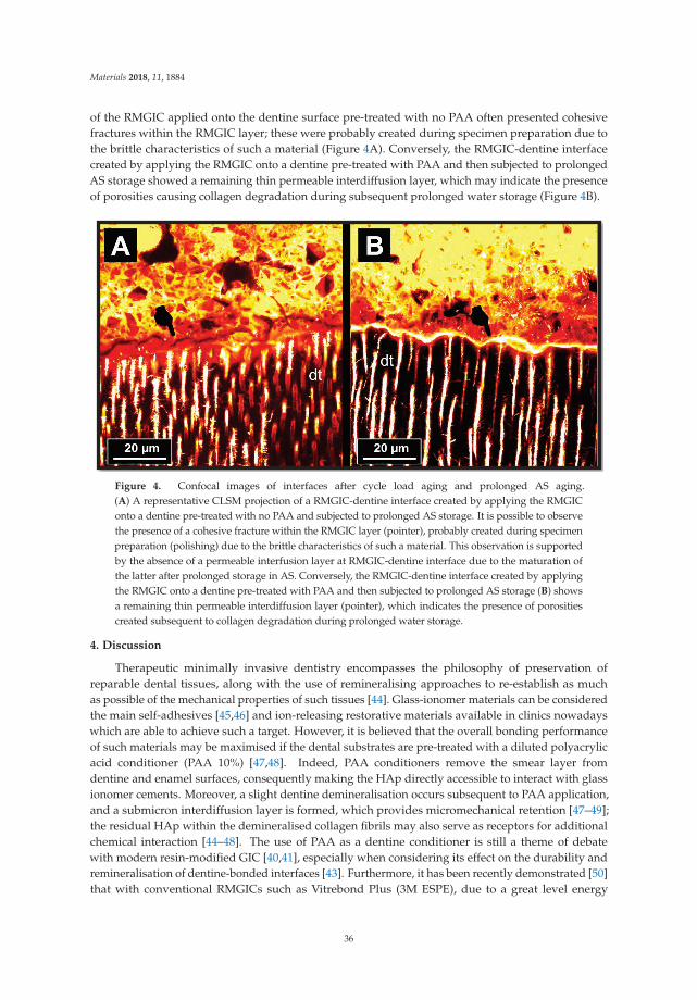
Materials 2018, 11, 1884
of the RMGIC applied onto the dentine surface pre-treated with no PAA often presented cohesivefractures within the RMGIC layer; these were probably created during specimen preparation due tothe brittle characteristics of such a material (Figure 4A). Conversely, the RMGIC-dentine interfacecreated by applying the RMGIC onto a dentine pre-treated with PAA and then subjected to prolongedAS storage showed a remaining thin permeable interdiffusion layer, which may indicate the presenceof porosities causing collagen degradation during subsequent prolonged water storage (Figure 4B).
Figure 4. Confocal images of interfaces after cycle load aging and prolonged AS aging.(A) A representative CLSM projection of a RMGIC-dentine interface created by applying the RMGIConto a dentine pre-treated with no PAA and subjected to prolonged AS storage. It is possible to observethe presence of a cohesive fracture within the RMGIC layer (pointer), probably created during specimenpreparation (polishing) due to the brittle characteristics of such a material. This observation is supportedby the absence of a permeable interfusion layer at RMGIC-dentine interface due to the maturation ofthe latter after prolonged storage in AS. Conversely, the RMGIC-dentine interface created by applyingthe RMGIC onto a dentine pre-treated with PAA and then subjected to prolonged AS storage (B) showsa remaining thin permeable interdiffusion layer (pointer), which indicates the presence of porositiescreated subsequent to collagen degradation during prolonged water storage.
4. Discussion
Therapeutic minimally invasive dentistry encompasses the philosophy of preservation ofreparable dental tissues, along with the use of remineralising approaches to re-establish as muchas possible of the mechanical properties of such tissues [44]. Glass-ionomer materials can be consideredthe main self-adhesives [45,46] and ion-releasing restorative materials available in clinics nowadayswhich are able to achieve such a target. However, it is believed that the overall bonding performanceof such materials may be maximised if the dental substrates are pre-treated with a diluted polyacrylicacid conditioner (PAA 10%) [47,48]. Indeed, PAA conditioners remove the smear layer fromdentine and enamel surfaces, consequently making the HAp directly accessible to interact with glassionomer cements. Moreover, a slight dentine demineralisation occurs subsequent to PAA application,and a submicron interdiffusion layer is formed, which provides micromechanical retention [47–49];the residual HAp within the demineralised collagen fibrils may also serve as receptors for additionalchemical interaction [44–48]. The use of PAA as a dentine conditioner is still a theme of debatewith modern resin-modified GIC [40,41], especially when considering its effect on the durability andremineralisation of dentine-bonded interfaces [43]. Furthermore, it has been recently demonstrated [50]that with conventional RMGICs such as Vitrebond Plus (3M ESPE), due to a great level energy
36

Materials 2018, 11, 1884
accumulation at the dentine bonding interface during cycle load, there was evident fluorescentpermeability associated with a lack of hermetic sealing.
The results of our study are in accordance those of Toledano at al. [50], as all the specimensapplied in dentine pre-treated with or without PAA showed remining permeability at the bondinginterface. Indeed, our current results demonstrated that the RMGIC tested in this study after theapplication on dentine without PAA pre-treatment presented a thin gap-free interface characterised bya thin layer of Rhodamine B absorbed through dentinal tubules (Figure 3A). In contrast, the interfaceof the RMGIC applied onto the dentine pre-treated with PAA was characterised by more Rhodamine Baccumulation, which was due to a lack of sealing of dentinal tubules (Figure 3B), as well as a thickerlayer of PAA-demineralised dentine, which remained non-infiltrated by the RMGIC [43,50].
However, after short-term load-cycle aging, the RMGIC-dentine specimens created usingthe RMGIC in dentine without PAA pre-treatment showed only a very thin interdiffusion layercharacterised by slight fluorescence signal at the interface. As described by Toledano at al. [50], such areduction in porosities at the bonding interface may have been due to apatite-like precipitation andremineralisation induced at the interface during mechanical cycling stress. Conversely, the specimenscreated with the RMGIC applied after PAA dentine pre-treatment also showed that the thicknessfluorescent signal at the interface was reduced compared to the same specimens at 24 h (Figure 3C),although such a porous layer was thicker than that observed when using no PAA dentine pre-treatment.In this case, it is possible that the level of mineral precipitation was not so suitable for remineralisingall the porosities within the interdiffusion layer, especially at its bottom. Indeed, Kim at al. [51] recentlyshown that GIC-based materials fail to completely remineralise apatite-depleted dentine due to a lackof nucleation of new apatite, even when biomimetic remineralising analogues were employed duringthe aging period [52].
It is important to highlight that such short-term load cycle aging was not able to induce anysignificant change in the microtensile bond strength, and no difference in the mode of failure in bothgroups of specimens (PAA vs. PAA dentine pre-treatment) was observed, compared to the controlspecimens at 24 h. Conversely, the results of this study showed that the specimens created with therepresentative RMGIC applied in dentine pre-treated with PAA showed a significant reduction in bondstrength after LC-AS aging compared to specimens tested after 24 h or after short-term load cycle aging.Moreover, the LC-AS aging protocol induce also a significant change in the mode of failure; RMGICapplied in dentine pre-treated with or without PAA presented more adhesive qualities compared to allthe other groups. Therefore, while the first null hypothesis is totally rejected, the second one that thedurability of RMGIC applied with or without the use of PAA conditioner would be affected by loadcycle followed by prolonged aging (8 months) in AS tested must be partially rejected.
Some of the current results are in accordance with those reported by Inoue et al. [45], who showedthat the use of PAA conditioner before application of GIC-based materials offered no significantincrease of the MTBS to dentine. Similar results were also recently reported by Sauro et al. [43], whoshowed that RMGIC applied onto dentine pre-treated with PAA showed significant μTBS reductionafter 6 months of AS storage alone or in combination with load cycling (p > 0.05). Moreover, they alsoshowed that for the RMGIC-dentine interface, specimens were affected by degradation/nanoleakageafter aging, unlike the interfaces created without the use of PAA conditioning, which showed signs ofremineralisation/maturation of the bonding interface.
The fractographic SEM analysis performed in this study showed that pre-treating the dentine withPAA could, in some cases, cause clear exposure of the collagen fibrils both before and after prolongedAS storage. Moreover, such fibrils were less abundant after prolonged AS storage compared to thoseobserved in the specimens aged in AS for 24 h (Figure 2F). These specimens also showed a significantincrease in the number of adhesive failures. In accordance with previously-published results [40,41,43],current fractographic SEM results showed that the specimens created with the use of no dentine PAAconditioner that failed in adhesive mode presented with dentine still covered by residual RMGIC,as the fracture occurred just above the dentine surface (Figure 2C).
37

Materials 2018, 11, 1884
It is hypothesised that such a result could be attributed to hydrolytic degradation processes thatoccurs over time within the collagen. Indeed, the use of PAA to pre-treat the dentine tissue mayhave demineralised the dentine collagen and activated endogenous matrix collagenolytic (MMP 1,MMP 8 and MMP 13) and gelatinolytic (MMP 2 and MMP 9) metalloproteinases [53]. It was alsodemonstrated [54] that high concentration of carboxylic groups in PAA acid conditioner may cause theformation a PAA-based polymeric gel layer within the bonding interface, which induces more watersorption at the interface. The RMGIC itself may also have degraded and become more porous overtime in AS, thereby facilitating diffusion of water towards the glass-ionomer–dentine interface andcausing an acceleration of the degradation processes [55].
The results of the ultramorphology and nanoleakage analysis of the RMGIC-dentine interfacesafter prolonged AS storage showed that when RMGIC were applied onto the dentine surface,pre-treated with no PAA, and then subjected to prolonged AS storage, a thin permeable interdiffusionlayer remained. This outcome may support the hypothesis that a bonding interface is usuallycharacterised by the presence of porosities created subsequent to collagen degradation duringprolonged water storage (Figure 4B). Conversely, the specimens created with the use of no PAAdentine conditioning showed that the absorbing layer at the interface seen at 24 h examination(Figure 3A) disappeared after prolonged AS storage (Figure 4A) due to the maturation of such areas [55],and possible remineralisation [43,56]. Indeed, the therapeutic properties (e.g., ion releasing) of RMGICmay have induced the growth of mineral crystals and remineralisation within the bonded-dentineinterface, which interfered with the proteolytic action of endogenous dentine metalloproteinases [56,57].This latter hypothesis is in accordance with previous studies that demonstrated the fluoride mightinhibit both pro- and active forms of MMP 2 and MMP 9 [58]. Moreover, Makowski & Ramsby [59]reported that mineral precipitation, as well as apatite formation, may inhibit MMP activity throughthe formation of [Ca-PO/MMP] complexes. Sauro et al. [43] have recently reported that such aremineralising potential of RMGIC may increase if they are applied onto dentine pre-treated withbioactive glass in air-abrasion systems, and then conditioned with or without the use of a PAAconditioning gel.
In conclusion, within the limitations of this in vitro study, it is possible to affirm that the clinicaldecision of using a PAA conditioner should be based upon the histological features of the dentineretained after cavity preparation (e.g., sound or caries-affected dentine). However, modern RMGICsmay be used for dentine restorations with or without the use of PAA pre-treatment, but it is importantto consider that such a type of acid etching procedure might increase the risk of degradation at thebonding interface after prolonged service in oral cavity under mechanical cycling stress and prolongedsaliva immersion. Conversely, in cases of no PAA dentine pre-treatment, it might be possible to havea synergic combination between GIC-based materials, saliva, and cycle occlusal load, which mayenhance the therapeutic properties of RMGIC to induce mineralisation and protection of the bondinginterface, thereby achieving more long-lasting restorations.
Author Contributions: For research articles with several authors, a short paragraph specifying their individualcontributions must be provided. The following statements should be used “Conceptualization, S.S. and I.M..;Methodology, V.F.M., J.M.N.M.; Validation, S.S., V.F.L.; Formal Analysis, R.G.M., J.A.G.B.; Investigation, S.S., I.M.,V.F.M., X.X.; Resources, V.F.L., I.M.; Data Curation, X.X.; Writing-Original Draft Preparation, S.S.; Writing-Review& Editing, S.S..; Supervision, V.F.L, S.S., I.M.; Project Administration, S.S., V.F.L., I.M.; Funding Acquisition, V.F.L,I.M.”, please turn to the CRediT taxonomy for the term explanation. Authorship must be limited to those whohave contributed substantially to the work reported.
Funding: This research received no external funding.
Acknowledgments: We declare that we have no proprietary, financial, professional or other personal interest ofany nature or kind in any product, service, and/or company that could be construed as influencing the positionpresented in, or the review of this manuscript. The authors of this article would like to thank SDI (Australia) fordonating us the RMGIC (RIVA LC) and the PAA conditioner used in this study.
Conflicts of Interest: The authors declare no conflict of interest.
38

Materials 2018, 11, 1884
References
1. Wilson, A.D.; Kent, B.E. The glass-ionomer cement, a new translucent dental filling material. J. Appl.Chem. Biotechnol. 1971, 21, 313. [CrossRef]
2. Wilson, A.D.; Nicholson, J.W. Acid-Base Cements: Their Biomedical and Industrial Applications; CambridgeUniversity Press: Cambridge, UK, July 2005; Volume 3.
3. Prosser, H.J.; Powis, D.R.; Wilson, A.D. Glass-ionomer cements of improved flexural strength. J. Dent. Res.1986, 65, 146–148. [CrossRef] [PubMed]
4. Alhalawani, A.M.; Curran, D.J.; Boyd, D.; Towler, M.R. The role of poly(acrylic acid) in conventional glasspolyalkenoate cements. J. Polym. Eng. 2016, 36, 221–237. [CrossRef]
5. Guggenberger, R.; May, R.; Stefan, K.P. New trends in glass-ionomer chemistry. Biomaterials 1998, 19, 479–483.[CrossRef]
6. McCabe, J.F.; Walls, A.W. (Eds.) Applied Dental Materials; John Wiley & Sons: Hoboken, NJ, USA, May 2013.7. Anusavice, K.J.; Shen, C.; Rawls, H.R. Phillips’ Science of Dental Materials; Elsevier Health Sciences:
Amsterdam, The Netherlands, 2013.8. Wilson, A.D. The chemistry of dental cements. Chem. Soc. Rev. 1978, 7, 265–296. [CrossRef]9. Nicholson, J.W. Chemistry of glass-ionomer cements: A review. Biomaterials 1998, 19, 485–494. [CrossRef]10. Zainuddin, N.; Karpukhina, N.; Hill, R.G.; Law, R.V. A long-term study on the setting reaction of glass
ionomer cements by 27 Al MAS-NMR spectroscopy. Dent. Mater. 2009, 25, 290–295. [CrossRef] [PubMed]11. Preston, A.J.; Higham, S.M.; Agalamanyi, E.A.; Mair, L.H. Fluoride recharge of aesthetic dental materials.
J. Oral Rehabil. 1999, 26, 936–940. [CrossRef] [PubMed]12. Forsten, L. Resin-modified glass ionomer cements: Fluoride release and uptake. Acta Odontol. Scand. 1995,
53, 222–225. [CrossRef] [PubMed]13. Forss, H.; Jokinen, J.; Spets-Happonen, S.; Seppä, L.; Luoma, H. Fluoride and mutans streptococci in plaque
grown on glass ionomer and composite. Caries Res. 1991, 25, 454–458. [CrossRef] [PubMed]14. Preston, A.J.; Mair, L.H.; Agalamanyi, E.A.; Higham, S.M. Fluoride release from aesthetic dental materials.
J. Oral Rehabil. 1999, 26, 123–129. [CrossRef] [PubMed]15. De Amorim, R.G.; Leal, S.C.; Frencken, J.E. Survival of atraumatic restorative treatment (ART) sealants and
restorations: A meta-analysis. Clin. Oral Investig. 2012, 16, 429–441. [CrossRef] [PubMed]16. Van Dijken, J.W. 3-Year clinical evaluation of a compomer, a resin-modified glass ionomer and a resin
composite in class III restorations. Am. J. Dent. 1996, 9, 195–198. [PubMed]17. Abdalla, A.I.; Alhadainy, H.A.; Garcia-Godoy, F. Clinical evaluation of glass ionomers and compomers in
class V carious lesions. Am. J. Dent. 1997, 10, 18–20. [PubMed]18. Leevailoj, C.; Platt, J.A.; Cochran, M.A.; Moore, B.K. In vitro study of fracture incidence and compressive
fracture load of all-ceramic crowns cemented with resin-modified glass ionomer and other luting agents.J. Prosthet. Dent. 1998, 80, 699–707. [CrossRef]
19. Pascotto, R.C.; de Lima Navarro, M.F.; Capelozza Filho, L.; Cury, J.A. In vivo effect of a resin-modified glassionomer cement on enamel demineralization around orthodontic brackets. Am. J. Orthod. Dentofac. 2004,125, 36–41. [CrossRef]
20. Jokstad, A.; Mjör, I.A. Ten years’ clinical evaluation of three luting cements. J. Dent. 1996, 24, 309–315.[CrossRef]
21. Murdoch-Kinch, C.A.; McLean, M.E. Minimally invasive dentistry. J. Am. Dent. Assoc. 2003, 134, 87–95.[CrossRef] [PubMed]
22. Peumans, M.; de Munck, J.; Mine, A.; van Meerbeek, B. Clinical effectiveness of contemporary adhesivesfor the restoration of non-carious cervical lesions. A systematic review. Dent. Mater. 2014, 30, 1089–1103.[CrossRef] [PubMed]
23. Terata, R.; Nakashima, K.; Kubota, M. Effect of temporary materials on bond strength of resin-modifiedglass-ionomer luting cements to teeth. Am. J. Dent. 2000, 13, 209–211. [PubMed]
24. McLean, J.W.; Powis, D.R.; Prosser, H.J.; Wilson, A.D. The use of glass-ionomer cements in bonding compositeresins to dentine. Br. Dent. J. 1985, 158, 410–414. [CrossRef] [PubMed]
25. Andersson-Wenckert, I.E.; Kieri, C. Durability of extensive class II open-sandwich restorations with aresin-modified glass ionomer cement after 6 years. Am. J. Dent. 2004, 17, 43–50. [PubMed]
39
Materials 2018, 11, 1884
26. Yamaga, R.; Nishino, M.; Yoshida, S.; Yokomizo, I. Diammine silver fluoride and its clinical application.J. Osaka Univ. Dent. Sch. 1972, 12, 1–20. [PubMed]
27. Sauro, S.; Osorio, R.; Watson, T.F.; Toledano, M. Influence of phosphoproteins’ biomimetic analogs onremineralization of mineral-depleted resin–dentin interfaces created with ion-releasing resin-based systems.Dent. Mater. 2015, 31, 759–777. [CrossRef] [PubMed]
28. Xie, D.; Brantley, W.A.; Culbertson, B.M.; Wang, G. Mechanical properties and microstructures ofglass-ionomer cements. Dent. Mater. 2000, 16, 129–138. [CrossRef]
29. Peutzfeldt, A.; Garcia-Godoy, F.; Asmussen, E. Surface hardness and wear of glass ionomers and compomers.Am. J. Dent. 1997, 10, 15–17. [PubMed]
30. Ahmed, N.; Zafar, M.S. Effects of wear on hardness and stiffness of restorative dental materials. Life Sci. J.2014, 11, 11–18.
31. Um, C.M.; Øilo, G. The effect of early water contact on glass-ionomer cements. Quintessence Int. 1992, 1, 23.32. Moshaverinia, A.; Roohpour, N.; Chee, W.W.L.; Schricker, S.R. A review of powder modifications in
conventional glass-ionomer dental cements. J. Mater. Chem. 2011, 21, 1319–1328. [CrossRef]33. Moshaverinia, A.; Ansari, S.; Moshaverinia, M.; Roohpour, N.; Darr, J.A.; Rehman, I. Effects of incorporation
of hydroxyapatite and fluoroapatite nanobioceramics into conventional glass ionomer cements (GIC).Acta Biomater. 2008, 4, 432–440. [CrossRef] [PubMed]
34. Moshaverinia, A.; Ansari, S.; Movasaghi, Z.; Billington, R.W.; Darr, J.A.; Rehman, I.U. Modification ofconventional glass-ionomer cements with N-vinylpyrrolidone containing polyacids, nano-hydroxy andfluorapatite to improve mechanical properties. Dent. Mater. 2008, 24, 1381–1390. [CrossRef] [PubMed]
35. Wilson, A.D. Resin-modified glass-ionomer cements. Int. J. Prosthodont. 1989, 3, 425–429.36. Soncini, J.A.; Maserejian, N.N.; Trachtenberg, F.; Tavares, M.; Hayes, C. The Longevity of amalgam versus
compomer/composite restorations in posterior primary and permanent teeth: Findings from the newengland children’s amalgam trial. J. Am. Dent. Assoc. 2007, 138, 763–772. [CrossRef] [PubMed]
37. McCabe, J.F. Resin-modified glass-ionomers. Biomaterials 1998, 19, 521–527. [CrossRef]38. Sauro, S.; Pashley, D. Strategies to stabilise dentine-bonded interfaces through remineralising operative
approaches - State of The Art. Int. J. Adhes. Adhes. 2016, 69, 39–57. [CrossRef]39. Tyas, M.J.; Burrow, M.F. Adhesive restorative materials: A review. Aust. Dent. J. 2004, 49, 112–121. [CrossRef]
[PubMed]40. Inoue, S.; Abe, Y.; Yoshida, Y.; De Munck, J.; Sano, H.; Suzuki, K.; Lambrechts, P.; Van Meerbeek, B. Effect of
conditioner on bond strength of glass-ionomer adhesive to dentin/enamel with and without smear layerinterposition. Oper. Dent. 2004, 29, 685–692. [PubMed]
41. De Munck, J.; Van Meerbeek, B.; Yoshida, Y.; Inoue, S.; Suzuki, K.; Lambrechts, P. Four-year water degradationof a resin-modified glass-ionomer adhesive bonded to dentin. Eur. J. Oral Sci. 2004, 112, 73–83. [CrossRef][PubMed]
42. Toledano, M.; Cabello, I.; Aguilera, F.S.; Osorio, E.; Osorio, R. Effect of in vitrochewing and bruxism eventson remineralization, at the resin-dentin interface. J. Biomech. 2015, 48, 14–21. [CrossRef] [PubMed]
43. Sauro, S.; Watson, T.; Moscardó, A.P.; Luzi, A.; Feitosa, V.P.; Banerjee, A. The effect of dentine pre-treatmentusing bioglass and/or polyacrylic acid on the interfacial characteristics of resin-modified glass ionomercements. J. Dent. 2018, 73, 32–39. [CrossRef] [PubMed]
44. Watson, T.F.; Atmeh, A.R.; Sajini, S.; Cook, R.J.; Festy, F. Present and future of glass ionomersandcalcium-silicate cements as bioactive materials in dentistry: biophotonics-based interfacial analyses inhealth and disease. Dent. Mater. 2014, 30, 50–61. [CrossRef] [PubMed]
45. Inoue, S.; Van Meerbeek, B.; Abe, Y.; Yoshida, Y.; Lambrechts, P.; Vanherle, G.; Sano, H. Effect of remainingdentin thickness and the use of conditioner on micro-tensile bond strength of a glass-ionomer adhesive.Dent. Mater. 2001, 17, 445–455. [CrossRef]
46. Yoshida, Y.; Van Meerbeek, B.; Nakayama, Y.; Snauwaert, J.; Hellemans, L.; Lambrechts, P.; Vanherle, G.;Wakasa, K. Evidence of chemical bonding at biomaterial–hard tissue interfaces. J. Dent. Res. 2000, 79, 709–714.[CrossRef] [PubMed]
47. Van Meerbeek, B.; Vargas, M.; Inoue, S.; Yoshida, Y.; Peumans, M.; Lambrechts, P.; Vanherle, G. Adhesivesand cements topromote preservation dentistry. Oper. Dent. 2001, 26, 119–144.
40

Materials 2018, 11, 1884
48. Van Meerbeek, B.; Inoue, S.; Perdigao, J.; Lambrechts, P.; Vanherle, G. Enamel and Dentin Adhesion;Summitt, J.B., Robbins, J.W., Schwartz, R.S, Eds.; Quintessence Publishing Co.: Chicago, IL, USA, 2001;pp. 178–235.
49. Lin, A.; McIntyre, N.; Davidson, R. Studies on the adhesion of glass ionomer cements to dentin. J. Dent. Res.1992, 71, 1836–1841. [CrossRef] [PubMed]
50. Toledano, M.; Osorio, R.; Osorio, E.; Cabello, I.; Toledano-Osorio, M.; Aguilera, F.S. In vitro mechanicalstimulation facilitates stress dissipation and sealing ability at the conventional glass ionomer cement-dentininterface. J. Dent. 2018, 73, 61–69. [CrossRef] [PubMed]
51. Kim, Y.K.; Yiu, C.K.; Kim, J.R.; Gu, L.; Kim, S.K.; Weller, R.N.; Pashley, D.H.; Tay, F.R. Failureof a glassionomer to remineralise apatite-depleted dentin. J. Dent. Res. 2010, 89, 230–235. [CrossRef] [PubMed]
52. Gu, L.S.; Kim, J.; Kim, Y.K.; Liu, Y.; Dickens, S.H.; Pashley, D.H.; Ling, J.Q.; Tay, F.R. Achemicalphosphorylation-inspired design for Type I collagen biomimeticremineralization. Dent. Mater. 2010, 26,1077–1089. [CrossRef] [PubMed]
53. Nishitani, Y.; Yoshiyama, M.; Wadgaonkar, B.; Breschi, L.; Mannello, F.; Mazzoni, A.; Carvalho, R.M.;Tjäderhane, L.; Tay, F.R.; Pashley, D.H. Activation of gelatinolytic/collagenolytic activity in dentin byself-etching adhesives. Eur. J. Oral Sci. 2006, 114, 160–166. [CrossRef] [PubMed]
54. Es-Souni, M.; Fischer-Brandies, H.; Zaporojshenko, V.; Es-Souni, M. On the interactionof polyacrylic acid asa conditionning agent with bovine enamel. Biomaterials 2002, 23, 2871–2878. [CrossRef]
55. Sidhu, S.K.; Watson, T.F. Interfacial characteristics of resin-modified glass-ionomermaterials: A study onfluid permeability using confocal fluorescence microscopy. J. Dent. Res. 1998, 77, 1749–1759. [CrossRef][PubMed]
56. Sauro, S.; Watson, T.F.; Thompson, I.; Toledano, M.; Nucci, C.; Banerjee, A. Influence of air-abrasion executedwith PAA-bioglass 45S5 on the bonding performance of a resin-modified glass ionomer cement. Eur. J.Oral Sci. 2012, 120, 168–177. [CrossRef] [PubMed]
57. Brackett, M.G.; Agee, K.A.; Brackett, W.W.; Key, W.O.; Sabatini, C.; Kato, M.T.; Buzalaf, M.A.; Tjäderhane, L.;Pashley, D.H. Effect of sodium fluoride on theendogenous MMP activity of dentin matrices. J. Nat. Sci. 2015,1, e118. [PubMed]
58. Tezvergil-Mutluay, A.; Seseogullari-Dirihan, R.; Feitosa, V.P.; Cama, G.; Brauer, D.S.; Sauro, S. Effects ofComposites Containing Bioactive Glasses on Demineralized Dentin. J. Dent. Res. 2017, 96, 999–1005.[CrossRef] [PubMed]
59. Makowski, G.S.; Ramsby, M.L. Differential effect of calcium phosphate andcalcium pyrophosphate onbinding of matrix metalloproteinases to fibrin: comparison to a fibrin-binding protease from inflammatoryjoint fluids. Clin. Exp. Immunol. 2004, 136, 176–187. [CrossRef] [PubMed]
© 2018 by the authors. Licensee MDPI, Basel, Switzerland. This article is an open accessarticle distributed under the terms and conditions of the Creative Commons Attribution(CC BY) license (http://creativecommons.org/licenses/by/4.0/).
41

materials
Article
Biomimetic Mineralizing Agents Recover the MicroTensile Bond Strength of Demineralized Dentin
Luiz Filipe Barbosa-Martins 1, Jossaria Pereira de Sousa 1, Lívia Araújo Alves 2,
Robert Philip Wynn Davies 3 and Regina Maria Puppin-Rontanti 4,*
1 Department of Pediatric Dentistry, Piracicaba Dental School, State University of Campinas,Piracicaba 13414-903; Brazil; [email protected] (L.F.B.-M.); [email protected] (J.P.d.S.)
2 Department of Oral Diagnosis, Piracicaba Dental School, State University of Campinas, Piracicaba 13414-903,Brazil; [email protected]
3 Division of Oral Biology, School of Dentistry, Faculty of Medicine & Health, University of Leeds,Leeds LS9 7TF, UK; [email protected]
4 Departments of Pediatric Dentistry and Restorative Dentistry, Piracicaba Dental School, University ofCampinas, Piracicaba 13414-903, Brazil
* Correspondence: [email protected]; Tel.: +55-19-2106-5286
Received: 10 July 2018; Accepted: 11 September 2018; Published: 14 September 2018
Abstract: Biomimetic remineralization is an approach that mimics natural biomineralization,and improves adhesive procedures. The aim of this paper was to investigate the influence of DentinCaries-like Lesions (DCLL)-Producing Model on microtensile bond strength (μTBS) of etch andrinse adhesive systems and investigate the effect of remineralizing agents such as Sodium Fluoride(NaF), MI Paste™ (MP) and Curodont™ Repair (CR) on caries-affected dentin (n = 6). Nine groupswere established: (1) Sound dentin; (2) Demineralized dentin/Chemical DCLL: (3) Demineralizeddentin/Biological DCLL; (4) Chemical/DCLL + NaF; (5) Chemical/DCLL + MP; (6) Chemical/DCLL +CR; (7) Biological/DCLL + NaF; (8) Biological/DCLL + MP; (9) Biological/DCLL + CR. Then all dentinblocks were subjected to a bonding procedure with Adper™ Single Bond 2 adhesive system/FiltekZ350XT 4 mm high block, following this they were immersed in deionized water/24 h and thensectioned with ∼=1 mm2 beams. The μTBS test was conducted at 1 mm/min/500 N loading.Failure sites were evaluated by SEM (scanning electron microscopy (150×). μTBS data weresubmitted to factorial ANOVA and Tukey’s test (p < 0.05). The highest values were found whendemineralized dentin was treated with MP and CR, regardless caries lesion depth (p < 0.05). There wasa predominance of adhesive/mixed in the present study. It was concluded that the use of theartificial dentin caries production models produces differences in the μTBS. Additionally MP and CRremineralizing agents could enhance adhesive procedures even at different models of caries lesion.
Keywords: dentin; desmineralization; microtensile bond strength
1. Introduction
During the execution of routine dental restorations, the hybrid structure formed by the dentalbonding procedure occurs through the interaction and subsequent polymerization of monomersaround the demineralized collagen matrix [1]. The oral cavity is a severe environment for theresin-dental bond to survive for a reasonable length of time, with thermomechanical changes,chemical attacks by acids and enzymes and other factors posing routine daily challenges. Therefore,to achieve effective and stable bonding, the preservation of dentin collagen is critical, since collagenrepresents the major organic component of the dentin matrix [2].
Caries is among the most common diseases worldwide [3], and the immediate bond strengths tocaries-affected dentin are commonly 20–50% lower than to sound dentin [4–6]. The restoration of the
Materials 2018, 11, 1733; doi:10.3390/ma11091733 www.mdpi.com/journal/materials42

Materials 2018, 11, 1733
normal conditions of the mineral content of the caries-affected dentin, prevents the action of enzymesin addition to providing an increased bond durability [7].
Biomimetic remineralization mimics the process of natural biomineralization by replacingdemineralized collagen matrix water with apatite crystallites [7]. Caries-affected dentin is comprised ofabout 14–53% of water compared with sound dentin, which exhibits a much lower value [8]. Therefore,by replacing water with minerals at the dentin–resin interface, this would increase the mechanicalproperties and inhibit water-related hydrolysis [9].
It has recently been demonstrated that the use of remineralizing agents in dentin could recover themechanical properties of the substrate [10]. In addition to sodium fluoride (NaF) and sodium phosphate(Na3PO4), casein phosphopeptide amorphous calcium phosphate (CPP-ACP), which is derived frommilk protein, can release calcium phosphate assisting in enamel and dentin remineralization [11]. It actsmainly by inhibiting demineralization and enzymatic degradation [12]. Furthermore, recent studieshave shown that the use CPP-ACP has no negative effect on bond strength [13,14]. The peptidicbiomimetic matrix ‘P11-4’, which has been incorporated into a clinical product (Curodont™ Repair) hasshown encouraging results in early clinical trials. It has been shown to improve the visual appearanceof carious lesions and increases the opacity on X-rays after treatment of proximal caries [15,16].Additionally, following the application of P11-4 and subsequent bonding procedures an increasedresin-dentin bond strength has been observed [17].
The restructured demineralized collagen matrix found in caries-affected dentin process byinterventions such as Sodium Fluoride (NaF), CPP-ACP contained in MI Paste™ (GC International)and P11-4 peptide contained in Curodont™ Repair (Credentis AG), prior to adhesive procedures by anetch-and-rinse adhesive system (Adper™ Single Bond 2 (3M ESPE) in the demineralized dentin couldbe a promising proposal for adhesive clinical procedures.
Clinical binding procedures simulated by mechanical methods (i.e., TMBS, μTBS) often useartificial demineralized dentin. However, the lack of standardization of caries lesions creates technicaldifficulties for evaluation [18]. In vitro models have been used to produce demineralized dentinunder controlled conditions [19–22]. Chemical methods provide superficial dentin demineralization,resulting in a substrate with similar hardness compared to natural caries-affected dentin [19].Conversely, the microbiological method promotes an excessive softening of dentin, but with amore comparable morphological pattern of collagen degradation to natural caries lesions [19–22].Pacheco et al., 2013 [23], evaluating molecular and structural lesions related to dental caries,produced by the chemical (GC), biological (GB), in situ (GIS) and natural (CNG) approaches,showed similar and lower surface hardness between CNG and GB of which GC and GIS, lower mineralcontent (Ca2+ and PO4
3−) for GB and GNC than GC and GIS. Therefore, the structure and mechanicalproperties are different with respect to the caries model production and the remineralizing agents mayact differently in the adhesion procedures depending on the model of caries lesions used.
To ascertain the potential efficacy of the bonding procedure carried out on remineralized dentinwe aimed to evaluate different remineralization treatments and the method used in producing thesimulated dentin-like caries lesions on the micro tensile bond strength of remineralized dentin.The hypothesis was that the Dentin Caries-like Lesions Producing Model and the remineralizingagents (Sodium Fluoride-(NaF), MI Paste™-(MP) and Curodont™ Repair-(CR)) affect microtensilebond strength-μTBS of etch-and-rinse adhesive system on caries-affected dentin.
2. Materials and Methods
Sixty-three sound human third molars were collected with patients’ informed consent,as approved by the Ethics Committee of Piracicaba Dental School, University of Campinas (Protocolnumber 37634814.5.0000.5418). The teeth were stored in 0.1% thymol solution at 4 ◦C for no longer than2 months after extraction. A 4.0 mm coronal dentin slice from each tooth was obtained by sectioning2.0 mm below cement-enamel junction (CEJ), and 2.0 mm above CEJ, using a slow-speed water-cooleddiamond saw (Isomet 1000, Buehler Ltd., Lake Bluff, IL, USA). Six dentin slices were used for sound
43

Materials 2018, 11, 1733
dentin (control group-CG), and the others were randomly assigned into 2 groups (n = 24), according tothe caries method production. The dentin surface of each specimen was wet polished with a 600-gritSiC paper (Arotec, São Paulo, Brazil) for 30 s to create a standardized smear layer. The dentin surfaceswere carefully examined under a stereomicroscope at ×50 magnification to confirm the absence ofenamel islets. The specimens were immediately subjected to production of caries in vitro. The groupdistribution is illustrated in Figure 1. To check the caries dentin depth, three teeth were chosen fromeach group and probed using a polarized light microscope (Figure 2).
Figure 1. Experimental design. DDC—demineralized dentin provided by chemical model;DDC/NaF-DDC + 2% NaF; DDC/MP-DDC + MI Paste™; DDC/CR-DDC + Curodont™ Repair;DDB—demineralized dentin provided by biological model; DDB/NaF-DDB + 2% NaF; DDB/MP-DDB+ MI Paste™; DDB/CR-DDB + Curodont™ Repair.
Figure 2. (A) Artificial caries lesions provided by Chemical Model; (B) Artificial caries lesions providedby Biological Model after removing the softened tissue; (C) Sound Dentin.
44

Materials 2018, 11, 1733
2.1. Artificial Dentin Caries-Like Lesions (DCLL) Production Protocols
Sixty dentin slices (54 teeth for μTBS and 6 for polarized light microscopy) were randomlyassigned into 2 groups according to dentin-like caries lesions producing models: chemical(carboxymethylcellulose acid gel) and biological (Streptococcus mutans—UA159 biofilm).
2.1.1. Chemical Model
The specimens were submerged in vials containing 5 mL of 6% carboxymethylcellulose acid gel(0.1 M lactic acid titrated to pH 5.0 in a KOH solution) at pH 5.0 and 37 ◦C. The specimens remainedin the gel for 48 h without renewal [23]. This model has been reported to supposedly provide ademineralized dentin similar to that of caries affected dentin.
2.1.2. Biological Model
The specimens were fixed with orthodontic wire on the lids of glass vials containing 250 mL ofsterile deionized water and were sterilized with gamma radiation (14.5 kGy dose) for 60 h (Pacheco etal., 2013). Then, they were transferred to another glass vial containing 250 mL of sterile brain-heartinfusion (BHI) broth (LabCenter, São Paulo, Brazil) supplemented with 0.5% yeast extract (LabCenter,São Paulo, Brazil), 0.5% glucose (LabCenter, São Paulo, Brazil), 1% sucrose (LabCenter, São Paulo,Brazil) and 2% S. mutans (UA159, ATCC, Oklahoma, OK, USA) incubated at 37 ◦C and supplementedwith 10% CO2, pH of around 4.0. Starter culture was transferred into 250 mL of fresh BHI and grownfor 4 h at 37 ◦C under aerobic conditions. Optical density at 550 nm (A550) of all bacterial suspensionswas adjusted to 0.05 prior to inoculation. Inoculation occurred only in the first day of the experiment,but the broth was renewed every 48 h over a 7-day period. The broth was Gram stained daily tomonitor contamination. The resulting biofilm formed over the teeth was removed with gauze and thesofter dentin layer was removed using #6 carbide drills; the removal ceased when dentin appearancewas like that of caries-affected dentin [23].
2.2. Polarized Light Microscopy (PLM)
After providing dentin caries-like lesions, three specimens of each dentin caries-likelesions-producing models and control group were sectioned perpendicular to the occlusal surface toobtain slices. The two more central slices of each tooth were selected and polished with #800, #1200,#2400 and #4000-grit silicon carbide (SiC) paper (Buehler, Lake Buff, IL, USA), obtaining a 0.15 μmdentin thickness. Dentin slices were analyzed for the depth of demineralization on PLM (Leica DMLP,Leica microsystems, Wetzlar, Germany). The dentin slices were kept in 100% humidity throughoutthe investigation. Depth caries lesions were measured in a PLM (Leica DMLP, Leica microsystems)using 20×/0.4 (corr.) objective. Standard settings for contrast, brightness and light were used for allimages. Four measurements were made in different parts of the same lesion from the lesion borderto the deepest part of the lesion, for each dentin caries-like lesion model. An average depth foreach specimen was calculated from the individual values based on depth difference between dentincaries-like lesions and sound dentin on the same specimen. Dentin caries-like lesions observed inthe specimens subjected to Chemical Model presented x = 12.69 μm average depth and BiologicalModel x = 148.55 μm, measured using PLM.
2.3. Dentin Surface Treatment
Thirty-six teeth were assigned to 6 groups according to the remineralization treatment:demineralized dentin by chemical model (DDC) + treated with 0.2% NaF Solution (1 min)—DDC/NaF;DDC + treated with MI Paste™ (1 min)—DDC/MP; and DDC + treated with Curodont™Repair—DDC/CR applied (5 min) plus Ca2+ and PO4
3− Solution (1 min); demineralized dentinby biological model (DDB) + 0.2% NaF Solution—DDB/NaF; DDB + treated with MI Paste™ applied
45

Materials 2018, 11, 1733
(1 min)—DDB/MP; and DDB + treated with Curodont™ Repair—DDB/CR applied (5 min) plus Ca2+
and PO43− Solution (1 min).
The specimens of groups DDC/NaF and DDB/NaF were remineralized using 0.1 mL of the 0.2%NaF. 0.2% NaF solution was applied on demineralized dentin surface and left dry for 1 min at roomtemperature for the groups DDC/NaF and DDB/NaF. 0.1 mL of the MI Paste™ was applied onto thesurface of the specimens from DDC/MP and DDB/MP groups with microbrush for 1 min at roomtemperature, the excess paste was removed by washing with deionized water. For DDC/CR andDDB/CR groups, 50 μL of the Curodont™ Repair was applied and left for 5 min, then, a Ca2+ andPO4
3− solution was applied and left onto surface for 1 min. For all treatments, the solution excess wasremoved with absorbent paper.
2.4. Bonding Procedures
A single operator applied the adhesive according to the manufacturer’s instruction (Table 1).An LED light-curing unit (Bluephase, Ivoclar Vivadent; Schaan, Liechtenstein) was set to the lowpower mode with a light intensity of 650 mW/cm2. A nanohybrid resin composite (Filtek Z350 XT,A2 (3M ESPE, St. Paul, MN, USA)) was used to create resin composite buildups in four layers of 1 mmeach [17]. Each layer was light cured for 20 s, followed by a final polymerization of 60 s. The specimenswere then stored at 100% humidity at 37 ◦C for 24 h.
Table 1. Materials, manufactures, components, batch numbers and application mode oftested materials.
Materials(Manufactures)
Main ComponentsBatch
NumberApplication Mode
0.2% NaF Solution 0.2 g of NaF in 100 mL deionized water Made in theLab *
1. Apply 1.0 mL of 0.2%NaF solution
Ca2+ and PO43−
Solution
Saturated solution of Ca2+ and PO43−
(1.5 mmol/L calcium, 0.9 mmol/Lphosphate, and 150 mmol/L KCl in
20 mmol/L cacodylic buffer, pH 7.0) [24].
Made inthe Lab
1. Apply 0.1 mL of Ca2+
and PO43− solution
MI™ Paste—GCInternacional,
Itabashi-ku, Tóquio,Japão
Glycerol, CPP-ACP, D-Sorbitol, Propyleneglycol, Titanium dioxide and silicon N2347319
1. Apply 0.1 mL of MI™Paste
Curodont™Repair—Credentis AG,Dorfstrasse, Windisch,
Switzerland
P11-4 peptide—amino acid sequence—(Ace-Gln-Gln-Arg-Phe-Glu-Trp-Glu-
Phe-Glu-Gln-Gln-NH2)N342x
1. Apply 50 μL ofCurodont™ Repair for5 min
2. Apply 0.1 mL of Ca2+
and PO43− solution
Scotchbond™Universal Etchant—3MESPE; St Paul, MN, USA
32% phosphoric acid N3451. Apply etchant for 15 s2. Rinse for 10 s
Adper Single Bond2.0—3M ESPE; St Paul,
MN, USA
HEMA, water, ethanol, Bis-GMA,dimethacrylates, amines, metacrylate
functional copolymer of polyacrylic andpolyitaconic acids, 10% by weight of 5
nanometer-diameter spherical sílicaparticles
N42912
3. Blot water excess4. Apply 2 consecutive
coats of adhesive for 15 swith gentle agitation
5. Gently air dry for 5 s6. Light-cure for 10 s
Filtek™ Z350 XT—3MESPE; St Paul, MN, USA
BIS-GMA, Bis-EMA, UDMA, TEG-DMA,camphorquinone, non-agglomerated
silica nanoparticlesN98354
1. Incremental insertion2 mm
2. Light-cure for 20 s
* Pediatric Dentistry Laboratory.
46

Materials 2018, 11, 1733
2.5. Microtensile Bond Strength Test (μTBS)
After storage, the specimens were sectioned perpendicularly to the resin/dentin interface toproduce dentin–resin beams with 1 mm2 at cross-sectional area, using a low speed diamond saw(ISOMET 1000, Buehler Ltd., Lake Buff, IL, USA). From six to eight beams were obtained pertooth, each beam was measured with a digital caliper (Mitutoyo; Kawasaki, Japan) to determinethe cross-sectional area. All beams were kept in deionized water for 24 h.
For μTBS measurements, each beam was fixed to a microtensile device with cyanocrylate glue(Super Bonder (#1883519), Loctite, Henkel Corp., Rocky Hill, CT, USA), and tested in a universaltesting machine (DL 2000, EMIC, Equipment and Systems Ltda., São José dos Pinhais, Brazil). The testwas carried out with a 500 N load at 1.0 mm/min cross speed until failure. The μTBS values wereexpressed in MPa.
2.6. Analysis of Failure Mode
All the fractured specimens from the microtensile bond strength analysis were assessed todetermine the failure mode using SEM at ×50 and ×150 magnifications. The fractured surfacesof the beams were paired, air dried, mounted on aluminum stubs, gold coated, and examined by SEM(JSM-5600LV, JEOL; Tokyo, Japan), operated at 15 kV. The failure patterns were classified accordingto the following categories: adhesive, mixed (involving resin composite, adhesive and/ or dentin),cohesive failure in the resin composite, and cohesive failure in dentin [17,25,26].
2.7. Statistical Analysis
Bond strength values for each group were analyzed by Shapiro–Wilk test (R Software version 3.4.3,The R Foundation for Statistical Computing, Vienna, Austria) in order to assess the normality of the datadistribution. Factorial ANOVA and post hoc Tukey test (R Software version 3.4.3, The R Foundationfor Statistical Computing, Vienna, Austria) were used to determine statistically significant differencesbetween factors: the dentin-like caries lesions model (two levels—chemical and biological models)and dentin remineralization treatment (four levels—SD, DD, NaF + DD, CPP-ACP + DD and P11-4 +DD) on dentin/resin bond strength, and additional Dunnett test to determine statistically significantdifferences between the experimental groups and the control group (sound dentin). The KruskalWallis test was used to evaluate the failure mode. The R Software version 3.4.3 (The R Foundation forStatistical Computing, Vienna, Austria), was used to perform the tests. Statistical difference was setat α = 5%.
3. Results
The images of the lesions observed in Chemical Model x = 12.69 (average depth) (A) and BiologicalModel x = 148.55 (B) using polarized light microscopy are displayed in Figure 2.
Factorial ANOVA revealed a significant interaction between studied factors: dentin caries-likelesion model and remineralization treatment (p < 0.001). In addition, there was a statistically significantdifference concerning artificial dentin caries-like lesion model (p < 0.001), and also between treatments(p < 0.001).
As shown in Table 2, a Tukey test (R Software version 3.4.3, The R Foundation for StatisticalComputing, Vienna, Austria) revealed that μTBS values of NaF to demineralized dentin for both,chemical and biological dentin caries-like lesion models, were significantly lower than otherremineralizing agents (p < 0.001). Biological DCLL model significantly reduced the microtensilebond strength when the dentin was treated by NaF and Curodont™ Repair (p < 0.001). Chemical DCLLmodel provided higher μTBS than the biological one, when demineralized dentin was treated by NaFand Curodont Repair (p < 0.05). In addition, dentin demineralized by the chemical DCLL treatedwith Curodont™ Repair provided the highest bond strength, and there was no significant differencefrom MI Paste™ for the same dentin condition, and they were significantly higher than sound dentin
47

Materials 2018, 11, 1733
(p < 0.05). However, when DCLL was treated with MI Paste™, there was no influence regardless ofthe DCLL model (p > 0.05) and they were significantly higher than sound dentin (p < 0.05). However,only when NaF was used there was an observed lower μTBS than sound dentin (p < 0.05). For bothDCLL models, demineralized dentin treated with NaF, MI Paste™ and Curodont™ Repair showedsignificant higher μTBS than demineralized dentin (p < 0.01). Demineralized dentin group showed thelowest bond strength for all groups (p < 0.01) (Table 2).
Table 2. Average and standard deviation of μTBS of demineralized dentin considering the ArtificialCaries Development Models.
Experimental GroupsArtificial Caries Development Models
Chemical Model Biological Model
Sound Dentin 43.32 ± 4.35Demineralized Dentin 21.96 ± 5.92 Ca * 22.89 ± 2.68 Da *
Demineralized Dentin + NaF 33.43 ± 10.42 Ba * 26.94 ± 6.70 Cb *Demineralized Dentin + MI Paste™ (CPP-ACP) 45.25 ± 8.83 Aa * 47.95 ± 6.69 Aa *
Demineralized Dentin + Curodont™ Repair (P11-4) 46.42 ± 12.03 Aa * 42.07 ± 7.83 Bb
Uppercase letters represent statistically significant difference in the column (p < 0.001). Lowercase letters representno statistically significant difference in the row (p > 0.05). * indicates statistically significant difference with thecontrol group (sound dentin) (p < 0.05) by additional Dunnett’s test.
The failure modes of specimens are shown in Figure 3. The failure modes of DDC (76%) and DDB(85%) specimens were predominantly adhesive failure; Mixed failure were found for DDC-NaF (84%),DDB-NaF (61%), DDC-MI Paste™ (54%), DDB-MI Paste™ (68%), DDC-Curodont™ Repair (71%) andDDB-Curodont™ Repair (70%); cohesive failure in composite resin was observed in the DDB-NaF(4%); cohesive failure in dentin was observed in the DDC-MI Paste™ (6%) DDB-MI Paste™ (2%) andDDB-Curodont™ Repair (5%) groups. The failure patterns were often adhesive and mixed for allgroups. There was no statistically significant difference between the fracture type by Kruskal-Wallis’stest, concerning DCLL model (p = 0.9967).
Figure 3. Distribution of failure modes. DDC—Demineralized Dentin by chemical model; DDB—Demineralized Dentin by biological model; NaF—Sodium Floride; MI Paste™-CPP-ACP—Caseinphospopeptide-amorphous calcium phosphate; Curodont™ Repair—P11-4—Peptide self-assembly.
Figure 4 shows representative SEMs for the fracture patterns observed for the differentgroups. Figure 3A—Cohesive failure on composite; 3B—Adhesive failure; 3C—Mixed failure;and 3D—cohesive failure on dentin.
48

Materials 2018, 11, 1733
Figure 4. SEM image of failure modes. (A) Cohesive failure in Resin Composite; (B) Adhesive failure;(C) Mixed failure and (D) Cohesive failure in Dentin. Abbreviations shows areas of Co. Composite;Ad. Adhesive; De. Dentin.
4. Discussion
Considering the studies of bond strength on resin/dentin interfaces, the quality of the dentinsubstrate can play a key role in the longevity of the bonding [24,27–29]. In the present study, we haveevaluated the influence of artificial caries development models (chemical and biological) and substrateconditions on resin/dentin bond strength. In this study, the first null hypothesis that there is adifference between DCLL models producing was proved, since there was no significant influenceof the DCLL model on μTBS of demineralized dentin. However, the second hypothesis that there isinfluence of DCLL model and demineralized dentin treatment on μTBS of an etch and rinse adhesivesystem was rejected, since the μTBS values were dependent of DCLL model and mineralizing agenttype. The highest μTBS were found when demineralized dentin was treated with for Curodont™Repair and NaF (p < 0.001), although there was no significant difference on μTBS when dentin wastreated with MI Paste (p > 0.05).
This study corroborates previous investigations demonstrating that the bonding procedures ondemineralized dentin [30–32] present lower μTBS when compared to a sound one, regardless of theartificial caries development model (Table 2). Morphological changes in the substrate provided bycaries production process can induce a decreased μTBS [20,33]. This reduction can be associated withchanges in physical and chemical properties of the demineralized substrate when compared to sounddentin [34]. Demineralized dentin provides a high porosity in the inter-tubular dentin, exposure ofcollagen fibers along with decrease in mineral content [35] and partial penetration of resin monomersand a non-homogeneous hybrid layer [36]. In a porous hybrid layer, over time, mineral and organicmatrix would be degraded giving rise to gaps which may be visible using a SEM which show ahigher rate of degradation [36]. In addition, the demineralization of dentin surface results in a morehydrophobic surface, avoiding the wettability of the adhesive [17].
The caries lesion provided by a biological model, which uses S. mutans biofilm, seems to bequite similar to the natural ones, based on molecular and structural evaluations [23]. Another modelused for providing dentin caries-like lesions is the chemical one, and it can be used to simulatecaries-affected dentin [5].
49

Materials 2018, 11, 1733
It is desirable that bonding between mineralized tooth tissues, such as dentin, and the restorativematerials must be sufficiently effective to resist varied challenges, such as biofilm attack, hydrolytic andenzymatic degradation, thermal and mechanical stress from repeated loading over many months oryears [37,38]. The reinforcement of demineralized collagen matrix can be achieved using remineralizingagents [10,39]. The current biomimetic remineralization approach provides a proof-of-concept thatutilizes nanotechnological principles to mimic natural biomineralization, extending the longevity ofresin–dentin bonds [10]. The mineral reinforcement of collagen matrix found in demineralized dentinappears to be a strategy that restores conditions found on sound dentin, as seen in the present study.In the present study the biomimetic remineralization strategy provided a higher (CPP-ACP + Chemicaland Biological DCCL models; P11-4 + Biological DCCL) or similar (P11-4) μTBS to demineralized dentinthan sound one, while the NaF remineralization provided higher μTBS values than demineralizeddentin, but lower than sound one. It can be observed that only for CCP-ACP, regardless DCLL model,the mineralizing agent provided a higher μTBS than sound dentin.
Despite it, concerning affected dentin, Bahari et al. (2014) [40] showed that 5 consecutivedays of CPP-ACP application for 15 min did not have any significant effect on μTBS of SB todemineralized dentin. However, it has been considered that the methodology used in that studywas quite different from that used in the present one. Firstly, according to the methodology description(Bahari et al, 2014) even sound dentin was submitted to the CPP-ACP action. Casein phosphopeptides(CPP) have been described to bind amorphous calcium phosphate, forming nano-complexes ofcasein phosphopeptide–amorphous calcium phosphate (CPP-ACP), thereby stabilizing in calciumphosphates [41,42]. Calcium and phosphate ions can easily diffuse into the porous lesion anddeposits in the partially demineralized crystals and rebuild hydroxy-apatite crystals [43]. This furthersubstantiates the theory that CPP-ACP is considered a biomaterial [44]. The presence of bioavailablecalcium and phosphate present the MI Paste™ can maintain a supersaturated state in dentalsubstrate [11]. Studies have demonstrated that CPP-ACP could reduce demineralization and increaseremineralization of dentin [44,45]. It is possible that in that study the CPP-ACP could have beenimpregnated onto caries-affected dentin and sound dentin [13,46] in the same way.
It is well-established that the collagen matrix serves as a scaffold for crystal deposition but doesnot provide a mechanism for orderly nucleation of hydroxyapatite [39]. The results of the present studycan be attributed to the ability of CPP-ACP to increase deposition of crystals on the dentin surface [47].Furthermore, the CPP also has the capacity to stabilize nano-ACP [48]. Therefore, the deposition andstabilization may result in a restructuring of the characteristics found in sound dentin, showing thehighest μTBS when demineralized dentin was treated with MI Paste. This reinforcement approach ofdemineralized collagen matrix structure may favor the bonding procedure. Further studies should becarried out to verify the stability of the bonding strength when CPP-ACP is used.
It is generally believed that extracellular matrix proteins, which play an important role incontrolling apatite nucleation and growth in the dentin remineralization process [49], mediate abiomineralization process. Biomimetic remineralization represents a different approach to this byattempting to backfill the demineralized dentin collagen with liquid-like ACP nanoprecursor particlesthat are stabilized by biomimetic analogs of noncollagenous proteins [10,50]. In this way, maybe thisparticular nucleation would provide a regular and feasible restructuration of the demineralized dentinand also would provide a favorable substrate for bonding, due to the more hydrophyllic nature of thesubstrate [23].
Another interesting finding of the present study was the fact that the μTBS means of MI Paste™group was higher than those found in sound dentin, and did not show a significant difference betweeneither artificial caries development models. The remineralization process and the artificial cariesdevelopment model, studied in this article, showed that the artificial caries development model affectsonly the μTBS of the demineralized dentin treated with NaF and P11-4.
Similar to CCP-ACP, the P11-4 approach using the DCLL chemical model provided significantlyhigher μTBS than sound dentin. The chemical model of DCLL provided a lower content of type I
50

Materials 2018, 11, 1733
collagen and higher content of calcium and phosphate ions, than the Biological one [23]. The collagenis the precursor for mineralization, acting as a scaffold for mineral aggregation. However, when NaFis used, only a deposition of ions and CaF formation occurs on dentin surface. Possibly the highcontent of mineral ions decreased the surface energy of the demineralized dentin [23]. The oppositecan be observed when biomimetic remineralization happens, using CPP-ACP or P11-4. In this case,the mineralization occurs by organized crystal formation guided by the scaffold. This kind of surfacecan experience a high surface energy and a high level of wettability by resin monomers providing thehighest μTBS [23].
The results of the study indicated that treatment with P11-4, with a single application showedsignificant improvement on μTBS. Despite the fact that there was significant difference betweenthe DCLL models, when P11-4 is used, the μTBS values still show high values. For the biologicalmodel when the peptide P11-4 was used no significant difference from sound dentin was observed.It is suggested that the use of the P11-4 is able to nucleate hydroxyapatite and to promote repair ofcaries-like lesions in vitro. We have no knowledge in the literature of any report in which treatmentwith P11-4 has been conducted in demineralized dentin, associating its effects with bonding procedures.However, research groups using other peptides, observed that this strategy mimics the functions ofnon-collagenous proteins (NCPs) [51,52]. This suggests that the action of the P11-4 peptide reflects thereinforcing of demineralized collagen matrix.
The potential for enamel lesion repair of P11-4 may mimic the functions of NPCs [15,53,54].Several studies indicate that P11-4 forms three dimensional fibrillar hierarchical structures resultingin gels in response to specific environmental triggers [53,54]. Assembled P11-4 forms scaffold-likestructures with negative charge domains, mirroring biological macromolecules in mineralized tissueextracellular matrices (ECM) [54].
Fluoride is well-known for its proved anti-cariogenic and antimicrobial capacity. Its ability toprevent demineralization and promote remineralization by calcium phosphate precipitation on dentalsurface by reducing the dissolution of hydroxyapatite [55]. Thus, the effect of demineralized dentintreated with NaF on the μTBS, improved the μTBS compared to demineralized dentin, but did notreach the sound dentin μTBS. It has to be considered that the dentin etching with 35% phosphoric acidincreases the bonding efficacy of dental adhesives and removes the smear layer and the superficialpart of the dentin, opening dentin tubules, demineralizing the dentin surface and increasing themicroporosity of the intertubular dentin [56]. Despite the phosphoric acid used in the etchingprocedure removing part of the mineral deposits, possibly the reinforcement provided by NaF ondemineralized dentin structure would improve the μTBS compared to demineralized dentin [57,58].However, the differences found with μTBS of Chemical and Biological models can be explainedby the deeper demineralization provided by biological model than the chemical one. Therefore,it is known that the mineral deposition of fluorides occurs on surface and generally causes hypermineralization of the dentin and in dentin tubules [10,59–61]. The disorganized precipitation anddeposit of mineral on dentin may mechanically obliterate the tubules reducing the performance of therestorative material [61], providing less efficiency of NaF treatment.
Another important aspect is that the NaF and Curodont Repair treatments showed a statisticallysignificant difference between the DCLL. With regard to the treatment with NaF, a previous study [62]showed that this treatment is able to reduce the subsurface dentin demineralization compared withthe control from 30 to 50 μm depth. At the other depths (60–220 μm) NaF showed no positiveeffect. Such results may be attributed to a possible reaction between NaF and demineralized dentin,producing soluble fluoride. In the present study, the demineralized dentin associated with NaFtreatment exhibit mixed failure type. This result may be associated with a surface reinforcement,which was partially removed by the etching with the phosphoric acid during bonding procedures.
With the exception of the treatment with NaF, demineralized dentin treated with Curodont Repairhas shown no negative influence on bond strength, as the biological DCLL did not differ from sounddentin. This behavior can be attributed to different interactions with the demineralized substrate,
51

Materials 2018, 11, 1733
since the caries depth produced by the biological model DCLL was higher than chemical one. It hasbeen reported that the P11-4 scaffold can act as nucleator for hydroxyapatite, infiltrating into the porouslesions and increase the mineral diffusion within the lesion, restructuring the affected tissue [15].
Moreover, regardless of the artificial caries development model, the results presented in this studysuggest that the use of remineralizing agents can reinforce the mechanical properties of demineralizeddentin and would favor the durability of resin-dentin bonds, since the treated demineralized substrateprovides an organized mineral surface. However, the degree of improvement in bonding strengthis dependent on the artificial caries development model and dentin treatment. Further studies haveto be carried out in order to observe the long-term efficacy of the remineralized dentin bonded toadhesive systems.
5. Conclusions
Based on the results of this study it can be concluded that:MI Paste™ and Curodont™ Repair recovered higher μTBS values when compared to sound
dentin. Each agent shows a different interaction with each artificial caries development model used.The remineralizing treatment of demineralized dentin is a potential approach for increasing bondstrength of etch and rinse adhesive systems.
Author Contributions: Conceptualization, L.F.B.-M. and R.M.P.-R.; Data curation, L.F.B.-M.; Formal analysis,L.F.B.-M., J.P.d.S. and R.M.P.-R.; Funding acquisition, R.M.P.-R.; Investigation, L.F.B.-M. and R.M.P.-R.;Methodology, L.F.B.-M., J.P.d.S., L.A.A. and R.M.P.-R.; Project administration, L.F.B.-M., J.P.d.S. and R.M.P.-R.;Resources, R.M.P.-R.; Supervision, R.P.W.D. and R.M.P.-R.; Validation, L.F.B.-M. and J.P.d.S.; Visualization,L.F.B.-M., J.P.d.S., L.A.A. and R.M.P.-R.; Writing: original draft, L.F.B.-M., J.P.d.S., L.A.A., R.P.W.D. and ReginaM.P.-R.; Writing: review and editing, L.F.B.-M., J.P.d.S., L.A.A., R.P.W.D. and R.M.P.-R.
Funding: This work was supported by grants of the Cordination for the Improvement of Higher EducationPersonnel (CAPES) and São Paulo Research Foundation (FAPESP) (2011/16634-3, 2015/12660-0) by the researchfinancial support.
Acknowledgments: The authors are grateful to Credentis (AG, Dorfstrasse, Windisch, Switzerland) for providingone of the materials tested.
Conflicts of Interest: The authors declare no conflict of interest.
References
1. Nakabayashi, N.; Kojima, K.; Masuhara, E. The promotion of adhesion by the infiltration of monomers intotooth substrates. J. Biomed. Mater. Res. 1982, 16, 265–273. [CrossRef] [PubMed]
2. Van, M.B.; Perdigão, J.; Lambrechts, P.; Vanherle, G. The clinical performance of adhesives. J. Dent. 1998,26, 1–20.
3. Kassebaum, N.J.; Bernabé, E.; Dahiya, M.; Bhandari, B.; Murray, C.J.; Marcenes, W. Global burden of untreatedcaries: A systematic review and metaregression. J. Dent. Res. 2015, 94, 650–658. [CrossRef] [PubMed]
4. Spencer, P.; Wang, Y. Adhesive phase separation at the dentin interface under wet bonding conditions.J. Biomed. Mater. Res. 2002, 62, 447–456. [CrossRef] [PubMed]
5. de Carvalho, F.G.; de Fucio, S.B.; Sinhoreti, M.A.; Correr-Sobrinho, L.; Puppin-Rontani, R.M. Confocal laserscanning microscopic analysis of the depth of dentin caries-like lesions in primary and permanent teeth.Braz. Dent. J. 2008, 19, 139–144. [CrossRef] [PubMed]
6. Perdigão, J.; Reis, A.; Loguercio, A.D. Dentin adhesion and MMPs: A comprehensive review. J. Esthet.Restor. Dent. 2013, 25, 219–241. [CrossRef] [PubMed]
7. Tjäderhane, L. Dentin bonding: Can we make it last? Oper. Dent. 2015, 40, 4–18. [CrossRef] [PubMed]8. Nakajima, M.; Kunawarote, S.; Prasansuttiporn, T.; Tagami, J. Bonding to caries-affected dentin. Jpn. Dent.
Sci. Rev. 2011, 47, 102–114. [CrossRef]9. Niu, L.N.; Zhang, W.; Pashley, D.H.; Breschi, L.; Mao, J.; Chen, J.H.; Tay, F.R. Biomimetic remineralization of
dentin. Dent. Mater. 2014, 30, 77–96. [CrossRef] [PubMed]10. Tay, F.R.; Pashley, D.H. Biomimetic remineralization of resin-bonded acid-etched dentin. J. Dent. Res. 2009,
88, 719–724. [CrossRef] [PubMed]
52

Materials 2018, 11, 1733
11. Reynolds, E.C. Calcium phosphate-based remineralization systems: Scientific evidence? Aust. Dent. J. 2008,53, 268–273. [CrossRef] [PubMed]
12. Brackett, M.G.; Agee, K.A.; Brackett, W.W.; Key, W.O.; Sabatini, C.; Kato, M.T.; Buzalaf, M.A.; Tjäderhane, L.;Pashley, D.H. Effect of Sodium Fluoride on the endogenous MMP Activity of Dentin Matrices. J. Nat. Sci.2015, 1, 1–11.
13. Borges, B.C.; Souza-Junior, E.J.; da Costa Gde, F.; Pinheiro, I.V.; Sinhoreti, M.A.; Braz, R.; Montes, M.A.Effect of dentin pre-treatment with a casein phosphopeptide-amorphous calcium phosphate (CPP-ACP)paste on dentin bond strength in tridimensional cavities. Acta Odontol. Scand. 2013, 71, 271–277. [CrossRef][PubMed]
14. Shafiei, F.; Derafshi, R.; Memarpour, M. Bond strength of self-adhering materials: Effect ofdentin-desensitizing treatment with a CPP-ACP paste. Int. J. Periodontics Restor. Dent. 2017, 37, 337–343.[CrossRef] [PubMed]
15. Brunton, P.A.; Davies, R.P.; Burke, J.L.; Smith, A.; Aggeli, A.; Brookes, S.J.; Kirkham, J. Treatment of earlycaries lesions using biomimetic self-assembling peptides—A clinical safety trial. Br. Dent. J. 2013, 215, 1–6.[CrossRef] [PubMed]
16. Schlee, M.; Schad, T.; Koch, J.H.; Cattin, P.C.; Rathe, F. Clinical performance of self-assembling peptide P11-4in the treatment of initial proximal carious lesions: A practice-based case series. J. Investig. Clin. Dent. 2018,9, e12286. [CrossRef] [PubMed]
17. Barbosa-Martins, L.F.; de Sousa, J.P.; de Castilho, A.R.F.; Puppin-Rontani, J.; Davies, R.P.W.;Puppin-Rontani, R.M. Enhancing bond strength on demineralized dentin by pre-treatment with selectiveremineralising agents. J. Mech. Behav. Biomed. Mater. 2018, 81, 214–221. [CrossRef] [PubMed]
18. Erhardt, M.C.; Rodrigues, J.A.; Valentino, T.A.; Ritter, A.V.; Pimenta, L.A. In vitro μTBS of one-bottle adhesivesystems: Sound versus artificially-created caries-affected dentin. J. Biomed. Mater. Res. B Appl. Biomater. 2008,86, 181–187. [CrossRef] [PubMed]
19. Marquezan, M.; Corrêa, F.N.; Sanabe, M.E.; Rodrigues Filho, L.E.; Hebling, J.; Guedes-Pinto, A.C.;Mendes, F.M. Artificial methods of dentine caries induction: A hardness and morphological comparativestudy. Arch. Oral Biol. 2009, 54, 1111–1117. [CrossRef] [PubMed]
20. Zanchi, C.H.; Lund, R.G.; Perrone, L.R.; Ribeiro, G.A.; Del Pino, F.A.; Pinto, M.B.; Demarco, F.F. Microtensilebond strength of two-step etch-and-rinse adhesive systems on sound and artificial caries-affected dentin.Am. J. Dent. 2010, 23, 152–156. [PubMed]
21. Sanabe, M.E.; de Souza Costa, C.A.; Hebling, J. Exposed collagen in aged resin–dentin bonds producedon sound and caries-affected dentin in the presence of chlorhexidine. J. Adhes. Dent. 2011, 13, 117–124.[PubMed]
22. Joves, G.J.; Inoue, G.; Nakashima, S.; Sadr, A.; Nikaido, T.; Tagami, J. Mineral density, morphology andbond strength of natural versus artificial caries-affected dentin. Dent. Mater. J. 2013, 32, 138–143. [CrossRef][PubMed]
23. Pacheco, L.F.; Banzi, É.; Rodrigues, E.; Soares, L.E.; Pascon, F.M.; Correr-Sobrinho, L.; Puppin-Rontani, R.M.Molecular and structural evaluation of dentin caries-like lesions produced by different artificial models.Braz. Dent. J. 2013, 24, 610–618. [CrossRef] [PubMed]
24. Zancopé, B.R.; Rodrigues, L.P.; Parisotto, T.M.; Steiner-Oliveira, C.; Rodrigues, L.K.A.; Nobre-dos-Santos, M.CO2 laser irradiation enhances CaF2 formation and inhibits lesion progression on demineralized dentalenamel—In vitro study. Lasers Med. Sci. 2016, 31, 539–547. [CrossRef] [PubMed]
25. Armstrong, S.; Breschi, L.; Özcan, M.; Pfefferkorn, F.; Ferrari, M.; Van Meerbeek, B. Academy of DentalMaterials guidance on in vitro testing of dental composite bonding effectiveness to dentin/enamel usingmicro-tensile bond strength (μTBS) approach. Dent. Mater. 2017, 33, 133–143. [CrossRef] [PubMed]
26. Bacchi, A.; Abuna, G.; Babbar, A.; Sinhoreti, M.A.C.; Feitosa, V.P. Influence of 3-month simulated pulpalpressure on the microtensile bond strength of simplified resin luting systems. J. Adhes. Dent. 2015, 17,265–271. [PubMed]
27. Peixoto, A.C.; Bicalho, A.A.; Isolan, C.P.; Maske, T.T.; Moraes, R.R.; Cenci, M.S.; Soares, C.J.; Faria-e-Silva, A.L.Bonding of adhesive luting agents to caries-affected dentin induced by a microcosm biofilm model.Oper. Dent. 2015, 40, 102–111. [CrossRef] [PubMed]
53

Materials 2018, 11, 1733
28. Qi, Y.P.; Li, N.; Niu, L.N.; Primus, C.M.; Ling, J.Q.; Pashley, D.H.; Tay, F.R. Remineralization of artificialdentinal caries lesions by biomimetically modified mineral trioxide aggregate. Acta Biomater. 2012, 8, 836–842.[CrossRef] [PubMed]
29. Robinson, C.; Brookes, S.J.; Kirkham, J.; Wood, S.R.; Shore, R.C. In vitro studies of the penetration of adhesiveresins into artificial caries-like lesions. Caries Res. 2001, 35, 136–141. [CrossRef] [PubMed]
30. Lenzi, T.L.; Calvo, A.F.; Tedesco, T.K.; Ricci, H.A.; Hebling, J.; Raggio, D.P. Effect of method of cariesinduction on aged resin–dentin bond of primary teeth. BMC Oral Health 2015, 15, 79. [CrossRef] [PubMed]
31. Yoshiyama, M.; Doi, J.; Nishitani, Y.; Itota, T.; Tay, F.R.; Carvalho, R.M.; Pashley, D.H. Bonding ability ofadhesive resins to caries-affected and caries-infected dentin. J. Appl. Oral Sci. 2004, 12, 171–176. [CrossRef][PubMed]
32. Doozandeh, M.; Firouzmandi, M.; Mirmohammadi, M. The simultaneous effect of extended etching timeand casein phosphopeptide-amorphous calcium phosphate containing paste application on shear bondstrength of etch-and-rinse adhesive to caries-affected dentin. J. Contemp. Dent. Pract. 2015, 16, 794–799.[CrossRef] [PubMed]
33. Ceballos, L.; Camejo, D.G.; Victoria Fuentes, M.; Osorio, R.; Toledano, M.; Carvalho, R.M.; Pashley, D.H.Microtensile bond strength of total-etch and self-etching adhesives to caries-affected dentine. J. Dent. 2003,31, 469–477. [CrossRef]
34. Fusayama, T.; Kurosaki, N. Structure and removal of carious dentin. Int. Dent. J. 1972, 22, 401–411. [PubMed]35. Wang, S.; Huang, C.; Zheng, T.L.; Zhang, Z.X.; Wang, Y.N.; Cheng, X.R. Microtensile bond strength and
morphological evaluations of total-etch and self-etch adhesives to caries-affected dentin. Zhonghua KouQiang Yi Xue Za Zhi 2006, 41, 323–326. [PubMed]
36. Sano, H.; Yoshiyama, M.; Ebisu, S.; Burrow, M.F.; Takatsu, T.; Ciucchi, B.; Carvalho, R.; Pashley, D.H.Comparative SEM and TEM observations of nanoleakage within the hybrid layer. Oper. Dent. 1995, 20,160–167. [PubMed]
37. Stanislawczuk, R.; Pereira, F.; Muñoz, M.A.; Luque, I.; Farago, P.V.; Reis, A.; Loguercio, A.D. Effects ofchlorhexidine-containing adhesives on the durability of resin–dentine interfaces. J. Dent. 2014, 42, 39–47.[CrossRef] [PubMed]
38. Nassar, M.; Hiraishi, N.; Shimokawa, H.; Tamura, Y.; Otsuki, M.; Kasugai, S.; Ohya, K.; Tagami, J.The inhibition effect of non-protein thiols on dentinal matrix metalloproteinase activity and HEMAcytotoxicity. J. Dent. 2014, 42, 312–318. [CrossRef] [PubMed]
39. Liu, Y.; Kim, Y.K.; Dai, L.; Li, N.; Khan, S.O.; Pashley, D.H.; Tay, F.R. Hierarchical and non-hierarchicalmineralisation of collagen. Biomaterials 2011, 32, 1291–1300. [CrossRef] [PubMed]
40. Bahari, M.; Savadi Oskoee, S.; Kimyai, S.; Pouralibaba, F.; Farhadi, F.; Norouzi, M. Effect of caseinphosphopeptide-amorphous calcium phosphate treatment on microtensile bond strength to carious affecteddentin using two adhesive strategies. J. Dent. Res. Dent. Clin. Dent. Prospects 2014, 8, 141–147. [PubMed]
41. Reynolds, E.C. Anticariogenic complexes of amorphous calcium phosphate stabilized by caseinphosphopeptides: A review. Spec. Care Dent. 1998, 18, 8–16. [CrossRef]
42. Cross, K.J.; Huq, N.L.; Reynolds, E.C. Casein phosphopeptides in oral health–chemistry and clinicalapplications. Curr. Pharm. Des. 2007, 13, 793–800. [CrossRef] [PubMed]
43. Kumar, V.L.; Itthagarun, A.; King, N.M. The effect of casein phosphopeptide-amorphous calcium phosphateon remineralization of artificial caries-like lesions: An in vitro study. Aust. Dent. J. 2008, 53, 34–40. [CrossRef][PubMed]
44. Cao, Y.; Mei, M.L.; Xu, J.; Lo, E.C.; Li, Q.; Chu, C.H. Biomimetic mineralisation of phosphorylated dentine byCPP-ACP. J. Dent. 2013, 41, 818–825. [CrossRef] [PubMed]
45. Rahiotis, C.; Vougiouklakis, G. Effect of a CPP-ACP agent on the demineralization and remineralization ofdentine in vitro. J. Dent. 2007, 35, 695–698. [CrossRef] [PubMed]
46. Sattabanasuk, V.; Burrow, M.F.; Shimada, Y.; Tagami, J. Resin bonding to dentine after caseinphosphopeptide-amorphous calcium phosphate (CPP-ACP) treatments. J. Adhes. Sci. Technol. 2009, 23,1149–1161. [CrossRef]
47. Adebayo, O.A.; Burrow, M.F.; Tyas, M.J. Resin-dentine interfacial morphology following CPP-ACP treatment.J. Dent. 2010, 38, 96–105. [CrossRef] [PubMed]
48. Rahiotis, C.; Vougiouklakis, G.; Eliades, G. Characterization of oral films formed in the presence of aCPP-ACP agent: An in situ study. J. Dent. 2008, 36, 272–280. [CrossRef] [PubMed]
54

Materials 2018, 11, 1733
49. Jee, S.S.; Thula, T.T.; Gower, L.B. Development of bone-like composites via the polymer-inducedliquid-precursor (PILP) process. Part 1: Influence of polymer molecular weight. Acta Biomater. 2010,6, 3676–3686. [CrossRef] [PubMed]
50. Kim, J.; Gu, L.; Breschi, L.; Tjäderhane, L.; Choi, K.K.; Pashley, D.H.; Tay, F.R. Implication of ethanolwet-bonding in hybrid layer remineralization. J. Dent. Res. 2010, 89, 575–580. [CrossRef] [PubMed]
51. Wang, D.Y.; Zhang, L.; Chen, J.H. The role of dentinal matrix metalloproteinases in collagenous degenerationof tooth tissue. Zhonghua Kou Qiang Yi Xue Za Zhi 2011, 46, 379–381. [PubMed]
52. Cao, Y.; Liu, W.; Ning, T.; Mei, M.L.; Li, Q.L.; Lo, E.C.; Chu, C.H. A novel oligopeptide simulating dentinematrix protein 1 for biomimetic mineralization of dentine. Clin. Oral. Investig. 2014, 18, 873–881. [CrossRef][PubMed]
53. Aggeli, A.; Bell, M.; Boden, N.; Carrick, L.M.; Strong, A.E. Self-assembling peptide polyelectrolyte beta-sheetcomplexes form nematic hydrogels. Angew. Chem. 2003, 42, 5761–5764. [CrossRef]
54. Kirkham, J.; Firth, A.; Vernals, D.; Boden, N.; Robinson, C.; Shore, R.C.; Brookes, S.J.; Aggeli, A.Self-assembling peptide scaffolds promote enamel remineralization. J. Dent. Res. 2007, 86, 426–430.[CrossRef] [PubMed]
55. ten Cate, J.M. Remineralization of caries lesions extending into dentin. J. Dent. Res. 2001, 80, 1407–1411.[CrossRef] [PubMed]
56. Marshall, G.W., Jr.; Balooch, M.; Kinney, J.H.; Marshall, S.J. Atomic force microscopy of conditioning agentson dentin. J. Biomed. Mater. Res. 1995, 29, 1381–1387. [CrossRef] [PubMed]
57. Kato, M.T.; Bolanho, A.; Zarella, B.L.; Salo, T.; Tjäderhane, L.; Buzalaf, M.A. Sodium fluoride inhibits MMP-2and MMP-9. J. Dent. Res. 2014, 93, 74–77. [CrossRef] [PubMed]
58. Altinci, P.; Mutluay, M.; Seseogullari-Dirihan, R.; Pashley, D.; Tjäderhane, L.; Tezvergil-Mutluay, A.NaF inhibits matrix-bound cathepsin-mediated dentin matrix degradation. Caries Res. 2016, 50, 124–132.[CrossRef] [PubMed]
59. Arends, J.; Christoffersen, J. Nature and role of loosely bound fluoride in dental caries. J. Dent. Res. 1990, 69,601–605. [CrossRef] [PubMed]
60. Arends, J.; Christoffersen, J.; Ruben, J.; Jongebloed, W.L. Remineralization of bovine dentine in vitro.The influence of the F content in solution on mineral distribution. Caries Res. 1989, 23, 309–314. [CrossRef][PubMed]
61. Prabhakar, A.R.; Manojkumar, A.J.; Basappa, N. In vitro remineralization of enamel subsurface lesionsand assessment of dentine tubule occlusion from NaF dentifrices with and without calcium. J. Indian Soc.Pedodontics Prev. Dent. 2013, 31, 29–35. [CrossRef] [PubMed]
62. Comar, L.P.; Souza, B.M.; Gracindo, L.F.; Buzalaf, M.A.; Magalhães, A.C. Impact of experimental nano-HAPpastes on bovine enamel and dentin submitted to a pH cycling model. Braz. Dent. J. 2013, 24, 273–278.[CrossRef] [PubMed]
© 2018 by the authors. Licensee MDPI, Basel, Switzerland. This article is an open accessarticle distributed under the terms and conditions of the Creative Commons Attribution(CC BY) license (http://creativecommons.org/licenses/by/4.0/).
55

materials
Article
Tuning Nano-Amorphous Calcium PhosphateContent in Novel Rechargeable AntibacterialDental Sealant
Maria Salem Ibrahim 1,2, Faisal D. AlQarni 3, Yousif A. Al-Dulaijan 3, Michael D. Weir 4,
Thomas W. Oates 4, Hockin H. K. Xu 4,5,6 and Mary Anne S. Melo 4,*
1 Program in Dental Biomedical Sciences, University of Maryland School of Dentistry,Baltimore, MD 21201, USA; [email protected]
2 Department of Preventive Dental Sciences, College of Dentistry, Imam Abdulrahman bin Faisal University,Dammam 34212, Saudi Arabia
3 Department of Substitutive Dental Sciences, College of Dentistry, Imam Abdulrahman bin Faisal University,Dammam 34212, Saudi Arabia; [email protected] (F.D.A.); [email protected] (Y.A.A-D.)
4 Department of Advanced Oral Sciences and Therapeutics, University of Maryland School of Dentistry,Baltimore, MD 21201, USA; [email protected] (M.D.W.); [email protected] (T.W.O.);[email protected] (H.H.K.X.)
5 Center for Stem Cell Biology & Regenerative Medicine, University of Maryland School of Medicine,Baltimore, MD 21201, USA
6 Marlene and Stewart Greenebaum Cancer Center, University of Maryland School of Medicine,Baltimore, MD 21201, USA
* Correspondence: [email protected]; Tel.: +1-410-706-8705
Received: 19 July 2018; Accepted: 20 August 2018; Published: 27 August 2018
Abstract: Dental sealants with antibacterial and remineralizing properties are promising forcaries prevention among children and adolescents. The application of nanotechnology andpolymer development have enabled nanoparticles of amorphous calcium phosphate (NACP)and dimethylaminohexadecyl methacrylate (DMAHDM) to emerge as anti-caries strategies viaresin-based dental materials. Our objectives in this study were to (1) incorporate different massfractions of NACP into a parental rechargeable and antibacterial sealant; (2) investigate the effects onmechanical performance, and (3) assess how the variations in NACP concentration would affect thecalcium (Ca) and phosphate (PO4) ion release and re-chargeability over time. NACP were synthesizedusing a spray-drying technique and incorporated at mass fractions of 0, 10, 20 and 30%. Flexuralstrength, flexural modulus, and flowability were assessed for mechanical and physical performance.Ca and PO4 ion release were measured over 70 days, and three ion recharging cycles were performedfor re-chargeability. The impact of the loading percentage of NACP upon the sealant’s performancewas evaluated, and the optimized formulation was eventually selected. The experimental sealantat 20% NACP had flexural strength and flexural modulus of 79.5 ± 8.4 MPa and 4.2 ± 0.4 GPa,respectively, while the flexural strength and flexural modulus of a commercial sealant control were70.7 ± 5.5 MPa (p > 0.05) and 3.3 ± 0.5 GPa (p < 0.05), respectively. A significant reduction in flowwas observed in the experimental sealant at 30% NACP (p < 0.05). Increasing the NACP mass fractionincreased the ion release. The sealant formulation with NACP at 20% displayed desirable mechanicalperformance and ideal flow and handling properties, and also showed high levels of long-term Caand PO4 ion release and excellent recharge capabilities. The findings provide fundamental data forthe development of a new generation of antibacterial and rechargeable Ca and PO4 dental sealants topromote remineralization and inhibit caries.
Keywords: dental sealant; resin sealant; calcium phosphate nanoparticles; long-term ion release;remineralization; ion recharge
Materials 2018, 11, 1544; doi:10.3390/ma11091544 www.mdpi.com/journal/materials56

Materials 2018, 11, 1544
1. Introduction
Dental caries is still a highly prevalent oral disease worldwide despite the various approachesthat have been used to prevent it [1,2]. These approaches include fluoride exposure, sugar intakecontrol, brushing and dental sealants [3]. Dental sealants help prevent caries in pits and fissuresof primary and permanent teeth, acting as a physical barrier for food accumulation and bacterialgrowth [4]. Accumulative evidence from epidemiologic findings has shown positive outcomes forcaries prevention when the teeth are sealed, in comparison to non-sealed teeth in children andadolescents [4,5]. It was found that sealants on permanent molars may reduce dental caries for up to24–48 months when compared to that of no sealant application [3,4]. In terms of sealants’ retention,resin-based sealants are the materials with a higher success rate [6]. Even so, findings have shown anincrease in dental resin-based sealants failures due to bacterial colonization under the restored fissures,thereby, initiating and progressing the carious lesion beneath the sealant [4].
Caries lesions at the sealed occlusal surfaces were initiated when the balance betweenthe remineralization and demineralization of the tooth structure was adversely affected, anddemineralization exceeded the remineralization abilities, resulting in the dissolution of hydroxyapatitecrystals [7]. To allow the remineralization process to occur, adequate levels of calcium (Ca) andphosphate (PO4) ions must be available [8]. Antibacterial and remineralizing resin-based sealants couldbe one of the most desirable approaches for management of dental caries in children and adolescentsdue to the potential of reduced bacteria and provide localized ion release near the tooth surface.
Recently, new fundamental research findings have highlighted the application of nanotechnologyand polymer development in dental materials, enabling nanoparticles of amorphous calcium phosphate(NACP) and quaternary ammonium methacrylate such as dimethylaminohexadecyl methacrylate(DMAHDM) to emerge as anti-caries strategies via resin-based materials [9,10]. Amorphous calciumphosphate (Ca3[PO4]2), as a precursor of the final crystalline hydroxyapatite, has been investigatedas a suitable remineralizing agent. There is a growing body of evidence suggesting that NACPcould enhance the remineralizing capacity due to a greater surface area-to-volume ratio [11,12].NACP had a relatively high specific surface area of 17.76 m2/g, compared to about 0.5 m2/g oftraditional micron-sized calcium phosphate particles used in dental resins [13,14]. Supported by thehigh performance of Ca and PO4 ion release provided by a nanostructured compound, NACP has ledto new possibilities for combating enamel demineralization [15].
Resin-based sealants with the ability to release Ca and PO4 ions are expected to suppressthe demineralization process and prevent dental caries. However, ion-depletion effect withloss of bioactivity over time (short-term ion release) has been a major drawback for calciumphosphate-containing resins [6,16]. Recently, significant levels of Ca and PO4 ions were releasedfrom NACP-containing resins that were sustainable over long periods of time and with rechargeablecapacity [17,18]. The rechargeable capability of NACP-containing dental materials have opened newhorizons and are expected to lead to relevant changes in remineralizing approaches [19]. The repeatablerecharge process to re-release Ca and PO4 ions can lead to supersaturation into the surroundingmicroenvironments under acidic attacks, such as enamel areas located within deep occlusal pitsand fissures with difficult access to brush. These ions can play a vital role in the precipitation ofcrystallites. This capability is highly desirable in a resin-based formulation for sealing the occlusalpits and fissures where high rates of demineralization happen and result in almost 50% of all caries inschool children [2,4].
Many of the innovative bioactive strategies and technologies require new dental materialswith new combinations of properties to meet the basic properties of the conventional polymericmaterials [13]. This is true for resin-based formulations that are needed for dental sealant applications.For example, regarding the chemical and physical characteristics, the resin-based sealants must possessa high degree of wettability, and flow and viscosity that allow the penetration between the occlusalfissures and grooves of the teeth [5]. Another important characteristic is the resistance to abrasion andfracture, which would demonstrate the adequate mechanical performance of the material [6]. This is a
57

Materials 2018, 11, 1544
challenging combination of characteristics when developing new resin-based formulations. Frequently,the incorporation of new agents in the resin-based system decreases the strength or bioactivity.
The present study reports the development of new antibacterial resin-based sealants that includeNACP for Ca and PO4 ion release and recharge properties. Our objectives were to (1) incorporatedifferent mass fractions of NACP into parental rechargeable antibacterial sealant; (2) investigate theeffects on mechanical performance; and (3) assess how the variations in NACP concentration wouldaffect the Ca and PO4 ions release and re-chargeability over time. It was hypothesized that adding anincreased percentage of NACP would have acceptable mechanical and physical performances, whileproducing substantial initial ion release and a long-term repeated recharge capability.
2. Results
Illustration of the rechargeable NACP approach to dealing with the dissolution-diffusion processof enamel demineralization around dental sealants is shown schematically in Figure 1. The ion rechargecycle diagram displayed the ion re-release from the exhausted and recharged NACP-containingresin-based sealants in Figure 1A. Three recharge/re-release cycles were performed, and each re-releasewas measured for 14 days. The ion re-release increased with increasing the NACP filler level. Figure 1Bshows the TEM image of NACP synthesized using the spray-drying technique having sizes of about100–300 nm. The structural model of amorphous calcium phosphate is exemplified in this image, andthe potential application for sealing the occlusal surface of the posterior teeth is illustrated.
Figure 1. Schematic diagram of the rechargeable nanoparticles of amorphous calcium phosphate(NACP) sealant approach to deal with enamel demineralization around dental sealants: In (A), therecharge cycle diagram illustrates the re-release from the exhausted and recharged NACP sealants.Three recharge/re-release cycles were performed, and each re-release was measured for 14 days. Theion re-release increased with increasing the NACP filler level. In (B), the TEM image of NACP from thespray-drying technique having sizes of about 100–300 nm.
Figure 2 describes the flexural strength and modulus of the antibacterial and rechargeableresin-based sealants (Mean ± SD; n = 8). In Figure 2A, the flexural strength showed a decreasing
58

Materials 2018, 11, 1544
trend with increasing NACP percentage, because the glass filler level was decreasing from 50% to20%. The experimental sealant at 20% NACP had the flexural strength and flexural modulus of79.5 ± 8.4 MPa and 4.2 ± 0.4 GPa, respectively. The commercial high viscosity sealant control showedflexural strength and flexural modulus of 70.7 ± 5.5 MPa (p > 0.05) and 3.3 ± 0.5 GPa (p < 0.05). Theseresults demonstrated that the rechargeable antibacterial sealant at 20% mass fraction of NACP hadmechanical properties similar to or higher than that of the commercial sealant/flowable compositecontrol. Only the sealant with 30% NACP had a significantly lower strength that those other groups(p < 0.05).
Figure 2. Bar graphs of (A) flexural strength and (B) flexural modulus (Mean ± SD; n = 8) of resin-basedsealants. The asterisk means that there was a statistically significant difference between the groups.
59

Materials 2018, 11, 1544
The flow results for the experimental and control resin-based sealants (Mean ± SD; n = 6) areplotted in Figure 3. NACP at a mass fraction of 30% compromised the flow of the sealant (p < 0.05).Experimental sealant with a mass fraction of 20% NACP had a flow that was not significantly differentfrom the control (p > 0.05).
Figure 3. Means ± SD of flow analysis of the resin-based sealant formulations (n = 6). The asteriskmeans that there was a statistically significant difference between the groups.
Figure 4 plots the Ca and PO4 initial ion release over time (Mean ± SD; n = 6). After 70 days ofion release, 30% NACP + 5% DMAHDM sealant had higher Ca ion release of 4.70 ± 0.95 mmol/L.This amount was significantly different from 20% NACP + 5% DMAHDM sealant that released3.64 ± 0.11 mmol/L (p < 0.05). The PO4 ion release had similar release behavior. The 30% NACP +5% DMAHDM sealant showed 4.25 ± 0.12 mmol/L of PO4 ion release, followed by the 20% NACP+ 5% DMAHDM with 3.41 ± 0.10 mmol/L of PO4 ion release (p < 0.05). During the 70 days, the pHincreased from approximately 4.5 to 6.5 for the groups of the formulations containing 20% and 30%NACP. These changes represent the neutralizing capabilities of calcium and phosphate ion release.
60

Materials 2018, 11, 1544
Figure 4. Cumulative initial ions release from sealants (Mean ± SD; n = 6). In (A), Calcium (Ca) ionand in (B) Phosphate ions. The dots for each group show the exact data and the dotted line is itsapproximate linear trend for each formulation.
61

Materials 2018, 11, 1544
The results of the three cycles of ion recharge and re-release are presented in Figure 5. The Caion re-releases after the first cycle were 0.46 ± 0.03, 0.73 ± 0.03, and 0.94 ± 0.04 for the experimentalsealants with 10%, 20% and 30% NACP, respectively (p < 0.05 between the three groups). On the otherhand, the PO4 ion re-release after the first cycle were 1.10 ± 0.01, 1.22 ± 0.02, and 1.25 ± 0.03 for theexperimental sealants with 10%, 20% and 30% NACP, respectively (p > 0.05 between the 20% and 30%NACP groups). All the experimental sealants showed good Ca and PO4 ion recharging abilities in thethree cycles tested.
Figure 5. Cumulative ions re-release from the recharged resin dental sealants (Mean ± SD; n = 3) afterthree cycles of ions recharge and re-release. The dots for each group show the exact data and the dottedline is its approximate linear trend for each formulation. In (A), Calcium ion and in (B) Phosphateions. There was no decrease in the ion re-release amounts with increasing the number of recharge andre-release cycles.
62

Materials 2018, 11, 1544
3. Discussion
Recent studies have developed rechargeable NACP resin-based materials for long-term Ca andPO4 ion release to combat tooth decay [17,19], but the effects of NACP filler level on antibacterialsealant properties had not been reported. Developing a new dental material for sealant applicationsmust meet the basic property requirements of clinicians. Thus, in the present study, an antibacterial andrechargeable Ca and PO4 releasing sealant was developed for the first time. The effects of NACP massfraction on mechanical and physical performance and Ca and PO4 ions initial release and re-releasewere determined to allow the development of ideal formulation with bioavailable ions for potentialenamel remineralization of occlusal pits and fissures.
In the case of the enlarged pit and fissure sealing, the mechanical properties of the materialbecome more important since the material can be placed onto areas that encounter mechanical stressesduring clenching [6]. In that situation, it appears clearly from the results that flowable resin compositeshave by far better flexural moduli than the pit and fissure sealants tested. Here, we assessed theflexural strength, flexural modulus and flow of different formulations and compared them to thecommercial resin-based sealant and flowable composite that did not contain NACP. The percentageload of the glass filler ranged from 50% in 5% DMAHDM + 0% NACP sealant to 20% in 5% DMAHDM+ 30% NACP formulation. Previous studies also revealed that the incorporation of a high content massfraction of nanoparticles, such as 40% might have a negative impact on material properties [20,21].Therefore, only three different mass fractions of NACP were used in this study: 5% DMAHDM +10% NACP, 5% DMAHDM + 20% NACP, and 5% DMAHDM + 30% NACP. However, the resultsobtained in this study revealed a significant interference of the NACP at the 30% mass fraction withthe flow of the experimental sealant. In deep fissures and grooves, the flow of the material is animportant characteristic to help seal the total surface [4]. It is expected that the sealant’s flow abilitycould be compromised when the experimental sealant incorporates the highest amount of NACP. Ourfindings suggest that the new formulation with 20% NACP did not negatively affect the mechanicaland physical properties, while providing substantial Ca and PO4 ions to inhibit caries.
Adding different caries-preventive measures and strategies to the sealant may increase its effecton caries prevention [5]. In previous studies, antibacterial resins containing quaternary ammoniummethacrylates with an alkyl chain length of 16 (DMAHDM) were synthesized and assessed with ahigher antibacterial potency against oral bacteria as an outcome. The mechanism of positively chargedquaternary amine disrupting the negatively charged bacterial membranes supported the decreasein bacterial coverage nearly 90% for resin formulations at 5% DMAHDM [17,19]. The long-termantibacterial activity of DMAHDM was demonstrated by Zhang et al. [20] and attributed to the factthat the antibacterial monomer was copolymerized with the resin by forming a covalent bondingwith the polymer network. Thus, the development of a resin-based dental sealant that also holdsremineralizing properties could be essential to improve the function of the resin-based dental sealanton posterior teeth fissures and grooves for caries prevention.
Regarding recharge ability, this study builds upon the methodology provided by Zhang et al. [21].Their report confirmed that the recharge of calcium and phosphate ions from resin-based formulationswas achievable, while also establishing some of the variables important for ion release andrecharge such as chemical composition of resin matrix and number of suitable cycles for rechargeof the specimens. For the tested formulations, the 20% NACP sealant showed a release of3.64 ± 0.11 mmol/L (p < 0.05) of Ca ions and 3.41 ± 0.10 mmol/L of PO4 ions during the 70-dayperiod. The effect of the NACP percentage loading within the resin-based antibacterial formulationcontaining 5% DMAHDM on the rate of Ca and PO4 is reported in Figure 4A,B. When comparingthe 20% NACP formulation and 10% NACP formulation, an approximate three times increase in Caand PO4 ions released was observed. However, as the NACP percentage load increased, the rateof diffusion of ions from the sealants increased approximately 5%. It appears that an increase inthe NACP concentration by a factor of 1.5 does not provide the same corresponding increase in Ca
63

Materials 2018, 11, 1544
and PO4 ion concentration over 70 days, which makes the 20%NACP formulations suitable for theproposed application.
Referring to previous studies from our group, this release concentration of Ca and PO4 ions wassimilar to release and recharge rates previously observed in a 30% NACP parental formulation withhigh inorganic filler content for reinforcement [17]. This variation could be attributed to the differencesin the filler level in each formulation since in order for these ions to be available for remineralization,they must diffuse through the resin matrix, in this case, 50% PEHB.
The chemical composition of the formulation has a unique role for rechargeability. Since one ofour objectives in this study was to develop ion-rechargeable resin-based sealants, PEHB resin matrixwas chosen. The PEHB resin had shown high ion-recharging abilities in previous reports [19,22,23]. Inthe present study, the sealant showed promising ion recharging abilities because of the great amountof re-released Ca and PO4 ions after each cycle of ion-recharge. This is likely due to the acidic adhesivemonomer PMGDM [18], which consists of a large part of the parental resin matrix PEHB. The suggestedrecharge mechanism of NACP is based on the ability of PMGDM chelate with the recharging Ca ionsduring the recharging process, and release the ions when it is exposed to the acidic environment, suchas pH 4. Further study is needed to investigate Ca and P ion recharge and re-release mechanisms insolutions that mimic the oral environment, such as artificial saliva.
4. Materials and Methods
4.1. Development of Dental Resin Sealants
The resin matrix consisted of 44.5% of pyromellitic glycerol dimethacrylate (PMGDM)(Hampford, Stratford, CT, USA), 39.5% of ethoxylated bisphenol a dimethacrylate (EBPADMA)(Sigma-Aldrich, St. Louis, MO, USA), 10% of 2-hydroxyethyl methacrylate (HEMA) (Esstech,Essington, PA, USA), and 5% of bisphenol a glycidyl dimethacrylate (Esstech) [17]. 1% of phenylbis(2,4,6-trimethylbenzoyl)-phosphine oxide (BAPO) (Sigma-Aldrich) was added as a photo-initiator.This resin matrix is referred to as PEHB. To formulate a sealant with antibacterial properties,dimethylaminohexadecyl methacrylate (DMAHDM) was synthesized via a modified Menschutkinreaction using a method as described previously [22].
NACP was synthesized by a spray-drying technique according to previous methodology [21].Briefly, calcium carbonate and dicalcium phosphate were dissolved in acetic acid to produce Caions with a concentration of 8 mmol/L and PO4 ions with a concentration of 5.3 mmol/L, then thissolution was sprayed in a heated chamber using a spray-drying machine. An electrostatic precipitatorwas used to harvest the NACP with a particle size of average ± 116 nm. The silanized bariumboroaluminosilicate glass particles were added for reinforcement purposes and had an average size of1.4 μm (Caulk/Dentsply, Milford, DE, USA). The fillers were incorporated into the resin matrix at afiller level of 50%. Two main types of resin-based materials were available as pit and fissure sealants:filled flowable composite and unfilled resin-based sealants, both were used as commercial controls inthis study. Hence, the following six sealants were tested.
1. Commercial Low-viscosity Sealant control termed “C-LV.” (FluroShield, Dentsply Caulk, Milford,DE, USA)”.
2. Commercial High-viscosity Sealant/Flowable Composite control termed “C-HV.” (Virtuoso,Den-Mat Holdings, Lompoc, CA, USA).
3. Experimental Sealant termed “5% DMAHDM + 0% NACP” (45% PEHB + 5% DMAHDM + 50%Glass + 0% NACP).
4. Experimental Sealant termed “5% DMAHDM + 10% NACP” (45% PEHB + 5% DMAHDM + 40%Glass + 10% NACP).
5. Experimental Sealant termed “5% DMAHDM + 20% NACP” (45% PEHB + 5% DMAHDM + 30%Glass + 20% NACP).
64

Materials 2018, 11, 1544
6. Experimental Sealant termed “5% DMAHDM + 30% NACP” (45% PEHB + 5% DMAHDM + 20%Glass + 30% NACP).
4.2. Flexural Strength and Flexural Modulus
Samples for flexural strength and flexural modulus testing were prepared using 2 × 2 × 25 mmstainless steel molds. Each paste was placed into the mold which was covered with Mylar strips andglass slides from both open sides of the mold then light-cured (500 mW/cm2, 60 s, Triad 2000, Dentsply,York, PA, USA) for on each open side. Samples were stored at 37 ◦C for 24 h. Flexural strength andflexural modulus were measured using three-point flexure with a 10 mm span at a crosshead-speed of1 mm/min on a computer-controlled universal testing machine (MTS 5500R, Cary, NC, USA) [24].
Flexural strength (F) was calculated by using the following formula:F = (3LS)/(2WH2), where L is the maximum load; S is the span; W is the width of the specimen
and H is the height.Flexural Modulus (M) was determined as:M = (LS33)/(4WH33d), where L is the maximum load; S is the span; W is the width of the
specimen, H is the height of the specimen, and d is the defluxion corresponding to the load L.
4.3. Flow Analysis
The recommendations of the ISO 6876/2012 and ANSI/ADA2000 standards were followed [25].Briefly, two glass plates of 40 mm × 40 mm and approximately 5 mm thickness were used. The weightof one glass plate was approximately 20 g. The paste of each sealant was placed in the center of one ofthe glass plates using a graduated syringe. The amount of sealant was approximately 0.05 mL. Thesecond glass plate was placed on top of the sealant; then a 100 g weight was used to make a totalweight of approximately 120 g. After 10 minutes, the weight was removed, and the largest and smallestdiameters of the discs formed by the compressed sealants were measured with the aid of a digitalcaliber (Mitutoyo MTI Corp., Huntersville, NC, USA) [26]. Six tests were done for each sealant.
4.4. Measurement of Initial Calcium and Phosphate Ions Release from NACP
Three specimens of approximately 2 × 2 × 12 mm were immersed in 50 mL of sodium chloride(NaCl) solution (133 mmol/L). The NaCl solution was previously buffered to pH 4 with 50 mmol/L oflactic acid to simulate a cariogenic low pH condition [27]. The specimen volume per solution ratio wasalmost 2.9 mm3/mL following previous study [22]. The tubes (n = 6) were kept in a 37 ◦C incubatorduring the experiment. Aliquots were collected at 1, 3, 5, 7, 9, 11, 14, 21, 28, 35, 42, 49, 56, 63 and 70days. The Ca and PO4 ions concentrations from collected aliquots were analyzed using SpectraMax®
M Series Multi-Mode Microplate Reader from Molecular Devices [28]. The absorbance was measuredusing known standards and calibration curves. After each collection, the NaCl solution was replacedby a fresh solution. All the groups were tested for the initial Ca and PO4 ions release [29].
4.5. Calcium and Phosphate Ions Recharge and Re-Release
After the calcium and phosphate ions released for 70 days, the specimens were stored for almost6 months before starting the ion recharging experiment to exhaust the ions in all the specimen andensure there is no additional ion release. The pH of the immersion solutions was assessed by the sameperiod of time. Then, these ion-exhausted specimens were used for the ion recharging experiment.The Ca ion recharging solution was made of 100 mmol/L CaCl2 and 50 mmol/L HEPES buffer [18].The PO4 ion recharge solution was made of 60 mmol/L KHPO4 and 50 mmol/L HEPES buffer. Bothsolutions were adjusted to pH 7 by the use of 1 mol/L KOH [19]. Three specimens of approx. 2 × 2 ×12 mm were immersed in 5 mL of the calcium ion or phosphate ion recharging solution and gentlyvortexed for 1 min using Analog Vortex Mixer, (Fisher Scientific, Waltham, MA, USA) to simulatethe action of using mouthwash [30]. Specimens were immersed three times for 1 minute each time.This is for a total of 3 min of ion recharge. After that, the recharged specimens were immersed in a
65

Materials 2018, 11, 1544
50 mL NaCl solution, which was adjusted to pH 4 as described in Section 4.4. This immersion was tomeasure the Ca and PO4 ion re-release on day 1, 2, 3, 5, 9, 11 and 14. After 14 days, the specimens wererecharged again, and then the ion re-release was measured for 14 days at day 1, 2, 3, 5, 7, 9, 11 and14 [31]. The same cycle or recharge and release were repeated for three times as described in Figure 1.The Ca and PO4 ion measurements were assessed in the same way as mentioned in Section 4.4.
4.6. Statistical Analysis
Kolmogorov-Smirnov test and Levene test were performed to confirm the normality and equalvariance of data. The results of flexural strength, flexural modulus, flow and Ca and PO4 ion releaseand re-release were analyzed using one-way analysis of variance (ANOVA). Multiple comparisonsbetween the different groups were conducted using Bonferroni’s multiple comparison tests. All thestatistical analyses were performed by SPSS 22.0 software (SPSS, Chicago, IL, USA) at an alpha of 0.05.
5. Conclusions
The effects of different percentage loading of the remineralizing agent, NACP in new dentalmaterial formulations have been studied in a thorough and systematic approach. Considering theclinically relevant properties for dental sealants, the formulation containing 20% of NACP was selectedas the optimal composition. The material flow was highly related to the mass fraction of the filler in theresin. At 20% NACP, the PEHB-based resin sealant provides high levels of Ca and PO4 ions release anddurable repeated recharge capability, with no negative effect on the mechanical and physical propertiesof the sealant. Therefore, this is a promising approach to provide long-term ion release to promoteremineralization and inhibit dental caries in occlusal surfaces of teeth in children and adolescents.
Author Contributions: Conceptualization, H.H.K.X., and M.A.S.M.; Methodology, H.H.K.X. and M.A.S.M.;Investigation, M.S.I., F.D.A., Y.A.A.-D.; Writing—M.S.I. and M.A.S.M.; Writing—Review & Editing, H.H.K.X.;Funding Acquisition, H.H.K.X., and M.A.S.M.; Resources, M.D.W., T.W.O.; Supervision, M.D.W. and T.W.O.;Project Administration, M.A.S.M. and M.D.W.
Funding: This study was supported by NIH R01DE17974 (HX) and a Seed Grant (HX, MM) from the Universityof Maryland School of Dentistry.
Acknowledgments: We thank Pei Feng, Associate Dean of Research, UMB School of Dentistry, and Nancy Lin,National Institute of Standards and Technology for comments that greatly improved the manuscript.
Conflicts of Interest: The authors declare no conflict of interest.
References
1. GBD 2015 Disease and Injury Incidence and Prevalence Collaborators. Global, regional, and nationalincidence, prevalence, and years lived with disability for 310 diseases and injuries, 1990–2015: A systematicanalysis for the Global Burden of Disease Study 2015. Lancet 2017, 388, 1545–1602.
2. Benzian, H.; Hobdell, M.; Mackay, J. Putting teeth into chronic diseases. Lancet 2011, 377, 464. [CrossRef]3. Skeie, M.S.; Klock, K.S. Dental caries prevention strategies among children and adolescents with
immigrant-or low socioeconomic backgrounds- do they work? A systematic review. BMC Oral Health2018, 18, 20. [CrossRef] [PubMed]
4. Ahovuo-Saloranta, A.; Forss, H.; Walsh, T.; Nordblad, A.; Mäkelä, M.; Worthington, H.V. Pit and fissuresealants for preventing dental decay in permanent teeth. Cochrane Database Syst. Rev. 2017, 7, 1–167.[CrossRef] [PubMed]
5. Beauchamp, J.; Caufield, P.W.; Crall, J.J.; Donly, K.; Feigal, R.; Gooch, B.; Ismail, A.; Kohn, W.; Siegal, M.;Simonsen, R. Evidence-based clinical recommendations for the use of pit-and-fissure sealants: A reportof the American Dental Association Council on Scientific Affairs. J. Am. Dent. Assoc. 2008, 139, 257–268.[CrossRef] [PubMed]
6. Poulsen, S.; Beiruti, N.; Sadat, N. A comparison of retention and the effect on caries of fissure sealing witha glass-ionomer and a resin-based sealant. Community Dent. Oral Epidemiol. 2001, 29, 298–301. [CrossRef][PubMed]
66

Materials 2018, 11, 1544
7. Cury, J.A.; Tenuta, L.M.A. Enamel remineralization: Controlling the caries disease or treating early carieslesions? Braz. Oral Res. 2009, 23, 23–30. [CrossRef] [PubMed]
8. Tenuta, L.M.; Del Bel Cury, A.A.; Bortolin, M.C.; Vogel, G.L.; Cury, J.A. Ca, Pi, and F in the fluid of biofilmformed under sucrose. J. Dent. Res. 2006, 85, 834–838. [CrossRef] [PubMed]
9. Melo, M.A.; Orrego, S.; Weir, M.D.; Xu, H.H.; Arola, D.D. Designing multi-agent dental materials forenhanced resistance to biofilm damage at the bonded interface. ACS Appl. Mater. Interfaces 2016, 8,11779–11787. [CrossRef] [PubMed]
10. Feng, X.; Zhang, N.; Xu, H.H.; Weir, M.; Melo, M.A.; Bai, Y.; Zhang, K. Novel orthodontic cement containingdimethylaminohexadecyl methacrylate with strong antibacterial capability. Dent. Mater. J. 2017, 36, 669–676.[CrossRef] [PubMed]
11. Weir, M.D.; Chow, L.C.; Xu, H.H.K. Remineralization of demineralized enamel via calcium phosphatenanocomposite. J. Dent. Res. 2012, 91, 979–984. [CrossRef] [PubMed]
12. Melo, M.A.S.; Weir, M.D.; Rodrigues, L.K.A.; Xu, H.H.K. Novel calcium phosphate nanocomposite withcaries-inhibition in a human in situ model. Dent. Mater. 2013, 29, 231–240. [CrossRef] [PubMed]
13. Melo, M.A.S.; Guedes, S.F.F.; Xu, H.H.K.; Rodrigues, L.K.A. Nanotechnology-based restorative materials fordental caries management. Trends Biotechnol. 2013, 31, 459–467. [CrossRef] [PubMed]
14. Cheng, L.; Zhang, K.; Weir, M.D.; Melo, M.A.; Zhou, X.; Xu, H.H. Nanotechnology strategies for antibacterialand remineralizing composites and adhesives to tackle dental caries. Nanomedicine 2015, 10, 627–641.[CrossRef] [PubMed]
15. Chole, D.; Lokhande, P.; Shashank, K.; Bakle, S.; Devagirkar, A.; Dhore, P. Comparative Evaluation of theFluoride Release and Recharge through Four Different Types of Pit and Fissure Sealants: An In Vitro Study.Int. J. Adv. Heal Sci. 2015, 2, 1–6.
16. Melo, M.A.S.; Powers, M.; Passos, V.F.; Weir, M.D.; Xu, H.H. Ph-activated nano-amorphous calciumphosphate-based cement to reduce dental enamel demineralization. Artif. Cells Nanomed. Biotechnol.2017, 45, 1778–1785. [CrossRef] [PubMed]
17. Al-Dulaijan, Y.A.; Cheng, L.; Weir, M.D.; Melo, M.A.S.; Liu, H.; Oates, T.W.; Wang, L.; Xu, H.H.K. Novelrechargeable calcium phosphate nanocomposite with antibacterial activity to suppress biofilm acids anddental caries. J. Dent. 2018, 72, 44–52. [CrossRef] [PubMed]
18. Melo, M.A.; Cheng, L.; Weir, M.D.; Hsia, R.C.; Rodrigues, L.K.; Xu, H.H. Novel dental adhesive containingantibacterial agents and calcium phosphate nanoparticles. J. Biomed. Mater. Res. B Appl. Biomater. 2013, 101,620–629. [CrossRef] [PubMed]
19. Li, F.; Weir, M.D.; Chen, J.; Xu, H.H.K. Effect of charge density of bonding agent containing a new quaternaryammonium methacrylate on antibacterial and bonding properties. Dent. Mater. 2014, 30, 433–441. [CrossRef][PubMed]
20. Zhang, K.; Cheng, L.; Wu, E.J.; Weir, M.D.; Bai, Y.; Xu, H.H. Effect of water-ageing on dentine bond strengthand anti-biofilm activity of bonding agent containing new monomer dimethylaminododecyl methacrylate.J. Dent. 2013, 41, 504–513. [CrossRef] [PubMed]
21. Zhang, L.; Weir, M.D.; Chow, L.C.; Antonucci, J.M.; Chen, J.; Xu, H.H.K. Novel rechargeable calciumphosphate dental nanocomposite. Dent. Mater. 2016, 32, 285–293. [CrossRef] [PubMed]
22. Wu, J.; Zhou, H.; Weir, M.D.; Melo, M.A.; Levine, E.D.; Xu, H.H. Effect of the dimethylaminohexadecylmethacrylate mass fraction on fracture toughness and antibacterial properties of CaP nanocomposite. J. Dent.2015, 43, 1539–1546. [CrossRef] [PubMed]
23. Zhang, N.; Melo, M.A.; Chen, C.; Liu, J.; Weir, M.D.; Bai, Y.; Xu, H.H. Development of a multifunctionaladhesive system for prevention of root caries and secondary caries. Dent. Mater. 2015, 31, 1119–1131.[CrossRef] [PubMed]
24. Xie, X.J.; Xing, D.; Wang, L.; Zhou, H.; Weir, M.D.; Bai, Y.X.; Xu, H.H. Novel rechargeable calcium phosphatenanoparticle-containing orthodontic cement. Int. J. Oral Sci. 2017, 9, 24–32. [CrossRef] [PubMed]
25. Melo, M.A.; Wu, J.; Weir, M.D.; Xu, H.H. Novel antibacterial orthodontic cement containing quaternaryammonium monomer dimethylaminododecyl methacrylate. J. Dent. 2014, 42, 1193–1201. [CrossRef][PubMed]
26. Zhang, N.; Melo, M.A.; Antonucci, J.; Lin, N.; Lin-Gibson, S.; Bai, Y.; Xu, H.H. Novel dental cement to combatbiofilms and reduce acids for orthodontic applications to avoid enamel demineralization. Materials 2016,9, 413. [CrossRef] [PubMed]
67

Materials 2018, 11, 1544
27. Zhang, N.; Weir, M.D.; Chen, C.; Melo, M.A.; Bai, Y.; Xu, H.H. Orthodontic cement with protein-repellent andantibacterial properties and the release of calcium and phosphate ions. J. Dent. 2016, 50, 51–59. [CrossRef][PubMed]
28. ISO 6876:2012. Dentistry–Root Canal Sealing Materials; International Organization for Standardization: Geneva,Switzerland, 2012.
29. Xie, X.; Wang, L.; Xing, D.; Arola, D.D.; Weir, M.D.; Bai, Y.; Xu, H.H. Protein-repellent and antibacterialfunctions of a calcium phosphate rechargeable nanocomposite. J. Dent. 2016, 52, 15–22. [CrossRef] [PubMed]
30. Melo, M.A.; Cheng, L.; Zhang, K.; Weir, M.D.; Rodrigues, L.K.; Xu, H.H. Novel dental adhesives containingnanoparticles of silver and amorphous calcium phosphate. Dent. Mater. 2013, 29, 199–210. [CrossRef][PubMed]
31. Zhang, L.; Weir, M.D.; Chow, L.C.; Reynolds, M.A.; Xu, H.H.K. Rechargeable calcium phosphate orthodonticcement with sustained ion release and re-release. Sci. Rep. 2016, 6, 36476. [CrossRef] [PubMed]
© 2018 by the authors. Licensee MDPI, Basel, Switzerland. This article is an open accessarticle distributed under the terms and conditions of the Creative Commons Attribution(CC BY) license (http://creativecommons.org/licenses/by/4.0/).
68

materials
Article
Preclinical Studies of the Biosafety and Efficacy ofHuman Bone Marrow Mesenchymal Stem CellsPre-Seeded into β-TCP Scaffoldsafter Transplantation
Mar Gonzálvez-García 1,2, Carlos M. Martinez 3, Victor Villanueva 2, Ana García-Hernández 1,
Miguel Blanquer 1, Luis Meseguer-Olmo 1, Ricardo E. Oñate Sánchez 4, José M. Moraleda 1 and
Francisco Javier Rodríguez-Lozano 1,4,5,*
1 Cell Therapy Unit, IMIB-University Hospital Virgen de la Arrixaca, Faculty of Medicin, University ofMurcia, 30120 Murcia, Spain; [email protected] (M.G.-G.); [email protected] (A.G.-H.);[email protected] (M.B.); [email protected] (L.M.-O.); [email protected] (J.M.M.)
2 Service of Oral and Maxillofacial Surgery, Clinical University Hospital Virgen de la Arrixaca, 30120 Murcia,Spain; [email protected]
3 Inflammation and Experimental Surgery Unit, CIBERehd, Institute for Bio-Health Research of Murcia,Clinical University Hospital Virgen de la Arrixaca, 30120 Murcia, Spain; [email protected]
4 School of Dentistry, University of Murcia, 30003 Murcia, Spain; [email protected] Unidad de Pacientes Especiales y Gerodontología, Universidad de Murcia, IMIB-Arrixaca,
Hospital Morales Meseguer, 30008 Murcia, Spain* Correspondence: [email protected]; Tel.: +34-868-889518; Fax: +34-968-369-088
Received: 21 July 2018; Accepted: 31 July 2018; Published: 3 August 2018
Abstract: Background: Cell-Based Therapies (CBT) constitute a valid procedure for increasing thequantity and quality of bone in areas with an inadequate bone volume. However, safety and efficacyshould be investigated prior to clinical application. The objective of this study was to evaluate thebiodistribution, safety and osteogenic capacity of bone marrow-derived human mesenchymal stemcells (hBMMSCs) pre-seeded into β-tricalcium phosphate (TCP) and implanted into NOD/SCIDmice at subcutaneous and intramuscular sites. Methods: hBMMSCs were isolated, characterized andthen cultured in vitro on a porous β-TCP scaffold. Cell viability and attachment were analyzed andthen hBMMSCs seeded constructs were surgically placed at subcutaneous and intramuscular dorsalsites into NOD/SCID mice. Acute and subchronic toxicity, cell biodistribution and efficacy wereinvestigated. Results: There were no deaths or adverse events in treated mice during the 48-hourobservation period, and no toxic response was observed in mice. In the 12-week subchronic toxicitystudy, no mortalities, abnormal behavioral symptoms or clinical signs were observed in the salinecontrol mice or the hBMMSCs/β-TCP groups. Finally, our results showed the bone-forming capacityof hBMMSCs/β-TCP since immunohistochemical expression of human osteocalcin was detected fromweek 7. Conclusions: These results show that transplantation of hBMMSCs/β-TCP in NOD/SCIDmice are safe and effective, and might be applied to human bone diseases in future clinical trials.
Keywords: preclinical biosafety; bone substitute; mesenchymal stem cells; β-tricalcium phosphate;tissue engineering
1. Introduction
Bone marrow is a source of mesenchymal stromal stem cells (MSCs) which have demonstratedin vivo and in vitro ability to differentiate into osteoblasts and chondrocytes, thus providing tissuerepair capacities [1,2]. Their functional properties have been confirmed in several studies using
Materials 2018, 11, 1349; doi:10.3390/ma11081349 www.mdpi.com/journal/materials69
Materials 2018, 11, 1349
autologous human bone marrow mesenchymal stem cells (hBMMSC) for bone repairing and tissuehealing [3]. hBMMSC represent a cell type with a high potential for bone regeneration [4] as a resultof their multipotential differentiation capacity, including differentiation into the osteogenic lineage,which constitutes a very valuable tool in medicine, specifically for tissue engineering in traumatologyor maxillofacial applications [5,6]. When hBMMSCs are seeded into a scaffold, the final product bringstogether the osteoinductive and osteoconductive properties of the biomaterial and the regenerative andhomeostatic properties of the cells. Therefore, this approach can provide an alternative to autogenousbone grafting that usually adds morbidity to the patients [7].
Cell-therapy approaches constitute one of the most promising instruments to enhance thereconstruction of both hard and soft tissues [8,9]. Nevertheless, cell dose and viability are always aproblem when we move from the bench to preclinical, or even further, to the clinical setting. Therefore,this point remains to be optimized [10].
Cell-Based Therapies (CBT) are a promising approach to a wide variety of medical conditionsthat currently do not have satisfactory treatments. However, differentiation and proliferation potentialof CBT involve new safety concerns that are not considered for conventional drug products [10].Preclinical studies are needed to address the safety and efficacy of an investigational stem cell-basedproduct before to move to the clinic. The development of new 3D scaffolds using advancedstrategies [11,12], the mechanism of action of the mesenchymal stem cells and the most efficientroute of administration have to be investigated in animal models that ideally should replicate humandisease without compromising the ability of human cells to engraft and survive [13]. One step higher,MSCs from different sources are currently being tested as investigational medicinal products inseveral clinical trials (clinicaltrials.gov) [13]. However, many clinical trials have failed to demonstrateefficacy results because, as we have previously mentioned, critical aspects such as cell dose, homing,engraftment, and biodistribution in vivo of these “living drugs” are difficult to extrapolate frompreclinical models [14]. In Europe, MSCs are somatic cell-therapy products, referred to as advancedtherapy medicinal products (ATMPs) and are subject to European Regulation No. 1394/2007 [15].
The aim of this study was to test the biodistribution and security profile of hBMMSCs pre-seededinto β-tricalcium phosphate (TCP) after subcutaneous/intramuscular transplantation. In addition,the safety in terms of toxicity of the procedure and its capacity of osteocalcin production was evaluated.
2. Material and Methods
2.1. Isolation and Culture of Bone Marrow-Derived hBMMSCs
Multipotent hMSCs were isolated from bone marrow as described previously [16]. The studywas approved by the Institutional Ethics Committee (Virgen de la Arrixaca University Hospital ID:101212/1/AEMPS), while all patients signed an informed consent. For isolation, the aspirated materialwas transferred into transfer bags containing heparin. The mononuclear cell fraction was obtainedusing Ficoll density gradient media and a cell washing closed automated SEPAX™ System (Biosafe,Eysines, Switzerland). After estimating the viability with trypan blue staining, cells were plated out in75 cm2 culture flasks (Sarstedt, Nümbrecht, Germany) with 10 mL of basal culture growth medium(GM). The GM used was α-MEM (Minimum Essential Media) medium (Invitrogen, Carlsbad, CA,USA), supplemented with 15% fetal bovine serum (FBS, Invitrogen), 100 mM L-ascorbic acid phosphate(Sigma-Aldrich, Steinheim, Germany) and antibiotics/antimycotics before incubating at 37 ◦C in 5%CO2. Cells in passage 3 were used for both in vitro and in vivo experiments.
2.2. Immunophenotypic Profiles of hBMMSC Cultures
hBMMSCs were analyzed by flow cytometry for mesenchymal (CD90, CD73), endothelial(CD105/endoglin,), hematopoietic (CD34, CD45) and HLA-DR stem cell (SC) markers, as previouslydescribed [17–19]. Single cell suspensions obtained by culture trypsinization were labelled orsurface markers with fluorochrome-conjugated antibodies: CD73-PE, CD90-APC, CD105-FITC,
70
Materials 2018, 11, 1349
HLA-DR-FITC, CD34-APC and CD45-FITC (Human MSC Phenotyping Cocktail, Miltenyi Biotec,Bergisch Gladbach, Germany).
2.3. Human Bone Marrow-Derived Mesenchymal Stem Cells (hBMMSCs) Seeded into Scaffold (hBMMSCs/β-TCP) Constructs Preparation
Synthetic β-Tricalcium phosphate (CellplexTM TCP, Wright Medical Technology, Inc., Arlington,TN, USA) with size of 0.7–1.4 mm, a porosity of 60%, and a pore size of 100–400 pm was used ascarrier. This dimension was appropriate for the specific application in the subcutaneous/intramuscularimplantation. Prior to cell seeding, sterile β-TCP granules were pre-wetted for 1 h in complete medium.For cell seeding in the study group, hBMMSCs were trypsinized, centrifuged and resuspended inan appropriate volume; after cell counting, the density of cells in suspension was adjusted to about1 × 106 cells. For the control group, β-TCP granules were pre-wetted with complete culture mediumfree of cells.
2.4. Cell Viability Assay
For this purpose, the MTT [3-(4,5-dimethylthiazol-2-yl)-2,5-diphenyltetrazolium bromide] assaywas used, as previously described [20]. The cells/scaffold constructs were initially loaded with1.0 × 104 cells/well in 96-well plates. After 1, 7 and 14 days, MTT (0.5 mg/mL in GM) was addedto each cell/scaffold construct. Cells were seeded on β-TCP, as described above and 3 to 5 granules,depending on the granule size, and incubated for 4 h at 37 ◦C and 5% CO2. The MTT insoluble formazanwas then dissolved by means of DMSO (Dimethyl sulfoxide) that was applied for 2–4 h to the constructsat 37 ◦C. The optical density (OD) was measured against blank (DMSO) at a wavelength of 570 nm anda reference filter of 690 nm by an automatic microplate reader (ELx800; Bio-Tek Instruments, Winooski,VT, USA). Cell-free scaffolds incubated under the same conditions were used as reference controls andtheir OD values were subtracted from those obtained from the corresponding hBMMSCs /scaffoldconstructs. Population doubling number (PDN) was then calculated for the cells from days 1 to 14using the cell number at day 1 as the seeding cell number (N0) and the day 14 as the harvested cellnumber (N1). The PDN was calculated with the following equation: Log10 (N1/N0) × 3.33 [21].
2.5. Scanning Electron Microscopy (SEM) Study of hBMMSCs Seeded on β-TCP
To evaluate the cell attachment of hBMMSCs adhered to β-TCP, study periods of 24 h, and 7 and15 days were established. Then, hBMMSCs were directly seeded onto β-TCP granules at a densityof 5 × 10⁴ cells/mL. After 24 h, 7 and 15 days of culture, the samples seeded with hBMMSCs wereprimarily fixed in a solution of 3% glutaraldehyde, 0.1 M Sucrose, 0.1 M sodium cacodylate for 45 min at4 ◦C. Then, they were rinsed again and dehydrated increasing concentrations (50–100% v/v) of ethanoland hexamethyldisilizane. The samples were dried in a critical point drier CPDO2 (Balzers Union,Liechtenstein, Germany) sputter-coated with a 20 nm thick layer of gold-palladium and observedunder a SEM (JSM-6390 LV, JEOL, Tokyo, Japan).
2.6. In vivo hBMMSCs/β-TCP Constructs Transplantation
Thirty female NOD/SCID mice (Charles River Laboratories, Inc., Wilmington, MA, USA) with anaverage age of 6 weeks were used in this study. All animal experiments were conducted in accordancewith the European Union guidelines for experimental animal use. The study protocol was approved bythe Ethical Committee for Animal Care of the University of Murcia, Murcia, Spain (101212/1/AEMPS).
Mice were anesthetized intraperitoneally with a solution of ketamine (Renaudin, Aïnhoa, France,100 mg/kg) and xylazine (Rompun, Bayer AG, Leverkusen, Germany, 10 mg/kg) and fixed on theboard. After an aseptic preparation was applied to the skin. A subcutaneous incision was made at themiddle of the dorsum.
The mice were randomly divided into two groups:
71

Materials 2018, 11, 1349
Group 1 formed by 25 NOD/SCID mice. A subcutaneous pocket was bluntly created in the leftparavertebral area. 5 granules of hBMMSCs/β-TCP constructs were transplanted into the pockets, andthe wound was suture closed. In addition, 5 granules of hBMMSCs/β-TCP construct was transplantedintramuscularly in the right paravertebral area.
Group 2 formed by 5 NOD/SCID mice. A subcutaneous pocket was bluntly created in the leftparavertebral area. 5 constructs (1%PBS/β-TCP) were transplanted into the pockets, and the woundwas suture closed. Also, 5 constructs (1%PBS/β-TCP) were transplanted intramuscularly in rightparavertebral area.
Food and water was given ad libitum and the individuals’ normal values for complications,abnormal locomotor activity, food and water consumption were recorded at different time points:1 day, 2 days, 1 week, 2 weeks, 5 weeks, 7 weeks, 9 weeks and 12 weeks; 14 organs (lung, heart, kidney,spleen, tibialis anterior muscle, brain, inguinal fat pad, bone marrow, stomach, intestine, liver, ovary,blood, knee joint) were harvested and frozen at −80 ◦C.
2.7. Acute and Subchronic Toxicity Study
To assess the acute toxicity, the animals from both groups were observed continuously beforesurgery, at each hour for the first 4 hours and then at 6 hours interval for the next 48 hours afterconstruct transplantation, to observe any deaths or abnormal locomotor activities. All mice werescored using a traditional welfare scoring system [22]. Values between 0–4 are considered a goodwelfare status, values of 5–9 indicate some kind of suffering, while 10–14 suggests that the mouse is ina state of considerable suffering. Finally, a score of between 15 and 19 (vocalization, self-mutilation,restlessness/stillness) is associated with intense pain and the animal should be sacrificed immediately.In addition, acute organ toxicity was evaluated by histological analysis 24 h and 48 h after surgery.
Subchronic toxicity was evaluated 1, 2, 5, 7, 9 and 12 weeks after surgery in all groups. The bodyweights and welfare status were recorded weekly. During the entire course of the study, animals wereobserved daily. In addition, subchronic organ toxicity was evaluated by histological analysis at thesame time points.
2.8. Biodistribution
hBMMSCs were detected in mouse tissues using the quantitative polymerase chain reaction (qPCR)technique described by François et al. [23]. Genomic DNA from fresh tissues was prepared usingthe QIAamp DNA Mini Kit from Qiagen according to the manufacturer’s instructions. The amountof human DNA in each sample was quantified by amplification of the human beta-globin gene,while endogenous mouse RAPSYN gene (Receptor-Associated Protein at the Synapse), served asinternal control. Absolute standard curves were generated for the human beta-globin and mouseRAPSYN genes. One hundred nanograms of purified DNA from several tissues was amplifiedusing Taqman Fast Advanced Master Mix and a Step-One Plus Real Time PCR (PolymeraseChain Reaction) system (Applied Biosytems, Foster City, CA, USA). The primers and probe forhuman beta-globin were: forward primer 5′GTGCACCTGACTCCTGAGGAGA3′ and reverse primer5′CCTTGATACCAACCTGCCCAGG3′; the probe labelled with fluorescent reporter and quencherwas 5′FAM-AAGGTGAACGTGGATGAAGTTGGTGG-TAMRA-3′. The primers and probe formouse RAPSYN gene were forward primer 5′ACCCACCCATCCTGCAAAT3′ and reverse primer5′ACCTGTCCGTGCTGCAGAA3′; the probe labelled with fluorescent reporter and quencher was5′FAM-CGGTGCCAGTGATGAGGTTGGTC-TAMRA-3′. Likewise, human DNA was isolated fromhMSC culture and used as a positive control [24].
2.9. Anatomic Pathology Examination
Representative samples from constructs and brain, lung, heart, liver, kidney, gut, spleen, lymphnode, bone marrow were fixed in 4% buffered formalin (Panreac Quimica, Barcelona, Spain) for48 h. Constructs and bone marrow were additionally decalcified in a formic-acid-based commercial
72

Materials 2018, 11, 1349
solution (TBD-2, Thermo, Madrid, Spain) for 12–16 h. Samples were then washed, processed andparaffin-embedded. Sections were obtained and stained with hematoxylin and eosin (H&E) forstandard histological analyses. To study the presence of human osteocalcin producer cells, a standardindirect ABC immunohistochemical staining was performed, using a specific polyclonal rabbit humananti-osteocalcin antibody (LsBio, Seattle, WA, USA) with a commercial kit EnVision FlexTM, (Dako,Carpinteria, CA, USA)). All samples were evaluated with a conventional light microscope (AxioScope AX10, Zeiss, Oberkochen, Germany), with attached digital camera (Axio Cam Icc3, Carl Zeiss,Jenna, Germany).
2.10. Statistics
Data were analyzed using the SPSS software (version 19, SPSS, Inc., Chicago, IL, USA). Statisticalanalysis was conducted using the Mann-Whitney U-test or Student’s t-test (others). p < 0.05 wasinterpreted as denoting statistical significance.
3. Results
3.1. Characterization of hBMMSCs In Vitro Experiments
The isolated hBMMSCs displayed a SC phenotype, and had a comparatively high purity;practically all cells showed a positive expression of the mesenchymal markers CD73, CD90 andCD105 (>95%) and lack expression of the hematopoietic markers, CD34, CD45 and HLA-DR (<5%)(Figure 1).
Cou
nt
10 0
10 1
10 2
10 3
10 4
0
14
29
43
57
HLA-DR FITC
Cou
nt
10 0
10 1
10 2
10 3
10 4
0
16
32
48
64
CD45 FITC
Cou
nt
10 0
10 1
10 2
10 3
10 4
0
14
28
41
55
CD105 FITC
Cou
nt
10 0
10 1
10 2
10 3
10 4
0
15
29
44
58
CD73 PE
Cou
nt
10 0
10 1
10 2
10 3
10 4
0
14
27
41
54
CD90 APC
Cou
nt
10 0
10 1
10 2
10 3
10 4
0
18
37
55
73
CD34 APC
Figure 1. Immunophenotypic characterization of hBMMSCs by flow cytometry for the expression ofmesenchymal (CD90, CD73, CD105/endoglin), hematopoietic (CD34, CD45) and HLA-DR markers(black line: unstained control; red, green and purple line: marker of interest). Results are means oftriplicates (±SD) of three independent experiments.
73

Materials 2018, 11, 1349
3.2. Cell Proliferation and Attachment
Figure 2A shows the proliferation of hBMMSCs on β-TCP after 1, 7 and 14 days, as assessed bythe MTT assay. hBMMSCs incubated in culture plates were monitored as positive control and cell-freescaffolds incubated under the same conditions were used as negative control. An MTT assay wasperformed at days 1, 7 and 14 after cell seeding into β-TCP to assess cell survival and proliferation.A significant increase in MTT reduction was seen at day 14 compared with days 1 and 7, indicatingthat hBMMSCs were able to survive and proliferate on β-TCP granules (p < 0.01). In addition, the PDNobtained with and without β-TCP was 2.22 ± 0.18 versus 2.09 ± 0.15, respectively.
Figure 2. (A) MTT assay results of hBMMSCs and β-TCP/hBMMSCs construct. Results are expressedas relative MTT activity compared with the control. Data were shown as mean ± SD from threeindependent experiments; (B) Cellular shape and adherence of hBMMSCs onto β-TCP by scanningelectron microscopy (SEM) 1, 7 and 14 days post-seeding on β-TCP. Scale bar: 100 μm.
SEM analyses revealed that small quantities of hBMMSCs were evenly attached to β-TCP granulesafter 24 h (Figure 2B). Importantly, at longer culture times (7 days) the hBMMSCs covered all thebiomaterial, exhibiting a fibroblastoid morphology with several cytoplasmatic prolongations thatallow the cells to anchor to β-TCP surface and establish intercellular connections. After 14 daysof culture, large amounts of hBMMSCs adhered to the β-TCP granules, appearing as multilayeredcultures. Moreover, calcified matrix deposition was detected on the surface of the cells.
3.3. Acute, Subchronic Toxicity Study
No death or clinical signs associated with toxicity occurred during the 48-hour observationperiod in animals. Mice exhibited normal behavior, without surgery complications or abnormallocomotor activities. No abnormal form or color was found in the animals’ feces. Body weightchanges were measured during this 2-day period. The welfare score of the 30 mice prior to andpost-implantation was 0. According to Figure 3A, no statistically significant weight loss was observedbetween hBMMSCs/β-TCP group and the physiological saline control group (p = 0.820).
Local and subchronic toxicity of hBMMSCs/β-TCP constructs were assessed in a 12-week toxicitystudy. No mortalities or adverse clinical signs were found in both groups (Figure 3B). There was nosignificant difference in body weight between groups in each week. Dose-related change in mean dailyfood or water consumption was not observed between the negative control and the hBMMSCs/β-TCP
74
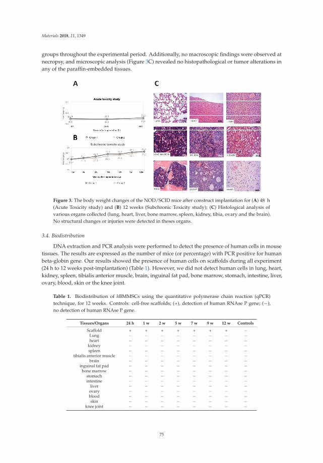
Materials 2018, 11, 1349
groups throughout the experimental period. Additionally, no macroscopic findings were observed atnecropsy, and microscopic analysis (Figure 3C) revealed no histopathological or tumor alterations inany of the paraffin-embedded tissues.
Figure 3. The body weight changes of the NOD/SCID mice after construct implantation for (A) 48 h(Acute Toxicity study) and (B) 12 weeks (Subchronic Toxicity study); (C) Histological analysis ofvarious organs collected (lung, heart, liver, bone marrow, spleen, kidney, tibia, ovary and the brain).No structural changes or injuries were detected in theses organs.
3.4. Biodistribution
DNA extraction and PCR analysis were performed to detect the presence of human cells in mousetissues. The results are expressed as the number of mice (or percentage) with PCR positive for humanbeta-globin gene. Our results showed the presence of human cells on scaffolds during all experiment(24 h to 12 weeks post-implantation) (Table 1). However, we did not detect human cells in lung, heart,kidney, spleen, tibialis anterior muscle, brain, inguinal fat pad, bone marrow, stomach, intestine, liver,ovary, blood, skin or the knee joint.
Table 1. Biodistribution of hBMMSCs using the quantitative polymerase chain reaction (qPCR)technique, for 12 weeks. Controls: cell-free scaffolds; (+), detection of human RNAse P gene; (−),no detection of human RNAse P gene.
Tissues/Organs 24 h 1 w 2 w 5 w 7 w 9 w 12 w Controls
Scaffold + + + + + + + −Lung − − − − − − − −heart − − − − − − − −
kidney − − − − − − − −spleen − − − − − − − −
tibialis anterior muscle − − − − − − − −brain − − − − − − − −
inguinal fat pad − − − − − − − −bone marrow − − − − − − − −
stomach − − − − − − − −intestine − − − − − − − −
liver − − − − − − − −ovary − − − − − − − −blood − − − − − − − −skin − − − − − − − −
knee joint − − − − − − − −
75

Materials 2018, 11, 1349
3.5. In Vivo Bone Formation
Next, the in vivo bone formation was analyzed. Histopathological analysis revealed signs oflamellar bone formation in both the subcutaneous (from week 7) and intramuscular (from week 9)constructs of Group 1 (hBMMSCs/β-TCP, Figure 4). No signs of lamellar bone neoformation wereobserved in the subcutaneous and intramuscular constructs of Group 2 at any time.
Figure 4. Representative images of subcutaneous and intramuscular constructs from Group 1 (a–d) andGroup 2 (e,f) at week 9 after constructs implantation. While there was signs of formation of lamellarbone (asterisks) with signs of functional lacunae (presence of nucleus, head arrows) interspersedwithin the construct matrix in subcutaneous (a,c) and intramuscular (b,d) constructs from Group 1,in subcutaneous (e) and intramuscular (f) constructs from Group 2 there was a infiltration of connectivetissue with trabecular disposition in which signs of a refringent material (+) could be identified withintrabeculae. (M): Skeletal muscle. Hematoxylin and eosin (H&E) stain. Magnifications: 100 × (a,b,e,f)and 200 × (c,d).
Immunohistochemical expression of human osteocalcin was detected only in bone marrowfrom mice of Group 1 from week 7 onwards (Figure 5). On the other hand, no signs of positiveimmunoreaction were observed in subcutaneous and intramuscular constructs from Group 2.
Quantitative results (Table 2) exhibited a significant difference in the osteocalcin expression amongthe subcutaneous/intramuscular group (Group 1) and control group (Group 2) (p < 0.05). There was nosignificant difference between the subcutaneous and intramuscular localizations. Overall, the resultsindicated that the hBMMSCs/β-TCP group can promote the formation of calcified matrix and theosteocalcin expression compared with the control group. While there is a strong positive expressionof osteocalcin in lacunae in human control bone, no positive reaction was observed either in bone orother tissues in the mouse (Figure 6).
76

Materials 2018, 11, 1349
Figure 5. Representative images of human osteocalcin expression of subcutaneous and intramuscularconstructs from Group 1 (a–f) and Group 2 (g,h) at week 9 after constructs implantation. Therewas positive expression of human osteocalcin in lamellar bone formations within subcutaneous orintramuscular constructs from Group 1 (a,b asterisks), particularly in functional lacunae (c,d, headarrows). Any sign of background was observed in negative controls of the same regions (e,f). On theother hand, no signs of positive immunoreaction were observed in subcutaneous and intramuscularconstructs from Group 2 (g,h). ABC anti-human osteocalcin stain. Magnifications: 100 × (a,b,e–h) and200 × (c,d).
Table 2. Frequency of human osteocalcin expression in mice after construct implantation comparedwith the control. ** p < 0.01.
Intramuscular Implant Subcutaneous Implant
Yes No Yes NoGroup I
(hBMMSCs/TCP) n = 18 ** n = 7 Group I(hBMMSCs/TCP) n = 17 ** n = 8
Group II(Cell free TCP) 0 n = 5 Group II
(Cell free TCP) 0 n = 5
77

Materials 2018, 11, 1349
Figure 6. Representative images of expression of human osteocalcin in human bone (positive control, a);and in mouse bone (b); brain (c); skin (d); lung (e); heart (f); spleen (g); liver (h); kidney (I) and skeletalmuscle (j). While there is a strong positive expression of osteocalcin in lacunae in human control bone(a, head arrows), no positive reaction was observed neither in bone (b), nor other tissues (c–j) in themouse. ABC anti-human osteocalcin stain. Magnifications: 100 × (a,b,e–j).
4. Discussion
Preclinical studies of the products for use in new CBT need to be carried out in animal modelsin order to verify their biosecurity and efficacy [25]. In fact, determining the distributive fate andretention of CBT products after administration is key part of characterizing their mechanism of actionand security profile [25,26]. The present study was prepared to analyze the biosafety of hBMMSCspre-seeded into TCP scaffolds after subcutaneous/intramuscular transplantation.
We reported that (i) hBMMSCs/β-TCP constructs did not cause acute or subchronic toxicities tothe mice (inspection of the health status of the operated mice and histologically analyses of severaltissue samples); (ii) human cells do not migrated into tissues distant from the implantation sites(expression of human globin gene, by quantitative PCR, in several tissues); (iii) hBMMSCs/β-TCPconstructs developed into bone tissue.
The limitation of this study was the animal model; immunocompetent animal model made theevaluation of the immune response of the implanted hBMMSCs under Good Laboratory Practice (GLP)
78

Materials 2018, 11, 1349
conditions difficult and could be more significant by investigating the impact of SCs in larger animalmodels. In contrast, subcutaneous implantation is an easy and non-invasive technique, and allowsperformance of several test items in the same animal [27].
New materials must first manifest their biocompatibility before cells can proliferate and producean extracellular mineralized matrix on a substrate [28]. For this purpose and to evaluate the possiblecytotoxicity of the β-TCP, we investigated the viability and cell attachment of hBMMSCs culturedon β-TCP by MTT assay and SEM, respectively. A similar level of cell viability to the controlwas seen after 14 days of culture. Previous studies using colorimetric assays demonstrated goodmetabolic cell activity, cell adhesion and cell morphology promoted by β-TCP [29–31]. SEM is themost commonly used electron microscopy approach to analyze morphological appearance of cellsseeded on certain biomaterials prior to implantation [32]. After 14 days of culture, we observed largeamounts of hBMMSCs adhering to the β-TCP granules, giving the appearance of multilayered cultures.Arpornmaeklong et al. [33] showed that β-TCP stimulates the attachment and differentiation of humanembryonic SCs (hESCs), especially the expression of genes related to neurogenesis (AP2a, FoxD3,HNK1, P75, Sox1, Sox10). Another recent study exhibited good morphology and cell attachment ofdental pulp SCs into the β-TCP scaffolds [34].
Therapies based on SCs have shown great potential in many clinical studies. However, noveltherapies using cell-based ATMPs require special safety testing strategies [27]. Thus, any additionalinformation showing toxicity tests can help guide the design of clinical trials [35]. In our study,the local and systemic toxicity of hBMMSCs intramuscular and subcutaneous transplanted wasmonitored for 12 weeks. No mortality, morbidity or abnormal clinical symptoms were found. Moreover,no hBMMSC-related changes were observed in histopathological lesions. In a previous study involvingmesenchymal progenitor cells derived from umbilical cord blood intravenously administered in mice,no toxicologically meaningful microscopic findings were observed in the animals [36]. Importantly wedid not observe any tumors in the sacrificed animals.
Due to the cell migration after local administration, biodistribution studies are key elements forunderstanding the physiological or pathological behavior of the cells before clinical use [37]. Ourbiodistribution results did not show any hBMMSCs in the tested organs (lung, heart, kidney, spleen,tibialis anterior muscle, brain, inguinal pad, bone marrow, stomach, intestine, liver, ovary, blood,knee joint) 12 weeks after transplantation, suggesting that cells stay where they are placed and donot invade other tissues. These data were consistent with those of a previous study in which nohDNA was detected in such major organs as the brain, heart, lungs, kidneys, spleen or liver of animalsafter intramuscular administration of hMSCs [25]. In the same line, Choi et al. [38] reported thatintracranially injected adipose mesenchymal SCs did not invade other tissues out of the brain innormal mice. However, after intra-articular injection, human Alu sequences were detected in severaltissues and organs [39]. This suggests that the biodistribution potential of mesenchymal SCs could beinfluenced by the route of administration.
Regarding the efficacy of hBMMSCs, subcutaneous ectopic bone formation models are commonlyused by CBT [40,41]. Previous reports have demonstrated that murine or human bone marrow stromalcells seeded on calcium phosphate (CaP) stimulate the bone formation implanted subcutaneously inimmune-compromised mice [42]. Our results showed the presence of signs of lamellar bone formationin both subcutaneous and intramuscular constructs of group β-TCP + hBMMSCs from week 7 in thosecases of subcutaneous implantation, and from week 9 in the intramuscular implants. While there wasa strong positive expression of osteocalcin in lacunae in human control bone, a positive reaction wasobserved neither in bone, nor other tissues in the mouse. In this context, other authors have shown thetherapeutic efficacy of BMMSCs/β-TCP in goat models of critical size bone defects [43].
79

Materials 2018, 11, 1349
5. Conclusions
Based on the data described in this work, it is concluded that transplantation of mesenchymalstem cell from bone marrow preseeded into β-TCP scaffolds in murine models is safe and effective.This results pave the way to perform “first in human” clinical trials to treat bone diseases in the future.
Author Contributions: M.G.-G., L.M.-O., A.G.H and F.J.R.L. designed the study. C.M.M, M.G.-G., and M.B.conducted the experiments; R.E.O.S. and F.J.R.-L. analysed the results. J.M.M. and F.J.R.L. contributed topreparation of the manuscript.
Funding: This work was supported by the Spanish Network of Cell Therapy (TerCel), RETICS subprogrammes ofthe I + D + I 2013–2016 Spanish National Plan. and projects ‘RD12/0019/0001’, ‘RD16/0011/0001’, ‘PI13/02699’and ‘EC11-009’ funded by Instituto de Salud Carlos III and cofunded by European Regional Development Fund.
Conflicts of Interest: The authors declare no conflict of interest.
References
1. Delorme, B.; Chateauvieux, S.; Charbord, P. The concept of mesenchymal stem cells. Regen. Med. 2006, 1,497–509. [CrossRef] [PubMed]
2. Moraleda, J.M.; Blanquer, M.; Bleda, P.; Iniesta, P.; Ruiz, F.; Bonilla, S.; Cabanes, C.; Tabares, L.; Martinez, S.Adult stem cell therapy: Dream or reality? Transpl. Immunol. 2006, 17, 74–77. [CrossRef] [PubMed]
3. Gonzálvez-García, M.; Rodríguez-Lozano, F.J.; Villanueva, V.; Segarra-Fenoll, D.; Rodríguez-González, M.A.;Oñate-Sánchez, R.; Blanquer, M.; Moraleda, J.M. Cell therapy in bisphosphonate-related osteonecrosis of thejaw. J. Craniofac. Surg. 2013, 24, 226–228. [CrossRef] [PubMed]
4. Qi, Y.; Niu, L.; Zhao, T.; Shi, Z.; Di, T.; Feng, G.; Li, J.; Huang, Z. Combining mesenchymal stem cell sheetswith platelet-rich plasma gel/calcium phosphate particles: A novel strategy to promote bone regeneration.Stem Cell Res. Ther. 2015, 6, 256. [CrossRef] [PubMed]
5. Cella, L.; Oppici, A.; Arbasi, M.; Moretto, M.; Piepoli, M.; Vallisa, D.; Zangrandi, A.; Di Nunzio, C.;Cavanna, L. Autologous bone marrow stem cell intralesional transplantation repairing bisphosphonaterelated osteonecrosis of the jaw. Head Face Med. 2011, 7, 16. [CrossRef] [PubMed]
6. Suenaga, H.; Furukawa, K.S.; Suzuki, Y.; Takato, T.; Ushida, T. Bone regeneration in calvarial defects in a ratmodel by implantation of human bone marrow-derived mesenchymal stromal cell spheroids. J. Mater. Sci.Mater. Med. 2015, 26, 254. [CrossRef] [PubMed]
7. Shamsul, B.S.; Tan, K.K.; Chen, H.C.; Aminuddin, B.S.; Ruszymah, B.H. Posterolateral spinal fusion withostegenesis induced BMSC seeded TCP/HA in a sheep model. Tissue Cell 2014, 46, 152–158. [CrossRef][PubMed]
8. Jimi, E.; Hirata, S.; Osawa, K.; Terashita, M.; Kitamura, C.; Fukushima, H. The current and future therapiesof bone regeneration to repair bone defects. Int. J. Dent. 2012, 2012, 148261. [CrossRef] [PubMed]
9. Sunil, P.; Manikandhan, R.; Muthu, M.; Abraham, S. Stem cell therapy in oral and maxillofacial region: Anoverview. J. Oral Maxillofac. Pathol. 2012, 16, 58–63. [CrossRef] [PubMed]
10. Basu, J.; Assaf, B.T.; Bertram, T.A.; Rao, M. Preclinical biosafety evaluation of cell-based therapies: Emergingglobal paradigms. Toxicol. Pathol. 2015, 43, 115–125. [CrossRef] [PubMed]
11. Patrício, T.; Domingos, M.; Gloria, A.; D’Amora, U.; Coelho, J.F.; Bártolo, P.J. Fabrication and characterisationof PCL and PCL/PLA scaffolds for tissue engineering. Rapid Prototyping J. 2014, 20, 145–156. [CrossRef]
12. Guarino, V.; Gloria, A.; Raucci, M.G.; De Santis, R.; Ambrosio, L. Bio-inspired composite and cell instructiveplatforms for bone regeneration. Int. Mater. Rev. 2013, 57, 256–275. [CrossRef]
13. Frey-Vasconcells, J.; Whittlesey, K.J.; Baum, E.; Feigal, E.G. Translation of stem cell research: Points toconsider in designing preclinical animal studies. Stem Cells Transl. Med. 2012, 1, 353–358. [CrossRef][PubMed]
14. Zhao, W.; Phinney, D.G.; Bonnet, D.; Dominici, M.; Krampera, M. Mesenchymal stem cell biodistribution,migration, and homing in vivo. Stem Cells Int. 2014, 2014, 292109. [CrossRef] [PubMed]
15. Rousseau, C.F.; Maciulaitis, R.; Sladowski, D.; Narayanan, G. Cell and Gene Therapies: European Viewon Challenges in Translation and How to Address Them. Front. Med. (Lausanne) 2018, 5, 158. [CrossRef][PubMed]
80

Materials 2018, 11, 1349
16. De Aza, P.N.; Garcia-Bernal, D.; Cragnolini, F.; Velasquez, P.; Meseguer-Olmo, L. The effects ofCa2SiO4-Ca3(PO4)2 ceramics on adult human mesenchymal stem cell viability, adhesion, proliferation,differentiation and function. Mater. Sci. Eng. C Mater. Biol. Appl. 2013, 33, 4009–4020. [CrossRef] [PubMed]
17. Dominici, M.; Le Blanc, K.; Mueller, I.; Slaper-Cortenbach, I.; Marini, F.; Krause, D.; Deans, R.;Keating, A.; Prockop, D.; Horwitz, E. Minimal criteria for defining multipotent mesenchymal stromalcells. The International Society for Cellular Therapy position statement. Cytotherapy 2006, 8, 315–317.[CrossRef] [PubMed]
18. Horwitz, E.M.; Le Blanc, K.; Dominici, M.; Mueller, I.; Slaper-Cortenbach, I.; Marini, F.C.; Deans, R.J.;Krause, D.S.; Keating, A.; Therapy, I.S.f.C. Clarification of the nomenclature for MSC: The InternationalSociety for Cellular Therapy position statement. Cytotherapy 2005, 7, 393–395. [CrossRef] [PubMed]
19. Rodriguez-Lozano, F.J.; Garcia-Bernal, D.; Onate-Sanchez, R.E.; Ortolani-Seltenerich, P.S.; Forner, L.;Moraleda, J.M. Evaluation of cytocompatibility of calcium silicate-based endodontic sealers and theireffects on the biological responses of mesenchymal dental stem cells. Int. Endod. J. 2017, 50, 67–76. [CrossRef][PubMed]
20. Llena, C.; Collado-Gonzalez, M.; Tomas-Catala, C.J.; Garcia-Bernal, D.; Onate-Sanchez, R.E.;Rodriguez-Lozano, F.J.; Forner, L. Human Dental Pulp Stem Cells Exhibit Different Biological Behaviours inResponse to Commercial Bleaching Products. Materials (Basel) 2018, 11, 1098. [CrossRef] [PubMed]
21. Eslaminejad, M.B.; Mirzadeh, H.; Nickmahzar, A.; Mohamadi, Y.; Mivehchi, H. Type I collagen gel in seedingmedium improves murine mesencymal stem cell loading onto the scaffold, increases their subsequentproliferation, and enhances culture mineralization. J. Biomed. Mater. Res. B Appl. Biomater. 2009, 90, 659–667.[CrossRef] [PubMed]
22. Lloyd, M.H.; Foden, B.W.; Wolfensohn, S.E. Refinement: Promoting the three Rs in practice. Lab. Anim. 2008,42, 284–293. [CrossRef] [PubMed]
23. Francois, S.; Bensidhoum, M.; Mouiseddine, M.; Mazurier, C.; Allenet, B.; Semont, A.; Frick, J.; Sache, A.;Bouchet, S.; Thierry, D.; et al. Local irradiation not only induces homing of human mesenchymal stem cellsat exposed sites but promotes their widespread engraftment to multiple organs: A study of their quantitativedistribution after irradiation damage. Stem Cells 2006, 24, 1020–1029. [CrossRef] [PubMed]
24. Francois, S.; Mouiseddine, M.; Allenet-Lepage, B.; Voswinkel, J.; Douay, L.; Benderitter, M.; Chapel, A.Human mesenchymal stem cells provide protection against radiation-induced liver injury by antioxidativeprocess, vasculature protection, hepatocyte differentiation, and trophic effects. Biomed. Res. Int. 2013, 2013,151679. [CrossRef] [PubMed]
25. Creane, M.; Howard, L.; O’Brien, T.; Coleman, C.M. Biodistribution and retention of locally administeredhuman mesenchymal stromal cells: Quantitative polymerase chain reaction-based detection of human DNAin murine organs. Cytotherapy 2017, 19, 384–394. [CrossRef] [PubMed]
26. Bailey, A.M.; Mendicino, M.; Au, P. An FDA perspective on preclinical development of cell-based regenerativemedicine products. Nat. Biotechnol. 2014, 32, 721–723. [CrossRef] [PubMed]
27. Zscharnack, M.; Krause, C.; Aust, G.; Thummler, C.; Peinemann, F.; Keller, T.; Smink, J.J.; Holland, H.;Somerson, J.S.; Knauer, J.; et al. Preclinical good laboratory practice-compliant safety study to evaluatebiodistribution and tumorigenicity of a cartilage advanced therapy medicinal product (ATMP). J. Transl.Med. 2015, 13, 160. [CrossRef] [PubMed]
28. Stratton, S.; Shelke, N.B.; Hoshino, K.; Rudraiah, S.; Kumbar, S.G. Bioactive polymeric scaffolds for tissueengineering. Bioact. Mater. 2016, 1, 93–108. [CrossRef] [PubMed]
29. Seebach, C.; Schultheiss, J.; Wilhelm, K.; Frank, J.; Henrich, D. Comparison of six bone-graft substitutesregarding to cell seeding efficiency, metabolism and growth behaviour of human mesenchymal stem cells(MSC) in vitro. Injury 2010, 41, 731–738. [CrossRef] [PubMed]
30. Xu, L.; Lv, K.; Zhang, W.; Zhang, X.; Jiang, X.; Zhang, F. The healing of critical-size calvarial bone defectsin rat with rhPDGF-BB, BMSCs, and beta-TCP scaffolds. J. Mater. Sci. Mater. Med. 2012, 23, 1073–1084.[CrossRef] [PubMed]
31. Xu, L.; Zhang, W.; Lv, K.; Yu, W.; Jiang, X.; Zhang, F. Peri-Implant Bone Regeneration Using rhPDGF-BB,BMSCs, and beta-TCP in a Canine Model. Clin. Implant Dent. Relat. Res. 2016, 18, 241–252. [CrossRef][PubMed]
81

Materials 2018, 11, 1349
32. Wu, H.; Kang, N.; Wang, Q.; Dong, P.; Lv, X.; Cao, Y.; Xiao, R. The Dose-Effect Relationship Between theSeeding Quantity of Human Marrow Mesenchymal Stem Cells and In Vivo Tissue-Engineered Bone Yield.Cell Transplant 2015, 24, 1957–1968. [CrossRef] [PubMed]
33. Arpornmaeklong, P.; Pressler, M.J. Effects of ss-TCP Scaffolds on neurogenic and osteogenic differentiationof Human Embryonic Stem Cells. Ann. Anat. 2017, 215, 52–62. [CrossRef] [PubMed]
34. Vina-Almunia, J.; Mas-Bargues, C.; Borras, C.; Gambini, J.; El Alami, M.; Sanz-Ros, J.; Penarrocha, M.; Vina, J.Influence of Partial O(2) Pressure on the Adhesion, Proliferation, and Osteogenic Differentiation of HumanDental Pulp Stem Cells on beta-Tricalcium Phosphate Scaffold. Int. J. Oral Maxillofac. Implants 2017, 32,1251–1256. [CrossRef] [PubMed]
35. He, J.; Ruan, G.P.; Yao, X.; Liu, J.F.; Zhu, X.Q.; Zhao, J.; Pang, R.Q.; Li, Z.A.; Pan, X.H. Chronic Toxicity Test inCynomolgus Monkeys For 98 Days with Repeated Intravenous Infusion of Cynomolgus Umbilical CordMesenchymal Stem Cells. Cell Physiol. Biochem. 2017, 43, 891–904. [CrossRef] [PubMed]
36. Yun, J.W.; Ahn, J.H.; Kwon, E.; Kim, S.H.; Kim, H.; Jang, J.J.; Kim, W.H.; Kim, J.H.; Han, S.Y.; Kim, J.T.;et al. Human umbilical cord-derived mesenchymal stem cells in acute liver injury: Hepatoprotectiveefficacy, subchronic toxicity, tumorigenicity, and biodistribution. Regul. Toxicol. Pharmacol. 2016, 81, 437–447.[CrossRef] [PubMed]
37. Reyes, B.; Coca, M.I.; Codinach, M.; Lopez-Lucas, M.D.; Del Mazo-Barbara, A.; Caminal, M.; Oliver-Vila, I.;Cabanas, V.; Lope-Piedrafita, S.; Garcia-Lopez, J.; et al. Assessment of biodistribution using mesenchymalstromal cells: Algorithm for study design and challenges in detection methodologies. Cytotherapy 2017, 19,1060–1069. [CrossRef] [PubMed]
38. Choi, S.A.; Yun, J.W.; Joo, K.M.; Lee, J.Y.; Kwak, P.A.; Lee, Y.E.; You, J.R.; Kwon, E.; Kim, W.H.; Wang, K.C.;et al. Preclinical Biosafety Evaluation of Genetically Modified Human Adipose Tissue-Derived MesenchymalStem Cells for Clinical Applications to Brainstem Glioma. Stem Cells Dev. 2016, 25, 897–908. [CrossRef][PubMed]
39. Toupet, K.; Maumus, M.; Peyrafitte, J.A.; Bourin, P.; van Lent, P.L.; Ferreira, R.; Orsetti, B.; Pirot, N.;Casteilla, L.; Jorgensen, C.; et al. Long-term detection of human adipose-derived mesenchymal stem cellsafter intraarticular injection in SCID mice. Arthritis Rheum. 2013, 65, 1786–1794. [CrossRef] [PubMed]
40. Suzuki, K.; Nagata, K.; Yokota, T.; Honda, M.; Aizawa, M. Histological evaluations of apatite-fiber scaffoldcultured with mesenchymal stem cells by implantation at rat subcutaneous tissue. Biomed. Mater. Eng. 2017,28, 57–64. [CrossRef] [PubMed]
41. Ismail, T.; Osinga, R.; Todorov, A., Jr.; Haumer, A.; Tchang, L.A.; Epple, C.; Allafi, N.; Menzi, N.; Largo, R.D.;Kaempfen, A.; et al. Engineered, axially-vascularized osteogenic grafts from human adipose-derived cells totreat avascular necrosis of bone in a rat model. Acta Biomater. 2017, 63, 236–245. [CrossRef] [PubMed]
42. Bouvet-Gerbettaz, S.; Boukhechba, F.; Balaguer, T.; Schmid-Antomarchi, H.; Michiels, J.F.; Scimeca, J.C.;Rochet, N. Adaptive immune response inhibits ectopic mature bone formation induced byBMSCs/BCP/plasma composite in immune-competent mice. Tissue Eng. Part A 2014, 20, 2950–2962.[CrossRef] [PubMed]
43. Chu, W.; Gan, Y.; Zhuang, Y.; Wang, X.; Zhao, J.; Tang, T.; Dai, K. Mesenchymal stem cells and porousbeta-tricalcium phosphate composites prepared through stem cell screen-enrich-combine(-biomaterials)circulating system for the repair of critical size bone defects in goat tibia. Stem Cell Res. Ther. 2018, 9, 157.[CrossRef] [PubMed]
© 2018 by the authors. Licensee MDPI, Basel, Switzerland. This article is an open accessarticle distributed under the terms and conditions of the Creative Commons Attribution(CC BY) license (http://creativecommons.org/licenses/by/4.0/).
82

materials
Article
Calcium Charge and Release of ConventionalGlass-Ionomer Cement Containing Nanoporous Silica
Koichi Nakamura 1,*, Shigeaki Abe 2, Hajime Minamikawa 3 and Yasutaka Yawaka 1
1 Department of Dentistry for Children and Disabled Person, Graduate School of Dental Medicine, HokkaidoUniversity, Kita 13 Nishi 7, Kita-ku, Sapporo 060-8586, Hokkaido, Japan; [email protected]
2 Department of Biomaterials and Bioengineering, Graduate School of Dental Medicine, Hokkaido University,Sapporo 060-8586, Hokkaido, Japan; [email protected]
3 Department of Dentistry for Molecular Cell Pharmacology, Graduate School of Dental Medicine, HokkaidoUniversity, Sapporo 060-8586, Hokkaido, Japan; [email protected]
* Correspondence: [email protected]; Tel.: +81-11-706-4292
Received: 5 July 2018; Accepted: 23 July 2018; Published: 27 July 2018
Abstract: The aim of this study was to evaluate calcium charge and release of conventionalglass-ionomer cement (GIC) containing nanoporous silica (NPS). Experimental specimens weredivided into two groups: the control (GIC containing no NPS) and GIC-NPS (GIC containing 10 wt %NPS). The specimens were immersed in calcium chloride solutions of 5 wt % calcium concentrationfor 24 h at 37 ◦C, whereupon the calcium ion release of the specimens was measured. The calcium ionrelease behavior of GIC-NPS after immersion in the calcium solution was significantly greater thanthat of the control. Scanning electron microscopy and electron-dispersive X-ray spectroscopy resultsindicated that calcium penetrated inside the GIC-NPS specimen, while the calcium was primarilylocalized on the surface of the control specimen. It was demonstrated that NPS markedly improvedthe calcium charge and release property of GIC.
Keywords: nanoporous silica; glass-ionomer cement; calcium
1. Introduction
The overall incidence of dental caries has gradually decreased, but it still occurs quitefrequently [1]. In the case of small dental carious lesions, the tooth is typically restored by a coronalrestoration material, which often requires a retreatment owing to circumstances such as the progressof the dental caries around the restoration or the dental restoration falling out [2,3]. The main causeof coronal restoration treatment failure is secondary caries, which requires coronal restoration orpulp treatment [4,5]. To avoid tooth loss, therefore, it is necessary to prevent secondary caries,for which fluoride takes an important role [6,7]. Fluoride ions are included in dental items suchas toothpaste and glass-ionomer cement (GIC) [8,9], and GIC is often used to prevent secondarycaries in children [10,11]. GIC is widely used in dentistry such as the luting cement [12], temporaryrestoration [13], and adhering orthodontic band [14]. The GIC supplemented with TiO2 nanoparticlesalso have the effect of antibacterial properties [15].
Calcium is an important component of the tooth. It is essential for the tooth mineralization thatthe calcium ion exists around the tooth. As one of the methods to supply calcium for teeth, somestudies proposed the addition of casein phosphopeptide amorphous calcium phosphate (CPP-ACP) toGIC [16–18]. Addition of CPP-ACP to GIC increased the release of calcium ions. However, CPP-ACPis an ingredient derived from milk, and clinicians should consider potential side effects from ingestionof casein derivative protein in people with immunoglobulin E allergies to milk proteins [19].
Nanoporous silica (NPS) has a structure that possesses a uniform pore size of about 1 nm witha large specific surface area and is attractive in applications such as a sustained drug release carrier
Materials 2018, 11, 1295; doi:10.3390/ma11081295 www.mdpi.com/journal/materials83

Materials 2018, 11, 1295
and catalyst support [20]. It is believed that NPS can adsorb formaldehyde gas and methylene bluepigments, and it has been confirmed that one of the properties of NPS is its ability to adsorb variousions and substances [21–23]. However, there are few studies on the application of NPS in dentistry.
The purpose of this study was to determine whether conventional GIC containing NPS wasable to charge and release calcium ions. Furthermore, the cross-section of the GIC-NPS specimenwas observed by scanning electron microscopy (SEM) and the compressive strength was measured.Our null hypotheses were that GIC containing the NPS did not charge and release calcium ions,and that there was no difference in the compressive strength of the GIC control and the GIC-NPS.
2. Materials and Methods
2.1. NPS Synthesis
The NPS was synthesized in accordance with the protocol described by Tagaya et al. [20]. In thisprocess, 1.37 mmol of cetyltrimethylammonium bromide (CTAB: Wako, Osaka, Japan) was addedto 120 mL of distilled water, to which 1.75 mL of 2.0 M sodium hydroxide (Wako) solution wasadded. The mixture was stirred for 30 min at 80 ◦C, whereupon 12.4 mmol of tetraethoxysilane(Wako) was added and the mixture stirred for 2 h at 80 ◦C. The resulting suspension was filtered anddried and, to remove the CTAB, the obtained particles were calcined at 550 ◦C for 4 h. The obtainedwhite particles were subsequently observed using an SEM (S-4800, HITACHI, Tokyo, Japan) and atransmission electron microscope (TEM: JEM-2010, JEOL, Tokyo, Japan).
2.2. Preparation of Test Specimens
The obtained NPS was added to the powder component of the GIC (GC Fuji II, GC, Tokyo, Japan;lot 1606141) at 10 wt % concentration, whereupon the test specimens were prepared by mixing thepowder component (with or without NPS) and the liquid component of the GIC (GC Fuji II, GC, Tokyo,Japan; lot 1607041) in accordance with the manufacturer specifications. The specimens were set intwo patterns, where the first was 10 mm in diameter and 1 mm in height for the calcium ion releasemeasurement and the SEM and electron-dispersive X-ray spectroscopy analysis, and the second was4 mm in diameter and 6 mm in height for the compressive testing. The experimental specimens weredivided into two groups comprising the control (no NPS) and the GIC-NPS (containing 10 wt % NPS).The specimens were wet ground via hand lapping using P400 grit silicon carbide abrasive papers(SANKYO, Saitama, Japan).
2.3. Calcium Charge and Release
Three 10 mm-diameter, 1 mm-height specimens were immersed in a calcium chloride solutionwith a 5 wt % calcium concentration (calcium 150 mg/3mL) for 24 h at 37 ◦C. After removal from thecalcium solution, the specimens were rinsed with distilled water and subsequently immersed in 5 mLof distilled water at 37 ◦C for 7 d. The distilled water was changed every day and was further analyzeddaily to determine the calcium ion concentration in the water. N = 6 samples (18 specimens) per groupwere used. The calcium ion concentration was measured using inductively coupled plasma atomicemission spectroscopy (ICPE-9000, SHIMADZU, Tokyo, Japan). The total weights of the calciumreleasing from the specimens were calculated from the calcium concentration. The preparation ofspecimens was carried out according to Bando’s conditions [24], but slightly modified.
2.4. Scanning Electron Microscopy (SEM) and Energy-Dispersive X-ray Spectroscopy (EDS)
Specimens containing calcium were analyzed using SEM and EDS (Genesis, EDAX Japan, Tokyo,Japan). The specimens were immersed in the 5 wt % calcium solution and dried. The specimens werecut perpendicularly, then the specimen surface and cross-section were observed after carbon coating.
84

Materials 2018, 11, 1295
2.5. Compressive Strength Test
The 4 mm-diameter, 6 mm-height test specimens were placed in a universal testing machine(Model 4204, Instron, Canton, OH, USA) with a cross-head speed of 1.0 mm/min. N = 12 specimensper group were used for this test. This procedure has been described in detail elsewhere [25].
2.6. Statistical Analysis
Statistical analysis was performed using IBM SPSS Statistics Version 21 (IBM Japan, Tokyo, Japan),and the results were analyzed statistically using the Mann-Whitney U test. The level of significancewas set at p < 0.05.
3. Results
3.1. Morphological Characteristics of NPS
The SEM and TEM images of NPS particles are shown in Figure 1. The NPS particles werespherical and approximately 200–300 nm in diameter, exhibiting pores a few nanometers in diameter.
Figure 1. Typical (A) SEM and (B) TEM images of nanoporous silica particles. The nanoporous silicaparticles showed sizes approximately 200–300 nm in diameter (A), exhibiting pores a few nanometersin diameter (B).
3.2. Calcium Charge and Release
Figure 2 shows the time-profile of the release of calcium from the specimens after immersionin the 5 wt % calcium chloride solution. The calcium release behavior of GIC-NPS was significantlygreater than that of the control (p < 0.05).
Figure 2. Time-profile of calcium release from the specimens after immersion in 5 wt % calcium chloridesolution. Significance was determined using the Mann-Whitney U test (p < 0.05).
85

Materials 2018, 11, 1295
3.3. EDS Analysis
Figure 3A–C show the results of SEM and EDS for the surface and the cross-section of thespecimens. As shown in Figure 3A, calcium was detected on the surfaces of both the control andGIC-NPS specimens; however, the specimen cross-sections exhibited significant amounts of calciumonly for the GIC-NPS specimen. Although calcium was localized primarily on the surface of thecontrol specimen, it was observed on the surface and throughout the inside of the GIC-NPS specimen(Figure 3B,C). In the surface of the control specimen (Figure 3B right and 3C right), calcium wasdistributed uniformly.
Figure 3. (A) Typical EDS spectra from the cross-section (upper) and surface (lower) of the GIC-NPS(left) and control (right) specimens, displaying the constitutive elements. (B) SEM images (lower) andEDS line analysis (upper) for the GIC-NPS cross-section (left), control cross-section (center), and controlsurface (right). (C) EDS element mapping of the Al (upper), Si (center), and Ca (lower) in the GIC-NPScross-section (left), control cross-section (center), and control surface (right).
86

Materials 2018, 11, 1295
3.4. Compressive Strength
The compressive strength of the control and GIC-NPS were 111.37 ± 28.75 MPa and100.32 ± 20.73 MPa (Mean ± SD, N = 12). There was no significant difference between the compressivestrength of the control and GIC-NPS (p = 0.319).
4. Discussion
In the present study, the amount of the calcium release from the glass-ionomer cement containingnanoporous silica (GIC-NPS) specimen was determined. The results suggested the existence of anNPS-induced calcium ion charge and release property of the GIC-NPS. Even when a concentrationof 10 wt % NPS was included in the GIC, the obtained specimens exhibited compressive strength,comparable to that of conventional GIC without NPS. It has been suggested that the possibility existsof NPS being used in dental materials as a calcium source, and it is therefore important that NPS didnot reduce significantly the strength properties of the dental materials in the present study. However,other properties (solubility, adhesive strength, etc.) of GIC-NPS still have not been clarified, and so it isnecessary for those concerned to hold further studies.
The nanopores in the NPS were approximately the size through which a calcium ion can pass [26].The EDS images of the GIC-NPS specimens indicated that calcium ions penetrated into the GIC-NPSafter immersion in the calcium solution. The NPS is well known as an adsorbent particle, and calciummight be adsorbed in the NPS pores in the same way other substances are adsorbed. It was notconfirmed, however, which component of the NPS combined with the calcium, but this should be thesubject of future research.
There were 150 mg of calcium in the immersed solution; nevertheless, the total amounts of thecalcium released from specimen of control and GIC-NPS were 17.21 ± 8.66 μg and 287.71 ± 56.60 μg,respectively. Almost all of the calcium remained in the immersed solution or was washed away byrinsing with water. The percentage of total calcium released from the specimen in GIC-NPS on thefirst day, the second day, and the third day were 86.6%, 6.5%, and 1.5%, respectively. In contrast,those percentages for the control on the first day, the second day, and the third day were 87.7%, 6.6%,and 3.3%, respectively. Though the total amount of calcium was different for both, the percentage ofcalcium released had a similar tendency in both.
In this study, NPS was found to not only adsorb calcium ion but also to release adsorbed calciumions when placed in an aqueous medium. The GIC-NPS could be expected to function as a sourceof calcium, and it was suggested that it could be useful to facilitate remineralization. Furthermore,the GIC used in this study was a fluoride-releasing material; thus, GIC-NPS may be useful forproducing fluoroapatite by allowing the coexistence of calcium and fluoride ions [27]. This is significantbecause fluoroapatite possesses a higher acid resistance than hydroxyapatite, the main component ofteeth. The amount of fluoride ions released from the GIC depends on the fluoroaluminosilicate glasscomposition included in the powder [28]. In this study, the inclusion of NPS decreases the relativeamount of the GIC powder, so it is likely that the amount of fluoride ions released may be slightlydecreased. This reduction may be negligible with a 10 wt % concentration of NPS, however, becauseof the amount of fluoride ions released from the GIC [29]. Furthermore, the specimens were immersedin calcium chloride solution in this study. Previous research has indicated that the surface hardness ofGIC increases with immersion in the calcium chloride solution [30]. The surface property of GIC mayhave changed by immersion of the solution.
The SEM/EDS analysis indicated that calcium penetrated to the interior of the GIC-NPS specimen.After immersion in the calcium solution, calcium was distributed homogenously on the surfaces of thecontrol specimen similar to the distribution of aluminum and silicon, which are the major constituentsof GIC (Figure 3C, right column). However, the distribution behavior in the depth direction wasclearly different in the two specimens. In the case of the control, calcium was localized on the surfaceof specimen (Figure 3C, middle column), while calcium was detected even inside of the GIC-NPS(Figure 3C, left column). The highest concentration of calcium was determined to be on the surface of
87

Materials 2018, 11, 1295
the GIC-NPS specimen, similar to that found for the control specimen. The EDS mapping suggeststhat the calcium concentration slightly decreased as a function of depth but persisted throughout theinterior of the GIC-NPS specimen.
The EDS spectral line analysis (Figure 3B) also revealed that the calcium concentration profile ofthe GIC-NPS specimen was clearly different than that of the control specimen. The latter exhibiteda sharp peak only around the specimen surface, while the former exhibited a calcium concentrationthat slightly decreased in a path from the surface to a depth of 0.1 mm, then remained approximatelyconstant up to 0.5 mm of depth.
Nanoporous silica is often applied as a film on the material surface and is rarely contained insidethe material [31]. Nevertheless, it was found herein that NPS was capable of the charge and release ofcalcium ions through the matrix even when the NPS was incorporated throughout the material.
Previous studies of powdered inorganic additives have shown the compressive strength increaseand decrease [25,32,33]. In this study, compressive strength has slightly decreased. It may be due tointerference of the NPS with the normal GIC reaction. By increasing of NPS component, compressivestrength might significantly decrease. However, under the conditions of the present study, there wereno significant differences between the two groups.
The appearance of secondary caries requires a certain period time. In the present study, however,the calcium charge and release properties of GIC-NPS were observed over only a week. If it isa repeated charge and release of the calcium, secondary caries were prevented in the long term.To confirm this presumption, further study is needed. We also measured the amount of calcium chargeand release; the study of the preventing effect for secondary caries may be needed in the model similarto the oral cavity.
In recent years, many studies of bioactive materials have been conducted [34–36]. The resultsof this study may be useful to the development of biomaterials. Firstly, the results herein may beextended to the development of remineralization-inducing materials by the uptake of phosphateions, which are essential for remineralization, e.g., pit and fissure sealants and restoration materials.Also, this study may inform the drug delivery system by the uptake of antibacterial agents such asCetylpyridinium Chloride (CPC). Furthermore, the NPS used in this study possesses a negative charge,and therefore cannot adsorb a negatively-charged fluoride ion. However, the NPS may be enabled toadsorb a fluoride ion by also causing it to retain a positive electric charge. This would give the abilityof fluoride ion release to the material without also requiring a composite resin that exhibits fluorideion release characteristics. We believe that it is necessary to study these possibilities in future work.
5. Conclusions
We demonstrated the capacity of GIC-NPS for calcium ion charge/release and contrasted itwith that of conventional GIC. The presence of NPS was found to markedly improve the calcium ioncharge/release property of GIC. Even for NPS concentrations up to 10 wt % in the GIC, the compressivestrength of the GIC was not changed significantly.
Supplementary Materials: The following are available online at http://www.mdpi.com/1996-1944/11/8/1295/s1, Table S1: Total calcium release, Table S2: Compressive strength.
Author Contributions: K.N., S.A., and H.M. performed the experiments and analyzed the data; Y.Y. contributedas advisors; and K.N. wrote the paper. All authors read and approved the final manuscript.
Funding: This work was supported by JSPS KAKENHI Grant Number 15K20575.
Conflicts of Interest: The authors declare no conflict of interest.
References
1. Ramos-Jorge, J.; Alencar, B.M.; Pordeus, I.A.; Soares, M.E.; Marques, L.S.; Ramos-Jorge, M.L.; Paiva, S.M.Impact of dental caries on quality of life among preschool children: Emphasis on the type of tooth and stagesof progression. Eur. J. Oral Sci. 2015, 123, 88–95. [CrossRef] [PubMed]
88

Materials 2018, 11, 1295
2. Metz, I.; Rothmaier, K.; Pitchika, V.; Crispin, A.; Hickel, R.; Garcia-Godoy, F.; Bücher, K.; Kühnisch, J. Riskfactors for secondary caries in direct composite restorations in primary teeth. Int. J. Paediatr. Dent. 2015,25, 451–461. [CrossRef] [PubMed]
3. Pallesen, U.; Van Dijken, J.W. A randomized controlled 27 years follow up of three resin composites in ClassII restorations. J. Dent. 2015, 43, 1547–1558. [CrossRef] [PubMed]
4. Brantley, C.F.; Bader, J.D.; Shugars, D.A.; Nesbit, S.P. Does the cycle of rerestoration lead to larger restorations?J. Am. Dent. Assoc. 1995, 126, 1407–1413. [CrossRef] [PubMed]
5. Hsu, C.Y.; Donly, K.; Drake, D.; Wefel, J. Effects of aged fluoride-containing restorative materials on recurrentroot caries. J. Dent. Res. 1998, 77, 418–425. [CrossRef] [PubMed]
6. Comar, L.P.; Wiegand, A.; Moron, B.M.; Rios, D.; Buzalaf, M.A.; Buchalla, W.; Magalhães, A.C. In situ effectof sodium fluoride or titanium tetrafluoride varnish and solution on carious demineralization of enamel.Eur. J. Oral Sci. 2012, 120, 342–348. [CrossRef] [PubMed]
7. Bonetti, D.; Clarkson, J.E. Fluoride Varnish for Caries Prevention: Efficacy and Implementation. Caries Res.2016, 50, 45–49. [CrossRef] [PubMed]
8. Kucukyilmaz, E.; Savas, S. Evaluation of shear bond strength, penetration ability, microleakage andremineralisation capacity of glass-ionomer-based fissure sealants. Eur. J. Paediatr. Dent. 2016, 17, 17–23.[PubMed]
9. Yönel, N.; Bikker, F.J.; Lagerweij, M.D.; Kleverlaan, C.J.; Van Loveren, C.; Özen, B.; Çetiner, S.; Van Strijp, A.J.Anti-erosive effects of fluoride and phytosphingosine: An in vitro study. Eur. J. Oral Sci. 2016, 124, 396–402.[CrossRef] [PubMed]
10. Goldman, A.S.; Chen, X.; Fan, M.; Frencken, J.E. Cost-effectiveness, in a randomized trial, of glass-ionomer-based and resin sealant materials after 4 yr. Eur. J. Oral Sci. 2016, 124, 472–479. [CrossRef][PubMed]
11. Zhao, I.S.; Mei, M.L.; Burrow, M.F.; Lo, E.C.; Chu, C.H. Prevention of secondary caries using silver diaminefluoride treatment and casein phosphopeptide-amorphous calcium phosphate modified glass-ionomercement. J. Dent. 2017, 57, 38–44. [CrossRef] [PubMed]
12. Heintze, S.D. Crown pull-off test (crown retention test) to evaluate the bonding effectiveness of luting agents.Dent. Mater. 2010, 26, 193–206. [CrossRef] [PubMed]
13. Coll, J.A.; Campbell, A.; Chalmers, N.I. Effects of glass ionomer temporary restorations on pulpal diagnosisand treatment outcomes in primary molars. Pediatr. Dent. 2013, 35, 416–421. [PubMed]
14. Millett, D.T.; Glenny, A.M.; Mattick, R.C.; Hickman, J.; Mandall, N.A. Adhesives for fixed orthodontic bands.Cochrane Database Syst. Rev. 2016, 10, CD004485. [CrossRef] [PubMed]
15. Garcia-Conreras, R.; Scougall-Vilchis, R.J.; Contreras-Bulner, R.; Sakagami, H.; Morales-Luckie, R.A.;Nakajima, H. Mechanical, antibacterial and bond strength properties of nano-titanium-enriched glassionomer cement. J. Appl. Oral Sci. 2015, 23, 321–328. [CrossRef] [PubMed]
16. Mazzaoui, S.A.; Burrow, M.F.; Tyas, M.J.; Dashper, S.G.; Eakins, D.; Reynolds, E.C. Incorporation of caseinphosphopeptide–amorphous calcium phosphate into a glass-ionomer cement. J. Dent. Res. 2003, 82, 914–918.[CrossRef] [PubMed]
17. Al Zraikat, H.; Palamara, J.E.; Messer, H.H.; Burrow, M.F.; Reynolds, E.C. The incorporation of caseinphosphopeptide–amorphous calcium phosphate into a glass ionomer cement. Dent. Mater. 2011, 27, 235–243.[CrossRef] [PubMed]
18. Zalizniak, I.; Palamara, J.E.; Wong, R.H.; Cochrane, N.J.; Burrow, M.F.; Reynolds, E.C. Ion release andphysical properties of CPP-ACP modified GIC in acid solutions. J. Dent. 2013, 41, 449–454. [CrossRef][PubMed]
19. Azarpazhooh, A.; Limeback, H. Clinical efficacy of casein derivatives: A systematic review of the literature.J. Am. Dent. Assoc. 2008, 139, 915–924. [CrossRef] [PubMed]
20. Tagaya, M.; Ikoma, T.; Yoshioka, T.; Motozuka, S.; Xu, Z.; Minami, F.; Tanaka, J. Synthesis and luminescenceproperties of Eu(III)-doped nanoporous silica spheres. J. Colloid Interface Sci. 1999, 218, 462–467. [CrossRef][PubMed]
21. Barbé, C.; Bartlett, J.; Kong, L.; Finnie, K.; Lin, H.Q.; Larkin, M.; Calleja, S.; Bush, A.; Calleja, G. Silica Particles:A Novel Drug-Delivery System. Adv. Mater. 2004, 16, 1959–1966. [CrossRef]
89

Materials 2018, 11, 1295
22. Fazaeli, Y.; Feizi, S.; Jalilian, A.R.; Hejrani, A. Grafting of [(64)Cu]-TPPF20 porphyrin complex onFunctionalized nano-porous MCM-41 silica as a potential cancer imaging agent. Appl. Radiat. Isotopes2016, 112, 13–19. [CrossRef] [PubMed]
23. Wang, L.; Chen, Q.; Li, C.; Fang, F. Nano-Web Cobalt Modified Silica Nanoparticles Catalysts for WaterOxidation and MB Oxidative Degradation. J. Nanosci. Nanotechnol. 2016, 16, 5364–5368. [CrossRef] [PubMed]
24. Bando, Y.; Nakanishi, K.; Abe, S.; Yamagata, S.; Yoshida, Y.; Iida, J. Electric charge dependence of controlleddye-release behavior in glass ionomer cement containing nano-poruos silica particles. J. Nanosci. Nanotechnol.2018, 18, 75–79. [CrossRef] [PubMed]
25. Elsaka, S.E.; Hamouda, I.M.; Swain, M.V. Titanium dioxide nanoparticles addition to a conventionalglass-ionomer restorative: Influence on physical and antibacterial properties. J. Dent. 2011, 39, 589–598.[CrossRef] [PubMed]
26. Hille, B. Ion Channels of Excitable Membranes, 3rd ed.; Sinauer Associates: Sunderland, MA, USA, 2001.27. Wegehaupt, F.J.; Tauböck, T.T.; Sener, B.; Attin, T. Long-term protective effect of surface sealants against
erosive wear by intrinsic and extrinsic acids. J. Dent. 2012, 40, 416–422. [CrossRef] [PubMed]28. Dennis, C.S. Development of glass-ionomer cement systems. Biomaterials 1998, 19, 467–478.29. Okte, Z.; Bayrak, S.; Fidanci, U.R.; Sel, T. Fluoride and aluminum release from restorative materials using
ion chromatography. J. Appl. Oral Sci. 2012, 20, 27–31. [CrossRef] [PubMed]30. Shiozawa, M.; Takahashi, H.; Iwasaki, N.; Wada, T.; Uo, M. Effect of immersion time of restorative glass
ionomer cements and immersion duration in calcium chloride solution on surface hardness. Dent. Mater.2014, 30, 377–383. [CrossRef] [PubMed]
31. Song, D.P.; Naik, A.; Li, S.; Ribbe, A.; Watkins, J.J. Rapid, Large-Area Synthesis of Hierarchical NanoporousSilica Hybrid Films on Flexible Substrates. J. Am. Chem. Soc. 2016, 138, 13473–13476. [CrossRef] [PubMed]
32. Xie, D.; Weng, Y.; Guo, X.; Zhao, J.; Gregory, R.L.; Zheng, C. Preparation and evaluation of a novelglass-ionomer cement with antibacterial functions. Dent. Mater. 2011, 27, 487–496. [CrossRef] [PubMed]
33. Bellis, C.A.; Nobbs, A.H.; O’sullivan, D.J.; Holder, J.A.; Barbour, M.E. Glass ionomer cements functionalisedwith a concentrated paste of chlorhexidine hexametaphosphate provides dose-dependent chlorhexidinerelease over at least 14 months. J. Dent. 2016, 45, 53–58. [CrossRef] [PubMed]
34. Lee, J.H.; Seo, S.J.; Kim, H.W. Bioactive glass-based nanocomposites for personalized dental tissueregeneration. Dent. Mater. J. 2016, 35, 710–720. [CrossRef] [PubMed]
35. Uo, M.; Wada, T.; Asakura, K. Structural analysis of strontium in human teeth treated with surface pre-reactedglass-ionomer filler eluate by using extended X-ray absorption fine structure analysis. Dent. Mater. J. 2017,36, 214–221. [CrossRef] [PubMed]
36. Gandolfi, M.G.; Iezzi, G.; Piattelli, A.; Prati, C.; Scarano, A. Osteoinductive potential and bone-bonding abilityof ProRoot MTA, MTA Plus and Biodentine in rabbit intramedullary model: Microchemical characterizationand histological analysis. Dent. Mater. 2017, 33, 221–238. [CrossRef] [PubMed]
© 2018 by the authors. Licensee MDPI, Basel, Switzerland. This article is an open accessarticle distributed under the terms and conditions of the Creative Commons Attribution(CC BY) license (http://creativecommons.org/licenses/by/4.0/).
90

materials
Article
Human Dental Pulp Stem Cells Exhibit DifferentBiological Behaviours in Response to CommercialBleaching Products
Carmen Llena 1, Mar Collado-González 2, Christopher Joseph Tomás-Catalá 2,
David García-Bernal 2, Ricardo Elías Oñate-Sánchez 2, Francisco Javier Rodríguez-Lozano 2,3,*
and Leopoldo Forner 1
1 Department of Stomatology, University de Valencia, 46010 Valencia, Spain; [email protected] (C.L.);[email protected] (L.F.)
2 School of Dentistry/Cellular Therapy and Hematopoietic Transplant Unit, Hematology Department,Virgen de la Arrixaca Clinical University Hospital, IMIB-Arrixaca, University of Murcia, 30120 Murcia,Spain; [email protected] (M.C.-G.); [email protected] (C.J.T.-C.); [email protected] (D.G.-B.);[email protected] (R.E.O.-S.)
3 School of Dentistry, Hospital Morales Meseguer 2pl. Av. Marqués de los Vélez s/n, University of Murcia,30008 Murcia, Spain
* Correspondence: [email protected]; Tel.: +34-86-888-9518
Received: 29 May 2018; Accepted: 22 June 2018; Published: 27 June 2018
Abstract: The purpose of this study was to evaluate the diffusion capacity and the biological effectsof different bleaching products on human dental pulp stem cells (hDPSCs). The bleaching gelwas applied for 90, 30 or 15 min to enamel/dentine discs that adapted in an artificial chamber.The diffusion of hydrogen peroxide (HP) was analysed by fluorometry and the diffusion productswere applied to hDPSCs. Cell viability, cell migration and cell morphology assays were performedusing the eluates of diffusion products. Finally, cell apoptosis and the expression of mesenchymalstem cell markers were analysed by flow cytometry. Statistical analysis was performed usinganalysis of variance and Kruskal–Wallis or Mann–Whitney tests (α < 0.05). Significant reductionsof approximately 95% in cell viability were observed for the 3 × 15 min groups (p < 0.001),while 1 × 30 min of PerfectBleach and 1 × 90 min of PolaNight resulted in reductions of 50% and60% in cell viability, respectively (p < 0.001). Similar results were obtained in the migration assay.Moreover, the 3 × 15 min group was associated with cell morphology alterations and reductionsof >70% in cell live. Finally, hDPSCs maintained their mesenchymal phenotype in all conditions.Similar concentrations of carbamide peroxide (CP) and HP in different commercial products exhibiteddifferent biological effects on hDPSCs.
Keywords: bleaching products; diffusion; cytotoxicity; dental pulp; stem cells
1. Introduction
The first study examining the chemical mechanism of hydrogen peroxide (HP) when coming intocontact with enamel was published in 1970, which demonstrated that HP reached the dentin structureto produce a whitening effect [1]. The permeability of hard dental tissues and the low molecularweight of HP explain HP diffusion [2,3]. The bleaching process occurs when the low molecular weightof HP diffuses through enamel and dentin as it releases reactive oxygen species (ROS) that react withother free or weakly bound substances before regaining molecular stability. This oxidant phenomenonmay explain the complex mechanism of dental bleaching [4]. The presence of oxidising agents and thepenetration capacity of HP are closely correlated with each other [5].
Materials 2018, 11, 1098; doi:10.3390/ma11071098 www.mdpi.com/journal/materials91

Materials 2018, 11, 1098
HP reaches the pulp chamber via the dentinal tubules, decreases cell metabolism and viability [6,7]and induces vascular permeability changes [8], DNA modifications and pulpal necrosis [9–12].
Dental pulp is a loose connective tissue that occupies the pulp chamber of the tooth,which originates from the embryonic dental papilla (ectomesenchymal tissue) [13]. It has a highcapacity for repair since it exhibits a significant regenerative response in reaction to injury or trauma,which causes the secretion of tertiary dentine and promotion of the differentiation of dental pulpstem cells (hDPSCs) into odontoblast-like cells. In addition, hDPSCs are able to differentiate towardosteogenic, myogenic and adipogenic lineages, melanocytes, Schwann cells and neurons [14].
Previous studies have analysed the biocompatibility of several bleaching agents, although thereare fewer investigations studying specific commercial products and the effect of different times ofapplications [15–19]. Furthermore, the different compositions of commercial bleaching productsare often unknown and commercial bleaching products have provided differing results for theirbiocompatibility in vitro and in vivo.
Cell cultures are suitable for evaluating the effects of different dental products and materials onpulp tissues [20] and the biological response of human pulp cells to these products and materials.The results of these in vitro studies are not directly comparable to those in humans, but these studiesprovide a good model with which to analyse different products and techniques and their potentialrisks [21].
The literature has questioned whether the use of bleaching products with a high concentrationof HP is necessary or even safe. However, these products have been applied and reapplied multipletimes in the same clinical session in order to increase the speed of changing the colour of the teeth.Although the whitening effect is known to be related to the diffusion of peroxide through the dentaltissues, studies suggest that the bleaching effect is not related to the constant reapplication of the gelbecause good results have been obtained with the technique of a single clinical application [22]. Thus,this finding might support the adoption of a new dosage that is based on a single application of thebleaching product. Thus, given that high concentrations of peroxide are potentially harmful to thepulp cells [7,23], a focus on the posology that is guided by the adoption of milder protocols is bothappealing and justifiable in an effort to find safe alternatives to bleaching.
In addition to the discrepancy in the clinical application protocols, the diffusion of and pulpdamage caused by different commercial products with similar HP concentrations is still not clear.
The present study evaluated the diffusion capacity and cytotoxicity of different high-concentrationcommercial bleaching products and different application protocols in vitro. The null hypothesis wasthat different commercial bleaching products with equal or similar concentrations of HP would notpromote different responses in dental pulp stem cells. This study was conducted in order to determineif other substances contained in the product are responsible for the cytotoxicity of the commercialbleaching product.
There are no studies that have determined the toxicity of the excipients contained in differentcommercial bleaching products with equal or similar HP concentrations.
2. Materials and Methods
2.1. Specimen Selection and Preparation
We selected freshly impacted mandibular third molars (n = 25) from patients aged 18 to 35 yearsto obtain a homogeneous sample. Selected teeth were not subjected to the oral environment, occlusalloads or other functional aggressions. The study was approved by the Ethics Committee of theUniversity of València (registration number H1443515306255).
Organic remains were removed from the teeth and the integrity of the dental structure wasconfirmed. Selected teeth were immersed in a 0.1% thymol solution and preserved at 4 ◦C for 48 h.Teeth were removed from the thymol solution and immersed in deionized water at 4 ◦C until use.
92

Materials 2018, 11, 1098
Twenty-five samples that contained 2 mm of enamel and 2 mm of dentin were prepared asfollows. Each tooth was cut mesio-distally with a diamond disk mounted on a hand piece withwater-cooling. Roots were discarded. The dentin surface was reduced with 400- and 600-grit siliconcarbide paper Sof-lex™ discs (3M Dental Products, St. Paul, MN, USA) until the dentin thicknesswas 2 mm. The sample dimensions were in the range of 0.5–0.7 cm × 0.4–0.6 cm. The dentin surfacewas treated with 0.5 M ethylene diamine tetra-acetic acid (EDTA) at a pH of 7.4 for 30 s, which wassubsequently rinsed with deionized water to remove the smear layer and stored in deionized water at4 ◦C until use.
An artificial pulp chamber was prepared with heavy silicone (Panasil R Putty, Kettenbach,Huntington Beach, CA, USA) with a capacity of 100 μL. To stabilize the sample, a heavy siliconering was fabricated to anchor the sample, which allowed the dentin to be in contact with the bufferand had an upper window for placing the study gel (Scheme 1). This ring fit perfectly in the artificialpulp chamber, which maintained the fixation of the sample, while additional sealing was performedbetween the sample and the ring by using wax.
Scheme 1. Schematic representation of the artificial pulp chamber. Heavy silicone (A,C) was utilizedto stabilize the sample (D), maintain the buffer (B) and prepare the artificial pulp chamber.
2.2. Diffusion Evaluation
The experimental specimens were randomized into five different groups (n = 5 each): group 1was exposed to a neutral pH gel of 37.5% HP -Pola Office + (PO)- (SDI, Bayswater, VIC, Australia)for 30 min (PO30); group 2 was exposed to the same product as group 1 but with 3 applicationsof 15 min (PO3x15); group 3 was exposed to neutral pH gel of 35% HP -PerfectBleach (PB)- (Voco,Cuxhaven, Germany) for 30 min (PB30); group 4 was exposed to the same product as group 3 butwith 3 applications of 15 min (PB3x15); and the group 5 was exposed to 16% carbamide peroxide (CP)-PolaNight (PN)- (SDI) for 90 min (PN90).
The diffusion of HP from different bleaching products was analysed using fluorimetry.The production of a homovanillic acid dimer from a reaction catalysed by peroxidase using HPas a substrate was measured as described by Barja [24]. Samples that had reacted with the peroxidasewere measured using a fluorimeter (model F-4500 fluorescence spectrophotometer, Hitachi, Japan).A standard fluorimetry signal curve of HP (H1009-100ML, Sigma-Aldrich, St. Louis, MO, USA)was generated. The same dilutions were measured in a Helios Alpha UV−vis spectrophotometer(Thermo Fisher Scientific Inc., Waltman, MA, USA). Therefore, the fluorimetry signal was related tothe concentration of the fluorescent dimer.
Phosphate-buffered saline (PBS; 400 μL) was placed in the buffer in the reservoir. Gel (0.5 μL) wasapplied to the external enamel surface and the film was coated (Parafilm M, Sigma-Aldrich, St. Louis,MO, USA). The HP in the PO15 and PB15 groups was replaced every 15 min without removal of thePBS from the reservoir. The HP remained for 30 min in the PO30 and PB30 groups and 90 min in the
93

Materials 2018, 11, 1098
PN90 group. The PBS containing the diffused HP from the reservoir was removed. The bleachingagent application process was performed at 37 ◦C.
A volume of 100 μL of the sample containing the diffused HP was removed and added to1400 μL of the reaction buffer. Glycine−EDTA buffer (500 μL) was added after 15 min. Fluorescenceintensity was measured, before the concentration value was extrapolated from the standard curvefrom the controls.
2.3. Biological Assays
2.3.1. Isolation and Culture of hDPSCs
hDPSCs were isolated and characterised as described previously [25]. Human DPSCs (hDPSCs)were obtained from impacted third molars collected from healthy subjects (n = 15). Donors providedwritten informed consent according to the guidelines of the Ethics Committee of our Institution. Briefly,the pulp chamber was exposed after removal of the tooth and the pulp tissue was retrieved, thoroughlyminced and digested by means of a collagenase I (3 mg/mL) and dispase II (4 mg/mL) buffer(Invitrogen, Karlsruhe, Germany) for 45 min at 37 ◦C. The cells were cultured with a-MEM (MinimumEssential Media) medium (Invitrogen), supplemented with 15% fetal bovine serum (FBS, Invitrogen),100 mM L-ascorbic acid phosphate (Sigma-Aldrich, Steinheim, Germany) and antibiotics/antimycotics(Complete Culture Medium, CCM) before incubating at 37 ◦C in 5% CO2. This study was performedusing hDPSCs from 4th passage of the culture.
2.3.2. Characterisation Assay
The mesenchymal immunophenotype of cultured human DPSCs was analyzed by flow cytometry.Single cell suspensions obtained by culture trypsinization were labelled or surface markers withfluorochrome-conjugated antibodies: CD73 (clone AD2), CD90 (clone DG3), CD105 (clone 43A4E1),CD14 (clone TÜK4), CD20 (clone LT20.B4), CD34 (clone AC136) and CD45 (clone 5B1) (HumanMesenchymal (MSC) Stem Cell Phenotyping Cocktail, Miltenyi Biotec, Bergisch-Gladbach, Germany),following the recommendations of the International Society of Cellular Therapy (ISCT) [26]. Flowcytometry analyses were performed using a FACSCanto II flow cytometer (BD Biosciences, San José,CA, USA).
2.3.3. Conditioned Medium
Culture medium conditioned with the bleaching products (BPs) was obtained as described byCavalcanti et al. [27] in compliance with the ISO 10993-12 [28] proceedings. Briefly, 100 μL of thesample containing the diffused HP was removed and was collected into aliquots, before dilutions of1%, 0.5% and 0.25% were created using cell culture medium. The conditioned medium was filtered ina sterile environment immediately and was used for cell culture exposure.
2.3.4. Cell Viability Assay
The metabolic activity of hDPSCs growing in the presence of different bleaching extracts wasevaluated using the MTT assay (MTT Cell Growth Kit, Chemicon, Rosemont, IL, USA). hDPSCs wereinitially loaded with 1 × 10³ cells per well and a volume of 180 μL in 96-well plates. Cells werestarved for 24 h in serum-free medium at 37 ◦C in a humidified 5% CO2 atmosphere prior to testing.The serum-free medium was replaced with different material elutes. Cells cultured in α-MEM mediumcontaining 10% FBS were the negative control. After 24, 48 and 72 h, MTT (1 mg/mL in CCM) wasadded after cell seeding and incubated for 4 h at 37 ◦C and 5% CO2. The MTT insoluble formazan wasthen dissolved by means of dimethyl sulfoxide DMSO that was applied for 2–4 h at 37 ◦C. The opticaldensity (OD) was measured against blank (DMSO) at a wavelength of 570 nm (Abs570) and a referencefilter of 690 nm by an automatic microplate reader (ELx800; Bio-Tek Instruments, Winooski, VT, USA)
94

Materials 2018, 11, 1098
2.3.5. Cell Migration
To study the chemotactic effect of bleaching extracts, hDPSCs were seeded on 12-well plasticdishes and incubated in culture medium for 24 h until until a monolayer formed. A pipette tipwas used to generate a cross-shaped scratch or “wound” and the cell debris was removed with PBS.Plated cells were incubated with various dilutions of the eluates up to 48 h to allow cell migrationback into the wounded area. Images of the scratched areas were captured at 0, 24 and 48 h usinga phase-contrast microscope (Nikon, Tokyo, Japan). Two images photomicrographs per well weretakenat each indicated time. ImageJ software (NIH, Bethesda, MD, USA) was used to evaluate thewound closure area.
2.3.6. Cell Morphology in Presence of Extracts
hDPSCs were plated on the glass coverslips at a low density and cultured in culture mediumcontaining different material extracts to investigate morphological changes. The cells were fixedwith 4% paraformaldehyde in phosphate-buffered saline (PBS) for 20 min at 2537 ◦C, permeabilizedwith 0.25% Triton X-100 (Sigma-Aldrich, St. Louis, MO, USA) for 3 min, and washed 3 times withPBS. For staining filamentous actin (F-actin), cells were incubated with CruzFluor594-conjugatedphalloidin (Santa Cruz Biotechnology, Dallas, TX, USA). Nuclei were counterstained with4,6-diamidino-2-phenylindole dihydrochloride (DAPI; Sigma-Aldrich). Fluorescent images wereobtained using a confocal microscopy (Zeiss, Oberkochen, Germany).
2.3.7. Analysis of the Expression of Mesenchymal Stem Cell Surface Markers on hDPSCs Exposed toBleaching Products Using Flow Cytometry
The expression of mesenchymal stem cell surface molecules was analysed in cultures of hDPSCsin 2nd–4th passages using flow cytometry. Briefly, cells were seeded at a density of 3 × 10⁴ cells/cm2
in 48-well plates and treated with the different eluates for 72 h at 37 ◦C. Cells were detached usinga 0.25% w/v trypsin-EDTA solution. This was then rinsed twice with PBS and incubated in the darkat 4 ◦C for 30 min with fluorescence-conjugated specific monoclonal antibodies for human CD73,CD90 and CD105 (MiltenyiBiotec, BergischGladbach, Germany), as recommended by the InternationalSociety of Cellular Therapy (ISCT), to confirm the mesenchymal phenotype of the cells (Dominici et al.,2006). The lack of expression of the hematopoietic markers CD14, CD20, CD34 and CD45 was alsoanalysed. Non-specific fluorescence was measured using specific-isotype monoclonal antibodies. Cellswere acquired using a BD FACS Canto flow cytometer (BD Biosciences) and analysed using Kaluzaanalysis software (Beckman Coulter, Inc., Brea, CA, USA).
2.3.8. Detection of Apoptosis and Necrosis Using Flow Cytometry (Annexin-V/7-AAD Staining)
hDPSCs were cultured in different bleaching extracts for 72 h, which was followed by doublestaining with PE-conjugated Annexin-V and 7-AAD (BD Biosciences, Pharmingen). Percentages of live(Annexin-V−/7-AAD−), early apoptotic (Annexin-V+/7-AAD−) or late apoptotic and necrotic cells(Annexin-V+/7-AAD+ and Annexin-V−/7-AAD+) were determined using flow cytometry, before thepercentages of each population were calculated.
2.4. Statistical Analysis
Two independent experiments were performed for all protocols in this study. Data were compiledand subjected to Levene’s test to verify the homogeneity of variance. Data data were compiled andsubjected to one-way ANOVA and Tukey’s test (* p < 0.05, ** p < 0.01 and *** p < 0.001.)
95

Materials 2018, 11, 1098
3. Results
3.1. HP/CP Diffusion
All bleached groups differed in HP diffusion. We created a 1:2 dilution of the productions of oneapplication, while we created 1:4 dilutions of the products of 3 applications to obtain unsaturatedmeasures of fluorometry. Our results demonstrated variation in the quantification of homovanillicdimer (HD) between bleaching products (Figure 1). Table 1 shows that HD concentrations with PNwere lower than those with the other products despite the longer exposure time (90 min). Pola Officeexhibited no differences with different numbers of applications. Perfect Bleach exhibited great diffusionwith three applications.
wavelength (nm)
380 400 420 440 460 480 500
Fluo
rimet
ry u
nits
0
2000
4000
6000
8000
Figure 1. Fluorometric spectrum of samples of PO30 diluted 1:2 (black), PO3x15 diluted 1:4 (red), PB30diluted 1:2 (green), PB3x15 diluted 1:4 (yellow) and PN90 diluted 1:2 (blue).
Table 1. Bleaching products diffusion.
Bleaching Products Applications Diffusion (mM)
Pola Office 1 × 30 38.4 ± 8.2Pola Office 3 × 15 39.3 ± 1.2
Perfect Bleach 1 × 30 28.2 ± 6.5Perfect Bleach 3 × 15 48.6 ± 1.7
Polanight 1 × 90 27.7 ± 0.4
3.2. Isolation and Characterisation of hDPSCs
Positive cell surface marker expression of the mesenchymal markers CD73, CD90 and CD105 andnegative expression of the hematopoietic markers CD14, CD20, CD34 and CD45 were evaluated onhDPSCs using flow cytometry. More than 95% of viable hDPSCs were positive for the mesenchymalmarkers CD73, CD90 and CD105 and negative for the hematopoietic markers CD14, CD20, CD34 andCD45 (Figure 2).
96

Materials 2018, 11, 1098
Figure 2. Immunophenotypic characterization of hDPSCs by flow cytometry for the expression ofmesenchymal (CD73, CD90 and CD105) and hematopoietic (CD14, CD20, CD34 and CD45) stem cell(SC) markers (grey histogram: unstained control, red histogram: marker of interest). Results are meansof triplicates of three independent experiments.
3.3. MTT Assays
MTT assays were performed to evaluate the effects of the eluates of different bleaching agentson hDPSC viability (Figure 3). The incubation of hDPSCs with 1% of each bleaching product yieldeda significant reduction in cell viability rates compared with the control group at the indicated times(*** p < 0.001). In contrast, the 1%, 0.5% and 0.25% PB30 or PN allowed a slight but significant recoveryof cell viability compared to PO30, PO3x15 or PB3x15 (ΔΔΔ p < 0.001). There was not difference in cellviability between 1 and 3 applications of the bleaching agents, except PO as there were significantdifferences in cell viability between 0.25% PO30 and PO3x15 (** p < 0.01). Our results revealeddifferences between bleaching agents with one application and PB30 produced better viability ratesthan PO30 and PN (*** p < 0.001).
ΔΔΔΔΔΔ
ΔΔΔ
ΔΔΔ
ΔΔΔΔΔΔ ΔΔΔΔΔΔ
ΔΔΔ
ΔΔΔ
ΔΔΔ
ΔΔΔΔΔΔ ΔΔΔ
ΔΔΔ
ΔΔΔ
ΔΔΔ
ΔΔΔ
Figure 3. Cell viability was determined using the 3-(4,5-dimethyl-thiazol)-2,5-diphenyl-tetrazoliumbromide (MTT) assay. The incubation of hDPSCs with 1% of each bleaching product revealeda significant reduction in cell viability rates compared with those in the control group at the indicatedtimes of culture (*** p < 0.001). In contrast, a slight but significant recovery of cell viability was observedwith 1%, 0.5% and 0.25% PB30 or PN compared to the others (ΔΔΔ p < 0.001).
3.4. Cell Migration
Compared to treatment with the complete medium (control), treatment with the different dilutionsof PB3x15, PB30 and PO3x15 (except 0.25%) for 24 h induced a significantly lower cell migration rate
97

Materials 2018, 11, 1098
(** p < 0.01 and *** p < 0.001; Figure 4). Cell migration with PN and PO30 was still impaired but slightlyhigher at 24 h. Extracts of PO30 significantly promoted wound closure after 48 h of treatment andachieved comparable levels to control extracts. The PN group exhibited a slightly faster cell migrationthan the PB30 group, but both groups exhibited larger wound openings than the control group. Takentogether, our results demonstrated that PO30 exerted a stronger effect on the migration ability ofhDPSCs, while PN and PB30 produced values that were similar to those of the controls.
24 HOURS 48 HOURS
Diluted 1/2 Diluted 1/4 Non diluted
0 HOURS
CN
PB 3x15
PB 30
PO 3X15
PO 30
PN 90
Diluted 1/2 Diluted 1/4 Non diluted
CN
PB 3x15
PB 30
PO 3X15
PO 30
PN 90
Diluted 1/2 Diluted 1/4 Non diluted
CN
PB 3x15
PB 30
PO 3X15
PO 30
PN 90
Figure 4. Wound-healing assay. Observation of wound closure change. Confluent hDPSCs werewounded and stimulated with bleaching extracts for up to 48 h. Cell migration is represented asthe percentage of the open wound area under each condition compared with the control (* p < 0.05;** p < 0.01; *** p < 0.001), which was analysed using one-way ANOVA.
3.5. Cell Morphology
The immunofluorescence assay was performed to detect changes in cellular morphology andcytoskeletal organisation of hDPSCs by phalloidin (red fluorescence) and DAPI (blue fluorescence).hDPSCs without bleaching extracts (control) showed a gradual increase in cell proliferation withfibroblastic morphology, a high expression of F-actin and confluency after 72 h of culture (Figure 5).Notably, treatment with extracts of PB3x15, PB30, PO3x15 and PO30 resulted in a small proportion ofcells in both groups that lacked F-actin cytoskeleton staining and exhibited condensed or fragmentednuclei, which is typical of apoptotic cells. PN, especially at 0.25% and 0.5% concentrations, exhibited
98

Materials 2018, 11, 1098
a similarly organised and stretched stress assembly of fibers compared with controls, which providesevidence for the optimal status of hDPSCs.
Figure 5. Representative immunofluorescence micrographs revealing the cytoskeletal organisation ofhDPSCs exposed to control and bleaching products. Cultured hDPSCs were treated with undilutedextracts of bleaching products for 24 h. For staining filamentous actin (F-actin), cells were incubatedwith CruzFluor594-conjugated phalloidin (Santa Cruz Biotechnology, Dallas, TX, USA). Nuclei werecounterstained with 4,6-diamidino-2-phenylindole dihydrochloride (DAPI) (blue). Scale bar = 150 μm.
99

Materials 2018, 11, 1098
3.6. Analysis of the Expression of Mesenchymal Stem Cell Surface Markers on hDPSCs Exposed to BleachingExtracts Using Flow Cytometry
Flow cytometry studies were performed to determine possible phenotypic changes in viablehDPSCs after culture with the eluates of the different bleaching agents. MSC surface moleculesCD73, CD90 and CD105 were expressed at levels greater than 95%, while the expression level ofthe hematopoietic markers CD14, CD20, CD34 and CD45 was lower than 5% (Figure 6). Notably,the incubation of hDPSCs with different dilutions of the extracts (1%, 0.5%, or 0.25%) did notsignificantly alter the percentage of positive expression of these mesenchymal markers compared tothat in untreated hDPSCs (controls).
Figure 6. Mesenchymal phenotype analysis of hDPSCs after culture with bleaching products usingflow cytometry. Cells were cultured for 72 h, detached and labelled with fluorescence-conjugatedspecific antibodies for the mesenchymal surface markers CD73, CD90 and CD105 and thehematopoietic markers CD14, CD20, CD34 and CD45. Inset numbers represent the mean fluorescenceintensity values of viable cells. Histograms show representative flow cytometry results from threeindependent experiments.
3.7. Apoptosis/Necrosis of hDPSCs in the Presence of Bleaching Extracts
Figure 7 shows the representative 2D dot plots of the distribution of viable(Annexin-V−/7-AAD−), early apoptotic (Annexin-V+/7-AAD−) or late apoptotic/necrotic(Annexin-V+/7-AAD+ and Annexin-V−/7-AAD+) cells among the untreated hDPSCs (controls) orcells exposed to different dilutions of the bleaching extracts. The PN extract was associated with morethan 90% of the cells remaining viable after 72 h. In contrast, the more concentrated eluates (1% and
100

Materials 2018, 11, 1098
0.5%) of the other bleaching agents decreased cell viability. This cytotoxic effect was minor in the mostdiluted extracts (0.25%).
94.97% 1.21%
2.59%1.24%Control DAA-7
Annexin-V
12.17%2.50%
0.25%
11.08%1.76% 4.50%0.32%
0.50% 1.00%
3x15 min
DAA-7
DAA-7
DAA-7
PerfectBleech
9.90% 75.43% 8.73% 78.44% 11.99% 83.19%
Annexin-V Annexin-V Annexin-V
0.25% 0.50% 1.00%
30 min
26.20% 66.86%
6.02%0.92%
Annexin-V
DAA-7
21.43% 60.29%
13.43%4.85%
Annexin-V
DAA-7
3.66% 84.97%
10.84%0.52%
0.50% 1.00%0.25%Annexin-V
DAA-7
Pola
3x15 min
11.02% 72.73%
13.38%2.86%D AA-7
7.96% 76.19%
7.96%1.42%DAA -7
18.84% 78.16%
2.84%0.17%DAA- 7
PolaOffice
30 min
Annexin-V Annexin-VAnnexin-V
5.53%2.54%DA A-7
0.25% 0.50%
4.87%1.51%DAA-7
DAA -7
11.86%3.26%
1.00%
Pola D4.39%1.54%
0.25%
D 3.94%1 24%
0.50%
D 4.33%1 36%
1.00%
65.22% 26.72%
Annexin-V
4.87% 63.74%
Annexin-V Annexin-V
13.65% 71.22%
PolaNight
90 min AA-7D
92.57% 1.50%
39%1.54%
Annexin-V
AA-7D
92.16% 2.66%
3.94%1.24%
Annexin-V
AA-7D
90.39% 3.91%
4.33%1.36%
Annexin-V
Figure 7. hDPSCs were cultured with bleaching extracts and plastic (control) for 72 h, before being labelledwith Annexin-V and 7-AAD and analysed using flow cytometry. Numbers within the different quadrantsrepresent the percentages of live (Annexin-V−/7-AAD−), early apoptotic (Annexin-V+/7-AAD−) orlate apoptotic and necrotic (Annexin-V+/7-AAD+ and Annexin-V−/7-AAD+) cells. Dot-plots displayrepresentative flow cytometry results from three independent experiments. comma in the figure shouldbe decimal point.
101
Materials 2018, 11, 1098
4. Discussion
To our knowledge, this is the first study to compare commercial bleaching products and theirbiological effects on stem cells from dental pulp. Little information on the diffusion capacity ofcommercial bleaching products and their effects on dental pulp tissue is available. Therefore,the present study quantified the diffusion of commercial bleaching products and their influenceon the biological responses of hDPSCs.
The trans-enamel and trans-dentinal diffusion of bleaching products was based on a previouslyreported method [24], which is more sensitive than spectrophotometry. Our results demonstrateda higher diffusion of bleaching products after 3 applications. These results corroborate previousstudies, which revealed that three applications of gels with greater HP concentrations produced greaterdiffusion of HP to the pulp chamber and increased damage to the dental pulp [11,12]. Therefore,the exposure time was less important than the number of applications. Our results demonstratedthat PN (90 min) exhibited lower rates of diffusion than the other bleaching products. This is the lessconcentrated product as 16% CP is equivalent to 5–6% HP, but its diffusion was similar to that obtainedwith PB30.
Pulpal inflammation associated with local tissue necrosis was demonstrated in vivo by theapplication of high-concentration HP bleaching gels (35–38%) to human teeth [7,29]. hDPSCs are usedas the cell line model to simulate clinical conditions because these cells play a major role in dental pulpinflammation and tooth hypersensitivity [25,30].
We analysed the biological cell responses of hDPSCs with respect to cell viability, cell migration,phenotype, apoptosis and morphology in the presence of three commercial bleaching products.The present investigation demonstrated that the PB3x15 and PO3x15 bleaching gel applicationswere cytotoxic to hDPSCs because cell viability was reduced. These results are clinically interestingbecause the number of applications may be involved with the occurrence of tooth sensitivity. In fact,Benetti et al. [31] demonstrated that major damage to rat dental pulp occurred after three applicationsof an HP gel, while de Oliveira et al. [6] also observed a significant reduction in cell viability followingthree applications of 10% HP gel for 15 min.
Previous studies demonstrated the healing of damaged pulp via the formation of a hard-tissuebarrier or reparative dentine. The migration of pulp stem cells to substitute for irreversibly damagedodontoblasts after differentiation is an important step in this process [25]. In general, three applicationsof the bleaching gel produced lower cell migration than PN or PO30. However, we observed better cellmigration in Pola Office groups than in Perfect bleach groups. Notably, Vaz et al. [32] reported thatpulp inflammation was related to increased macrophage migration. These authors observed highermacrophage migration with in-office bleaching compared to at-home bleaching.
Optimising the state of dental pulp stem cells contributes to cell migration and dental pulprepair [21]. Immunofluorescence staining showed that undiluted extracts of cells treated with PNproduced no morphological alterations and no changes in cytoskeletal organisation patterns comparedto cells treated with the other bleaching products. Again, we observed better cell morphology in PO30than PB30.
Analysis of the expression of mesenchymal stem cell markers is important in regenerativedentistry [33], which was performed in this study. The ISCT states that multipotent mesenchymalstromal cells must express CD105, CD73 and CD90 and should be devoid of the expression ofhematopoietic markers, such as CD45, CD34, CD14 and CD11b [26]. We characterised the surfaceexpression pattern of these markers using flow cytometry to evaluate possible changes in the expression.With commercial bleaching products, the MSC surface molecules CD73, CD90 and CD105 wereexpressed at levels greater than 95% following commercial bleaching, while the expression level of thehematopoietic markers CD34 and CD45 was less than 5%. Therefore, the bleaching products used inthis study maintained the mesenchymal phenotype of hDPSCs.
Apoptosis plays an important role in the formation of reparative dentin by providing room for newdentin and preventing inflammation. Apoptosis also leads to the formation of mature dental pulp when
102

Materials 2018, 11, 1098
exposed to extraneous stimuli [34]. Our results revealed that PN did not induce apoptosis, which isconsistent with the results from Benneti et al. [31] as they demonstrated that the effects on pulp tissuevaried with the HP concentration. These authors observed that higher HP concentrations producedpulp necrosis and had a prolonged effect on the apoptotic process, while lower HP concentrationsinduced moderate inflammation, cell proliferation and apoptosis. Nevertheless, in the present study,also in the apoptosis experiment, bleaching products with 35% HP concentration were more cytotoxicthan 37% HP and caused membrane permeability-related apoptosis and necrosis. Thus, this effect ismore dependent on the commercial product than on the composition.
5. Conclusions
In general, a low concentration of bleaching products, such as PN applied for 90 min, was lesscytotoxic than other commercial bleaching products with 35–37.5% HP concentration. This effectoccurred independently of the application protocol. For high concentration products, bleachingproducts with 35% HP concentration were more cytotoxic than 37% HP, which suggests that unknownagents in bleaching products could play an important role in determining their level of toxicity.
Author Contributions: C.L., M.C.-G., C.J.T.-C., D.G.-B., R.O.-S., F.J.R.-L., and L.F. conceived of and designedthe experiments; M.C.-G., C.J.T.-C., and D.G.-B. performed the experiments; C.L. and F.J.R.-L. analysed thedata; R.O.-S., F.J.R.-L., D.G.-B., and L.F. contributed materials/analysis tools; C.L., F.J.R.-L., and L.F. wrotethe manuscript.
Funding: Research was funded by by the Spanish Net of Cell Therapy (TerCel), RETICS subprograms of theI+D+I 2013–2016 Spanish National Plan, and projects “RD12/0019/0001” and “RD16/0011/0001” funded by theInstituto de Salud Carlos III to JMM and co-funded by the European Regional Development Fund.
Conflicts of Interest: The authors explicitly state that there are no conflicts of interest related to this article.
References
1. Cohen, S.C. Human pulpal response to bleaching procedures on vital teeth. J. Endod. 1979, 5, 134–138.[CrossRef]
2. Pugh, G., Jr.; Zaidel, L.; Lin, N.; Stranick, M.; Bagley, D. High levels of hydrogen peroxide in overnighttooth-whitening formulas: Effects on enamel and pulp. J. Esthet. Restor. Dent. 2005, 17, 40–45, discussion46–47. [PubMed]
3. Kwon, S.R.; Li, Y.; Oyoyo, U.; Aprecio, R.M. Dynamic model of hydrogen peroxide diffusion kinetics into thepulp cavity. J. Contemp. Dent. Pract. 2012, 13, 440–445. [PubMed]
4. Azer, S.S.; Machado, C.; Sanchez, E.; Rashid, R. Effect of home bleaching systems on enamel nanohardnessand elastic modulus. J. Dent. 2009, 37, 185–190. [CrossRef] [PubMed]
5. Sulieman, M. An overview of bleaching techniques: I. History, chemistry, safety and legal aspects.Dent. Update 2004, 31, 608–610, 612–604, 616. [CrossRef] [PubMed]
6. De Oliveira Duque, C.C.; Soares, D.G.; Basso, F.G.; Hebling, J.; de Souza Costa, C.A. Influence ofenamel/dentin thickness on the toxic and esthetic effects of experimental in-office bleaching protocols.Clin. Oral Investig. 2017, 21, 2509–2520. [CrossRef] [PubMed]
7. Soares, D.G.; Basso, F.G.; Hebling, J.; de Souza Costa, C.A. Immediate and late analysis of dental pulp stemcells viability after indirect exposition to alternative in-office bleaching strategies. Clin. Oral Investig. 2015,19, 1013–1020. [CrossRef] [PubMed]
8. Ferreira, V.G.; Nabeshima, C.K.; Marques, M.M.; Paris, A.F.; Gioso, M.A.; dos Reis, R.S.; Machado, M.E. Toothbleaching induces changes in the vascular permeability of rat incisor pulps. Am. J. Dent. 2013, 26, 298–300.[PubMed]
9. Lima, A.F.; Marques, M.R.; Soares, D.G.; Hebling, J.; Marchi, G.M.; de Souza Costa, C.A. Antioxidant therapyenhances pulpal healing in bleached teeth. Restor. Dent. Endod. 2016, 41, 44–54. [CrossRef] [PubMed]
10. Costa, C.A.; Riehl, H.; Kina, J.F.; Sacono, N.T.; Hebling, J. Human pulp responses to in-office tooth bleaching.Oral Surg. Oral Med. Oral Pathol. Oral Radiol. Endod. 2010, 109, e59–e64. [CrossRef] [PubMed]
103

Materials 2018, 11, 1098
11. Cintra, L.T.A.; Ferreira, L.L.; Benetti, F.; Gastelum, A.A.; Gomes-Filho, J.E.; Ervolino, E.; Briso, A.L.F. The effectof dental bleaching on pulpal tissue response in a diabetic animal model. Int. Endod. J. 2017, 50, 790–798.[CrossRef] [PubMed]
12. Ferreira, L.L.; Gomes-Filho, J.E.; Benetti, F.; Carminatti, M.; Ervolino, E.; Briso, A.L.F.; Cintra, L.T.A. The effectof dental bleaching on pulpal tissue response in a diabetic animal model: A study of immunoregulatorycytokines. Int. Endod. J. 2018, 51, 347–356. [CrossRef] [PubMed]
13. Rodríguez-Lozano, F.J.; Insausti, C.L.; Iniesta, F.; Blanquer, M.; Ramírez, M.D.; Meseguer, L.;Meseguer-Henarejos, A.B.; Marín, N.; Martínez, S.; Moraleda, J.M. Mesenchymal dental stem cells inregenerative dentistry. Med. Oral Patol. Oral Cir. Bucal 2012, 17, e1062–e1067. [CrossRef] [PubMed]
14. Pisciotta, A.; Bertoni, L.; Riccio, M.; Mapelli, J.; Bigiani, A.; La Noce, M.; Orciani, M.; de Pol, A.; Carnevale, G.Use of a 3D Floating Sphere Culture System to Maintain the Neural Crest-Related Properties of HumanDental Pulp Stem Cells. Front. Physiol. 2018, 9, 547. [CrossRef] [PubMed]
15. Cintra, L.T.; Benetti, F.; Ferreira, L.L.; Rahal, V.; Ervolino, E.; Jacinto Rde, C.; Gomes Filho, J.E.; Briso, A.L.Evaluation of an experimental rat model for comparative studies of bleaching agents. J. Appl. Oral Sci. 2016,24, 171–180. [CrossRef] [PubMed]
16. De Almeida, L.C.; Soares, D.G.; Gallinari, M.O.; de Souza Costa, C.A.; Dos Santos, P.H.; Briso, A.L.Color alteration, hydrogen peroxide diffusion, and cytotoxicity caused by in-office bleaching protocols.Clin. Oral Investig. 2015, 19, 673–680. [CrossRef] [PubMed]
17. Dias Ribeiro, A.P.; Sacono, N.T.; Lessa, F.C.; Nogueira, I.; Coldebella, C.R.; Hebling, J.; de Souza Costa, C.A.Cytotoxic effect of a 35% hydrogen peroxide bleaching gel on odontoblast-like mdpc-23 cells. Oral Surg. OralMed. Oral Pathol. Oral Radiol. Endod. 2009, 108, 458–464. [CrossRef] [PubMed]
18. Hanks, C.T.; Fat, J.C.; Wataha, J.C.; Corcoran, J.F. Cytotoxicity and dentin permeability of carbamide peroxideand hydrogen peroxide vital bleaching materials, in vitro. J. Dent. Res. 1993, 72, 931–938. [CrossRef][PubMed]
19. Kinomoto, Y.; Carnes, D.L., Jr.; Ebisu, S. Cytotoxicity of intracanal bleaching agents on periodontal ligamentcells in vitro. J. Endod. 2001, 27, 574–577. [CrossRef] [PubMed]
20. Peters, O.A. Research that matters—Biocompatibility and cytotoxicity screening. Int. Endod. J. 2013,46, 195–197. [CrossRef] [PubMed]
21. Tomas-Catala, C.J.; Collado-Gonzalez, M.; Garcia-Bernal, D.; Onate-Sanchez, R.E.; Forner, L.; Llena, C.;Lozano, A.; Moraleda, J.M.; Rodriguez-Lozano, F.J. Biocompatibility of new pulp-capping materials neomtaplus, mta repair hp, and biodentine on human dental pulp stem cells. J. Endod. 2018, 44, 126–132. [CrossRef][PubMed]
22. Marson, F.C.; Sensi, L.G.; Vieira, L.C.; Araujo, E. Clinical evaluation of in-office dental bleaching treatmentswith and without the use of light-activation sources. Oper. Dent. 2008, 33, 15–22. [CrossRef] [PubMed]
23. Soares, D.G.; Ribeiro, A.P.; Lima, A.F.; Sacono, N.T.; Hebling, J.; de Souza Costa, C.A. Effect of fluoride-treatedenamel on indirect cytotoxicity of a 16% carbamide peroxide bleaching gel to pulp cells. Braz. Dent. J. 2013,24, 121–127. [CrossRef] [PubMed]
24. Barja, G. The quantitative measurement of H2O2 generation in isolated mitochondria. J. Bioenerg. Biomembr.2002, 34, 227–233. [CrossRef] [PubMed]
25. Collado-Gonzalez, M.; Pecci-Lloret, M.R.; Tomas-Catala, C.J.; Garcia-Bernal, D.; Onate-Sanchez, R.E.;Llena, C.; Forner, L.; Rosa, V.; Rodriguez-Lozano, F.J. Thermo-setting glass ionomer cements promotevariable biological responses of human dental pulp stem cells. Dent. Mater. 2018, 34, 932–943. [CrossRef][PubMed]
26. Dominici, M.; Le Blanc, K.; Mueller, I.; Slaper-Cortenbach, I.; Marini, F.; Krause, D.; Deans, R.;Keating, A.; Prockop, D.; Horwitz, E. Minimal criteria for defining multipotent mesenchymal stromal cells.The international society for cellular therapy position statement. Cytotherapy 2006, 8, 315–317. [CrossRef][PubMed]
27. Cavalcanti, B.N.; Rode, S.M.; Marques, M.M. Cytotoxicity of substances leached or dissolved from pulpcapping materials. Int. Endod. J. 2005, 38, 505–509. [CrossRef] [PubMed]
28. International Organization for Standardization. 10993-12. Biological evaluation of Medical Devices—Part 12:Sample Preparation and Reference Materials; International Organization for Standardization: Geneva,Switzerland, 2007.
104

Materials 2018, 11, 1098
29. De Almeida, L.C.; Soares, D.G.; Azevedo, F.A.; Gallinari Mde, O.; Costa, C.A.; dos Santos, P.H.; Briso, A.L.At-home bleaching: Color alteration, hydrogen peroxide diffusion and cytotoxicity. Braz. Dent. J. 2015,26, 378–383. [CrossRef] [PubMed]
30. Jha, N.; Ryu, J.J.; Choi, E.H.; Kaushik, N.K. Generation and role of reactive oxygen and nitrogen speciesinduced by plasma, lasers, chemical agents, and other systems in dentistry. Oxid. Med. Cell. Longev. 2017,2017, 7542540. [CrossRef] [PubMed]
31. Benetti, F.; Gomes-Filho, J.E.; Ferreira, L.L.; Ervolino, E.; Briso, A.L.F.; Sivieri-Araujo, G.; Dezan-Junior, E.;Cintra, L.T.A. Hydrogen peroxide induces cell proliferation and apoptosis in pulp of rats after dentalbleaching in vivo: Effects of the dental bleaching in pulp. Arch. Oral Biol. 2017, 81, 103–109. [CrossRef][PubMed]
32. Vaz, M.M.; Lopes, L.G.; Cardoso, P.C.; Souza, J.B.; Batista, A.C.; Costa, N.L.; Torres, E.M.; Estrela, C.Inflammatory response of human dental pulp to at-home and in-office tooth bleaching. J. Appl. Oral Sci.2016, 24, 509–517. [CrossRef] [PubMed]
33. Rodríguez-Lozano, F.J.; Bueno, C.; Insausti, C.L.; Meseguer, L.; Ramírez, M.C.; Blanquer, M.; Marín, N.;Martínez, S.; Moraleda, J.M. Mesenchymal stem cells derived from dental tissues. Int. Endod. J. 2011,44, 800–806. [CrossRef] [PubMed]
34. Wu, T.T.; Li, L.F.; Du, R.; Jiang, L.; Zhu, Y.Q. Hydrogen peroxide induces apoptosis in human dental pulpcells via caspase-9 dependent pathway. J. Endod. 2013, 39, 1151–1155. [CrossRef] [PubMed]
© 2018 by the authors. Licensee MDPI, Basel, Switzerland. This article is an open accessarticle distributed under the terms and conditions of the Creative Commons Attribution(CC BY) license (http://creativecommons.org/licenses/by/4.0/).
105

materials
Article
Physicochemical and Microbiological Assessment ofan Experimental Composite Doped withTriclosan-Loaded Halloysite Nanotubes
Diana A. Cunha 1, Nara S. Rodrigues 1, Lidiane C. Souza 1, Diego Lomonaco 1,2,
Flávia P. Rodrigues 1,3, Felipe W. Degrazia 4, Fabrício M. Collares 4, Salvatore Sauro 5,6,* and
Vicente P. A. Saboia 1,7
1 Post-Graduate Programme in Dentistry, Federal University of Ceará, Rua Monsenhor Furtado S/N,Rodolfo Teófilo, Fortaleza 60430-355, Ceará, Brazil; [email protected] (D.A.C.);[email protected] (N.S.R.); [email protected] (L.C.S.); [email protected] (D.L.);[email protected] (F.P.R); [email protected] (V.P.A.S.)
2 Department of Organic and Inorganic Chemistry, Federal University of Ceará, Fortaleza 60440-900,Ceará, Brazil
3 School of Dentistry, Paulista University—UNIP, R. Dr. Bacelar 1212, Vila Clementino, São Paulo 04026-002,SP, Brazil
4 Laboratório de Materiais Dentários, Faculdade de Odontologia, Universidade Federal do Rio Grande do Sul,Rua Ramiro Barcelos, 2492, Rio Branco, Porto Alegre 90035-003, Rio Grande do Sul, Brazil;[email protected] (F.W.D.); [email protected] (F.M.C.)
5 Departamento de Odontología, Facultad de Ciencias de la Salud, Universidad CEU-Cardenal Herrera,C/Del Pozos/n, Alfara del Patriarca, 46115 Valencia, Spain
6 Tissue Engineering and Biophotonics Research Division King’s College London Dental Institute (KCLDI),London SE1 9RT, UK
7 Department of Restorative Dentistry, School of Dentistry, of Ceará, Fortaleza 60430-355, Ceará, Brazil* Correspondence: [email protected]; Tel.: +34-6348-68517
Received: 6 May 2018; Accepted: 22 June 2018; Published: 25 June 2018
Abstract: This study is aimed at evaluating the effects of triclosan-encapsulated halloysite nanotubes(HNT/TCN) on the physicochemical and microbiological properties of an experimental dentalcomposite. A resin composite doped with HNT/TCN (8% w/w), a control resin compositewithout nanotubes (HNT/TCN-0%) and a commercial nanofilled resin (CN) were assessed fordegree of conversion (DC), flexural strength (FS), flexural modulus (FM), polymerization stress(PS), dynamic thermomechanical (DMA) and thermogravimetric analysis (TGA). The antibacterialproperties (M) were also evaluated using a 5-day biofilm assay (CFU/mL). Data was submitted toone-way ANOVA and Tukey tests. There was no significant statistical difference in DC, FM andRU between the tested composites (p > 0.05). The FS and CN values attained with the HNT/TCNcomposite were higher (p < 0.05) than those obtained with the HNT/TCN-0%. The DMA analysisshowed significant differences in the TAN δ (p = 0.006) and Tg (p = 0) between the groups. TGAcurves showed significant differences between the groups in terms of degradation (p = 0.046) andweight loss (p = 0.317). The addition of HNT/TCN induced higher PS, although no significantantimicrobial effect was observed (p = 0.977) between the groups for CFUs and (p = 0.557) dryweight. The incorporation of HNT/TCN showed improvements in physicochemical and mechanicalproperties of resin composites. Such material may represent an alternative choice for therapeuticrestorative treatments, although no significance was found in terms of antibacterial properties.However, it is possible that current antibacterial tests, as the one used in this laboratory study,may not be totally appropriate for the evaluation of resin composites, unless accompanied withaging protocols (e.g., thermocycling and load cycling) that allow the release of therapeutic agentsincorporated in such materials.
Keywords: mechanical properties; nanotubes; resin composite; Streptococcus mutans; triclosan
Materials 2018, 11, 1080; doi:10.3390/ma11071080 www.mdpi.com/journal/materials106

Materials 2018, 11, 1080
1. Introduction
Resin dental composites (RDCs) have been widely modified in the last few decades. Indeed,in order to increase their clinical performance, especially in terms of wear resistance and lowerpolymerization shrinkage, modern RDCs are formulated with a high amount of glass/ceramic fillers(60–80 wt.%) [1]. Moreover, inorganic nanoparticles and nanofibers have also been incorporated withinthe composition of RDCs to advance their mechanical and esthetic properties [2], as well as theirbiological and bioactive properties [3].
Nowadays, the formation of secondary caries remains one of the main reasons for the replacementof resin composite restorations [4–6]. Carious lesions along the margins of our restorations presentan important causal relationship with the accumulation of a cariogenic biofilm; this is probablyfacilitated by gaps formed at the tooth-restoration interface, as well as by excessive roughness ofresin composite [7]. Streptococcus mutans is one of the main species of a cariogenic biofilm responsiblefor secondary caries [8], hence, restorative materials with bioactive and antimicrobial properties areneeded in order to improve the clinical outcome in daily practice [9].
Triclosan (TCN) is a well-known antibacterial agent used in a wide range of products suchas toothpastes and mouthwashes. Several studies demonstrated the efficiency of such therapeuticsubstances against gram-positive microorganisms, (e.g., Streptococcus mutans [10,11], Staphylococcusaureus [12], Lactobacillus spp. [13], and Actinomyces spp. [14]). Indeed, due to such antimicrobialpotential, along with its low molecular weight, uses and applications of TCN have radically increasedin the last 30 years [12]. However, neat TCN incorporation in resin-based materials could lead to ahigh leachability that can cause a rapid decrease of the antimicrobial properties of such materials.To overcome this issue, TCN was previously incorporated in specific “vehicles” known as nanotubes,in order to achieve a slower and more controlled release of such an antibacterial agent [3,11].
Halloysite nanotubes (HNT) are natural aluminosilicates with a hollow tubular structure [3],which are typically used as a reinforcing nano-filler to improve some mechanical properties ofresin-composites [15] such as tensile strength, flexural strength, storage modulus [16], as well asmicrohardness and bond strength [17]. HNT is a “green” biocompatible nanomaterial characterized byvery low cytotoxicity [18]. Furthermore, it acts as “biologically-safe” reservoirs for the encapsulationand controlled release of a variety of therapeutic drugs [15,19], bioactive molecules [20] and matrixmetalloproteinase inhibitors [21,22]. Active principles released by halloysite may last 30 to 100 timesmore than when these were incorporated alone or using some different nanocarriers in polymernanocomposite [23,24].
Thus, the aim of the present study was to evaluate in vitro the effects of triclosan-encapsulatedhalloysite nanotubes (HNT/TCN) on the physical-chemical and microbiological properties of anexperimental micro-hybrid resin dental composite.
The null hypothesis tested in this study was that the inclusion of HNT-TCN would not influencethe physicochemical and microbiological properties of such an experimental resin composite.
2. Material and Methods
2.1. Material
Halloysite nanotubes (Al2Si2O5(OH)4·2H2O) with a diameter of 30–70 nm and length of1–3 μm (Sigma-Aldrich, St. Louis, MO, USA) were treated with a silane solution (5 wt.% of3-metacryloxypropyltrimetoxysilane and 95 wt.% acetone) at 110 ◦C for 24 h. Subsequently, these weremixed [1:1 ratio] with 2,4,4-Trichloro-2-hydroxydiphenyl ether (TCN: Triclosan, Fagron, Rotterdam,SH, The Netherlands) under constant shaking for 1 h [3]. The mixture was then dispersed in 95 wt.%pure ethanol (0.03 mg/mL−1) and ultasonicated for 1 h. The nanoparticles were finally desiccated for10 days at 30 ◦C to ensure complete evaporation of the residual solvents. The HNT/TCN nanoparticles
107

Materials 2018, 11, 1080
obtained after such processing method were finally prepared and characterized using a TransmissionElectron Microscope (TEM) JEM 120 Exll (JEOL, Tokyo, Japan) at 80 kV at a magnification X 300,000.
An experimental resin composite was created by mixing 75 wt.% Bis-GMA (2,2-bis-[4-(hydroxyl-3-methacryloxy-propyloxy)phenyl]propane) and 25 wt.% triethylene glycol dimethacrylate(TEGDMA) (Sigma-Aldrich, St. Louis, MO, USA) under continuous agitation andsonication for 30 min. Camphorquinone (CQ), ethyl4-dimethylaminobenzoate (EDAB), anddiphenyliodoniumhexafluorophosphate (DPIHFP; Milwaukee, MI, USA) were also added at 1 mol %to obtain a light-curable resin-based material. Incorporation of 8 wt.% of HNT/TCN and 72 wt.% silicamicro-hybrid filler was performed by stirring for 12 h under continuous sonication of the experimentalresin composite.
The control experimental resin composite was formulated with the same organic matrix andsilica micro-hybrid filler (80 wt.%), but without the use of the HNT/TCN nanotubes (HNT/TCN-0%)(Table 1).
Table 1. Composition of the resin composite used in this study.
Name of theComposite
Type of Composite Manufacturer/Lot No. Composition
HNT/TCN-0%Experimental
resin-composite—–
Organic matrix: Bis-GMA, TEGDMA.
Filler type: Silica micro-hybrid filler80 wt.%
Filler content: 80 wt.%
HNT/TCNExperimental
resin-composite—–
Organic matrix: Bis-GMA, TEGDMA.
Filler type: Silica micro-hydrid filler72 wt.% and halloysite nanotubes8 wt.%.
Filler content: 80 wt.%
Filtek Z-350XT(Shade A1D)
Commercialnano-filledcomposite
3M ESPE (St Paul, MN,USA)/N702257
Organic matrix: Bis-GMA, Bis-EMA,UDMA, TEGDMA
Filler type: Silica and zirconiananofillers, agglomeratedzirconia-silica nanoclusters
Filler content: 82 wt.%
A commercial nano-filled resin composite was used in this study as a control group. According tothe manufacturer (3M ESPE), this material has nanoclusters of zirconia (4–11 nm) and silica (20 nm)nanoparticles, along with micro-filled silica/zirconia particles (0.6 mm) (Table 1).
2.2. Degree of Conversion (DC)
Three discs were created for each composite used in this study using teflon molds (6 mm in diameter× 2 mm thick) and light-cured in the absence of oxygen for 40 s under an acetate transparent stripusing a light-curing system (1200 mW/cm2, Bluephase, Ivoclar Vivadent, Schaan, Liechtenstein),at a standardized 2-mm distance. The degree of conversion (DC) of each material tested in thisstudy was evaluated through micro-Raman spectroscopy (Xplora Horiba, Paris, France) in the rangebetween 1590 and 1670 cm−1 using the 638 nm laser emission wavelength, 5 s acquisition time and10 accumulations [11,25]. Three specimens for each group were analyzed at a standardized roomtemperature of 23 ± 1 ◦C. DC was calculated as described in a previous study by Rodrigues et al. [25]on the intensity of the C=C stretching vibrations (peakheight) at 1635 cm−1 and using the symmetricring stretching at 1608 cm−1 from the polymerized and non-polymerized specimens.
DC% =
(1 −
(R cured
R uncured
))× 100 (1)
108

Materials 2018, 11, 1080
2.3. Flexural Strength and Flexural Modulus
The flexural strength (FS) and flexural modulus (FM) (n = 5) evaluation was performed accordingto ISO 4049/2000 [26] using a universal mechanical testing machine (Instron 3345, Canton, MA, EUA).Specimens with standard dimensions of 25 × 2 × 2 mm were prepared using a Teflon split mold.A polyester strip and a glass slide covered the resin-composite, and the light tip guide was placed overthe center of the mold, to light-cure the specimens for 40 s as described in Section 2.2. After irradiation,the specimens were removed from the molds and carefully polishedusing a 320 grit SiC abrasive paper.All of the specimens were stored in water at 37 ◦C for 24 h. The specimens were positioned in a 3-pointbending apparatus with 2 parallel supports with a distance of 20 mm. The specimens were loadeduntil fracture with a 500 Kgf load cell at a cross-head speed of 0.05 mm/min. The flexural strength(MPa) was calculated using the following formula:
σ =3L × Fmax
2w × h2 (2)
L is the distance between the parallel supports (mm); Fmax is the load at fracture (N); w is thewidth (mm), and h is the height (mm).
The flexural modulus (GPa) was calculated using the following formula:
Ef =F × L3
4wh3d(3)
F and d stands for the load and deflection increment, respectively, between 2 specific points in theelastic portion of the curves (N and mm); L is the distance between the parallel supports (mm); w is thewidth (mm), h is the height (mm).
2.4. Dynamic Thermomechanical Analysis (DMA)
Three specimens (8 mm × 2 mm × 2 mm) were prepared for each group (Table 1) and light-curedas previously described. A DMA system (Mettler Toledo, Columbus, OH, USA), equipped witha single bending cantilever was used to determine the mechanical properties in clamped mode.The viscoelastic properties were characterized by applying a sinusoidal deformation force to thematerial under dynamic conditions: Temperature, time, frequency, stress, or a combination of theseparameters. The storage modulus (E′), glass transition temperature (Tg) and tangent delta (TAN-δ) ofthe tested materials were evaluated at different temperatures under cyclic stress (frequency of 2.0 Hzand amplitude of 10 μm) and from 50 to 800 ◦C at the heating rate of 2 ◦C min−1. The TAN-δ valuerepresents the damping properties of the material, serving as an indicator of all types of molecularmotions and phase transitions.
2.5. Thermogravimetric Analysis (TGA)
A further three specimens were prepared for each group (Table 1) (mass of 10 mg). A thermogravimetricanalysis was performed to determine the thermal degradation and the weight percentage of fillersresin-composites. A thermal program from 30 to 193 ◦C at the heating rate of 2 ◦C min−1 in nitrogenatmosphere determined the weight changes as a function of time and temperature. Thermogravimetricanalysis was performed using the Pyris 1 TGA (SDTA851—Mettler Toledo) thermal analyzer.
2.6. Polymerization Stress Measurements (PS)
Poly(methyl methacrylate) rods, 5 mm in diameter and 13 or 28 mm in length, had one oftheir flat surfaces sandblasted with 250 μm alumina. On the shorter rod, to allow for the highestpossible light transmission during photoactivation, the opposite surface was polished with siliconeSiC papers (600, 1200, and 2000 grit) followed by felt disks with 1μm alumina paste (Alumina 3,ATM, Altenkirchen, Germany). The sandblasted surfaces received a layer of methylmethacrylate
109

Materials 2018, 11, 1080
(JET Acrilico Auto Polimerizante, Sao Paulo, Brazil), followed by two thin layers of unfilled resin(Scotchbond Multi-Purpose Plus, bottle 3, 3M ESPE).
The resin composite was light-cured as previously described for 40 s. The rods were attached tothe opposing clamps of a universal testing machine (Instron 5565, Canton, MA, USA) with the treatedsurfaces, facing each other with a 1-mm gap. The resin composite was inserted into the gap and shapedinto a cylinder following the perimeter of the rods. An extensometer (0.1 μm resolution), attachedto the rods (Instron 2630-101, Bucks, UK) in order to assess the height of the specimen, provided thefeedback to the testing machine to keep the height constant. Therefore, the force registered by theload cell was necessary to counteract the polymerization shrinkage to maintain the specimen’s initialheight. A hollow stainless steel fixture with a lateral slot attached the short rod to the testing machine,allowing the tip of the light guide to be positioned in contact with the polished surface of the rod.Force development was monitored for 10 min from the beginning of the photoactivation; the nominalstress was calculated by dividing the maximum force value by the cross-section area of the rod. Fivespecimens were tested for each tested material [27].
2.7. Microbiology Assay
Streptococcus mutans (S. mutans) UA159 (ATTCC) was obtained from single colonies isolatedon blood agar plates, inoculated in Tryptone yeast-extract broth containing 1% glucose (w/v) andincubated for 18 h at 37 ◦C under micro-aerophilic conditions in partial atmosphere of 5% CO2.
To analyze antimicrobial effects, blocks (4 × 4 × 2 mm) of each group were produced (Table 1).Materials were dispensed in a silicone mold, covered with a polyester tape and then submitted todigital pressure for 2 s to better accommodate the material, with curing light being activated for 40 s.Specimens were sterilized by exposure to Plasma Hydrogen Peroxide before starting biofilm formation.Mono-species S. mutans biofilms were formed on blocks placed in bath cultures at 37 ◦C in 5 % CO2 upto 5 days in 24-well polystyrene plates. The biofilms grew in tryptone yeast-extract broth containing1% sucrose (w/w) and were kept undisturbed for 24 h to allow an initial biofilm formation. Duringthe biofilm formation period, once daily the discs were dip-washed three times in a plate containingNaCl 0.89% solution to remove the loosely bound biofilm and they were transferred to new 24-wellplates with sterile medium. The blocks of each experimental group were removed after 5 days of initialbiofilm formation and transferred to pre-weighed microtubes containing 1 mL of NaCl 0.89 % solution.Biofilms were then dispersed with 3 pulses of 15 s with 15 s of interval at a 7-W output (BransonSonifier 150; Branson Ultrasonics, Danbury, CT, USA). An aliquot (0.05 mL) of the homogenized biofilmwas serially diluted (10−1–10−7) and plated onto blood agar plates. Plates were then incubated at37 ◦C, 5% CO2 for 48 h, before enumerating viable microorganisms. Results were expressed as colonyforming units (CFU)/mL and transformed in log10 CFU to reduce variance heterogeneity [28].
To determine the biofilm dry weight, 200 μL aliquots of the initial biofilm suspension weretransferred to pre-weighed tubes and dehydrated with ethanol solutions (99 %). The tubes werecentrifuged, and the supernatants were discarded before the pellet was dried into a desiccator (P2O5)for 24 h and weighted (±0.00001 mg). The dry weight of the biofilm was determined by calculatingthe weight in the tube (initial weight − final weight) and in the original suspension (dry weight in1 mL = dry weight in 200 μL × 5) [29].
2.8. Statistical Analysis
Physicochemical properties data was submitted to analysis of variance with one factor (Oneway-ANOVA), followed by Tukey test. For analyzing antimicrobial effects was performed analysis ofvariance with one factor (One way-ANOVA). Significance level was set at 5%. The program used toperform the analyses was IBM SPSS Statistics Version 20.0 (Armonk, NY, USA).
110

Materials 2018, 11, 1080
3. Results
TEM analysis showed that TCN was successfully deposited inside the lumen of the HNTs(Figure 1A,B). Means and standard deviations of physicochemical properties of the tested materialsare presented in Tables 2 and 3. To summarize, the degree of conversion (DC) test showed thatthere was no significant difference between the tested materials (p = 0.879). The flexural strength ofHNT/TCN and that of Z350XT was greater than attained with the control HNT/TCN-0% (p = 0.005)composite. However, no significant difference was encountered between the storage modulus (Ef) ofthe three tested groups (Figure 2). The maximum polymerization stress obtained in the specimenscreated with HNT/TCN composite was significantly (p < 0.05) greater than that observed in the controlHNT/TCN-free resin composite. The DMA assessment showed no significant differences betweenthe three tested composites for the TAN δ at Tg (p = 0.006) and Tg (p = 0) (Table 3). The TGA curves(Figure 3) obtained in nitrogen atmosphere showed that there was no significant difference betweenthe three tested composites on the first degradation step (p > 0.05): HNT/TCN (296 ◦C), HNT/TCN-0%(301 ◦C) and the commercial resin composite (286 ◦C). Moreover, on the second degradation step, nosignificant difference was observed: HNT/TCN (419 ◦C), HNT/TCN-0% (426 ◦C) and the commercialresin composite (415 ◦C). There was no difference (p = 0.317) between the weight loss of the compositesHNT/TCN (25.7%), HNT/TCN (24.5%) and the commercial resin composite (30%) (Table 3).
Figure 1. (A) TEM image that shows the presence of TCN nanoparticles with diameter of 5–10 nm.(B) TEM image of a nanotube with its inner-surface of 40–50 nm diameter range and outer-surface of90 nm diameter.
Table 2. Results Degree of Conversion (DC), Flexural Modulus (E) and Flexural Strength (FS), Maximumpolymerization stress (PS).
Composite DC (%) E (GPa) FS (MPa) PS (MPa)
HNT/TCN-0% 75.9 (5.4) A 6.8 (0.9) A 75.9 (10.1) B 3.6 (0.3) B
HNT/TCN 78.5 (2.2) A 7.5 (0.2) A 107.2 (6.6) A 5.4 (0.9) A
Z350XT 72.5 (10.6) A 6.8 (0.4) A 101.4 (18.4) A 3.6 (0.3) B
* Different capital letters in column indicate statistical difference (p < 0.05).
111

Materials 2018, 11, 1080
Table 3. Results Glass Transition Temperature (Tg), TAN-δ and Thermogravimetric analysis (TGA).
Composite Tg (◦C)Tanδ (×103)
at TgTGA Weight
Loss (%)
TGA Temperature ofthe First
Degradation Step (◦C)
TGA Temperature ofthe Second
Degradation Step (◦C)
HNT/TCN-0% 102 (6.56) B 156.7 (0.15) A 27.3 (0.58) A 301 (1.73) A 426 (3.6) A
HNT/TCN 154 (4.36) A 106.7 (0.11) B 26.3 (0.58) A 296 (1.0) B 419 (1.15) A,B
Z350XT 105.3 (3.51) B 103.3 (0.15) B 27 (1.0) A 286 (2.64) C 415 (5.72) B
* Different capital letters in column indicate statistical difference (p < 0.05).
Figure 2. DMA curves of the specimens showed that the MPa, TAN δ at Tg for HNT/TCN resincomposite presented a lower TAN δ at Tg when compared to the control composite containingno HNT/TCN-0%. The glass transition temperature (Tg) of HNT/TCN is higher than the otherresin composite.
The results of the microbiological test are presented in Table 4. There were no statisticallysignificant differences (p = 0.977) between the experimental groups for CFUs and (p = 0.557) fordry weight.
Table 4. Results microbiological tests, as colony forming units after 5 days (CFU)/mL/mm2 and biofilmdry weight. This test showed no statistical difference between experimental groups and commercialresin composite (p = 0.977).
Composite (CFU)/mL/mm2 Dry Weight (g)
HNT/TCN-0% 6.9267 (0.35) A 0.0004 (0.00025) A
HNT/TCN 6.8733 (0.28) A 0.0008 (0.00082) A
Z350XT 6.8867 (0.28) A 0.0003 (0.00006) A
* The same capital letters indicate absence of statistical difference (p < 0.05).
112

Materials 2018, 11, 1080
Figure 3. TGA curves of the specimens. Curve of the weight loss (%) showing that HNT/TCN resincomposite presented a lower weight loss than other groups. The second curve below shows that therewere two steps of degradation for each group.
4. Discussion
Recent studies have demonstrated that the incorporation of HNTs (5 wt.%) in resin-based materials(i.e., dental adhesive systems and enamel infiltrants) could improve their micro-hardness, flexuralstrength [15] and maximum polymerization rate [11]. However, if the concentration of HNTs ishigher than 10 wt.% it is likely to attain a decrease of both flexural strength [15] and maximumpolymerization rate [11]. The reason of such outcomes has been attributed to the behavior of suchnanotubes to agglomerate in micro-cluster; this causes interference in the mechanism of interactionbetween nanotubes and the polymer matrix [30]. The null hypothesis that the inclusion of HNT-TCN atconcentration of 8% into an experimental resin composite would have increased the physicochemicaland microbiological properties must be partially accepted since some physicochemical properties wereimproved, although no significant differences were attained in terms of antibacterial activity (CFUs)and dry weight after 5 days of initial biofilm formation.
The physicochemical results obtained in this study are in agreement with those recently presentedby Degrazia et al. [11]. Indeed, the organic matrix structure and the characteristic of fillers employedin the formulation of composite materials exert a direct influence on the surface roughness, degreeof conversion, finishing, and polishing procedures; this may influence the surface quality of resincomposite when applied in clinical scenarios [31]. Moreover, it is believed that if a higher degree ofconversion is achieved, it is possible to extend the long-term stability and longevity of resin composites,especially in those cases when relatively “short” light-curing periods are performed [32]. However,despite the absence of statistical significance, the HNT/TCN composite presented greater numericaldegree of conversion compared to the two control composites tested in this study. It is importantto consider that for monomers with the same functionality, the higher the conversion of doublebond monomers, the greater the mechanical strength of the cured resin [33]. Indeed, it has beenalready demonstrated that during the light-curing process HNT/TCN nanotubes may increase the
113

Materials 2018, 11, 1080
intermolecular interactions between monomers during polymerization [30], due to the C=O andAl-O-H groups present on the inner and outer surface of the HNTs [34]. In general, in case of greatercross-link conversion degree, the conversion of monomers may advance and increase the density ofthe polymer network [35].
The dynamic mechanical analysis is usually employed for the evaluation of the glass transitiontemperature (Tg) of resin-based materials, since this allows a more “in-depth” knowledge of thenetwork homogeneity and cross-linking density [36]. As previously stated, the addition of nanotubesin resin-based materials may cause the formation of intermolecular interactions (hydrogen bondformation between hydroxyl groups) between the outer surface of HNTs and Bis-GMA molecule byhydroxyl groups [30].
It has been reported that, low values of TAN δ at specific Tg values indicate a better interfacialadhesion between the organic matrix and the filler [35]. In the present study, the glass transitiontemperature of the tested materials showed that the HNT/TCN composite presented a lower TAN δ
at Tg when compared to the control composite containing no HNT. This outcome indicates that thenanotubes may enhance the chemical interaction during polymerization reaction between the organicmatrix and the inorganic fillers (Figures 2 and 3).
Furthermore, the strength, modulus and impact resistance of resin-based materials can beincreased when these are doped with HNTs, even at relative low concentration (5 wt.%) [23]. Accordingto the ISO 4049/2000 standard [26], the flexural strengths of universal resin-based restorative materialsshould be higher than 80 MPa. The Flexural strength test (Table 2) showed that FS of the commercialnanofilled composite and that of the experimental composite HNT/TCN were statistically higher thanFS obtained with the control composite HNT/TCN-0%; the latter did not meet the minimal requiredvalues of ISO 4049/2000. High flexural strength is necessary to prevent cohesive fractures withinthe bulk material, especially when considering posterior restorations [37,38]. However, HNT/TCNpresented FS values similar to that of the modern commercial composite used in this study. However,although the similarity in flexural strength, the elastic modulus of the Filtek Z350 XT attained in ourstudy was lower than that reported by Rosa et al., 2012 [39]. The main reason for such a difference canbe attributed to the different light-curing protocol used in our study.
A further important characteristic that may influence the stress development is the flexuralmodulus (FM); this is often associated with the composition of the tested material. In this study, theFM tests showed no significant difference between the tested groups (Table 2). Correlation between theFM and the polymerization stress values is a valid simplified approach [38]. However, the limitationsof this in vitro test are related to the fact that the light needed to pass through the acrylic rod, as wellas the 1 mm thick material before reaching the upper rod in order to create the bonding betweenthe materials and the rods [40]. Indeed, in this study the high standard deviation of polymerizationstress test obtained in the HNT/TCN resin composite may be related to the compliance of the test.Tensilometer test configurations can also present some limitations when considering a clinical scenarioand the configuration of the cavities [41]. Nonetheless, it was previously reported that it is a validmethod to simulate the conditions present in small cavities, where the volume of the resin compositeis restricted; this may represent the clinical situation in minimally invasive restorative procedures [42].
Resin composites with lower values of the polymerization stress are those having the lowestelastic modulus; this is probably due to the greater deformation capacity of these latter materials [43].The addition of HNT/TCN in the experimental resin composite resulted in a significant increase inPS (Table 2). It is known that resin-composites with high modulus of elasticity may generate stifferrestorations; such conditions may increase the effect of the polymerization stress on the tooth-compositeinterfaces [44]. Conversely, resin composites with a lower elastic modulus create less stress on theinterface, although these may lack full dimensional recovery to withstand the masticatory load [44].Indeed, it can be observed that despite the higher polymerization stress value of HNT/TCN, FMvalues had the tendency to be higher than the other two materials tested in this study. Anotherreason HNT/TCN composite showed higher polymerization stress may be associated with the lower
114
Materials 2018, 11, 1080
micro-hybrid silica filler content (72%) compared to the HNT-free control composite, which had80 wt.% of micro-hybrid filler.
To test the antibacterial effect, a test was carried out with 5 days of biofilm growth. Degrazia andcollaborators [11], showed that their resin-based material doped with HNT/TCN had antibacterialeffect up to 72h; antibacterial properties can be also evaluated after 24 h of biofilm growth. It issuggested that the antibacterial effect can be obtained by direct contact of S. mutans with the inhibitoryagent present in the resin composite (TCN in case of the present work). In agreement with thisidea, Feitosa and collaborators [22] demonstrated strong antibacterial activity when S. mutans was indirect contact with doxycycline-encapsulated nanotube-modified dentin adhesive after 24 h. In thepresent study, no statistically significant difference was found between the CFUs values of the 3 testedcomposites biofilm formed over a 5-day period (p = 0.977) (Table 4). It seems that the decrease in theinhibitory antibacterial effect overtime might be related to the inability of such agent from the resincomposite to reach the whole thickness of the plaque, in particular the outer layers of bacteria. Probably,the absence in degradation and wearing of the material seems to be considered as an advantage. Dueto the interestingly low CFU of the HNT/TCN, in the present work, we believe that the daily removalof the biofilm can reduce its thickness and allow the direct contact of the agent with the bacteria andpromoting a more effective antibacterial action. Furthermore, it is possible that current antibacterialtests performed in a laboratory may not be totally appropriate for the evaluation of resin composites,unless accompanied with aging protocols that allow the release of therapeutic agents such as the TCN.
Future studies are suggested to improve the development of antimicrobial materials and theunderstanding of the relationship between their formulations, morphology and properties, to promotethe longevity (shelf-life and ageing) of resin-based materials restorations. Moreover, studies includingits rheological properties with alternative methods for synthesis and nanotubes can extend the rangeof application of such materials as well the satisfaction of as patients and clinicians.
5. Conclusions
It can be concluded that:
• Incorporation of 8 wt.% seems to be a satisfactory formulation of halloysite nanotube for achievingappropriate mechanical properties for an experimental resin micro-hybrid composite withoutaffecting the viscosity and the material and increase the risk for phase separation;
• The experimental resin composite containing 8 wt.% halloysite nanotube doped with triclosan,from a physicochemical point of view, seems to be a suitable restorative material such as thecurrent commercial nano-filled resin-composite;
• Incorporation of triclosan seems to be test-dependent, since it showed no response in maturebiofilms as used in this study.
Author Contributions: D.A.C. performed the experiments and wrote the manuscript; N.S.R. and L.C.S. performedthe experiments; D.L. analyzed the data and contributed materials/analysis tools; F.P.R. wrote part of themanuscript and revised the manuscript; F.W.D. revised the manuscript; F.M.C. analyzed the data and revisedthe manuscript; S.S. conceived of and designed the experiments. Revised the manuscript at all the steps; V.P.A.S.conceived of and designed the experiments.
Acknowledgments: We would like to acknowledge the “Departamento de Biomateriais e Biologia Oral”from Universidade de São Paulo for all the technical support offered on polymerization stress measurementstest. In addition to Coordenação de Aperfeiçoamento de Pessoal de Nível Superior—CAPES, by grantingthe scholarship.This work was also supported by the research grant INDI—“Programa de Consolidación deIndicadores: Fomento Plan Estatal CEU-UCH” 2017–2018 to Salvatore Sauro. We would also like to thank allthose members of the group “Dental Biomaterials Science & Research (https://www.facebook.com/groups/985792791507045/?ref=bookmarks) who helped us in enriching the background and the references of our study.
Conflicts of Interest: The authors declare no conflict of interest.
115

Materials 2018, 11, 1080
References
1. Padovani, G.C.; Feitosa, V.P.; Sauro, S.; Tay, F.R.; Durán, G.; Paula, A.J.; Durán, N. Advances in dentalmaterials through nanotechnology: Facts, perspectives and toxicological aspects. Trends Biotechnol. 2015,33, 621–636. [CrossRef] [PubMed]
2. Wang, X.; Cai, Q.; Zhang, X.; Wei, Y.; Xu, M.; Yang, X.; Ma, Q.; Cheng, Y.; Deng, X. Improved performance ofBis-GMA/TEGDMA dental composites by net-like structures formed from SiO2 nanofiber fillers. Mater. Sci.Eng. C 2016, 59, 464–470. [CrossRef] [PubMed]
3. Degrazia, F.W.; Leitune, V.C.B.; Takimi, A.S.; Collares, F.M.; Sauro, S. Physicochemical and bioactiveproperties of innovative resin-based materials containing functional halloysite-nanotubes fillers. Dent. Mater.2016, 32, 1133–1143. [CrossRef] [PubMed]
4. Murdoch-Kinch, C.A.; McLean, M.E. Minimally invasive dentistry. J. Am. Dent. Assoc. 2003, 134, 87–95.[CrossRef] [PubMed]
5. Bernardo, M.; Luis, H.; Martin, M.D.; Leroux, B.G.; Rue, T.; Leitão, J.; DeRouen, T.A. Survival and reasonsfor failure of amalgam versus composite posterior restorations placed in a randomized clinical trial. J. Am.Dent. Assoc. 2007, 138, 775–783. [CrossRef] [PubMed]
6. Demarco, F.F.; Corrêa, M.B.; Cenci, M.S.; Moraes, R.R.; Opdam, N.J.M. Longevity of posterior compositerestorations: Not only a matter of materials. Dent. Mater. 2012, 28, 87–101. [CrossRef] [PubMed]
7. Khvostenko, D.; Hilton, T.J.; Ferracane, J.L.; Mitchell, J.C.; Kruzic, J.J. Bioactive glass fillers reduce bacterialpenetration into marginal gaps for composite restorations. Dent. Mater. 2016, 32, 73–81. [CrossRef] [PubMed]
8. Loesche, W.J. Role of streptococcus mutans in human dental decay. Microbiol. Rev. 1986, 50, 353–380.[PubMed]
9. Fugolin, A.P.P.; Pfeifer, C.S. New resins for dental composites. J. Dent. Res. 2017, 96, 1085–1091. [CrossRef][PubMed]
10. Wu, H.-X.; Tan, L.; Tang, Z.-W.; Yang, M.-Y.; Xiao, J.-Y.; Liu, C.-J.; Zhuo, R.-X. Highly efficient antibacterialsurface grafted with a triclosan-decorated poly(n-hydroxyethylacrylamide) brush. ACS Appl. Mater. Interfaces2015, 7, 7008–7015. [CrossRef] [PubMed]
11. Degrazia, F.W.; Genari, B.; Leitune, V.C.B.; Arthur, R.A.; Luxan, S.A.; Samuel, S.M.W.; Collares, F.M.; Sauro, S.Polymerisation, antibacterial and bioactivity properties of experimental orthodontic adhesives containingtriclosan-loaded halloysite nanotubes. J. Dent. 2017. [CrossRef] [PubMed]
12. Kaffashi, B.; Davoodi, S.; Oliaei, E. Poly(ε-caprolactone)/triclosan loaded polylactic acid nanoparticlescomposite: A long-term antibacterial bionanocomposite with sustained release. Int. J. Pharm. 2016, 508, 10–21.[CrossRef] [PubMed]
13. Rathke, A.; Staude, R.; Muche, R.; Haller, B. Antibacterial activity of a triclosan-containing resin compositematrix against three common oral bacteria. J. Mater. Sci. Mater. Med. 2010, 21, 2971–2977. [CrossRef][PubMed]
14. Wicht, M.; Haak, R.; Kneist, S.; Noack, M. A triclosan-containing compomer reduces spp. predominant inadvanced carious lesions. Dent. Mater. 2005, 21, 831–836. [CrossRef] [PubMed]
15. Feitosa, S.A.; Münchow, E.A.; Al-Zain, A.O.; Kamocki, K.; Platt, J.A.; Bottino, M.C. Synthesis andcharacterization of novel halloysite-incorporated adhesive resins. J. Dent. 2015, 43, 1316–1322. [CrossRef][PubMed]
16. Guimarães, L.; Enyashin, A.N.; Seifert, G.; Duarte, H.A. Structural, electronic, and mechanical properties ofsingle-walled halloysite nanotube models. J. Phys. Chem. C 2010, 114, 11358–11363. [CrossRef]
17. Bottino, M.C.; Batarseh, G.; Palasuk, J.; Alkatheeri, M.S.; Windsor, L.J.; Platt, J.A. Nanotube-modified dentinadhesive—Physicochemical and dentin bonding characterizations. Dent. Mater. 2013, 29, 1–8. [CrossRef][PubMed]
18. Cavallaro, G.; Lazzara, G.; Milioto, S.; Parisi, F. Halloysite nanotubes for cleaning, consolidation andprotection. Chem. Rec. 2018. [CrossRef] [PubMed]
19. Alkatheeri, M.S.; Palasuk, J.; Eckert, G.J.; Platt, J.A.; Bottino, M.C. Halloysite nanotube incorporation intoadhesive systems—Effect on bond strength to human dentin. Clin. Oral Investig. 2015, 19, 1905–1912.[CrossRef] [PubMed]
116

Materials 2018, 11, 1080
20. Ghaderi-Ghahfarrokhi, M.; Haddadi-Asl, V.; Zargarian, S.S. Fabrication and characterization ofpolymer-ceramic nanocomposites containing drug loaded modified halloysite nanotubes. J. Biomed. Mater.Res. Part A 2018. [CrossRef] [PubMed]
21. Palasuk, J.; Windsor, L.J.; Platt, J.A.; Lvov, Y.; Geraldeli, S.; Bottino, M.C. Doxycycline-loadednanotube-modified adhesives inhibit MMP in a dose-dependent fashion. Clin. Oral Investig. 2017. [CrossRef][PubMed]
22. Feitosa, S.A.; Palasuk, J.; Kamocki, K.; Geraldeli, S.; Gregory, R.L.; Platt, J.A.; Windsor, L.J.; Bottino, M.C.Doxycycline-encapsulated nanotube-modified dentin adhesives. J. Dent. Res. 2014, 93, 1270–1276. [CrossRef][PubMed]
23. Liu, M.; Jia, Z.; Jia, D.; Zhou, C. Recent advance in research on halloysite nanotubes-polymer nanocomposite.Prog. Polym. Sci. 2014, 39, 1498–1525. [CrossRef]
24. Svizero Nda, R.; Silva, M.S.; Alonso, R.C.; Rodrigues, F.P.; Hipólito, V.D.; Carvalho, R.M.; D’Alpino, P.H.Effects of curing protocols on fluid kinetics and hardness of resin cements. Dent. Mater. J. 2013, 32, 32–41.[CrossRef] [PubMed]
25. Rodrigues, N.S.; de Souza, L.C.; Feitosa, V.P.; Loguercio, A.D.; D’Arcangelo, C.; Sauro, S.; Saboia, V.d.P.A.Effect of different conditioning/deproteinization protocols on the bond strength and degree of conversion ofself-adhesive resin cements applied to dentin. Int. J. Adhes. Adhes. 2017. [CrossRef]
26. ISO 4049. Dentistry—Polymer-Based Filling, Restorative and Luting Materials; 7.10 Depth of cure, Class 2 materials;International Organization for Standardization: Geneva,Switzerland, 2000.
27. Gonçalves, F.; Boaro, L.C.; Ferracane, J.L.; Braga, R.R. A comparative evaluation of polymerization stressdata obtained with four different mechanical testing systems. Dent. Mater. 2012, 28, 680–686. [CrossRef][PubMed]
28. Duarte, S.; Gregoire, S.; Singh, A.P.; Vorsa, N.; Schaich, K.; Bowen, W.H.; Koo, H. Inhibitory effects of cranberrypolyphenols on formation and acidogenicity of streptococcus mutans biofilms. FEMS Microbiol. Lett. 2006,257, 50–56. [CrossRef] [PubMed]
29. Peralta, S.L.; Carvalho, P.H.A.; van de Sande, F.H.; Pereira, C.M.P.; Piva, E.; Lund, R.G. Self-etchingdental adhesive containing a natural essential oil: Anti-biofouling performance and mechanical properties.Biofouling 2013, 29, 345–355. [CrossRef] [PubMed]
30. Chen, Q.; Zhao, Y.; Wu, W.; Xu, T.; Fong, H. Fabrication and evaluation of Bis-GMA/TEGDMA dentalresins/composites containing halloysite nanotubes. Dent. Mater. 2012, 28, 1071–1079. [CrossRef] [PubMed]
31. Abzal, M.; Rathakrishnan, M.; Prakash, V.; Vivekanandhan, P.; Subbiya, A.; Sukumaran, V. Evaluation ofsurface roughness of three different composite resins with three different polishing systems. J. Conserv. Dent.2016, 19. [CrossRef]
32. Sauro, S.; Osorio, R.; Watson, T.F.; Toledano, M. Therapeutic effects of novel resin bonding systems containingbioactive glasses on mineral-depleted areas within the bonded-dentine interface. J. Mater. Sci. Mater. Med.2012, 23, 1521–1532. [CrossRef] [PubMed]
33. Braga, R.R.; Ballester, R.Y.; Ferracane, J.L. Factors involved in the development of polymerization shrinkagestress in resin-composites: A systematic review. Dent. Mater. 2005, 21, 962–970. [CrossRef] [PubMed]
34. Luo, R.; Sen, A. Rate enhancement in controlled radical polymerization of acrylates using recyclableheterogeneous lewis acid. Macromolecules 2007, 40, 154–156. [CrossRef]
35. Karabela, M.M.; Sideridou, I.D. Synthesis and study of properties of dental resin composites with differentnanosilica particles size. Dent. Mater. 2011, 27, 825–835. [CrossRef] [PubMed]
36. Bacchi, A.; Yih, J.A.; Platta, J.; Knight, J.; Pfeifer, C.S. Shrinkage/stress reduction and mechanical propertiesimprovement in restorative composites formulated with thio-urethane oligomers. J. Mech. Behav. Biomed.Mater. 2018, 78, 235–240. [CrossRef] [PubMed]
37. Aydınoglu, A.; Yoruç, A.B.H. Effects of silane-modified fillers on properties of dental composite resin.Mater. Sci. Eng. C 2017, 79, 382–389. [CrossRef] [PubMed]
38. Boaro, L.C.C.; Gonalves, F.; Guimarães, T.C.; Ferracane, J.L.; Versluis, A.; Braga, R.R. Polymerizationstress, shrinkage and elastic modulus of current low-shrinkage restorative composites. Dent. Mater. 2010,26, 1144–1150. [CrossRef] [PubMed]
39. Rosa, R.S.; Balbinot, C.E.; Blando, E.; Mota, E.G.; Oshima, H.M.; Hirakata, L.; Pires, L.A.; Hübler, R.Evaluation of mechanical properties on three nanofilled composites. Stomatologija 2012, 14, 126–130.[PubMed]
117

Materials 2018, 11, 1080
40. Gonçalves, F.; Boaro, L.C.C.; Miyazaki, C.L.; Kawano, Y.; Braga, R.R. Influence of polymeric matrix on thephysical and chemical properties of experimental composites. Braz. Oral Res. 2015, 29, 1–7. [CrossRef][PubMed]
41. Boaro, L.C.C.; Fróes-Salgado, N.R.; Gajewski, V.E.S.; Bicalho, A.A.; Valdivia, A.D.C.M.; Soares, C.J.;Miranda Júnior, W.G.; Braga, R.R. Correlation between polymerization stress and interfacial integrityof composites restorations assessed by different in vitro tests. Dent. Mater. 2014, 30, 984–992. [CrossRef][PubMed]
42. Rodrigues, F.P.; Lima, R.G.; Muench, A.; Watts, D.C.; Ballester, R.Y. A method for calculating the complianceof bonded-interfaces under shrinkage: Validation for Class i cavities. Dent. Mater. 2014, 30, 936–944.[CrossRef] [PubMed]
43. Labella, R.; Lambrechts, P.; Van Meerbeek, B.V.G. Polymerization shrinkage and elasticity of flowableadhesives and filled composites. Dent. Mater. 1999, 15, 128–137. [CrossRef]
44. Rosatto, C.M.P.; Bicalho, A.A.; VerÃssimo, C.; Bragança, G.F.; Rodrigues, M.P.; Tantbirojn, D.; Versluis, A.;Soares, C.J. Mechanical properties, shrinkage stress, cuspal strain and fracture resistance of molars restoredwith bulk-fill composites and incremental filling technique. J. Dent. 2015, 43, 1519–1528. [CrossRef] [PubMed]
© 2018 by the authors. Licensee MDPI, Basel, Switzerland. This article is an open accessarticle distributed under the terms and conditions of the Creative Commons Attribution(CC BY) license (http://creativecommons.org/licenses/by/4.0/).
118

materials
Article
Properties of Experimental Dental CompositesContaining Antibacterial Silver-Releasing Filler
Robert Stencel 1, Jacek Kasperski 2, Wojciech Pakieła 3, Anna Mertas 4, Elzbieta Bobela 4,
Izabela Barszczewska-Rybarek 5 and Grzegorz Chladek 3,*
1 Private Practice, Center of Dentistry and Implantology, ul. Karpinskiego 3, 41-500 Chorzów, Poland;[email protected]
2 Department of Prosthetic Dentistry, School of Medicine with the Division of Dentistry in Zabrze,Medical University of Silesia, pl. Akademicki 17, 41-902 Bytom, Poland; [email protected]
3 Faculty of Mechanical Engineering, Institute of Engineering Materials and Biomaterials, Silesian Universityof Technology, ul. Konarskiego 18a, 44-100 Gliwice, Poland; [email protected]
4 Chair and Department of Microbiology and Immunology, School of Medicine with the Division of Dentistryin Zabrze, Medical University of Silesia in Katowice, ul. Jordana 19, 41-808 Zabrze, Poland;[email protected] (A.M.); [email protected] (E.B.)
5 Department of Physical Chemistry and Technology of Polymers, Silesian University of Technology,44-100 Gliwice, Poland; [email protected]
* Correspondence: [email protected]; Tel.: +48-32-237-2907
Received: 20 May 2018; Accepted: 11 June 2018; Published: 18 June 2018
Abstract: Secondary caries is one of the important issues related to using dental compositerestorations. Effective prevention of cariogenic bacteria survival may reduce this problem. The aimof this study was to evaluate the antibacterial activity and physical properties of composite materialswith silver sodium hydrogen zirconium phosphate (SSHZP). The antibacterial filler was introducedat concentrations of 1%, 4%, 7%, 10%, 13%, and 16% (w/w) into model composite material consistingof methacrylate monomers and silanized glass and silica fillers. The in vitro reduction in the numberof viable cariogenic bacteria Streptococcus mutans ATCC 33535 colonies, Vickers microhardness,compressive strength, diametral tensile strength, flexural strength, flexural modulus, sorption,solubility, degree of conversion, and color stability were investigated. An increase in antimicrobialfiller concentration resulted in a statistically significant reduction in bacteria. There were nostatistically significant differences caused by the introduction of the filler in compressive strength,diametral tensile strength, flexural modulus, and solubility. Statistically significant changes in degreeof conversion, flexural strength, hardness (decrease), solubility (increase), and in color were registered.A favorable combination of antibacterial properties and other properties was achieved at SSHZPconcentrations from 4% to 13%. These composites exhibited properties similar to the control materialand enhanced in vitro antimicrobial efficiency.
Keywords: dental composites; antibacterial properties; silver; mechanical properties; degree ofconversion; sorption; solubility; color stability
1. Introduction
Worldwide, around 2.4 billion people (33% of the population) suffer from dental caries inpermanent teeth, and the percentage of this chronic disease increased between 2005 and 2015 by 14% [1].Moreover, in some countries like Poland, more than 90% of the adult population has experienceddental caries and use dental fillings or dentures [2]. These facts illustrate the progressive extent of thedemand for dental materials and the role of constant development in this specific field of materialscience. Dental caries, but sometimes also dental trauma or extensive wear caused, e.g., by bruxism,
Materials 2018, 11, 1031; doi:10.3390/ma11061031 www.mdpi.com/journal/materials119

Materials 2018, 11, 1031
may lead to the loss of hard tissues of the teeth. One of the strategies allowing reconstruction of theteeth structure is using direct restorative materials, which are shaped intraorally to create restorationsdirectly in teeth cavities [3]. Currently, the most common of them are photopolymerizable resin-basedcomposites, introduced few decades ago as a substitute for amalgams [4]. This type of material isalso considered to be the most prospective, which has resulted in a growing number of new productson the market and numerous investigations in this area. In comparison with other direct restorativematerials, composites show optimal esthetic properties, which are related to possibilities of colormatching (translucency, shades), satisfying color stability and polishability [5]. Composites are alsoreasonably easy to use and need less invasive preparation techniques than amalgams [6], which shouldbe considered as additional clinical advantages. As a result of many years of evolution, moderncomposites show good mechanical and physical properties [7], with wear rates similar to humanenamel [8] as well as suitable biocompatibility [9]. Nevertheless, use of resin composites may still leadto higher failure rates in comparison to amalgams [10,11]. The two most frequent reasons for compositefailures are fractures and secondary caries [12,13]. Pereira-Cenci et al. [14], in their extensive review,concluded that secondary caries is the cause of up to 55% of resin composite filling replacements.It is defined as “positively diagnosed carious lesion, which occurs at the margins of an existingrestoration” [15]. However, currently, it is commonly accepted that it is a primary carious lesionof teeth at the margin of a filling, but it occurs after some time from placing the restoration [15,16],in contrast to the remaining caries, which are caused by incomplete elimination of infected tooth tissuesduring cavity preparation [15]. Secondary caries is often linked to the presence of microleakage causedby various factors [17–19], which may be the reason for the occurrence of liquids, chemical substances,and finally bacteria between the tooth and the restoration [20,21]. Regardless of the doubts about theetiology of caries after the placement of fillings, it is recognized as a serious and widespread clinicalproblem. Moreover, composites accumulate more biofilm and plaque than other direct restorativematerials [22]. For this reason, it is believed that the perfect resin composite filling should not only havesuitable mechanical and esthetic properties but also ought to possess antibacterial properties to avoidcolonization of the tooth/restoration interface by pathogenic bacteria, such as Streptococcus mutans(S. mutans) [23,24].
Diverse research with different additives has been carried out to develop effectiveantibacterial composites. Numerous experiments have focused on resins containingpolymerizable antibacterial additives, such as quaternary ammonium dimethacrylate (QADM) [25],12-methacryloyloxydodecylpyridinium bromide (MDPB) [26], dimethylaminohexadecyl methacrylate(DMAHDM) [27], dimethyl-hexadecyl-methacryloxyethyl-ammonium iodide (DHMAI) [28],or dimethylaminododecyl and dimethylaminohexadecyl methacrylates [29]. Other organic materialsincluding quaternary ammonium polyethylenimine (PEI) nanoparticles [30], chlorhexidine [31,32],triclosan [33], chitosan [34], and benzalkonium chloride and acrylic acid [35] were also testedwith varying degrees of success. The use of different experimental fillers is another importantstrategy for developing antimicrobial composites. Tavassoli Hojati et al. [36], Kasraei et al. [37],and Aydin Sevinç et al. [38] reported the reduction of cariogenic bacteria after incorporation ofzinc oxide nanoparticles, probably due to the mechanism of production of active oxygen species,such as H2O2 or the possible leaching of Zn2+ ions. Khvostenko et al. [39] used bioactive glass(65% SiO2, 31% CaO, 4% P2O5) and obtained a 61% reduction of S. mutans penetration of the gapdepth under laboratory conditions, which suggests that the release of ions from glass into thegap may help control the local chemistry by creating an antimicrobial environment that reducesbiofilm propagation. Łukomska-Szymanska et al. [20] noted that composites additionally filledwith calcium fluoride had shown a significant reduction of S. mutans and L. acidophilus, which wasprobably related with creating hydrofluoric acid that can penetrate the bacterial membrane, generateacidification of cytoplasm, and inhibit enzymes. Sodagar et al. [40] modified the commerciallyavailable orthodontic composite with titanium dioxide nanoparticles and proved inhibition ofS. mutans and S. sanguinis growth. The most widely tested materials in previous years were those
120

Materials 2018, 11, 1031
containing silver. Niu et al. [41] successfully applied tetrapod-like zinc oxide whiskers to increaseantibacterial resistance. Chatzistavrou et al. [42] confirmed a significant reduction of S. mutans forAg-doped bioactive glass and additional bioactivity of tested materials. Ai et al. [43] investigatedcomposite resin reinforced with silver nanoparticle-laden hydroxyapatite nanowires, where nanowireswere used as reinforcement and nanosilver as an antimicrobial agent. The reduction of microorganismswas noted, however, only when the experimental filler was added into the matrix and its concentrationwas limited to 10%, so those interesting results needs confirmation in follow-up experiments onmaterials with typical reinforcing fillers. Łukomska-Szymanska et al. [44], reported a viabilityof S. mutans from 48% to 87% in comparison to control samples on the surface of experimentalcomposites with the addition of silver particles alone and combined titanium dioxide, silica dioxide,and zirconium dioxide nanoparticles or microparticles. Kasraei et al. [37] and Azarsina et al. [45]modified commercially available composites with silver nanoparticles and noted a reduction ofbacterial colonies. However, amber to brown discoloration of materials with nanosilver has been noted,which is a limitation for an esthetic material [37,44,45]. Also, the inhibitory effect against S. mutansof resin composites with silver-containing inorganic particles like silica gel have been confirmed,which the authors linked not with silver ion release but with the presence of active oxygen, includinghydroxyl radicals, created by the catalytic action of silver during photoactivation or contact with waterat polar surfaces [46]. Additionally, simultaneous effects of silver nanoparticles with hydroxyapatitenanoparticles [47] or antimicrobial monomers [27,48] were also investigated.
Silver sodium hydrogen zirconium phosphate (SSHZP) is a silver-releasing ceramic.This submicron-sized antimicrobial material is white and stable, so as opposed to silver nanoparticles,it should not cause the typical initial amber or brown discoloration due to the plasmon effect [49],which is problematic in the case of dental materials. However, the question of further color changesrelated with silver ion release during contact with a wet environment and its oxidation remains open.
So far, SSHZP has been reported as an additive into a polymethyl methacrylate (PMMA)denture base material [50] and a polydimethylsiloxane-based soft denture lining [51]. SSHZP wasalso previously investigated as an antimicrobial additive into chitosan and alginate fibers [52,53].Moreover, it is incorporated into some currently available alginate and carboxymethylcellulose wounddressings [54,55]. In this study, we report the use of SSHZP as antibacterial filler for a distinctly differentmaterial—a experimental direct restorative photopolymerizable resin-based composite, reinforced withvaried filler types at high concentrations. Therefore, the aim of the presented work was to investigatethe impact of the proposed filler (SSHZP), introduced into resin-based composites intended as directrestorative materials, for its antimicrobial effectiveness, mechanical properties, degree of conversion,sorption, solubility, and color changes. Our hypothesis was that composites additionally filled withSSHZP would show antimicrobial effectiveness against cariogenic bacteria and suitable properties fordental restorative materials.
2. Materials and Methods
2.1. Materials Preparation
The matrix consisted of three mixed monomers: bisphenol A glycidyl methacrylate (bis-GMA),urethane-dimethacrylate (UDMA), and triethylene glycol dimethacrylate (TEGDMA) at a weightratio of 42:38:20, respectively (all purchased form Sigma-Aldrich, St. Louis, MO, USA). Additionally,0.4% (w/w) of camphorquinone (CQ, Sigma-Aldrich, St. Louis, MO, USA) as the photosensitizerand 1% (w/w) of N,N-dimethylaminoethyl methacrylate (DMAEMA) as a photoaccelerator (bothSigma-Aldrich, St. Louis, MO, USA) were introduced. The reinforcing fillers were two silanizedbarium borosilicate glass fillers (Esschem, Linwood, PA, USA), with a mean particle size declaredby the manufacturer of 2 μm (G1) or 0.7 μm (G2), and silanized silica nanofiller Aerosil R7200(AR) (Evonic Industries, Essen, Germany), used at a weight ratio of 50:35:15, respectively. Silversodium hydrogen zirconium phosphate containing approximately 10% of silver (w/w), with molecular
121

Materials 2018, 11, 1031
formula Ag0.46 Na0.29 H0.25 Zr2 (PO4)3 [56] (Milliken Chemical, Spartanburg, SC, USA) was used as anantimicrobial filler. The SSHZP was compounded at concentrations of 1%, 4%, 7%, 10%, 13%, and 16%(w/w), and the masses necessary to prepare the composites were calculated according to the equation:
mSSHZP =cSSHZP × mMRF
1 − cSSHZP(1)
where mSSHZP was the SSZHP g; cSSHZP was the SSZHP concentration, % (w/w); and mMRF was thematrix with reinforcing filler mass (always constant).
The fillers were compounded into a matrix in 50 mL glass Griffin form beakers at roomtemperature in the following order: SSHZP, G1, G2, and AR as the last one. All compositeswere prepared in standardized portions based on the same masses of matrix and reinforcing fillers.The compositions of standardized portions of tested materials are listed in Table 1. The introducingprocess was carried out gradually in standard portions of 1 g (SSHZP, G1, G2) or 0.5 g (AR). For thelowest concentration of SSHZP, or when the last portion of particular fillers was added, they weresmaller. Compounding was effected by multiple spreadings and mixings of materials with a stainlesssteel spatula on the wall of the beaker to apply shear forces. The subsequent doses of fillers were addedwhen a homogeneous consistency for the previous dose was achieved. The process of compoundingfor one material took about 2.5–4 h; the longer time was needed for materials with higher fillerconcentrations due to their increasing viscosity. The obtained compositions were placed under thepressure of 80 mbar for 25 min in a modified vacuum stirrer (Twister evolution, Renfert GmbH,Hilzingen, Germany). All materials were polymerized with a DY400-4 LED lamp (Denjoy Dental,Changsha, China), power 5 W, intensity 1400–2000 mW/cm2, optical wave length 450–470 nm.
Table 1. Compositions of investigated materials with the masses of components needed to preparestandard portions.
Code Matrix, g Matrix, % (w/w) RF, g RF, % (w/w) SSHZP, g SSHZP, % (w/w) TF, % (w/w)
Control 15.00 35.00 27.86 65.00 0 0 65.00AC 1 15.00 34.35 27.86 64.65 0.43 1 65.35AC 4 15.00 33.60 27.86 62.40 1.76 4 66.40AC 7 15.00 32.55 27.86 60.45 3.22 7 67.45
AC 10 15.00 31.50 27.86 58.50 4.76 10 68.50AC 13 15.00 30.45 27.86 56.55 6.40 13 69.55AC 16 15.00 29.40 27.86 54.60 8.16 16 70.60
AC—antibacterial composite, RF—reinforcing fillers, SSHZP—silver sodium hydrogen zirconium phosphate,TF—total concentration of compounded fillers.
2.2. Scanning Electron Microscopy (SEM) Investigations
Fillers were added to 99.8% ethanol, ultrasonically homogenized, and dropped on carbon tape.Polymerized samples for composite morphology observations measured 10 × 2 × 2 mm. Two typesof specimens were used. The first type was subjected to the standard procedure which involvedwet-grinding and polishing using diamond pastes. The other type was immersed in liquid nitrogen andbroken. Composite samples after polishing were also etched with orthophosphoric acid. All sampleswere sputtered with gold. Observations were performed using a Zeiss SUPRA 35 scanning electronmicroscope (Zeiss, Oberkochen, Germany) at accelerating voltages from 3 kV to 20 kV.
2.3. Antibacterial Test
Specimens measured 11 mm in diameter and 2 mm in thickness and were prepared in Teflonmolds. The mold was placed at a microscope slide covered with 50 μm thick polyester foil. The materialwas placed into the mold and covered with the foil and microscope slide. Then, the upper microscopeslide was manually pressed and taken away. When the sample was polymerized, the polyester foil wasremoved. The molds with samples were wet-ground sequentially with P800- and P1200-grit abrasive
122

Materials 2018, 11, 1031
papers to remove excess of material and to standardize the surface. Next, the samples were rinsedwith distilled water and pushed out of the molds.
The in vitro reduction of bacteria was examined according to the previously describedmethod [51,57,58] with some modifications. The standard strain of bacterium Streptococcus mutansATCC 33535 was used. Sterilized samples of composites were immersed individually in 2 mLof bacterial suspensions in tryptone water, which contained approximately 1.5 × 10⁵ CFU/mL(CFU—colony forming units) of S. mutans. A suspension of bacteria in tryptone water was tested as apositive control. Pure tryptone water was tested as a negative control. Incubation was carried out in ashaking incubator for 17 h at 37 ◦C. After incubation, 20 μL of suspension was seeded onto Columbiaagar (bioMerieux, Marcy l’Etoille, France) with 5% sheep blood plates. The cultured plates were finallyincubated at 37 ◦C for 24 h, and the numbers of bacterial colonies were counted. The relative reductionin the number of viable bacteria colonies (RB) was calculated according to the equation:
RB =Vc − Vt
Vc× 100% (2)
where Vc was the number of viable microorganism colonies of the positive control (BLANK) and Vt
was the number of viable microorganism colonies of the test specimen.
2.4. Compressive Strength
Compressive strength was examined according to the method presented by Mota et al. [59],with some necessary specifications concerning sample preparation. Cylindrical specimens (3 mm indiameter and 6 mm in height) were prepared as described for the microbiological test. However, dueto their height, polymerization was carried out at the top and at the bottom before the removal ofthe polyester foil. Furthermore, after removing them from the mold, the samples were cured on fourlateral surfaces, according to the recommendation of Galvão et al. [60]. Ten samples were preparedfrom each composite. The samples were conditioned in distilled water at 37 ± 1 ◦C for 24 h. Tests wereconducted using a universal testing machine (Zwick Z020, Zwick GmbH & Com, Ulm, Germany) at across-head speed of 0.5 mm/min. Compressive strength was calculated according to the equation:
σcs =FA
(3)
where σcs was the compressive strength, MPa; F was force at fracture, N; and A was the initialcross-sectional area of specimen, mm2.
2.5. Diametral Tensile Strength
The samples for the diametral tensile strength (DTS) tests (6 mm in diameter and 3 mm inheight) [61] were prepared with a method similar to the microbiological test, but irradiation wascarried out at the top and at the bottom before removing the polyester foil. Ten samples wereprepared from each composite. The samples were conditioned in distilled water at 37 ± 1 ◦C for24 h [61]. Compressive load was applied on the lateral surface of the samples at a cross-head speedof 0.5 mm/min [20] using a universal testing machine Zwick Z2.5. The DTS values were calculatedaccording to the equation:
DTS =2Fπdh
(4)
where DTS was the ultimate diametral tensile strength, MPa; F was the force at fracture, N; d was thediameter, mm; and h was the thickness, mm.
2.6. Flexural Strength
Three-point bending tests were carried out using a universal testing machine Zwick Z2.5 inaccordance with the ISO 4049 standard [62], with specifications concerning sample preparation.Specimens measuring 25 × 2 × 2 mm were prepared using silicone (Zetalabor Platinum 85Touch,
123

Materials 2018, 11, 1031
Zhrmack SpA, Badia Polesine, Italy) molds placed in a stainless-steel frame. Materials were packedinto a mold and polymerized by a method similar to the previous test, but five overlapping irradiationswere carried out, starting from the center of the sample. After curing, samples were taken out of themold, the excess of material was cut off with a scalpel, and the specimens were then wet-ground withP800- and P1200-grit abrasive papers. Ten samples were prepared from each composite. The sampleswere stored in distilled water at 37 ± 1 ◦C for 24 h. The test was performed at a cross-head speedof 0.75 mm/min and the distance between the supports was 20 mm. Flexural strength and flexuralmodulus were calculated according to the equations:
σf l =3Pl2bh2 (5)
E =P1l3
4bh3δ(6)
where σfl was flexural strength, MPa; E was flexural modulus, GPa; l was distance between thesupports, mm; b and h were the specimen width and height, mm; P was maximal force, N; P1 wasthe load at chosen point at the elastic region of the stress-strain plot, kN; and δ was the deflection atP1, mm.
2.7. Vickers Hardness
Vickers microhardness was measured on specimens like for DTS, however, samples afterwet-grinding were also polished with 6-μm and 3-μm diamond suspensions (Struers GmbH, Willich,Germany). Three samples were made from each composite. The samples were stored in distilled waterat 37 ± 1 ◦C for 24 h. Hardness was measured 10 times for each specimen at randomly chosen locationsusing the microhardness tester (Future-Tech FM-700, Future-Tech Corp, Tokyo, Japan) at a 100-g loadand a loading time of 15 s [63]. Vickers hardness was calculated according to the equation:
E =1.8544 × F
d2 (7)
where F was the load, N, and d was the average length of the diagonal left by the indenter, mm.
2.8. Degree of Conversion
The degree of conversion (DC) was determined using the method described by Atira et al. [64]with modifications made during sample preparation. Specimens, measuring 5 mm in diameter and2 mm in height, were prepared in Teflon molds as previously described, but irradiation was carried outonly at the top. The samples were removed from the molds and dried in desiccators with freshly driedsilica gel at 37 ± 1 ◦C for 24 h. Spectra were recorded by a Fourier transform infrared spectroscopy(FTIR) spectrophotometer (Perkin Elmer Spectrum Two, Perkin Elmer, Waltham, MA, USA), equippedwith an attenuated total reflectance (ATR) crystal. The absorption intensity of selected peaks wasmeasured in the range of 1800–1500 cm−1 and recorded with 128 scans at a resolution of 1 cm−1.The DC was calculated from the decrease of the absorption band at 1637 cm−1, referring to the C=Cstretching vibration (AC=C) in relation to the peak at 1608 cm−1, and assigned to the aromatic stretchingvibrations (AAr) in accordance with the equation [65]:
DC(%) = (1 − (AC=C/AAr)a f ter curing
(AC=C/AAr)be f ore curing)× 100 (8)
2.9. Sorption and Solubility
The specimens measuring 15 mm in diameter and 1 mm in height were prepared using Teflonmolds [66] and polymerized at nine overlapping irradiation zones in accordance with the methoddescribed in the ISO standard [62]. After curing, they were ground with P1200-grit abrasive paper toremove excess material with potentially poorly polymerized layers [67] and to standardize the surface.
124

Materials 2018, 11, 1031
Then, the samples were removed from the molds. Five test samples of each material were made.The measurement of sorption and solubility was performed in accordance with ISO 4049. The sampleswere dried inside desiccators with freshly dried silica gel in a dryer at 37 ± 1 ◦C and weighed daily(AS 110/C/2, Radwag, Radom, Poland) with an accuracy of 0.1 mg. When the changes in mass wereno higher than 0.1 mg, the mass values were recorded as m1, and the thickness and diameter weremeasured with a digital caliper with an accuracy of 0.1 mm. Each sample was placed in 10 mL ofdistilled water for 7 days at 37 ± 1 ◦C. After storing, the samples were removed from water withtweezers, dried from visible moisture with filter paper, kept at room temperature for 15 s, and weighed(m2 mass values were denoted). The drying process was repeated as described above, and stable masswas denoted as m3. Sorption and solubility were calculated using equations:
wsp =m2 − m3
V(9)
wsl =m1 − m3
V(10)
where wsp was sorption, wsl was solubility, ml was the initial mass of dried sample, μg; m2 was themass after storing, μg, and m3 was the mass after the second drying, μg; and V was the volume of thesample, mm3.
2.10. Color Change Measurement
To evaluate the color changes, the specimens measuring 7 mm in diameter and 3 mm in thicknesswere prepared in Teflon molds. The mold was placed on a microscope slide. The material was placedinto the mold, covered with polyester foil and finally with second microscope slide. Then, the uppermicroscope slide was manually pressed and taken away. The form prepared in this way was inverted(the slide was on top, foil on the bottom). This was important to do because during polymerization,the elastic foil allowed the material to move due to polymerization shrinkage (typical meniscus wasformed), while the working surface of the composite in contact with the slide adhered to it andremained flat. The cured sample was pushed out of the mold. Five samples were prepared from eachmaterial. After preparation, samples were stored in dry and dark conditions at 37 ◦C for 24 h and nextwere immersed in 10 mL of distilled water in darkness at 37 ± 1 ◦C. Distilled water was replaced afterthe second and fourth day. Color measurements were obtained 24 h after polymerization (baseline)and after 7 days of immersion. A spectrophotometer (CM2600d, Konica Minolta, Takyo, Japan) wasused to record the CIE L*a*b* parameters with a D65 illuminant on a white ceramic tile. The CIELabsystem is composed of three axes: L* is the lightness from 0 (black) to 100 (white), a* represents the red(+a* value)—green (−a* value) axis, and b* represents the blue (−b* value)—yellow (+b* value) axis.The color change (ΔE*) was calculated using the equation [68]:
ΔE∗ =
√(ΔL∗)2 + (Δa∗)2 + (Δb∗)2 (11)
where ΔL* = L(7 days) − L(baseline); Δa* = a(7 days) − a(baseline); and Δb* = b(7 days) − b(baseline).
2.11. Statistical Analysis
Statistical analysis of the results was done with the use of the Statistica software (softwareversion 13.1, TIBCO Software Inc., Palo Alto, CA, USA). The distributions of the residuals weretested with the Shapiro–Wilk test, and the equality of variances was tested with the Levene test.When the distribution of the residuals was normal and the variances were equal, the one-way ortwo-way ANOVA with Tukey HSD post hoc tests were used (α = 0.05), otherwise the nonparametricKruskal–Wallis test (α = 0.05) was used. Regression analysis was performed to determine thecorrelation between DC and hardness (α = 0.05).
125

Materials 2018, 11, 1031
3. Results
3.1. Scanning Electron Microscopy Investigations
Figure 1 presents the morphologies of the used fillers. For both glass fillers (Figure 1a,b), numerousparticles showed a much smaller (starting from 50 nm) or larger (up to 8 μm) size than the meansize declared by the manufacturer (2 μm and 0.7 μm). The shapes of the particles were irregular.Nanoparticle aggregations measuring up to 50 nm were noted for silica filler (Figure 1c). For SSHZPparticles measured approximately from 100 nm to 500 nm (Figure 1d) but also larger structures,consisting of particles connected to each other, were observed (Figure 1e).
(a)
(b)
(c)
(d)
(e)
Figure 1. Scanning electron microscopy images presenting the morphologies of used fillers: glass fillerswith a mean particle size of 0.7 μm (a); 2 μm (b); silica nanofiller (c); and silver sodium hydrogenzirconium phosphate (d,e).
126

Materials 2018, 11, 1031
SEM images illustrating the morphologies of composite reinforced with glass and silica fillersare presented in Figure 2a,b. The morphologies of materials with additional antibacterial filler arepresented in Figure 2c–f. Good distribution of silica nanoparticles between glass submicroparticles andmicroparticles in the matrix was observed (Figure 2b). Large aggregations of AR were not detected.The SSHZP was also well distributed up to the highest concentrations. Single particles were clearlyvisible, however, clusters measuring up to 2 μm were also noted. Observations for frozen-broken butnot etched samples (Figure 2e,f) showed good contact between the particles and the matrix.
(a)
(b)
(c)
(d)
(e)
(f)
Figure 2. Representative SEM images presenting the morphologies of the cured base compositecompounded with: reinforcing fillers (a,b); addition of 7% (c) and 16% (c–f) of silver sodium hydrogenzirconium phosphate; (a–d)—wet-ground, polished, etched samples (e,f)—frozen-broken but notetched samples, black arrows (c,d) indicate the gaps between SSZHP and matrix after etching.
127
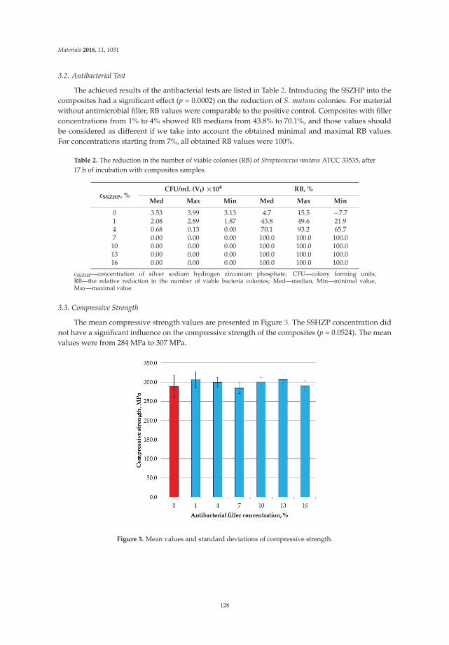
Materials 2018, 11, 1031
3.2. Antibacterial Test
The achieved results of the antibacterial tests are listed in Table 2. Introducing the SSZHP into thecomposites had a significant effect (p = 0.0002) on the reduction of S. mutans colonies. For materialwithout antimicrobial filler, RB values were comparable to the positive control. Composites with fillerconcentrations from 1% to 4% showed RB medians from 43.8% to 70.1%, and those values shouldbe considered as different if we take into account the obtained minimal and maximal RB values.For concentrations starting from 7%, all obtained RB values were 100%.
Table 2. The reduction in the number of viable colonies (RB) of Streptococcus mutans ATCC 33535, after17 h of incubation with composites samples.
cSSZHP, %CFU/mL (Vt) ×104 RB, %
Med Max Min Med Max Min
0 3.53 3.99 3.13 4.7 15.5 −7.71 2.08 2.89 1.87 43.8 49.6 21.94 0.68 0.13 0.00 70.1 93.2 65.77 0.00 0.00 0.00 100.0 100.0 100.010 0.00 0.00 0.00 100.0 100.0 100.013 0.00 0.00 0.00 100.0 100.0 100.016 0.00 0.00 0.00 100.0 100.0 100.0
cSSZHP—concentration of silver sodium hydrogen zirconium phosphate; CFU—colony forming units;RB—the relative reduction in the number of viable bacteria colonies; Med—median, Min—minimal value,Max—maximal value.
3.3. Compressive Strength
The mean compressive strength values are presented in Figure 3. The SSHZP concentration didnot have a significant influence on the compressive strength of the composites (p = 0.0524). The meanvalues were from 284 MPa to 307 MPa.
Figure 3. Mean values and standard deviations of compressive strength.
128

Materials 2018, 11, 1031
3.4. Diametral Tensile Strength
The mean diametral tensile strength values are presented in Figure 4. The SSHZP concentrationdid not have a significant influence on the compressive strength of the composites (p = 0.2986).The mean values were from 40.3 MPa to 43.1 MPa.
Figure 4. Mean values and standard deviations of diametral tensile strength.
3.5. Flexural Strength
The mean flexural strength values are presented in Figure 5a. The SSHZP introduction had asignificant influence on flexural strength (p = 0.0178). The post hoc test showed a significant (p < 0.05)decrease in flexural strength for the composite with the antibacterial filler concentration of 16%(88 MPa). However, these values were not significantly different (p > 0.05) in comparison to the resultsobtained for other materials with SSHZP. The highest mean flexural strength value was registered forthe control material (96 MPa).
(a)
(b)
Figure 5. Mean flexural strength (a) and flexural modulus (b) values with standard deviations; differentlowercase letters show significantly different results at the p < 0.05 level.
129

Materials 2018, 11, 1031
The mean flexural modulus values are presented in Figure 5b. The SSHZP concentration did nothave a significant effect on the flexural modulus (p = 0.5351). The mean values were from 5.6 GPa to6.1 GPa.
3.6. Vickers Hardness
The mean Vickers hardness values are presented in Figure 6. The SSHZP introduction had asignificant influence on flexural strength (p < 0.0001), and the post hoc test showed a significant(p < 0.05) decrease in hardness starting from the antibacterial filler concentration of 4%. However, thevalues for SSHZP concentration from 4% to 13% and from 7% to 16% were not significantly different.The highest mean hardness value was registered for the control material (52.7 HV0.1), and the lowestvalue was for a composite with 16% of SSHZP (48.2 HV0.1).
Figure 6. Mean Vickers microhardness values with standard deviations; different lowercase lettersshow significantly different results at the p < 0.05 level.
3.7. Degree of Conversion
The mean degrees of conversion values are presented in Figure 7. At the top of the samples(Figure 7a) the degree of conversion significantly decreased (p < 0.0001) with increasing SSHZPconcentrations, from 68.7% for the control material to 58.7% for the composite with an SSHZPconcentration of 16%. The post hoc test showed that the results for concentrations from 1% to 7%, from4% to 13%, and from 7% to 16% were not significantly different. Degrees of conversion values obtainedat the top were significantly lower in comparison to the values registered at the bottom of the samples(p < 0.0001). At the bottom of the samples (Figure 7b), the degree of conversion significantly decreased(p < 0.0134) with increasing SSHZP concentrations, from 53.3% for the control material to 47.6% for thecomposite with an SSHZP concentration of 16%. However, the post hoc test showed that the resultsfor concentrations from 1% to 13% were not significantly different, and only the mean value for thematerial with the highest SSHZP concentration was significantly lower.
130

Materials 2018, 11, 1031
(a)
(b)
Figure 7. Mean degree of conversion values with standard deviations at the top (a); and at the bottom(b) of the samples, different lowercase letters show significantly different results at the p < 0.05 level.
3.8. Sorption and Solubility
The mean sorption values are presented in Figure 8a. SSHZP introduction had a significantinfluence on sorption values (p < 0.0004). The post hoc test showed a significant increase in sorptionvalues for composites with 13% and 16% of SSHZP. The mean sorption for the composite with thehighest SSHZP concentration was 44% greater than for the control material.
(a)
(b)
Figure 8. Mean values with standard deviations of sorption (a); and solubility (b), different lowercaseletters show significantly different results at the p < 0.05 level.
The mean solubility values are presented in Figure 8b, and there were no statistically significantdifferences (p = 0.4185) between the results obtained for the investigated materials.
3.9. Color Measurement
The results of initial color measurements are presented in Table 3. The obtained L* axis valuesshowed a significant increase (p < 0.0001) with the increasing SSHZP concentration. The a* and b* axisvalues showed a significant decrease with the increasing SSHZP concentration (p < 0.0001).
131
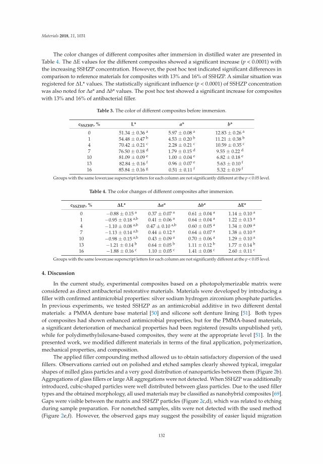
Materials 2018, 11, 1031
The color changes of different composites after immersion in distilled water are presented inTable 4. The ΔE values for the different composites showed a significant increase (p < 0.0001) withthe increasing SSHZP concentration. However, the post hoc test indicated significant differences incomparison to reference materials for composites with 13% and 16% of SSHZP. A similar situation wasregistered for ΔL* values. The statistically significant influence (p < 0.0001) of SSHZP concentrationwas also noted for Δa* and Δb* values. The post hoc test showed a significant increase for compositeswith 13% and 16% of antibacterial filler.
Table 3. The color of different composites before immersion.
cSSZHP, % L* a* b*
0 51.34 ± 0.36 a 5.97 ± 0.08 a 12.83 ± 0.26 a
1 54.48 ± 0.47 b 4.53 ± 0.20 b 11.21 ± 0.38 b
4 70.42 ± 0.21 c 2.28 ± 0.21 c 10.59 ± 0.35 c
7 76.50 ± 0.18 d 1.79 ± 0.15 d 9.55 ± 0.22 d
10 81.09 ± 0.09 e 1.00 ± 0.04 e 6.82 ± 0.18 e
13 82.84 ± 0.16 f 0.96 ± 0.07 e 5.63 ± 0.10 f
16 85.84 ± 0.16 g 0.51 ± 0.11 f 5.32 ± 0.19 f
Groups with the same lowercase superscript letters for each column are not significantly different at the p < 0.05 level.
Table 4. The color changes of different composites after immersion.
cSSZHP, % ΔL* Δa* Δb* ΔE*
0 −0.88 ± 0.15 a 0.37 ± 0.07 a 0.61 ± 0.04 a 1.14 ± 0.10 a
1 −0.95 ± 0.18 a,b 0.41 ± 0.06 a 0.64 ± 0.04 a 1.22 ± 0.13 a
4 −1.10 ± 0.08 a,b 0.47 ± 0.10 a,b 0.60 ± 0.05 a 1.34 ± 0.09 a
7 −1.13 ± 0.14 a,b 0.44 ± 0.12 a 0.64 ± 0.07 a 1.38 ± 0.10 a
10 −0.98 ± 0.15 a,b 0.43 ± 0.09 a 0.70 ± 0.06 a 1.29 ± 0.10 a
13 −1.21 ± 0.14 b 0.64 ± 0.05 b 1.11 ± 0.12 b 1.77 ± 0.14 b
16 −1.88 ± 0.16 c 1.10 ± 0.05 c 1.41 ± 0.08 c 2.60 ± 0.11 c
Groups with the same lowercase superscript letters for each column are not significantly different at the p < 0.05 level.
4. Discussion
In the current study, experimental composites based on a photopolymerizable matrix wereconsidered as direct antibacterial restorative materials. Materials were developed by introducing afiller with confirmed antimicrobial properties: silver sodium hydrogen zirconium phosphate particles.In previous experiments, we tested SSHZP as an antimicrobial additive in two different dentalmaterials: a PMMA denture base material [50] and silicone soft denture lining [51]. Both typesof composites had shown enhanced antimicrobial properties, but for the PMMA-based materials,a significant deterioration of mechanical properties had been registered (results unpublished yet),while for polydimethylsiloxane-based composites, they were at the appropriate level [51]. In thepresented work, we modified different materials in terms of the final application, polymerization,mechanical properties, and composition.
The applied filler compounding method allowed us to obtain satisfactory dispersion of the usedfillers. Observations carried out on polished and etched samples clearly showed typical, irregularshapes of milled glass particles and a very good distribution of nanoparticles between them (Figure 2b).Aggregations of glass fillers or large AR aggregations were not detected. When SSHZP was additionallyintroduced, cubic-shaped particles were well distributed between glass particles. Due to the used fillertypes and the obtained morphology, all used materials may be classified as nanohybrid composites [69].Gaps were visible between the matrix and SSHZP particles (Figure 2c,d), which was related to etchingduring sample preparation. For nonetched samples, slits were not detected with the used method(Figure 2e,f). However, the observed gaps may suggest the possibility of easier liquid migration
132

Materials 2018, 11, 1031
between SSHZP and the matrix than between silanized glass and the matrix. For frozen-brokennonetched samples, glass particles were usually not visible, which suggests their good connectionwith the matrix and is related to the salinization process used by the manufacturer. Large aggregationsof SSHZP were not observed, which was favored because of their potential influence on the propertiesof the composites [70]. However, some structures consisting of connected particles, probably comingfrom the used antimicrobial filler (Figure 1e), were observed. Observations have also shown some airbubbles in the polymerized composites. They were probably caused by the used procedure of manualpreparation of composite and/or by the process of sample preparation. Bubbles measured from a fewup to 50 μm. Those structural defects might decrease the mechanical properties because they may actas stress concentrators. In the future, the bubbles can also have a negative effect on the mechanicalproperties at the bonded interface.
In previous works related with dental materials, the antimicrobial effectiveness of SSHZPagainst Candida albican (C. albicans), Staphylococcus aureus (S. aureus), and Escherichia coli (E. coli) wasconfirmed [50,51]. However, only two tested microorganisms (C. albicans, S. aureus) had clinicallyproven relevance, which is related to using partial or complete dentures [71–74], but none of them wasassociated with tooth decay. The problem of caries appearing between teeth and composite restorationsis widely disputed in the literature, and its mechanisms are probably multifactorial. Bacterial speciesassociated with secondary caries and primary caries seem to be the same. However, a higher proportionof caries-related bacteria (mutans streptococci, lactobacilli) was found on restored surfaces than onunrestored dentin or enamel [75], which is an additional argument for the development of antibacterialmaterials. Despite the fact that both mutans streptococci and lactobacilli have a confirmed role in dentalcaries, the S. mutans strains are usually investigated in the context of antimicrobial composites, so thisbacterium was also used in our experiment.
Due to differences in microbiological test protocols, the results obtained for the antimicrobialfillers mentioned in the introduction cannot be directly compared to one another or to the results of thepresent study. In our experiment, samples were stored in an S. mutans suspension. All specimens werefinally finished with P1200-grit abrasive paper, which gives well standardized and smooth surfaces.This may be confusing in the context of the recognized fact that higher values of roughness promotebacterial adhesion and dental plaque retention [76–78]. However, in our study, adherence of bacteriaand biofilm formation were not investigated. Samples were immersed in bacteria suspension in ashaking incubator, and the changes in the number of bacteria were investigated. In this experiment,silver ions released into the environment determined the reduction of bacteria, so using a smoothsurface would have created stricter test conditions due to the smaller surface area responsible for therelease of antibacterial ions.
After incubation, the reduction of the bacteria population in the environment was registered forall materials, but starting from a concentration of 7%, it was complete. Antimicrobial properties ofthe used filler are initiated in humid environments by the mechanism of silver ion release from aninorganic, insoluble carrier, which was described by Kampmann et al. [79]. With time, this mechanismmay lead to the loss of antibacterial properties due to the continuous silver ion release, so furtherinvestigations in this context should be made. Additionally, restorative materials in oral cavities aresubject to tribological processes. This, on the one hand, may be the reason for the selective removalof particles from the matrix, but on the other hand, it can also cause “refreshment” of antimicrobialproperties by gradual abrasion of the surface with fillers. Both mentioned conceptions may be checkedin future experiments.
The microbiological properties of the newly developed antibacterial composites are regularlytested, whereas their mechanical and functional properties are much less frequently reported.Compressive strength, flexural strength, flexural modulus, diametral tensile strength, and hardnessare frequently tested mechanical properties for dental restorative materials.
In the present study, we used different curing protocols for each mechanical evaluation. It wasjustified by the varied sample dimensions, which were dictated by the requirements of the procedures.
133
Materials 2018, 11, 1031
It is known that light intensity, polymerization time, and curing depth determine whether or not dentalcomposites are properly cured [80]. Mechanical tests as well as sorption/solubility and antibacterialtests require samples having a length or diameter much larger than the area effectively covered bythe used lamp. The use of overlapping light-curing areas for flexural test samples has been subjectto criticism due to the risk of preparation of nonhomogeneous specimens [81], although the effect ofthat method on the flexural properties has been questioned [82]. The height of the samples is equallyimportant due to the expected decrease of the degree of conversion with the depth [83]. A thickness of2 mm can be considered in that context as a safe value [84]. Moreover, Koran et al. [85] establishedthat if the total dose of light intensity (interpreted as light intensity in exposure time) delivered tothe photopolymerizable dental composite is high enough to achieve complete polymerization, thesurface hardness, as well residual monomer concentration, tends to remain constant. This shows thatthe best way to standardize samples is to use overlapping areas of irradiation on both the bottom andtop surface, and for samples higher than 4 mm, to use additional irradiations on the lateral surface.However, such an approach is a simplification because it does not take into account other changesoccurring in the material during the polymerization and postirradiation polymerization [86–88].
Composites often replace a large bulk of the teeth structure, so the dental restorative materialsare usually subjected to compressive forces generated during mastication [89]. If we consider that thecompressive strength and plastic limit of tooth tissues [3,90] may be recommended as a standard for thestrength of composites [60], we can accept a 230 MPa as a secure value for a composite. The obtainedresults were higher and were additionally comparable with values reported for numerous commerciallyavailable materials with BisGMA, TEGDMA, and UDMA matrices and similar filler content [59,91],including those releasing fluoride [92]. Compressive strength after antimicrobial filler addition wasinvestigated in only a few previous works. The effect of the used additives was varied. An increase ofcompressive strength values at low concentrations and a decrease of them at larger concentrations wasnoted for nanosilver [93], zinc oxide [36], and tetretrapod-like zinc oxide whisker [41]. Yoshida et al. [74]have shown no effect of silver-containing ceramic microparticles. In our study, the filler addition alsohad no effect on compressive strength.
Stress analyses have shown that restorative composites can fracture under tension [94] and tensilestrength data may have equal, if not greater, importance than compressive strength, especially in thearea near the teeth–composite interface [95,96]. The diametral tensile strength test is an alternativemethod to evaluate the tensile strength of brittle materials, and it is the default for investigatingdental restorative materials. Nevertheless, it gives correct results only if minimal or no plasticdeformations occur and when deformations at fracture are small because the area of contact is stillnear to theoretical [61]. For this reason, this test should not be used for resins or experimentalcomposites with a low filler concentration because of their stress-strain characteristics. UsuallyDTS values for different types of commercially available composite materials range from 25 MPato 50 MPa [97–99], but for modern nanocomposites, they may reach over 80 MPa [100]. For allinvestigated materials, mean DTS values were above 40 MPa, which can be considered to be satisfactoryvalues. The antibacterial filler introduction had no statistically significant effect on DTS, althoughthe mean value for the control group was the lowest. Those findings are in opposition to the resultsobtained by Łukomska-Szymanska et al. [101], Diaz et al. [102], and Sokołowski et al. [103], wherenonfunctionalized calcium fluoride microparticles, zinc oxide three-dimensional microstructures,nanosilver, and nanogold decreased the DTS values of modified composites.
Flexural strength and flexural modulus have been reported as indicators of clinical wear ofcomposites in some studies [104,105]. Composite fillings are also exposed to flexural stress, especiallyin stress-bearing cavities for restoration classes I, II, and IV [82]. The flexural test is also indicated asa method that relates well to tensile failure [104]. Flexural strength is the only mechanical propertyspecified by the ISO 4049 standard for composite restorative materials, which requires minimal valuesof 80 MPa for occlusal tooth surface restorations and 50 MPa for others [62], so all investigatedmaterials meet these requirements. The obtained results were additionally comparable to other
134

Materials 2018, 11, 1031
materials with similar matrix or filler concentrations [4,106–108]. A parallel situation was noted forflexural moduli [109], the values of which are not defined by the standard and may be diversifiedfor different clinical situations. Cervical cavities demand composites characterized by a relativelylower modulus to flex with the teeth, but posterior composites need a high modulus to withstand theocclusal forces [110]. In the presented study, the flexural strength significantly decreased only for thehighest antimicrobial filler concentration, whereas the flexural modulus values were stable. Similartrends were noted for other experimental materials compounded with antibacterial particles [36,111].However, for composites with calcium fluoride, after 24 h storing in wet conditions, a significantdeterioration of flexural properties has been registered [112]. The addition of zinc oxide whiskers [41]and nanoparticle-laden hydroxyapatite nanowires [36] caused increases in flexural strength andmodulus, but for the concentration of 10%, properties decreased for both fillers. This suggests thatthe obtained effects are related to both filler type and filler loading. The decrease in flexural strengthnoted in our study for the highest SSHZP concentration should be treated with some caution, alsoin the context of the obtained compressive and tensile strength values. For one sample, the flexuralstrength was 77 MPa. If we deleted this result, we would have a mean value of 90.1 MPa, and thisvalue is not statistically different from any other. After consideration, we decided not to remove thatresult because samples for flexural strength for this material were the most difficult to prepare duethe increasing viscosity of composition in combination with a small working area of silicone mold(2 × 25 mm), which created an increased risk of making structural defects during the packing ofthe material. The registered statistical difference can be an indicator of those problems, especially ifwe consider that flexural strength has been noted to be more sensitive to subtle changes in materialsubstructure than, for example, compressive strength test [113].
The Vickers microhardness test is a known method used to compare composite resins, especiallyin the context of their wear resistance prediction [50,114]. Direct restorative composites used indentistry demonstrate Vickers hardness starting from 40 kgf/mm2, but for some materials, valuesexceed 100 kgf/mm2 [10,114–116]. The values obtained in this work were within this range but wererather at the lower limit. Increasing antibacterial filler concentration caused a small but systematiclowering of microhardness. This is in opposition to some findings for commercially available materials,where higher filler content was correlated with higher microhardness [117], but is in accordance withsome research, where introducing antibacterial additives decreased microhardness [44,101,111].
Degree of conversion is an important property of restorative composites due to the potential riskof biological responses related to monomer release and affection of pulp tissues [118]. The obtainedvalues were in agreement with the findings from other studies, performed on similar dimethacrylatesystems, and measured with the same method [64]. The reduction of DC values at the bottom of thesamples was also expected because when light moves through a material with increasing density,its intensity is reduced. The reduction of DC values with increasing SSHZP content was probablyrelated to the effect of light scattering by particles. Moreover, some reports showed larger scatteringwhen the particle size is circa one half or close to that of the curing light wavelength [119,120]. In thepresented study, this situation took place because the optical wave length was 450–470 nm, so it wassimilar to the observed SSHZP particles size, which may explain unfavorable DC changes. Additionally,the DC values at the top of the samples were well correlated (R2 = 0.9058, p = 0.001) with microhardnessresults. This is in accordance with other reports, where surface microhardness has been identified as agood indicator of DC changes [121,122].
The solubility and sorption properties are important from the viewpoint of biocompatibilityconcerns over monomer releasing and in relation to the stability of the composites due to degradationfrom the uptake of solvents and the wash-out of ingredients of materials [104]. Sorption valuesfor all samples were lower than 40 μg/mm3, so all investigated materials met the requirements ofthe ISO standard. Solubility values’ samples were lower or, for one sample, equal to 7.5 μg/mm3,so all materials also met the requirements of the ISO standard. The sorption values were comparableto numerous commercially available materials [123,124] but increased for the highest antimicrobial
135

Materials 2018, 11, 1031
filler concentration. The solubility for all composites, including the control group, was generallyhigher than for most modern materials, however, for some of them, a similar value has alsobeen registered [66,123,124]. The increasing solubility with SSHZP content was not statisticallysignificant. The principal factor influencing sorption and solubility of dental restorative materials isthe composition of polymer matrix [67]. The sorption is influenced by the polarity of the molecularstructure, the presence of hydroxyl groups (which may crate hydrogen bonds), and with the degreeof crosslinking of the matrix [125]. Water may penetrate into the free volume between the chainsand nanopores formed during polymerization, or it can be successively attached to polymer chainsvia hydrogen bonds [123]. In this study, a hydrophilic monomers system (Bis-GMA, TEGDMA,UDMA) [123] was used, from which TEGDMA and Bis-GMA create networks characterized byhigher sorption due to the presence of the ether linkages and hydroxyl groups, respectively [66,126].Thus, those kinds of matrices usually show relatively large values of water sorption and solubility.Additionally, after the introduction of nonsilanized SSHZP, water would migrate in the interfacebetween the filler particles/particle aggregations and the matrix, which may explain the enhancedsorption values for the higher concentration. Also, decreased DC may have some influence onwater sorption. The uptake of water also allows diffusion out (into the storage medium) to residualmonomers, fillers, degradation products, and other leachable components, so sorption is oftencorrelated with solubility [66]. This was not found in this study, although a statistically insignificantincrease in solubility was registered. This may suggest that an experiment should be prepared infuture with longer storage periods to allow for more extensive changes in the materials, which will beeasier to detect, or with more sensitive methods.
The introduction of SSHZP caused significant changes in composite color. The materialswith increasing antimicrobial filler concentrations show a narrow whitening effect, representedby increasing L* axis values. Composites have also shown a reduction of reddish and yellowcoloration related to decreasing positive values of a* and b*, respectively. These changes, at thestarting point, might be considered as beneficial. After being stored in distilled water, colorchanges expressed by ΔE values can be classified as noticeable only for an experienced observer(ΔE values from 1 to 2) for all materials, excluding the highest concentration for which unexperiencedobserver may notice the difference (ΔE values from 2 to 2.5) [127]. However, all achieved ΔE valueswere comparable to the results obtained for commercial [128] and experimental [129,130] materialsafter a similar period of storing in distilled water. For all composites, the darkening, reddening,and yellowing effects were registered, which were also registered for other photopolymerizableresin-based composites [120,128,130]. The reasons for the noted color changes might be multifactorial.De Oliwiera et al. [120] suggest that reduced monomer conversion can lead to poorer color stabilityin the composites during storing due to the oxidation of the amines or monomers. In our study, thereduction of DC values was also noted, which may partially be the reason for the reddening andyellowing of the materials. However, the slight, progressive changes in DC values cannot explainmuch of the decreased color stability of the materials for the two highest concentrations of SSHZP.The effect of color changes can be also associated with the leaching of components [130], includingthe antibacterial filler. The silver ions released from the filler, their deposition on the surface, andfurther oxidation may be considered as the reason for the darkening, reddening, and yellowing of thecomposites. However, additional investigations ought to be conducted to clarify this behavior.
The findings presented here should be enhanced with further in vitro investigations. As anespecially important part of future research, the cytotoxic potential of composites should be examined.The toxicological data for the used SSHZP [56] let us suppose that materials should not presentunfavorable properties in that aspect, although toxic effects in dental materials with silver have beenregistered [70]. The ion release into the environment and the dynamics of this process should beinvestigated, as an indication of the persistence of the material’s antibacterial activity. The SSHZPintroduced into the PMMA denture base material in a previous work have shown antimicrobialproperties decreasing with time during a three-month experiment [50], so it should be expected that
136

Materials 2018, 11, 1031
the durability of the antibacterial activity of the tested composites will also be limited. The usedantimicrobial test additionally did not reflect real conditions, especially in the context of the expectedlifetime of the dental composites and the processes occurring in the interface area between tooth tissuesand composite. Nevertheless, the presented results of antimicrobial tests are a promising base forfurther experiments. All properties studied here may also be tested with long-term experiments.
Another limitation of the present study was that the mechanical properties of the proposedcomposites were not evaluated after biofilm exposure. Acid production by bacteria during metabolicprocesses may be the cause of changes in some mechanical properties of composites. Microhardnessis usually stable after exposure to S. mutans biofilm [77,131], but Fúcio et al. [131] have shown that itcan be reduced for particular restorative composites. Moreover, the presence of biofilm influencesthe mechanical properties of resin–dentin bonds. Melo et al. [132], in self-designed experimentswith quasi-static and fatigue performance tests, have shown that the S. mutans growth may be thecause of the reduction in the mechanical properties of the bonded interface. The degradation in thedentin–composite interface in the biofilm environment was confirmed by other researchers [133,134],who indicated that the studied experimental composites should be examined in the future in thisrespect. The problem of acid production by bacteria may also be important in the context ofthe observed gaps between SSZHP particles and the matrix after the etching of samples for SEMinvestigations. This may suggest the possibility of accelerated changes in this area due to the influenceof the acidic environment. Depending on whether these changes occur as a result of the presence of abiofilm, this phenomenon could negatively affect the mechanical properties of materials due to thecreation and propagation of structural defects.
In the present study, we used quasi-static tests for the mechanical properties’ evaluation,which was sufficient at the planned initial stage. However, fatigue tests in the past decade havegained increased importance because dental materials under clinical conditions are subject to cyclicloading [135], caused by thousands of cycles of mastication per day. It has been proven that flexuralstatic strength is higher than values obtained after flexural cyclic loading [136]. Cyclic loading alsoreduces fracture toughness [137]. Fatigue tests are also used to evaluate mechanical properties ofresin–dentin bonds [138,139], also in the context of bacteria presence [133]. These results suggest thedesirability of conducting research in this direction, including comparative studies with commerciallyavailable composites.
5. Conclusions
Within the limits of this study, it can be concluded that the experimental composites showed a highinitial reduction of bacteria colonies for the tested S. mutans strain. The satisfactory combination of thereduction of bacteria colonies with physical properties was achieved for filler concentrations rangingfrom 4% to 13%. Those materials exhibited mechanical properties similar to the base material, as well asthe degree of conversion, sorption, solubility, and color stability at acceptable levels. The cytotoxic testsand long-term investigations, including silver ion release into the environment, need to be performedin future experiments.
Author Contributions: Conceptualization, R.S. and G.C.; Formal analysis, R.S.; Investigation, R.S., W.P., A.M.,E.B., I.B.-R. and G.C.; Methodology, R.S., W.P., A.M., I.B.-R. and G.C.; Resources, R.S. and G.C.; Supervision, G.C.;Visualization, R.S.; Writing—original draft, R.S. and G.C.; Writing—review & editing, R.S., J.K. and G.C.
Funding: This work was financially supported with statutory funds of Faculty of Mechanical Engineering ofSilesian University of Technology in 2018 and by Robert Stencel.
Acknowledgments: The authors would like to thank the Konica Minolta Poland for help during thecolor measurements.
Conflicts of Interest: The authors declare no conflict of interest.
137

Materials 2018, 11, 1031
References
1. Vos, T.; Allen, C.; Arora, M.; Barber, R.M.; Brown, A.; Carter, A.; Casey, D.C.; Charlson, F.J.; Chen, A.Z.;Coggeshall, M.; et al. Global, regional, and national incidence, prevalence, and years lived with disability for310 diseases and injuries, 1990–2015: A systematic analysis for the Global Burden of Disease Study 2015.Lancet Lond. Engl. 2016, 388, 1545–1602. [CrossRef]
2. Polish Ministry of Health. Monitoring the Oral Health of the Polish Population in the Years 2016–2020; PolishMinistry of Health: Warsaw, Poland, 2015. (In Polish)
3. Anusavice, K.; Shen, C.; Rawls, H.R. Phillips’ Science of Dental Materials—12th Edition, 12th ed.; Saunders:St. Louis, MO, USA, 2013; ISBN 978-0-32-324205-9.
4. Jayanthi, N.; Vinod, V. Comparative evaluation of compressive strength and flexural strength of conventionalcore materials with nanohybrid composite resin core material an in vitro study. J. Indian Prosthodont. Soc.2013, 13, 281–289. [CrossRef] [PubMed]
5. Ilie, N.; Hickel, R. Resin composite restorative materials. Aust. Dent. J. 2011, 56 (Suppl. 1), 59–66. [CrossRef][PubMed]
6. Ástvaldsdóttir, Á.; Dagerhamn, J.; van Dijken, J.W.V.; Naimi-Akbar, A.; Sandborgh-Englund, G.; Tranæus, S.;Nilsson, M. Longevity of posterior resin composite restorations in adults—A systematic review. J. Dent.2015, 43, 934–954. [CrossRef] [PubMed]
7. Alzraikat, H.; Burrow, M.; Maghaireh, G.; Taha, N. Nanofilled Resin Composite Properties and ClinicalPerformance: A Review. Oper. Dent. 2018, in press. [CrossRef] [PubMed]
8. Lambrechts, P.; Goovaerts, K.; Bharadwaj, D.; De Munck, J.; Bergmans, L.; Peumans, M.; Van Meerbeek, B.Degradation of tooth structure and restorative materials: A review. Wear 2006, 261, 980–986. [CrossRef]
9. Chan, K.H.S.; Mai, Y.; Kim, H.; Tong, K.C.T.; Ng, D.; Hsiao, J.C.M. Review: Resin Composite Filling. Materials2010, 3, 1228–1243. [CrossRef]
10. Rasines Alcaraz, M.G.; Veitz-Keenan, A.; Sahrmann, P.; Schmidlin, P.R.; Davis, D.; Iheozor-Ejiofor, Z. Directcomposite resin fillings versus amalgam fillings for permanent or adult posterior teeth. Cochrane DatabaseSyst. Rev. 2014, 3, CD005620. [CrossRef] [PubMed]
11. Hurst, D. Amalgam or composite fillings—Which material lasts longer? Evid. Based Dent. 2014, 15, 50–51.[CrossRef] [PubMed]
12. Demarco, F.F.; Corrêa, M.B.; Cenci, M.S.; Moraes, R.R.; Opdam, N.J.M. Longevity of posterior compositerestorations: Not only a matter of materials. Dent. Mater. Off. Publ. Acad. Dent. Mater. 2012, 28, 87–101.[CrossRef] [PubMed]
13. Opdam, N.J.M.; van de Sande, F.H.; Bronkhorst, E.; Cenci, M.S.; Bottenberg, P.; Pallesen, U.; Gaengler, P.;Lindberg, A.; Huysmans, M.C.; van Dijken, J.W. Longevity of posterior composite restorations: A systematicreview and meta-analysis. J. Dent. Res. 2014, 93, 943–949. [CrossRef] [PubMed]
14. Pereira-Cenci, T.; Cenci, M.S.; Fedorowicz, Z.; Azevedo, M. Antibacterial agents in composite restorations forthe prevention of dental caries. In The Cochrane Library; John Wiley & Sons, Ltd.: Hoboken, NJ, USA, 2013.
15. Lai, G.; Li, M. Secondary Caries. In Contemporary Approach to Dental Caries; InTech: Rijeka, Croatia, 2012;pp. 403–422. ISBN 978-9-53-510305-9.
16. Mjör, I.A.; Toffenetti, F. Secondary caries: A literature review with case reports. Quintessence Int. Berl. Ger.1985 2000, 31, 165–179.
17. Shih, W.-Y. Microleakage in different primary tooth restorations. J. Chin. Med. Assoc. 2016, 79, 228–234.[CrossRef] [PubMed]
18. Gale, M.S.; Darvell, B.W. Thermal cycling procedures for laboratory testing of dental restorations. J. Dent.1999, 27, 89–99. [CrossRef]
19. Nedeljkovic, I.; Teughels, W.; De Munck, J.; Van Meerbeek, B.; Van Landuyt, K.L. Is secondary caries withcomposites a material-based problem? Dent. Mater. Off. Publ. Acad. Dent. Mater. 2015, 31, e247–e277.[CrossRef] [PubMed]
20. Łukomska-Szymanska, M.; Zarzycka, B.; Grzegorczyk, J.; Sokołowski, K.; Półtorak, K.; Sokołowski, J.;Łapinska, B. Antibacterial Properties of Calcium Fluoride-Based Composite Materials: In VitroStudy. BioMedRes. Int. 2016, 2016, 1048320. [CrossRef] [PubMed]
138

Materials 2018, 11, 1031
21. Chatzistavrou, X.; Lefkelidou, A.; Papadopoulou, L.; Pavlidou, E.; Paraskevopoulos, K.M.; Fenno, J.C.;Flannagan, S.; González-Cabezas, C.; Kotsanos, N.; Papagerakis, P. Bactericidal and Bioactive DentalComposites. Front. Physiol. 2018, 9, 103. [CrossRef] [PubMed]
22. Imazato, S. Antibacterial properties of resin composites and dentin bonding systems. Dent. Mater. Off. Publ.Acad. Dent. Mater. 2003, 19, 449–457. [CrossRef]
23. Chen, L.; Shen, H.; Suh, B.I. Antibacterial dental restorative materials: A state-of-the-art review. Am. J. Dent.2012, 25, 337–346. [PubMed]
24. Maas, M.S.; Alania, Y.; Natale, L.C.; Rodrigues, M.C.; Watts, D.C.; Braga, R.R. Trends in restorative compositesresearch: What is in the future? Braz. Oral Res. 2017, 31, e55. [CrossRef] [PubMed]
25. Cheng, L.; Zhang, K.; Zhou, C.-C.; Weir, M.D.; Zhou, X.-D.; Xu, H.H.K. One-year water-ageing of calciumphosphate composite containing nano-silver and quaternary ammonium to inhibit biofilms. Int. J. Oral Sci.2016, 8, 172–181. [CrossRef] [PubMed]
26. Imazato, S.; Torii, M.; Tsuchitani, Y.; McCabe, J.F.; Russell, R.R. Incorporation of bacterial inhibitor into resincomposite. J. Dent. Res. 1994, 73, 1437–1443. [CrossRef] [PubMed]
27. Zhang, K.; Li, F.; Imazato, S.; Cheng, L.; Liu, H.; Arola, D.D.; Bai, Y.; Xu, H.H.K. Dual antibacterial agentsof nano-silver and 12-methacryloyloxydodecylpyridinium bromide in dental adhesive to inhibit caries.J. Biomed. Mater. Res. B Appl. Biomater. 2013, 101, 929–938. [CrossRef] [PubMed]
28. Cherchali, F.Z.; Mouzali, M.; Tommasino, J.B.; Decoret, D.; Attik, N.; Aboulleil, H.; Seux, D.; Grosgogeat, B.Effectiveness of the DHMAI monomer in the development of an antibacterial dental composite. Dent. Mater.Off. Publ. Acad. Dent. Mater. 2017, 33, 1381–1391. [CrossRef] [PubMed]
29. Rego, G.F.; Vidal, M.L.; Viana, G.M.; Cabral, L.M.; Schneider, L.F.J.; Portela, M.B.; Cavalcante, L.M.Antibiofilm properties of model composites containing quaternary ammonium methacrylates after surfacetexture modification. Dent. Mater. Off. Publ. Acad. Dent. Mater. 2017, 33, 1149–1156. [CrossRef] [PubMed]
30. Beyth, N.; Yudovin-Farber, I.; Bahir, R.; Domb, A.J.; Weiss, E.I. Antibacterial activity of dental compositescontaining quaternary ammonium polyethylenimine nanoparticles against Streptococcus mutans. Biomaterials2006, 27, 3995–4002. [CrossRef] [PubMed]
31. Zhang, J.F.; Wu, R.; Fan, Y.; Liao, S.; Wang, Y.; Wen, Z.T.; Xu, X. Antibacterial dental composites withchlorhexidine and mesoporous silica. J. Dent. Res. 2014, 93, 1283–1289. [CrossRef] [PubMed]
32. Leung, D.; Spratt, D.A.; Pratten, J.; Gulabivala, K.; Mordan, N.J.; Young, A.M. Chlorhexidine-releasingmethacrylate dental composite materials. Biomaterials 2005, 26, 7145–7153. [CrossRef] [PubMed]
33. Rathke, A.; Staude, R.; Muche, R.; Haller, B. Antibacterial activity of a triclosan-containing resin compositematrix against three common oral bacteria. J. Mater. Sci. Mater. Med. 2010, 21, 2971–2977. [CrossRef][PubMed]
34. Ali, S.; Sangi, L.; Kumar, N. Exploring antibacterial activity and hydrolytic stability of resin dental compositerestorative materials containing chitosan. Technol. Health Care Off. J. Eur. Soc. Eng. Med. 2017, 25, 11–18.[CrossRef] [PubMed]
35. Wang, J.; Dong, X.; Yu, Q.; Baker, S.N.; Li, H.; Larm, N.E.; Baker, G.A.; Chen, L.; Tan, J.; Chen, M. Incorporationof antibacterial agent derived deep eutectic solvent into an active dental composite. Dent. Mater. Off. Publ.Acad. Dent. Mater. 2017, 33, 1445–1455. [CrossRef] [PubMed]
36. Tavassoli Hojati, S.; Alaghemand, H.; Hamze, F.; Ahmadian Babaki, F.; Rajab-Nia, R.; Rezvani, M.B.;Kaviani, M.; Atai, M. Antibacterial, physical and mechanical properties of flowable resin compositescontaining zinc oxide nanoparticles. Dent. Mater. Off. Publ. Acad. Dent. Mater. 2013, 29, 495–505. [CrossRef][PubMed]
37. Kasraei, S.; Sami, L.; Hendi, S.; AliKhani, M.-Y.; Rezaei-Soufi, L.; Khamverdi, Z. Antibacterial properties ofcomposite resins incorporating silver and zinc oxide nanoparticles on Streptococcus mutans and Lactobacillus.Restor. Dent. Endod. 2014, 39, 109–114. [CrossRef] [PubMed]
38. Aydin Sevinç, B.; Hanley, L. Antibacterial activity of dental composites containing zinc oxide nanoparticles.J. Biomed. Mater. Res. B Appl. Biomater. 2010, 94, 22–31. [CrossRef] [PubMed]
39. Khvostenko, D.; Hilton, T.J.; Ferracane, J.L.; Mitchell, J.C.; Kruzic, J.J. Bioactive glass fillers reduce bacterialpenetration into marginal gaps for composite restorations. Dent. Mater. Off. Publ. Acad. Dent. Mater. 2016, 32,73–81. [CrossRef] [PubMed]
139

Materials 2018, 11, 1031
40. Sodagar, A.; Akhoundi, M.S.A.; Bahador, A.; Jalali, Y.F.; Behzadi, Z.; Elhaminejad, F.; Mirhashemi, A.H. Effectof TiO2 nanoparticles incorporation on antibacterial properties and shear bond strength of dental compositeused in Orthodontics. Dent. Press J. Orthod. 2017, 22, 67–74. [CrossRef] [PubMed]
41. Niu, L.N.; Fang, M.; Jiao, K.; Tang, L.H.; Xiao, Y.H.; Shen, L.J.; Chen, J.H. Tetrapod-like zinc oxide whiskerenhancement of resin composite. J. Dent. Res. 2010, 89, 746–750. [CrossRef] [PubMed]
42. Chatzistavrou, X.; Velamakanni, S.; DiRenzo, K.; Lefkelidou, A.; Fenno, J.C.; Kasuga, T.; Boccaccini, A.R.;Papagerakis, P. Designing dental composites with bioactive and bactericidal properties. Mater. Sci. Eng. CMater. Biol. Appl. 2015, 52, 267–272. [CrossRef] [PubMed]
43. Ai, M.; Du, Z.; Zhu, S.; Geng, H.; Zhang, X.; Cai, Q.; Yang, X. Composite resin reinforced with silvernanoparticles-laden hydroxyapatite nanowires for dental application. Dent. Mater. Off. Publ. Acad. Dent.Mater. 2017, 33, 12–22. [CrossRef] [PubMed]
44. Łukomska-Szymanska, M.M.; Kleczewska, J.; Bielinski, D.M.; Jakubowski, W.; Sokołowski, J. Bactericidalproperties of experimental dental composites based on dimethacrylate resins reinforced by nanoparticles.Eur. J. Chem. 2014, 5, 419–423. [CrossRef]
45. Azarsina, M.; Kasraei, S.; Yousef-Mashouf, R.; Dehghani, N.; Shirinzad, M. The antibacterial properties ofcomposite resin containing nanosilver against Streptococcus mutans and Lactobacillus. J. Contemp. Dent. Pract.2013, 14, 1014–1018. [CrossRef] [PubMed]
46. Yoshida, K.; Tanagawa, M.; Matsumoto, S.; Yamada, T.; Atsuta, M. Antibacterial activity of resin compositeswith silver-containing materials. Eur. J. Oral Sci. 1999, 107, 290–296. [CrossRef] [PubMed]
47. Sodagar, A.; Akhavan, A.; Hashemi, E.; Arab, S.; Pourhajibagher, M.; Sodagar, K.; Kharrazifard, M.J.;Bahador, A. Evaluation of the antibacterial activity of a conventional orthodontic composite containingsilver/hydroxyapatite nanoparticles. Prog. Orthod. 2016, 17, 40. [CrossRef] [PubMed]
48. Cheng, L.; Weir, M.D.; Xu, H.H.K.; Antonucci, J.M.; Kraigsley, A.M.; Lin, N.J.; Lin-Gibson, S.; Zhou, X.Antibacterial amorphous calcium phosphate nanocomposites with a quaternary ammonium dimethacrylateand silver nanoparticles. Dent. Mater. 2012, 28, 561–572. [CrossRef] [PubMed]
49. Fan, C.; Chu, L.; Rawls, H.R.; Norling, B.K.; Cardenas, H.L.; Whang, K. Development of an antimicrobialresin—A pilot study. Dent. Mater. Off. Publ. Acad. Dent. Mater. 2011, 27, 322–328. [CrossRef] [PubMed]
50. Chladek, G.; Basa, K.; Mertas, A.; Pakieła, W.; Zmudzki, J.; Bobela, E.; Król, W. Effect of Storage in DistilledWater for Three Months on the Antimicrobial Properties of Poly(methyl methacrylate) Denture Base MaterialDoped with Inorganic Filler. Materials 2016, 9, 328. [CrossRef] [PubMed]
51. Jabłonska-Stencel, E.; Pakieła, W.; Mertas, A.; Bobela, E.; Kasperski, J.; Chladek, G. Effect of Silver-EmittingFiller on Antimicrobial and Mechanical Properties of Soft Denture Lining Material. Materials 2018, 11, 318.[CrossRef] [PubMed]
52. Qin, Y. Silver-containing alginate fibres and dressings. Int. Wound J. 2005, 2, 172–176. [CrossRef] [PubMed]53. Qin, Y.; Zhu, C. Antimicrobial Properties of Silver-Containing Chitosan Fibers. In Medical and Healthcare
Textiles; Woodhead Publishing Series in Textiles; Woodhead Publishing: Sawston, UK, 2010; pp. 7–13. ISBN978-1-84-569224-7.
54. Ågren, M. Wound Healing Biomaterials—Volume 2: Functional Biomaterials; Woodhead Publishing: Sawston,UK, 2016; ISBN 978-0-08-100606-1.
55. Qin, Y. Antimicrobial dressings for the management of wound infection. In Medical Textile Materials;Woodhead Publishing Series in Textiles; Woodhead Publishing: Sawston, UK, 2016; pp. 145–160.ISBN 978-0-08-100618-4.
56. National Industrial Chemicals Notification and Assessment Scheme (Nicnas)-Silver Sodium HydrogenZirconium Phosphate. Available online: https://www.pharosproject.net/uploads/files/sources/1185/alphasan-std1081fr.pdf (accessed on 11 March 2004).
57. Melaiye, A.; Sun, Z.; Hindi, K.; Milsted, A.; Ely, D.; Reneker, D.H.; Tessier, C.A.; Youngs, W.J.Silver(I)-imidazole cyclophane gem-diol complexes encapsulated by electrospun tecophilic nanofibers:Formation of nanosilver particles and antimicrobial activity. J. Am. Chem. Soc. 2005, 127, 2285–2291.[CrossRef] [PubMed]
58. Xu, X.; Yang, Q.; Wang, Y.; Yu, H.; Chen, X.; Jing, X. Biodegradable electrospun poly(L-lactide) fiberscontaining antibacterial silver nanoparticles. Eur. Polym. J. 2006, 42, 2081–2087. [CrossRef]
140
Materials 2018, 11, 1031
59. Mota, E.G.; Weiss, A.; Spohr, A.M.; Oshima, H.M.S.; de Carvalho, L.M.N. Relationship between filler contentand selected mechanical properties of six microhybrid composites. Rev. Odonto Ciênc. 2011, 26, 151–155.[CrossRef]
60. Galvão, M.R.; Caldas, S.G.F.R.; Calabrez-Filho, S.; Campos, E.A.; Bagnato, V.S.; Rastelli, A.N.S.; Andrade, M.F.Compressive strength of dental composites photo-activated with different light tips. Laser Phys. 2013,23, 045604. [CrossRef]
61. Penn, R.W.; Craig, R.G.; Tesk, J.A. Diametral tensile strength and dental composites. Dent. Mater. 1987, 3,46–48. [CrossRef]
62. International Organization for Standardization (ISO). EN ISO 4049:2009 Dentistry—Polymer-Based RestorativeMaterials; ISO: Geneva, Switzerland, 2009.
63. Chladek, G.; Basa, K.; Zmudzki, J.; Malara, P.; Nowak, A.J.; Kasperski, J. Influence of aging solutions on wearresistance and hardness of selected resin-based dental composites. Acta Bioeng. Biomech. 2016, 18, 43–52.[PubMed]
64. Atria, P.J.; Sampaio, C.S.; Cáceres, E.; Fernández, J.; Reis, A.F.; Giannini, M.; Coelho, P.G.; Hirata, R.Micro-computed tomography evaluation of volumetric polymerization shrinkage and degree of conversionof composites cured by various light power outputs. Dent. Mater. J. 2018, 37, 33–39. [CrossRef] [PubMed]
65. Barszczewska-Rybarek, I.; Jurczyk, S. Comparative Study of Structure-Property Relationships in PolymerNetworks Based on Bis-GMA, TEGDMA and Various Urethane-Dimethacrylates. Materials 2015, 8, 1230–1248.[CrossRef] [PubMed]
66. Leal, J.P.; da Silva, D.J.; Leal, R.F.M.; Oliveira-Júnior, C.D.C.; Prado, V.L.G.; Vale, G.C. Effect of Mouthwasheson Solubility and Sorption of Restorative Composites. Int. J. Dent. 2017, 2017, 5865691. [CrossRef] [PubMed]
67. Toledano, M.; Osorio, R.; Osorio, E.; Fuentes, V.; Prati, C.; Garcia-Godoy, F. Sorption and solubility ofresin-based restorative dental materials. J. Dent. 2003, 31, 43–50. [CrossRef]
68. Vichi, A.; Ferrari, M.; Davidson, C.L. Color and opacity variations in three different resin-based compositeproducts after water aging. Dent. Mater. Off. Publ. Acad. Dent. Mater. 2004, 20, 530–534. [CrossRef] [PubMed]
69. Swift, E.J.; Swift, E.J. Nanocomposites. J. Esthet. Restor. Dent. 2005, 17, 3–4. [CrossRef]70. Nuñez-Anita, R.E.; Acosta-Torres, L.S.; Vilar-Pineda, J.; Martínez-Espinosa, J.C.; de la Fuente-Hernández, J.;
Castaño, V.M. Toxicology of antimicrobial nanoparticles for prosthetic devices. Int. J. Nanomed. 2014, 9,3999–4006. [CrossRef]
71. Scully, C. Oral and Maxillofacial Medicine—E-Book: The Basis of Diagnosis and Treatment, 3rd ed.; ChurchillLivingstone Elsevier: Edinburgh, UK, 2013; ISBN 978-0-70-205205-7.
72. Pinto, T.M.S.; Neves, A.C.C.; Leão, M.V.P.; Jorge, A.O.C. Vinegar as an antimicrobial agent for control ofCandida spp. in complete denture wearers. J. Appl. Oral Sci. Rev. FOB 2008, 16, 385–390. [CrossRef]
73. Gupta Effect of Comonomer of Methacrylic Acid on Flexural Strength and Adhesion of Staphylococcusaureus to Heat Polymerized Poly (Methyl Methacrylate) Resin: An in Vitro Study. Available online:http://www.j-ips.org/article.asp?issn=0972-4052;year=2017;volume=17;issue=2;spage=149;epage=155;aulast=Gupta#ref12 (accessed on 12 January 2018).
74. Sumi, Y.; Miura, H.; Sunakawa, M.; Michiwaki, Y.; Sakagami, N. Colonization of denture plaque byrespiratory pathogens in dependent elderly. Gerodontology 2002, 19, 25–29. [CrossRef] [PubMed]
75. Thomas, R.Z.; van der Mei, H.C.; van der Veen, M.H.; de Soet, J.J.; Huysmans, M.C. Bacterial compositionand red fluorescence of plaque in relation to primary and secondary caries next to composite: An in situstudy. Oral Microbiol. Immunol. 2008, 23, 7–13. [CrossRef] [PubMed]
76. Tanner, J.; Carlén, A.; Söderling, E.; Vallittu, P.K. Adsorption of parotid saliva proteins and adhesion ofStreptococcus mutans ATCC 21752 to dental fiber-reinforced composites. J. Biomed. Mater. Res. B Appl. Biomater.2003, 66, 391–398. [CrossRef] [PubMed]
77. Beyth, N.; Bahir, R.; Matalon, S.; Domb, A.J.; Weiss, E.I. Streptococcus mutans biofilm changessurface-topography of resin composites. Dent. Mater. Off. Publ. Acad. Dent. Mater. 2008, 24, 732–736.[CrossRef] [PubMed]
78. Cazzaniga, G.; Ottobelli, M.; Ionescu, A.; Garcia-Godoy, F.; Brambilla, E. Surface properties of resin-basedcomposite materials and biofilm formation: A review of the current literature. Am. J. Dent. 2015, 28, 311–320.[PubMed]
141

Materials 2018, 11, 1031
79. Kampmann, Y.; De Clerck, E.; Kohn, S.; Patchala, D.K.; Langerock, R.; Kreyenschmidt, J. Study on theantimicrobial effect of silver-containing inner liners in refrigerators. J. Appl. Microbiol. 2008, 104, 1808–1814.[CrossRef] [PubMed]
80. Mahn, E. Clinical criteria for the successful curing of composite materials. Rev. Clín. Periodoncia Implantol.Rehabil. Oral 2013, 6, 148–153. [CrossRef]
81. Yap, A.U.J.; Teoh, S.H. Comparison of flexural properties of composite restoratives using the ISO andmini-flexural tests. J. Oral Rehabil. 2003, 30, 171–177. [CrossRef] [PubMed]
82. Chang, M.; Dennison, J.; Yaman, P. Physical property evaluation of four composite materials. Oper. Dent.2013, 38, E144–E153. [CrossRef] [PubMed]
83. Gonçalves, F.; Campos, L.M.P.; Rodrigues-Júnior, E.C.; Costa, F.V.; Marques, P.A.; Francci, C.E.; Braga, R.R.;Boaro, L.C.C. A comparative study of bulk-fill composites: Degree of conversion, post-gel shrinkage andcytotoxicity. Braz. Oral Res. 2018, 32, e17. [CrossRef] [PubMed]
84. Monte Alto, R.V.; Guimarães, J.G.A.; Poskus, L.T.; da Silva, E.M. Depth of cure of dental composites submittedto different light-curing modes. J. Appl. Oral Sci. Rev. FOB 2006, 14, 71–76. [CrossRef]
85. Koran, P.; Kürschner, R. Effect of sequential versus continuous irradiation of a light-cured resin compositeon shrinkage, viscosity, adhesion, and degree of polymerization. Am. J. Dent. 1998, 11, 17–22. [CrossRef][PubMed]
86. Catelan, A.; Mainardi, M.D.C.A.J.; Soares, G.P.; de Lima, A.F.; Ambrosano, G.M.B.; Lima, D.A.N.L.;Marchi, G.M.; Aguiar, F.H.B. Effect of light curing protocol on degree of conversion of composites. ActaOdontol. Scand. 2014, 72, 898–902. [CrossRef] [PubMed]
87. Yamamoto, T.; Hanabusa, M.; Kimura, S.; Momoi, Y.; Hayakawa, T. Changes in polymerization stress andelastic modulus of bulk-fill resin composites for 24 hours after irradiation. Dent. Mater. J. 2018, 37, 87–94.[CrossRef] [PubMed]
88. Achilias, D.S. A Review of Modeling of Diffusion Controlled Polymerization Reactions. Macromol. TheorySimul. 2007, 16, 319–347. [CrossRef]
89. Korkut, E.; Torlak, E.; Altunsoy, M. Antimicrobial and mechanical properties of dental resin compositecontaining bioactive glass. J. Appl. Biomater. Funct. Mater. 2016, 14, e296–e301. [CrossRef] [PubMed]
90. Chun, K.; Choi, H.; Lee, J. Comparison of mechanical property and role between enamel and dentin in thehuman teeth. J. Dent. Biomech. 2014, 5, 1758736014520809. [CrossRef] [PubMed]
91. Moezzyzadeh, M. Evaluation of the Compressive Strength of Hybrid and Nanocomposites. Shahid BeheshtiUniv. Dent. J. 2012, 30, 23–28.
92. Xu, X.; Burgess, J.O. Compressive strength, fluoride release and recharge of fluoride-releasing materials.Biomaterials 2003, 24, 2451–2461. [CrossRef]
93. Das Neves, P.B.A.; Agnelli, J.A.M.; Kurachi, C.; de Souza, C.W.O. Addition of silver nanoparticles tocomposite resin: Effect on physical and bactericidal properties in vitro. Braz. Dent. J. 2014, 25, 141–145.[CrossRef] [PubMed]
94. Williams, P.D.; Smith, D.C. Measurement of the tensile strength of dental restorative materials by use of adiametral compression test. J. Dent. Res. 1971, 50, 436–442. [CrossRef] [PubMed]
95. Ausiello, P.; Ciaramella, S.; Fabianelli, A.; Gloria, A.; Martorelli, M.; Lanzotti, A.; Watts, D.C. Mechanicalbehavior of bulk direct composite versus block composite and lithium disilicate indirect Class II restorationsby CAD-FEM modeling. Dent. Mater. Off. Publ. Acad. Dent. Mater. 2017, 33, 690–701. [CrossRef] [PubMed]
96. Dejak, B.; Młotkowski, A. A comparison of stresses in molar teeth restored with inlays and direct restorations,including polymerization shrinkage of composite resin and tooth loading during mastication. Dent. Mater.Off. Publ. Acad. Dent. Mater. 2015, 31, e77–e87. [CrossRef] [PubMed]
97. Sakaguchi, R.; Powers, J. Craig’s Restorative Dental Materials, 13th ed.; Elsevier Mosby: Philadelphia, PA,USA, 2012; ISBN 978-0-32-308108-5.
98. Casselli, D.S.M.; Worschech, C.C.; Paulillo, L.A.M.S.; Dias, C.T.D.S. Diametral tensile strength of compositeresins submitted to different activation techniques. Braz. Oral Res. 2006, 20, 214–218. [CrossRef] [PubMed]
99. Della Bona, A.; Benetti, P.; Borba, M.; Cecchetti, D. Flexural and diametral tensile strength of compositeresins. Braz. Oral Res. 2008, 22, 84–89. [CrossRef] [PubMed]
100. Mitra, S.B.; Wu, D.; Holmes, B.N. An application of nanotechnology in advanced dental materials. J. Am.Dent. Assoc. 1939 2003, 134, 1382–1390. [CrossRef]
142

Materials 2018, 11, 1031
101. Łukomska-Szymanska, M.; Kleczewska, J.; Nowak, J.; Prylinski, M.; Szczesio, A.; Podlewska, M.;Sokołowski, J.; Łapinska, B. Mechanical Properties of Calcium Fluoride-Based Composite Materials. BioMedRes. Int. 2016, 2016, 2752506. [CrossRef] [PubMed]
102. Dias, H.B.; Bernardi, M.I.B.; Ramos, M.A.D.S.; Trevisan, T.C.; Bauab, T.M.; Hernandes, A.C.;de Souza Rastelli, A.N. Zinc oxide 3D microstructures as an antimicrobial filler content for compositeresins. Microsc. Res. Tech. 2017, 80, 634–643. [CrossRef] [PubMed]
103. Sokołowski, J.; Szynkowska, M.I.; Kleczewska, J.; Kowalski, Z.; Sobczak-Kupiec, A.; Pawlaczyk, A.;Sokołowski, K.; Łukomska-Szymanska, M. Evaluation of resin composites modified with nanogold andnanosilver. Acta Bioeng. Biomech. 2014, 16, 51–61.
104. Ferracane, J.L. Resin-based composite performance: Are there some things we can’t predict? Dent. Mater.Off. Publ. Acad. Dent. Mater. 2013, 29, 51–58. [CrossRef] [PubMed]
105. Peutzfeldt, A.; Asmussen, E. Modulus of resilience as predictor for clinical wear of restorative resins. Dent.Mater. Off. Publ. Acad. Dent. Mater. 1992, 8, 146–148. [CrossRef]
106. Feiz, A.; Samanian, N.; Davoudi, A.; Badrian, H. Effect of different bleaching regimens on the flexuralstrength of hybrid composite resin. J. Conserv. Dent. 2016, 19, 157–160. [CrossRef] [PubMed]
107. Pala, K.; Tekçe, N.; Tuncer, S.; Demirci, M.; Öznurhan, F.; Serim, M. Flexural strength and microhardness ofanterior composites after accelerated aging. J. Clin. Exp. Dent. 2017, 9, e424–e430. [CrossRef] [PubMed]
108. Kumar, G.; Shivrayan, A. Comparative study of mechanical properties of direct core build-up materials.Contemp. Clin. Dent. 2015, 6, 16–20. [CrossRef] [PubMed]
109. Randolph, L.D.; Palin, W.M.; Leloup, G.; Leprince, J.G. Filler characteristics of modern dental resincomposites and their influence on physico-mechanical properties. Dent. Mater. Off. Publ. Acad. Dent.Mater. 2016, 32, 1586–1599. [CrossRef] [PubMed]
110. Rodrigues Junior, S.A.; Zanchi, C.H.; de Carvalho, R.V.; Demarco, F.F. Flexural strength and modulus ofelasticity of different types of resin-based composites. Braz. Oral Res. 2007, 21, 16–21. [CrossRef] [PubMed]
111. Brandão, N.L.; Portela, M.B.; Maia, L.C.; Antônio, A.; Silva, V.L.M.E.; Silva, E.M.D. Model resin compositesincorporating ZnO-NP: Activity against S. mutans and physicochemical properties characterization. J. Appl.Oral Sci. Rev. FOB 2018, 26, e20170270. [CrossRef] [PubMed]
112. Łapinska, B.; Łukomska-Szymanska, M.; Sokołowski, J.; Nowak, J. Experimental composite material modifiedwith calcium fluoride—Three-point bending flexural test. J. Achiev. Mater. Manuf. Eng. 2016, 74, 72–77.[CrossRef]
113. Azillah, M.A.; Anstice, H.M.; Pearson, G.J. Long-term flexural strength of three direct aesthetic restorativematerials. J. Dent. 1998, 26, 177–182. [CrossRef]
114. Poggio, C.; Lombardini, M.; Gaviati, S.; Chiesa, M. Evaluation of Vickers hardness and depth of cure of sixcomposite resins photo-activated with different polymerization modes. J. Conserv. Dent. 2012, 15, 237–241.[CrossRef] [PubMed]
115. Abed, Y.A.; Sabry, H.A.; Alrobeigy, N.A. Degree of conversion and surface hardness of bulk-fill compositeversus incremental-fill composite. Tanta Dent. J. 2015, 12, 71–80. [CrossRef]
116. Roy, K.K.; Kumar, K.P.; John, G.; Sooraparaju, S.G.; Nujella, S.K.; Sowmya, K. A comparative evaluation ofeffect of modern-curing lights and curing modes on conventional and novel-resin monomers. J. Conserv.Dent. 2018, 21, 68–73. [CrossRef] [PubMed]
117. Schmidt, C.; Ilie, N. The effect of aging on the mechanical properties of nanohybrid composites based onnew monomer formulations. Clin. Oral Investig. 2013, 17, 251–257. [CrossRef] [PubMed]
118. Randolph, L.D.; Palin, W.M.; Bebelman, S.; Devaux, J.; Gallez, B.; Leloup, G.; Leprince, J.G. Ultra-fastlight-curing resin composite with increased conversion and reduced monomer elution. Dent. Mater. Off.Publ. Acad. Dent. Mater. 2014, 30, 594–604. [CrossRef] [PubMed]
119. AlShaafi, M.M. Factors affecting polymerization of resin-based composites: A literature review. Saudi Dent. J.2017, 29, 48–58. [CrossRef] [PubMed]
120. De Oliveira, D.C.R.S.; de Menezes, L.R.; Gatti, A.; Correr Sobrinho, L.; Ferracane, J.L.; Sinhoreti, M.A.C.Effect of Nanofiller Loading on Cure Efficiency and Potential Color Change of Model Composites. J. Esthet.Restor. Dent. 2016, 28, 171–177. [CrossRef] [PubMed]
121. AlShaafi, M.M. Effects of Different Temperatures and Storage Time on the Degree of Conversion andMicrohardness of Resin-based Composites. J. Contemp. Dent. Pract. 2016, 17, 217–223. [CrossRef] [PubMed]
143

Materials 2018, 11, 1031
122. Catelan, A.; de Araújo, L.S.N.; da Silveira, B.C.M.; Kawano, Y.; Ambrosano, G.M.B.; Marchi, G.M.;Aguiar, F.H.B. Impact of the distance of light curing on the degree of conversion and microhardness of acomposite resin. Acta Odontol. Scand. 2015, 73, 298–301. [CrossRef] [PubMed]
123. Bociong, K.; Szczesio, A.; Sokolowski, K.; Domarecka, M.; Sokolowski, J.; Krasowski, M.;Lukomska-Szymanska, M. The Influence of Water Sorption of Dental Light-Cured Composites on ShrinkageStress. Materials 2017, 10, 1142. [CrossRef] [PubMed]
124. Boaro, L.C.; Gonçalves, F.; Guimarães, T.C.; Ferracane, J.L.; Pfeifer, C.S.; Braga, R.R. Sorption, solubility,shrinkage and mechanical properties of “low-shrinkage” commercial resin composites. Dent. Mater. Off.Publ. Acad. Dent. Mater. 2013, 29, 398–404. [CrossRef] [PubMed]
125. Mortier, E.; Gerdolle, D.A.; Jacquot, B.; Panighi, M.M. Importance of water sorption and solubility studiesfor couple bonding agent—Resin-based filling material. Oper. Dent. 2004, 29, 669–676. [PubMed]
126. Sideridou, I.; Tserki, V.; Papanastasiou, G. Study of water sorption, solubility and modulus of elasticity oflight-cured dimethacrylate-based dental resins. Biomaterials 2003, 24, 655–665. [CrossRef]
127. Mokrzycki, W.S.; Tatol, M. Color difference ΔE: A survey. Mach. Graph. Vis. 2011, 20, 383–411.128. De Oliveira, D.C.R.S.; Ayres, A.P.A.; Rocha, M.G.; Giannini, M.; Puppin Rontani, R.M.; Ferracane, J.L.;
Sinhoreti, M.A.C. Effect of Different In Vitro Aging Methods on Color Stability of a Dental Resin-BasedComposite Using CIELAB and CIEDE2000 Color-Difference Formulas. J. Esthet. Restor. Dent. 2015, 27,322–330. [CrossRef] [PubMed]
129. Jeong, T.-S.; Kang, H.-S.; Kim, S.-K.; Kim, S.; Kim, H.-I.; Kwon, Y.H. The effect of resin shades onmicrohardness, polymerization shrinkage, and color change of dental composite resins. Dent. Mater. J.2009, 28, 438–445. [CrossRef] [PubMed]
130. Albuquerque, P.P.A.C.; Bertolo, M.L.; Cavalcante, L.M.A.; Pfeifer, C.; Schneider, L.F.S. Degree of conversion,depth of cure, and color stability of experimental dental composite formulated with camphorquinone andphenanthrenequinone photoinitiators. J. Esthet. Restor. Dent. 2015, 27 (Suppl. 1), S49–S57. [CrossRef][PubMed]
131. Fúcio, S.B.P.; Carvalho, F.G.; Sobrinho, L.C.; Sinhoreti, M.A.C.; Puppin-Rontani, R.M. The influence of30-day-old Streptococcus mutans biofilm on the surface of esthetic restorative materials—An in vitro study.J. Dent. 2008, 36, 833–839. [CrossRef] [PubMed]
132. Melo, M.A.; Orrego, S.; Weir, M.D.; Xu, H.H.K.; Arola, D.D. Designing Multiagent Dental Materials forEnhanced Resistance to Biofilm Damage at the Bonded Interface. ACS Appl. Mater. Interfaces 2016, 8,11779–11787. [CrossRef] [PubMed]
133. Mutluay, M.M.; Zhang, K.; Ryou, H.; Yahyazadehfar, M.; Majd, H.; Xu, H.H.K.; Arola, D. On the fatiguebehavior of resin-dentin bonds after degradation by biofilm. J. Mech. Behav. Biomed. Mater. 2013, 18, 219–231.[CrossRef] [PubMed]
134. Li, Y.; Carrera, C.; Chen, R.; Li, J.; Lenton, P.; Rudney, J.D.; Jones, R.S.; Aparicio, C.; Fok, A. Degradation inthe dentin-composite interface subjected to multi-species biofilm challenges. Acta Biomater. 2014, 10, 375–383.[CrossRef] [PubMed]
135. Drummond, J.L. Degradation, fatigue and failure of resin dental composite materials. J. Dent. Res. 2008, 87,710–719. [CrossRef] [PubMed]
136. Lohbauer, U.; Belli, R.; Ferracane, J.L. Factors involved in mechanical fatigue degradation of dental resincomposites. J. Dent. Res. 2013, 92, 584–591. [CrossRef] [PubMed]
137. Ravindranath, V.; Gosz, M.; De Santiago, E.; Drummond, J.L.; Mostovoy, S. Effect of cyclic loading andenvironmental aging on the fracture toughness of dental resin composite. J. Biomed. Mater. Res. B Appl.Biomater. 2007, 80, 226–235. [CrossRef] [PubMed]
144
Materials 2018, 11, 1031
138. Mutluay, M.M.; Yahyazadefar, M.; Ryou, H.; Majd, H.; Do, D.; Arola, D. Fatigue of the Resin-Dentin Interface:A New Approach for Evaluating the Durability of Dentin Bonds. Dent. Mater. Off. Publ. Acad. Dent. Mater.2013, 29, 437–449. [CrossRef] [PubMed]
139. Yahyazadehfar, M.; Mutluay, M.M.; Majd, H.; Ryou, H.; Arola, D. Fatigue of the resin-enamel bondedinterface and the mechanisms of failure. J. Mech. Behav. Biomed. Mater. 2013, 21, 121–132. [CrossRef][PubMed]
© 2018 by the authors. Licensee MDPI, Basel, Switzerland. This article is an open accessarticle distributed under the terms and conditions of the Creative Commons Attribution(CC BY) license (http://creativecommons.org/licenses/by/4.0/).
145

materials
Article
Modified Polymeric Nanoparticles Exert In VitroAntimicrobial Activity Against Oral Bacteria
Manuel Toledano-Osorio 1, Jegdish P. Babu 2, Raquel Osorio 1,*, Antonio L. Medina-Castillo 3,
Franklin García-Godoy 2 and Manuel Toledano 1
1 Dental School, University of Granada, Campus de Cartuja s/n, 18071 Granada, Spain;[email protected] (M.T.-O.); [email protected] (M.T.)
2 College of Dentistry, University of Tennessee Health Science Center, 875 Union Avenue,Memphis, TN 381632110, USA; [email protected] (J.P.B.); [email protected] (F.G.-G.)
3 NanoMyP, Spin-Off Enterprise from University of Granada, Edificio BIC-Granada, Av. Innovación 1,Armilla, 18016 Granada, Spain; [email protected]
* Correspondence: [email protected]; Tel.: +34-958-243-789; Fax: +34-958-240-908
Received: 16 May 2018; Accepted: 13 June 2018; Published: 14 June 2018
Abstract: Polymeric nanoparticles were modified to exert antimicrobial activity against oral bacteria.Nanoparticles were loaded with calcium, zinc and doxycycline. Ions and doxycycline release weremeasured by inductively coupled plasma optical emission spectrometer and high performance liquidchromatography. Porphyromonas gingivalis, Lactobacillus lactis, Streptoccocus mutans, gordonii andsobrinus were grown and the number of bacteria was determined by optical density. Nanoparticleswere suspended in phosphate-buffered saline (PBS) at 10, 1 and 0.1 mg/mL and incubated with 1.0 mLof each bacterial suspension for 3, 12, and 24 h. The bacterial viability was assessed by determiningtheir ability to cleave the tetrazolium salt to a formazan dye. Data were analyzed by ANOVA andScheffe’s F (p < 0.05). Doxycycline doping efficacy was 70%. A burst liberation effect was producedduring the first 7 days. After 21 days, a sustained release above 6 μg/mL, was observed. Calcium andzinc liberation were about 1 and 0.02 μg/mL respectively. The most effective antibacterial materialwas found to be the Dox-Nanoparticles (60% to 99% reduction) followed by Ca-Nanoparticlesor Zn-Nanoparticles (30% to 70% reduction) and finally the non-doped nanoparticles (7% to 35%reduction). P. gingivalis, S. mutans and L. lactis were the most susceptible bacteria, being S. gordoniiand S. sobrinus the most resistant to the tested nanoparticles.
Keywords: antibacterial; calcium; doxycycline; nanoparticles; zinc
1. Introduction
Bacteria are the main cause of prevalent oral diseases as caries and periodontitis. Oral administrationof antibacterial agents presents an important limitation, as it is accessing the dentin interface,the radicular canal or the subgingival pockets where these bacteria grow. In these cases, benefits oflocal versus systemic delivery routes are clear [1]. This work explores the design of nanoparticles (NPs)that locally administered will exert therapeutic antibacterial properties against oral bacteria.
Resin-based restorative materials are commonly employed in clinical treatments to seal theinterfaces and as a result, bonding to dentin is challenging [2]. Gaps at these bonded interfaces leadto microleakage, which also facilitate the invasion of cariogenic pathogens to cause secondary cariesinfections [2,3]. Therefore, in order to minimize the incidence of secondary caries, it would be desirablethe existence of an antibacterial agent able to inhibit cariogenic pathogens at the dentin interface [4].For this purpose, studying antibacterial effects against Streptococcus mutans (Sm), Streptococcusgordonii (Sg), Streptococcus sobrinus (Ss), and Lactobacillus lactis (Ll) have been recommended [3].
Materials 2018, 11, 1013; doi:10.3390/ma11061013 www.mdpi.com/journal/materials146

Materials 2018, 11, 1013
In endodontic treatment, elimination of bacteria in the root canal system, is a major challenge.Microorganisms remained after canal treatment will impair periapical healing and will facilitatedeveloping apical lesions [5]. Porphyromonas gingivalis (Pg) is a major etiologic agent not only in therecurrent infections after endodontic treatment [6], but also in the development and progression ofperiapical lesions and periodontitis [7]. Biomaterials design leading to minimize the incidence ofpersistent or recurrent infections of the root canal system and apical periodontitis would also bedesirable [8]. It is important to note that the biosafety of sodium hypochlorite during root canaltreatment has been recently questioned [9].
Endogenous matrix metalloproteinases (MMPs) are interstitial collagenases present in radiculardentin, periodontal tissue and periapical bone [10–14]. MMPs have been related to chronicinflammation processes and abscesses at apical level [10,12]. Then MMPs inhibition will improve theprognosis of endodontic treatments [11]. Moreover, if resin-based materials are used for dentin bondingand tooth restoring, collagen degradation by MMPs will occur at the dentin interface jeopardizingrestorations longevity [15]. When bonding to dentin in restorative dentistry, if dentin is infiltrated byMMP inhibitors, crystallite-sparse collagen fibrils of the scaffold could be protected from degradationfacilitating further remineralization [2,16,17]. Metal nanoparticles (i.e., silver, gold, or zinc oxide . . . )have been previously introduced in restorative dentistry, mainly due to their antibacterial or MMPsinhibition properties [18–20].
Novel polymeric nanoparticles (NPs), about 100 nm in diameter, have been synthetized andpreviously tested at the resin-dentin bonded interface [17,21]. NPs have been shown to inhibit dentinMMPs collagen degradation [22], and to facilitate mineral growth at the interface without impairingbond strength [21,22]. Sequences of anionic carboxylate (i.e., COO−) are along the backbone ofthe polymeric NPs. These functional groups permit the possibility of calcium and zinc quelation(1 μg Ca/mg NPs and 2.2 μg Zn/mg NPs) [23]. Cationic metals, loaded onto particles surfaces,if released, may provide for antimicrobial activity. Both metal cations have been demonstrated tohave significant antibacterial effects [24,25]. Moreover, when NPs are larger than 10 nm, they donot penetrate bacteria membranes and are thought to exert further antimicrobial effects throughaccumulation on cell membranes [26]. At this stage, the bacterial membrane permeability may becomecompromised rendering the cell unable to regulate transport through it, and eventually causing celldeath [27]. Doxycycline hyclate is also an antibacterial [28] and potent MMPs inhibitor [29] that isproposed to be immobilized on presented NPs.
Proposed NPs may be employed in three different dental clinical applications: (1) during thedentin bonding process, to exert antibacterial activity at the resin–dentin bonded interface [17,21];(2) inside the radicular canal, during the endodontic treatment, to facilitate bacterial elimination; and(3) at the periodontal pocket, onto the cementum surface, to directly exert antibacterial activity [23].
Thus, the purpose of this in vitro study was to design and synthetize NPs doped with calcium/zincions or with immobilized doxycycline able to exert antibacterial activity against Sm, Sg, Ss, Ll and Pg.The null hypotheses to be tested are that: (1) Calcium, zinc and doxycycline are not liberated fromNPs, and (2) NPs, calcium, zinc and doxycycline doped NPs do not affect bacterial viability.
2. Results
1. Loading efficacy and release of doxycycline hyclate from NPs: The amount of doxycycline in theaqueous solution before NPs immersion was 1333 μg/mL (per mg of NPs). In the supernatant,after NPs immersion, doxycycline concentration was 399.5 μg/mL (per mg of NPs). Loading efficacywas around 70%. Mean and standard deviation of doxycycline liberation (μg/mL) and cumulativeliberation (%), per 10 mg of NPs at each time point are presented in Table 1. Doxycycline liberationwas 106 μg/mL (per mg of NPs) at 12 h. A burst effect with rapid doxycycline liberation was observedfrom 12 h until the first week of storage. After 7 days, the antibiotic release was above 20 μg/mL(per mg of NPs). After 21 days, doxycycline liberation was stably sustained, being 8 and 6 μg/mL(per mg of NPs) at 21 and 28 days, respectively. After 24 h, a 57% of the immobilized doxycycline was
147
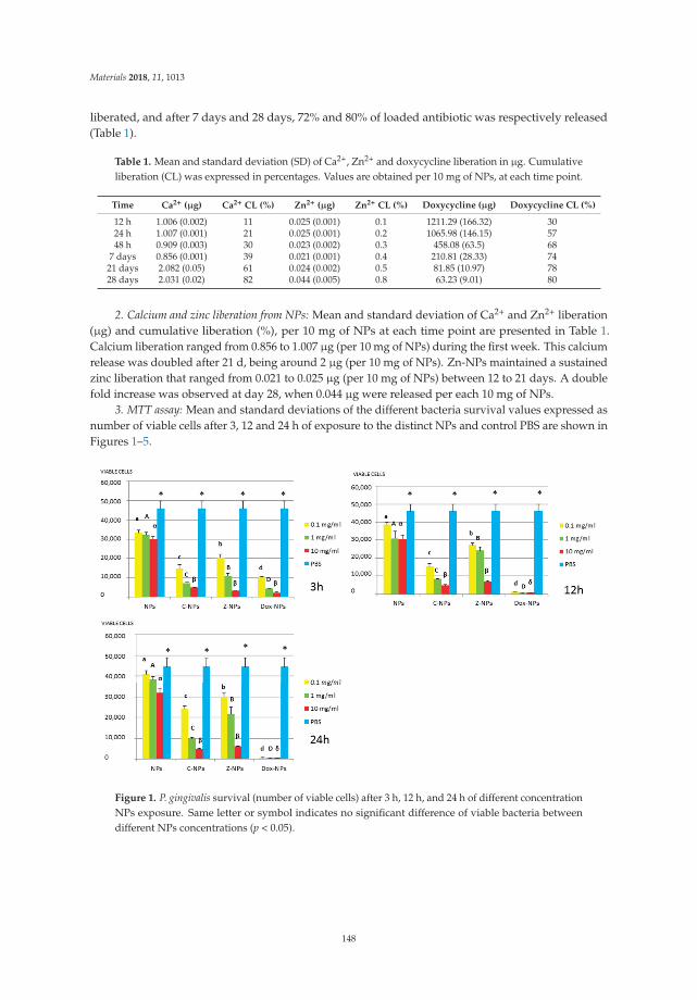
Materials 2018, 11, 1013
liberated, and after 7 days and 28 days, 72% and 80% of loaded antibiotic was respectively released(Table 1).
Table 1. Mean and standard deviation (SD) of Ca2+, Zn2+ and doxycycline liberation in μg. Cumulativeliberation (CL) was expressed in percentages. Values are obtained per 10 mg of NPs, at each time point.
Time Ca2+ (μg) Ca2+ CL (%) Zn2+ (μg) Zn2+ CL (%) Doxycycline (μg) Doxycycline CL (%)
12 h 1.006 (0.002) 11 0.025 (0.001) 0.1 1211.29 (166.32) 3024 h 1.007 (0.001) 21 0.025 (0.001) 0.2 1065.98 (146.15) 5748 h 0.909 (0.003) 30 0.023 (0.002) 0.3 458.08 (63.5) 68
7 days 0.856 (0.001) 39 0.021 (0.001) 0.4 210.81 (28.33) 7421 days 2.082 (0.05) 61 0.024 (0.002) 0.5 81.85 (10.97) 7828 days 2.031 (0.02) 82 0.044 (0.005) 0.8 63.23 (9.01) 80
2. Calcium and zinc liberation from NPs: Mean and standard deviation of Ca2+ and Zn2+ liberation(μg) and cumulative liberation (%), per 10 mg of NPs at each time point are presented in Table 1.Calcium liberation ranged from 0.856 to 1.007 μg (per 10 mg of NPs) during the first week. This calciumrelease was doubled after 21 d, being around 2 μg (per 10 mg of NPs). Zn-NPs maintained a sustainedzinc liberation that ranged from 0.021 to 0.025 μg (per 10 mg of NPs) between 12 to 21 days. A doublefold increase was observed at day 28, when 0.044 μg were released per each 10 mg of NPs.
3. MTT assay: Mean and standard deviations of the different bacteria survival values expressed asnumber of viable cells after 3, 12 and 24 h of exposure to the distinct NPs and control PBS are shown inFigures 1–5.
Figure 1. P. gingivalis survival (number of viable cells) after 3 h, 12 h, and 24 h of different concentrationNPs exposure. Same letter or symbol indicates no significant difference of viable bacteria betweendifferent NPs concentrations (p < 0.05).
148

Materials 2018, 11, 1013
Figure 2. S. mutans survival (number of viable cells) after 3 h, 12 h, and 24 h of different concentrationNPs exposure. Same letter or symbol indicates no significant difference of viable bacteria betweendifferent NPs concentrations (p < 0.05).
Figure 3. L. lactis survival (number of viable cells) after 3 h, 12 h, and 24 h of different concentrationNPs exposure. Same letter or symbol indicates no significant difference of viable bacteria betweendifferent NPs concentrations (p < 0.05).
149

Materials 2018, 11, 1013
Figure 4. S. gordonii survival (number of viable cells) after 3 h, 12 h, and 24 h of different concentrationNPs exposure. Same letter or symbol indicates no significant difference of viable bacteria betweendifferent NPs concentrations (p < 0.05).
Figure 5. S. sobrinus survival (number of viable cells) after 3 h, 12 h, and 24 h of different concentrationNPs exposure. Same letter or symbol indicates no significant difference of viable bacteria betweendifferent NPs concentrations (p < 0.05).
150

Materials 2018, 11, 1013
In general, all tested NPs affected the viability of bacterial suspension. The most effective were theDox-NPs followed by Ca-NPs or Zn-NPs and finally non-doped NPs that attained the most variableand least reduction in bacterial survival (8% to 70% after 24 h).
The viability of tested bacteria following the incubation with NPs depends on the type of NPs.The two bacteria, S. gordonii and S. sobrinus were found to be the most resistant to the tested NPs.After 24 h, they were only affected by Zn-NPs (70% reduction in bacterial viability) and by Dox-NPs(60% reduction). For P. gingivalis, S. mutans and L. lactis, Dox-NPs reduced the bacterial viability by60% to 99%, after 24 h depending on the concentration of doxycycline. Meanwhile the reduction inbacterial viability were from 20% to 60% for S. gordonii and S. sobrinus. The P. gingivalis, L. lactis andS. mutans Dox-NPs effect was not variable during the time of the study. Only in the cases of S. gordoniiand S. sobrinus cultures a drop in Dox-NPs efficacy was observed after 24 h. At 24 h, for P. gingivalisall tested concentrations of Dox-NPs attained above 98% bacterial death. In general, most effectivedosage of Dox-NPs was found to be 10 mg/mL.
When testing Ca, Zn-doped or even undoped-NPs for S. mutans and L. lactis, bacterial viabilitywas significantly affected in doses and time dependent manners. After 24 h, only those NPs contained10 mg/mL were effective. Both bacteria were equally susceptible to Zn-NPs (68% cells reduction).When considering Ca-NPs or undoped-NPs, L. lactis was more susceptible (reduction values forCa-NPs: 90%, for unloaded NPs: 70%) than S. mutans (reduction values for Ca-NPs: 60%, for unloadedNPs: 50%).
Testing of NPs doped with Ca and Zn-doped NPs at 0.1 mg/mL against P. gingivalis, bacterialviability was significantly affected and bacterial death ranges between 55% to 27%. However, at themost effective concentration −10 mg/mL, bacterial reduction ranges were from 80% to 93%, withoutsignificant differences between both ion-doped NPs. P. gingivalis incubated with unloaded NPs attainedlow but dose and time-dependent percentages of bacterial survival reduced, from 34.3% to 7.2%.
3. Discussion
There are several in vitro testing models for the efficacy of antibacterial agents, which may involvesingle or multispecies bacteria. This microcosm model is the most clinically relevant, but attainedresults are often difficult to interpret as there is no way to control for the behavior of individual bacterialspecies. It is also difficult to decide which species are appropriate in each experiment and their relativeamounts [3]. Using biofilm models is also challenging as the results may also be different on variousmaterials surfaces with different chemistry and/or topography [30]. Therefore, when analyzing novelantibacterial agents planktonic monoculture tests are necessary to facilitate results interpretation.Further studies need to be conducted on clinical isolates and multi-species biofilms on differentmaterial surfaces or interfaces, which may express resistance trait against tested antibacterial effect.
P. gingivalis was selected for the present study as it is one of the most frequently detectedanaerobic microorganisms in subgingival plaque samples from periodontal-endodontic combinedlesions and necrotic pulp [6]. S. mutans, S. gordonii, S. sobrinus and L. lactis were used as are themost frequently detected microorganisms in cariogenic plaque [3]. P. gingivalis is a Gram-negativebacteria, S. mutans, S. gordonii, S. sobrinus and L. lactis are Gram positive. P. gingivalis has an asymmetricdistribution of lipids at their cell walls, the outer face contains lipopolysaccharide (LPS), and theinner face has phospholipids [8]. S. gordonii, S. sobrinus S. mutans and L. lactis also have LPS attheir membranes, which exhibits anionic charge, as a result it may facilitate cationic groups to bondand exert antimicrobial activity [8]. This may be a reason for observing low antibacterial activity oftested non-loaded NPs, as they are also anionic (potential zeta is −41 ± 5 mV measured in water atpH = 7) [20], and will not be attracted to tested bacteria which posse a zeta potential of approximately−25 mV at pH = 7 [31].
Ca-NPs and Zn-NPs exerted antibacterial activity, at 10 mg/mL 80% to 93% bacterial reductionafter 48 h, was encountered (Figure 1) as a possible result of liberated calcium and zinc fromNPs. After 48 h, 0.9 and 0.02 μg per 10 mg of NPs of calcium and zinc are respectively released
151

Materials 2018, 11, 1013
(Table 1). Cationic metals as calcium or zinc have been shown to be potent antimicrobials [24,25,27].Calcium release from NPs was estimated to be 0.08 and 0.1 μg/mL (per mg of NPs) from 12 hup to 7 days, while zinc release was around 0.02 μg/mL (per mg of NPs) at the same time-points.Cummulative liberation of both ions is 30% for calcium and 0.3% for zinc after 48 h. It has been shownthat lipopolysaccharides at the outer membrane of Gram-negative bacteria possess magnesium andcalcium ions that bridge to negatively-charged phosphor-sugars [8]. Therefore, cationic elementsmay also displace these metal ions damaging the outer membrane, leading to cell death [8,26]. It hasalso been previously shown that zinc ions markedly enhanced the adhesion and accumulation ofsalivary and serum proteins on cells of P. gingivalis and inhibited their coaggregation when growingon biofilms [27].
Zinc has a known inhibitory effect on glycolysis and proteinase activity in many oralbacteria [27]. Zinc may affect S. mutans viability by inhibiting glycolysis [32]. Kinetic studies ofthe glucosyltransferases of S. sobrinus by Devulapalle and Mooser [33] showed that the Zn ion acts as areversible, competitive inhibitor at the fructose subsite within the active site of the glucosyltransferase.This observation may well explain the reported dose-dependent effects of zinc on the tested bacteria.Even when the exact antibacterial mechanism of zinc has not been clearly identified yet, covalently oroxidatively induced damage has been claimed [32]. Zinc ions are considered useful for limiting thesettlement/colonization of P. gingivalis in the gingival sulcus with the goal of preventing periodontaldisease [27] and in the case of S. mutans preventing carious disease [32]. Zinc has long been knownas a plaque-inhibiting compound and also can influence acid production by different microbes [32].In addition, zinc is able to depolarize the membrane potential, it does not always cause the bacterialcell membrane to rupture and leak, but alters permeability that is closely related to the sensitivityof bacteria to ionic environment [30]. Ion homeostasis affects the proliferation, communication andmetabolism of bacteria; then, zinc may sometimes produce an inhibitive instead of destructive effectagainst bacteria [30].
Dox-NPs exerted the highest antibacterial activity to all the tested concentrations (80% to 97%bacterial reduction after 24 h). Following our results, doxycycline was found to be released at sustainedlevels for over 28 days, with a significant burst effect at 24 h. It is liberated at concentrations highabove to those considered effective against bacteria at any time point of the present study. For each mgof NPs 121, 106 and 46 μg/mL of doxycycline will be liberated at 12, 24 and 48 h, respectively. A bursteffect with rapid doxycycline liberation was observed from 12 h until the first week of storage. After7 days time-point, antibiotic release was maintained above 20 μg/mL (per mg of NPs). As bacterialsusceptibility to doxycycline is obtained around 0.1 to 0.2 μg/mL [28], doxycycline is then liberatedfrom NPs at concentrations high above to those considered effective against most of the tested bacteria.It was shown that doxycycline at a concentration between 0.5 and 1 μg/mL is bactericidal againstdifferent Pg strains [34], and between 0.1 and 6.0 μg/mL is effective against Pg and other putativeperiodontal pathogens [35,36]. It should be stressed that tested Dox-NPs after 28 days are able toliberate doxycycline concentrations above 6 μg/mL.
Doxycycline is a polar and amphoteric compound. Doxycycline as a salt (hyclate) is watersoluble. Doxycycline is known to act against most bacteria by inhibiting the microbial proteinsynthesis that requires access into the cell wall and lipid solubility [37]. Doxycycline binds theribosome to prevent ribonucleic acid synthesis by avoiding addition of more amino acid to thepolypeptide [37]. Doxycycline is also known to provoke a potent and long-lasting inhibition of dentinmatrix metalloproteinases [29] that are related to chronic inflammation processes and abscessesat apical level [10]. It may explain how long-term administration of a sub-antimicrobial doseof doxycycline, to dogs with periodontitis, is regarded as an effective treatment for periodontalinflammation, even when it does not induce antimicrobial effects [38]. It is also important to notethat MMPs inhibition may also prevent collagen degradation at the resin bonded dentin interface [15].It will also probably reduce secondary caries formation, as MMPs activity is augmented at cariesaffected dentin [39].
152

Materials 2018, 11, 1013
The reported doxycycline liberation data are high and sustained, if compared to the releaseprofile of other previously proposed compounds as a cellulose-acetate-loaded doxycycline formulationstudied by Tonetti et al. [40]. Kim et al. [28] introduced a biodegradable doxycycline gel and reporteda mean local concentration of 20 mg/mL, after 15 min; values that were lowered to 577 μg/mLafter 3 days and to 16 μg/mL after 12 days. Deasy et al. [41] used tetracycline hydrochloride inpoly(hydroxybutyric acid) as a biodegradable polymer matrix and showed sustained release just over4 to 5 days, with a significant burst effect at 24 h. Previously introduced materials are then able toliberate doxycycline at higher concentrations, but in shorter periods of time, denoting accentuatedburst effects.
In general, tested NPs had little effect on the growth of (S. sobrinus and S. gordonii) and speciallyDox-NPs after 24 h, which greatly affected P. gingivalis, S. mutans and L. lactis survival rates (at leastat the evaluated time points and concentrations). Recent results on advanced caries lesions in younghuman teeth, using bacterial sequence analysis methods are consistent and indicate that S. gordoniidiminishes greatly in caries-associated plaque biofilm, while S. mutans persists [42]. It means thatNPs may selectively inhibit cariogenic and periodontal bacteria, while leaving commensal microbes.However, it should be assayed in properly designed multibacteria biofilms models in future studies.It is to be noted that the tested NPs are biocompatible against human fibroblasts [23], and theapplication of antibacterials is crucial if regenerative/revascularization processes are performedfor the endodontic treatment [43], these NPs may be an interesting tool.
Two important limitations are recognized for the present Dox-NPs: (1) antibiotics may producebacterial strain resistance, which is a current global concern; therefore, further research is needed.(2) The bacteria grow in biofilms, and are known to be more resistant to antimicrobial treatmentthan the planktonic cultures used for the present antimicrobial susceptibility testing [44]. Then, it isimperative to include the biofilm mode of growth of bacteria when testing treatments for bondeddentin interfaces, endodontic and periapical diseases. But these tests are difficult to control, in terms ofknowing specifically how bacteria are involved in the process [3]. It will not be possible to ascertain ifa specific toxicity of NPs against individual bacteria is being produced [44], or just a biofilm disruptioninterfering with first colonizers bacteria attachment to dentin.
It may be concluded after this in vitro study that experimental NPs loaded with zinc, calciumor doxycycline are effective to eradicate tested oral bacteria. For clinical applications, using theseNPs at the resin-bonded interface as cavity liners may be recommended. As it was shown before,that NPs do not affect bond efficacy and improve dentin remineralization [17,21,22]. The samebeneficial effect may be found if NPs are used in endodontics, before resin sealant cement application.However, as recognized in the study limitations, further investigations into antibacterial effects throughbiofilm models of multiple bacterial species should be implemented.
4. Material and Methods
1. Preparation of Nanoparticles (NPs): PolymP-n Active NPs were acquired (NanoMyP, Granada,Spain). Particles are fabricated trough polymerization precipitation. Main components of NPs are2-hydroxyethyl methacrylate, ethylene glycol dimethacrylate and methacrylic acid; these compoundsare the backbone monomer, the cross-linker and the functional monomer respectively.
Calcium-loaded NPs (Ca-NPs) and Zinc-loaded NPs (Zn-NPs) were produced. Zinc and calciumcomplexation was obtained immersing 30 mg of NPs during 3 days, under continuous shaking inaqueous solutions of ZnCl2 or CaCl2, at room temperature. Fifteen mL of the solutions containingzinc or calcium at 40 ppm were employed (pH 6.5). Then, the adsorption equilibrium of metal ionswas reached [23]. To separate the NPs from the supernatant, the suspensions were centrifuged.0.96 ± 0.04 μg Ca/mg NPs and 2.15 ± 0.05 μg Zn/mg NPs were the attained ion complexationvalues [23]. NPs loaded with doxycycline hyclate were also produced. An 18-mL aqueous solution,with 40 mg/mL of doxycycline hyclate (Sigma-Aldrich, Darmstadt, Germany) was prepared, and 30 mgof NPs were immersed in the solution for 4 h, under continuous shaking. Then, to separate NPs from
153

Materials 2018, 11, 1013
the supernatant the suspensions were centrifuged. Following groups were tested: (1) NPs (NPs),(2) NPs loaded with Ca (Ca-NPs), (3) NPs loaded with Zn (Zn-NPs), and (4) NPs with immobilizeddoxycycline hyclate (Dox-NPs).
2. Loading efficacy and release of doxycycline from NPs: For loading efficacy 18 mL of 40 mg/mLaqueous solution of doxycycline hyclate was prepared and the amount of doxycycline in the initialaqueous solution was assessed in triplicate samples of 100 μL and recorded as initial doxycyclineconcentration (1333 μg Dox/mL). Three different samples containing 1 mg of NPs and 0.6 mL ofthe doxycycline solution were incubated for 4 h, under continuous shaking. Then, the suspensionswere centrifuged and the particles were separated from the supernatant, 100 μL of each supernatantwas analyzed for final doxycycline concentration. Final doxycycline concentration was subtractedfrom initial values to calculate loading efficacy [45]. To ascertain for doxycycline liberation, 30 mgof doxycycline loaded-NPs were suspended in 3 mL of phosphate buffer saline (PBS, pH 7.4, FisherScientific SL, Madrid, Spain), three different eppendorf tubes containing 1 mL of the Dox-NPssuspension were stored at 37 ◦C. After 12 h, suspensions were centrifuged and the particles wereseparated from the supernatant. An aliquot (0.1 mL) of each supernatant was analyzed for doxycyclineconcentration. NPs were washed and 1 mL of fresh PBS was used to resuspend the NPs at 10 mg/mLuntil the next supernatant collection. Seven different time-points were tested: 12, 24, 48 h, 7, 14, 21and 28 days. Supernatans were stored at −20 ◦C until doxycycline concentration measuring [45].The amount of doxycycline was assayed by high performance liquid chromatography (HPLC) (WatersAlliance 2690, Waters Corporation, Milford, MA, USA) equipped with a UV-Vis detector. A binarymobile phase consisting of solvent systems A and B was used in an isocratic elution with 80:20 A:B.Mobile phase A was 50 mM KHPO4 in distilled H20 and mobile phase B was 100% acetonitrile.The HPLC flow rate was 1.0 mL/min and the total run time was 10 min. The retention time was4.85 min. The concentration of doxycycline was calculated based on a standard curve of known levelsof doxycycline at 273 nm [45].
3. Calcium and zinc liberation from NPs: 150 mg of zinc and 150 mg of calcium loaded-NPs weresuspended in 15 mL of deionized water. Three different eppendorf tubes containing 5 mL of theCa-NPs suspensions and other 3 with Zn-NPs were stored at 37 ◦C. After 12 h, suspensions werecentrifuged and the particles separated from the supernatant; 5 mL of each supernatant was analyzedfor calcium and zinc concentration. NPs were washed and 5 mL of fresh deionized water was usedto resuspend the NPs at 10 mg/mL until the next supernatant collection. Seven different time pointswere tested: 12 h, 24 h, 48 h, 7 d, 14 d, 21 d and 28 d. Supernatans were stored at −20 ◦C until testing.Calcium and zinc concentrations were analyzed through an inductively coupled plasma (ICP) opticalemission spectrometer (ICP-OES Optima 8300, Perkin-Elmer, MA, USA) [23].
4. Bacteria: P. gingivalis 33,277, S. mutans 700,610, S. sobrinus 33,478, S. gordonii 10,558 and L. lactis12,315 were obtained from ATCC (Bethesda, MD, USA). The anaerobic organism, Pg was grownin Tryptic Soy broth (TSB) supplemented with yeast extract (5 g/L), Hemin (5 mg/L), Menadione(1 mg/L), for 72 h. Strict anaerobic conditions were employed, Thermo Scientific Oxoid AnaeroGen(Thermo Fisher Scientific, Waltham, MA, USA) was used in an anaerobic jar, which provides 7–15%CO2 and <0.1% O2. The remaining test bacteria were grown in TSB for 24 h at 37 ◦C. The bacterial cellswere harvested by centrifugation and re-suspended in the same growth media. The number of bacteriaper mL was determined by measuring the optical density at 600 nm and adjusting it to a standardbacterial suspension of 1 × 10⁷ CFU/mL [46].
5. MTT assay: The NPs were suspended in PBS at three different concentrations (10 mg/mL,1 mg/mL and 0.1 mg/mL). NPs were placed into Eppendorf tubes with bacterial broths(1 × 10⁷ CFU/mL for each 0.45 mL of NPs suspensions) and incubated for 3, 12 and 24 h at37 ◦C. Sterile pipetting was used throughout the study. Susceptibility testing of P. gingivaliswas conducted in an anaerobic jar as described above. At the end of each incubation period,the effect of the NPs on bacteria was evaluated by the ability of viable bacteria to cleave thetetrazolium salt (3-[4,5-dimethylthiazol-2-yl]-2,5-diphenyl tetrazolium bromide) (MTT) to a formazan
154

Materials 2018, 11, 1013
dye (Sigma-Aldrich, Darmstadt, Germany). 96-well flat-bottom microtiter plates were used to placethe suspensions in. The plates were incubated for 4 h at 37 ◦C, after the MTT labeling agent additionto each culture well. Then, the solubilizing agent that was provided by the manufacturer was addedand an overnight incubation at room temperature was performed. An enzyme-linked immunosorbentassay (ELISA) reader (Spectrostar Nano, BMG Labtech, Cary, NC, USA) was employed, the purpleformazan color that was produced from the MTT by viable cells, was read (560 nm) [46]. Assays wereperformed with three determinants, and experiments were performed in triplicate. Data expressed asmean ± standard deviation were analyzed by one-way analysis of variance (ANOVA) and the posthoc comparisons Scheffe’s F tests, at p < 0.05, using SPSS Statistic 20.
Author Contributions: Conceptualization, M.T.-O., J.B., F.G., M.T. and R.O.; Methodology, M.T.-O., J.B., F.G.,A.M., M.T. and R.O.; Software, M.T.-O., J.B.; Formal Analysis, M.T.-O., J.B., F.G., A.M., M.T. and R.O.; Investigation,M.T.-O., J.B., F.G., A.M., M.T. and R.O.; Resources, M.T.-O., J.B., A.M.; Data Curation, M.T.-O., J.B., F.G., A.M.,M.T. and R.O.; Writing—Original Draft Preparation, M.T.-O., J.B., F.G., A.M., M.T. and R.O.; Writing—Review &Editing, M.T.-O., J.B., F.G., M.T. and R.O.; Supervision, J.B., F.G., M.T. and R.O; Project Administration, R.O. andM.T.; Funding Acquisition, M.T. and R.O.
Funding: This research was funded by the Spanish Ministry of Economy and Competitiveness (MINECO) andEuropean Regional Development Fund (FEDER) grant number MAT2017-85999-P.
Acknowledgments: The research project MAT2017-85999-P was funded by the Spanish Ministry of Economy andCompetitiveness (MINECO) and European Regional Development Fund (FEDER). No funds were received tocover publication costs.
Conflicts of Interest: The authors declare no conflicts of interest. The founding sponsors had no role in the designof the study; in the collection, analyses, or interpretation of data; in the writing of the manuscript, and in thedecision to publish the results.
References
1. Chaves, P.; Oliveira, J.; Haas, A.; Beck, R.C.R. Applications of polymeric nanoparticles in oral diseases:A review of recent findings. Curr. Pharm. Des. 2018. [CrossRef] [PubMed]
2. Spencer, P.; Ye, Q.; Park, J.; Topp, E.M.; Misra, A.; Marangos, O.; Wang, Y.; Bohaty, B.S.; Singh, V.; Sene, F.; et al.Adhesive/Dentin Interface: The Weak Link in the Composite Restoration. Ann. Biomed. Eng. 2010,38, 1989–2003. [CrossRef] [PubMed]
3. Ferracane, J.L. Models of Caries Formation around Dental Composite Restorations. J. Dent. Res. 2017,96, 364–371. [CrossRef] [PubMed]
4. Hirose, N.; Kitagawa, R.; Kitagawa, H.; Maezono, H.; Mine, A.; Hayashi, M.; Haapasalo, M.; Imazato, S.Development of a Cavity Disinfectant Containing Antibacterial Monomer MDPB. J. Dent. Res. 2016,95, 1487–1493. [CrossRef] [PubMed]
5. Albuquerque, M.T.P.; Evans, J.D.; Gregory, R.L.; Valera, M.C.; Bottino, M.C. Antibacterial TAP-mimicElectrospun Polymer Scaffold—Effects on P. gingivalis-Infected Dentin Biofilm. Clin. Oral Investig. 2016,20, 387–393. [CrossRef] [PubMed]
6. Kim, R.J.; Kim, M.O.; Lee, K.S.; Lee, D.Y.; Shin, J.H. An in vitro evaluation of the antibacterial propertiesof three mineral trioxide aggregate (MTA) against five oral bacteria. Arch. Oral Biol. 2015, 60, 1497–1502.[CrossRef] [PubMed]
7. Rôças, I.N.; Siqueira, J.F.; Santos, K.R.; Coelho, A.M. “Red complex” (Bacteroides forsythus, Porphyromonasgingivalis, and Treponema denticola) in endodontic infections: A molecular approach. Oral Surg. Oral Med.Oral Pathol. Oral Radiol. Endod. 2001, 91, 468–471. [CrossRef] [PubMed]
8. Wang, L.; Xie, X.; Weir, M.D.; Fouad, A.F.; Zhao, L.; Xu, H.H. Effect of bioactive dental adhesive onperiodontal and endodontic pathogens. J. Mater. Sci. Mater. Med. 2016, 27, 168. [CrossRef] [PubMed]
9. Gu, L.; Huang, X.; Griffin, B.; Bergeron, B.R.; Pashley, D.H.; Niu, L.; Tay, F.R. Primum non nocere—The effectsof sodium hypochlorite on dentin as used in endodontics. Acta Biomater. 2017, 61, 144–156. [CrossRef][PubMed]
10. Letra, A.; Ghaneh, G.; Zhao, M. MMP-7 and TIMP-1, New Targets in Predicting Poor Wound Healing inApical Periodontitis. J. Endod. 2013, 39, 1141–1146. [CrossRef] [PubMed]
155

Materials 2018, 11, 1013
11. Menezes-Silva, R.; Khaliq, S.; Deeley, K.; Letra, A.; Vieira, A.R. Genetic Susceptibility to Periapical Disease:Conditional Contribution of MMP2 and MMP3 Genes to the Development of Periapical Lesions and HealingResponse. J. Endod. 2012, 38, 604–607. [CrossRef] [PubMed]
12. Paula-Silva, G.F.W.; Bezerra da Silva, L.A.; Kapila, L.Y. Matrix Metalloproteinase Expression in Teeth withApical Periodontitis Is Differentially Modulated by the Modality of Root Canal Treatment. J. Endod. 2010,36, 231–237. [CrossRef] [PubMed]
13. Ahmed, G.M.; El-Baz, A.A.; Hashem, A.A.; Shalaan, A.K. Expression levels of matrix metalloproteinase-9and gram-negative bacteria in symptomatic and asymptomatic periapical lesions. J. Endod. 2013, 39, 444–448.[CrossRef] [PubMed]
14. Accorsi-Mendonca, T.; Silva, E.J.; Marcaccini, A.M.; Gerlach, R.F.; Duarte, K.M.; Pardo, A.P.; Line, S.R.;Zaia, A.A. Evaluation of gelatinases, tissue inhibitor of matrix metalloproteinase-2, and myeloperoxidaseprotein in healthy and inflamed human dental pulp tissue. J. Endod. 2013, 39, 879–882. [CrossRef] [PubMed]
15. Osorio, R.; Yamauti, M.; Osorio, E.; Ruiz-Requena, M.E.; Pashley, D.; Tay, F.R.; Toledano, M. Effect of dentinetching and chlorhexidine application on metalloproteinase-mediated collagen degradation. Eur. J. Oral Sci.2011, 119, 79–85. [CrossRef] [PubMed]
16. Liu, Y.; Mai, S.; Li, N. Differences between top-down and bottom-up approaches in mineralizing thick,partially-demineralized collagen scaffolds. Acta Biomater. 2011, 7, 1742–1751. [CrossRef] [PubMed]
17. Toledano, M.; Osorio, R.; Osorio, E.; Medina-Castillo, A.L.; Toledano-Osorio, M.; Aguilera, F.S. Ions-modifiednanoparticles affect functional remineralization and energy dissipation through the resin-dentin interface.J. Mech. Behav. Biomed. Mater. 2017, 68, 62–79. [CrossRef] [PubMed]
18. Zhu, J.; Liang, R.; Sun, C.; Xie, L.; Wang, J.; Leng, D.; Wu, D.; Liu, W. Effects of nanosilver and nanozincincorporated mesoporous calcium-silicate nanoparticles on the mechanical properties of dentin. PLoS ONE2017, 12, e0182583. [CrossRef] [PubMed]
19. Zhang, K.; Cheng, L.; Imazato, S.; Antonucci, J.M.; Lin, N.J.; Lin-Gibson, S.; Bai, Y.; Xu, H.H.K. Effects ofdual antibacterial agents MDPB and nano-silver in primer on microcosm biofilm, cytotoxicity and dentinbond properties. J. Dent. 2013, 41, 464–474. [CrossRef] [PubMed]
20. Hashimoto, M.; Sasaki, J.I.; Yamaguchi, S.; Kawai, K.; Kawakami, H.; Iwasaki, Y.; Imazato, S. GoldNanoparticles Inhibit Matrix Metalloproteases without Cytotoxicity. J. Dent. Res. 2015, 94, 1085–1091.[CrossRef] [PubMed]
21. Osorio, R.; Cabello, I.; Medina-Castillo, A.L.; Osorio, E.; Toledano, M. Zinc-modified nanopolymers improvethe quality of resin-dentin bonded interfaces. Clin. Oral Investig. 2016, 20, 2411–2420. [CrossRef] [PubMed]
22. Osorio, R.; Osorio, E.; Medina-Castillo, A.L.; Toledano, M. Polymer nanocarriers for dentin adhesion.J. Dent. Res. 2014, 93, 1258–1263. [CrossRef] [PubMed]
23. Osorio, R.; Alfonso-Rodríguez, C.A.; Medina-Castillo, A.L.; Alaminos, M.; Toledano, M. Bioactive PolymericNanoparticles for Periodontal Therapy. PLoS ONE 2016, 7, e0166217. [CrossRef] [PubMed]
24. Munchow, E.A.; Albuquerque, M.T.; Zero, B.; Kamocki, K.; Piva, E.; Gregory, R.L.; Bottino, M.C. Developmentand characterization of novel ZnO-loaded electrospun membranes for periodontal regeneration. Dent. Mater.2015, 31, 1038–1051. [CrossRef] [PubMed]
25. Munchow, E.A.; Pankajakshan, D.; Albuquerque, M.T.; Kamocki, K.; Piva, E.; Gregory, R.L.; Bottino, M.C.Synthesis and characterization of CaO-loaded electrospun matrices for bone tissue engineering.Clin. Oral Investig. 2016, 27, 1921–1933. [CrossRef] [PubMed]
26. Vargas-Reus, M.A.; Memarzadeh, K.; Huang, J.; Ren, G.G.; Allaker, R.P. Antimicrobial activity ofnanoparticulate metal oxides against peri-implantitis pathogens. Int. J. Antimicrob. Agents 2012, 40, 135–139.[CrossRef] [PubMed]
27. Tamura, M.; Ochiai, K. Zinc and copper play a role in coaggregation inhibiting action of Porphyromonasgingivalis. Mol. Oral Microbiol. 2009, 24, 56–63. [CrossRef]
28. Kim, T.S.; Bürklin, T.; Schacher, B.; Ratka-Krüger, P.; Schaecken, M.T.; Renggli, H.H.; Fiehn, W.; Eickholz, P.Pharmacokinetic profile of a locally administered doxycycline gel in crevicular fluid, blood, and saliva.J. Periodontol. 2002, 73, 1285–1291. [CrossRef] [PubMed]
29. Osorio, R.; Yamauti, M.; Osorio, E.; Ruiz-Requena, M.E.; Pashley, D.H.; Tay, F.R.; Toledano, M. Zinc reducescollagen degradation in demineralized human dentin explants. J. Dent. 2011, 39, 148–153. [CrossRef][PubMed]
156

Materials 2018, 11, 1013
30. Fan, W.; Sun, Q.; Li, Y.; Tay, F.R.; Fan, B. Synergistic mechanism of Ag+–Zn2+ in anti-bacterial activity againstEnterococcus faecalis and its application against dentin infection. J. Nanobiotechnol. 2018, 16, 10. [CrossRef][PubMed]
31. Cowan, M.M.; Van der Mei, H.C.; Stokroos, I.; Busscher, H.J. Heterogeneity of surfaces of subgingivalbacteria as detected by zeta potential measurements. J. Dent. Res. 1992, 71, 1803–1806. [CrossRef] [PubMed]
32. Wunder, D.; Bowen, W.H. Action of agents on glucosyltransferases from Streptococcus mutans in solutionand adsorbed to experimental pellicle. Arch. Oral Biol. 1999, 44, 203–214. [CrossRef]
33. Devulapalle, K.S.; Mooser, G. Subsite specificity of the active site of glucosyltransferases from Streptococcussobrinus. J. Biol. Chem. 1994, 269, 11967–11971. [PubMed]
34. Larsen, T. Susceptibility of Porphyromonas gingivalis in biofilms to amoxicillin, doxycycline and metronidazole.Oral Microbiol. Immunol. 2002, 17, 267–271. [CrossRef] [PubMed]
35. Slots, J.; Rams, T.E. Antibiotics in periodontal therapy: Advantages and disadvantages. J. Clin. Periodontol.1990, 17, 479–493. [CrossRef] [PubMed]
36. Larsen, T. In vitro release of doxycycline from bioabsorbable materials and acrylic strips. J. Periodontol. 1990,61, 30–34. [CrossRef] [PubMed]
37. Gamal, A.Y.; Kumper, R.M.A. Novel Approach to the Use of Doxycycline-Loaded Biodegradable Membraneand EDTA Root Surface Etching in Chronic Periodontitis: A Randomized Clinical Trial. J. Periodontol. 2012,83, 1086–1094. [CrossRef] [PubMed]
38. Kim, S.E.; Hwang, S.Y.; Jeong, M. Clinical and microbiological effects of a subantimicrobial dose of oraldoxycycline on periodontitis in dogs. Vet. J. 2016, 208, 55–59. [CrossRef] [PubMed]
39. Toledano, M.; Nieto-Aguilar, R.; Osorio, R.; Campos, A.; Osorio, E.; Tay, F.R.; Alaminos, M. Differentialexpression of matrix metalloproteinase-2 in human coronal and radicular sound and carious dentine. J. Dent.2010, 38, 635–640. [CrossRef] [PubMed]
40. Tonetti, M.S.; Lang, N.P.; Cortellini, P.; Suvan, J.E.; Eickholz, P.; Fourmousis, I.; Topoll, H.; Vangsted, T.;Wallkamm, B. Effects of a single topical doxycycline administration adjunctive to mechanical debridement inpatients with persistent/recurrent periodontitis but acceptable oral hygiene during supportive periodontaltherapy. J. Clin. Periodontol. 2012, 39, 475–482. [CrossRef] [PubMed]
41. Deasy, P.B.; Collins, A.E.; MacCarthy, D.J.; Russell, R.J. Use of strips containing tetracycline hydrochloride ormetronidazole for the treatment of advanced periodontal disease. J. Pharm. Pharmacol. 1989, 41, 694–699.[CrossRef] [PubMed]
42. Gross, E.L.; Leys, E.J.; Gasparovich, S.R.; Firestone, N.D.; Schwartzbaum, J.A.; Janies, D.A.; Asnani, K.;Griffen, A.L. Bacterial 16S sequence analysis of severe caries in young permanent teeth. J. Clin. Microbiol.2010, 48, 4121–4128. [CrossRef] [PubMed]
43. Yassen, G.H.; Vail, M.M.; Chu, T.G.; Platt, J.A. The effect of medicaments used in endodontic regeneration onroot fracture and microhardness of radicular dentine. Int. Endod. J. 2013, 46, 688–695. [CrossRef] [PubMed]
44. Garrett, T.R.; Bhakoo, M.; Zhang, Z.B. Bacterial adhesion and biofilms on surfaces. Prog. Nat. Sci. 2008,18, 1049–1056. [CrossRef]
45. Palasuk, J.; Windsor, L.J.; Platt, J.A.; Lvov, Y.; Geraldeli, S.; Bottino, M.C. Doxycycline-loadednanotube-modified adhesives inhibit MMP in a dose-dependent fashion. Clin. Oral Investig. 2018,22, 1243–1252. [CrossRef] [PubMed]
46. Banzi, E.C.; Costa, A.R.; Puppin-Rontani, R.M.; Babu, J.P.; García-Godoy, F. Inhibitory effects of a curedantibacterial bonding system on viability and metabolic activity of oral bacteria. Dent. Mater. 2014,30, e238–e244. [CrossRef] [PubMed]
© 2018 by the authors. Licensee MDPI, Basel, Switzerland. This article is an open accessarticle distributed under the terms and conditions of the Creative Commons Attribution(CC BY) license (http://creativecommons.org/licenses/by/4.0/).
157

materials
Article
Dental Resin Cements—The Influence of WaterSorption on Contraction Stress Changes andHydroscopic Expansion
Grzegorz Sokolowski 1, Agata Szczesio 2, Kinga Bociong 2, Karolina Kaluzinska 2,
Barbara Lapinska 3, Jerzy Sokolowski 3, Monika Domarecka 3 and
Monika Lukomska-Szymanska 3,*
1 Department of Prosthetic Dentistry, Medical University of Lodz, 251 Pomorska St., 92-213 Lodz, Poland;[email protected]
2 University Laboratory of Materials Research, Medical University of Lodz, 251 Pomorska St., 92-213 Lodz,Poland; [email protected] (A.S.); [email protected] (K.B.);[email protected] (K.K.)
3 Department of General Dentistry, Medical University of Lodz, 251 Pomorska St., 92-213 Lodz, Poland;[email protected] (B.L.); [email protected] (J.S.);[email protected] (M.D.)
* Correspondence: [email protected]; Tel.: +48-42-675-74-64
Received: 14 May 2018; Accepted: 6 June 2018; Published: 8 June 2018
Abstract: Resin matrix dental materials undergo contraction and expansion changes due topolymerization and water absorption. Both phenomena deform resin-dentin bonding and influencethe stress state in restored tooth structure in two opposite directions. The study tested three compositeresin cements (Cement-It, NX3, Variolink Esthetic DC), three adhesive resin cements (Estecem,Multilink Automix, Panavia 2.0), and seven self-adhesive resin cements (Breeze, Calibra Universal,MaxCem Elite Chroma, Panavia SA Cement Plus, RelyX U200, SmartCem 2, and SpeedCEM Plus).The stress generated at the restoration-tooth interface during water immersion was evaluated. Theshrinkage stress was measured immediately after curing and after 0.5 h, 24 h, 72 h, 96 h, 168 h, 240 h,336 h, 504 h, 672 h, and 1344 h by means of photoelastic study. Water sorption and solubility werealso studied. All tested materials during polymerization generated shrinkage stress ranging from4.8 MPa up to 15.1 MPa. The decrease in shrinkage strain (not less than 57%) was observed afterwater storage (56 days). Self-adhesive cements, i.e., MaxCem Elite Chroma, SpeedCem Plus, PanaviaSA Plus, and Breeze exhibited high values of water expansion stress (from 0 up to almost 7 MPa).Among other tested materials only composite resin cement Cement It and adhesive resin cementPanavia 2.0 showed water expansion stress (1.6 and 4.8, respectively). The changes in stress value(decrease in contraction stress or built up of hydroscopic expansion) in time were material-dependent.
Keywords: resin cements; shrinkage stress; water sorption; hydroscopic expansion;photoelastic investigation
1. Introduction
Resin composite cements have been widely used with ceramic, resin, or metal alloy-basedprosthodontic restorations [1]. The cementation technique used in adhesive dentistry is one of the majorfactors, which exerts influence on the clinical success of indirect restorative procedures. Cement isused to bond tooth and restoration simultaneously creating a barrier against microbial leakage [2]. Theuniversal cement that can be applied in all indirect restorative procedures has not been introduced intothe market yet. Therefore, clinicians should understand the influence of applied material propertiesand preparation design on the clinical performance of the restoration [3].
Materials 2018, 11, 973; doi:10.3390/ma11060973 www.mdpi.com/journal/materials158

Materials 2018, 11, 973
The composition of resin composite cements is almost the same as of resin composites [4]. Resincements mainly consist of various methacrylate resins and inorganic fillers which are often coatedwith organic silanes to provide adhesion between the filler and the matrix. These materials ofteninclude bonding agents to promote the adhesion between resin cement and tooth structure. Mainmonomers are, i.e., hydroxyethyl methacrylate (HEMA), 4-methacryloyloxyethy trimellitate anhydride(4-MET), carboxylic acid, and organophospate 10-methacryloxydecyl dihydrogen phosphate (10-MDP)(Figure 1). The acidic group bonds calcium ions in the tooth structure [5,6].
Figure 1. Monomers used as bonding agents in resin cements.
Resin composite cements are used in combination with adhesive systems. This procedureaims at creating micro-mechanical retention to both enamel and dentin. The material may alsoform a strong adhesion to an adequately-treated surface of the composite, ceramic, and metallicrestorations [7]. Taking into account the surface preparation before the cementation process, resincements can be divided into: (1) composite resin cement (used with total-etch adhesive systems);(2) adhesive resin cement (used with separate self-etching adhesive systems); and (3) self-adhesiveresin cement (containing a self-adhesive system) [1].
The application of resin matrix-based cements is time-consuming and susceptible to manipulationerrors [8]. The self-adhesive resin cements are proposed to simplify the restoration procedure. Thesematerials bond dentin in one step without any surface conditioning or pre-treatment (priming) [9,10].
All currently available resin-based materials exhibit polymerization shrinkage. Moreover, resincements are generally applied as a thin layer, particularly when used to lute posts, inlays, and crowns.In the aforementioned clinical cases, the cavity design has a high C-factor (high number of bondedsurfaces and a low number of un-bonded surfaces) [11]. Additionally, low-viscosity compositesexhibit a relatively high shrinkage amounting up to 6% (comparable to resin cements) [12,13]. Thesefactors may generate sufficient stress resulting in debonding of the luting material, thereby increasingmicroleakage [14]. Nevertheless, there is little data on the stress generated by these materials. Thesorption characteristic of resin-based dental cements has been extensively evaluated [15–17]. However,the analysis of the influence of water sorption on the change in contraction stress is inadequate. Thepurpose of this study was to evaluate the development of the stress state, i.e., the contraction stressgenerated during photopolymerization and hydroscopic expansion within different types of resincements which undergo water ageing by means of photoelastic analysis.
2. Materials and Methods
The composition of investigated resin cements and bonding systems is presented in Tables 1and 2.
159

Materials 2018, 11, 973
Ta
ble
1.
The
com
posi
tion
ofre
sin
cem
ents
.
Mate
rial
Ty
pe
Co
mp
osi
tio
nC
uri
ng
Tim
e(s
)M
an
ufa
ctu
rer
Cem
ent-
ItC
ompo
site
resi
nce
men
tbi
s-G
MA
,UD
MA
,HD
DM
A,P
EGD
MA
,bar
ium
-bor
o-si
licat
egl
ass
(65
wt%
)20
Jene
ric
Pent
ron
(Wal
lingf
ord,
CT,
USA
)
NX
3C
ompo
site
resi
nce
men
tTE
GD
MA
,bis
-GM
A,fl
uoro
-alu
min
osili
cate
glas
s(6
7.5
wt%
/47
vol%
),ac
tivat
ors,
stab
ilize
rs,r
adio
paqu
eag
ent
20K
err
(Ora
nge,
CA
,USA
)
Var
iolin
kEs
thet
icD
CC
ompo
site
resi
nce
men
tU
MD
Aan
dfu
rthe
rm
etha
cryl
ate
mon
omer
s,yt
terb
ium
trifl
uori
de,s
pher
oid
mix
edox
ide
(67
wt%
/38
vol
%),
init
iato
rs,s
tabi
lizer
san
dpi
gmen
ts10
Ivoc
lar
Viv
aden
t(E
llwan
gen,
Ger
man
y)
Este
cem
Adh
esiv
ere
sin
cem
ent
bis-
GM
A,T
EGD
MA
,bis
-MPE
PP,s
ilica
-zir
coni
afil
ler
(74
wt%
),ca
mph
orqu
inon
e20
Toku
yam
aD
enta
l(T
aito
u,Ja
pan)
Mul
tilin
kA
utom
ixA
dhes
ive
resi
nce
men
tdi
met
hacr
ylat
ean
dH
EMA
,bar
ium
glas
san
dsi
lica
fille
r,yt
terb
ium
trifl
uori
de(6
8w
t%),
cata
lyst
s,st
abili
zers
,pig
men
ts10
Ivoc
lar
Viv
aden
t(E
llwan
gen,
Ger
man
y)
Pana
via
2.0
Adh
esiv
ere
sin
cem
ent
10-M
DP,
BPED
MA
,hyd
roph
obic
alip
hatic
met
ahry
late
s,hy
drop
hilic
alip
hatic
met
ahry
late
,sila
nate
dsi
lica
fille
r,si
lana
ted
bari
umgl
ass
fille
r,so
dium
fluor
ide
(70.
8w
t%)
20K
urar
ay(O
saka
,Jap
an)
Bree
zeSe
lf-a
dhes
ive
resi
nce
men
tbi
s-G
MA
,UD
MA
,TEG
DM
A,H
EMA
,4-M
ET,s
ilane
trea
ted
bari
umgl
ass,
silic
a,Bi
OC
l,C
a-A
l-F-
silic
ate,
curi
ngsy
stem
20Je
neri
cPe
ntro
n(W
allin
gfor
d,C
T,U
SA)
Cal
ibra
Uni
vers
alSe
lf-a
dhes
ive
resi
nce
men
t
UD
MA
,tri
met
hylo
lpro
pane
trim
etha
cryl
ate
TMPT
MA
,bis
-EM
A—
Bisp
heno
lAet
hoxy
late
dim
etha
cryl
ate,
TEG
DM
A,H
EMA
,3-(
acry
loyl
oxy)
-2-h
ydro
xypr
opyl
met
hacr
ylat
e,ur
etha
nem
odifi
edbi
s-G
MA
,PEN
TA,
sila
nate
dba
rium
glas
s,fu
med
silic
a(4
8vo
l%)
10D
ents
ply
Siro
na(Y
ork,
PA,U
SA)
Max
Cem
Elit
eC
hrom
aSe
lf-a
dhes
ive
resi
nce
men
tH
EMA
,GD
M,U
DM
A,1
,1,3
,3-t
etra
met
hylb
utyl
hydr
oper
oxid
eTE
GD
MA
,fluo
roal
umin
osili
cate
glas
s,G
PDM
,bar
ium
glas
sfil
ler,
fum
edsi
lica
(69
wt%
)10
Ker
r(O
rang
e,C
A,U
SA)
Pana
via
SAC
emen
tPlu
sSe
lf-a
dhes
ive
resi
nce
men
tbi
s-G
MA
,TEG
DM
A,H
EMA
,10-
MD
P,hy
drop
hobi
car
omat
icdi
met
hacr
ylat
e,hy
drop
hobi
cal
ipha
tic
dim
etha
cryl
ate,
sodi
umflu
orid
e,si
lana
ted
bari
umgl
ass
fille
r,si
lana
ted
collo
idal
silic
a(7
0w
t%/4
0vo
l%)
10K
urar
ay(O
saka
,Jap
an)
Rel
yXU
200
Self
-adh
esiv
ere
sin
cem
ent
met
hacr
ylat
em
onom
ers
cont
aini
ngph
osph
oric
acid
grou
ps,m
etha
cryl
ate
mon
omer
s,si
lana
ted
fille
rs(7
0w
t%/4
3vo
l%),
init
iato
rco
mpo
nent
s,st
abili
zers
,rhe
olog
ical
addi
tive
s,al
kalin
e(ba
sic)
init
iato
rco
mpo
nent
s,st
abili
zers
,pig
men
ts20
3MES
PE(S
t.Pa
ul,
MN
,USA
)
Smar
tCem
2Se
lf-a
dhes
ive
resi
nce
men
tU
DM
A,u
reth
ane
mod
ified
bis-
GM
A,T
EGD
MA
,PEN
TA,d
imet
hacr
ylat
ere
sins
,bar
ium
boro
nflu
oroa
lum
inos
ilica
tegl
ass
amor
phou
ssi
lica
(69
wt%
/46
vol%
)10
Den
tspl
ySi
rona
(Yor
k,PA
,USA
)
Spee
dCEM
Plus
Self
-adh
esiv
ere
sin
cem
ent
UD
MA
,TEG
DM
A,P
EGD
MA
,met
hacr
ylat
edph
osph
oric
acid
este
r,1,
10-d
ecan
diol
dim
etha
cryl
ate,
copo
lym
ers,
dibe
nzoy
lper
oxid
e,yt
terb
ium
trifl
uori
de,b
ariu
mgl
ass,
silic
ondi
oxid
e(7
5w
t%/4
5vo
l%)
20Iv
ocla
rV
ivad
ent
(Ellw
ange
n,G
erm
any)
bis-
GM
A—
bisp
heno
lA
glyc
old
imet
hacr
ylat
e,U
DM
A—
ure
than
ed
imet
hacr
ylat
e,
TE
GD
MA
—tr
ieth
ylen
egl
ycol
dim
etha
cryl
ate,
GD
M—
glyc
erol
1,3-
dim
etha
cryl
ate,
GP
DM
—gl
ycer
olp
hosp
hate
dim
etha
crya
lte,
bis-
MP
EP
P—
bisp
heno
lA
pol
yeth
oxy
met
hacr
ylat
e,H
EM
A—
hyd
roxy
ethy
lm
etha
cryl
ate,
PE
GD
MA
—p
olye
thyl
ene
glyc
old
imet
hacr
ylat
e,N
PG
DM
A—
neop
enty
ldim
etha
cryl
ate,
10-M
DP
—10
-met
hacr
ylox
ydec
yld
ihyd
roge
np
hosp
hate
,B
PE
DM
A—
bisp
heno
l-A
-pol
yeth
oxy
dim
etha
cryl
ate,
PE
NTA
—d
ipen
taer
ythr
itol
pen
ttac
ryla
tem
onop
hosp
hate
,H
DD
MA
—1,
6-he
xane
dio
ld
imet
hacr
ylat
e,4-
ME
T—4-
met
hacr
yloy
loxy
ethy
trim
ellit
ate
anhy
dri
de,
MA
C-1
0—11
-met
hacr
yloy
loxy
-1,1
-und
ecan
edic
arbo
xylic
acid
,TM
PTM
A—
trim
ethy
lolp
ropa
netr
imet
hacr
ylat
e.
160

Materials 2018, 11, 973
Table 2. The curing time of bonding systems.
Bonding system Manufacturer Curing Time (s) Bonding System Dedicated to
Bond-1 C&BPrimer/Adhesive
Jeneric Pentrton(Wallingford, CT, USA) 10 Cement It, Breeze
Clearfil SE bond Kuraray (Osaka, Japan) 10 Panavia 2.0, Panavia SACement Plus
Easy Bond 3M ESPE (St. Paul,MN, USA) 10 RelyX U200
Estelink Tokuyam Dental(Taitou, Japan) 10 Estecem
Monobond Plus Ivoclar Vivadent(Ellwangen, Germany) 10 Variolink Esthetic DC, Multilink
Automix, SpeedCEM Plus
OptiBond XRT Kerr (Orange, CA, USA) 10 NX3, MaxCem Elite Chroma
Prime&Bond ElectUniversal
Dentsply Sirona (York,PA, USA) 10 SmartCem 2, Calibra Universal
2.1. Absorbency Dynamic Study
Absorbency dynamic was determined by means of procedure as described by Bociong et al. [18].The samples were prepared according to ISO 4049 [19]. Curing time was consistent with themanufacturer’s instructions (Table 2).
In order to characterize absorbency dynamic, the cylindrical samples with dimensions of 15 mmin diameter and of 1 mm in width were prepared. The tested materials were applied in one layer andcured with LED light lamp (Mini L.E.D., Acteon, Mérignac Cedex, France) in nine zones partiallyoverlapping each other with direct contact of optical fiber with the material surface. Exposure timewas applied according to the manufacturer’s instructions (Table 1).
Five samples were prepared for each tested cement. The samples’ weight was determined(RADWAG AS 160/C/2, Poland) immediately after preparation and then for 30 consecutive days,and after 1344 h (56 days) and 2016 h (84 days). The absorbency was calculated according to theEquation (1) [20]:
A =mi − m0
m0·100% (1)
where A is the absorbency of water, m0 is the mass of the sample in dry condition, and mi is the massof the sample after storage in water for a specified (i) period of time.
2.2. Water Sorption and Solubility
Water sorption and solubility were investigated according to ISO 4049 [19]. The detailed procedureof tests has been described extensively in the previously published literature [18,21]. Curing time wasconsistent with the manufacturer’s instructions (Table 1).
Water sorption and solubility were calculated according to following equations:
Wsp =m2 − m3
V(2)
Wsl =m1 − m3
V(3)
where: Wsp is the water sorption, Wsl is the water solubility, m1 is the initial constant mass (μg), m2 isthe mass after seven days of water immersion (μg), m3 is the final constant mass (μg), V is the specimenvolume (mm3).
161

Materials 2018, 11, 973
2.3. Photoelastic Study
Photoelastic analysis allows for quantitative measurement and visualization of stressconcentration that develops during photopolymerization or water sorption of resin composites [22,23].The modified method enables analysis of the relationship between water sorption and the change ofstress state (contraction or expansion) of resin materials. This test was described extensively in ourprevious articles [18,24]. Photoelastically-sensitive plates of epoxy resin (Epidian 53, Organika-SarzynaSA, Nowa Sarzyna, Poland) were used in this study. Calibrated orifices of 3 mm in diameter and of4 mm in depth were prepared in resin plates in order to mimic an average tooth cavity. The generatedstrains in the plates were visualized in circular transmission polariscope FL200 (Gunt, Barsbüttel,Germany) and photoelastic strain calculations were based on the Timoshenko equation [25].
3. Results
3.1. Absorbency Dynamic Study
Water absorbency and contraction stress mean values were presented in Figures 2–14. The waterimmersion resulted in an increase in weight of all tested materials. The water sorption (wt%) increasedfor Breeze up to three times. The lowest value of absorbency after 2016 h (84 days) was observed forVariolink Esthetic DC.
3.2. Water Sorption and Solubility
Mean values of water sorption and solubility were presented in Table 3. Maxcem Elite Chromaand Breeze exhibited the highest, while Estecem showed the lowest values of water sorption.
Table 3. Stress state before and after 2016 h (84 days) of water immersion, contraction stress drop,absorbency, and solubility of tested materials.
MaterialStress State (MPa) Contraction Stress
Drop (%)Sorption(μg/mm3)
Solubility(μg/mm3)
0.5 h 2016 h
Cement It 10.9 ± 2.2 −1.6 ± 0.4 115 * 27.8 ± 0.8 1.9 ± 0.4NX3 6.3 ± 0.1 1.6 ± 0.1 79 23.8 ± 0.6 3.7 ± 1.2
Variolink Esthetic 10.9 ± 0.4 4.7 ± 0.1 57 22.4 ± 0.8 10.0 ± 2.0
Estecem 6.8 ± 0.9 1.6 ± 0.2 76 12.5 ± 2.2 4.6 ± 1.9Multilink Automix 12.5 ± 0.4 2.1 ± 0.9 83 25.3 ± 1.5 2.2 ± 0.8
Panavia 2.0 5.3 ± 1.8 −4.8 ± 0.4 191 * 33,9 ± 1.7 11.1 ± 1.0
Breeze 7.8 ± 1.6 −6.3 ± 1.6 180 * 47.7 ± 3.1 3.1 ± 0.5Calibra Universal 11.1 ± 0.7 0.0 ± 0.8 100 30.9 ± 1.5 5.0 ± 2.6
MaxCem Elite Chroma 10.4 ± 0.9 −6.3 ± 0.3 160 * 50.4 ± 1.3 8.5 ± 1.3Panavia SA Plus 4.8 ± 0.4 −1.6 ± 0.2 133 * 26.4 ± 1.3 1.7 ± 0.4
RelyX U200 13.5 ± 0.8 2.6 ± 0.9 81 29.6 ± 1.3 0.4 ± 0.2SmartCem 2 15.1 ± 0.9 1.6 ± 0.9 89 33.0 ± 0.9 4.9 ± 1.2
SpeedCEM Plus 11.9 ± 1.1 −1.6 ± 0.4 113 * 28.2 ± 0.5 2.5 ± 0.4
* represents materials with over-compensated polymerization stress due to water expansion.
3.3. Photoelastic Study
All materials exhibited shrinkage and the associated contraction stress during hardening process.The significant reduction in contraction stress was observed due to hygroscopic expansion of cements(Figures 2–16). Water ageing of six cements resulted in additional stress characterized by the oppositedirection of forces. The investigated materials exhibited various contraction stress values.
162

Materials 2018, 11, 973
00.20.40.60.811.21.41.61.8
-4-202468
1012
0 500 1000 1500 2000
abso
rben
cy
stre
ss s
tate
[MPa
]
time [h]stress stateabsorbency
Figure 2. The influence of water sorption (2016 h water ageing) on the absorbency and contractionstress generated during the photopolymerization of Cement It.
00.10.20.30.40.50.60.70.80.91
0123456789
0 500 1000 1500 2000ab
sorb
ency
[wt.
%]
stre
ss s
tate
[MPa
]
time [h] stress stateabsorbency
Figure 3. The influence of water sorption (2016 h water ageing) on the absorbency and contractionstress generated during the photopolymerization of NX3.
00.050.10.150.20.250.30.350.40.45
0
2
4
6
8
10
12
0 500 1000 1500 2000
abso
rben
cy [w
t.%]
stre
ss s
tate
[MPa
]
time [h] stress stateabsorbency
Figure 4. The influence of water sorption (2016 h water ageing) on the absorbency and contractionstress generated during the photopolymerization of Variolink Esthetic DC.
163

Materials 2018, 11, 973
0
0.1
0.2
0.3
0.4
0.5
0.6
0.7
012345678
0 500 1000 1500 2000
abso
bren
cy [w
t. %
]
stre
ss s
tate
[MPa
]
time [h] stress stateabsorbency
Figure 5. The influence of water sorption (2016 h water ageing) on the absorbency and contractionstress generated during the photopolymerization of Estecem.
0
0.2
0.4
0.6
0.8
1
1.2
1.4
0
2
4
6
8
10
12
14
0 500 1000 1500 2000ab
sorb
ency
[wt.%
]
stre
ss s
tate
[MPa
]
time [h] stress stateabsorbency
Figure 6. The influence of water sorption (2016 h water ageing) on the absorbency and contractionstress generated during the photopolymerization of Multilink Automix.
0
0.2
0.4
0.6
0.8
1
1.2
1.4
-6
-4
-2
0
2
4
6
8
0 500 1000 1500 2000
abso
rben
cy [w
t. %
]
stre
ss s
tate
[MPa
]
time [h]stress state
absorbency
Figure 7. The influence of water sorption (2016 h water ageing) on the absorbency and contractionstress generated during the photopolymerization of Panavia 2.0.
164

Materials 2018, 11, 973
00.511.522.533.54
-8-6-4-202468
10
0 500 1000 1500 2000
abso
rben
cy [w
t. %
]
stre
ss s
tate
[MPa
]
time [h] stress stateabsorbency
Figure 8. The influence of water sorption (2016 h water ageing) on the absorbency and contractionstress generated during the photopolymerization of Breeze.
0
0.2
0.4
0.6
0.8
1
1.2
1.4
0
2
4
6
8
10
12
14
0 500 1000 1500 2000ab
sorb
ency
[wt.
%]
stre
ss s
tate
[MPa
]
time [h] stress state
absorbency
Figure 9. The influence of water sorption (2016 h water ageing) on the absorbency and contractionstress generated during the photopolymerization of Calibra Universal.
00.20.40.60.811.21.41.61.8
-8-6-4-202468
1012
0 500 1000 1500 2000
abso
rben
cy [w
t. %
]
stre
ss s
tate
[MPa
]
time [h]stress state
absorbency
Figure 10. The influence of water sorption (2016 h water ageing) on the absorbency and contractionstress generated during the photopolymerization of Maxcem Elite Chroma.
165

Materials 2018, 11, 973
0
0.2
0.4
0.6
0.8
1
1.2
1.4
-3-2-10123456
0 500 1000 1500 2000
abso
rben
cy [w
t. %
]
stre
ss s
tate
[MPa
]
time [h]stress state
absorbency
Figure 11. The influence of water sorption (2016 h water ageing) on the absorbency and contractionstress generated during the photopolymerization of Panavia SA Plus.
00.20.40.60.811.21.41.61.82
0
2
4
6
8
10
12
14
16
0 500 1000 1500 2000
abso
rben
cy [w
t. %
]
stre
ss s
tate
[MPa
]
time [h] stress state
absorbency
Figure 12. The influence of water sorption (2016 h water ageing) on the absorbency and contractionstress generated during the photopolymerization of Rely U200.
0
0.2
0.4
0.6
0.8
1
1.2
02468
10121416
0 500 1000 1500 2000
abso
rben
cy [w
t. %
]
stre
ss s
tate
[MPa
]
time [h] stress stateabsorbency
Figure 13. The influence of water sorption (2016 h water ageing) on the absorbency and contractionstress generated during the photopolymerization of SmartCem 2.
166

Materials 2018, 11, 973
00.20.40.60.811.21.41.6
-4-202468
101214
0 500 1000 1500 2000
abso
rben
cy [w
t. %
]
stre
ss s
tate
[MPa
]
time [h]
stress state
absorbency
Figure 14. The influence of water sorption (2016 h water ageing) on the absorbency and contractionstress generated during the photopolymerization of SpeedCEM Plus.
0.5 h 24 h 96 h 168h 240 h
336 h 504 h 672 h 1344 h 2016 h
Figure 15. Isochromes in an epoxy plate around Maxcem Elite Chroma restoration before and afterwater storage; 0.5–2016 h.
0.5 h 24 h 96 h 168h 240 h
336 h 504 h 672 h 1344 h 2016 h
Figure 16. Isochromes in an epoxy plate around NX3 restoration before and after water storage;0.5–2016 h.
167

Materials 2018, 11, 973
The lowest contraction stress from tested materials exhibited Panavia SA Plus. The contractionstress decreased from 4.8 up to 0.0 MPa after 504 h (21 days) of water conditioning (Figure 11). Furtherwater ageing resulted in additional stress: after 2016 h (84 days) stress level increased up to −1.6 MPa(Figure 11).
The highest contraction stress was observed for SmartCem 2 amounting up to 15.1 MPa. Thecontraction stress of SmartCem 2 after 2016 h (84 days) of water storage reduced up to 1.6 MPa(Figure 13).
4. Discussion
High configuration factor (C-factor) and the low viscosity of resin cements may generate relativelyhigh contraction stress. This stress may cause debonding of the luting material, thereby increasingmicroleakage [14]. Our previous study (using an epoxy resin plate) [18,24], demonstrated that thephotoelastic method can be used to evaluate the effect of water sorption on stress reduction at thetooth-restoration interface. This method shows that the contraction stress of dental resins may bepartially relieved by the water uptake [18]. However, the in-depth analysis of the shrinkage stressvalues in various resin cement materials after water ageing is also highly demanded.
The overall results showed the development of the initial stress in the compressive directionduring photopolymerization. The composition of resin cements affected the sorption and solubilityprocesses which, in turn, exerted influence on the hygroscopic expansion and plasticization. Thus, thecompensatory effect was composition-dependent [26]. This study confirmed the lower water sorptionfor composite resin cements as compared with self-adhesive resin cements that underwent waterageing. The presence of hydroxyl, carboxyl, and phosphate groups in monomers made them morehydrophilic and, supposedly, more prone to water sorption [27].
Variolink Esthetic DC and NX3 do not contain adhesive monomers. In the present study theyexhibited sorption similar to composite materials [18]. Variolink Esthetic DC showed a high solubilityvalue and the lowest decrease in contraction stress (of about 70%). The water immersion of resinmaterials might result in dissolving and leaching of some components (unreacted monomers or fillers)out of the material [28]. Variolink Esthetic DC contains a modified polymer matrix and nanofiller. Thelowest contraction stress might result from a small hydrolysis and plasticization effect of the modifiedresin matrix.
Cement It is a composite resin cement which, in comparison with NX3 and Variolink Esthetic DC,showed higher values of water sorption and its total value of stress changes was 12.5 MPa. This couldbe explained by the composition of the polymer matrix containing bis-GMA and HDDMA. Thesemonomers showed comparable characteristics: high water sorption values [29,30], similar polymernetworks, and susceptibility to hydrolysis [31].
The four tested materials did not meet the requirements of ISO 4049, as they showed sorptionvalues above 40 μg/mm3 (Breeze and MaxCem Elite Chroma) or the solubility value above 7.5 μg/mm3
(Variolink Esthetic DC and Panavia 2.0). The differences in water absorption of the polymer networkdepending on monomer type were reported. The highest water sorption was observed for MaxCemElite Chroma (Figure 15). This material consists of HEMA and GDM that have one of the highesthydrophilicities among dental resins. HEMA was shown to induce water sorption, leading to theexpansion of the polymer matrix [32]. Resin-modified glass-ionomer cements (RMGICs) absorbedmore water due to hydroxyethyl methacrylate content, present in the hydrogel form in the polymerizedmatrices [33]. HEMA might be present either as a separate component or a grafted component intothe structure of the polyacrylic acid backbone. The polymerized matrices of these materials were veryhydrophilic and might include an interpenetrating network of poly(HEMA), copolymers of graftedHEMA, and polyacid salts that were more prone to water uptake [34]. Park et al. [35] showed that GDMexhibited the highest water sorption in comparison to bis-GMA, HEMA, EGDM, DEGDM, TEGDMA,GDM, and GTM. In the present study, the decrease in contraction stress after 2016 h (84 days) of waterimmersion varied significantly between tested materials. Figures 2–16 showed that the expansion
168
Materials 2018, 11, 973
dynamics also differed substantially. All studied materials exhibited contraction stress relief duringwater immersion. The value of stress decreased up to 0 MPa in different times depending on thematerial and its composition.
To sum up, the phenomenon of hydroscopic expansion after compensation of contraction stressshould be emphasized more and evaluated. Such over-compensation could lead to internal expansionstress [36]. Hygroscopic stress could result in micro-cracks or even cusp fractures in the restoredtooth [37], poorer mechanical properties [38,39], hydrolytic degradation of bonds particularly at theresin–filler interface [40], polymer plasticization leading to hardness reduction and glass transitiontemperature [40], and impaired wear resistance [41]. Excessive water sorption is not desired as itcauses an outward movement of residual monomers and ions due to material solubility. Furthermore,water sorption might generate peeling stress in bonded layers of polymers that may cause seriousclinical consequences [42], which may occur especially when prosthetic restorations are adhesivelycemented [42].
The present study demonstrated that self-adhesive cements, i.e., Maxcem Elite Chroma, SpeedCem Plus, Cement It, Panavia SA Plus, Breeze, and Panavia 2.0 exhibited high stress values due to waterexpansion (from 0 up to almost 7 MPa). Water expansion stress of Maxcem Elite Chroma and Breezeamounted to up to ~6–7 MPa which could be associated with their composition, particularly with acidicmonomer 4-MET in Breeze and HEMA, and GDM and tetramethylbutyl hydroperoxide in MaxcemElite Chroma. The monomers, mentioned above, were responsible for water uptake and stress built-upassociated with hydroscopic expansion [35]. According to the literature such high stress values werenot desirable. Huang et al. [33] found that a giomer material exhibited extensive hygroscopic expansion(due to osmotic effect) enabling enclosing glass cylinders to crack after two weeks of immersion inwater. Cusp fracture in endodontically-treated teeth was attributed to hygroscopic expansion of atemporary filling material [33], while cracks in all-ceramic crowns were associated with hygroscopicexpansion of compomer and resin-modified glassionomer materials used as core build-up and/orluting cements [33]. Three-year clinical performance study also suggested hygroscopic expansion as apossible cause of cusp fracture in 19% of teeth restored with an ion-releasing composite [43].
Thus, the positive influence of water sorption on contraction stress relief in the case of lutingcements, particularly self-adhesive materials, should be considered carefully. Shrinkage occurredwithin seconds, but water sorption took days and weeks. The rate of hygroscopic shear stress reliefdepended on the resin volume and its accessibility to water [11]. The contraction stress relief ratesobserved in luting resin cements could be much lower than in composite resin restorative materials.The composite restorations usually have a relatively large surface exposed to water in comparison tothe overall surface of the luting material. As far as luting cements are concerned, the surface exposedto oral fluids is extremely small, while the pathway is extremely long. The consequences (slowercompensation of contraction stress) might be less severe if water sorption is also possible from thedentin (dentin exposed to oral environment) [26].
The precise effect of water absorption depends on many factors including not only the materialcharacteristics, the rate and amount of water absorbed, but also the mechanism of absorption [44].Absorption leads to dimensional changes and has potentially important clinical implications. Thepositive effect of water absorption on composite restorative materials can be described as themechanism for the compensation of polymerization shrinkage and the relaxation of stress. In clinicalconditions, water absorption may help in the closure of contraction gaps around composite fillingmaterials. It is worth emphasizing that the absorption can, in some cases, result in significanthydroscopic expansion and, thus, be damaging to the resin material and bonded tooth structure.
5. Conclusions
Among all studied resin cements, self-adhesive cements exhibited the highest water sorption dueto acid monomer content, which affected the formation of hydroscopic expansion stress. The presence
169

Materials 2018, 11, 973
of this type of stress might pose a threat to prosthetic restorations. Therefore, there is still a need forresearch that would precisely illustrate the generated stress in clinical conditions.
Tested resin cements generated differentiated contraction stress during photopolymerization. Thedynamic of hydroscopic compensation (resulting from water sorption) or over-compensation of thecontraction stress is also material characteristic-dependent.
Author Contributions: M.-L.S., K.B., and J.S. conceived and designed the experiments; A.S., B.L., and K.K.performed the experiments; M.D. and G.S. analyzed the data; and G.S., A.S., and K.B. wrote the paper.
Funding: The financial support of this work by Medical University of Lodz, Poland (grant no.502-03/2-148-03/502-24-075) is gratefully acknowledged.
Conflicts of Interest: The authors declare no conflict of interest. The founding sponsors had no role in the designof the study; in the collection, analyses, or interpretation of data; in the writing of the manuscript; or in thedecision to publish the results.
References
1. Christensen, G.J. Why use resin cements? J. Am. Dent. Assoc. 2010, 141, 204–206. [CrossRef] [PubMed]2. Diaz-Arnold, A.M.; Vargas, M.A.; Haselton, D.R. Current status of luting agents for fixed prosthodontics.
J. Prosthet. Dent. 1999, 81, 135–141. [CrossRef]3. Radovic, I.; Monticelli, F.; Goracci, C.; Vulicevic, Z.R.; Ferrari, M. Self-adhesive resin cements: A literature
review. J. Adhes. Dent. 2008, 10, 251–258. [CrossRef] [PubMed]4. Sunico-Segarra, M.; Segarra, A. A Practical Clinical Guide to Resin Cements; Springer: Berlin/Heidelberg,
Germany, 2015; pp. 9–23.5. Rawls, H.R.; Shen, C.; Anusavice, K.J. Dental Cements. In Phillips’ Science of Dental Materials; Saunders:
Philadelphia, PA, USA, 2013; pp. 307–339.6. Hill, E.E.; Lott, J. A clinically focused discussion of luting materials. Aust. Dent. J. 2011, 56 (Suppl. S1), 67–76.
[CrossRef] [PubMed]7. El-Mowafy, O. The use of resin cements in restorative dentistry to overcome retention problems. J. Can.
Dent. Assoc. 2001, 67, 97–102. [PubMed]8. Vrochari, A.D.; Eliades, G.; Hellwig, E.; Wrbas, K.T. Curing efficiency of four self-etching, self-adhesive resin
cements. Dent. Mater. 2009, 25, 1104–1108. [CrossRef] [PubMed]9. Turkistani, A.; Sadr, A.; Shimada, Y.; Nikaido, T.; Sumi, Y.; Tagami, J. Sealing performance of resin cements
before and after thermal cycling: Evaluation by optical coherence tomography. Dent. Mater. 2014, 30,993–1004. [CrossRef] [PubMed]
10. Hitz, T.; Stawarczyk, B.; Fischer, J.; Hämmerle, C.H.F.; Sailer, I. Are self-adhesive resin cements a validalternative to conventional resin cements? A laboratory study of the long-term bond strength. Dent. Mater.2012, 28, 1183–1190. [CrossRef] [PubMed]
11. Feilzer, A.J.; De Gee, A.J.; Davidson, C.L. Increased Wall-to-Wall Curing Contraction in Thin Bonded ResinLayers. J. Dent. Res. 1989, 68, 48–50. [CrossRef] [PubMed]
12. Al Sunbul, H.; Silikas, N.; Watts, D.C. Polymerization shrinkage kinetics and shrinkage-stress in dentalresin-composites. Dent. Mater. 2016, 32, 998–1006. [CrossRef] [PubMed]
13. Spinell, T.; Schedle, A.; Watts, D.C. Polymerization shrinkage kinetics of dimethacrylate resin-cements.Dent. Mater. 2009, 25, 1058–1066. [CrossRef] [PubMed]
14. Frassetto, A.; Navarra, C.O.; Marchesi, G.; Turco, G.; Di Lenarda, R.; Breschi, L.; Ferracane, J.L.; Cadenaro, M.Kinetics of polymerization and contraction stress development in self-adhesive resin cements. Dent. Mater.2012, 28, 1032–1039. [CrossRef] [PubMed]
15. Shiozawa, M.; Takahashi, H.; Asakawa, Y.; Iwasaki, N. Color stability of adhesive resin cements afterimmersion in coffee. Clin. Oral Investig. 2015, 19, 309–317. [CrossRef] [PubMed]
16. Petropoulou, A.; Vrochari, A.D.; Hellwig, E.; Stampf, S.; Polydorou, O. Water sorption and water solubilityof self-etching and self-adhesive resin cements. J. Prosthet. Dent. 2015, 114, 674–679. [CrossRef] [PubMed]
17. Marghalani, H.Y. Sorption and solubility characteristics of self-adhesive resin cements. Dent. Mater. 2012, 28,e187–e198. [CrossRef] [PubMed]
170

Materials 2018, 11, 973
18. Bociong, K.; Szczesio, A.; Sokolowski, K.; Domarecka, M.; Sokolowski, J.; Krasowski, M.;Lukomska-szymanska, M. The Influence of Water Sorption of Dental Light-Cured Composites on ShrinkageStress. Materials 2017, 10, 1142. [CrossRef] [PubMed]
19. Stomatologia—Materiały Polimerowe do Odbudowy; PN-EN ISO 4049:2010; Polski Komitet Normalizacyjny:Warszawa, Poland, 2010.
20. Li, L.; Chen, M.; Zhou, X.; Lu, L.; Li, Y.; Gong, C.; Cheng, X. A case of water absorption and water/fertilizerretention performance of super absorbent polymer modified sulphoaluminate cementitious materials.Constr. Build. Mater. 2017, 150, 538–546. [CrossRef]
21. Müller, J.A.; Rohr, N.; Fischer, J. Evaluation of ISO 4049: Water sorption and water solubility of resin cements.Eur. J. Oral Sci. 2017, 125, 141–150. [CrossRef] [PubMed]
22. Bociong, K. Naprezenia skurczowe generowane podczas fotoutwardzania eksperymentalnego kompozytustomatologicznego. Cz. II. Przem. Chem. 2017, 1, 72–74. [CrossRef]
23. Bociong, K.; Krasowski, M.; Domarecka, M.; Sokołowski, J. Wpływ metody fotopolimeryzacji kompozytówstomatologicznych na bazie zywic dimetakrylanowych na naprezenia skurczowe oraz wybrane własciwosciutwardzonego materiału. Polimery 2016, 61, 499–508. [CrossRef]
24. Domarecka, M.; Sokołowski, K.; Krasowski, M.; Szczesio, A.; Bociong, K.; Sokołowski, J.;Łukomska-Szymanska, M. Influence of water sorption on the shrinkage stresses of dental composites.J. Stomatol. 2016, 69, 412–419.
25. Timoshenko, S.; Goodier, J.N. Theory of Elasticity, 2nd ed.; McGraw-Hill: New York, NY, USA, 1951.26. Feilzer, A.J.; de Gee, A.J.; Davidson, C.L. Relaxation of polymerization contraction shear stress by hygroscopic
expansion. J. Dent. Res. 1990, 69, 36–39. [CrossRef] [PubMed]27. Santerre, J.P.; Shajii, L.; Leung, B.W. Relation of dental composite formulations to their degradation and
the release of hydrolyzed polymeric-resin-derived products. Crit. Rev. Oral Biol. Med. 2001, 12, 136–151.[CrossRef] [PubMed]
28. Mahajan, R.P.; Shenoy, V.U.; MV, S.; Walzade, P.S. Comparative Evaluation of Solubilities of Two NanohybridComposite Resins in Saliva Substitute and Distilled Water: An in vitro Study. J. Contemp. Dent. 2017, 7,82–85.
29. Siswomihardjo, W.; Sunarintyas, S.; Matinlinna, J.P. The influence of resin matrix on the water sorption offiber-reinforced composites for DENTAL use. J. Eng. Appl. Sci. 2016, 11, 2678–2682.
30. Sideridou, I.; Tserki, V.; Papanastasiou, G. Study of water sorption, solubility and modulus of elasticity oflight-cured dimethacrylate-based dental resins. Biomaterials 2003, 24, 655–665. [CrossRef]
31. Ling, L.; Xu, X.; Choi, G.Y.; Billodeaux, D.; Guo, G.; Diwan, R.M. Novel F-releasing composite with improvedmechanical properties. J. Dent. Res. 2009, 88, 83–88. [CrossRef] [PubMed]
32. Malacarne, J.; Carvalho, R.M.; de Goes, M.F.; Svizero, N.; Pashley, D.H.; Tay, F.R.; Yiu, C.K.; de OliveiraCarrilho, M.R. Water sorption/solubility of dental adhesive resins. Dent. Mater. 2006, 22, 973–980. [CrossRef][PubMed]
33. Huang, C.; Kei, L.; Wei, S.H.Y.; Cheung, G.S.P.; Tay, F.R.; Pashley, D.H. The influence of hygroscopic expansionof resin-based restorative materials on artificial gap reduction. J. Adhes. Dent. 2002, 4, 61–71. [PubMed]
34. Wilson, A.D. Resin-modified glass-ionomer cements. Int. J. Prosthodont. 1990, 3, 425–429. [PubMed]35. Park, J.; Eslick, J.; Ye, Q.; Misra, A.; Spencer, P. The influence of chemical structure on the properties in
methacrylate-based dentin adhesives. Dent. Mater. 2013, 31, 1713–1723. [CrossRef] [PubMed]36. Wei, Y.J.; Silikas, N.; Zhang, Z.T.; Watts, D.C. Diffusion and concurrent solubility of self-adhering and new
resin–matrix composites during water sorption/desorption cycles. Dent. Mater. 2011, 21, 97–205. [CrossRef][PubMed]
37. Rüttermann, S.; Krüger, S.; Raab, W.H.M.; Janda, R. Polymerization shrinkage and hygroscopic expansionof contemporary posterior resin-based filling materials—A comparative study. J. Dent. 2007, 35, 806–813.[CrossRef] [PubMed]
38. Musanje, L.; Shu, M.; Darvell, B.W. Water sorption and mechanical behaviour of cosmetic direct restorativematerials in artificial saliva. Dent. Mater. 2001, 17, 394–401. [CrossRef]
39. Bastioli, C.; Romano, G.; Migliaresi, C. Water sorption and mechanical properties of dental composites.Biomaterials 1990, 11, 219–223. [CrossRef]
40. Ferracane, J.L. Hygroscopic and hydrolytic effects in dental polymer networks. Dent. Mater. 2006, 22,211–222. [CrossRef] [PubMed]
171

Materials 2018, 11, 973
41. Göhring, T.N.; Besek, M.J.; Schmidlin, P.R. Attritional wear and abrasive surface alterations of compositeresin materials in vitro. J. Dent. 2002, 30, 119–127. [CrossRef]
42. Kalachandra, S.; Turner, D.T. Water sorption of polymethacrylate networks: Bis-GMA/TEGDM copolymers.J. Biomed. Mater. Res. 1987, 21, 329–338. [CrossRef] [PubMed]
43. Versluis, A.; Tantbirojn, D.; Lee, M.S.; Tu, L.S.; Delong, R. Can hygroscopic expansion compensatepolymerization shrinkage? Part I. Deformation of restored teeth. Dent. Mater. 2011, 27, 126–133. [CrossRef][PubMed]
44. McCabe, J.F.; Rusby, S. Water absorption, dimensional change and radial pressure in resin matrix dentalrestorative materials. Biomaterials 2004, 25, 4001–4007. [CrossRef] [PubMed]
© 2018 by the authors. Licensee MDPI, Basel, Switzerland. This article is an open accessarticle distributed under the terms and conditions of the Creative Commons Attribution(CC BY) license (http://creativecommons.org/licenses/by/4.0/).
172

materials
Article
Effects of Three Calcium Silicate Cements onInflammatory Response and Mineralization-InducingPotentials in a Dog Pulpotomy Model
Chung-Min Kang 1,2,†, Jiwon Hwang 1,†, Je Seon Song 1,3, Jae-Ho Lee 1,3, Hyung-Jun Choi 1,3 and
Yooseok Shin 3,4,*
1 Department of Pediatric Dentistry, College of Dentistry, Yonsei University, Seoul, 03722, Korea;[email protected] (C.-M.K.); [email protected] (J.H.); [email protected] (J.S.S.); [email protected] (J.-H.L.);[email protected] (H.-J.C.)
2 Department of Pharmacology, College of Medicine, Yonsei University, Seoul 03722, Korea3 Oral Science Research Center, College of Dentistry, Yonsei University, Seoul 03722, Korea4 Department of Conservative Dentistry, College of Dentistry, Yonsei University, 50-1 Yonseiro,
Seodaemun-Gu, Seoul 03722, Korea* Correspondence: [email protected]; Tel.: +82-2-2228-3149; Fax: +82-2-313-7575† These authors contributed equally to this work.
Received: 27 April 2018; Accepted: 25 May 2018; Published: 27 May 2018
Abstract: This beagle pulpotomy study compared the inflammatory response and mineralization-inducing potential of three calcium silicate cements: ProRoot mineral trioxide aggregate (MTA)(Dentsply, Tulsa, OK, USA), OrthoMTA (BioMTA, Seoul, Korea), and Endocem MTA (Maruchi,Wonju, Korea). Exposed pulp tissues were capped with ProRoot MTA, OrthoMTA, or Endocem MTA.After 8 weeks, we extracted the teeth, then performed hematoxylin-eosin and immunohistochemicalstaining with osteocalcin and dentin sialoprotein. Histological evaluation comprised a scoring systemwith eight broad categories and analysis of calcific barrier areas. We evaluated 44 teeth capped withProRoot MTA (n = 15), OrthoMTA (n = 18), or Endocem MTA (n = 11). Most ProRoot MTA specimensformed continuous calcific barriers; these pulps contained inflammation-free palisading patternsin the odontoblastic layer. Areas of the newly formed calcific barrier were greater with ProRootMTA than with Endocem MTA (p = 0.006). Although dentin sialoprotein was highly expressedin all three groups, the osteocalcin expression was reduced in the OrthoMTA and Endocem MTAgroups. ProRoot MTA was superior to OrthoMTA and Endocem MTA in all histological analyses.ProRoot MTA and OrthoMTA resulted in reduced pulpal inflammation and more complete calcificbarrier formation, whereas Endocem MTA caused a lower level of calcific barrier continuity withtunnel defects.
Keywords: calcium silicate cements; pulpal response; mineralization; calcific barrier; inflammation;odontoblastic layer
1. Introduction
Vital pulp therapy consists of apexogenesis, pulpotomy, pulpal debridement, indirect pulpcapping, and direct pulp capping [1]. The aim of these treatments includes maintenance of vitalityand preservation of the remaining pulp to enable adequate healing of the pulp-dentin complex [2,3].These procedures offer a more conservative approach to root canal therapy for traumatic and cariouslyexposed pulps [4,5]. ProRoot mineral trioxide aggregate (PMTA) (Dentsply, Tulsa, OK, USA) has beenrecognized as an appropriate material for vital pulp therapy, and has provided higher clinical successrates in a long-term study [6].
Materials 2018, 11, 899; doi:10.3390/ma11060899 www.mdpi.com/journal/materials173

Materials 2018, 11, 899
Although PMTA possesses bioactive and antibacterial activities with high sealing properties [7–10],it has some drawbacks such as a prolonged setting time, discoloration potential, and difficulthandling properties [11]. Recently, calcium silicate cement development has helped to overcomethese disadvantages in the search for bioactive dental materials. These include OrthoMTA (OMTA;BioMTA) and Endocem MTA (EMTA; Maruchi, Wonju, Korea).
OMTA contains lower concentrations of heavy metals for greater biocompatibility [12] and has alow expansion rate and a good sealing ability [13]. According to the manufacturer, OMTA preventsmicroleakage by forming a layer of hydroxyapatite that acts as an interface between the material andthe canal wall [14]. Moreover, it has been reported to create a favorable environment for bacterialentombment through intratubular mineralization [15]. Clinical studies reported that both PMTAand OMTA had high success rates in pulpotomy and partial pulpotomy when used in primary andpermanent teeth [16,17]. However, OMTA showed lower biocompatibility compared with PMTA inan in vitro study [18]. EMTA has been introduced as an MTA-derived pozzolan cement. EMTA hasa short setting time (around 4 min) without the addition of a chemical accelerator because it uses apozzolanic reaction [19]. It is convenient to handle based on its adequate consistency and washoutresistance [20]. Indeed, EMTA demonstrated good biocompatibility, osteogenicity, and odontogeniceffects similar to PMTA in an in vitro study [19].
Despite the increased use of various calcium silicate cements in vital pulp therapy, their effects afterpulpotomy in an in vivo animal model have not yet been investigated. In addition, most studies havehad short durations of approximately 4 weeks. Accordingly, the present study aimed to evaluate andcompare levels of calcific barrier formation, inflammation reaction, and hard tissue barrier formationhistologically following the application of PMTA, OMTA, and EMTA for 8 weeks in a beagle pulpotomymodel. The null hypothesis is that there is no significant difference in inflammatory response andmineralization-inducing potentials among the three materials; both OMTA and EMTA have similarbiologic effects to PMTA.
2. Materials and Methods
2.1. Animal Model
The present study used a beagle dog pulpotomy model. The inclusion criteria were non-damageddentition and healthy periodontium. The teeth of two dogs were numbered and randomly dividedinto three groups with a table of random sampling numbers; the three calcium silicate material groupsare shown in Table 1. We performed animal selection, control, the surgical operation, and preparationas per the procedures approved by the Yonsei University Health System’s Institutional Animal Careand Use Committee (certification #2013-0317-4). These experiments were performed in accordancewith the National Institutes of Health Guide for the Care and Use of Laboratory Animals.
Table 1. The chemical compositions of the calcium silicate cements tested in this study.
Materials Composition Setting Time
PMTA
Tricalcium silicate
Initial setting time: 78 min (±5 min)Final setting time: 261 min (±21 min)
Tricalcium aluminateDicalcium silicate
Tetracalcium aluminoferriteGypsum
Free calcium oxideBismuth oxide
OMTA
Tricalcium silicate
Initial setting time: 180 minFinal setting time: 360 min (±21 min)
Dicalcium silicateTricalcium aluminate
Tetracalcium aluminoferriteFree calcium oxide
Bismuth oxide
174

Materials 2018, 11, 899
Table 1. Cont.
Materials Composition Setting Time
EMTA
Calcium oxide
Initial setting time: 2 min (±30 s)Final setting time: 4 min (±30 s)
Silicon dioxideBismuth oxide
Aluminum oxideH2O/CO2
Magnesium oxideSulfur trioxideFerrous oxide
Titanium dioxide
PMTA, ProRoot MTA® (Dentsply Tulsa, OK, USA); OMTA, Ortho MTA® (BioMTA, Seoul, Korea); EMTA, EndocemMTA® (Maruchi, Wonju, Korea).
2.2. Surgical Procedure
We performed all operations in a clean, sterilized room. We administered intravascularinjections of tiletamine/zolazepam (Zoletile® 5 mg/kg; Virbac Korea, Seoul, Korea) and xylazine(Rompun® 0.2 mg/kg; Bayer Korea, Seoul, Korea), and used the inhalational anesthetic isoflurane(Gerolan®, Choongwae Pharmaceutical, Seoul, Korea) for general anesthesia induction. To preventinfection, we injected enfloxacin (5 mg/kg) subcutaneously immediately before and after the operation.We administered amoxicillin clavulanate (12.5 mg/kg) orally for 5 to 7 days after the operation.
2.3. Pulpotomy Procedure
We used lidocaine hydrochloride (2%) with 1:100,000 epinephrine (Kwangmyung Pharmaceutical,Seoul, Korea) for local anesthesia. After forming a cavity on the occlusal surface using a high-speedcarbide bur #330 (H7 314 008, Brasseler, Lemgo, Germany), we mechanically exposed the pulp. Weremoved the crown of the pulp at the level of the cementoenamel junction and stopped bleeding byinjecting sterile saline and applying slight pressure with sterile cotton pellets. We applied the MTA tothe top of the cut pulp as per the manufacturer’s guidelines in each group. When we applied the MTAto the pulp area, we used cotton balls soaked in saline. We performed the final cavity restoration usingthe self-curing glass ionomer cement Ketac-Molar (3M ESPE; St. Paul, MN, USA). Eight weeks afterthe operation, we euthanized the dogs by over-sedation.
2.4. Histological Analysis
We extracted the teeth with forceps and removed one-third of the root using a high-speed bur.We fixed the specimens in 10% neutral-buffered formalin (Sigma-Aldrich, St. Louis, MO, USA) for 48 h,and demineralized them in ethylenediaminetetraacetic acid (EDTA; pH 7.4; Fisher Scientific, Houston,TX, USA) for 6 weeks before embedding them in paraffin. For each specimen, we created 3-μmcontinuous sections in the buccolingual direction and stained them subsequently with hematoxylinand eosin (HE). We used InnerView 2.0 software (InnerView Co., Seongnam-si, Gyeonggi-do, Korea) forimage analysis. Five observers blinded to the group allocations examined the specimens. We evaluatedthe specimens for calcific barrier formation, dental pulp inflammation, and odontoblastic cell layerformation with a scoring system reported in a previous study [21–23] (Table 2). We adopted the scoreagreed upon by at least three of the five observers who were blinded to specimen grouping. In addition,we measured the area of the newly formed hard tissue using ImageJ version 1.48 (National Institute ofHealth, Bethesda, MD, USA).
2.5. Immunohistochemistry
For immunohistochemistry (IHC), we deparaffinized 3-μm cross-sections with xylene, rehydratedthem, and rinsed them with distilled water. For antigen retrieval, we used protease K (Dako NorthAmerica Inc., Carpinteria, CA, USA) for osteocalcin (OC) and dentin sialoprotein (DSP) staining.
175

Materials 2018, 11, 899
To activate endogenous peroxidase, we added 3% hydrogen peroxide, while preventing non-specificbinding by incubating sections in 5% bovine serum albumin (Sigma-Aldrich). Subsequently, we incubatedsections overnight with the following primary antibodies: anti-OC antibody (rabbit polyclonal, Ab109112,1:10,000; Abcam, Cambridge, UK) or anti-DSP antibody (rabbit polyclonal, sc-33586, 1:500; SantaCruz Biotechnology, Santa Cruz, CA, USA). Subsequently, we applied EnVision + System-Horseradishperoxidase (HRP)-Labeled Polymer anti-rabbit (K4003, Dako) for 20 min. After developing color usingthe labeled streptavidin biotin kit (Dako) as per the manufacturer’s guidelines, we counterstained thesections with Gill’s hematoxylin (Sigma-Aldrich).
Table 2. Scores used during the histological analysis of calcific barriers and dental pulp.
Scores Calcific Barrier Continuity
1 Complete calcific barrier formation
2Partial/incomplete calcific barrier formation extending to more than one-half of the exposure site but
not completely closing the exposure site3 Initial calcific barrier formation extending to no more than one-half of the exposure site4 No calcific barrier formation
Scores Calcific barrier morphology1 Dentin or dentin-associated with irregular hard tissue2 Only irregular hard tissue deposition3 Only a thin layer of hard tissue deposition4 No hard tissue deposition
Scores Tubules in calcific barrier1 No tubules present2 Mild (tubules present in less than 30% of the calcific barrier)3 Moderate to severe (tubules present in more than 30% of the calcific barrier)4 No hard tissue deposition
Scores Inflammation intensity1 Absent or very few inflammatory cells2 Mild (an average of <10 inflammatory cells)3 Moderate (an average of 10–25 inflammatory cells)4 Severe (an average >25 inflammatory cells)
Scores Inflammation extensity1 Absent2 Mild (inflammatory cells next to the dentin bridge or area of pulp exposure only)3 Moderate (inflammatory cells observed in one-third or more of the coronal pulp or in the mid pulp)4 Severe (all of the coronal pulp is infiltrated or necrotic)
Scores Inflammation type1 No inflammation2 Chronic inflammation3 Acute and chronic inflammation4 Acute inflammation
Scores Dental pulp congestion1 No congestion2 Mild (enlarged blood vessels next to the dentin bridge or area of pulp exposure only)3 Moderate (enlarged blood vessels observed in one-third or more of the coronal pulp or in the mid pulp)4 Severe (all of the coronal pulp is infiltrated with blood cells)
Scores Odontoblastic cell layer1 Complete palisading cell pattern2 Partial/incomplete palisading cell pattern3 Presence of odontoblast-like cells only4 Absent
2.6. Statistical Analysis
We performed statistical analyses using SPSS version 23 software (SPSS, Chicago, IL, USA).To analyze the area of the newly formed calcific barrier, we applied a one-way analysis of variance(ANOVA) (significance at p < 0.05) and the post hoc Scheffé test (Bonferroni correction; p < 0.017).
176

Materials 2018, 11, 899
3. Results
The present study used two male beagle dogs aged 18–24 months that weighed approximately12 kg. We selected 60 teeth from the two dogs. Among the 60 teeth, we excluded five teeth from thePMTA group, two from the OMTA group, and nine from the EMTA group that failed during toothremoval or specimen production; we evaluated only 44 specimens in the final analysis. Eventually,we analyzed the PMTA (n = 15), OMTA (n = 18), and EMTA (n = 11) specimens histologically.
3.1. Calcific Barrier Formation
Most specimens in the PMTA group formed a complete calcific barrier, while some in theOMTA and EMTA groups produced a partially discontinuous calcific barrier (Figure 1 and Table 3).We observed no calcific barrier in only one specimen from the EMTA group (Figure 2B). In addition,the PMTA group produced hard tissue most similar to the dentin, while we observed a partiallyirregular or thinly formed calcific layer in the OMTA and EMTA groups. Although three groupscontained calcific barriers with low tubule formation (Figure 2D), moderate to severe tubule formationin calcific barrier was found in an OMTA specimen (Figure 2C). We compared the areas of the formedcalcific barriers; the calcific barrier in the PMTA group was the widest, followed by those in the OMTAand EMTA groups. There was a statistically significant difference between the PMTA and EMTAgroups (p = 0.006; Figure 3E). When the calcific barriers were standardized with coronal pulpal widthaccording to tooth type, PMTA also had a significantly higher area than EMTA (p = 0.0114; Figure 3F).
Figure 1. Hematoxylin-eosin staining for the evaluation of the histomorphologic characteristics ofthe newly formed calcific barrier (CB) after 8 weeks. Most PMTA and OMTA specimens formedcontinuous CBs and the pulps contained palisading patterns in the odontoblastic layer that were freefrom inflammation. However, EMTA specimens showed less favorable odontoblastic layer formation((A–C): scale bars = 250 μm, (D–F): scale bars = 50 μm).
177

Materials 2018, 11, 899
Table 3. Score percentages for calcific barriers.
GroupsCalcific Barrier Continuity (%)
1 2 3 4
PMTA 100 (15/15) * - - -OMTA 66.67 (12/18) 16.67 (3/18) 16.67 (3/18) -EMTA 45.45 (5/11) 18.18 (2/11) 27.27 (3/11) 9.09 (1/11)
GroupsCalcific Barrier Morphology (%)
1 2 3 4
PMTA 86.67 (13/15) 13.33 (2/15) - -OMTA 38.89 (7/18) 27.78 (5/18) 33.33 (6/18) -EMTA 45.45 (5/11) 18.18 (2/11) 27.27 (3/11) 9.09 (1/11)
GroupsTubules in Calcific Barrier (%)
1 2 3 4
PMTA 60 (9/15) 33.33 (5/15) 6.67 (1/15) -OMTA 61.11 (11/18) 27.78 (5/18) 11.11 (2/18) -EMTA 63.64 (7/11) 18.18 (2/11) 9.09 (1/11) 9.09 (1/11)
PMTA, ProRoot MTA®; OMTA, Ortho MTA®; EMTA, Endocem MTA®. * (number of teeth receiving the score/totalnumber of teeth evaluated).
Figure 2. (A) An example of a pulpotomy procedure. The site of pulpotomy (yellow arrow) wasan upper part of the calcific barrier (red arrow). MTA was filled in the pulp exposure area, but thematerial is not visible due to partial loss during specimen processing (blue arrow); (B) An EMTAspecimen without a calcific barrier (yellow arrow). This corresponds to score 4 in the calcific barriercontinuity category; (C) Moderate to severe tubule formation in calcific barrier of an OMTA specimen(red arrow); (D) A PMTA specimen with complete calcific barrier formation and no tubule in barrier(red arrow). It also has complete palisading cell pattern in odontoblastic cell layer (yellow arrow);(E) An EMTA specimen with absent odontoblastic cell layer under calcific barrier. This corresponds toscore 4 in the odontoblastic cell layer category; (F–H) Inflammatory cells that are observed in coronaland middle pulps.
178

Materials 2018, 11, 899
Figure 3. The area of the newly formed calcific barrier for each material after 8 weeks. (A) Thedistributions of tooth types among the three test groups; (B) An example measuring coronal pulpalwidth and calcific barrier thickness; Although the thickness of the calcific barriers did not vary accordingto tooth type (C); the width of the coronal pulp differed by tooth type (p = 0.0126) (D). Thus, the area ofthe calcific barrier was calculated by dividing by the horizontal width of the coronal pulp. The barsrepresent the mean ± the standard deviation; (E) In the PMTA group, the calcific barrier is widest,followed by those in the OMTA and EMTA groups. There is a statistically significant difference betweenthe PMTA and EMTA groups (p = 0.006); (F) When the calcific barriers were standardized by coronalpulpal width, PMTA also had a significantly higher area than EMTA (p = 0.0114). We performedstatistical analyses with a one-way ANOVA and the post hoc Scheffé test. The number of specimens isn = 15 in the PMTA group, n = 18 in the OMTA group, and n = 11 in the EMTA group.
3.2. Pulpal Reaction
We observed mononuclear inflammatory cells in 26.67% of the specimens from the PMTA group,44.44% from the OMTA group, and 63.64% from the EMTA group (Table 4). When we compared theextent of inflammation among the groups, inflammation was mild or almost absent in the PMTA andOMTA groups. In 9% of the EMTA specimens, we observed inflammatory cells in one-third or more ofthe coronal pulp or middle pulp. While there was almost no acute inflammation in any group, a fewcases demonstrated chronic inflammation exclusively. The pulpal congestion reaction was mild inthe PMTA group and most severe in the EMTA group. We observed no pulpal congestion above themoderate level in any of the three groups.
179

Materials 2018, 11, 899
Table 4. Score percentages for inflammatory responses.
GroupsInflammation Intensity (%)
1 2 3 4
PMTA 73.33 (11/15) * 26.67 (4/15) - -OMTA 55.56 (10/18) 44.44 (8/18) - -EMTA 36.36 (4/11) 63.64 (7/11) - -
GroupsInflammation Extensity (%)
1 2 3 4
PMTA 73.33 (11/15) 26.67 (4/15) - -OMTA 55.56 (10/18) 44.44 (8/18) - -EMTA 36.36 (4/11) 54.55 (6/11) 9.09 (1/11) -
GroupsInflammation Type (%)
1 2 3 4
PMTA 73.33 (11/15) 26.67 (4/15) - -OMTA 55.56 (10/18) 44.44 (8/18) - -EMTA 36.36 (4/11) 63.64 (7/11) - -
GroupsDental Pulp Congestion (%)
1 2 3 4
PMTA 40 (6/15) 53.33 (8/15) 6.67 (1/15) -OMTA 27.78 (5/18) 61.11 (11/18) 11.11 (2/18) -EMTA 18.18 (2/11) 63.64 (7/11) 18.18 (2/11) -
* (number of teeth receiving the score/total number of teeth evaluated).
3.3. Odontoblastic Cell Layer
In the PMTA group, a complete palisading cell pattern was visible in 60% of the specimens, with26.67% showing Partial/incomplete palisading cell pattern (Table 5). The OMTA and EMTA groupsshowed less favorable results compared with the PMTA group: all OMTA specimens showed anodontoblastic cell layer. In contrast, approximately 9.09% of the EMTA group specimens showed noodontoblastic cell layer.
Table 5. Score percentages for the odontoblastic cell layer.
GroupsOdontoblastic Cell Layer (%)
1 2 3 4
PMTA 60 (9/15) * 26.67 (4/15) 13.33 (4/15) -OMTA 33.33 (6/18) 50 (9/18) 16.67 (3/18) -EMTA 45.45 (5/11) 18.18 (2/11) 27.27 (3/11) 9.09 (1/11)
* (number of teeth receiving the score/total number of teeth evaluated).
3.4. Immunohistochemistry
DSP and OC staining indicated hard tissue formation in all three groups. The DSP was highlyexpressed in all three groups, which indicated odontogenic differential potential (Figure 4A–F).Although OC was also expressed in all three groups, its expression was relatively less in the EMTAgroup (Figure 4G–L).
180

Materials 2018, 11, 899
Figure 4. Immunohistochemical staining of dentin sialoprotein (DSP) and osteocalcin (OC). The DSP ishighly expressed in all three groups. Although OC is expressed most clearly in odontoblast-like cells inthe PMTA group, its expression was reduced in the EMTA group. Yellow arrows indicate cells with apositive signal. ((A–C,G–I): scale bars = 150 μm; (D–F,J–L): scale bars = 50 μm).
4. Discussion
Many attempts have been made to improve the clinical application of MTA by modifyingchemical components or adding a setting accelerator; however, it is important to note that changes inMTA components may cause adverse effects with respect to physical and biological properties [24].We recommend that in vivo studies explore the pulpal response when using MTA for vital pulptherapy. We recently reported the pulpal responses to pulpotomy with RetroMTA, TheraCal (Bisco Inc.,Schamburg, IL, USA), and PMTA [22], and compared the biological efficacies of Endocem Zr (Maruchi,Wonju, Korea) and PMTA at 4 weeks in an in vivo study [23]. We performed this study to compareand evaluate the pulpal response associated with PMTA, OMTA, and EMTA over 8 weeks in a beagledog pulpotomy model. To our knowledge, this is the first study to examine the biological responsefrom OMTA compared with those from EMTA and PMTA simultaneously in an in vivo model.
Overall, both PMTA and OMTA showed favorable pulpal reactions and formed an almostcomplete calcific barrier. EMTA induced a slightly greater inflammatory reaction than the othertwo MTAs and had a lower amount of calcific barrier formation with tunnel defects. These resultssuggested that EMTA had relatively less biocompatibility than PMTA or OMTA. In addition, PMTA
181

Materials 2018, 11, 899
and OMTA formed favorable palisading patterns in odontoblast cells, which demonstrate that thesematerials had stronger odontogenic differentiation potential.
We suggest several reasons for the low biocompatibility of EMTA observed in this study.A study reported that EMTA resulted in lower cell viability than PMTA immediately after mixingbecause of its high pH and the heat from the cement surface [25]. This initial cytotoxic effect mightcontribute to denaturation of adjacent cells [26]. EMTA was also known to express a lower levelof osteopontin, a specific bone mineralization marker, compared with other MTAs [27]. Reducedosteopontin production could be a result of lower hard tissue formation. However, these resultswere not in accordance with the results of a previous in vitro study, which reported that OMTA wassignificantly more cytotoxic than PMTA and EMTA [27]. The difference may result from interactionswith living cells; chemical reactions in the dentin-MTA interfacial layer might have caused thedifferences in results between the in vivo and in vitro study models. In addition, while PMTA andOMTA exhibit similar components, EMTA contains different chemicals, such as small particles ofpozzolan. A pozzolan is a siliceous and aluminous material that reacts chemically with calciumhydroxide to form calcium silicate hydrate in the presence of water [28]. Unlike the initially highpH levels after mixing, EMTA showed a significantly lower pH value than PMTA and OMTA [29].A previous study found that an acidic environment adversely affected the physical properties andhydration behavior of MTA [30]. We recommend further study because EMTA had inferior cell viabilityand a low pH level after setting, but was not significantly different from the other two MTAs.
Despite the small number of reports regarding OMTA and EMTA, various in vivo and clinicalstudies have demonstrated successful formation of calcific barriers in vital pulp therapy withPMTA [16,17,22,23,31,32]. It is still controversial whether calcific barrier formation at the interfacebetween the pulp and material indicates the success of the treatment. Therefore, to judge the efficacyof different MTAs, it is important to analyze the presence or absence of inflammation (the typeand severity) and the continuity, structure, and tubule formation of the formed calcific barrier [33].This study interpreted the calcific barrier as a sign of healing and a positive reaction to stimulation.In the three MTA groups, the calcific barrier interfaces had columnar cells projecting into the bridgewith polarized nuclei, which indicates the formation of odontoblast cells and reparative dentinsynthesis. This study also confirmed odontogenic properties with IHC by using OC and DSP. OC is aspecific marker of the late odontoblastic development pathway and DSP is a marker of odontoblastregulation in reparative dentin mineralization [34]. In regards to several molecular mechanisms,in vitro studies with PMTA showed continuously increasing transcription of mRNA for DSP [35].Further, upregulation of OC mRNA confirmed the odontoblastic pathway of cell differentiation,indicating that cells enter a quiescent phase in another calcium silicate cement, Biodentine [36]. IHCrevealed that PMTA had the potential to induce greater odontoblastic differentiation than OMTA orEMTA. In the analysis of the mean calcific barrier areas, the areas of newly formed calcific barrier weresignificantly greater in the PMTA group than in the EMTA group. When comparing the area of newlyformed calcific barrier in this study statistically, the size of the pulp would be different according tothe type of tooth. Although we tried to distribute the tooth types evenly among the experimentalgroups, we excluded some specimens during tooth removal and specimen production, especially inthe EMTA group. Further, the PMTA was regarded as a positive control, but there was no negativecontrol group in this experiment. We recommend further study with a larger sample and the inclusionof a negative control.
In the present study, we evaluated the pulpal response in pulpotomy model for a period of8 weeks. Other studies used the same time interval [37,38]. In one study, osteodentin matrix formationoccurred during the first 2 weeks; after 3 weeks, a complete layer of reparative dentin was formedat the capping site [39]. Another study reported the presence of a calcified bridge in all specimens at5 weeks after capping with MTA [34]. Notably, we designed the study to compare pulpal responses ina previous study with a shorter time interval (4 weeks) to determine whether there was any difference
182

Materials 2018, 11, 899
in response, compared with an 8-week interval. At 8 weeks, hard tissue formation at the exposure sitewas thicker with less inflammation than in our previous in vivo study at 4 weeks [22,23].
The results of the animal model may not correspond with those of human teeth. A completehard tissue barrier appeared 1 week after pulp capping in canine teeth [33], whereas the initiation ofhard tissue formation has been reported to start as early as 2 weeks after pulp capping in humans [40].Most studies reported that it took 30 to 42 days to form a hard tissue barrier in humans [41,42].In addition, we performed the evaluation of the pulpal response on healthy, intact teeth from dogs.Therefore, these results do not necessarily reflect the effects of newly developed MTAs on inflamedpulps. We advise clinicians to place a wet cotton pellet over the MTA in the first visit, followed byreplacement with a permanent restoration at the second visit. In this experiment under G/A, the longsetting time of PMTA and OMTA was a limitation. Accordingly, careful consideration is necessarywhen adapting these results to clinical situations.
In conclusion, the results of the present in vivo study demonstrated that PMTA and OMTA hadfavorable outcomes when used as pulpotomy materials. Both materials showed biocompatibilityand induced high-quality calcific barrier formation at the interface with the pulp tissue. However,EMTA has appropriate characteristics and clinical advantages of a shorter setting time and no toothdiscoloration. Regarding the application of these results on pulpotomy in healthy canine pulps withno inflammation, we recommend further clinical studies using human teeth for an evaluation of thebiological efficacy of these materials.
Author Contributions: C.-M.K. performed data curation, formal analysis and contributed in writing originaldraft; J.H. contributed in formal analysis and writing original draft; J.S.S. performed conceptualization, projectadministration, and visualization; J.-H.L. contributed in supervision and validation. H.-J.C. contributed in projectadministration and supervision; Y.S. performed conceptualization, funding acquisition, and resources; C.-M.K.and J.H. contributed equally as first authors.
Acknowledgments: This study was supported by a faculty research grant of Yonsei university new facultyresearch fund (6-2017-0009).
Conflicts of Interest: The authors declare no conflict of interest.
References
1. Dahlkemper, P.; Dan, B.; Ang, D.; Goldberg, R.; Rubin, R.; Schultz, G.; Sheridan, B.; Slingbaum, J.; Stevens, M.;Powell, W. Guide to Clinical Endodontics; American Association of Endodontics: Chicago, IL, USA, 2013.
2. Zhang, W.; Yelick, P.C. Vital pulp therapy—Current progress of dental pulp regeneration andrevascularization. Inter. J. Dent. 2010, 2010, 856087. [CrossRef] [PubMed]
3. Asgary, S.; Hassanizadeh, R.; Torabzadeh, H.; Eghbal, M.J. Treatment Outcomes of 4 Vital Pulp Therapies inMature Molars. J. Endod. 2018, 44, 529–535. [CrossRef] [PubMed]
4. Parirokh, M.; Torabinejad, M.; Dummer, P. Mineral trioxide aggregate and other bioactive endodonticcements: An updated overview—Part I: Vital pulp therapy. Inter. Endod. J. 2018, 51, 177–205. [CrossRef][PubMed]
5. Endodontology, E.S.O. Quality guidelines for endodontic treatment: Consensus report of the EuropeanSociety of Endodontology. Inter. Endod. J. 2006, 39, 921–930. [CrossRef]
6. Torabinejad, M.; Parirokh, M. Mineral trioxide aggregate: A comprehensive literature review—Part II:Leakage and biocompatibility investigations. J. Endod. 2010, 36, 190–202. [CrossRef] [PubMed]
7. Barrieshi-Nusair, K.M.; Hammad, H.M. Intracoronal sealing comparison of mineral trioxide aggregate andglass ionomer. Quintessence Int. 2005, 36, 539–545. [PubMed]
8. Iwamoto, C.E.; Adachi, E.; Pameijer, C.H.; Barnes, D.; Romberg, E.E.; Jefferies, S. Clinical and histologicalevaluation of white ProRoot MTA in direct pulp capping. Am. J. Dent. 2006, 19, 85–90. [PubMed]
9. Parirokh, M.; Torabinejad, M. Mineral trioxide aggregate: A comprehensive literature review—Part I:Chemical, physical, and antibacterial properties. J. Endod. 2010, 36, 16–27. [CrossRef] [PubMed]
10. Schmalz, G.; Widbiller, M.; Galler, K. Material tissue interaction—From toxicity to tissue regeneration.Oper. Dent. 2016, 41, 117–131. [CrossRef] [PubMed]
183

Materials 2018, 11, 899
11. Parirokh, M.; Torabinejad, M. Mineral trioxide aggregate: A comprehensive literature review—Part III:Clinical applications, drawbacks, and mechanism of action. J. Endod. 2010, 36, 400–413. [CrossRef] [PubMed]
12. Kum, K.Y.; Zhu, Q.; Safavi, K.; Gu, Y.; Bae, K.S.; Chang, S.W. Analysis of six heavy metals in Ortho mineraltrioxide aggregate and ProRoot mineral trioxide aggregate by inductively coupled plasma-optical emissionspectrometry. Aust. Endod J. 2013, 39, 126–130. [CrossRef] [PubMed]
13. Chang, S.-W.; Baek, S.-H.; Yang, H.-C.; Seo, D.-G.; Hong, S.-T.; Han, S.-H.; Lee, Y.; Gu, Y.; Kwon, H.-B.;Lee, W.; et al. Heavy Metal Analysis of Ortho MTA and ProRoot MTA. J. Endod. 2011, 37, 1673–1676.[CrossRef] [PubMed]
14. Physicochemical Analysis. Available online: www.biomta.com (accessed on 26 May 2018).15. Yoo, J.S.; Chang, S.W.; Oh, S.R.; Perinpanayagam, H.; Lim, S.M.; Yoo, Y.J.; Oh, Y.R.; Woo, S.B.; Han, S.H.;
Zhu, Q.; et al. Bacterial entombment by intratubular mineralization following orthograde mineral trioxideaggregate obturation: A scanning electron microscopy study. Int. J. Oral Sci. 2014, 6, 227–232. [CrossRef][PubMed]
16. Kang, C.M.; Kim, S.H.; Shin, Y.; Lee, H.S.; Lee, J.H.; Kim, G.T.; Song, J.S. A randomized controlled trial ofProRoot MTA, OrthoMTA and RetroMTA for pulpotomy in primary molars. Oral Dis. 2015, 21, 785–791.[CrossRef] [PubMed]
17. Kang, C.M.; Sun, Y.; Song, J.S.; Pang, N.S.; Roh, B.D.; Lee, C.Y.; Shin, Y. A randomized controlled trialof various MTA materials for partial pulpotomy in permanent teeth. J. Dent. 2017, 60, 8–13. [CrossRef][PubMed]
18. Lee, B.N.; Son, H.J.; Noh, H.J.; Koh, J.T.; Chang, H.S.; Hwang, I.N.; Hwang, Y.C.; Oh, W.M. Cytotoxicity ofnewly developed ortho MTA root-end filling materials. J. Endod. 2012, 38, 1627–1630. [CrossRef] [PubMed]
19. Park, S.J.; Heo, S.M.; Hong, S.O.; Hwang, Y.C.; Lee, K.W.; Min, K.S. Odontogenic effect of a fast-settingpozzolan-based pulp capping material. J. Endod. 2014, 40, 1124–1131. [CrossRef] [PubMed]
20. Choi, Y.; Park, S.-J.; Lee, S.-H.; Hwang, Y.-C.; Yu, M.-K.; Min, K.-S. Biological effects and washout resistanceof a newly developed fast-setting pozzolan cement. J. Endod. 2013, 39, 467–472. [CrossRef] [PubMed]
21. Nowicka, A.; Lipski, M.; Parafiniuk, M.; Sporniak-Tutak, K.; Lichota, D.; Kosierkiewicz, A.; Kaczmarek, W.;Buczkowska-Radlinska, J. Response of human dental pulp capped with biodentine and mineral trioxideaggregate. J. Endod. 2013, 39, 743–747. [CrossRef] [PubMed]
22. Lee, H.; Shin, Y.; Kim, S.O.; Lee, H.S.; Choi, H.J.; Song, J.S. Comparative Study of Pulpal Responses toPulpotomy with ProRoot MTA, RetroMTA, and TheraCal in Dogs’ Teeth. J. Endod. 2015, 41, 1317–1324.[CrossRef] [PubMed]
23. Lee, M.; Kang, C.M.; Song, J.S.; Shin, Y.; Kim, S.; Kim, S.O.; Choi, H.J. Biological efficacy of two mineraltrioxide aggregate (MTA)-based materials in a canine model of pulpotomy. Dent. Mater. J. 2017, 36, 41–47.[CrossRef] [PubMed]
24. Kogan, P.; He, J.; Glickman, G.N.; Watanabe, I. The effects of various additives on setting properties of MTA.J. Endod. 2006, 32, 569–572. [CrossRef] [PubMed]
25. Song, M.; Yoon, T.S.; Kim, S.Y.; Kim, E. Cytotoxicity of newly developed pozzolan cement and other root-endfilling materials on human periodontal ligament cell. Restor. Dent. Endod. 2014, 39, 39–44. [CrossRef][PubMed]
26. De Deus, G.; Ximenes, R.; Gurgel-Filho, E.D.; Plotkowski, M.C.; Coutinho-Filho, T. Cytotoxicity of MTA andPortland cement on human ECV 304 endothelial cells. Int. Endod. J. 2005, 38, 604–609. [CrossRef] [PubMed]
27. Kim, M.; Yang, W.; Kim, H.; Ko, H. Comparison of the biological properties of ProRoot MTA, OrthoMTA,and Endocem MTA cements. J. Endod. 2014, 40, 1649–1653. [CrossRef] [PubMed]
28. Koseoglu, S.; Pekbagr Yan, K.T.; Kucukyilmaz, E.; Saglam, M.; Enhos, S.; Akgun, A. Biological response ofcommercially available different tricalcium silicate-based cements and pozzolan cement. Microsc. Res. Tech.2017, 80, 994–999. [CrossRef] [PubMed]
29. Che, J.-L.; Kim, J.-H.; Kim, S.-M.; Choi, N.-k.; Moon, H.-J.; Hwang, M.-J.; Song, H.-J.; Park, Y.-J. Comparisonof setting time, compressive strength, solubility, and pH of four kinds of MTA. Korean J. Dent. Mater. 2016,43, 61–71. [CrossRef]
30. Lee, Y.-L.; Lee, B.-S.; Lin, F.-H.; Lin, A.Y.; Lan, W.-H.; Lin, C.-P. Effects of physiological environments on thehydration behavior of mineral trioxide aggregate. Biomaterials 2004, 25, 787–793. [CrossRef]
184

Materials 2018, 11, 899
31. De Rossi, A.; Silva, L.A.; Gaton-Hernandez, P.; Sousa-Neto, M.D.; Nelson-Filho, P.; Silva, R.A.;de Queiroz, A.M. Comparison of pulpal responses to pulpotomy and pulp capping with biodentine andmineral trioxide aggregate in dogs. J. Endod. 2014, 40, 1362–1369. [CrossRef] [PubMed]
32. Nair, P.N.; Duncan, H.F.; Pitt Ford, T.R.; Luder, H.U. Histological, ultrastructural and quantitativeinvestigations on the response of healthy human pulps to experimental capping with mineral trioxideaggregate: A randomized controlled trial. Int. Endod. J. 2008, 41, 128–150. [PubMed]
33. Parirokh, M.; Asgary, S.; Eghbal, M.J.; Stowe, S.; Eslami, B.; Eskandarizade, A.; Shabahang, S. A comparativestudy of white and grey mineral trioxide aggregate as pulp capping agents in dog's teeth. Dent. Traumatol.2005, 21, 150–154. [CrossRef] [PubMed]
34. Bronckers, A.L.; Farach-Carson, M.C.; Van Waveren, E.; Butler, W.T. Immunolocalization of osteopontin,osteocalcin, and dentin sialoprotein during dental root formation and early cementogenesis in the rat. J. BoneMiner. Res. 1994, 9, 833–841. [CrossRef] [PubMed]
35. Widbiller, M.; Lindner, S.; Buchalla, W.; Eidt, A.; Hiller, K.-A.; Schmalz, G.; Galler, K. Three-dimensionalculture of dental pulp stem cells in direct contact to tricalcium silicate cements. Clin. Oral Investig. 2016, 20,237–246. [CrossRef] [PubMed]
36. Zanini, M.; Sautier, J.M.; Berdal, A.; Simon, S. Biodentine induces immortalized murine pulp celldifferentiation into odontoblast-like cells and stimulates biomineralization. J. Endod. 2012, 38, 1220–1226.[CrossRef] [PubMed]
37. Zarrabi, M.H.; Javidi, M.; Jafarian, A.H.; Joushan, B. Histologic Assessment of Human Pulp Response toCapping with Mineral Trioxide Aggregate and a Novel Endodontic Cement. J. Endod. 2010, 36, 1778–1781.[CrossRef] [PubMed]
38. Asgary, S.; Parirokh, M.; Eghbal, M.J.; Ghoddusi, J. SEM evaluation of pulp reaction to different pulp cappingmaterials in dog’s teeth. Iran. Endod. J. 2006, 1, 117–123. [PubMed]
39. Simon, S.; Cooper, P.; Smith, A.; Picard, B.; Naulin Ifi, C.; Berdal, A. Evaluation of a new laboratory modelfor pulp healing: Preliminary study. Inter. Endod. J. 2008, 41, 781–790. [CrossRef] [PubMed]
40. AlShwaimi, E.; Majeed, A.; Ali, A.A. Pulpal Responses to Direct Capping with Betamethasone/GentamicinCream and Mineral Trioxide Aggregate: Histologic and Micro-Computed Tomography Assessments. J. Endod.2016, 42, 30–35. [CrossRef] [PubMed]
41. Eskandarizadeh, A.; Shahpasandzadeh, M.H.; Shahpasandzadeh, M.; Torabi, M.; Parirokh, M. A comparativestudy on dental pulp response to calcium hydroxide, white and grey mineral trioxide aggregate as pulpcapping agents. J. Conserv. Dent. 2011, 14, 351–355. [PubMed]
42. Shahravan, A.; Jalali, S.P.; Torabi, M.; Haghdoost, A.A.; Gorjestani, H. A histological study of pulp reaction tovarious water/powder ratios of white mineral trioxide aggregate as pulp-capping material in human teeth:A double-blinded, randomized controlled trial. Int. Endod. J. 2011, 44, 1029–1033. [CrossRef] [PubMed]
© 2018 by the authors. Licensee MDPI, Basel, Switzerland. This article is an open accessarticle distributed under the terms and conditions of the Creative Commons Attribution(CC BY) license (http://creativecommons.org/licenses/by/4.0/).
185

materials
Article
Antibacterial, Hydrophilic Effect and MechanicalProperties of Orthodontic Resin Coated withUV-Responsive Photocatalyst
Akira Kuroiwa 1, Yoshiaki Nomura 2,*, Tsuyoshi Ochiai 3,4,5, Tomomi Sudo 1, Rie Nomoto 6,
Tohru Hayakawa 6, Hiroyuki Kanzaki 1, Yoshiki Nakamura 1 and Nobuhiro Hanada 2
1 Department of Orthodontics, Tsurumi University School of Dental Medicine, Yokohama 230-8501, Japan;[email protected] (A.K.); [email protected] (T.S.); [email protected] (H.K.);[email protected] (Y.N.)
2 Department of Translational Research, Tsurumi University School of Dental Medicine,Yokohama 230-8501, Japan; [email protected]
3 Photocatalyst Group, Research and Development Department, Local Independent Administrative AgencyKanagawa Institute of industrial Science and TEChnology (KISTEC), 407 East Wing, Innovation CenterBuilding, KSP, 3-2-1 Sakado, Takatsu-ku, Kawasaki, Kanagawa 213-0012, Japan; [email protected]
4 Materials Analysis Group, Kawasaki Technical Support Department, KISTEC, Ground Floor East Wing,Innovation Center Building, KSP, 3-2-1 Sakado, Takatsu-ku, Kawasaki, Kanagawa 213-0012, Japan
5 Photocatalysis International Research Center, Tokyo University of Science,2641 Yamazaki, Noda, Chiba 278-8510, Japan
6 Department of Dental Engineering, Tsurumi University School of Dental Medicine,Yokohama 230-8501, Japan; [email protected] (R.N.); [email protected] (T.H.)
* Correspondence: [email protected]; Tel.: +81-045-580-8462
Received: 18 April 2018; Accepted: 21 May 2018; Published: 25 May 2018
Abstract: Photocatalysts have multiple applications in air purifiers, paints, and self-cleaning coatingsfor medical devices such as catheters, as well as in the elimination of xenobiotics. In this study,a coating of a UV-responsive photocatalyst, titanium dioxide (TiO2), was applied to an orthodonticresin. The antibacterial activity on oral bacteria as well as hydrophilic properties and mechanicalproperties of the TiO2-coated resin were investigated. ultraviolet A (UVA) (352 nm) light was used asthe light source. Antibacterial activity was examined with or without irradiation. Measurements ofearly colonizers and cariogenic bacterial count, i.e., colony forming units (CFU), were performed afterirradiation for different time durations. Hydrophilic properties were evaluated by water contact anglemeasurements. While, for the assessment of mechanical properties, flexural strength was measuredby the three-point bending test. In the coat(+)light(+) samples the CFU were markedly decreasedcompared to the control samples. Water contact angle of the coat(+)light(+) samples was decreasedafter irradiation. The flexural strength of the specimen irradiated for long time showed a highervalue than the required standard value, indicating that the effect of irradiation was weak. We suggestthat coating with the ultraviolet responsive photocatalyst TiO2 is useful for the development oforthodontic resin with antimicrobial properties.
Keywords: orthodontic resin; photocatalyst TiO2; antibacterial; cariogenic; early colonizer; hydrophilicproperties; irradiation
1. Introduction
Maintenance of good oral hygiene after an active orthodontic treatment is one of the mostimportant procedures. In general, after an active orthodontic treatment, moved teeth and jawboneare retained by acrylic resin based retainer [1]. The retainer is typically used for at least two years.
Materials 2018, 11, 889; doi:10.3390/ma11060889 www.mdpi.com/journal/materials186

Materials 2018, 11, 889
The longer the retainer is used, the more unhygienic it becomes. Gradually, micro-organisms colonizeon the surfaces of retainers, just as on dentures, and they often cause stomatitis, dental caries,periodontal disease, chronic atrophic or candidiasis [2–11]. Porosities on the outer and inner surfacesof retainer also provide favorable conditions for microbial colonization [12]. Therefore, prevention ofmicro-organism colonization is essential for the maintenance of good oral hygiene and prevention oforal diseases.
Management of oral biofilm is important to maintain a good oral status. Common oral diseases,dental caries and periodontitis, are caused by an imbalance between biofilms and host defenses [13].The initial process in the oral biofilm formation starts with pellicle, covering the tooth surface withina few minutes after mechanical cleaning. The pellicle plays a major role in the development andmaintenance of bacterial communities. Then, the complex bacterial communities develop on thepellicle within a few days, and their components can be divided into two categories—early colonizers,and late colonizers [14]. Early colonizers that directly adhere to the pellicles are predominantlystreptococci [15]. Streptococci constitute 60% to 90% of the bacteria that colonize on the teeth in the first4 h after professional cleaning [16]. These species are mainly Gram-positive and have minor pathogeniceffects on periodontal tissue. Late colonizers, such as Fusobacterium nucleatum, Porphyromonas gingivalis,Tannerella forsythia, Treponema denticola, and Aggregatibacter actinomycetemcomitans tend to be morepathogenic than the early colonizers. The late colonizers alone cannot form a biofilm on the toothsurface, but they form a biofilm by their parasitical adherence to the early colonizers [14].
In the formation of a biofilm it is inevitable that colonizing bacteria primarily adhere to thesurface of the retainer [17]. Therefore, it is critical to prevent the bacteria from adhering to theretainers. As the orthodontic acrylic resin and denture base acrylic resin have similar requirementsfor clinical use (ISO 20795), the results for the denture base resin are also applicable for orthodonticacrylic resin. Various approaches have been employed for the prevention of microbial biofilms.These include dental disinfection, denture cleaner, mixing resin with antibacterial agents, and coatingresin with antiseptic [6,18–32]. However, the biofilms are resistant to antibacterial and antifungalsagents [33,34]. Moreover, a long-term use of denture cleanser corrodes metals such as clasps [35,36].Denture cleanser affects the color stability of the denture base acrylic resin [37]. Mechanical cleaningwith the adjunctive use of antimicrobial solutions is helpful in reducing biofilm growth or preventingits formation. However, such approaches rely primarily on a patient’s compliance, and may becompromised in pediatric, geriatric, and handicapped individuals. Silver nanoparticles impregnatedin acrylic resin make the appliance antibacterial, but releasing silver nanoparticles from the resin isa limiting factor [24,25,27,31]. Incorporating fluorine and silver ions into resin elutes antimicrobialsbut it is available only for the first few weeks [27]. Moreover, elution of antimicrobial agents mayresult in deterioration of the mechanical properties of the retainer over time. This reduction rendersthe appliance more susceptible to fracture, due to its low resistance to impact, low flexural strength,or low fatigue strength [38]. Hence, novel and alternative methods to prevent the micro-organismcolonization are required.
Application of a photocatalyst is one of the effective and safe approaches to remove biofilmfrom the dentures or retainer [39,40]. Photocatalyst reaction is defined as a photocatalyst promotedreaction on a solid surface, usually a semiconductor [41]. Titanium dioxide (TiO2) is the moststudied photocatalyst [39,41–50]. TiO2 is biocompatible, nontoxic, and inexpensive. TiO2 generatesreactive oxygen species (ROSs) upon ultraviolet A (UVA) irradiation, and its strong oxidative powerdecomposes micro-organisms and organic materials [33,42–44,46,50]. Further, the photocatalystreaction can obtain superhydrophilic properties by UV irradiation, and superhydrophilicity preventsdirt accumulation on the device.
The purpose of the present study was to test the clinical applications TiO2 coated orthodonticresin based retainer. To this end, we investigated the antibacterial effects as well as mechanical,and hydrophilic properties of acrylic based orthodontic resin coated with the photocatalyst TiO2 afterirradiation with UVA. Thus, we determined its clinical suitability for use as an orthodontic retainer
187

Materials 2018, 11, 889
material. First, we examined the effect of the orthodontic resin coated with TiO2 on bacteria forvarious irradiation durations. Further, we investigated the antimicrobial effect against early colonizers,the bacteria which are first attached to the appliances. The effects on S. mutans and S. sobrinus, whichare the most well-known cariogenic bacteria, were investigated. Second, mechanical properties of theorthodontic resin are evaluated by irradiating for about 2 years, which is the recommended usageperiod of the orthodontic retainer. Third, the hydrophilic properties were investigated as one of thephotocatalytic effects. Acquiring hydrophilic properties by decreasing the contact angle could lead tothe prevention of bacterial adhesion and a self-cleaning function.
2. Materials and Methods
Autopolymerizable orthodontic acrylic resin (Ortho Crystal, Nissin Co., Tokyo, Japan), which consistedof a liquid component and a powder component, was used for this study. In the following sections,autopolymerizable orthodontic acrylic resin are referred to as ‘resin’.
2.1. Sample Preparation
According to the manufacturer’s instructions, a powder-to-liquid ratio of 10 g:4.5 mL was used.Powder and liquid components were mixed under vibration for homogenization and removal ofthe trapped air. The slurry was poured into an aluminum open mold and pressed using a pairof glass plates to fabricate the specimens of different dimensions: 50 mm × 50 mm × 3.0 mm(n = 60), 50 mm × 50 mm × 3.0 mm (n = 20), and 64 mm × 10 mm × 3.5 mm (n = 35) forthe antibacterial properties tests, photoinduced hydrophilic tests, and mechanical properties tests,respectively. The slurry resin was immediately transferred in the polymerization equipment for a dentaltechnique at 40 ◦C (manufacturer’s recommendation: 30–40 ◦C), and 0.25 MPa for 30 min to enhancecuring (Fit Resin Multicure, Shofu Inc., Kyoto, Japan). Following preparation, each specimen was keptat room temperature in water for 12 h to eliminate the residual monomer. All test specimens weregradually grinded with waterproof polishing paper, having a grain size of approximately 30 μm (P500),18 μm (P1000), and 15 μm (P1200). The specimens were then divided into four test groups for theassessment of the antibacterial properties and water contact angle measurement of base resin coatedwith thin film of photocatalytic TiO2. Uncoated resin and non-lighted resin were used as controlgroups for their respective experimental group.
A spin-coating methods was used to apply ultraviolet-light-responsive photocatalytic titaniumdioxide (UV-TiO2) to the surface of the materials. The surface modification of the specimen withcommercialized photocatalytic TiO2 (NRC 350A and 360C, Nippon Soda Co., Ltd., Tokyo, Japan) wascarried out by a sol-gel thin film spin-coating method according to the manufacturer’s instructions.NRC 350A was coated to the surface of the materials. After coating, they were dried in a desiccatorunder 30 ◦C for 48 h. Then NRC 360Cwas coated, and dried in a desiccator in the same way.After the coating, the surface conditions of the materials were observed by a scanning electronmicroscope (SEM; JSM-5600LV, JEOL, Tokyo, Japan) at an accelerating voltage of 15 kV. Specimens weresputter-coated with Au prior to the SEM observations.
We covered samples with a glass to prevent drying, and irradiation was carried out from above.UVA from a black light source (wavelength: 352 nm, FL15BLB, Toshiba Co., Tokyo, Japan) was selectedas the light source for catalytic excitation. The irradiation was performed at a distance of 10 cm(1.0 mW/cm2 under the glass).
2.2. Bacterial Strains
Streptococcus mutans ATCC 25175 (S. mutans), Streptococcus sobrinus ATCC33478 (S. sobrinus),Streptococcus gordonii ATCC 10558 (S. gordonii), Streptococcus oralis ATCC 35037 (S. oralis ATCC),Streptococcus oralis GTC 276 (S. oralis GTC), Streptococcus sanguinis ATCC 10556 (S. sanguinis),and Streptococcus mitis MRS 08-31 (S. mitis) were used in this study. S. mutans and S. sobrinusare cariogenic bacteria. S. gordonii, S. oralis ATCC, S. oralis GTC, S. sanguinis, and S. mitis are
188
Materials 2018, 11, 889
the early colonizers. These bacterial species were inoculated into 4 mL of Tryptic Soy (TS) broth(Becton, Dickinson and Company, Sparks, MD, USA) and were cultured aerobically at 37 ◦C for 16 h.They were harvested by centrifugation at 3000× rpm for 15 min and then suspended with phosphatebuffered saline (PBS) resulting in an optical density at 540 nm (OD540 equal to 1.0).
2.3. Antibacterial Test
A micro-organism suspension (500 μL) adjusted to OD540 = 1.0 was dropped directly on the surfaceof the coated and non-coated specimen on ice, and UV irradiation was performed for 0, 15, 30, 60, 90, 120,150, and 180 min. After irradiation, each bacterial cell pellet was suspended in 1 mL PBS and subjected toserial 10-fold dilutions in PBS. The dilutions of each bacteria were inoculated on MS agar (Difico MitisSalivarius Agar (semi-selective medium for streptococci); BD Biosciences, Flanklin Lakes, NJ, USA) inpetri dishes with spiral plating equipment (Eddy Jet, IUL SA, Barcelona, Spain), and petri dishes wereincubated under anaerobic conditions in an AnaeroPack-Anaero box (AnaeroPack System, Mitsubishi GasChemical Co., Inc., Tokyo, Japan) at 37 ◦C for 48 h. The number of colonies was counted in accordancewith the spiral plater instruction manual. Each measurement was repeated three times.
2.4. Bending Test
The rectangular plates (64 mm × 10 mm × 3.5 mm) were immersed in distilled water at 37 ◦C for 0,200, 400, 600, 800, 1000, 1200 h under UVA irradiation from a distance of 10 cm. The three-point bendingtest was conducted using a universal testing machine (EZ Test 500 N, Shimadzu Co., Kyoto, Japan) ata crosshead speed of 5 mm/min and a span length of 50.0 mm (n = 5). Then, load-displacement curveswere plotted to measure bending strength, elastic modulus, and toughness. The test was conductedaccording to ISO20795-2 standard.
2.5. Water Contact Angle Measurement
The rectangular plates (50 mm× 50 mm× 3.0 mm) were prepared as indicated above and spin-coatedwith 125 μL of the experimental coating materials on each sample. The water contact angles weremeasured using a contact angle device (FTA125, First Ten Ångstroms, Portsmouth, VA, USA) at 25 ◦C.For surface analysis of the hydrophilic characteristics, 3.5 μL of deionized water (Milli-Q Plus system,Japan Millipore, Tokyo, Japan) was dropped on the surface, and video images were taken. Video imageswere automatically inputted to an attached computer in which the contact angles were measured usingan image analysis program (FTA32 video, First Ten Ångstroms). Water contact angles were measuredevery 30 min for a period of 20 s at 25 ◦C.
2.6. Statistical Analysis
Antibacterial effects were analyzed by three factors: TiO2 coating, presence of UVA, and irradiatedtime (n = 3). The significance of differences in the antibacterial effects was examined using three-wayANOVA or two-way ANOVA. Mechanical properties were examined using one-way ANOVA,with irradiated time included as a factor (n = 5). Tukey’s HSD test was then used to determinethe positions of significance. All statistical analyses were performed using IBM SPSS Statistics version22.0 (IBM, Tokyo, Japan). A significance level of p < 0.05 was used.
3. Results
Antibacterial Test
The antimicrobial activity of the resin coated with TiO2 was examined by bacterial count of the earlycolonizers and cariogenic bacteria under UV irradiation. Figure 1 shows the antibacterial activity of theTiO2-coated resin surfaces against S. gordonii ATCC 10558, and S. oralis ATCC 35037 after irradiation.The coat(−)light(−) group was used as control group. The coat(+)light(−) group showed no significantdifferences compared with the control group. Hence, there was no effect with the coat alone. We also
189

Materials 2018, 11, 889
examined the activity with UV alone. Coat(−)light(+) induced a significant reduction in the numberof colony. The number of S. gordonii was reduced from 1.6 × 10⁷ colony-forming units/mL (CFU/mL)to 1.6 × 10⁶ CFU/mL (after 120 min of UV irradiation), and that of S. oralis ATCC was reduced from6.5 × 105 CFU/mL to 3.1 × 10⁴ CFU/mL (after 90 min of UV irradiation). Consequently, about 90%,95% colonies of S. gordonii and S. oralis ATCC were not formed on the coated plates upon irradiation(Figure 1A,B). On the other hand, coat(+)light(+) group showed a significant reduction in the CFU.The number of S. gordonii was reduced from 1.6 × 10⁷ CFU/mL to 2.7 × 10⁴ CFU/mL (after 120 min ofUV irradiation) and that of S. oralis ATCC from 6.5 × 10⁵ CFU/mL to 5.2 × 10³ CFU/mL (after 90 min ofUV irradiation). Thus, about 99.9% or more colonies of both S. gordonii and S. oralis ATCC were not formedon the coated plates upon irradiation (Figure 1A,B). Hence, coat(+)light(+) group showed a hundred timeshigher antibacterial effect, when compared with coat(−)light(+).
(A) (B)
Figure 1. Antibacterial effects of TiO2 photocatalysis against (A) Streptococcus gordonii; (B) Streptococcusoralis ATCC. coat(+)light(+): experiment group containing powdered TiO2 with irradiation.
coat(−)light(−): control group without both TiO2 and irradiation. coat(+)light(−): experimentgroup in the presence of powdered TiO2 without irradiation. coat(−)light(−): experiment groupwithout TiO2, but with irradiation.
Compared with the TiO2-noncoated samples, TiO2-coated samples showed a rapid decrease inthe level of CFU of Streptococcus gordonii, particularly in the early stages (90 min and 120 min) ofirradiation. Therefore, other strains were also examined at 90 and 120 min (Figure 2A,B). The cellviability on each sample before UV irradiation was set as 100%. The coat(+)light(+) clearly showeda great reduction in the cell viability. Cell viability was reduced to 0.2% for S. sobrinus, 0.9% for S. oralisGTC, 5.4% for S. mutans, and 9.9% for S. sanguinis. Clearly, the coat(+)light(+) samples exhibitedthe best antimicrobial performance in all microbes after 90 min UV irradiation. Similar results wereobserved even after 120 min of irradiation.
190

Materials 2018, 11, 889
(A)
(B)
Figure 2. Antibacterial effects on various specimens of Streptococcus sobrinus, Streptococcus oralis GTC,Streptococcus mutans, and Streptococcus sanguinis at (A) 90 min, and (B) 120 min of ultraviolet A(UVA) irradiation.
Table 1 shows explanatory variables related to bacterial counts by three way ANOVA.The coefficients of bacterial counts in light(+) and interaction of light(+) and coat(+) were all negative.The coefficients of light(+) for S. oralis and S. sobrinus were higher than those by interaction of light(+)and coat(+). In contrast, the coefficients of light(+) of S. gordonii, S. mutans, and S. sanguinis were higherthan those by interaction of light(+) and coat(+). The coefficient of both light(+) and interaction oflight(+) and coat(+) of S. mutans was the highest in all the bacteria. The coefficient for irradiation time of120 min were significantly different in all bacterial species. In all the bacteria, the coefficient of coat(+)was almost 0. On the other hand, the coefficient of light(+) and interaction of light(+) and coat(+) werestatistically significant. These results indicated that the antibacterial effect of the photocatalyst wasexerted by UVA irradiation. It also showed differences in susceptibility of oral bacteria to UVA.
191

Materials 2018, 11, 889
Ta
ble
1.
Mod
els
ofth
ree
way
AN
OVA
for
chan
ges
inba
cter
ialc
ount
s.p-
valu
esle
ssth
an0.
05w
ere
cons
ider
edst
atis
tica
llysi
gnifi
cant
.
S.go
rdon
iiA
TC
C10558
S.m
itis
S.or
alis
AT
CC
35037
S.or
alis
GTC
276
Co
effi
cien
t95%
CI
p-V
alu
eC
oeffi
cien
t95%
CI
p-V
alu
eC
oeffi
cien
t95%
CI
p-V
alu
eC
oeffi
cien
t95%
CI
p-V
alu
e
Inte
rcep
t6.
563
6.04
–7.0
80.
999<
8.26
77.
57–8
.96
0.99
9<6.
584
6.18
–6.9
80.
999<
8.84
08.
30–9
.37
0.99
9<
tim
e
30m
in−0
.061
−0.6
7–0.
550.
845
−0.3
06−1
.13–
0.52
0.46
2−0
.372
−0.8
4–0.
100.
123
−0.4
98−1
.13–
0.13
0.12
260
min
−0.2
38−0
.85–
0.37
0.44
4−0
.753
−1.5
7–0.
070.
073
−0.6
16−1
.09–−0
.14
0.01
2−0
.700
−1.3
3–−0
.06
0.03
190
min
−0.4
58−1
.07–
0.15
0.14
3−1
.246
−2.0
7–−0
.42
<0.0
01−0
.846
−1.3
2–−0
.37
<0.0
01−0
.877
−1.5
1–−0
.24
<0.0
0112
0m
in−1
.001
−1.6
1–−0
.38
<0.0
01−1
.671
−2.4
9–−0
.84
<0.0
01−1
.003
−1.4
7–−0
.52
<0.0
01−1
.260
−1.8
9–−0
.62
<0.0
0115
0m
in−1
.175
−1.7
9–−0
.55
<0.0
01−1
.935
−2.7
5–−1
.10
<0.0
01−1
.231
−1.7
0–−0
.75
<0.0
01−1
.576
−2.2
1–−0
.94
<0.0
0118
0m
in−1
.654
−2.2
7–−1
.03
<0.0
01−2
.432
−3.2
5–−1
.60
<0.0
01−1
.491
−1.9
6–−1
.01
<0.0
01−1
.820
−2.4
5–−1
,18
<0.0
01
coat
(+)
−0.0
01−0
.46–
0.46
0.99
6−0
.015
−0.6
3–0.
600.
963
0.01
7−0
.34–
0.37
0.92
5−0
.008
−0.4
8–0.
470.
975
light
(+)
−0.6
95−1
.16–−0
.22
<0.0
01−1
.457
−2.0
8–−0
.83
<0.0
01−1
.247
−1.6
0–−0
.88
<0.0
01−1
.338
−1.8
1–−0
.85
<0.0
01in
tera
ctio
nof
light
and
coat
−1.2
16−1
.87–−0
.55
<0.0
01−1
.845
−2.7
2–−0
.96
<0.0
01−0
.727
−1.2
3–−0
.21
0.00
6−1
.206
−1.8
8–−0
.52
<0.0
01
S.m
utan
sS.
sobr
inus
S.sa
ngui
nis
Co
effi
cien
t95%
CI
p-V
alu
eC
oeffi
cien
t95%
CI
p-V
alu
eC
oeffi
cien
t95%
CI
p-V
alu
e
Inte
rcep
t5.
609
5.38
–5.8
30.
999<
11.0
6610
.68–
11.4
40.
999<
9.28
08.
90–9
.65
0.99
9<
tim
e
30m
in−0
.119
−0.3
8–0.
140.
376
−0.5
85−1
.03–−0
.13
0.01
1−0
.105
−0.5
4–0.
330.
638
60m
in−0
.272
−0.5
3–−0
.01
0.04
4−0
.938
−1.3
8–−0
.49
<0.0
01−0
.231
−0.6
7–0.
200.
299
90m
in−0
.347
−0.6
1–−0
.82
0.01
1−1
.121
−1.5
6–−0
.67
<0.0
01−0
.348
−0.7
8–0.
090.
120
120
min
−0.5
12−0
.77–−0
.24
<0.0
01−1
.146
−1.5
9–−0
.70
<0.0
01−0
.718
−1.1
5–−0
.27
0.00
215
0m
in−0
.512
−0.7
7–−0
.24
<0.0
01−1
.404
−1.8
5–−0
.95
<0.0
01−0
.848
−1.2
8–−0
.40
<0.0
0118
0m
in−0
.908
−1.1
7–−0
.64
<0.0
01−1
.555
−2.0
0–−1
.10
<0.0
01−1
.239
−1.6
7–−0
.79
<0.0
01
coat
(+)
<0.0
01−0
.20–
0.20
0.99
80.
001
−0.3
3–0.
330.
995
<0.0
01−0
.33–
0.33
0.99
9<lig
ht(+
)−0
.480
−0.6
7–−0
.27
<0.0
01−1
.587
−1.9
2–−1
.25
<0.0
01−0
.514
−0.8
4–−0
.18
0.00
3in
tera
ctio
nof
light
and
coat
−0.5
57−0
.84–−0
.27
<0.0
01−0
.680
−1.1
5–−0
.20
0.00
6−0
.948
−1.4
1–−0
.47
<0.0
01
192

Materials 2018, 11, 889
SEM was used to observe the cross section of coating; cross sectional photographs are shown inFigure 3.
Figure 3. Scanning electron microscopy of cross sectional photographs of the TiO2 coating.
The Flexural strength (Fs) and flexural modulus (Fm) of resin plates after UV irradiation areshown in Figure 4A,B, respectively. There was no difference between the irradiated test pieces, and allirradiated specimens fulfilled the requirements of the ISO 20795-2:2010 standard for Fs testing after1200 h of UV irradiation (>65 MPa) (Figure 4A).
In the same way, all irradiated TiO2-coated specimens fulfilled the requirements of the ISO20795-2:2010 standard for Fm testing after 1200 h of UV irradiation (>2000 MPa) (Figure 4B).
(A) (b)
Figure 4. Flexural strength (A) and Flexural modulus (B) of the TiO2-coated resin plates uponUV irradiation.
Figure 5A shows the water contact angle for TiO2-noncoated groups and TiO2-coated groups.Figure 5B shows the images illustrating the wettability of water on TiO2-coated specimens orTiO2-noncoated specimens after 120 min of UV irradiation.
The water contact angle of TiO2-coated specimen was very small from the start of experiment,compared to that of TiO2-noncoated and it gradually decreased with time.
193

Materials 2018, 11, 889
(A)
(B)
Figure 5. (A) Water contact angle of the resin plates with TiO2-coating upon UV irradiation for 0, 30, 60,and 120 min. (B) Image shows contact angle of a water droplet on non-coated (left) and TiO2-coatedresin plate (right) after 120 min of UV irradiation.
4. Discussion
This study demonstrates the antibacterial effects of TiO2-coating on S. mutans, S. sobrinus, and earlycolonizers upon UVA irradiation. When TiO2-coating and UVA were used together, a significantreduction in the microbial count was observed. In a previous study, TiO2 coated orthodontic archwires showed the photocatalytic antibacterial effects on S. mutans, and its reduction rate was morethan 99.99% by bacterial count after 1 h irradiation [51]. Also, a tissue conditioner containing a TiO2
photocatalyst decreased bacterial counts for Escherichia coli (about 90%), Staphylococcus aureus (>99.99%),and S. mutans (about 90%) after 2 h of irradiation [52]. Moreover, the photocatalytic antibacterialeffects of metal specimens coated with two crystalline forms of TiO2 by thermal and anodic oxidationdecreased bacterial counts for S. mutans (about 90%) after 60 min irradiation [43]. Furthermore,titanium disks coated with anatase-rich titanium dioxide (TiO2) reduced amount of viable cells ofS. oralis by 40% after 24 h UVA exposure [53]. Interestingly, anodized titanium (AO) decreased survivalratio of S. sanguinis (70%) upon 2 h of UV irradiation [50]. Also, after 20 min of UV exposure to TiO2
surfaces, viabilities of S. mutans were reduced by 65% [54]. There were a few reports on S. gordonii andS. sobrinus. Finally, our results showed the bacterial count reduction rate of S. gordonii (>90%) (Figure 1),S. mutans (87%), S. sobrinus (>99%), S. oralis (98%), S. sanguinis (90%), and S. mitis (>99%) (Figure 2)after 90 min irradiation. The reduction rates of S. oralis, and S. sanguinis were better than those of paststudy [50,53]. The reduction rate of S. mutans was similar as previous studies [43,52–54]. However,a greater reduction rates of bacteria than those in our study were reported [51]. Although factors thatcontribute to the difference in the reduction rate are unknown, this may be due to the difference in thecomponents of the photocatalyst used. This may also be due to the difference in the surface propertiesof the coating. Similar to the previous reports, the photocatalytic reaction induced a relatively mild
194

Materials 2018, 11, 889
decrease in the bacterial counts after the first 20 min of irradiation and showed a rapid decrease uponsubsequent irradiation (Figure 1A) [55]. There is a difference in reaction by bacteria, but only uponirradiation for at least 90 min. Also, compared to conventional cleaning methods, cleaning is facilitatedby coating TiO2. This may lead to improvement in patient’s compliance, reduced cost for equipmentcleaning, and prevention of unpleasant odors.
UVA irradiation alone showed a decrease in bacterial counts. Furthermore, the photocatalyticactivities of TiO2 coating decreased significantly in bacterial counts of S. gordonii, S. oralis ATCC 35037(Figure 1), S. sobrinus, S. mutans, S. oralis GTC, S. sanguinis, and S. mitis (Figure 2). When uncoatedresins were irradiated with UVA light, all the bacteria reduced in counts. These reductions in thelight(+)coat(−) groups were expected outcomes and followed a similar trend as a previous study,which reported that the viability of S. mutans decreased significantly after 60 min of UVA irradiationas compared to the control which was not irradiated [43]. The decrease may be associated with thecell-damaging effect of UVA. Coat(+)light(+) group showed a higher antibacterial effect, as comparedto the coat(−)light(+) group. The difference of antibacterial effect between these group was explainedin the Table 1.
The primary step in photocatalytic decomposition consists of hydroxyl radical attack on thebacterial cell wall [48]. This leads to increased permeability which allows radicals to reach and damagethe cytoplasmic membrane causing lipid peroxidation and thereby causing membrane disorder [48].The antibacterial effect of TiO2 is associated to this disorder of cytoplasmic membrane [48,56].We propose that the observed bacterial type-dependent variation in the antimicrobial effects may bedue to differential effects of hydroxyl radicals on distinct bacterium species [57,58]. The antibacterialeffect of the TiO2 coating for various organisms is determined primarily by the complexity and densityof the cell walls, as well as by the types of micro-organisms [59].
In this study, we observed that UVA irradiation has antibacterial effect. In cariogenic bacteria,S. mutans was more resistant to UVA than S. sobrinus. This may be because of the higher GC content ofS. sobrinus than S. mutans. It has been proposed that species with genomes exhibiting a high GC contentare more susceptible to UV-induced mutagenesis [60]. Also, our results showed that the coefficientsfor S. sanguinis and S. gordonii in light(+) treatment were low. Consistently, it has been demonstratedthat S. sanguinis and S. gordonii have higher resistance to H2O2 [61].
In all bacteria, S. mutans was the most resistant to UVA. Conversely, S. sobrinus and S. oralis werehighly susceptible to UVA. Several factors may contribute to the cause of these variable responses toUVA among species. For instance, the production of various ROSs involved in inducing UV damagemay vary among species. In addition, the method of defending and repairing DNA damage differsamong bacteria [61–63]. Also, the oxidative damage to biomolecules and counteracting protectivemechanisms underlie the variability in UVA sensitivity among different bacterial species [64]. However,the reason of higher sensitivity of S. oralis and S. sobrinus to UV irradiation is unknown (Table 1).Further experiments are needed to understand the mechanistic basis for the variable susceptibility oforal bacteria to UVA.
In general, orthodontic patients are young, and oral care is a major problem for these patients.S. mutans and S. sobrinus are the most harmful cariogenic bacteria and TiO2 coating have shown to beeffective against them.
Bacteria form biofilms on the surface of the device. During biofilm formation, adherent bacteriaproduce a polymer matrix in which the community becomes embedded and biofilm bacteria arenotoriously resistant to antimicrobial substances [65]. Once they are established on the exposedsurface of a dental device, removal of biofilms can be extremely difficult. Effective methodologyfor cleaning of dental device is not well established. Application of photocatalyst is consideredone of the potential strategy to overcome these problems. Therefore, further study is necessary forunderstanding the effects of TiO2 on various organisms not only in vitro conditions but also in vivoand intraoral conditions.
195

Materials 2018, 11, 889
With regard to flexural strength, there was no significant difference in bending strength evenafter irradiation for a long time. UV irradiated specimens fulfilled the requirements of the ISO 20795-2standard for Fs testing (Figure 4A). Similarly, regarding strength modulus, there was no difference inbending strength even after irradiation for a long time (Figure 4B). These data imply that the resin canwithstand irradiation for a long time to ensure a long-term clinical use of orthodontics. Even whenirradiating for about 2 years, which is the recommended use period of the retainer, the durability wassatisfactory. It was shown that clinical application is achievable.
Moreover, TiO2-coating improved the hydrophilic properties of the surface of the denture baseacrylic resin (Figure 5A,B). A previous study reported that the water contact angle of surfaces inTiO2-coated resin was 68.1 ± 3.4 degrees [66]. However, TiO2-coating makes the resin surface morehydrophilic, with a water contact angle of 25.4 ± 2.1 degrees (Figure 5). It has been reported that TiO2
coating applied to acrylic resin inhibits the adhesion of S. sanguinis and C. albicans organisms [67,68].In this study, enhancement in the hydrophilic properties of acrylic resin based orthodontic resin surfacesuppresses the adhesion of early colonizer, the subsequent adhesion of other microbes, which couldreduce the total number of microbes adhering to orthodontic resin. Suppression of early colonizercould reduce further bacterial adhesion thereby reducing the risk of systemic disease. Moreover,improvement in the hydrophilic properties of orthodontic resin surface can suppress adhesion of otherdirt such as food debris. Even without irradiation, the coat(+) group showed higher hydrophilicitymaking it easier to remove dirt.
We would like to establish the novel home care method for orthodontic retainer, with use ofTiO2-coating and UV irradiation. As one of the clinical applications, patients place the retainerunder the UV lamp and they can also easily clean it at home. This cleaning methods is very simple.In addition, cleaning up the device can be carried out by a professional at the time of visit to theclinic. Our method can be applied not only to a retainer but also to other orthodontic appliances(expansion plate, functional orthodontic appliance), as well as to denture base and occlusal splint.
One of the important aspect of our method is biocompatibility. It has been reported that inanimals the TiO2-coated resin has no irritation to the oral mucosa, nor does it cause skin sensitization.Any elution of components from the coating has no deleterious effects on the tissues [69].
Overall, we demonstrate that the TiO2-coated resin exposed to UVA irradiation shows greatreduction of microbial counts when compared with uncoated and coated without UVA-exposedsamples. In addition, the durability of the specimen showed a higher value than the requiredstandard value, indicating that the effect of irradiation was small. In conclusion, the results ofthis preliminary study suggest that the antibacterial effect of TiO2-coated resin can be beneficial inlong-lasting orthodontic treatments.
5. Conclusions
The antimicrobial activity of the resin coated with TiO2 was examined by bacterial count of theearly colonizers and cariogenic bacteria under UV irradiation. The results of present study suggestthat coating with the ultraviolet responsive photocatalyst TiO2 is useful for antimicrobial properties ofremovable orthodontic resin based retainer.
Author Contributions: A.K., Y.N., T.O., T.S., R.N., T.H., H.K., Y.N., and N.H. conceived of and designed theexperiments; A.K. and T.S. performed the experiments; A.K. and Y.N. analyzed the data; Y.N., T.O., R.N., and T.H.contributed materials/analysis tools; A.K., Y.N., T.O., and Y.N. wrote the manuscript.
Funding: This research received no external funding.
Acknowledgments: The authors are grateful to Nihon Sotatu Co., for supplying the TiO2 coating.
Conflicts of Interest: The authors declare no conflict of interest.
196
Materials 2018, 11, 889
References
1. Littlewood, S.J.; Millett, D.T.; Doubleday, B.; Bearn, D.R.; Worthington, H.V. Retention procedures for stabilisingtooth position after treatment with orthodontic braces. Cochrane Database Syst. Rev. 2006, 51, 94–95.
2. Batoni, G.; Pardini, M.; Giannotti, A.; Ota, F.; Giuca, M.R.; Gabriele, M.; Campa, M.; Senesi, S. Effect ofremovable orthodontic appliances on oral colonisation by mutans streptococci in children. Eur. J. Oral Sci.2001, 109, 388–392. [CrossRef] [PubMed]
3. Bjerklin, K.; Gärskog, B.; Rönnerman, A. Proximal caries increment in connection with orthodontic treatmentwith removable appliances. Br. J. Orthod. 1983, 10, 21–24. [CrossRef] [PubMed]
4. Hibino, K.; Wong, R.W.; Hagg, U.; Samaranayake, L.P. The effects of orthodontic appliances on candida inthe human mouth. Int. J. Paediatr. Dent. 2009, 19, 301–308. [CrossRef] [PubMed]
5. Kiyoko, T.; Kazuhiko, N.; Sonoko, M.; Atsuko, T.; Takashi, O. Clinical and microbiological evaluations ofacute periodontitis in areas of teeth applied with orthodontic bands. Pediatr. Dent. J. 2005, 15, 212–218.
6. Kuroki, K.; Hayashi, T.; Sato, K.; Asai, T.; Okano, M.; Kominami, Y.; Takahashi, Y.; Kawai, T. Effect ofself-cured acrylic resin added with an inorganic antibacterial agent on Streptococcus mutans. Dent. Mater. J.2010, 29, 277–285. [CrossRef] [PubMed]
7. Madurantakam, P.; Kumar, S. Fixed and removable orthodontic retainers and periodontal health.Evid. Based Dent. 2017, 18, 103–104. [CrossRef] [PubMed]
8. Ramage, G.; Tomsett, K.; Wickes, B.L.; López-Ribot, J.L.; Redding, S.W. Denture stomatitis: A role for candidabiofilms. Oral Surg. Oral Med. Oral Pathol. Oral Radiol. Endodontol. 2004, 98, 53–59. [CrossRef]
9. Turkoz, C.; Canigur Bavbek, N.; Kale Varlik, S.; Akca, G. Influence of thermoplastic retainers onStreptococcus mutans and Lactobacillus adhesion. Am. J. Orthod. Dentofac. Orthop. 2012, 141, 598–603.[CrossRef] [PubMed]
10. Zharmagambetova, A.; Tuleutayeva, S.; Akhmetova, S. Microbiological aspects of the orthodontic treatment.Georgian Med. News 2017, 264, 39–43.
11. Zingler, S.; Pritsch, M. Association between clinical and salivary microbial parameters during treatment withremovable orthodontic appliances with or without use of fluoride mouth rinse. Eur. J. Paediatr. Dent. 2016,17, 181–187. [PubMed]
12. Shay, K. Denture hygiene: A review and update. J. Contemp. Dent. Pract. 2000, 1, 28–41. [PubMed]13. Song, W.S.; Lee, J.K.; Park, S.H.; Um, H.S.; Lee, S.Y.; Chang, B.S. Comparison of periodontitis-associated
oral biofilm formation under dynamic and static conditions. J. Periodontal Implant Sci. 2017, 47, 219–230.[CrossRef] [PubMed]
14. Kolenbrander, P.E.; Andersen, R.N.; Blehert, D.S.; Egland, P.G.; Foster, J.S.; Palmer, R.J.Communication among oral bacteria. Microbiol. Mol. Biol. Rev. 2002, 66, 486–505. [CrossRef] [PubMed]
15. Teles, F.R.; Teles, R.P.; Sachdeo, A.; Uzel, N.G.; Song, X.Q.; Torresyap, G.; Singh, M.; Papas, A.; Haffajee, A.D.;Socransky, S.S. Comparison of microbial changes in early redeveloping biofilms on natural teeth and dentures.J. Periodontol. 2012, 83, 1139–1148. [CrossRef] [PubMed]
16. Nyvad, B.; Kilian, M. Microbiology of the early colonization of human enamel and root surfaces in vivo.Scand. J. Dent. Res. 1987, 95, 369–380. [CrossRef] [PubMed]
17. Verran, J.; Motteram, K.L. The effect of adherent oral streptococci on the subsequent adherence of candidaalbicans to acrylic in vitro. J. Dent. 1987, 15, 73–76. [CrossRef]
18. Da Silva, P.M.; Acosta, E.J.; Pinto Lde, R.; Graeff, M.; Spolidorio, D.M.; Almeida, R.S.; Porto, V.C.Microscopical analysis of candida albicans biofilms on heat-polymerised acrylic resin after chlorhexidinegluconate and sodium hypochlorite treatments. Mycoses 2011, 54, e712–e717. [CrossRef] [PubMed]
19. De Andrade, I.M.; Cruz, P.C.; da Silva, C.H.; de Souza, R.F.; Paranhos Hde, F.; Candido, R.C.; Marin, J.M.;de Souza-Gugelmin, M.C. Effervescent tablets and ultrasonic devices against candida and mutansstreptococci in denture biofilm. Gerodontology 2011, 28, 264–270. [CrossRef] [PubMed]
20. De Freitas Fernandes, F.S.; Pereira-Cenci, T.; da Silva, W.J.; Ricomini Filho, A.P.; Straioto, F.G.; Cury, A.A.D.B.Efficacy of denture cleansers on Candida spp. Biofilm formed on polyamide and polymethyl methacrylateresins. J. Prosthet. Dent. 2011, 105, 51–58. [CrossRef]
21. De Souza, R.F.; de Freitas Oliveira Paranhos, H.; Lovato da Silva, C.H.; Abu-Naba’a, L.; Fedorowicz, Z.;Gurgan, C.A. Interventions for cleaning dentures in adults. Cochrane Database Syst. Rev. 2009, CD007395.[CrossRef] [PubMed]
197

Materials 2018, 11, 889
22. Dhamande, M.M.; Pakhan, A.J.; Thombare, R.U.; Ghodpage, S.L. Evaluation of efficacy of commercialdenture cleansing agents to reduce the fungal biofilm activity from heat polymerized denture acrylic resin:An in vitro study. Contemp. Clin. Dent. 2012, 3, 168–172. [CrossRef] [PubMed]
23. Farhadian, N.; Usefi Mashoof, R.; Khanizadeh, S.; Ghaderi, E.; Farhadian, M.; Miresmaeili, A.Streptococcus mutans counts in patients wearing removable retainers with silver nanoparticles vs. those wearingconventional retainers: A randomized clinical trial. Am. J. Orthod. Dentofac. Orthop. 2016, 149, 155–160.[CrossRef] [PubMed]
24. Monteiro, D.R.; Gorup, L.F.; Takamiya, A.S.; de Camargo, E.R.; Filho, A.C.; Barbosa, D.B. Silver distributionand release from an antimicrobial denture base resin containing silver colloidal nanoparticles. J. Prosthodont.2012, 21, 7–15. [CrossRef] [PubMed]
25. Oei, J.D.; Zhao, W.W.; Chu, L.; DeSilva, M.N.; Ghimire, A.; Rawls, H.R.; Whang, K. Antimicrobial acrylicmaterials with in situ generated silver nanoparticles. J. Biomed. Mater. Res. B Appl. Biomater. 2012, 100, 409–415.[CrossRef] [PubMed]
26. Regis, R.R.; Zanini, A.P.; Della Vecchia, M.P.; Silva-Lovato, C.H.; Oliveira Paranhos, H.F.; de Souza, R.F.Physical properties of an acrylic resin after incorporation of an antimicrobial monomer. J. Prosthodont. 2011,20, 372–379. [CrossRef] [PubMed]
27. Shinonaga, Y.; Arita, K. Antibacterial effect of acrylic dental devices after surface modification by fluorineand silver dual-ion implantation. Acta Biomater. 2012, 8, 1388–1393. [CrossRef] [PubMed]
28. Sodagar, A.; Kassaee, M.Z.; Akhavan, A.; Javadi, N.; Arab, S.; Kharazifard, M.J. Effect of silver nano particleson flexural strength of acrylic resins. J. Prosthodont. Res. 2012, 56, 120–124. [CrossRef] [PubMed]
29. Sousa, F.A.; Paradella, T.C.; Koga-Ito, C.Y.; Jorge, A.O. Effect of sodium bicarbonate on candida albicansadherence to thermally activated acrylic resin. Braz. Oral Res. 2009, 23, 381–385. [CrossRef] [PubMed]
30. Vieira, A.P.; Senna, P.M.; Silva, W.J.; Del Bel Cury, A.A. Long-term efficacy of denture cleansers in preventingCandida spp. Biofilm recolonization on liner surface. Braz. Oral Res. 2010, 24, 342–348. [CrossRef] [PubMed]
31. Wady, A.F.; Machado, A.L.; Zucolotto, V.; Zamperini, C.A.; Berni, E.; Vergani, C.E. Evaluation of candidaalbicans adhesion and biofilm formation on a denture base acrylic resin containing silver nanoparticles.J. Appl. Microbiol. 2012, 112, 1163–1172. [CrossRef] [PubMed]
32. Wolff, M.S.; Larson, C. The cariogenic dental biofilm—Good, bad or just something to control? Braz. Oral. Res.2009, 23, 31–38. [CrossRef] [PubMed]
33. Chandra, J.; Mukherjee, P.K.; Leidich, S.D.; Faddoul, F.F.; Hoyer, L.L.; Douglas, L.J.; Ghannoum, M.A.Antifungal resistance of candidal biofilms formed on denture acrylic in vitro. J. Dent. Res. 2001, 80, 903–908.[CrossRef] [PubMed]
34. Jagger, D.C.; Harrison, A. Denture cleaning the best approach. Br. Dent. J. 1995, 178, 413–417. [CrossRef][PubMed]
35. Budtz-Jørgensen, E. Materials and methods for cleaning dentures. J. Prosthet. Dent. 1979, 42, 619–623.[CrossRef]
36. Fitjer, L.C.; Jonas, I.E.; Kappert, H.F. Corrosion susceptibility of lingual wire extensions in removableappliances. An in vitro study. J. Orofac. Orthop. 2002, 63, 212–226. [CrossRef] [PubMed]
37. Hong, G.; Murata, H.; Li, Y.; Sadamori, S.; Hamada, T. Influence of denture cleansers on the color stability ofthree types of denture base acrylic resin. J. Prosthet. Dent. 2009, 10, 205–213. [CrossRef]
38. Rantala, L.I.; Lastumäki, T.M.; Peltomäki, T.; Vallittu, P.K. Fatigue resistance of removable orthodonticappliancereinforced with glass fibre weave. J. Oral Rehabil. 2003, 30, 501–506. [CrossRef] [PubMed]
39. Yamada, Y.; Yamada, M.; Ueda, T.; Sakurai, K. Reduction of biofilm formation on titanium surface withultraviolet-c pre-irradiation. J. Biomater. Appl. 2014, 29, 161–171. [CrossRef] [PubMed]
40. Tomomi, S.; Susumu, I.; Tsuyoshi, O.; Hiroyuki, K.; Yusuke, M.; Nobuhiro, H.; Yoshiki, N. Evaluation ofantibacterial activity of visible light-responsive TiO2-based photocatalyst coating on orthodontic materialsagainst cariogenic bacteria. Asian Pacfic. J. Dent. 2016, 16, 15–22.
41. Fujishima, A.; Honda, K. Electrochemical photolysis of water at a semiconductor electrode. Nature 1972, 238, 37–38.[CrossRef] [PubMed]
42. Cho, M.; Chung, H.; Choi, W.; Yoon, J. Linear correlation between inactivation of E. coli and oh radicalconcentration in TiO2 photocatalytic disinfection. Water Res. 2004, 38, 1069–1077. [CrossRef] [PubMed]
43. Choi, J.Y.; Chung, C.J.; Oh, K.T.; Choi, Y.J.; Kim, K.H. Photocatalytic antibacterial effect of TiO2 film of tiagon streptococcus mutans. Angle Orthod. 2009, 79, 528–532. [CrossRef]
198

Materials 2018, 11, 889
44. Choi, J.Y.; Kim, K.H.; Choy, K.C.; Oh, K.T.; Kim, K.N. Photocatalytic antibacterial effect of TiO2 film formedon ti and tiag exposed to lactobacillus acidophilus. J. Biomed. Mater. Res. B Appl. Biomater. 2007, 80, 353–359.[CrossRef] [PubMed]
45. Ochiai, T.; Fujishima, A. Photoelectrochemical properties of TiO2 photocatalyst and its applications forenvironmental purification. J. Photochem. Photobiol. C Photochem. Rev. 2012, 13, 247–262. [CrossRef]
46. Hoffmann, M.R.; Martin, S.T.; Choi, W. Environmental applications of semiconductor photocatalysis.Chem. Rev. 1995, 95, 69–96. [CrossRef]
47. Hosseinpour, S.; Tang, F.; Wang, F.; Livingstone, R.A.; Schlegel, S.J.; Ohto, T.; Bonn, M.; Nagata, Y.; Backus, E.Chemisorbed and physisorbed water at the TiO2/water interface. J. Phys. Chem. Lett. 2017, 8, 2195–2199.[CrossRef] [PubMed]
48. Sunada, K.; Watanabe, T.; Hashimoto, K. Studies on photokilling of bacteria on TiO2 thin film. J. Photochem.Photobiol. A Chem. 2003, 156, 227–233. [CrossRef]
49. Takeuchi, M.; Sakamoto, K.; Martra, G.; Coluccia, S.; Anpo, M. Mechanism of photoinducedsuperhydrophilicity on the TiO2 photocatalyst surface. J. Phys. Chem. B 2005, 109, 15422–15428. [CrossRef][PubMed]
50. Unosson, E.; Tsekoura, E.K.; Engqvist, H.; Welch, K. Synergetic inactivation of staphylococcus epidermidisand streptococcus mutansin a TiO2/H2O2/uv system. Biomatter 2013, 3, e26727. [CrossRef] [PubMed]
51. ÖZyildiz, F.; Uzel, A.; Hazar, A.S.; GÜDen, M.; ÖLmez, S.; Aras, I.; Karaboz, I. Photocatalytic antimicrobialeffect of TiO2 anatase thin-film–coated orthodontic arch wires on 3 oral pathogens. Turkish J. Biol. 2014, 38,289–295. [CrossRef]
52. Uchimaru, M.; Sakai, T.; Moroi, R.; Shiota, S.; Shibata, Y.; Deguchi, M.; Sakai, H.; Yamashita, Y.; Terada, Y.Antimicrobial and antifungal effects of tissue conditioners containing a photocatalyst. Dent. Mater. J. 2011,30, 691–699. [CrossRef] [PubMed]
53. Westas, E.; Hayashi, M.; Cecchinato, F.; Wennerberg, A.; Andersson, M.; Jimbo, R.; Davies, J.R. Bactericidaleffect of photocatalytically-active nanostructured TiO2 surfaces on biofilms of the early oral colonizer,streptococcus oralis. J. Biomed. Mater. Res. A 2017, 105, 2321–2328. [CrossRef] [PubMed]
54. Ahn, S.; Han, J.; Lim, B.; Lim, Y. Comparison of ultraviolet light-induced photocatalytic bactericidal effect onmodified titanium implant surfaces. Int. J. Oral Maxillofac. Implants 2011, 26, 39–44. [PubMed]
55. Kayano, S.; Yoshihiko, K.; Kazuhito, H.; Akira, F. Batericidal and detoxification effects of TiO2 filmphotocatalysts. Environ. Sci. Technol. 1998, 32, 726–728.
56. Saito, T.; Iwase, T.; Horie, J.; Morioka, T. Mode of photocatalytic bactericidal action of powderedsemiconductor TiO2 on mutans streptococci. J. Photochem. Photobiol. B 1992, 14, 369–379. [CrossRef]
57. Dongari, A.I.; Miyasaki, K.T. Sensitivity of actinobaciiius actinomycetemcomitans and haemophiiusapiiropiiiius to oxicjative killing. Oral Microbiol. Immunol. 1991, 6, 363–372. [CrossRef] [PubMed]
58. Jagger, J. Near-uv radiation effects on microorganisms. Photochem. Photobiol. 1981, 34, 761–768. [CrossRef][PubMed]
59. Maness, P.; Smolinski, S.; Blake, D.; Huang, Z.; Wolfrum, E.; Jacoby, W. Bactericidal activity of photocatalyticTiO2 reaction: Toward an understanding of its killing mechanism. Appl. Environ. Microbiol. 1999, 65, 4094–4098.[PubMed]
60. Matallana-Surget, S.; Meador, J.; Joux, F.; Douki, T. Effect of the gc content of DNA on the distribution ofuvb-induced bipyrimidinephotoproducts. Photochem. Photobiol. Sci. 2008, 7, 794–801. [CrossRef] [PubMed]
61. Arrieta, J.M.; Weinbauer, M.G.; Herndl, G.J. Interspecific variability in sensitivity to uv radiation andsubsequent recovery in selected isolates of marine bacteria. Appl. Environ. Microbiol. 2000, 66, 1468–1473.[CrossRef] [PubMed]
62. Matallana-Surget, S.; Douki, T.; Cavicchioli, R.; Joux, F. Remarkable resistance to uvb of the marine bacteriumphotobacterium angustum explained by an unexpected role of photolyase. Photoch. Photobio. Sci. 2009, 8,1313–1320. [CrossRef] [PubMed]
63. Santos, A.L.; Lopes, S.; Baptista, I.; Henriques, I.; Gomes, N.C.; Almeida, A.; Correia, A.; Cunha, A.Diversity in uv sensitivity and recovery potential among bacterioneuston and bacterioplankton isolates.Lett. Appl. Microbiol. 2011, 52, 360–366. [CrossRef] [PubMed]
64. Santos, A.L.; Oliveira, V.; Baptista, I.; Henriques, I.; Gomes, N.C.; Almeida, A.; Correia, A.; Cunha, A.Wavelength dependence of biological damage induced by uv radiation on bacteria. Arch. Microbiol. 2013,195, 63–74. [CrossRef] [PubMed]
199

Materials 2018, 11, 889
65. Gilbert, P.; Das, J.; Foley, I. Biofilm susceptibility to antimicrobials. Adv. Dent. Res. 1997, 11, 160–167.[CrossRef] [PubMed]
66. Kado, D.; Sakurai, K.; Sugiyama, T.; Ueda, T. Evaluation of cleanability of a titanium dioxide (TiO2)-coatedacrylic resin denture base. Prosthodont. Res. Pract. 2005, 4, 69–76. [CrossRef]
67. Arai, T.; Ueda, T.; Sugiyama, T.; Sakurai, K. Inhibiting microbial adhesion to denture base acrylic resin bytitanium dioxide coating. J. Oral Rehabil. 2009, 36, 902–908. [CrossRef] [PubMed]
68. Obata, T.; Ueda, T.; Sakurai, K. Inhibition of denture plaque by TiO2 coating on denture base resins in themouth. J. Prosthet. Dent. 2017, 118, 759–764. [CrossRef] [PubMed]
69. Tsuji, M.; Ueda, T.; Sawaki, K.; Kawaguchi, M.; Sakurai, K. Biocompatibility of a titanium dioxide-coatingmethod for denture base acrylic resin. Gerodontology 2016, 33, 539–544. [CrossRef] [PubMed]
© 2018 by the authors. Licensee MDPI, Basel, Switzerland. This article is an open accessarticle distributed under the terms and conditions of the Creative Commons Attribution(CC BY) license (http://creativecommons.org/licenses/by/4.0/).
200

materials
Correction
Correction: Stencel, R., et al. Properties ofExperimental Dental Composites ContainingAntibacterial Silver-Releasing Filler. Materials2018, 11, 1031
Robert Stencel 1, Jacek Kasperski 2, Wojciech Pakieła 3, Anna Mertas 4, Elzbieta Bobela 4,
Izabela Barszczewska-Rybarek 5 and Grzegorz Chladek 3,*
1 Private Practice, Center of Dentistry and Implantology, ul. Karpinskiego 3, 41-500 Chorzów, Poland;[email protected]
2 Department of Prosthetic Dentistry, School of Medicine with the Division of Dentistry in Zabrze,Medical University of Silesia, pl. Akademicki 17, 41-902 Bytom, Poland; [email protected]
3 Faculty of Mechanical Engineering, Institute of Engineering Materials and Biomaterials,Silesian University of Technology, ul. Konarskiego 18a, 44-100 Gliwice, Poland; [email protected]
4 Chair and Department of Microbiology and Immunology, School of Medicine with the Division of Dentistryin Zabrze, Medical University of Silesia in Katowice, ul. Jordana 19, 41-808 Zabrze, Poland;[email protected] (A.M.); [email protected] (E.B.)
5 Department of Physical Chemistry and Technology of Polymers, Silesian University of Technology,44-100 Gliwice, Poland; [email protected]
* Correspondence: [email protected]; Tel.: +48-32-237-2907
Received: 21 September 2018; Accepted: 25 September 2018; Published: 2 November 2018
In the published article, “Properties of Experimental Dental Composites Containing AntibacterialSilver-Releasing Filler” [1], we found two editing errors. The Vickers hardness was calculated accordingto equation 7 of reference [1], but HV should be in the place of E in reference [1]:
HV =1.8544 × F
d2 (7)
Moreover, F was the load in kgf, not in N, as it was previously described in reference [1].We also found an editing error in Figure 6 of reference [1]. The axis should be described as
“Vickers microhardness, kgf/mm2”, not “Vickers microhardness, MPa”.
Figure 6. Mean Vickers microhardness values with standard deviations; different lowercase lettersshow significantly different results at the p < 0.05 level.
Materials 2018, 11, 2173; doi:10.3390/ma11112173 www.mdpi.com/journal/materials201

Materials 2018, 11, 2173
The changes do not affect the results. The values were correct. We apologize for the inconveniencethis has caused and we would like to thank the editorial office for publishing the correction.The manuscript will be updated and the original will remain online on the article webpage, with areference to this Correction.
References
1. Stencel, R.; Kasperski, J.; Pakieła, W.; Mertas, A.; Bobela, E.; Barszczewska-Rybarek, I.; Chladek, G. Propertiesof experimental dental composites containing antibacterial silver-releasing filler. Materials 2018, 11, 1031.[CrossRef] [PubMed]
© 2018 by the authors. Licensee MDPI, Basel, Switzerland. This article is an open accessarticle distributed under the terms and conditions of the Creative Commons Attribution(CC BY) license (http://creativecommons.org/licenses/by/4.0/).
202

MDPISt. Alban-Anlage 66
4052 BaselSwitzerland
Tel. +41 61 683 77 34Fax +41 61 302 89 18
www.mdpi.com
Materials Editorial OfficeE-mail: [email protected]
www.mdpi.com/journal/materials


MDPI St. Alban-Anlage 66 4052 Basel Switzerland
Tel: +41 61 683 77 34 Fax: +41 61 302 89 18
www.mdpi.com ISBN 978-3-03921-420-4