Kinesin family member 12 is a candidate polycystic kidney disease modifier in the cpk mouse
Upload
independentCategory
view
0download
0
Conventional Kinesin Holoenzymes Are Composed of Heavy andLight Chain Homodimers†
Scott R. DeBoer‡,§, YiMei You‡,§, Anita Szodorai∥, Agnieszka Kaminska§, GustavoPigino§, Evelyn Nwabuisi§, Bin Wang§, Tatiana Estrada-Hernandez§, Stefan Kins∥, Scott T.Brady§, and Gerardo Morfini*,§Department of Anatomy and Cell Biology, University of Illinois at Chicago, Chicago, Illinois 60612,and Center for Molecular Biology, University of Heidelberg (ZMBH), Heidelberg, Germany
AbstractConventional kinesin is a major microtubule-based motor protein responsible for anterogradetransport of various membrane-bounded organelles (MBO) along axons. Structurally, this molecularmotor protein is a tetrameric complex composed of two heavy (kinesin-1) chains and two light chain(KLC) subunits. The products of three kinesin-1 (kinesin-1A, -1B, and -1C, formerly KIF5A, -B,and -C) and two KLC (KLC1, KLC2) genes are expressed in mammalian nervous tissue, but thefunctional significance of this subunit heterogeneity remains unknown. In this work, we examine allpossible combinations among conventional kinesin subunits in brain tissue. In sharp contrast withprevious reports, immunoprecipitation experiments here demonstrate that conventional kinesinholoenzymes are formed of kinesin-1 homodimers. Similar experiments confirmed previous findingsof KLC homodimerization. Additionally, no specificity was found in the interaction betweenkinesin-1s and KLCs, suggesting the existence of six variant forms of conventional kinesin, as definedby their gene product composition. Subcellular fractionation studies indicate that such variantsassociate with biochemically different MBOs and further suggest a role of kinesin-1s in the targetingof conventional kinesin holoenzymes to specific MBO cargoes. Taken together, our data address thecombination of subunits that characterize endogenous conventional kinesin. Findings on thecomposition and subunit organization of conventional kinesin as described here provide a molecularbasis for the regulation of axonal transport and delivery of selected MBOs to discrete subcellularlocations.
Molecular motors of the kinesin and dynein superfamilies are responsible for microtubule-(MT-) based motility in cells. Approximately 40−45 kinesin-related polypeptides have beenidentified in mouse and human (1), with 25 or more being expressed in the developing nervoussystem (2). From these, conventional kinesin is the most abundant kinesin family member inthe adult nervous system (3). Biochemical (4) and electron microscopic studies (5) indicatedthat the native conventional kinesin holoenzyme exists as a tetramer consisting of two kinesinlight chain (KLCs)1 and two kinesin heavy chain (kinesin-1, KHC, KIF5s) subunits (6).
†This work was supported by grants from the Huntington's Disease Society of America (HDSA) and ALSA (to G.M.), grants fromNINDS (NS23868, NS23320, NS41170 and NS43408), MDA, and ALSA (to S.T.B.), and the Fitz-Thyssen Foundation and DFG (toS.K.).* To whom correspondence should be addressed. Phone: (312) 996−6791. Fax: (312) 413−0354. E-mail: [email protected]..‡These authors contributed equally to this paper.§University of Illinois at Chicago.∥University of Heidelberg.1Abbreviations: KHC, kinesin heavy chain; KLC, kinesin light chain; TR, tandem repeats; PIPES, 1,4-piperazinediethanesulfonic acid;HEPES, N-(2-hydroxyethyl)piperazine-N′-2-ethanesulfonic acid; AMP-PNP, adenosine 5′-(β,γ-imino)triphosphate; GST, glutathione S-transferase; SDS–PAGE, sodium dodecyl sulfate–polyacrylamide gel electrophoresis; ECL, enchanced chemiluminescence; PVDF, poly(vinylidene difluoride); MBO, membrane-bound organelle.
NIH Public AccessAuthor ManuscriptBiochemistry. Author manuscript; available in PMC 2009 February 18.
Published in final edited form as:Biochemistry. 2008 April 15; 47(15): 4535–4543. doi:10.1021/bi702445j.
NIH
-PA Author Manuscript
NIH
-PA Author Manuscript
NIH
-PA Author Manuscript

Following the agreed nomenclature for kinesins, the term “conventional kinesin” herein refersto the tetrameric motor protein complex (heavy and light chains), whereas “kinesin-1” refersexclusively to the heavy chain subunits (7). Experimental evidence indicates that KLCs playa role in the binding (8) and targeting (9) of conventional kinesin to MBOs through interactionsinvolving their tandem repeat (TR) domain (10) and their alternatively spliced carboxyterminus (8,11,12), respectively. Kinesin-1s, on the other hand, are responsible for themechanochemical properties of the conventional kinesin holoenzyme, containing both MTbinding and ATPase domains at their amino terminus (4). Following the amino-terminal motordomain, a hinge, a stalk, and a globular tail are found toward the carboxy terminus of kinesin-1s(13). While the stalk region mediates their interaction with KLCs (14), the variable globulartail of kinesin-1 has been proposed to play a role in the regulation and cargo targeting ofconventional kinesin (9,13) and to provide an interaction site for other proteins, such as myosinV (15). Although ultrastructural studies suggest an association of both the kinesin-1 tail domainand KLCs with their transported cargoes (16), little is known about the precise roles that eachsubunit plays in this process (13).
In neuronal cells, conventional kinesin is a major MT-based motor responsible for theanterograde transport of various membrane-bound organelles (MBOs) from the neuronal cellbody to their final sites of utilization in axons (17,18). MBOs associated with conventionalkinesin include mitochondria, synaptic vesicle precursors, lysosomes, and post-Golgi vesiclecarriers (19-21). Intriguingly, these MBOs differ significantly in their biochemical compositionand transport rates (18). Moreover, different MBO cargoes often need to be delivered todistinct, specialized axonal subdomains. Neurotransmitter-bearing synaptic vesicles and theirprecursors, for example, are delivered in a regulated fashion to presynaptic terminals, whereasvesicles bearing specific sodium channels need to be selectively delivered to nodes of Ranvier(22). These observations suggest the existence of molecular mechanisms that allow for thetargeting of conventional kinesin to biochemically heterogeneous MBO cargoes and for theregulation of their delivery to specific axonal domains (23).
Recently, genetic information revealed a significant heterogeneity among the composingsubunits of conventional kinesin (2). Specifically, three kinesin-1 genes [kinesin-1A,kinesin-1B, and kinesin-1C, formerly known as KIF5A, -B, and -C (7)] and two KLC genes[KLC1 and KLC2 (24)] have been identified in mammalian nervous tissue. Although thebiological significance of this heterogeneity in conventional kinesin subunits is unknown, itmight play a role in the selective targeting of conventional kinesin to different cargoes (13)and in the differential regulation of their transport by effector proteins (25). Earlier studiesprovided partial information on the interaction among selected subunits of conventional kinesin(24,26,27). However, the combination of subunits that generates biochemically heterogeneousforms of conventional kinesin has not yet been addressed.
To gain novel insights on the biochemical heterogeneity of conventional kinesin, we performedimmunoprecipitation experiments using well-validated, highly specific antibodies thatselectively recognize each kinesin-1 and KLC subunit. Data presented here demonstrates thatendogenous conventional kinesin from brain is exclusively composed of kinesin-1 and KLChomodimers. No selectivity was found in the interaction between kinesin-1 and KLChomodimers, suggesting the existence of six subunit combinations that give rise tobiochemically heterogeneous forms of conventional kinesin. Subcellular fractionation studiesalso indicated that different subunit variants of conventional kinesin associate with differentMBOs and suggested a potential role of kinesin-1s in their MBO targeting. Our findings onsubunit-dependent heterogeneity of conventional kinesin and their homodimerizationproperties provide a molecular basis for the transport regulation of selected MBOs in neurons.
DeBoer et al. Page 2
Biochemistry. Author manuscript; available in PMC 2009 February 18.
NIH
-PA Author Manuscript
NIH
-PA Author Manuscript
NIH
-PA Author Manuscript
EXPERIMENTAL PROCEDURESRecombinant Proteins
Full-length cDNAs coding for mouse kinesin-1A, -1B, and -1C were subcloned into pcDNA-myc-6His plasmid vector (Invitrogen, San Diego, CA). COS-7 cells were transientlytransfected using Lipofectamine reagent (28) for 24 h, as indicated by the manufacturer. AGST-tagged polypeptide comprising amino acids 1−535 of murine kinesin-1B (GST-KIF5B1−535; a generous gift from P. Ferreira) was expressed and purified as described before (25).
Antibody ProductionPeptide sequences unique for each kinesin-1 (see Table 1) were identified and synthesized(Biosynthesis) with an additional cysteine residue located at the amino terminus. Thesepeptides were conjugated to keyhole lympet hemocyanin and injected into rabbits or mice(Pierce). Rabbit polyclonal anti-KIF5B sera were generated in rabbits (Cocalico) and affinitypurified from whole serum using recombinant GST-KIF5B1−535 conjugated to cyanogenbromide-activated Sepharose beads (25). Monoclonal antibodies against kinesin-1C (uKHC)and KLC2 (B2A5) were produced as described before (10,29).
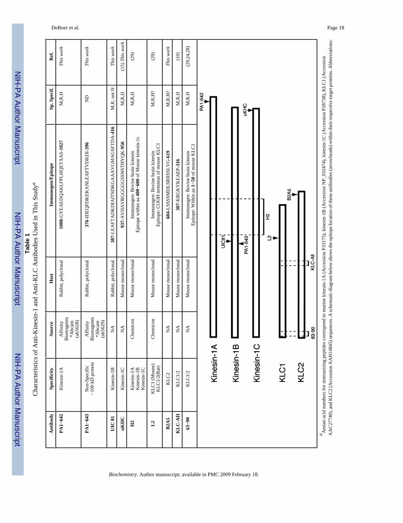
AntibodiesA detailed description of anti-kinesin-1 and anti-KLC antibodies used here is provided in Table1. Unless indicated, antibody stocks were prepared at 1 mg/mL concentration and used at thefollowing concentrations: anti-KIF5A, 1:1000 (Affinity BioReagents catalog no. PA1−642);anti-KIF5B, 1:1000 (Affinity BioReagents catalog no. PA1−643); anti-KIF5B (UIC 81), 1:500;anti-KIF5C (uKHC), 1:1000; anti-(pan) kinesin-1 (H2, Chemicon), 1:1000; anti-pan KLC (63−90), 1:500; anti-pan KLC (KLC-All), 1:500; anti-KLC1 (L2, 1:500; anti-KLC2 (B2A5),1:500; anti-SNAP-25, 1:2500 (Santa Cruz sc-20048); anti-6-His tag (Qiagen), 1:500; rabbitanti-synapsin-1, 1:5000 (BioTrend); mouse anti-synaptophysin, 1:500 (Sigma); mouse anti-APP, 1:5000 (22C11, Roche); and rabbit anti-synaptotagmin (Sigma), 1:5000. The followingsecondary antibodies were used: Jackson 111−035−045 HRP-conjugated goat anti-rabbit,1:15000, and Jackson 115−035−146 HRP-conjugated goat anti-mouse IgG, 1:15000.
ImmunoblotsProteins were separated by SDS–PAGE on 4−12% Bis-Tris gels (NuPage minigels; Invitrogen)using MOPS running buffer (Invitrogen) and transferred to PVDF using Towbin buffersupplemented with 10% (v/v) methanol (90 min at 400 mA using a Hoeffer TE22 apparatus).Immunoblots were blocked with 1% (w/v) non-fat dried milk in PBS (12 mM sodiumphosphate, pH 7.4, 1.4 mM potassium phosphate, 2.7 mM potassium chloride, and 140 mMNaCl). Membranes were incubated with primary antibodies overnight at 4 °C in 1% IgG-freeBSA and washed four times with 0.1% Tween-20 in PBS. Primary antibody binding wasdetected with HRP-conjugated anti-mouse or anti-rabbit antibody (Jackson Immunoresearch)and visualized by chemiluminescence (ECL; Amersham). The molecular mass marker wasfrom Invitrogen (Blue Plus2 prestained standard no. LC5925).
Microtubule Pellet PreparationConventional kinesin-enriched microtubule (MT) pellets were prepared as described before(30). Mouse brains were dissected and quickly homogenized in BRB80 buffer (80 mM PIPES,pH 6.8, 1 mM MgCl2, and 1 mM EDTA, pH 7) containing mammalian protease inhibitorcocktail (1:100 dilution; Sigma). This homogenate was centrifuged at 12500gmax for 20 minat 4 °C. The supernatant fraction was transferred to a new tube and centrifuged at125000gmax for 5 min at 4 °C in a TL100.3 rotor (Beckman). The resulting supernatant (cytosol)was transferred to a new tube and adjusted to 20 μM Taxol and either 2 mM ATP or 2 mM
DeBoer et al. Page 3
Biochemistry. Author manuscript; available in PMC 2009 February 18.
NIH
-PA Author Manuscript
NIH
-PA Author Manuscript
NIH
-PA Author Manuscript

AMP-PNP. After 30 min at 37 °C, MT-containing cytosolic fractions were loaded on top of a20% sucrose cushion prepared in BRB80 buffer plus 20 μM Taxol and centrifuged at 35000rpm (131438gmax) in a MLS-50 rotor (Beckman) for 10 min. The resulting MT pellets wereresuspended in BRB80 using a 27 gauge syringe. Pellets and supernatant fractions wereadjusted to 1× gel loading buffer (GLB) using a 5× GLB stock [0.35 M Tris-HCl, pH 6.8, 10%(w/v) SDS (Pierce, Sequanal grade), 36% glycerol, 5% β-mercaptoethanol, 0.01%bromophenol blue], as previously described (31).
Immunoprecipitation ExperimentsMouse brains were homogenized in lysis buffer [LB; 25 mM Tris-HCl, pH 7.4, 150 mM NaCl,1% Triton X-100, and 1/100 dilution of mammalian protease inhibitor cocktail (Sigma)], aspreviously described (32). Lysates were centrifuged twice for 5 min at 55000 rpm(163640gmax) using a TLA 100.3 rotor (Beckman Instruments, Palo Alto, CA). The resultingsupernatant fractions were precleared using a mixture of protein G–agarose beads (Pierce) andnonimmune mouse IgG-conjugated Sepharose beads (Jackson Immunoresearch) for 1 h atroom temperature. Precleared brain lysate (400 μg) was brought to 1 mL with LB and incubatedwith 5 μg of the appropriate antibody plus 10 μL of protein G–agarose beads at 4 °C for 3 h.Immunocomplexes were recovered by centrifugation (3000gmax for 30 s) and washed fourtimes with 1 mL of LB and once with 50 mM HEPES, pH 7.4. Immunoclomplexes wereresuspended in GLB. For immunodepletion experiments in Figure 3B, precleared mouse brainlysates were subjected to three cycles of immunoprecipitation as described above. A 50 μLaliquot of each resultant supernatant was saved for immunoblot analysis after eachimmunoprecipitation cycle.
Subcellular Fractionation ProceduresA mouse brain was homogenized in 4 mL of HB (0.32 M sucrose, 10 mM HEPES, 5 mMEDTA, pH 7.4). Whole homogenate was centrifuged for 5 min at 1200gmax, 5000gmax, and10000gmax (10 min each) using a Beckman TLA100.3 rotor. The resulting pellets werediscarded, and the final supernatant was centrifuged at 100000gmax for 30 min in a SorvalS45A rotor. The membrane pellet obtained [V1 fraction (23,31)] was carefully resuspended in0.5 mL of HB plus 50% iodixanol using a 27 gauge syringe. Resuspended membranes wereplaced in a 13 mL centrifuge tube, and a premixed linear gradient from 5% to 23% iodixanol(prepared in HB) was loaded on top. Samples were centrifuged at 150000gmax for 3 h using aSW40Ti rotor (Beckman Instruments, Palo Alto, CA). Fifteen fractions were collected frombottom to top using a peristaltic pump. Equal volumes of each fraction were analyzed by SDS–PAGE and immunoblotting. Quantitative immunoblotting was performed as described before(33).
RESULTSCharacterization of Anti-Kinesin-1 Antibodies
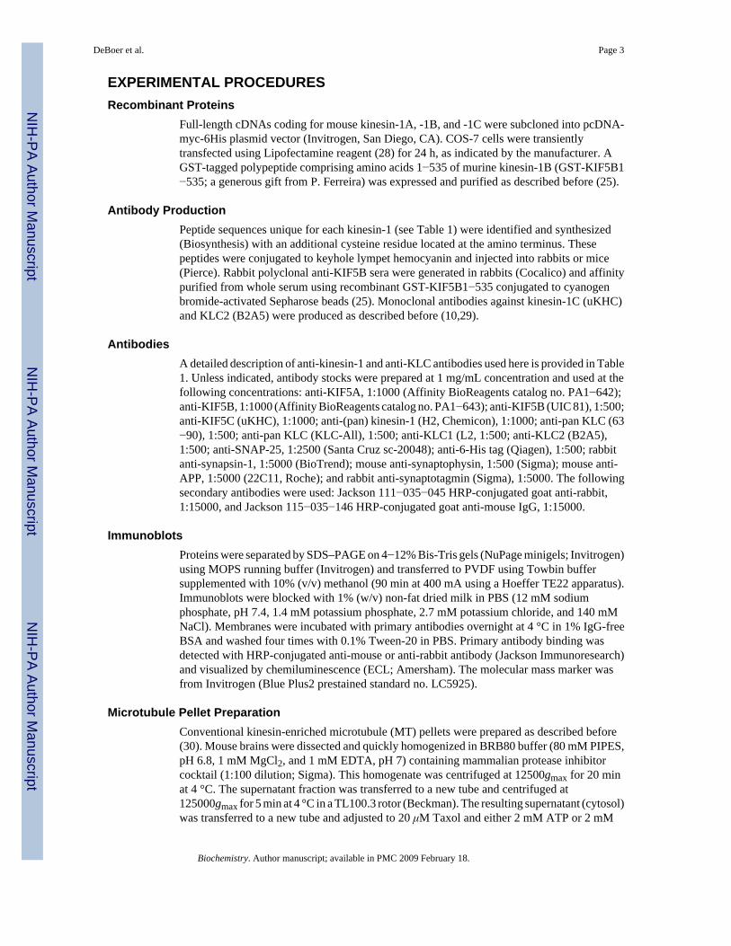
Both novel and previously described antibodies recognizing specific subunit isotypes ofconventional kinesin were rigorously characterized (Table 1). Major differences in the aminoand carboxy terminus of KLCs facilitated selection of peptide sequences specific for KLC1and KLC2. However, the selection of peptide sequences unique to each kinesin-1 was limitedby their high degree of sequence homology (26). When tested against whole mouse brainlysates, anti-KLC1 (L2) and anti-KLC2 (B2A5) antibodies recognized single bands at 64 and57 kDa, respectively, whereas the 63−90 antibody recognized both KLC1 and KLC2 (10,24),and these bands comigrated with those recognized by L2 and B2A5, respectively (Figure 1A).Anti-kinesin-1 antibodies recognized immunoreactive bands at the molecular mass expectedfor kinesin-1s (Figure 1A). The slightly lower mobility of kinesin-1A makes it distinguishablefrom bands corresponding to kinesin-1B and -1C under these electrophoretic and
DeBoer et al. Page 4
Biochemistry. Author manuscript; available in PMC 2009 February 18.
NIH
-PA Author Manuscript
NIH
-PA Author Manuscript
NIH
-PA Author Manuscript

immunoblotting conditions. H2 antibody recognized a doublet corresponding to kinesin-1A(upper band) and kinesin-1B and -1C combined (lower band) (18,29,34). Longer exposureswith H2 antibody were needed to visualize kinesin-1A, suggesting its levels in whole brainlysates were lower than the combined levels of kinesin-1B and kinesin-1C. Significantly,addressing the identity of kinesin-1s recognized by H2 allowed for the unequivocalidentification of kinesin-1A from kinesin-1B/C in fast axonal transport studies previously doneby our group (18).
The high degree of homology among kinesin-1s prompted us to confirm the specificity of anti-kinesin-1 antibodies taking advantage of a major biochemical characteristic of conventionalkinesin: its ability to form a rigor structure with MTs in the presence of AMP-PNP (anonhydrolyzable analogue of ATP) but not ATP (35). Mouse brain cytosolic fractions wereincubated with Taxol and either 2 mM ATP or 2 mM AMP-PNP. After 20 min incubation,samples were spun to produce a MT-enriched pellet and a corresponding supernatant (Figure1B) (36). Aliquots of each fraction obtained were analyzed by immunoblotting.Immunoreactive bands recognized by H2, PA1−642, UIC 81, uKHC, L2, B2A5, and KLC-Allantibodies were all enhanced in MT-enriched pellets prepared in the presence of AMP-PNP,compared to those prepared in the presence of ATP. Since these properties are identical to thosepreviously reported for conventional kinesin, we concluded that these antibodies effectivelyrecognized their specific target and not other proteins with a similar molecular mass. The onlyexception was PA1−643, a commercially available antibody against kinesin-1B. PA1−643recognized a major band with a molecular mass similar to that of kinesin-1B, but this band waspresent at similar levels in both ATP and AMP-PNP MT-enriched pellets. After long exposures,a very faint band was visualized which behaved as expected for kinesin-1B (data not shown),but this band was undetectable in total brain lysates. As a result of this cross-reactivity, thePA1−643 antibody is not well suited for analysis of kinesin-1B in cell or tissue samples. Thisobservation highlights the importance of proper antibody validation in studies involving highlyhomologous polypeptides. We next examined potential cross-reactivities among kinesin-1antibodies. The production of bacterially expressed full-length recombinant kinesin-1polypeptides in bacteria mainly resulted in the formation of truncated species of lowermolecular mass (37), thus complicating the identification of properly folded, full-lengthkinesin-1 polypeptides that would enable appropriate antibody characterization. To circumventthese issues, anti-kinesin-1 antibodies were reacted with lysates derived from COS-7 cellstransfected with cDNAs coding for 6-His-tagged, full-length mouse kinesin-1A, kinesin-1B,or kinesin-1C (Figure 1C). COS-7 cell lysates were normalized to similar levels ofoverexpressed kinesin-1s using an antibody against their 6-His tag. Whereas endogenouskinesin-1 levels were close to the levels of detection (38), exogenously expressed kinesin-1smigrated as single full-length polypeptide bands of approximately 100−110 kDa (Figure 1C).From this approach, antibodies specific for each kinesin-1 were identified (PA1−642, UIC 81,and uKHC for kinesin-1A, -1B, and -1C, respectively). In agreement with previous reports(26), the widely used monoclonal H2 antibody was found to recognize all kinesin-1s, albeitwith different affinities (1A > 1C >> 1B; data not shown).
Conventional Kinesin Is Composed of Kinesin-1 HomodimersDetermining all potential combinations among conventional kinesin subunits has been limitedby the lack of well-characterized antibodies that selectively recognize specific subunits. Anti-kinesin-1 antibodies described above were used to immunoprecipitate conventional kinesinfrom mouse brain under native conditions (26) (32) (Figure 2). As expected, each anti-kinesin-1antibody immunoprecipitated its corresponding kinesin-1 target protein (Figure 2A). The onlyexception was the H2 antibody, which immunoprecipitated all kinesin-1s, albeit with differentaffinities (data not shown). Significantly, we found no evidence of kinesin-1 heterodimers, asonly one kinesin-1 type could be detected in these immunoprecipitates with kinesin-1-specific
DeBoer et al. Page 5
Biochemistry. Author manuscript; available in PMC 2009 February 18.
NIH
-PA Author Manuscript
NIH
-PA Author Manuscript
NIH
-PA Author Manuscript
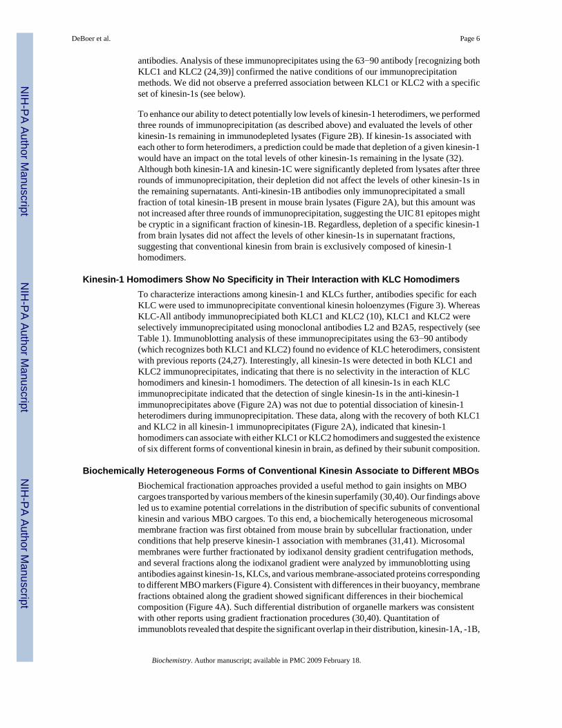
antibodies. Analysis of these immunoprecipitates using the 63−90 antibody [recognizing bothKLC1 and KLC2 (24,39)] confirmed the native conditions of our immunoprecipitationmethods. We did not observe a preferred association between KLC1 or KLC2 with a specificset of kinesin-1s (see below).
To enhance our ability to detect potentially low levels of kinesin-1 heterodimers, we performedthree rounds of immunoprecipitation (as described above) and evaluated the levels of otherkinesin-1s remaining in immunodepleted lysates (Figure 2B). If kinesin-1s associated witheach other to form heterodimers, a prediction could be made that depletion of a given kinesin-1would have an impact on the total levels of other kinesin-1s remaining in the lysate (32).Although both kinesin-1A and kinesin-1C were significantly depleted from lysates after threerounds of immunoprecipitation, their depletion did not affect the levels of other kinesin-1s inthe remaining supernatants. Anti-kinesin-1B antibodies only immunoprecipitated a smallfraction of total kinesin-1B present in mouse brain lysates (Figure 2A), but this amount wasnot increased after three rounds of immunoprecipitation, suggesting the UIC 81 epitopes mightbe cryptic in a significant fraction of kinesin-1B. Regardless, depletion of a specific kinesin-1from brain lysates did not affect the levels of other kinesin-1s in supernatant fractions,suggesting that conventional kinesin from brain is exclusively composed of kinesin-1homodimers.
Kinesin-1 Homodimers Show No Specificity in Their Interaction with KLC HomodimersTo characterize interactions among kinesin-1 and KLCs further, antibodies specific for eachKLC were used to immunoprecipitate conventional kinesin holoenzymes (Figure 3). WhereasKLC-All antibody immunoprecipiated both KLC1 and KLC2 (10), KLC1 and KLC2 wereselectively immunoprecipitated using monoclonal antibodies L2 and B2A5, respectively (seeTable 1). Immunoblotting analysis of these immunoprecipitates using the 63−90 antibody(which recognizes both KLC1 and KLC2) found no evidence of KLC heterodimers, consistentwith previous reports (24,27). Interestingly, all kinesin-1s were detected in both KLC1 andKLC2 immunoprecipitates, indicating that there is no selectivity in the interaction of KLChomodimers and kinesin-1 homodimers. The detection of all kinesin-1s in each KLCimmunoprecipitate indicated that the detection of single kinesin-1s in the anti-kinesin-1immunoprecipitates above (Figure 2A) was not due to potential dissociation of kinesin-1heterodimers during immunoprecipitation. These data, along with the recovery of both KLC1and KLC2 in all kinesin-1 immunoprecipitates (Figure 2A), indicated that kinesin-1homodimers can associate with either KLC1 or KLC2 homodimers and suggested the existenceof six different forms of conventional kinesin in brain, as defined by their subunit composition.
Biochemically Heterogeneous Forms of Conventional Kinesin Associate to Different MBOsBiochemical fractionation approaches provided a useful method to gain insights on MBOcargoes transported by various members of the kinesin superfamily (30,40). Our findings aboveled us to examine potential correlations in the distribution of specific subunits of conventionalkinesin and various MBO cargoes. To this end, a biochemically heterogeneous microsomalmembrane fraction was first obtained from mouse brain by subcellular fractionation, underconditions that help preserve kinesin-1 association with membranes (31,41). Microsomalmembranes were further fractionated by iodixanol density gradient centrifugation methods,and several fractions along the iodixanol gradient were analyzed by immunoblotting usingantibodies against kinesin-1s, KLCs, and various membrane-associated proteins correspondingto different MBO markers (Figure 4). Consistent with differences in their buoyancy, membranefractions obtained along the gradient showed significant differences in their biochemicalcomposition (Figure 4A). Such differential distribution of organelle markers was consistentwith other reports using gradient fractionation procedures (30,40). Quantitation ofimmunoblots revealed that despite the significant overlap in their distribution, kinesin-1A, -1B,
DeBoer et al. Page 6
Biochemistry. Author manuscript; available in PMC 2009 February 18.
NIH
-PA Author Manuscript
NIH
-PA Author Manuscript
NIH
-PA Author Manuscript
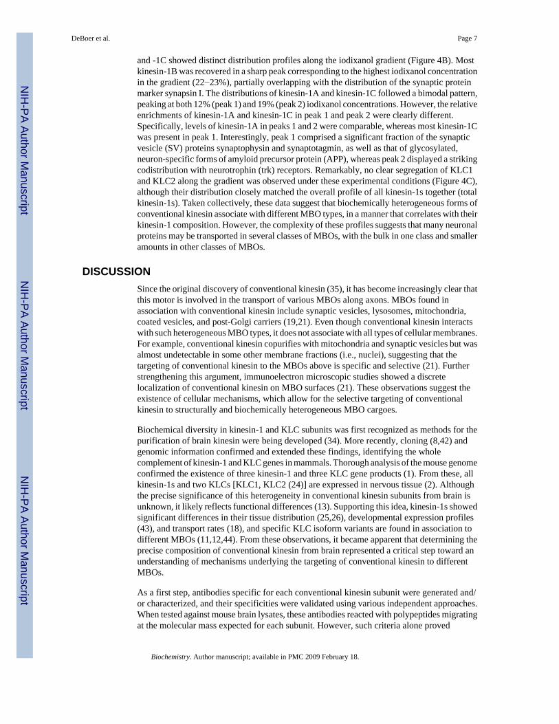
and -1C showed distinct distribution profiles along the iodixanol gradient (Figure 4B). Mostkinesin-1B was recovered in a sharp peak corresponding to the highest iodixanol concentrationin the gradient (22−23%), partially overlapping with the distribution of the synaptic proteinmarker synapsin I. The distributions of kinesin-1A and kinesin-1C followed a bimodal pattern,peaking at both 12% (peak 1) and 19% (peak 2) iodixanol concentrations. However, the relativeenrichments of kinesin-1A and kinesin-1C in peak 1 and peak 2 were clearly different.Specifically, levels of kinesin-1A in peaks 1 and 2 were comparable, whereas most kinesin-1Cwas present in peak 1. Interestingly, peak 1 comprised a significant fraction of the synapticvesicle (SV) proteins synaptophysin and synaptotagmin, as well as that of glycosylated,neuron-specific forms of amyloid precursor protein (APP), whereas peak 2 displayed a strikingcodistribution with neurotrophin (trk) receptors. Remarkably, no clear segregation of KLC1and KLC2 along the gradient was observed under these experimental conditions (Figure 4C),although their distribution closely matched the overall profile of all kinesin-1s together (totalkinesin-1s). Taken collectively, these data suggest that biochemically heterogeneous forms ofconventional kinesin associate with different MBO types, in a manner that correlates with theirkinesin-1 composition. However, the complexity of these profiles suggests that many neuronalproteins may be transported in several classes of MBOs, with the bulk in one class and smalleramounts in other classes of MBOs.
DISCUSSIONSince the original discovery of conventional kinesin (35), it has become increasingly clear thatthis motor is involved in the transport of various MBOs along axons. MBOs found inassociation with conventional kinesin include synaptic vesicles, lysosomes, mitochondria,coated vesicles, and post-Golgi carriers (19,21). Even though conventional kinesin interactswith such heterogeneous MBO types, it does not associate with all types of cellular membranes.For example, conventional kinesin copurifies with mitochondria and synaptic vesicles but wasalmost undetectable in some other membrane fractions (i.e., nuclei), suggesting that thetargeting of conventional kinesin to the MBOs above is specific and selective (21). Furtherstrengthening this argument, immunoelectron microscopic studies showed a discretelocalization of conventional kinesin on MBO surfaces (21). These observations suggest theexistence of cellular mechanisms, which allow for the selective targeting of conventionalkinesin to structurally and biochemically heterogeneous MBO cargoes.
Biochemical diversity in kinesin-1 and KLC subunits was first recognized as methods for thepurification of brain kinesin were being developed (34). More recently, cloning (8,42) andgenomic information confirmed and extended these findings, identifying the wholecomplement of kinesin-1 and KLC genes in mammals. Thorough analysis of the mouse genomeconfirmed the existence of three kinesin-1 and three KLC gene products (1). From these, allkinesin-1s and two KLCs [KLC1, KLC2 (24)] are expressed in nervous tissue (2). Althoughthe precise significance of this heterogeneity in conventional kinesin subunits from brain isunknown, it likely reflects functional differences (13). Supporting this idea, kinesin-1s showedsignificant differences in their tissue distribution (25,26), developmental expression profiles(43), and transport rates (18), and specific KLC isoform variants are found in association todifferent MBOs (11,12,44). From these observations, it became apparent that determining theprecise composition of conventional kinesin from brain represented a critical step toward anunderstanding of mechanisms underlying the targeting of conventional kinesin to differentMBOs.
As a first step, antibodies specific for each conventional kinesin subunit were generated and/or characterized, and their specificities were validated using various independent approaches.When tested against mouse brain lysates, these antibodies reacted with polypeptides migratingat the molecular mass expected for each subunit. However, such criteria alone proved
DeBoer et al. Page 7
Biochemistry. Author manuscript; available in PMC 2009 February 18.
NIH
-PA Author Manuscript
NIH
-PA Author Manuscript
NIH
-PA Author Manuscript

insufficient, given the high degree of homology among kinesin-1 polypeptides. Antibodieswere validated, taking advantage of the well-established biochemical property of conventionalkinesin to form a rigor structure with MTs in the presence of the nonhydrolyzable ATP analogueAMP-PNP (35). Immunoreactive bands recognized by these antibodies were significantlyenriched in MT pellets prepared in the presence of AMP-PNP, but not ATP, thus validatingPA1−642, UIC 81, uKHC, L2, and B2A5 antibodies. Importantly, not all antibodies tested metthe criteria above. For example, a commercially available polyclonal antibody againstkinesin-1B (PA1−643) recognized a prominent band at approximately 100 kDa in mouse brainlysates, but this band partitioned similarly to MT-enriched pellets in a manner independent ofthe nucleotide present. Finally, potential cross-reactivity among anti-kinesin-1 antibodies wasevaluated by immunoblotting using lysates derived from COS-7 cells transfected with full-length cDNA constructs coding for kinesin-1A, -1B, and -1C. COS-7 cells were chosenbecause, unlike bacteria, these cells efficiently express transfected full-length kinesin-1s (38).Also, these cells express low levels of endogenous kinesin-1. Supporting this idea, longexposures were needed with the high-affinity H2 antibody to detect kinesin-1 in untransfectedCOS cells (data not shown). This strategy allowed us to confirm the selectivity of anti-kinesin-1antibodies against their specific kinesin-1 target.
Having identified antibodies that selectively recognize a conventional kinesin subunit, weevaluated all possible subunit combinations present in endogenous conventional kinesin frombrain using immunoprecipitation approaches (32) (45). As expected, each anti-kinesin-1antibody immunoprecipitated their appropriate target kinesin-1, albeit with different affinities.Coimmunoprecipitation of KLCs with the kinesin-1 antibodies confirmed the native,nondenaturing conditions of our immunoprecipitation procedure (32). Significantly, nokinesin-1 other than the one specific for each antibody could be detected, indicating thatconventional kinesin holoenzymes precipitated by these antibodies are composed of kinesin-1homodimers. Immunodepletion experiments further tested this possibility. If a significantfraction of endogenous kinesin-1s can associate to form heterodimers in vivo, a prediction couldbe made that depletion of a specific kinesin-1 form from brain lysates would impact on thelevels of other kinesin-1s remaining in these lysates. Significantly, nearly complete depletionof kinesin-1A and kinesin-1C from mouse lysates did not affect the total levels of otherkinesin-1s. Although kinesin-1B could not be completely immunodepleted, its levels wereunaffected by the depletion of kinesin-1A and kinesin-1C. Taken together, these resultsindicated that endogenous conventional kinesin from brain is composed exclusively ofkinesin-1 homodimers.
Our findings on kinesin-1 homodimerization here sharply contrast with a previous reportshowing that different kinesin-1s can associate to form heterodimers (26). This notion,however, was inconsistent with the large differences in tissue distribution, relative levels, andcellular functions proposed for each kinesin-1 (26), as well as differences in their transportrates (18), the phenotypic manifestation of each kinesin-1 knockout (26,46,47), and thepreferred association of selected kinesin-1s to various protein effectors (25), all observationswhich strongly suggested unique roles for each kinesin-1 in the nervous system. Differencesin antibody specificities could explain these disparate results.
Previous reports indicated that KLCs interact with each other to form homodimers of the sameKLC type (24) and isoform (27). These findings were confirmed and further extended by thestudies here. Specifically, we found that all three kinesin-1s were coimunoprecipitated witheach KLC, indicating that KLC1 and KLC2 homodimers can interact with all three possiblekinesin-1 homodimers. These data confirmed previous reports of KLC homodimerization(24) and were consistent with the high degree of homology found in the KLC-binding domainamong kinesin-1s. Throughout our studies, some small variability was observed in the amountsof each kinesin-1 coimmunoprecipitated with KLC1 and KLC2 (data not shown). Potential
DeBoer et al. Page 8
Biochemistry. Author manuscript; available in PMC 2009 February 18.
NIH
-PA Author Manuscript
NIH
-PA Author Manuscript
NIH
-PA Author Manuscript

sources of variability include the relative abundance of different kinesin-1s in different areasof the brain (data not shown). Because immunoprecipitations were done using whole brainlysates, small variations in homogenization could contribute to the variability above. Ourresults allowed us to conclude unequivocally that each kinesin-1 has the ability to bind to eitherKLC1 or KLC2 but do not permit definitive statements on the relative affinities of each KLCfor each kinesin-1. This later issue clearly requires different experimental approaches, currentlyongoing in our laboratory. Taken together, data obtained from kinesin-1 and KLCimmunoprecipitation experiments suggest the existence of six variants of conventional kinesin,as defined by their subunit compositions (kinesin-1A/KLC1, kinesin-1A/KLC2, kinesin-1B/KLC1, kinesin-1B/KLC2, kinesin-1C/KLC1, and kinesin-1C/KLC2). However, KLC1transcripts have been shown to undergo alternative splicing that results in the generation ofnumerous KLC1 isoforms (8,11), so the actual combination of kinesin subunits that mayproduce functionally diverse holoenzymes exceeds the number estimated from the geneproduct composition alone and may expand the repertoire of functions possible for a givenkinesin-1.
We next examined preferred associations between biochemically heterogeneous MBOs andspecific conventional kinesin subunits using subcellular fractionation approaches.Significantly, kinesin-1A, -1B, and -1C showed different distribution profiles along continuousiodixanol gradients, which were consistent with association of specific kinesin-1s tobiochemically heterogeneous MBOs. In contrast to kinesin-1s, KLC1 and KLC2 displayednearly identical distribution profiles under these experimental conditions. Consistent with theresults above, kinesin-1s, but not KLCs, displayed major differences in their transport kineticsin vivo, and these differences correlated with association to different MBOs (18). Takentogether, these data suggested that kinesin-1s play a role in the targeting of conventional kinesinvariants to different MBOs. Such a claim is consistent with ultrastructural observationsshowing extensive association of conventional kinesin's heavy chains with their membranouscargoes (16). Although differences in the subcellular distribution of KLC1 and KLC2 were notobvious in subcellular fractionation experiments here, immunolocalization experiments incultured cells showed different MBO localizations for each KLC1 isoform (refs 11 and 12;Stenoien and Brady, unpublished observations), suggesting a role for the variable COOHterminus of KLCs in the targeting of conventional kinesin to selected MBOs (13). Although arole of the KLC's TR domain in the tight binding of conventional kinesin to membranes is wellestablished (10), it is unlikely that this domain plays a role in the targeting of conventionalkinesin, given its invariable presence in all KLCs (9,10). The TR domain likely contributes tothe tight binding of conventional kinesins to MBOs. The available experimental evidencesuggests that both the variable globular tail domain of kinesin-1s and the extreme carboxytermini of KLCs play a role in the selective targeting of conventional kinesin variants to selectedMBOs (9,13). Further work is needed to establish the precise functional role of each subunitin this process.
The localized delivery of selected MBOs at specific neuronal and axonal subdomains underliesthe ability of neurons to receive and transmit chemical information. Significantly, our dataprovide a conceptual basis for the transport regulation of specific MBO populations conveyedby conventional kinesin in neurons (23). Indeed, various reports indicate the existence of bothkinesin-1 and KLC-specific regulatory mechanisms, which are consistent with our findings ofkinesin-1 and KLC homodimerization properties described here. For example, various proteinshave been reported which selectively associate to specific kinesin-1s (15,25). Also, variousprotein kinases have been identified, which selectively regulate kinesin-1 (33) or KLC-relatedactivities (48), and these kinases phosphorylate selected kinesin-1 and KLC polypeptides(Morfini and Brady, unpublished observations). Subunit-specific regulatory mechanisms couldallow for the selective, nonpromiscuous regulation of conventional kinesin variants inassociation to specific MBO cargoes, as well as the delivery of such cargoes to discrete
DeBoer et al. Page 9
Biochemistry. Author manuscript; available in PMC 2009 February 18.
NIH
-PA Author Manuscript
NIH
-PA Author Manuscript
NIH
-PA Author Manuscript
subcellular locations (23). Finally, our findings here also have implications for selected aspectsof neuronal dysfunction in the context of human diseases, since alterations in regulatorypathways for conventional kinesin-based motility are being increasingly recognized asimportant pathogenic lesions in various neurodegenerative diseases (49-51).
ACKNOWLEDGMENTThe authors thank Dongyan Huang and Oscar Buzzio for excellent technical assistance.
REFERENCES1. Miki H, Setou M, Kaneshiro K, Hirokawa N. All kinesin superfamily protein, KIF, genes in mouse
and human. Proc. Natl. Acad. Sci. U.S.A 2001;98:7004–7011. [PubMed: 11416179]2. Miki H, Setou M, Hirokawa N. Kinesin superfamily proteins (KIFs) in the mouse transcriptome.
Genome Res 2003;13:1455–1465. [PubMed: 12819144]3. Wagner MC, Pfister KK, Brady ST, Bloom GS. Purification of kinesin from bovine brain and assay
of microtubule-stimulated ATPase activity. Methods Enzymol 1991;196:157–175. [PubMed:1851939]
4. Bloom GS, Wagner MC, Pfister KK, Brady ST. Native structure and physical properties of bovinebrain kinesin and identification of the ATP-binding subunit polypeptide. Biochemistry 1988;27:3409–3416. [PubMed: 3134048]
5. Hirokawa N, Pfister KK, Yorifuji H, Wagner MC, Brady ST, Bloom GS. Submolecular domains ofbovine brain kinesin identified by electron microscopy and monoclonal antibody decoration. Cell1989;56:867–878. [PubMed: 2522351]
6. Kuznetsov SA, Vaisberg EA, Shanina NA, Magretova NA, Chernyak NM, Gelfand VI. The quatenarystructure of bovine brain kinesin. EMBO J 1988;7:353–356. [PubMed: 3130248]
7. Lawrence CJ, Dawe RK, Christie KR, Cleveland DW, Dawson SC, Endow SA, Goldstein LS, GoodsonHV, Hirokawa N, Howard J, Malmberg RL, McIntosh JR, Miki H, Mitchison TJ, Okada Y, ReddyAS, Saxton WM, Schliwa M, Scholey JM, Vale RD, Walczak CE, Wordeman L. A standardizedkinesin nomenclature. J. Cell Biol 2004;167:19–22. [PubMed: 15479732]
8. Cyr JL, Pfister KK, Bloom GS, Slaughter CA, Brady ST. Molecular genetics of kinesin light chains:Generation of isoforms by alternative splicing. Proc. Natl. Acad. Sci. U.S.A 1991;88:10114–10118.[PubMed: 1946431]
9. Wozniak MJ, Allan VJ. Cargo selection by specific kinesin light chain 1 isoforms. EMBO J2006;25:5457–5468. [PubMed: 17093494]
10. Stenoien DS, Brady ST. Immunochemical analysis of kinesin light chain function. Mol. Biol. Cell1997;8:675–689. [PubMed: 9247647]
11. Khodjakov A, Lizunova EM, Minin AA, Koonce MP, Gyoeva FK. A specific light chain of kinesinassociates with mitochondria in cultured cells. Mol. Biol. Cell 1998;9:333–343. [PubMed: 9450959]
12. Gyoeva FK, Bybikova EM, Minin AA. An isoform of kinesin light chain specific for the Golgicomplex. J. Cell. Sci 2000;113(Part 11):2047–2054. [PubMed: 10806115]
13. Brady ST. A kinesin medley: Biochemical and functional heterogeneity. Trends Cell Biol1995;5:159–164. [PubMed: 14732151]
14. Diefenbach RJ, Mackay JP, Armati PJ, Cunningham AL. The C-terminal region of the stalk domainof ubiquitous human kinesin heavy chain contains the binding site for kinesin light chain.Biochemistry 1998;37:16663–16670. [PubMed: 9843434]
15. Huang JD, Brady ST, Richards BW, Stenoien D, Resau JH, Copeland NG, Jenkins NA. Directinteraction of two different transport motors and implications for vesicle transport. Nature1999;397:267–270. [PubMed: 9930703]
16. Hirokawa N, Bloom GS, Vallee RB. Cytoskeletal architecture and immunocytochemical localizationof microtubule-associated proteins in regions of axons associated with rapid axonal transport: thebeta,beta′-iminodipropionitrile-intoxicated axon as a model system. J. Cell Biol 1985;101:227–239.[PubMed: 2409096]
17. Morfini, G.; Stenoien, DL.; Brady, ST. Basic Neurochemistry. Vol. 7th ed.. 2005. Publisher, Address
DeBoer et al. Page 10
Biochemistry. Author manuscript; available in PMC 2009 February 18.
NIH
-PA Author Manuscript
NIH
-PA Author Manuscript
NIH
-PA Author Manuscript
18. Elluru R, Bloom GS, Brady ST. Fast axonal transport of kinesin in the rat visual system: functionalityof the kinesin heavy chain isoforms. Mol. Biol. Cell 1995;6:21–40. [PubMed: 7538359]
19. Jaulin F, Xue X, Rodriguez-Boulan E, Kreitzer G. Polarization-dependent selective transport to theapical membrane by KIF5B in MDCK cells. Dev. Cell 2007;13:511–522. [PubMed: 17925227]
20. Feiguin F, Ferreira A, Kosik KS, Caceres A. Kinesin-mediated organelle translocation revealed byspecific cellular manipulations. J. Cell Biol 1994;127:1021–1039. [PubMed: 7962067]
21. Leopold PL, McDowall AW, Pfister KK, Bloom GS, Brady ST. Association of kinesin withcharacterized membrane-bounded organelles. Cell Motil. Cytoskeleton 1992;23:19–33. [PubMed:1382871]
22. Boiko T, Rasband MN, Levinson SR, Caldwell JH, Mandel G, Trimmer JS, Matthews G. Compactmyelin dictates the differential targeting of two sodium channel isoforms in the same axon. Neuron2001;30:91–104. [PubMed: 11343647]
23. Morfini G, Szebenyi G, Richards B, Brady ST. Regulation of kinesin: implications for neuronaldevelopment. Dev. Neurosci 2001;23:364–376. [PubMed: 11756752]
24. Rahman A, Friedman DS, Goldstein LS. Two kinesin light chain genes in mice. J. Biol. Chem1998;273:15395–15403. [PubMed: 9624122]
25. Cai Y, Singh BB, Aslanukov A, Zhao H, Ferreira PA. The docking of kinesins, KIF5B and KIF5C,to Ran-binding protein 2 (RanBP2) is mediated via a novel RanBP2 domain. J. Biol. Chem2001;276:41594–41602. [PubMed: 11553612]
26. Kanai Y, Okada Y, Tanaka Y, Harada A, Terada S, Hirokawa N. KIF5C, a novel neuronal kinesinenriched in motor neurons. J. Neurosci 2000;20:6374–6384. [PubMed: 10964943]
27. Gyoeva FK, Sarkisov DV, Khodjakov AL, Minin AA. The tetrameric molecule of conventionalkinesin contains identical light chains. Biochemistry 2004;43:13525–13531. [PubMed: 15491159]
28. Pigino G, Morfini G, Mattson MP, Brady ST, Busciglio J. Alzheimer's presenilin 1 mutations impairkinesin-based axonal transport. J. Neurosci 2003;23:4499–4508. [PubMed: 12805290]
29. Pfister KK, Wagner MC, Stenoien D, Bloom GS, Brady ST. Monoclonal antibodies to kinesin heavyand light chains stain vesicle-like structures, but not microtubules, in cultured cells. J. Cell Biol1989;108:1453–1463. [PubMed: 2522455]
30. Morfini G, Quiroga S, Rosa A, Kosik K, Caceres A. Suppression of KIF2 in PC12 cells alters thedistribution of a growth cone nonsynaptic membrane receptor and inhibits neurite extension. J. CellBiol 1997;138:657–669. [PubMed: 9245793]
31. Morfini, G.; Tsai, M.; Szebenyi, G.; Brady, ST. Approaches to study interactions between kinesinmotors and membranes. In: Vernos, I., editor. Kinesin Protocols. Humana Press; Totowa, NJ: 2000.p. 147-162.
32. Lazarov O, Morfini GA, Lee EB, Farah MH, Szodorai A, Koliatsos V, Kins S, Lee VM-Y, WongPCY, Price DL, Brady ST, Sisodia SS. Axonal transport, amyloid precursor protein, kinesin-1, andthe processing apparatus: revisited. J. Neurosci 2005;25:2386–2395. [PubMed: 15745965]
33. Morfini G, Pigino G, Szebenyi G, You Y, Pollema S, Brady ST. JNK mediates pathogenic effects ofpolyglutamine-expanded androgen receptor on fast axonal transport. Nat. Neurosci 2006;9:907–916.[PubMed: 16751763]
34. Wagner MC, Pfister KK, Bloom GS, Brady ST. Copurification of kinesin polypeptides withmicrotubule-stimulated Mg-ATPase activity and kinetic analysis of enzymatic processes. Cell Motil.Cytoskeleton 1989;12:195–215. [PubMed: 2524282]
35. Brady ST. A novel brain ATPase with properties expected for the fast axonal transport motor. Nature1985;317:73–75. [PubMed: 2412134]
36. Lasek RJ, Brady ST. Adenylyl imidodiphosphate (AMPPNP), a nonhydrolyzable analogue of ATP,produces a stable intermediate in the motility cycle of fast axonal transport. Biol. Bull 1984;167:503.
37. Cho KI, Cai Y, Yi H, Yeh A, Aslanukov A, Ferreira PA. Association of the kinesin-binding domainof RanBP2 to KIF5B and KIF5C determines mitochondria localization and function. Traffic2007;8:1722–1735. [PubMed: 17887960]
38. Verhey KJ, Lizotte DL, Abramson T, Barenboim L, Schnapp BJ, Rapoport TA. Light chain-dependentregulation of kinesin's interaction with microtubules. J. Cell Biol 1998;143:1053–1066. [PubMed:9817761]
DeBoer et al. Page 11
Biochemistry. Author manuscript; available in PMC 2009 February 18.
NIH
-PA Author Manuscript
NIH
-PA Author Manuscript
NIH
-PA Author Manuscript

39. Pigino G, Pelsman A, Mori H, Busciglio J. Presenilin-1 mutations reduce cytoskeletal association,deregulate neurite growth, and potentiate neuronal dystrophy and tau phosphorylation. J. Neurosci2001;21:834–842. [PubMed: 11157069]
40. Okada Y, Yamazaki H, Sekine-Aizawa Y, Hirokawa N. The neuron-specific kinesin superfamilyprotein KIF1A is a unique monomeric motor for anterograde axonal transport of synaptic vesicleprecursors. Cell 1995;81:769–780. [PubMed: 7539720]
41. Tsai M-Y, Morfini G, Szebenyi G, Brady ST. Modulation of kinesin-vesicle interactions by Hsc70:Implications for regulation of fast axonal transport. Mol. Biol. Cell 2000;11:2161–2173. [PubMed:10848636]
42. Aizawa H, Sekine Y, Takemura R, Zhang Z, Nangaku M, Hirokawa N. Kinesin superfamily in murinecentral nervous system. J. Cell Biol 1992;119:1287–1296. [PubMed: 1447303]
43. Vignali G, Lizier C, Sprocati MT, Sirtori C, Battaglia G, Navone F. Expression of neuronal kinesinheavy chain is developmentally regulated in the central nervous system of the rat. J. Neurochem1997;69:1840–1849. [PubMed: 9349526]
44. Brady, ST.; Pfister, KK. Motor Proteins. Kendrick-Jones, J.; Cross, RA., editors. The Company ofBiologists, Ltd.; Cambridge, England: 1991. p. 103-108.
45. Morfini G, Pigino G, Brady ST. Approaches to kinesin-1 phosphorylation. Methods Mol. Biol2007;392:51–69. [PubMed: 17951710]
46. Xia CH, Roberts EA, Her LS, Liu X, Williams DS, Cleveland DW, Goldstein LS. Abnormalneurofilament transport caused by targeted disruption of neuronal kinesin heavy chain KIF5A. J. CellBiol 2003;161:55–66. [PubMed: 12682084]
47. Tanaka Y, Kanai Y, Okada Y, Nonaka S, Takeda S, Harada A, Hirokawa N. Targeted disruption ofmouse conventional kinesin heavy chain, kif5B, results in abnormal perinuclear clustering ofmitochondria. Cell 1998;93:1147–1158. [PubMed: 9657148]
48. Morfini G, Szebenyi G, Elluru R, Ratner N, Brady ST. Glycogen synthase kinase 3 phosphorylateskinesin light chains and negatively regulates kinesin-based motility. EMBO J 2002;23:281–293.[PubMed: 11823421]
49. Morfini G, Pigino G, Beffert U, Busciglio J, Brady ST. Fast axonal transport misregulation andAlzheimer's disease. Neuromol. Med 2002;2:89–99.
50. Morfini G, Pigino G, Brady ST. Polyglutamine expansion diseases: Failing to deliver. Trends Mol.Med 2005;11:64–70. [PubMed: 15694868]
51. Morfini G, Pigino G, Opalach K, Serulle Y, Moreira JE, Sugimori M, Llinas RR, Brady ST. 1-Methyl-4-phenylpyridinium affects fast axonal transport by activation of caspase and protein kinaseC. Proc. Natl. Acad. Sci. U.S.A 2007;104:2442–2447. [PubMed: 17287338]
DeBoer et al. Page 12
Biochemistry. Author manuscript; available in PMC 2009 February 18.
NIH
-PA Author Manuscript
NIH
-PA Author Manuscript
NIH
-PA Author Manuscript

Figure 1.Characterization of anti-kinesin-1-specific antibodies. (A) Immunoblot analysis of wholemouse brain lysates using anti-kinesin-1 and anti-KLC antibodies. Anti-kinesin-1A, -1B, and-1C antibodies recognized a single band at the expected molecular mass size (approximately100−110 kDa). Note the slightly lower mobility of kinesin-1A under these SDS–PAGEconditions. H2 antibody against all kinesin-1s recognizes a band doublet corresponding tokinesin-1A (upper band, arrowhead) and kinesin-1C/B (lower band, arrow). Anti-kinesin-1BPA1−643 antibody recognized a major band with a slower mobility to those recognized byother kinesin-1 antibodies, raising questions against its specificity (see text). (B) Aliquots ofmouse brain cytosol (Cyt) were incubated with Taxol and either 2 mM ATP or 2 mM AMP-PNP, a nonhydrolyzable ATP analogue. After 20 min incubation, samples were spun to producea MT-enriched pellet (P) and a corresponding supernatant (S). In the presence of AMP-PNP,this procedure allows for quantitative recovery of conventional kinesin in association to MT-enriched pellets. Aliquots of each fraction were analyzed by immunoblotting using H2antibody, and various novel antibodies against kinesin-1s and KLC2 (see Table 1). Note thatwith the only exception of PA1−643 antibody, all antibodies tested recognized polypeptideswhich biochemically behave as expected for conventional kinesin. (C) COS-7 cells weretransfected with plasmids encoding His-tagged, full-length versions of kinesin-1A, -1B, and-1C and lysed for 24 h after transfection. COS-7 lysates were first normalized to similarkinesin-1 levels using anti-His antibody (Tet-His) and then analyzed by immunoblot usinganti-kinesin-1A (PA1−642), anti-kinesin-1B (UIC 81), and anti-kinesin-1C (uKHC) (see Table1). These antibodies recognized a single band at the expected molecular mass size(approximately 100−110 kDa), and no cross-reactivity with other kinesin-1s was observed.The monoclonal antibody H2 recognized all kinesin-1s, albeit with different affinities (1A >1C >> 1B). After very long exposure of film, a very faint band was recognized by H2 antibodyin untransfected (Neg) cells (data not shown). Arrowheads and arrows indicate the positionsof kinesin 1A and kinesin-1B/C, respectively.
DeBoer et al. Page 13
Biochemistry. Author manuscript; available in PMC 2009 February 18.
NIH
-PA Author Manuscript
NIH
-PA Author Manuscript
NIH
-PA Author Manuscript

Figure 2.Kinesin-1s exist as homodimers in brain tissue. (A) Validated anti-kinesin-1 antibodies wereused for immunoprecipitation from mouse brain lysates. Non-immune IgGs were used ascontrols for immunoprecipitation specificity. A lane loaded with mouse brain lysate (Input) isshown. Immunoblot analysis indicates that each antibody exclusively immunoprecipitatedtheir respective antigen. As expected from its specificity, H2 antibody immunoprecipitated allkinesin-1s. The presence of KLC1 and KLC2 in these immunoprecipitates confirmed the nativeconditions of the methods herein. Key: NR IgG, normal rabbit IgG; NM IgG, normal mouseIgG. (B) Three rounds of immunoprecipitation were carried out as described in (A), and aliquotsof the lysate supernatants obtained after each round were analyzed by immunoblot. Depletionof each kinesin-1A and kinesin-1C from brain lysates does not significantly affect the levelsof all other kinesin-1 proteins, indicating that kinesin-1s exist primarily as homodimers.
DeBoer et al. Page 14
Biochemistry. Author manuscript; available in PMC 2009 February 18.
NIH
-PA Author Manuscript
NIH
-PA Author Manuscript
NIH
-PA Author Manuscript

Figure 3.Kinesin-1 homodimers interact with both KLC1 and KLC2 homodimers. Immunoblots with63−90 antibody show that anti-KLC1 (L2) and anti-KLC2 (B2A5) antibodies selectivelyimmunoprecipitated their corresponding antigen, whereas KLC-All antibody raised against theTR domain common to all KLCs immunoprecipitated both. These data confirmed previousreports of KLC homodimerization. Significantly, kinesin-1A, -1B, and -1C could all be foundin both anti-KLC1 and anti-KLC2 immunoprecipitates, suggesting there is no specificity inthe interactions between KLCs and kinesin-1 homodimers.
DeBoer et al. Page 15
Biochemistry. Author manuscript; available in PMC 2009 February 18.
NIH
-PA Author Manuscript
NIH
-PA Author Manuscript
NIH
-PA Author Manuscript
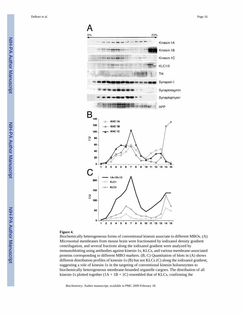
Figure 4.Biochemically heterogeneous forms of conventional kinesin associate to different MBOs. (A)Microsomal membranes from mouse brain were fractionated by iodixanol density gradientcentrifugation, and several fractions along the iodixanol gradient were analyzed byimmunoblotting using antibodies against kinesin-1s, KLCs, and various membrane-associatedproteins corresponding to different MBO markers. (B, C) Quantitation of blots in (A) showsdifferent distribution profiles of kinesin-1s (B) but not KLCs (C) along the iodixanol gradient,suggesting a role of kinesin-1s in the targeting of conventional kinesin holoenzymes tobiochemically heterogeneous membrane-bounded organelle cargoes. The distribution of allkinesin-1s plotted together (1A + 1B + 1C) resembled that of KLCs, confirming the
DeBoer et al. Page 16
Biochemistry. Author manuscript; available in PMC 2009 February 18.
NIH
-PA Author Manuscript
NIH
-PA Author Manuscript
NIH
-PA Author Manuscript

heterotetrameric composition of conventional kinesin in association to these MBOs. IU =intensity units.
DeBoer et al. Page 17
Biochemistry. Author manuscript; available in PMC 2009 February 18.
NIH
-PA Author Manuscript
NIH
-PA Author Manuscript
NIH
-PA Author Manuscript
NIH
-PA Author Manuscript
NIH
-PA Author Manuscript
NIH
-PA Author Manuscript
DeBoer et al. Page 18Ta
ble
1C
hara
cter
istic
s of A
nti-K
ines
in-1
and
Ant
i-KLC
Ant
ibod
ies U
sed
in T
his S
tudy
a
Ant
ibod
ySp
ecifi
city
Sour
ceH
ost
Imm
unog
en/E
pito
peSp
. Spe
cif.
Ref
.
PA1−
642
Kin
esin
-1A
Aff
inity
Bio
reag
ents
*Abc
am(a
b562
8)
Rab
bit,
poly
clon
al10
08-G
YEA
EDQ
AK
LFPL
HQ
ETA
AS-
1027
M,R
,HTh
is w
ork
PA1−
643
Non
-Spe
cific
∼10
0 kD
pro
tein
Aff
inity
Bio
reag
ents
*Abc
am(a
b562
9)
Rab
bit,
poly
clon
al37
6-ID
EQFD
KEK
AN
LEA
FTV
DK
DI-
396
ND
This
wor
k
UIC
81
Kin
esin
-1B
NA
Rab
bit,
poly
clon
al38
7-LE
AFT
AD
KD
IAIT
SDK
GA
AA
VG
MA
GSF
TDA
-416
M,R
, not
HTh
is w
ork
uKH
CK
ines
in-1
CN
AM
ouse
mon
oclo
nal
937-
AV
HA
VR
GG
GG
GSS
NST
HY
QK
-956
M,R
,H(1
5) T
his w
ork
H2
Kin
esin
-1A
Kin
esin
-1B
Kin
esin
-1C
Che
mic
onM
ouse
mon
oclo
nal
Imm
unog
en: B
ovin
e br
ain
kine
sin
Epito
pe w
ithin
aa
400−
600
of M
ouse
kin
esin
-1s
M,R
,H(2
9)
L2
KLC
1 (M
ouse
)K
LC1/
2(R
at)
Che
mic
onM
ouse
mon
oclo
nal
Imm
unog
en: B
ovin
e br
ain
kine
sin
Epito
pe: C
OO
H te
rmin
us o
f mou
se K
LC1
M,R
,H?
(29)
B2A
5K
LC2
NA
Mou
se m
onoc
lona
l60
4-LS
SSSM
DLS
RR
SSLV
G-6
19M
,R,H
?Th
is w
ork
KL
C-A
IIK
LC1/
2N
AM
ouse
mon
oclo
nal
307-
KR
GK
YK
EAEP
-316
M,R
,H(1
0)
63−9
0K
LC1/
2N
AM
ouse
mon
oclo
nal
Imm
unog
en: B
ovin
e br
ain
kine
sin
Epito
pe: W
ithin
aa
1−50
of m
ouse
KLC
1M
,R,H
(29,
24,2
8)
a Am
ino
acid
num
bers
for i
mm
uniz
ing
pept
ides
cor
resp
ond
to m
urin
e ki
nesi
n-1A
(Acc
essi
on P
3317
5), k
ines
in-1
B (A
cces
sion
NP_
0324
74),
kine
sin-
1C (A
cces
sion
P28
738)
, KLC
1 (A
cces
sion
AA
C27
740)
, and
KLC
2 (A
cces
sion
AA
H14
845)
sequ
ence
s. A
sche
mat
ic d
iagr
am b
elow
show
s the
epito
pe lo
catio
n of
thes
e ant
ibod
ies (
arro
whe
ads)
with
in th
eir r
espe
ctiv
e tar
get p
rote
ins.
Abb
revi
atio
ns:
Biochemistry. Author manuscript; available in PMC 2009 February 18.

NIH
-PA Author Manuscript
NIH
-PA Author Manuscript
NIH
-PA Author Manuscript
DeBoer et al. Page 19Sp
. Spe
cif.,
spec
ies s
peci
ficity
; Ref
., R
efer
ence
s; N
A, n
ot c
omm
erci
ally
ava
ilabl
e; N
D, n
ot d
eter
min
ed; M
, mou
se; R
, rat
; H, h
uman
. A q
uest
ion
mar
k in
dica
tes a
spec
ies f
or w
hich
the
spec
ifici
ty o
f agi
ven
antib
ody
has n
ot b
een
esta
blis
hed.
Ast
eris
ks (*
) ind
icat
e th
at p
olyc
lona
l ant
ibod
ies g
ener
ated
aga
inst
the
indi
cate
d pe
ptid
e ar
e al
so a
vaila
ble
from
Abc
am.
Biochemistry. Author manuscript; available in PMC 2009 February 18.
Copyright © 2022 FDOKUMEN