Development of advanced phospholipid vesicles loaded with ...
Upload
independentCategory
view
4download
0
The Rockefeller University Press, 0021-9525/98/11/777/18 $2.00The Journal of Cell Biology, Volume 143, Number 3, November 2, 1998 777–794http://www.jcb.org 777
Overexpression of Tau Protein Inhibits Kinesin-dependentTrafficking of Vesicles, Mitochondria, and EndoplasmicReticulum: Implications for Alzheimer’s Disease
A. Ebneth, R. Godemann, K. Stamer, S. Illenberger, B. Trinczek, E.-M. Mandelkow, and E. Mandelkow
Max-Planck Unit for Structural Molecular Biology, D-22607 Hamburg, Germany
Abstract.
The neuronal microtubule-associated pro-tein tau plays an important role in establishing cell po-larity by stabilizing axonal microtubules that serve as tracks for motor-protein–driven transport processes. To investigate the role of tau in intracellular transport, we studied the effects of tau expression in stably trans-fected CHO cells and differentiated neuroblastoma N2a cells. Tau causes a change in cell shape, retards cell growth, and dramatically alters the distribution of vari-ous organelles, known to be transported via microtu-bule-dependent motor proteins. Mitochondria fail to be transported to peripheral cell compartments and clus-ter in the vicinity of the microtubule-organizing center. The endoplasmic reticulum becomes less dense and no
longer extends to the cell periphery. In differentiated N2a cells, the overexpression of tau leads to the disap-pearance of mitochondria from the neurites. These ef-fects are caused by tau’s binding to microtubules and slowing down intracellular transport by preferential im-pairment of plus-end–directed transport mediated by kinesin-like motor proteins. Since in Alzheimer’s dis-ease tau protein is elevated and mislocalized, these ob-servations point to a possible cause for the gradual de-generation of neurons.
Key words: mitochondria • transport • endoplasmic reticulum • exocytosis • Alzheimer’s disease
T
HE
microtubule network plays an important role inmaintaining cellular morphology, in mitotic pro-cesses, in intracellular trafficking, and in establish-
ing cellular polarity and neurite outgrowth in differentiat-ing neurons (Drubin and Nelson, 1996; Li and Black, 1996;Goodson et al., 1997). Microtubules serve as tracks for themovement of mitochondria (Nangaku et al., 1994; Morrisand Hollenbeck, 1995; Rickard and Kreis, 1996), lyso-somes (Hollenbeck and Swanson, 1990), peroxisomes(Wiemer et al., 1997) and various other organelles such asendocytotic or exocytotic vesicles (Scales et al., 1997). Fur-thermore, they are important in establishing and maintain-ing the functionality of the endoplasmic reticulum (Water-man-Storer et al., 1995). Microtubule (MT)
1
-dependenttransport is achieved through motor proteins such asdynein or kinesin and their relatives (Waterman-Storer
and Salmon, 1997; Hirokawa, 1998; Lippincott-Schwartz,1998). Kinesin is a plus-end–directed motor, whereasdynein-related motor proteins move in the opposite di-rection (Brady, 1995; Vallee and Sheetz, 1996). The or-ganelles are linked to the motors via membrane/organelle-bound adaptor proteins such as kinectin or dynactin(Kumar et al., 1995; Burkhardt et al., 1997).
The microtubule tracks are stabilized by microtubule-associated proteins (MAPs) such as MAP2, tau, MAP4,and others. These are mostly filamentous proteins thatbind to the microtubule surface and promote microtubuleassembly (for reviews, see Hirokawa, 1994; Mandelkowand Mandelkow, 1995). The developmental regulation ofMAPs, for instance in neuronal differentiation, suggestsan important role in organizing the microtubule cytoskele-ton (Matus, 1994). Tau is enriched in axons, MAP2 in thesomatodendritic compartment of neurons (Binder et al.,1985), whereas MAP4 can be found in many types of tis-sues (Chapin and Bulinski, 1991; West et al., 1991). MAP2,tau, and MAP4 share a remarkable sequence homology inthe conserved repeat regions located in the COOH-termi-nal microtubule-binding domain (Chapin and Bulinski,1992). The repeated motifs are preceded by an NH
2
-termi-nal projection domain that has been suggested to serve asa spacer between microtubules (Chen et al., 1992) or as ananchor for cellular enzymes (Obar et al., 1989).
Address all correspondence to Eckhard Mandelkow or Andreas Ebneth,MPG-ASMB, Notkestrasse 85, D-22607 Hamburg, Germany. Tel.: 49-40-8998-2810. Fax: 49-40-8971-6822. E-mail: [email protected] or [email protected]
1.
Abbreviations used in this paper
: AD, Alzheimer’s disease; EGFP, en-hanced green fluorescent protein; MAP, microtubule-associated protein;MT, microtubule; MTOC, microtubule organizing center; TMR, tetrame-thylrhodamine.

The Journal of Cell Biology, Volume 143, 1998 778
Since motor proteins and MAPs both interact with themicrotubule surface, the question arises whether MAPs in-terfere with the movement of motor proteins. Severalstudies have suggested that this is the case for certainMAPs, especially those of higher molecular weight. Thus,MAP2 can inhibit the motility of dynein or kinesin in vitro(Paschal et al., 1989; Von Massow et al., 1989; Lopez andSheetz, 1993; Hagiwara et al., 1994), while smaller frag-ments had only a weak effect. This could be accounted forin several ways: (
a
) MAPs could have a general stericblocking effect of the extended NH
2
-terminal projectiondomain; (
b
) MAPs could compete with motors for com-mon binding sites, probably on beta-tubulin (Hagiwara etal., 1994); and (
c
) MAPs could affect transport processesindirectly by altering microtubule dynamics (Waterman-Storer and Salmon, 1997).
When MAP4 is overexpressed in mouse L
tk
2
cells, it in-terferes with motor-protein–dependent organelle motilityand trafficking in vivo (Bulinski et al., 1997). Furthermore,overexpression of tau or MAP2c in COS-fibroblasts indi-cates that both MAPs might function as phosphorylation-dependent regulators of organelle transport (Sato-Haradaet al., 1996). The phosphorylation and dephosphorylationof microtubule-associated proteins is believed to be a keyfactor regulating the affinity to microtubules, thereby fa-cilitating and supporting vital cellular processes such asproliferation, differentiation, and trafficking through sta-bilization of the tracks for motor proteins (Lopez andSheetz, 1995; Mandell and Banker, 1996; Sato-Harada et al.,1996; Drewes et al., 1997). Indeed, MAPs isolated fromcells are phosphorylated and it is well documented thatthis phosphorylation interferes with binding to microtu-bules in vitro and in vivo (Drechsel et al., 1992; Biernat et al.,1993; Illenberger et al., 1996).
In axonal growth, where a dynamic microtubule array isindispensable for differentiation, the phosphorylation anddistribution of tau are correlated (Mandell and Banker,1996; Li and Black, 1996), suggesting a regulated balancebetween kinases and phosphatases, as well as a finelytuned gradient of tau concentration. In Alzheimer’s dis-ease (AD), this balance between kinases and phosphatasesseems to be disturbed (Trojanowski and Lee, 1995; Man-delkow et al., 1995). Neurofibrillary tangles, one of thehallmarks of this disease, consist of paired helical fila-ments, which are mainly composed of hyperphosphory-lated microtubule-associated protein tau (Wischik et al.,1988). Furthermore, tau in AD mislocalizes to the somato-dendritic compartment of neurons (Braak et al., 1994) andtau concentration has been reported to be elevated in ADbrain to a significant extent (Khatoon et al., 1992). Thisfurther strengthens the importance of proper control ofposttranslational modifications, localization, and intracel-lular concentration of microtubule-associated proteins invivo.
These observations lead us to investigate whether theoverexpression of tau protein had an effect on organelleand vesicle trafficking in cultured cells. This approach hadbeen useful for studying the distribution of exogenousMAPs (Barlow et al., 1994; Olson et al., 1995; Bulinski etal., 1997; Kaech et al., 1996). In previous studies, we hadshown that CHO and neuroblastoma N2a cells can serveas models for the state of MAP phosphorylation during
the cell cycle, and that the phosphorylation of tau by thekinase MARK leads to microtubule breakdown (Preuss etal., 1995; Drewes et al., 1997; Illenberger et al., 1998). Herewe demonstrate that the overexpression of tau leads to achange in cell shape, loss of polarization, and the retarda-tion of cell growth. One of the most visible consequencesis the dramatically altered distribution of mitochondria.Instead of being dispersed throughout the cytoplasm, themitochondria become clustered near the nucleus at the mi-crotubule organizing center (MTOC). In differentiatedneuroblastoma cells, the neurites become nearly devoid ofmitochondria. The endoplasmic reticulum becomes lessdense and retracts from the cell periphery or from neu-rites. Transferrin endocytosed by clathrin-coated vesiclesaccumulates near the MTOC and recycling is significantlyslowed down. These effects can be explained by assumingthat tau interferes with MT-driven transport, in the case ofmitochondria preferentially with the plus-end–directedtransport (by kinesin-like motors) so that the minus-end–directed transport (by dynein) predominates. This resultsin a net inward transport of mitochondria and other or-ganelles such as peroxisomes towards the MTOC, wherethe microtubule minus-ends are organized. The perturba-tions of the balance in microtubule-dependent transport,especially in neurites, would be consistent with the ob-served impairment of axonal traffic in Alzheimer’s diseaseneurons, their loss of polarity, and their subsequent degen-eration.
Materials and Methods
Cell Culture
CHO cells were grown in HAM’s F12 medium supplemented with 10%fetal calf serum and 2 mM glutamine (Biochrom, Berlin, Germany). Forimmunofluorescence, cells were seeded onto coverslips at a density of 2
3
10
4
cells/cm
2
in 24-well culture dishes and grown overnight at 37
8
C with5% CO
2
. N2a neuroblastoma cells were grown in MEM Earle supple-mented with 10% fetal calf serum, 2 mM glutamine (Biochrom) and 1%nonessential amino acids (Sigma Chemical Co., Deisenhofen, Germany).Differentiation of N2a cells was achieved by addition of 1
m
M retinoicacid (Sigma Chemical Co.) for 24–48 h in the presence of 0.1% fetal calfserum. For stable transfection, plasmids derived from pcDNA3 (Invitro-gen Corp., Leek, The Netherlands) coding for the longest human tau-isoform htau40 under the control of the cytomegalovirus promoter wereintroduced into the cells with the DOTAP-method (Boehringer Mann-heim, Mannheim, Germany) according to the manufacturer’s instructions.Stable transfectants were selected in the presence of 800
m
g/ml (CHO) or1 mg/ml geneticin (N2a) as described (Preuss et al., 1995), and subse-quently recloned.
Antibodies and Dyes
The following antibodies were used: rat monoclonal antitubulin antibod-ies YL1/2 and mouse monoclonal antibody DM1A were purchased fromSerotec (Oxford, UK) and Sigma Chemical Co., respectively. Polyclonalrabbit anti–tau antibody K9JA was from DAKOPATTS (Hamburg, Ger-many), polyclonal rabbit antibody used as an ER marker was a gift fromDr. D. Louvard (Institut Curie, Paris; Louvard et al., 1982) and polyclonalanticatalase antibody was from Dr. W. Just (University of Heidelberg,Heidelberg, Germany). Antivimentin antibody V9 was from BoehringerMannheim. All fluorescently (TRITC, FITC, and AMCA) labeled sec-ondary antibodies were from DIANOVA (Hamburg, Germany). Fluores-cent dyes DiOC
6
(3), MitoTracker™ Green, MitoTracker™ Red, andrhodamine-labeled transferrin were purchased from Molecular Probes,Inc. (Eugene, OR). DiOC
6
(3) was used as a stain for in vivo observationof the ER at 2.5
m
g/ml.

Ebneth et al.
Tau Inhibits Kinesin-dependent Vesicle Trafficking
779
Immunofluorescence
Cells were fixed with 2% paraformaldehyde or methanol, and incubationwith antibodies was performed essentially as described (Preuss et al.,1995). Tau isoforms tightly bound to microtubules can be visualized bymethanol fixation (which allows unbound protein to diffuse out, see Fig.2
c
). By contrast, weakly bound constructs (e.g., K10, K12) cannot be visu-alized by methanol fixation (since they would diffuse away). Instead, it isnecessary to use paraformaldehyde, which fixes all cellular proteins buttends to dissociate tau from microtubules (see Fig. 3, for details see Illen-berger et al., 1998). To visualize mitochondria, MitoTracker™ Green wasadded at a concentration of 400 nM 30 min before fixation. CHO cells sta-bly transfected with human tau40 or mock-transfected cells were incu-bated for 1 h with 5
m
M nocodazole (Sigma Chemical Co.) or 6 h with1–10
m
M taxotere (Rhone-Poulence Rorer, Vitry, France). Cells werefixed before and 1 or 4 h after nocodazole, and 6 h after taxotere treat-ment, respectively. Cells were examined with an Axioplan fluorescencemicroscope (Carl Zeiss Jena GmbH, Jena, Germany) equipped with a100
3
oil-immersion objective and filters optimized for triple-label exper-iments. Pictures were taken with a cooled CCD camera (Visicam; Visitron,Puchheim, Germany) and analyzed using the MetaMorph
®
software pack-age (Visitron). The fraction of the cell area containing mitochondria wasquantified as follows: after fixation and immunofluorescence labeling, thepictures were recorded and the areas containing mitochondria were circum-scribed manually (omitting the nucleus) and calculated. The same was donewith the total cell area visible in the microtubule stain. A minimum of 30cells were measured per experiment and the ratios plotted (e.g., see Fig. 11).
Microinjection
Microinjection experiments were performed as described (Preuss et al.,1997). In brief, cells were seeded onto coverslips at a density of 2
3
10
4
cells in 35-mm dishes and incubated overnight. Recombinant protein, pre-pared as described (Biernat et al., 1993; Gustke et al., 1994) was concen-trated in Microcon microconcentrators (Amicon Inc., Beverly, MA) ac-cording to the manufacturer’s instructions. During this procedure, thebuffer was changed to 25 mM HEPES, pH 7.2, 1 mM PMSF, and proteinwas then microinjected with an Eppendorf Micromanipulator 5171 at con-stant pressure within 15 min. MitoTracker™ Red was added 30 min be-fore fixation at a concentration of 200 nM, and cells were finally fixed withparaformaldehyde at given time points and prepared for immunofluores-cence. For quantitative analysis of the intracellular concentration of tauprotein in the tau40-stable cell line, 1.9, 0.95, and 0.38 mg/ml purified re-combinant tau protein, as well as buffer only, were microinjected intoCHO cells. Cells were then fixed in parallel with the tau-stable cell lineand stained with a polyclonal anti–tau antibody. The fluorescence of thetau stain was then quantified using the CCD camera and the MetaMorph
®
software package and normalized to the cell dimensions. A minimum of80 cells were analyzed per microinjection and at least 100 tau-stable cellswere scored. Regression analysis of the normalized fluorescence intensi-ties of microinjected cells was performed with the program Sigma Plot(
R
5
0.9995), and the mean intracellular tau concentration of the stablecell line was calculated.
Monitoring Mitochondrial Distribution and Endoplasmic Reticulum In Vivo
For the visualization of mitochondria in vivo, cells were seeded ontoLabTek chambered cover glass (NUNC, Inc., Naperville IL.) at 70% con-fluency. 30 min before immunofluorescence examination, MitoTracker™Green or Red was added to cells at a final concentration of 200 nM, cellswere then rinsed once with medium. For monitoring mitochondrial redis-tribution tau-stable CHO cells were incubated for 1 h with 5
m
M nocoda-zole and 200 nM MitoTracker™ Green. Disassembly of the microtubulenetwork was confirmed by fixation and tubulin staining in immunofluores-cence microscopy. Nocodazole was then removed and cells were exam-ined with the cooled CCD camera on an inverted microscope (Axiovert10; Carl Zeiss Jena GmbH) using standard filters for FITC fluorescence.Staining of the endoplasmic reticulum was achieved with DiOC
6
(3) at aconcentration of 2.5
m
g/ml, 30-min incubation at 37
8
C, and rinsing cellsonce with medium. Immunofluorescence microscopy was then performedas described above.
Transferrin Uptake and Recycling
Cells were grown in 35-mm six-well tissue culture plates for 2 d to
z
80%
confluency at 37
8
C and 5% CO
2
. Supernatant was then replaced by se-rum-free medium, and 2 h later tetramethylrhodamine (TMR)-labeledtransferrin was added to a final concentration of 380 nM. Cells were fur-ther incubated for 1 h to achieve maximal transferrin uptake. Mediumcontaining excess or recycled transferrin was removed and new mediumcontaining 60
m
g/ml partially Fe
2
1
-saturated transferrin (Sigma ChemicalCo.) was added. Recycling was then monitored by again removing me-dium at given time points and quantitating fluorescence of TMR-labeledtransferrin in a Fluoro Max spectrofluorometer (Spex Industries Inc., Edi-son, NJ) at 575 nm emission and 552 nm excitation wavelength. Cells werethen trypsinized, lysed, and protein concentration was determined to nor-malize fluorescent signals to total protein present in the culture dishes,representing cell density. At least five independent experiments were per-formed with tau-stable and mock-transfected CHO cells to ensure statisti-cal significance. To quantitate residual intracellular transferrin, cells wereseeded onto coverslips, grown overnight, and subsequently saturated withTMR transferrin (see above). Cells were then fixed in methanol either im-mediately or 7 min after removal of TMR transferrin. Residual transferrinfluorescence was quantitated as above using standard filters for rho-damine fluorescence. Pictures were corrected by background subtractionand shadowing correction provided with the MetaMorph
®
software pack-age (Visitron). At least 300 different cells per time point and cell line werequantified by thresholding.
Tau Isoforms and Constructs
Human brains contain mainly six isoforms of tau generated by alternativesplicing (Goedert et al., 1989). The present work focuses on the largest(tau40, 441 residues) and smallest (tau23, 352 residues) isoform, contain-ing four or three repeats in their microtubule-binding region (Fig. 1). Clon-ing, bacterial expression, and purification was done as described (Biernatet al., 1993; Gustke et al., 1994). Other tau constructs were also generatedas described, such as K12 and K10 (derived from htau23, three repeatswith the COOH-terminal flanking region or the whole COOH-terminaltail), K35 (which comprises the microtubule-binding domain of tau, in-cluding the proline-rich region upstream of the repeats), and K23 (a vari-
Figure 1. Domain structure of tau isoforms and constructs. Tau40is the longest tau isoform in the human CNS, occurring mostly inaxons (Goedert et al., 1989). It consists of 441 amino acid resi-dues and can be divided into an NH2-terminal projection domainand a COOH-terminal microtubule binding domain containingfour imperfect repeats (R1–R4) and basic proline-rich sequenceson either side that act as microtubule targeting domains (shadeddark, P1, and P2). The repeats show high homology between tau,MAP2, and MAP4. The shaded NH2-terminal inserts and repeattwo can be alternatively spliced, resulting in six different tau iso-forms. The predominantly embryonic isoform tau23 lacks allthree inserts (352 residues). Tau constructs K12 and K10 are de-rived from tau23, they contain three repeats plus either a COOH-terminal flanking region (K12) or the whole COOH-terminal tail(K10). K35 contains three repeats and the basic proline-rich re-gions on either side, including the COOH-terminal tail. K23 con-tains all of tau23 except the repeats.

The Journal of Cell Biology, Volume 143, 1998 780
ant of tau23 lacking the three repeats). The dissociation constants of theseconstructs and their binding to cellular microtubules were reported previ-ously (Gustke et al., 1994; Preuss et al., 1997): tau40 (
K
d
5
1.1
m
M), tau23(2.5
m
M), K35 (1.0
m
M), K23 (6.5
m
M), K10 (18.5
m
M), and K12 (7.1
m
M).
Inhibition of Dynein-dependent Transport by Overexpression of Dynamitin
Dynamitin (p50) expression plasmid was transfected into tau-stable CHOcells with the DOTAP-method. 6 h after transfection, cells were fixed inparaformaldehyde as described (see above). Mitochondria were stainedwith MitoTracker™ Red (Molecular Probes, Inc.), tubulin with rat mono-clonal antitubulin antibody (YL1/2; Serotec) and transfected p50-dyna-mitin with mouse monoclonal antidynamitin 50-I. Immunofluorescenceanalysis was performed as described above. The p50 expression plasmidand antibody were generous gifts from C. Echeverri and R. Vallee(Worcester Foundation, Shrewsbury, MA) (Burkhardt et al., 1997).
Monitoring Transport of Individual Mitochondria After Nocodazole Removal
Cells were seeded onto LabTek chambered cover glass (NUNC, Inc.) at70% confluency. Nocodazole was then added at a final concentration of1
m
M for 2 h and subsequently removed. 30 min before removal, Mi-toTracker™ Red was added at a concentration of 200 nM to visualize mi-tochondria. After an additional 30 min to allow recovery of the microtu-bule cytoskeleton, reaggregation of the organelles was monitored in vivoon an inverted microscope with a cooled CCD camera (see above) by re-cording pictures every second for 2 min with the following constraint: onlymitochondria translocated with a minimum distance of 1
m
m and a veloc-ity of 0.5
m
m/s were taken into consideration to exclude diffusional pro-cesses. By using the nucleus as a reference, transported mitochondriawere grouped into three categories: movement towards the nucleus (cen-tripetal; denoted as
2
in Fig. 7) was regarded as minus-end directed,movement towards the cell periphery (centrifugal; denoted as
1
) as plus-end directed and all other movements as circumferential (denoted as
2
/
1
).
Results
Cell Shape and Mitochondrial Distribution Is Strongly Altered in Tau-overexpressing CHO Cells
To investigate the effects of human tau expression in vivo,we established CHO cells stably transfected with tau40, anisoform containing four repeats (Preuss et al., 1995, andthis report). Immunofluorescent staining of microtubulesshowed that their network was normal and containedbound tau (Fig. 2), consistent with similar results onMAP4-transfected cells (Olson et al., 1995; Nguyen et al.,1997). In particular, no bundling of microtubules was seen,contrary to experiments with transient transfection (Kanaiet al., 1989; Lee and Rook, 1992) or with microinjection(Preuss et al., 1997), where higher cellular concentrationsof MAPs are obtained. To calibrate the intracellular tauconcentration, we microinjected different amounts of puri-fied, recombinant tau protein into CHO cells, and pre-pared them for immunofluorescence analysis in parallelwith the tau-stable cells (see Materials and Methods). Thisyielded an average concentration of 0.04
m
g/ml tau in thestably transfected cells, corresponding to
z
1
m
M. Sincethe cellular tubulin concentration is
z
40
m
M (Hiller andWeber, 1978), the ratio of tau to tubulin remained low,
z
1:40, similar to that reported for PC12 (Drubin et al.,1985) or HeLa cells (Bulinsky and Borisy, 1979). Further-more, stable expression of tau in PC12 cells under controlof the cytomegalovirus promoter (which was used in ourexperiments) resulted in only a twofold increase of intra-cellular tau-protein level (Brandt et al., 1995). Taken to-gether, these data show that the concentration of tau in the
stably transfected CHO and N2a cells is in the range of en-dogenous MAP levels in the control cells and increases theamount of total MAP only about two- to threefold.
In spite of the low cellular concentration, the expressionof tau leads to conspicuous changes in cell shape andgrowth behavior. Whereas control cells were elongated,tau-transfected CHO cells rounded up (compare Fig. 2,
b
vs.
e
). This phenotype can be evoked in several ways, butone possibility is the inhibition of the motor protein kine-sin as described by Rodionov et al. (1993), and a similar in-terpretation is appropriate in our case as well (see below).A second noticeable effect was that overexpression of tauin CHO cells slowed down proliferation approximatelytwofold (data not shown), similar to observations withMAP4-transfected cells (Nguyen at al., 1997).
We wanted to investigate whether the observed changesin shape and proliferation were indeed related to microtu-bule-dependent transport and therefore analyzed the cellswith several markers for organelles. This revealed a re-markable clustering of mitochondria in tau40-expressingCHO cells near the nucleus in the vicinity of the microtu-bule organizing center (Fig. 2,
a
and
c
). This effect was re-produced in two independently generated tau40-stable celllines, thus ruling out recloning artifacts. In contrast, mock-transfected CHO cells showed that mitochondria weredispersed throughout the entire cytoplasm (Fig. 2
d
), fre-quently aligned with microtubules, suggesting a micro-tubule-dependent organization of mitochondria. The frac-tion of the cell area containing mitochondria decreasedfrom 66.1% in mock-transfected cells to 27.7% in the tau-stable cell line (see Fig. 10). To exclude the possibility of afixation artifact, we established CHO cells stably trans-fected with an enhanced green fluorescent protein(EGFP)–tau fusion protein that allowed us to monitortau’s effect on mitochondrial distribution in living cells.EGFP tau was tightly associated with microtubules (asseen by EGFP fluorescence) and led to the same effects onmitochondrial clustering (data not shown).
Since there are several reports (e.g., Summerhayes et al.,1983) linking the distribution of mitochondria to the integ-rity of the intermediate filament system, we investigatedwhether the vimentin network was altered through tauoverexpression. In tau40-stable cells, the extension of thisfilamentous network is significantly reduced and becomeslocated in a perinuclear area (Trinczek, B., A. Ebneth,E.-M. Mandelkow, and E. Mandelkow, manuscript in prep-aration). Thus, one might speculate that tau’s effects onthe intermediate filament system may account for the ag-gregation of mitochondria at the MTOC. However, whentreating cells with nocodazole to disrupt microtubules, mi-tochondria began to disperse again (see below), whereasthe extension of the intermediate filament network wasnot altered in the tau-stable CHO cells despite disruptionof microtubules. It has been shown that the disruption ofmicrotubules leads to a collapse of the intermediate fila-ment system (Klymkowski et al., 1989). Removal of no-codazole again did not affect the extension of the vimentinnetwork (data not shown), but led to a reclustering of mi-tochondria, which indicates that the integrity of the micro-tubule system, not the intermediate filament system, is re-sponsible for the observed phenotype of mitochondrialclustering in tau40-stable cells.

Ebneth et al.
Tau Inhibits Kinesin-dependent Vesicle Trafficking
781
Interestingly, inhibition of kinesin motors by microinjec-tion of an inhibitory antibody into fibroblasts led to astrikingly similar phenotype of a perinuclear localizationof the vimentin system (Gyoeva and Gelfand, 1991).
Mitochondria are organelles that can disappear fromcellular regions with lowered metabolic demand (e.g.,Morris and Hollenbeck, 1993). This raises the possibilitythat clustering of mitochondria was not due to an impair-ment of the cell’s transport, but to a change in the meta-bolic activity in the periphery due to the overexpression oftau. For this reason, we looked for other organelles knownto be transported via microtubule-dependent motors andasked if they were clustered, too. This was indeed the case.When staining tau-stable cells with an antibody raisedagainst catalase, an enzyme present in the matrix of perox-isomes, the organelles were significantly enriched in theperinuclear region, whereas in control cells they were
evenly distributed (data not shown). This argues that or-ganelle clustering near the MTOC is caused by a generalinfluence of tau on microtubule-dependent plus-end–directed transport, rather than a selective retraction of mi-tochondria in response to altered metabolic demands.
To identify the domains of tau responsible for the clus-tering of mitochondria, we studied CHO cells stably trans-fected with several tau isoforms or constructs whose be-havior had already been characterized in vitro (Gustke et al.,1994; Fig. 1): human tau40 (the longest isoform in the cen-tral nervous system), tau23 (the shortest isoform, foundpredominantly in embryonic brain), K12 (a tau constructcontaining only the three repeats plus the COOH-terminalflanking region), K10 (containing three repeats plus thewhole COOH-terminal rest of the molecule), K35 (con-taining three repeats and both flanking regions), and K23(a variant of tau23 without the repeats). The isoforms
Figure 2. Mitochondria accumulate near theMTOC in CHO cells overexpressing tau. Cellsstably transfected with tau40 (a–c) and mock-transfected CHO cells (d–f). Microtubules werestained with antibody DM1A (b and e) and tauwith polyclonal anti–tau antibody K9JA (c andf). Mitochondria were visualized with Mito-Tracker™ Green (a and d). Cells were fixedin methanol 30 min after addition of Mito-Tracker™ Green. In tau-stable cells (a–c), thereis a striking clustering of mitochondria near theMTOC (seen near the nucleus where the micro-tubule stain is most intense), whereas in mock-transfected cells (d–f) the mitochondria are dis-persed throughout the cytoplasm. In addition,the tau-stable cells are more rounded up com-pared with control cells. Bar, 20 mm.
The Journal of Cell Biology, Volume 143, 1998 782
tau40, tau23, and K35 induced pronounced mitochondrialclustering around the MTOC (Figs. 2 and 3,
a–d
, and datanot shown), whereas K10 (data not shown), K12, and K23did not show the effect (Fig. 3,
e–h
). This correlates wellwith the strengths of binding (dissociation constants
z
1–2
m
M for tau40, tau23, and K35, compared with
z
7–20
m
M
Figure 3. Effects of different tau constructs on mitochondrial dis-tribution in CHO cells. Mitochondrial distribution in CHO cellsstably transfected with different tau constructs was examined af-ter paraformaldehyde fixation. This allows the visualization of to-tal tau in the cells, but tends to dissociate tau from the microtu-bules (see Materials and Methods). Human tau23, the shortesttau-isoform (a and b), as well as K35 (c and d), a tau constructlacking nearly the entire projection domain (see Fig. 1) both ledto the phenotype of mitochondrial clustering near the MTOC asseen in tau40-stable cells. On the other hand, K23 (e and f), a tauconstruct lacking the microtubule-binding region and K12 (g andh), consisting mainly of this MT-binding region (Fig. 1), did notalter the distribution of mitochondria. This argues for a tightbinding of tau to microtubules as a prerequisite for impairment ofplus-end–directed transport of these organelles and excludes thepossibility that tau might inhibit kinesin by directly interactingwith the motor protein. Mitochondria were stained with Mi-toTracker™ Red (a, c, e, and g) and transfected tau with poly-clonal anti–tau antibody K9JA (b, d, f, and h). Bar, 20 mm.
Figure 4. Microinjection of recombinant tau protein leads to mi-tochondrial clustering near the MTOC. Wild-type CHO cells weremicroinjected with 1.3 mg/ml (z1.2 mM) of purified recombinanttau protein and incubated for 4 h. After addition of Mi-toTracker™ Red (a and b), cells were fixed with paraformalde-hyde and stained with antibodies against tubulin (c and d) andtau (e and f). Mitochondrial clustering is visible even within 4 hafter microinjection. Note that the microtubule network retainsits normal appearance and that mitochondria in microinjectedcells similar as in the tau-stable CHO and N2a cells tend to clus-ter at one side of the nucleus in the vicinity of the MTOC. Thecell periphery of microinjected cells is outlined for clarity. Bar,10 mm.

Ebneth et al. Tau Inhibits Kinesin-dependent Vesicle Trafficking 783
for the other constructs; Gustke et al., 1994). K35 bindstightly but lacks parts of the NH2-terminal projection do-main so that this domain appears to be of lesser impor-tance for mitochondrial transport. Conversely, K10 andK12 contain the repeats but lack the NH2-terminal projec-tion and flanking domains. Thus, their binding to microtu-bules is weak and this explains why they have no effect onmitochondrial distribution. K23 is a special case because itcontains the projection domain, the two flanking domains,but not the repeats. It binds to microtubules with reason-able affinity (Kd z7 mM), but still does not influence mi-crotubule-based traffic. These observations argue that ahigh affinity of tau to microtubules is the major determi-nant for observing the effects of tau overexpression andthat it is highly unlikely that tau inhibits plus-end–directedtransport by directly binding to kinesins.
Microinjection of Tau into CHO Cells Leads to Mitochondrial Clustering at the MTOC
Next we asked whether the elevation of tau protein was di-rectly responsible for the phenotype of mitochondrial clus-tering at the MTOC, or whether it was due to other factorsnot related to the protein level. Recombinant tau40 was
microinjected into mock-transfected CHO cells to a finalconcentration of z1.2 mM where the microtubule networkis not affected (Preuss et al., 1997). Nevertheless, mito-chondria began to accumulate near the nucleus within 4 h(Fig. 4). Microinjection of mouse serum at the same con-centration as a control on the other hand had no effect onmitochondrial distribution (data not shown). Thus, thephenotype can be generated without affecting the micro-tubule cytoskeleton, suggesting that tau protein has a di-rect effect on the redistribution of mitochondria, presum-ably by retarding their plus-end directed movement (seebelow).
Nocodazole and Taxotere Reverse the Effect of Tau on Mitochondrial Distribution
To investigate whether the organization and integrity ofmicrotubules was important for the clustering of mito-chondria, we applied two microtubule drugs, nocodazoleand taxotere. Nocodazole causes the disassembly of micro-tubules, taxotere (a taxol derivative) causes their assemblyand stabilization (Jordan and Wilson, 1998). In the pres-ence of taxotere, new microtubules are nucleated ran-domly in the cytoplasm, independently of the MTOC, so
Figure 5. Disruption of microtubules by no-codazole leads to a random distribution ofmitochondria in tau-stable CHO cells. Tau-stable cells (a and b) and mock-transfectedCHO cells (c and d). Cells were treated withnocodazole to disrupt the MT cytoskeleton,fixed with methanol, and immunolabeledwith antibodies against tubulin (b and d).In both cell lines, mitochondria, visualizedby addition of MitoTracker™ Green beforefixation (a and c), are now visible through-out the entire cytoplasm even in the cell pe-riphery, suggesting the existence of an intactMT network as a prerequisite for mitochon-drial clustering. Bar, 20 mm.

The Journal of Cell Biology, Volume 143, 1998 784
that the defined polarity and orientation is lost (McNivenand Porter, 1986; Weisshaar et al., 1992). If microtubulesmediated the relocalization of mitochondria towards thecell center, one would expect that disruption of the micro-tubule network in the tau-overexpressing cells might leadto a wild-type distribution. This was indeed observed. Fig.5 shows the disruption of microtubules in tau-stable CHOcells treated with nocodazole, which resulted in an in-creased cytosolic background staining arising from disas-sembled tubulin dimers. Within 1 h after nocodazole treat-ment, the mitochondria were redistributed to peripheralregions of the cell. The mitochondria-containing areathereby increases from 27.7 to 63.9%, which reaches thevalue of control cells within the error margin (see Fig. 10).Conversely, when nocodazole was removed again, an ap-parently normal microtubule network reappeared withinabout 15 min (data not shown), originating at the MTOC,and the phenotype of mitochondrial clustering, clearly vis-ible already within z1 h, was fully restored after z4 h, in-dicating that the organelles were actively transported to-wards the MTOC (see Figs. 6 and 10). Taken together,these data suggest that a shift in the balance between plus-and minus-end–directed motor activities through tau-overexpression is responsible for the phenotypic effects.Thus, the ratio of mitochondria actively transported to-wards the cell periphery (that is plus-end directed) or to-wards the cell center (the MTOC, minus-end directed) intau-stable versus control cells is expected to be altered bytau overexpression. To investigate this in further detail,cells were treated with nocodazole to achieve an even dis-tribution of mitochondria in both cell lines. The microtu-bule array was then allowed to recover by washing out thedrug and the direction of the movement of individual mi-tochondria was analyzed. Although transport in general isaffected by tau overexpression (Fig. 7), irrespective of thekind of motor activity, plus-end–directed transport de-creased z10-fold, whereas minus-end–directed transportdropped only about twofold, resulting in a net inwardtransport of mitochondria in tau-stable cells comparedwith control cells.
These results demonstrate that microtubule-dependentprocesses are responsible for the clustering of mitochon-
dria. It also means that mitochondria can become dis-persed independently of microtubules, presumably by dif-fusion (in contrast to the ER, see below). The fact thatreconstitution of the MT network leads to a reclustering ofmitochondria near the cell center suggests that the overex-pression of tau impairs predominantly the plus-end–directed transport of these organelles (see below). If thiswere true, one would expect that the normal distributionof mitochondria could be restored even in tau-overex-pressing cells by abolishing the polar organization of mi-crotubules. This can be achieved by treatment of cells withtaxotere (at final concentrations of up to 10 mM), whichleads to microtubule nucleation with random orientationsthroughout the cytoplasm. Fig. 8 shows that taxotere-
Figure 6. Reassembly ofmicrotubules after removalof nocodazole restores theclustering of mitochondria.The microtubule cytoskele-ton of tau-transfected CHOcells was disrupted by addi-tion of 5 mM nocodazole for2 h. Pictures of living cellswere then taken after re-moval of nocodazole andreassembly of microtubulesat the indicated timepoints. Mitochondria werestained with MitoTracker™Green. Note that, concomi-
tant with microtubule reassembly, the phenotype of mitochondrial clustering at the MTOC near the nucleus begins to reappearafter z1 h. The cell periphery is outlined for clarity. Bar, 10 mm.
Figure 7. The ratio betweenplus- and minus-end–directedtransport of mitochondria isaltered by tau overexpres-sion. Tau-stable and controlcells were treated with no-codazole to disrupt the mi-crotubule cytoskeleton andto achieve an even distribu-tion of mitochondria in bothcell lines. The drug waswashed out after 2 h of incu-bation and mitochondria
were stained with MitoTracker™ Red. Translocations of the or-ganelles were then recorded for 2 min with a time interval of 1 s.The direction of movement was regarded as plus-end directedwhen the organelles were transported to the periphery (1), mi-nus-end directed when transport was towards the nucleus (2),and neutral (2/1) when movement was vertical to the axis be-tween nucleus and periphery of the cells. Note that the relativenumber of transported mitochondria is significantly lower for allthree groups in the tau-stable cell line (P , 0.0001, two tailedStudent’s t test). However, in tau-stable cells, the plus-end–directed movement is significantly more affected (z10-fold)than the reverse direction (2-fold). This leads to the phenotypeof mitochondrial clustering. At least 40 cells were analyzed forquantitation, the total number of mitochondria ranged be-tween 100 and 700 in both cell lines.

Ebneth et al. Tau Inhibits Kinesin-dependent Vesicle Trafficking 785
treatment has a similar effect as nocodazole in restoringthe dispersed distribution of mitochondria. Again, themitochondria-containing area within the tau-stable cellsincreases to 58.1%, comparable with the 66.1% of mock-transfected cells (see Fig. 10). This result seems counterin-tuitive since the two drugs have opposite effects on micro-tubules, but it becomes understandable by consideringthat taxotere randomizes microtubules (at the micromolarconcentrations used here), and thus destroys the basis for
directed transport. In other words, a phenotype caused byimpairment of plus-end–directed transport can manifest it-self only in the presence of a polarized microtubule net-work where microtubule minus ends converge on theMTOC. Randomizing microtubule polarity by addition oftaxotere allows transport of mitochondria to the cell pe-riphery by minus-end–directed motors and thus leads to aredispersion of these organelles.
Inhibition of Dynein by Overexpression ofp50-Dynamitin Reverses the Effect of Mitochondrial Clustering by Tau
The minus-end–directed transport of mitochondria is thoughtto depend on dynein (Hirokawa et al., 1990; Vallee et al.,1995), and therefore one would expect that inhibition ofdynein would counteract the clustering of mitochondriainduced by overexpression of tau. We inhibited dynein bytransient transfection of the tau-overexpressing cells withdynamitin, a subunit of the dynactin complex that is cru-cial for the attachment between organelles and microtu-bules (Burkhardt et al., 1997). Overexpression of dyna-mitin in tau-stable cells significantly reduced the clusteringof mitochondria, whereas EGFP transfection as a controlhad no effect on the mitochondrial distribution (Fig. 9).The area containing mitochondria within the cells aftertransfection increased from z28% in EGFP-transfectedtau-stable cells to 47.9% in dynamitin-overexpressing cells(P , 0.0001, two-tailed Student’s t test; Fig. 10). The fact
Figure 8. Randomization of microtubule polarity by taxotere al-lows clustered mitochondria to disperse again. Tau-stable cellswere treated with 10 mM taxotere for 6 h, which leads to an al-tered microtubule organization. Note that the MTOC in taxo-tere-treated cells is barely visible in the tubulin stain (b), in con-trast to nontreated cells (compare Fig. 2). Microtubules arenucleated randomly throughout the cytoplasm, thus abolishingthe role of the MTOC as an organizing element and thereforeabolishing the polarity of microtubules. Minus-end–directed mo-tors are now able to transport mitochondria back to peripheralregions in the tau-stable cells. Cells were fixed in methanol andmitochondria were stained with MitoTracker™ Green (a), tubu-lin was immunostained with DM1A (b). Bar, 10 mm.
Figure 9. Dynamitin overex-pression restores the wild-type distribution of mito-chondria. Tau-stable cellswere transfected with dyna-mitin (p50), a subunit of thedynactin complex. 6 h aftertransfection, the mitochon-dria began to disperse again,visualized with MitoTracker™Red after paraformaldehydefixation (a). Note that thecell transfected with tauand dynamitin (arrow-heads) shows dispersed mi-tochondria, whereas theneighboring tau-stable cellsshow the typical clusteringnear the MTOC. Dynamitinwas stained with antidyna-mitin 50-1 antibody (b) andtubulin with YL1/2 anti-body (c). Bar, 20 mm.

The Journal of Cell Biology, Volume 143, 1998 786
that mitochondria were not completely redispersed (incontrast to the nocodazole-treated cells, where dispersal iscomplete) is probably due to fixation of cells 6 h aftertransfection. Since the overexpression of dynamitin af-fects the organization of the MTOC (Burkhardt et al.,1997), which in turn interferes with directed microtubule-dependent transport of mitochondria, we analyzed onlythose cells whose microtubule network was clearly unaf-fected. However, the experiment demonstrates that dy-nein motors are actively involved in the mitochondrialtransport and argues against different types of mecha-nisms that operate in other situations (e.g., tethering toshrinking microtubules; Lombillo et al., 1995).
Shape and Extension of the Endoplasmic Reticulum Is Altered by the Overexpression of Tau
Thus far, we concentrated on the microtubule-dependenttransport of mitochondria in transfected CHO cells. How-ever, microtubules are responsible for transporting othervesicles and organelles as well, notably the endoplasmicreticulum (Terasaki and Reese, 1994). Its distribution ap-pears to depend both on kinesin-dependent transport andon an intact microtubule network (Vale and Hotani, 1988;Hamm-Alvarez et al., 1993; Feiguin et al., 1994). To inves-tigate if overexpression of tau would affect the ER, westained living cells with DiOC6(3), a fluorescent markerfor the ER. Overexpression of tau caused pronouncedchanges in shape and extension of the ER (Fig. 11). First,in tau-stable cells, the extension of the ER was strongly re-duced so that it no longer reached the cell periphery. Sec-ondly, the density and branching of the ER was signifi-cantly decreased. As in the case of mitochondria, thisresult is consistent with the assumption that plus-end–directed transport along microtubules is perturbed.
On the other hand, observations with the microtubuledrugs taxotere and nocodazole showed that the behaviorof the ER is more complex. Taxotere partially restored the
wild-type distribution, although the ER did not fully reachthe cell periphery (data not shown). In contrast, after dis-ruption of microtubules with nocodazole, the ER did notexpand again. These results confirm that organized micro-tubules are necessary for the general constitution of theER, in addition to kinesin (Lippincott-Schwartz, 1998).
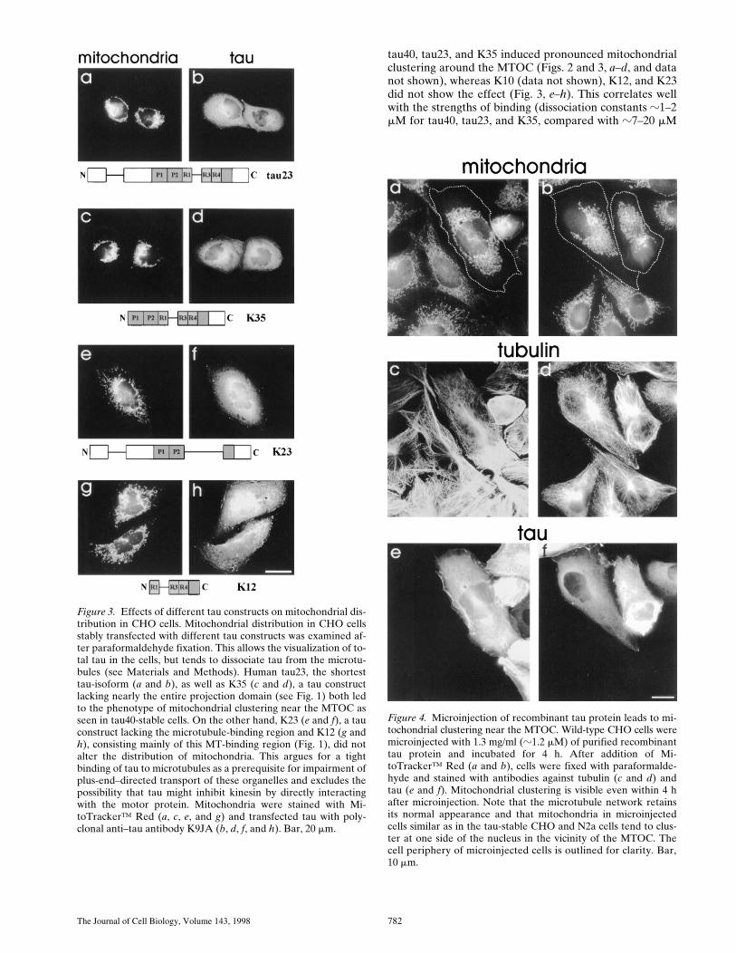
Recycling of Transferrin in Tau-overexpressing CHO Cells Is Slowed Down
Endo- and exocytotic processes depend on an intact mi-crotubule cytoskeleton. We therefore asked whether theoverexpression of tau influences these processes as well,using rhodamine-labeled transferrin as a marker. Trans-ferrin is endocytosed via transferrin receptors and clath-rin-coated vesicles and is actively transported along micro-tubules by motor proteins through early and sortingendosomes to recycling compartments in the pericentri-olar region of the cell. It is finally released by exocytosis,again via microtubule-dependent transport processes (Mar-tys et al., 1995). The efflux of transferrin was measured byremoval of the supernatant at different time points and byquantitating the exocytosed transferrin by fluorescence(Fig. 12 a). At early time points (a few minutes after re-moval of excess transferrin) the tau-stable cell linesshowed a significantly lower level of exocytosed transfer-rin than the control cells, but this difference disappearedlater on (z20 min). Thus, the outward flow of exocyticvesicles was retarded but not interrupted.
This observation was confirmed by quantitating the im-munofluorescence of residual TRITC-labeled transferrinin methanol-fixed tau-stable cells and mock-transfectedcontrols. Methanol-fixed tau-stable cells yielded a signifi-cantly higher amount of transferrin 7 min after removal oftransferrin-containing media compared with mock-trans-fected cells (Fig. 12 a; P , 0.0001, two-tailed Student’s ttest), mostly in the vicinity of the MTOC (the region of therecycling compartment; Martys et al., 1995). Whereasz50% of the initial intracellular transferrin had been exo-cytosed in mock-transfected cells, only 10% had been re-cycled in tau-stable cells during the same time period. Thiscorrelates well with the experiment shown in Fig. 12 b,where, conversely, the exocytosed transferrin was quanti-tated. Again, after z7 min, only 50% of transferrin wasexocytosed in tau-stable cells compared with control cells.
Transport of Mitochondria and EndoplasmicReticulum into the Neurites of DifferentiatedN2a-Neuroblastoma Cells Is Impaired by Overexpression of Tau
In the experiments described so far, we focussed on CHOcells to investigate tau’s effects on transport processes.The chief reasons were that CHO cells are well suited forstudying transport due to their flattened cell form andtheir size, which facilitates observation and microinjec-tion. Secondly, CHO cells do not normally express tau sothat the discrimination between endogenous MAPs (e.g.,MAP4) and stably introduced tau is much easier than inneuroblastoma cells. On the other hand, tau is expressedmainly in neuronal tissue, raising the question whether theeffects of tau overexpression manifest themselves in neu-ronal cells as well. For this reason, we stably transfected
Figure 10. Quantitation ofmitochondrial clustering af-ter overexpression of tau,drug treatment, and inhibi-tion of dynein. The distribu-tion of mitochondria wasquantified as described inMaterials and Methods. Aminimum of 30 cells were an-alyzed per experiment (n). Inmock-transfected cells, mito-chondria occupy up to 65%of the total cell area. The
same is true in cells stably transfected with K23, a tau constructlacking the microtubule-binding repeats (CHO-K23). This valuedrops to z30% in tau40-stable cells (CHO-tau). Treatment of thestably transfected cells with either nocodazole or taxotere re-stores the wild-type distribution of mitochondria (CHO-tau no-codazole or CHO-tau taxotere, respectively). The same effect canbe seen after transfection with dynamitin (CHO-tau transfectiondynamitin), but not after transfection of EGFP as a control pro-tein (CHO-tau transfection EGFP; P , 0.0001, two-tailed Stu-dent’s t test). Removal of nocodazole again leads to clustering ofmitochondria at the nucleus (CHO-tau noc. removal; 4 h).

Ebneth et al. Tau Inhibits Kinesin-dependent Vesicle Trafficking 787
human tau40 into the mouse neuroblastoma cell line N2a.Although the cell body of N2a cells is very compact andsmall (z10–20 mm, compared with z40 mm for CHO cells)so that it is difficult to detect differences in mitochondriallocalization, the organelles appear to be clustered in thevicinity of the nucleus in tau-stable compared with controlcells. Transport processes in the neurites of differentiatedN2a cells on the other hand can be monitored much moreeasily because transport deficits in these structures accu-mulate to a more significant extent. As shown by severalreports, tau protein is important for the generation of neu-rites, and transport of material into the neurites dependson kinesin (Baas et al., 1991; Feiguin et al., 1994).
Differentiated wild-type N2a cells contain abundant mi-tochondria in their neurites, extending up to the tip (Fig.13 d). In striking contrast, the neurites of differentiatedN2a cells transfected with tau showed very few mitochon-dria (Fig. 13 a and Table I). The remarkable depletion ofmitochondria from neuritic extensions in these cells indi-cates that tau overexpression in neuronal cells leads to the
same phenotype as seen in CHO cells, and again can beexplained by an impairment of plus-end–directed microtu-bule-dependent transport of mitochondria. The absence ofthese organelles would severely limit the energy supply ofneurites and could cause their degeneration. These obser-vations have implications for the cellular causes of Alz-heimer’s disease where elevated and mislocalized tau occursconcomitant with neuronal degeneration (as discussedlater).
Since the distribution of mitochondria was impaired indifferentiated N2a neuroblastoma cells overexpressingtau, we asked whether the ER was affected as well (judg-ing by immunofluorescence with an ER-specific antibody;Louvard et al., 1982). Control cells showed ER elementsthroughout the cell body mostly surrounding the nucleus,but visible in the neuritic structures. By contrast, the ma-jority of tau-overexpressing cells (z82%, Table I) showedthat the ER was collapsed towards the cell center, withoutextending around the nucleus (Fig. 14 a). The overall in-tensity of ER-staining remained the same in the two cell
Figure 11. The ER organiza-tion is altered in CHO cellsoverexpressing tau. Tau40-stable (a and b) and mock-transfected CHO cells (cand d) were stained withDiOC6(3) to visualize theER. In tau-stable cells, theER is less branched and doesnot reach the cell periphery,while in control cells the ERextends almost to the plasmamembrane and displays amore complex structure.Note that, although the ex-tension of the ER of thesmaller cell in Fig. 11 b seemsto be less effected when com-pared with the larger cells,the branching and complex-ity of the endoplasmic reticu-lum is significantly reducedwhen compared with mock-transfected cells in c and d.The cell periphery is outlinedfor clarity. Bar, 10 mm.
The Journal of Cell Biology, Volume 143, 1998 788
types. In particular, the ER extending into the neuriteswas strongly reduced in the tau-stable cells (compare Fig.14, a and d). Thus, an elevated level of tau in differentiatedN2a cells causes a retraction of the ER structure in the cellbody similar to the effect seen in CHO cells, leading to adecrease of the ER in neurites, presumably by affectingintracellular transport processes. Consistent with this,the neurites of tau-overexpressing cells are significantlyshorter than those of control cells (e.g., the number of neu-rites extending $30 mm decreases approximately three-fold, Table I).
Discussion
Tau Retards Plus-End–directed Transport of Mitochondria, ER, and Transport Vesicles Along Microtubules
In this study, we have investigated how an increased levelof tau protein (generated by overexpression or microinjec-tion) affects intracellular transport in CHO and neuroblas-toma cells. The main results can be summarized as follows:(a) increased tau leads to a clustering of mitochondria andperoxisomes near the MTOC (Fig. 2), to a contraction ofthe intermediate filament system and the ER (Fig. 11),and to a reduced rate of exocytosis (e.g., of transferrin re-ceptor, Fig. 12). The cells lose their elongated shape andbecome rounded (Fig. 2), and their growth rate slowsdown. Mitochondria and ER are decreased in neuritic pro-cesses (Figs. 13 and 14; Table I). (b) All of the above ef-fects are observed at relatively low levels of tau overex-pression where the microtubule network is not impaired
(no bundling, no detachment from MTOC); in fact, theydepend on the integrity of the microtubule network. Theeffects also depend on a strong binding of tau to microtu-bules (Kd z1 mM, provided by the repeats and proline-richflanking domain). (c) If the microtubule network is dis-rupted by drugs (i.e., disassembly by nocodazole, Fig. 5),or detachment of microtubules from MTOC and random-ization by taxotere (Fig. 8), the tau-induced changes in thedistribution of mitochondria are reversed so that the or-ganelles become dispersed throughout the cell again (Fig.10). (d) Inhibition of dynein (via overexpression of dyna-mitin) reverses the effects of tau overexpression and al-lows the dispersion of mitochondria (Figs. 9 and 10).
These effects can be explained by the assumption thattau interferes preferentially with the plus-end–directedtransport along microtubules involving kinesin, whilethe minus-end transport involving dynein is less affected.The directionality of organelle transport observed withthe tau-transfected cells is consistent with a numberof reports showing that plus-end–directed transport isachieved mainly by kinesin-like motors, and the reversetransport by dynein (Vallee and Sheetz, 1996; Rickard andKreis, 1996; Hirokawa, 1998; Lippincott-Schwartz, 1998).For example, mitochondria are transported by the kinesinmotors KIF1B (Nangaku et al., 1994) and KIF5B (Tanaka etal., 1998), or in the opposite direction by dynein (Hirokawa etal., 1990), and, therefore, inhibition of kinesin leads to anet retrograde transport of mitochondria. Rodionov et al.(1993) have shown that microinjection of an inhibitory ki-nesin antibody, which cross-reacts with several membersof the kinesin superfamily, leads to a strikingly similarphenotype of mitochondrial clustering. Furthermore, thetargeted disruption of mouse conventional kinesin heavychain KIF5B results in the perinuclear clustering of mito-chondria (Tanaka et al., 1998). Similarly, the extension ofthe ER and the intermediate filament system or the traf-ficking of vesicles is determined by a balance between ki-nesin- and dynein-dependent movements so that the ERand the vimentin-network collapses when kinesin is sup-pressed (Feiguin et al., 1994; Gyoeva and Gelfand, 1991).(The term “anterograde” is used for the plus-end–directedaxonal transport along microtubules, but also for the mi-nus-end–directed transport from ER to Golgi, so that weprefer the term “plus-end–directed” for the present dis-cussion.)
The function of tau or MAPs is usually discussed interms of their role in microtubule stabilization (Matus,1994), the generation of cell processes (Kosik and McCon-logue, 1994), in their spacer function between microtu-bules (Chen et al., 1992), or as anchoring sites for otherproteins (Obar et al., 1989; Ookata et al., 1995). The datapresented here point to a new function of tau; that is, regu-lation of transport within cells by the variation of the intra-cellular concentration of tau on the surface of microtu-bules. Since related effects have been observed withMAP4 (Bulinski et al., 1997), this may be a general func-tion of MAPs. In this context, it is of interest to note that,although MAP4 efficiently stabilizes microtubules in vitroand in vivo, the mitochondrial distribution in MAP4-over-expressing cells has been reported to not be altered(Nguyen et al., 1997). This argues against the possibilitythat a change in lifetime of microtubules accounts for the
Figure 12. Transferrin recycling is retarded by tau overexpres-sion. Cells were seeded on cover slips and preincubated withTMR-labeled transferrin in serum-free medium for 2 h. Excesstransferrin was removed and tau-stable and mock-transfectedcells were fixed in methanol. Residual transferrin fluorescencewas then quantitated and plotted (a). Note that, at early timepoints (7 min), the transferrin fluorescence in tau-stable cells isdecreased by z10%, whereas in control cells 50% of transferrinis exocytosed in the same time interval. 300 cells were quantitatedper time point and cell line. (b) Time course of the exocytosis oftransferrin after equilibration of tau-stable and mock-transfectedcells with fluorescently labeled transferrin. A significant decreaseof exocytosed transferrin can be seen 5–10 min after start of theexperiment in tau-stable compared with control cells. Fluores-cent signals were normalized to total cell numbers in each ex-periment. Data represent averages of four independent experi-ments.

Ebneth et al. Tau Inhibits Kinesin-dependent Vesicle Trafficking 789
effect of mitochondrial clustering, because the lifetimewould be increased both by MAP4 and tau. More impor-tantly, the comparison perhaps points to different func-tions of tau and MAP4 in vivo.
Inhibition of Motor Activity by MAPs
The question of whether and how MAPs and motors couldinterfere with each other on a microtubule has been thesubject of extended discussions, mainly because experi-mental evidence obtained in vitro remained ambiguous(e.g., Paschal et al., 1989; Von Massow et al., 1989; Rodi-onov et al., 1990; Lopez and Sheetz, 1993; Hagiwara et al.,1994). The results depended not only on the type of MAPsand motors used, their effective concentrations (in solu-tion or bound to a surface), but also on the experimentaldesign (microtubule gliding or bead movement assay). Themajority of the in vitro results suggested that tight bindingof MAPs to microtubules via the microtubule-binding do-
main and a large projection domain was a prerequisite forobserving any inhibition in motility. Thus, MAP2 could re-tard kinesin or dynein, while smaller MAPs (e.g., tau,MAP2c) had a much smaller effect or none at all (VonMassow et al., 1989; Lopez and Sheetz, 1993).
Conceptually, one can analyze the interference betweenMAPs and motors in terms of competition for commonbinding sites on a microtubule and/or in terms of sterichindrance (Lopez and Sheetz, 1993). A purely competitivemechanism would require only the microtubule-bindingdomain of MAPs (repeats plus flanking regions), whilesteric hindrance would depend on both microtubule bind-ing plus the NH2-terminal projection domain. Since sev-eral of the earlier studies had emphasized that largeprojection domains of MAPs were necessary for motor in-hibition, we were surprised to find that a small MAP suchas tau, overexpressed only moderately in cells, could be in-hibitory for plus-end–directed transport. This could beachieved even with constructs having no projection do-
Figure 13. Transport of mitochondria into neu-rites of differentiated neuroblastoma cells is im-paired by overexpression of tau. Tau40-stable(a–c) and mock-transfected N2a cells (d–f) weredifferentiated for 2 d by addition of 1 mM reti-noic acid into medium containing 0.1% fetalcalf serum and fixed in methanol. Mitochon-dria were visualized with MitoTracker™ Green(a and d), immunostaining was done with anti-bodies against tubulin (b and e) and tau (c andf). It can be clearly seen that significantly fewermitochondria are present in the neurites of tau-stable cells in contrast to control cells (comparea and d, and Table I). In addition, mitochon-dria in tau-stable cells appear to be more clus-tered in the vicinity of the MTOC (a), similar totau-stable CHO cells. The comparable stainingintensity of the cell bodies indicates that theclustering effect is not due to a lower numberof mitochondria in the tau-transfected cell. Bar,10 mm.

The Journal of Cell Biology, Volume 143, 1998 790
main (K35). Constructs with lower affinity generally hadno effect (K10, K12), even when the projection domainwas present, as in K23 (Fig. 3). Obviously the cellular envi-ronment reacts more sensitively to MAPs than the experi-mental systems used in vitro. This could be explained byseveral factors: in the crowded environment of a cell, evenshorter MAPs could have noticeable effects on transport,which would be most apparent for small motors (note thatKIF1B, responsible for mitochondrial transport, is only35-nm long, compared with 75 nm for conventional kine-sin; Nangaku et al., 1994; Scholey et al., 1989). Secondly,the periods of observation are long, compared with the invitro experiments, so that small differences in transportvelocities can accumulate to a measurable extent. Thirdly,the cellular effects result from an interplay between plus-and minus-end motors, which are differently affected byMAPs, while in vitro experiments were done with one typeof motor only. Finally, the in vitro studies are usually donewith an excess of motors that tend to overwhelm the resis-tance that MAPs might offer. A comparison between thedifferent isoforms and constructs of tau shows that the in-hibition of vesicle movement is dominated by the competi-tion between tau and motors for binding sites on the mi-crotubules, while steric hindrance is of lesser importance.
The direction of microtubule-based transport is thoughtto be determined by particular motor proteins anchored atthe organelle by means of adaptor complexes (e.g., kinec-tin, dynactin; Kumar et al., 1995; Burkhardt et al., 1997).The variety of motor proteins in cells makes it possible, inprinciple, for each type of vesicle or organelle to select oneor more type of motor, depending on the required destina-tion (Hirokawa, 1998; Hamm-Alvarez et al., 1993). In spiteof attached motors, the movement of vesicles is not simplyunidirectional, but rather saltatory, with frequent detach-ments and even reversals (Wacker et al., 1997). The netmovement is therefore the result of many trials, biased in aparticular direction. Inside a cell, MAPs could inhibit theactivity of a motor in several ways; e.g., by preventing theinitial attachment, by reducing the velocity, or by reducingthe run length. In the present context, the important pointis that tau most likely affects the balance between differ-
ent directions, impairing the plus-end–directed move-ments of mitochondria more than the minus-end–directedones, and this results in the clustering of these organellesnear the MTOC, whereas the phenotype of ER retractionor the slow down in the export of transferrin might beachieved by simply reducing the overall transport pro-cesses or influencing the run length of motor proteins onmicrotubules.
Besides saltatory particle movement with directionalbias, one should consider other mechanisms as well. Forexample, extension of the ER network can be driven bymicrotubule polymerization where the tips of microtu-bules are attached to the membrane by special attachmentcomplexes (Allan and Vale, 1994; Waterman-Storer et al.,1995), and anaphase transport of chromosomes can bedriven by microtubule depolymerization (Lombillo et al.,1995; Merdes et al., 1997). The attachment complexes cancontain motor-like proteins that, in this case, operate asmicrotubule anchors rather than as independent motors.In practice, motor- and assembly-driven mechanisms maycoexist within the cell (Rodionov and Borisy, 1997). How-ever, in our case, the direct motor-dependent processesappear to be the dominant ones, as shown by the dy-namitin-induced inhibition of dynein (Figs. 9 and 10;Burkhardt et al., 1997). Moreover, since tau stabilizes mi-crotubules, it would be expected to enhance plus-end–directed transport if this were assembly driven, rather thaninhibiting it as observed. Finally, we note that the distribu-tion of organelles in the cytoplasm can be influenced bytwo further mechanisms, diffusion and actomyosin-basedmovement. The latter appears to be restricted to special-ized regions (e.g., near the cell cortex or the axon; Morrisand Hollenbeck, 1995; Mermall et al., 1998), but a diffu-sional component is likely to operate at all times. It ex-plains the dispersion of clustered mitochondria upon mi-crotubule disassembly by nocodazole, but this is not likelyto be affected by tau.
Having determined the effects of tau overexpression inCHO cells, an obvious question was whether analogous ef-fects would be observable in cells of neuronal origin. Inthis case, the experimental approaches are more limitedbecause of the smaller size of the cells. For example, a re-distribution of organelles in the cell body is difficult to ob-serve and the level of endogenous tau is too low for manybiochemical assays. On the other hand, vesicles and or-ganelles are easily detectable in the long neuritic processesof differentiated cells. Thus, the neurites of control N2acells show staining with the markers of mitochondria andthe ER, but N2a cells overexpressing tau only at a signifi-cantly lower level. In agreement with the previous inter-pretation, this means that plus-end–directed transport, atleast in the case of mitochondria, is impaired, leading to anaccumulation of these elements in the cell body.
The effect of tau on vesicle trafficking described hererepresents a new function that has attracted little attentionthus far. This was due in part to the fact that the influenceof MAPs on the activity of motor proteins were mainly in-vestigated in vitro, showed only weak effects, visible onlyat high MAP concentrations, and were apparently re-stricted to high molecular weight MAPs (Paschal et al.,1989; Von Massow et al., 1989; Lopez and Sheetz, 1993;Hagiwara et al., 1994). Nevertheless, in the context of
Table I. Quantitation of the Phenotypic Effects of Tau Overexpression in N2a-Neuroblastoma Cells
N2a tau40 N2a mock
Neurites 5.4% 15%(length .30 mm)Mitochondria in neurites 17% 94%(.6 per neurite)ER distribution 82% 23%(clustered)
A minimum of 150 cells were analyzed in four (neurite length and mitochondria) orthree (ER distribution) independent experiments. In differentiated, mock-transfectedcells, 15% of the cells developed neurites longer than 30 mm, whereas, in tau-stablecells, this value drops to only 5.4%. The effect of tau-overexpression becomes evenmore impressive when analyzing the mitochondria present in the neuritic structures:only 17% of tau-stable cells transported more than six mitochondria into their neu-rites, compared with 94% of control cells. The average number of mitochondria in thecontrol cells was z10–20 per neurite (data not shown). The ER was regarded as clus-tered when it did not surround the nucleus of the cells. In tau-stable cells, up to 82%displayed this phenotype. In contrast, in mock-transfected cells, only 23% of cells hada similar appearance.

Ebneth et al. Tau Inhibits Kinesin-dependent Vesicle Trafficking 791
cells, the subtle effects become magnified and visible as re-distribution of cellular components. The most importantprerequisite for tau’s influence on intracellular traffic isthe tight binding to microtubules due to the repeats andthe flanking domains (z1–2 mM) while the projection do-main has only a minor effect. This implies that the phos-phorylation of tau should play an important role since thisregulates tau’s affinity to microtubules. One can broadlydistinguish two types of phosphorylation of tau, proline di-rected (affecting Ser-Pro or Thr-Pro motifs and inducedby proline-directed kinases such as MAP kinase, GSK3,cdk5) and non–proline directed (induced by second-mes-senger–dependent kinases such as PKA, PKC, CaMK,MARK; for review see Mandelkow et al., 1995). Proline-directed phosphorylation has only a moderate influence ontau-microtubule interactions (Biernat et al., 1993), whereastwo other sites potently disrupt the interaction, Ser262(phosphorylated by MARK; Drewes et al., 1997) and
Ser214 (phosphorylated by PKA; Zheng-Fischhöfer et al.,1998; both these sites are enhanced in Alzheimer’s tau).Thus far, such sites were mainly seen as modulators ofmicrotubule stability. However, in the light of our presentresults, one should also view the MAP-microtubule affin-ity-regulating phosphorylation reactions as modulators ofintracellular traffic. In this scenario, tau bound to microtu-bules would present resistance to plus-end–directed move-ment, which would be relieved by tau’s phosphorylationand detachment from microtubules. Since the outwardflow of material is a prerequisite for cell polarity and dif-ferentiation (Feiguin et al., 1994; Bradke and Dotti, 1997),the activity of tau kinases would become important at thispoint. It is interesting to note that vesicle transport can beinhibited by MAP2, but relieved by a kinase that is proba-bly related to MARK (Drewes et al., 1997; Lopez andSheetz, 1995), a kinase that phosphorylates MAP2c andMAP4 in vitro (Illenberger et al., 1996), and therefore can
Figure 14. The expansion of the endoplasmicreticulum in differentiated neuroblastoma cells issignificantly reduced by overexpression of tau.Tau-stable N2a cells (a–c) or mock-transfectedcells (d–f) were induced to differentiate, andthen fixed in paraformaldehyde and triple-la-beled with antibodies against the ER (a and d),tubulin (b and e), and tau (c and f). In contrast tocontrol cells, z82% of tau-stable neuroblastomacells show the ER clustered at one side of the nu-cleus (a), whereas in mock-transfected cells theER surrounds the entire nucleus (d; compare Ta-ble I). Furthermore, the expansion of the ERinto the neurites in control cells (d) can clearlybe seen. In tau-stable cells, only a faint signal ofER-immunoreactivity is visible in neuritic pro-cesses. The total intensity of the ER stain is thesame in both cells. Arrowheads in a and d are in-dicating the position of the neurite relative to thecell body. Bar, 10 mm.

The Journal of Cell Biology, Volume 143, 1998 792
be expected to regulate the affinity of MAPs to microtu-bules in general. This points to another interesting obser-vation: it has been shown that MAP1A can compensatefor the lack of tau in knock-out mice (Harada et al., 1994),suggesting that the function of MAPs is partially redun-dant. Whereas axonal elongation was not affected in theseanimals, microtubule stability was decreased in some ax-ons. It is conceivable that different MAPs regulate thetransport of different organelles and in different cell com-partments. Thus, it would be interesting to correlate thedistribution of organelles and MAPs in mouse modelslacking or overexpressing MAPs to get further clues forthe functions of MAPs in intracellular transport.
Implications for Neuronal Degeneration inAlzheimer’s Disease
The effects of tau overexpression on mitochondrial dis-tribution, vesicle-trafficking, and the ER have importantimplications for Alzheimer’s disease. In AD, tau is ab-normally phosphorylated, aggregated into paired helicalfilaments, and mislocalized from the axon to the somato-dendritic compartment (Binder et al., 1985; Wischik et al.,1988; Braak et al., 1994). In addition, it has been reportedthat intracellular tau-protein concentration in AD brain iselevated (Khatoon et al., 1992). In neurons, this mislocal-ization and elevated intracellular concentration of tauwould affect the transport of mitochondria or ER to themost peripheral regions of the axons and would lead to adecrease in glucose metabolism and ATP synthesis inthese compartments, causing energy deficits, loss of Ca21
homeostasis, or lipid synthesis (Futerman and Banker,1996; Mattson and Guo, 1997). Finally, this may lead todegeneration of the neuron, presumably beginning fromthe most distal regions, leading to the so called “dyingback” of axons (Braak et al., 1994; Trojanowski and Lee,1995). In this context, it is of special interest that compro-mised mitochondrial function has indeed been shown toassociate with late onset Alzheimer’s disease (Davis et al.,1997). In addition, a slowing down of transport processescould affect the proper distribution of proteins, particu-larly of amyloid precursor protein (APP), another keyplayer in the progression of Alzheimer’s disease that is re-routed by transcytosis (Simons et al., 1995), and whoseproteolytic processing to the amyloidogenic peptide Ab1-42takes place in the endoplasmic reticulum (Cook et al.,1997; Hartmann et al., 1997). Thus, a transport inhibitiondue to the overexpression and mislocalization of tau inneurons might increase production of Ab1-42. Conversely,overproduction of amyloidogenic variants of APP corre-lates with an increased level of tau protein and tau mRNA(Lee et al., 1996), and, in the Alzheimer’s-related Downsyndrome, APP and tau are both upregulated (Oyama et al.,1994). Finally, the mutant presenilins PS1 and PS2 that areresponsible for familial Alzheimer’s disease have beenshown to interact with APP in the endoplasmic reticulum,thereby enhancing the production of Ab (Xia et al., 1997).Thus, future work might give new clues to an understand-ing of Alzheimer’s disease since it might experimentallylink the function of AD-specific gene products, such asAPP and the presenilins, with mitochondrial energy sup-ply and tau protein.
We thank I. Thielke and K. Alm for excellent technical assistance, and J.Biernat for generously providing the tau constructs. We gratefully ac-knowledge the gift of ER antibody from D. Louvard (Institut Curie, Paris,France), the gift of dynamitin (p50) expression plasmid, and the antidyna-mitin 50-1 antibody from Dr. R.B. Vallee (Worcester Foundation,Shrewsbury, MA), and the gift of the anticatalase antibody from Dr. W.Just. This article is based in part on the doctoral thesis by R. Godemannand K. Stamer in the Faculty of Biology, University of Hamburg (Ham-burg, Germany).
This project was supported by grants from the Deutsche Forschungsge-meinschaft.
Received for publication 10 April 1998 and in revised form 11 September1998.
References
Allan, V.J., and R.D. Vale. 1994. Movement of membrane tubules along micro-tubules in vitro: evidence for specialized sites of motor attachment. J. CellSci. 107:1885–1897.
Baas, P.W., T. Slaughter, A. Brown, and M.M. Black. 1991. Microtubule dy-namics in axons and dendrites. J. Neurosci. Res. 30:134–153.
Barlow, S., M.L. Gonzalezgaray, R.R. West, J.B. Olmsted, and F. Cabral. 1994.Stable expression of heterologous microtubule-associated proteins (MAPs)in chinese-hamster ovary cells—evidence for differing roles of MAPs in mi-crotubule organization. J. Cell Biol. 126:1017–1029.
Biernat, J., N. Gustke, G. Drewes, E.-M. Mandelkow, and E. Mandelkow. 1993.Phosphorylation of Ser262 strongly reduces binding of tau to microtubules:distinction between PHF-like immunoreactivity and microtubule binding.Neuron. 11:153–163.
Binder, L.I., A. Frankfurter, and L.I. Rebhun. 1985. The distribution of tau inthe mammalian central nervous system. J. Cell Biol. 101:1371–1378.
Braak, H., E. Braak, and E.-M. Mandelkow. 1994. A sequence of cytoskeletonchanges related to the formation of neurofibrillary tangles and neuropilthreads. Acta Neuropathol. 87:554–567.
Bradke, F., and C. Dotti. 1997. Neuronal polarity: vectorial cytoplasmic flowprecedes axon formation. Neuron. 19:1175–1186.
Brady, S.T. 1995. Biochemical and functional diversity of microtubule motors inthe nervous system. Curr. Opin. Neurobiol. 5:551–558.
Brandt, R., J. Leger, and G. Lee. 1995. Interaction of tau with the neural plasmamembrane mediated by tau’s amino-terminal projection domain. J. CellBiol. 131:1327–1340.
Bulinski, J.C., and G.G. Borisy. 1979. Self-assembly of microtubules in extractsof cultured HeLa cells and the identification of HeLa microtubule-associ-ated proteins. Proc. Natl. Acad. Sci. USA. 76:293–297.
Bulinski, J.C., T.E. McGraw, D. Gruber, H.-L. Nguyen, and M.P. Sheetz. 1997.Overexpression of MAP4 inhibits organelle motility and trafficking in vivo.J. Cell Sci. 110:3055–3064.
Burkhardt, J.K., C.J. Echeverri, T. Nilsson, and R.B. Vallee. 1997. Overexpres-sion of the dynamitin (p50) subunit of the dynactin complex disrupts dynein-dependent maintenance of membrane organelle distribution. J. Cell Biol.139:469–484.
Chapin, S.J., and J.C. Bulinski. 1991. Non-neuronal 2103103 Mr microtubuleassociated protein (MAP4) contains a domain homologous to the microtu-bule-binding domains of neuronal MAP2 and tau. J. Cell Sci. 98:27–36.
Chapin, S.J., and J.C. Bulinski. 1992. Microtubule stabilization by assembly-promoting microtubule-associated proteins: a repeat performance. Cell Mo-til. Cytoskelet. 23:236–243.
Chen, J., Y. Kanai, and N. Hirokawa. 1992. Projection domains of MAP2 andtau determine spacings between microtubules in dendrites and axons. Na-ture. 360:674–677.
Cook, D.G., M.S. Forman, J.C. Sung, S. Leight, D.L. Kolson, T. Iwatsubo,V.M.-Y. Lee, and R.W. Doms. 1997. Alzheimer’s Abeta(1-42) is generatedin the endoplasmic reticulum/intermediate compartment of NT2N cells. Nat.Med. 3:1021–1023.
Davis, E.D., S. Miller, C. Herrnstadt, S.S. Ghosh, E. Fahy, L.A. Shinobu, D.Galasko, L.J. Thal, M.F. Beal, N. Howell, and W.D. Parker, Jr. 1997. Muta-tions in mitochondrial cytochrome c oxidase genes segregate with late-onsetAlzheimer’s disease. Proc. Natl. Acad. Sci. USA. 94:4526–4531.
Drechsel, D.N., A.A. Hyman, M.H. Cobb, and M.W. Kirschner. 1992. Modula-tion of the dynamic instability of tubulin assembly by the microtubule-asso-ciated protein tau. Mol. Cell. Biol. 3:1141–1154.
Drewes, G., A. Ebneth, U. Preuss, E.-M. Mandelkow, and E. Mandelkow. 1997.MARK, a novel family of protein kinases that phosphorylate microtubule-associated proteins and trigger microtubule disruption. Cell. 89:297–308.
Drubin, D.G., S.C. Feinstein, E.M. Shooter, and M.W. Kirschner. 1985. Nervegrowth factor-induced neurite outgrowth in PC12 cells involves the coordi-nate induction of microtubule assembly and assembly-promoting factors. J.Cell Biol. 101:1799–1807.
Drubin, D.G., and W.J. Nelson. 1996. Origins of cell polarity. Cell. 84:335–344.Feiguin, F., A. Ferreira, K.S. Kosik, and A. Caceres. 1994. Kinesin-mediated or-

Ebneth et al. Tau Inhibits Kinesin-dependent Vesicle Trafficking 793
ganelle translocation revealed by specific cellular manipulations. J. Cell Biol.127:1021–1039.
Futerman, A.H., and G.A. Banker. 1996. The economics of neurite out-growth—the addition of new membrane to growing axons. Trends Neurosci.19:144–149.
Goedert, M., M.G. Spillantini, M.C. Potier, J. Ulrich, and R.A. Crowther. 1989.Cloning and sequencing of the cDNA encoding an isoform of microtubule-associated protein tau containing four tandem repeats: differential expres-sion of tau protein mRNAs in human brain. EMBO (Eur. Mol. Biol. Organ.)J. 8:393–399.
Goodson, H.V., C. Valetti, and T.E. Kreis. 1997. Motors and membrane traffic.Curr. Opin. Cell Biol. 9:18–28.
Gustke, N., B. Trinczek, J. Biernat, E.-M. Mandelkow, and E. Mandelkow.1994. Domains of tau protein and interactions with microtubules. Biochemis-try. 33:9511–9522.
Gyoeva, F.K., and V.I. Gelfand. 1991. Coalignment of vimentin intermediatefilaments with microtubules depends on kinesin. Nature. 353:445–448.
Hagiwara, H., H. Yorifuji, R. Sato-Yoshitake, and N. Hirokawa. 1994. Compe-tition between motor molecules (kinesin and cytoplasmic dynein) and fi-brous microtubule-associated proteins in binding to microtubules. J. Biol.Chem. 269:3581–3589.
Hamm-Alvarez, S.F., P.Y. Kim, and M.P. Sheetz. 1993. Regulation of vesicletransport in CV-1 cells and extracts. J. Cell Sci. 106:955–966.
Harada, A., K. Oguchi, S. Okabe, J. Kuno, S. Terada, T. Ohshima, R. Sato-Yoshitake, Y. Takei, T. Noda, and N. Hirokawa. 1994. Altered microtubuleorganization in small-calibre axons of mice lacking tau protein. Nature. 369:488–491.
Hartmann, T., S.C. Bieger, B. Bruhl, J.P. Tienari, N. Ida, D. Allsop, G.W. Rob-erts, C.L. Masters, C.G. Dotti, K. Unsicker, and K. Beyreuther. 1997. Dis-tinct sites of intracellular production for Alzheimer’s disease Abeta40/42amyloid peptides. Nat. Med. 3:1016–1020.
Hiller, G., and K. Weber. 1978. Radioimmunoassay for tubulin: a quantitativecomparison of the tubulin content of different established tissue culture cellsand tissues. Cell. 14:795–804.
Hirokawa, N., R. Sato-Yoshitake, T. Yoshida, and T. Kawashima. 1990. Braindynein (MAP1c) localizes on both anterogradely and retrogradely trans-ported membranous organelles in vivo. J. Cell Biol. 111:1027–1037.
Hirokawa, N. 1994. Microtubule-organization and dynamics dependent on mi-crotubule-associated proteins. Curr. Opin. Cell Biol. 6:74–81.
Hirokawa, N. 1998. Kinesin and dynein superfamily proteins and the mecha-nism of organelle transport. Science. 279:519–526.
Hollenbeck, P., and J. Swanson. 1990. Radial extension of macrophage tubularlysosomes supported by kinesin. Nature. 346:864–866.
Illenberger, S., G. Drewes, B. Trinczek, J. Biernat, H. Meyer, J. Olmsted, E.-M.Mandelkow, and E. Mandelkow. 1996. Phosphorylation of microtubule-associated proteins MAP2 and MAP4 by the protein kinase p110mark. J.Biol. Chem. 271:10834–10843.
Illenberger, S., Q. Zheng-Fischhöfer, U. Preuss, K. Stamer, K. Baumann, B.Trinczek, J. Biernat, R. Godemann, E.-M. Mandelkow, and E. Mandelkow.1998. The endogenous and cell-cycle dependent phosphorylation of tau pro-tein in living cells: implications for Alzheimer’s disease. Mol. Biol. Cell.9:1495–1512.
Jordan, M.A., and L. Wilson. 1998. Microtubules and actin filaments: dynamictargets for cancer chemotherapy. Curr. Opin. Cell Biol. 10:123–130.
Kaech, S., B. Ludin, and A. Matus. 1996. Cytoskeletal plasticity in cells express-ing neuronal microtubule-associated proteins. Neuron. 17:1189–1199.
Kanai, Y., R. Takemura, T. Oshima, H. Mori, Y. Ihara, M. Yanagisawa, T.Masaki, and N. Hirokawa. 1989. Expression of multiple tau isoforms and mi-crotubule bundle formation in fibroblasts transfected with a single taucDNA. J. Cell Biol. 109:1173–1184.
Khatoon, S., I. Grundke-Iqbal, and K. Iqbal. 1992. Brain levels of microtubule-associated protein tau are elevated in Alzheimer’s disease: a radioimmuno-slot-blot assay for nanograms of the protein. J. Neurochem. 59:750–753.
Klymkowski, M., J. Bachant, and A. Domingo. 1989. Functions of intermediatefilaments. Cell Motil. Cytoskelet. 14:309–331.
Kosik, K.S., and L. McConlogue. 1994. Microtubule-associated protein func-tion: lessons from expression in spodoptera frugiperda cells. Cell Motil. Cy-toskelet. 28:195–198.
Kumar, J., H. Yu, and M.P. Sheetz. 1995. Kinectin, an essential anchor for kine-sin-driven vesicle motility. Science. 267:1834–1837.
Lee, G., and S. Rook. 1992. Expression of tau protein in non-neuronal cells: mi-crotubule binding and stabilization. J. Cell Sci. 102:227–237.
Lee, R.K.K., R.J. Wurtman, B.P.S. Yang, and R. Neve. 1996. Increased tau pro-tein in primary cortical astrocytes expressing human Ab(1-42) and APP751.Soc. Neurosci. Abstr. 22:1419.
Li, Y., and M.M. Black. 1996. Microtubule assembly and turnover in growingaxons. J. Neurosci. 16:531–544.
Lippincott-Schwartz, J. 1998. Cytoskeletal proteins and Golgi dynamics. Curr.Opin. Cell Biol. 10:52–59.
Lombillo, V.A., R.J. Stewart, and J.R. McIntosh. 1995. Minus-end-directed mo-tion of kinesin-coated microspheres driven by microtubule depolymeriza-tion. Nature. 373:161–164.
Lopez, L., and M. Sheetz. 1993. Steric inhibition of cytoplasmic dynein and ki-nesin motility by MAP2. Cell Motil. Cytoskelet. 24:1–16.
Lopez, L.A., and M.P. Sheetz. 1995. A microtubule-associated protein (MAP2)
kinase restores microtubule motility in embryonic brain. J. Biol. Chem. 270:12511–12517.
Louvard, D., H. Reggio, and G. Warren. 1982. Antibodies to the Golgi complexand the rough endoplasmic reticulum. J. Cell Biol. 92:92–107.
Mandelkow, E., and E.-M. Mandelkow. 1995. Microtubules and microtubule-associated proteins. Curr. Opin. Cell Biol. 7:72–81.
Mandelkow, E.-M., J. Biernat, G. Drewes, N. Gustke, B. Trinczek, and E. Man-delkow. 1995. Tau domains, phosphorylation, and interactions with microtu-bules. Neurobiol. Aging. 16:355–362.
Mandell, J.W., and G. Banker. 1996. A spatial gradient of tau protein phosphor-ylation in nascent axons. J. Neurosci. 16:5727–5740.
Martys, J.L., T. Shevell, and T.E. McGraw. 1995. Studies of transferrin recy-cling reconstituted in streptolysin O permeabilized Chinese hamster ovarycells. J. Biol. Chem. 270:25976–25984.
Mattson, M.P., and Q. Guo. 1997. Cell and molecular neurobiology of preseni-lins: a role for the endoplasmic reticulum in the pathogenesis of Alzheimer’sdisease? J. Neurosci. Res. 50:505–513.
Matus, A. 1994. Stiff microtubules and neuronal morphology. Trends Neurosci.17:19–22.
McNiven, M.A., and K.R. Porter. 1986. Microtubule polarity confers directionto pigment transport in chromatophores. J. Cell Biol. 103:1547–1555.
Merdes, A., and D.W. Cleveland. 1997. Pathways of spindle pole formation: dif-ferent mechanisms; conserved components. J. Cell Biol. 138:953–956.
Mermall, V., P.L. Post, and M.S. Mooseker. 1998. Unconventional myosins incell movement, membrane traffic, and signal transduction. Science. 279:527–533.
Morris, R.L., and P.J. Hollenbeck. 1993. The regulation of bidirectional mito-chondrial transport is coordinated with axonal outgrowth. J. Cell Sci. 104:917–927.
Morris, R.L., and P.J. Hollenbeck. 1995. Axonal transport of mitochondriaalong microtubules and F-actin in living vertebrate neurons. J. Cell Biol. 131:1315–1326.
Nangaku, M., R. Sato-Yoshitake, Y. Okada, Y. Noda, R. Takemura, H.Yamazaki, and N. Hirokawa. 1994. KIF1B, a novel microtubule plus end-directed monomeric motor protein for transport of mitochondria. Cell. 79:1209–1220.
Nguyen, H.-L., S. Chari, D. Gruber, C.-M. Lue, S.J. Chapin, and J.C. Bulinski.1997. Overexpression of full- or partial-length MAP4 stabilizes microtubulesand alters cell-growth. J. Cell Sci. 110:281–294.
Obar, R.A., J. Dingus, H. Bayley, and R.B. Vallee. 1989. The RII subunit ofcAMP-dependent protein kinase binds to a common amino-terminal domainin microtubule-associated proteins 2A, 2B, and 2C. Neuron. 3:639–645.
Olson, K.R., J.R. McIntosh, and J.B. Olmsted. 1995. Analysis of MAP 4 func-tion in living cells using green fluorescent protein (GFP) chimeras. J. CellBiol. 130:639–650.
Ookata, K., S. Hisanaga, J.C. Bulinski, H. Murofushi, H. Aizawa, T.J. Itoh, H.Hotani, E. Okumura, K. Tachibana, and T. Kishimoto. 1995. Cyclin B inter-action with microtubule-associated protein 4 (MAP4) targets p34cdc2 kinaseto microtubules and is a potential regulator of M-phase microtubule dynam-ics. J. Cell Biol. 128:849–862.
Oyama, F., N.J. Cairns, H. Shimada, R. Oyama, K. Titani, and Y. Ihara. 1994.Down’s syndrome: up-regulation of beta-amyloid protein precursor and taumRNAs and their defective coordination. J. Neurochem. 62:1062–1066.
Paschal, B.M., R.A. Obar, and R.B. Vallee. 1989. Interaction of brain cytoplas-mic dynein and MAP2 with a common sequence at the C terminus of tubu-lin. Nature. 342:569–572.
Preuss, U., F. Doring, S. Illenberger, and E.-M. Mandelkow. 1995. Cell-cycledependent phosphorylation and microtubule-binding of tau-protein stablytransfected into Chinese-hamster ovary cells. Mol. Biol. Cell. 6:1397–1410.
Preuss, U., J. Biernat, E.-M. Mandelkow, and E. Mandelkow. 1997. The ‘jaws’model of tau-microtubule interaction examined in CHO cells. J. Cell Sci.110:789–800.
Rickard, J.E., and T.E. Kreis. 1996. Clips for organelle-microtubule interac-tions. Trends Cell Biol. 6:178–183.
Rodionov, V., F. Gyoeva, A. Kashina, S. Kuznetsov, and V.I. Gelfand. 1990.Microtubule-associated proteins and microtubule-based translocator havedifferent binding-sites on tubulin molecule. J. Biol. Chem. 265:5702–5707.
Rodionov, V.I., F.K. Gyoeva, E. Tanaka, A.D. Bershadsky, J.M. Vasiliev, andV.I. Gelfand. 1993. Microtubule-dependent control of cell-shape andpseudopodial activity is inhibited by the antibody to kinesin motor domain.J. Cell Biol. 123:1811–1820.
Rodionov, V.I., and G.G. Borisy. 1997. Microtubule treadmilling in vivo. Sci-ence. 275:215–218.
Sato-Harada, R., S. Okabe, T. Umeyama, Y. Kanai, and N. Hirokawa. 1996.Microtubule-associated proteins regulate microtubule function as the trackfor intracellular membrane organelle transports. Cell Struct. Funct. 21:283–295.
Scales, S.J., R. Pepperkok, and T.E. Kreis. 1997. Visualization of ER-to-Golgitransport in living cells reveals a sequential mode of action for COPII andCOPI. Cell. 90:1137–1148.
Scholey, J.M., J. Heuser, J.T. Yang, and L.S. Goldstein. 1989. Identification ofglobular mechanochemical heads of kinesin. Nature. 338:355–357.
Simons, M., E. Ikonen, P.J. Tienari, A. Cid-Arregui, U. Monning, K. Bey-reuther, and C. Dotti. 1995. Intracellular routing of human amyloid proteinprecursor: axonal delivery followed by transport to the dendrites. J. Neuro-sci. Res. 41:121–128.

The Journal of Cell Biology, Volume 143, 1998 794
Summerhayes, I.C., D. Wong, and L.B. Chen. 1983. Effect of microtubules andintermediate filaments on mitochondrial distribution. J. Cell Sci. 61:87–105.
Tanaka, Y., Y. Kanai, Y. Okada, S. Nonaka, S. Takeda, A. Harada, and N. Hi-rokawa. 1998. Targeted disruption of mouse conventional kinesin heavychain, kif5B, results in abnormal perinuclear clustering of mitochondria.Cell. 93:1147–1158.
Terasaki, M., and T.S. Reese. 1994. Interactions among endoplasmic reticulum,microtubules, and retrograde movements of the cell surface. Cell Motil. Cy-toskelet. 29:291–300.
Trojanowski, J.Q., and V.M.-Y. Lee. 1995. Phosphorylation of paired helical fil-ament tau in Alzheimer’s disease neurofibrillary lesions: focusing on phos-phatases. FASEB J. 9:1570–1576.
Vale, R.D., and H. Hotani. 1988. Formation of membrane networks in vitro bykinesin-driven microtubule movement. J. Cell Biol. 107:2233–2241.
Vallee, R.B., K.T. Vaughan, and C.J. Echeverri. 1995. Targeting of cytoplasmicdynein to membranous organelles and kinetochores via dynactin. ColdSpring Harbor Symp. Quant. Biol. 60:803–811.
Vallee, R.B., and M.P. Sheetz. 1996. Targeting of motor proteins. Science. 271:1539–1544.
Von Massow, A., E.-M. Mandelkow, and E. Mandelkow. 1989. Interaction be-tween kinesin, microtubules, and microtubule-associated protein 2. Cell Mo-til. Cytoskelet. 14:562–571.
Wacker, I., C. Kaether, A. Kromer, A. Migala, W. Almers, and H.H. Gerdes.1997. Microtubule-dependent transport of secretory vesicles visualized inreal time with a GFP-tagged secretory protein. J. Cell Sci. 110:1453–1463.
Waterman-Storer, C.M., J. Gregory, S.F. Parsons, and E.D. Salmon. 1995.Membrane/microtubule tip attachment complexes (TACs) allow the assem-
bly dynamics of plus ends to push and pull membranes into tubulovesicularnetworks in interphase Xenopus egg extracts. J. Cell Biol. 130:1161–1169.
Waterman-Storer, C., and E.D. Salmon. 1997. Microtubule dynamics: tread-milling comes around again. Curr. Biol. 7:R369–R372.
Weisshaar, B., T. Doll, and A. Matus. 1992. Reorganization of the microtubularcytoskeleton by embryonic microtubule-associated protein 2 (MAP2c). De-velopment (Camb.). 116:1151–1161.
West, R.R., K.M. Tenbarge, and J.B. Olmsted. 1991. A model for microtubule-associated protein 4 structure. Domains defined by comparisons of human,mouse, and bovine sequences. J. Biol. Chem. 266:21886–21896.
Wiemer, E.A.C., T. Wenzel, T.J. Deerinck, M.H. Ellisman, and S. Subramani.1997. Visualization of the peroxisomal compartment in living mammaliancells: dynamic behavior and association with microtubules. J. Cell Biol. 136:71–80.
Wischik, C.M., M. Novak, H.C. Thogersen, P.C. Edwards, M.J. Runswick, R.Jakes, J.E. Walker, C. Milstein, M. Roth, and A. Klug. 1988. Isolation of afragment of tau derived from the core of the paired helical filament ofAlzheimer’s disease. Proc. Natl. Acad. Sci. USA. 85:4506–4510.
Xia, W., J. Zhang, R. Perez, E. Koo, and D.J. Selkoe. 1997. Interaction betweenamyloid precursor protein and presenilins in mammalian cells: implicationsfor the pathogenesis of Alzheimer’s disease. Proc. Natl. Acad. Sci. USA. 94:8208–8213.
Zheng-Fischhöfer, Q., J. Biernat, E.-M. Mandelkow, S. Illenberger, R. Gode-mann, E. Mandelkow. 1998. Sequential phosphorylation of tau-protein byGSK-3b and protein kinase A at Thr212 and Ser214 generates the Alz-heimer-specific epitope of antibody AT100 and requires a paired helical fila-ment-like conformation. Eur. J. Biochem. 252:542–552.
Copyright © 2022 FDOKUMEN