The kinesin KIF1C and microtubule plus ends regulate podosome dynamics in macrophages
49
The kinesin KIF1C and microtubule plus-ends regulate podosome dynamics in macrophages Petra Kopp * , Reiner Lammers † , Martin Aepfelbacher ‡ , Günther Woehlke § , Thomas Rudel ║ , Nikolaus Machuy ║ , Walter Steffen Π , and Stefan Linder *# * Institut für Prophylaxe und Epidemiologie der Kreislaufkrankheiten, Ludwig-Maximilians- Universität, 80336 München, Germany, † Medizinische Klinik IV, Universität Tübingen, 72076 Tübingen, Germany, ‡ Institut für Infektionsmedizin, Universitätsklinikum Hamburg Epppendorf, 20246 Hamburg, Germany, § Institut für Zellbiologie, Ludwig-Maximilians- Universität, 80336 München, Germany, ║ Max-Planck-Institut für Infektionsbiologie, Schumannstr. 21/22, 10117 Berlin, Germany, Π Institut für Molekular- und Zellphysiologie, Medizinische Hochschule Hannover, 30625 Hannover, Germany running head : KIF1C and microtubules regulate podosome dynamics key words: podosomes, microtubules, kinesin, actin cytoskeleton # corresponding author: Stefan Linder Institut für Prophylaxe und Epidemiologie der Kreislaufkrankheiten, Ludwig-Maximilians- Universität, Pettenkoferstr. 9, 80336 München, Germany Tel.: +49 89 5160 4371 Fax: +49 89 5160 4382 email : [email protected] http://www.molbiolcell.org/content/suppl/2006/03/22/E05-11-1010.DC1.html Supplemental Material can be found at:
-
Upload
independent -
Category
Documents
-
view
0 -
download
0
Transcript of The kinesin KIF1C and microtubule plus ends regulate podosome dynamics in macrophages

The kinesin KIF1C and microtubule plus-ends regulate podosome
dynamics in macrophages
Petra Kopp*, Reiner Lammers†, Martin Aepfelbacher‡, Günther Woehlke§, Thomas Rudel║,
Nikolaus Machuy║, Walter SteffenΠ, and Stefan Linder*#
*Institut für Prophylaxe und Epidemiologie der Kreislaufkrankheiten, Ludwig-Maximilians-
Universität, 80336 München, Germany, †Medizinische Klinik IV, Universität Tübingen,
72076 Tübingen, Germany, ‡Institut für Infektionsmedizin, Universitätsklinikum Hamburg
Epppendorf, 20246 Hamburg, Germany, §Institut für Zellbiologie, Ludwig-Maximilians-
Universität, 80336 München, Germany, ║Max-Planck-Institut für Infektionsbiologie,
Schumannstr. 21/22, 10117 Berlin, Germany, ΠInstitut für Molekular- und Zellphysiologie,
Medizinische Hochschule Hannover, 30625 Hannover, Germany
running head: KIF1C and microtubules regulate podosome dynamics
key words: podosomes, microtubules, kinesin, actin cytoskeleton
#corresponding author:
Stefan Linder
Institut für Prophylaxe und Epidemiologie der Kreislaufkrankheiten, Ludwig-Maximilians-
Universität, Pettenkoferstr. 9, 80336 München, Germany
Tel.: +49 89 5160 4371
Fax: +49 89 5160 4382
email : [email protected]
http://www.molbiolcell.org/content/suppl/2006/03/22/E05-11-1010.DC1.htmlSupplemental Material can be found at:

2
abbreviations:
AMP-PNP adenosine-5´-(β,γ-imido)triphosphate
CLIP Cytoplasmic linker protein
EHNA erythro-9-[3-(2-hydroxynonyl)]adenine
KAP kinesin-associated protein
KIF kinesin-like-family
KLC kinesin light chain
MALDI-TOF matrix assisted laser desorption ionization- time of flight
mRFP monomeric Red Fluorescent Protein
MTOC microtubule-organizing center
PBD PTPD-binding domain
PTPD protein tyrosine phosphatase
siRNA small interfering RNA
shRNA short hairpin RNA

3
ABSTRACT
Microtubules are important for the turnover of podosomes, dynamic, actin-rich adhesions
implicated in migration and invasion of monocytic cells. The molecular basis for this
functional dependency, however, remained unclear. Here, we show that contact by
microtubule plus-ends critically influences the cellular fate of podosomes in primary human
macrophages. In particular, we identify the kinesin KIF1C, a member of the Kinesin-3 family,
as a plus-end-enriched motor which targets regions of podosome turnover. Expression of
mutation constructs or siRNA-/shRNA-based depletion of KIF1C resulted in decreased
podosome dynamics and ultimately in podosome deficiency. Importantly, protein interaction
studies showed that KIF1C binds to non- muscle myosin IIA via its PTPD-binding domain,
thus providing an interface between the actin and tubulin cytoskeletons, which may facilitate
the subcellular targeting of podosomes by microtubules. This is the first report to implicate a
kinesin in podosome regulation and also the first to describe a function for KIF1C in human
cells.

4
INTRODUCTION
Crosstalk between cytoskeletal systems is essential for processes such as cell motility, wound
healing or cell division. In particular, functional coupling of the actin and microtubule
cytoskeletons has been the focus of much interest in recent years (reviewed in Goode et al.,
2000; Rodriguez et al., 2003; Schliwa and Woehlke, 2003). Here, we investigate the
regulatory relationship between actin-based podosomes and microtubule plus-ends, as well as
of motor proteins which may allow the functional linkage of both structures.
Podosomes are actin-rich adhesions typical for cells of the monocytic lineage, but are also
formed in other cell types (reviewed in Linder and Aepfelbacher, 2003; Buccione et al.,
2004). Prominent features that distinguish podosomes (and the related invadopodia) from
other adhesions are their two-part architecture - a core of F-actin and associated proteins
embedded in a ring structure containing plaque proteins such as paxillin or vinculin – and
their ability to concentrate matrix metalloproteases and actively engage in matrix degradation
(reviewed in Linder and Kopp, 2005). Podosomes are thought to play important roles in
migration and invasion of cells. Podosome-mediated adhesion provides local anchorage and
may thus stabilize cellular protrusions and support directional migration. Importantly,
podosome-localized release of matrix metalloproteases probably contributes to the invasive
abilities of podosome-forming cells in physiological and pathological conditions.
Podosomes are highly dynamic organelles with a half-life of 2-12 min (Kanehisa et al., 1990;
Destaing et al., 2003), and their regulation comprises multiple signal transduction pathways,
including Src kinase activity (Marchisio et al., 1988), RhoGTPase signaling and actin-
regulatory mechanisms (Linder et al., 1999; Burns et al., 2001). Importantly, microtubules
have been shown to influence the dynamics (Evans et al., 2003), positioning (Babb et al.,
1997; Destaing et al., 2005) and formation (Linder et al., 2000) of podosomes. However,
despite the observed close association of podosomes and microtubules, it remained unclear

5
whether both structures indeed interact in living cells. Moreover, the molecular basis for this
functional dependency remained unresolved.
Here, we show that the kinesin KIF1C, a member of Kinesin-3 family (Lawrence et al., 2004),
is critically involved in the microtubule-dependent regulation of podosome dynamics in
macrophages. KIF1C has been implicated in Golgi-ER transport (Dorner et al., 1998) and in
mouse macrophage resistance to anthrax toxin (Watters et al., 2001). Knock-out experiments
in mice, however, suggested that KIF1C activity is dispensable for Golgi-ER transport
(Nakajima et al., 2002). So far, no function for KIF1C has been described in human cells.
We now show that KIF1C is enriched at a subset of microtubule plus-ends which contact
podosomes. Contact by KIF1C is followed by enhanced podosome dynamics, i.e. fission or
dissolution. Conversely, expression of mutation or truncation constructs of KIF1C resulted in
decreased podosome dynamics and ultimately in podosome deficiency. We further identify
non-muscle myosin IIA as an interaction partner of KIF1C. Myosin IIA is enriched in regions
of high podosome turnover, and its binding to KIF1C may contribute to the efficient targeting
of podosomes by microtubule plus-ends.

6
MATERIALS AND METHODS
Cell isolation and cell culture
Human peripheral blood monocytes were isolated and differentiated into macrophages as
described previously (Linder et al., 1999).
Microinjection of proteins
Cells for microinjection experiments were cultured for 5-8 d. Proteins were expressed in
Escherichia coli, as described (Linder et al., 2000). For microinjection, proteins were dialyzed
against microinjection buffer (50 mM TrisHCl, pH 7.4, 150 mM NaCl, 5 mM MgCl2),
concentrated in Centricon filters (Millipore, Bedford, MA, USA), shock-frozen and stored at -
80°C. Microinjection was performed using transjector 5246 (Eppendorf) and a Compic Inject
micromanipulator (Cell Biology Trading, Hamburg, Germany). GST-KIF1C-PBD was
injected into the cytoplasm at 1, 2 and 3 µg/µl. (Inhibitory) antibodies m74-2, SUK4
(Cytoskeleton, Denver, USA) and K2.4 (HiSS Diagnostics, Freiburg, Germany) against
dynein heavy chain, KIF5B or KIF3B, respectively, were injected at 4 µg/µl or 5 µg/µl
(K2.4). Control experiments were performed with GST or microinjection buffer. Injected cells
were identified by labeling coinjected rat IgG (5 mg/ml, Dianova) with FITC-labeled goat
anti-rat IgG antibody (Dianova).
Transfection of cells
Cells were transiently transfected using a Nucleofector I (amaxa, Köln, Germany) according
to the manufacturer´s instructions and seeded on coverslips at a density of 5x10⁵.

7
Immunofluorescence and microscopy
Cells were fixed for 10 min in 3.7% formaldehyde solution and permeabilized for 1 min in
ice-cold acetone. Actin was stained with Alexa 568-labeled phalloidin (Molecular Probes,
Leiden, Netherlands), KIF1C with specific primary ab (Cytoskeleton, Denver, USA),
mitochondria with OxPhos-ab against cytochrome-c-oxidase subunit I (Molecular Probes,
Leiden, The Netherlands), myosin IIA with specific primary polyclonal ab (Dako Cytomation,
Glostrup, Denmark) and tubulin with specific primary polyclonal ab (Cytoskeleton, Denver,
USA; Note: This antibody also works sufficiently with formaldehyde fixation, which was
used when costaining of other proteins in tubulin-specific fixations such as
glutaraldehyde/PHEM was not feasible). Secondary antibodies were Alexa 488- or Alexa
568-labeled goat anti-mouse, goat-anti-rabbit or goat anti-sheep (Molecular Probes, Leiden,
The Netherlands). Coverslips were mounted in Mowiol (Calbiochem, Darmstadt, Germany)
containing p-phenylendiamine (Sigma-Aldrich, St. Louis, USA) as anti-fading reagent and
sealed with nail polish.
Microscopy was performed as described (Linder et al., 2000). Immunofluorescence-stained
preparations were observed with a Leica DM RBE microscope using a 100x plan fluotar
objective. Images were captured with a Spot-camera (Leica). Confocal microscopy was
performed with a Leica DM IRB confocal scanning microscope and a 100 x plan apo
objective.
Live Cell Imaging
Confocal time lapse microscopy of transiently transfected cells was performed using an
UltraView LCI Live Cell Imaging-System with UltraView Software version 5.5.0.2 (Perkin
Elmer, Wellesley, USA), images were captured using a scan interline digital CCD camera,
model Orca ER (Hamamatsu, Herrsching, Germany) with a resolution of 1344x1024 pixels
and converted to 12-bit-images. Filter settings were 488 nm excitation, 525/50nm emission,

8
568 nm excitation, 697/45 nm emission. Cells were seeded on Glass Bottom Dishes (MatTek,
Ashland, USA) at a density of 4-8x10⁵ and incubated 20 h before the start of the experiment.
During the experiment, cells were incubated in a chamber connected to an incubator
controller (EMBL, Heidelberg, Germany) at 37°C and 5% CO2.
To be scored as a contact between podosomes (labeled with mRFP-actin) and microtubule
plus ends (labeled with GFP-CLIP170 or KIF1C-GFP), the respective mRFP and GFP signals
had to be directly adjacent or overlapping, without intermediate black pixels. Contact in most
cases included i) apparently directed, not random, movement of plus ends towards
podosomes, ii) a momentary stop of movement at podosomes and iii) a subsequent change or
reversal of direction. A correlation between microtubule contact and podosome fate was
scored if an alteration of podosome behavior (fission, dissolution) was discernible within 1-2
min after contact by a microtubule plus-end (most alterations were observed within shorter
periods).
Plasmid construction and mutagenesis
For cloning of GST-KIF1C-P, part of the coding sequence of human KIF1C was amplified
and cloned into pGEX-1λT, resulting in a construct coding for aa 713-811. For cloning and
expression of GST-KIF1C-U, the coding sequence of KIF1C was amplified and cloned into
pGEX5X-3, resulting in a construct coding for aa 441-623. For cloning and expression of
wild type GFP-KIF1C, the 5´ half of the coding sequence up to nucleotide 1847 was
amplified from pRKS-KIF1C generating a 5´XhoI and a 3´EcoRI-restriction site. The 3´half
of the KIF1C coding sequence was amplified generating a 5´EcoRI and a 3´BamHI restriction
site. The two regions were cloned successively into vector pEGFP-N1 (Clontech, Palo Alto,
USA). GFP-KIF1C-K103A was generated by removing the newly generated EcoRI-
restriction site and by introducing a K103A mutation using QuikChange Site-Directed
Mutagenesis Kit (Stratagene, La Jolla, USA).
9
Vector encoding GFP-KIF3BΔNT was a kind gift from V. Gelfand, MBP-KAP3 was a kind
gift from Y. Takai, GFP-KAP3-wt and GFP-KAP3ΔArm5 were kind gifts from T. Akiyama,
KIF4 was a kind gift from Y. Lee, Myc-KHC wt, Myc-KHC H582, HA-KLC wt, HA-KLC-
TPR6 and HA-KLC-L176 were kind gifts from B. Schnapp, FLAG-KLC2 and FLAG-KLC2
were kind gifts from T. Ichimura, DsRed-EB1 was a kind gift from A. Barth, GFP-CLIP 170
was a kind gift from N. Galjart.
GST pull down assay
GST pull downs were prepared as described previously (Linder et al, 1999). Briefly, 12x 106
cells cultured for 6-12 d were lysed by addition of 1.2 ml of lysis buffer (10 mM Tris-HCl pH
8, 1% Triton X-100, 75 mM NaCl, 5 mM EDTA, 1 mM DTT), containing Complete Mini
Protease Inhibitors (Roche, Penzberg, Germany) and incubated for 30 min at 4°C. After
centrifugation at 3500 x g for 10 min at 4°C, the lysate was added to glutathione Sepharose
beads (Amersham Biosciences, Little Chalfont, UK), previously incubated with 150 µg of
GST-fusion proteins or GST at comparable molar ratios. Beads were incubated with lysate for
1 h at 4°C, washed 3 times with lysis buffer, pelleted and mixed with 100 µl SDS-sample
buffer.
Immunoblotting
Immunolabelling was performed by standard procedure, using the above mentioned primary
antibodies. Secondary antibodies were horse-radish-peroxidase-coupled anti-mouse or anti-
rabbit IgG (Dianova, Hamburg, Germany). Protein bands were visualized by using Super
Signal kit (Pierce, Rockford, USA) and X-Omat AR film (Kodak, Stuttgart, Germany).

10
Validation of siRNA by quantitative real time PCR
siRNAs were validated by quantitative real time PCR (q-PCR) using lysates of transfected
HeLa cells, as previously described (Machuy et al., 2005). Briefly, 0.1 to 0.25 µg siRNA
(final concentration 80 to 200 nM) directed against KIF1C, KIF3A, KAP3A, KIF5B, or
Luciferase as control and 2 µl Transmessenger reagent (Qiagen) were added to 10 x 104 cells
seeded in 96 well plates. RNA was isolated 48 h later using the RNeasy® 96 BioRobot®
8000 system (Qiagen). The relative amount of target mRNA was determined by q-PCR using
QuantitectTM SYBR® Green RT-PCR Kit following manufacturer’s instructions (Qiagen).
siRNA/shRNA knockdown experiments
650 ng of duplex siRNA were transfected transiently in primary human macrophages. For
visualization of transfected cells, 1.2 µg of pEGFP-N1-vector was cotransfected, and cells
were cultured for 24 to 72 h. For vector-encoded shRNA, different target regions in the
KIF1C coding sequence were chosen, and cloned into psiSTRIKE U6 (Promega Madison,
USA). Cells were transfected and cultured for additional 24 to 72. Target sequence
corresponds to the nucleotides 426 to 442 in KIF1C. As a control, a scrambled sequence was
used.
Reverse Transcriptase Reaction
6x10⁶ cells were cultured for 7 d, and mRNA was isolated using QuickPrep TM Micro
mRNA Purification Kit (Amersham Biosciences, Little Chalfont, UK). DNA was removed by
DNAse digestion (Novagen, Madison, USA). For cDNA-synthesis, 1 µg Random Primer
(Promega, Madison, USA) was annealed to 2 µg of RNA for 5 min at 70°C, and second
strand synthesis was performed using M-MLV Reverse Transcriptase (Promega, Madison,
USA) using an oligonucleotide primer pair corresponding to nucleotides 3030-3059 and 3369-
3397 of the KIF1C coding sequence, respectively. As a control for quantitative removal of

11
residual DNA, oligonucleotide primers specific for an exon in the human β-actin gene were
used, corresponding to nucleotides 1161-1142 and 716-735, respectively.
Podosome reformation
Podosomes were disrupted by addition of tyrosine kinase inhibitor PP2 (Sigma) at 25 µM for
1h with subsequent wash out. Podosome disruption/reformation does not interfere with
microtubule integrity or distribution (Linder et al., 2000). Macrophages normally display 50-
150 podosomes per cell. The threshold value for a macrophage containing podosomes was set
to 10. This allowed an immediate and clear distinction between between cells “containing” or
“not containing” podosomes.
Immunoprecipitation
Immunoprecipitations were performed with the µMACS-Protein Isolation Kit (Miltenyi
Biotec, Bergisch-Gladbach, Germany). 7-14 d old macrophages were lysed in lysis buffer A
(50 mM TRIS HCl pH 7.5, 100mM KCl, 0.5% Triton X-100) or B (20 mM HEPES pH 7.5,
150 mM NaCl, 250 mM sucrose, 1mM Na-molybdate, 1% Igepal), both with Complete
protease inhibitors (Roche). 2 µg of antibody were added to 50 µl of protein-A-coupled
magnetic beads, incubated with the cleared lysate for 1 h and applied to the µMACS
separation column. After washing 4 times with washing buffer (20 mM HEPES, 150 mM
NaCl, 2 mM peroxovanadate, 0,1% Triton X-100, 10 % glycerol) the immunoprecipitate was
eluted with 1 x Laemmli-buffer. Immunoprecipitations of GFP fusion proteins were
performed using the GFP-µMACS Epitope Tag Protein Isolation Kit (Miltenyi Biotec,
Bergisch-Gladbach, Germany) according to the manufacturer´s protocol. The cells were lysed
in (50 mM Tris HCl pH 8,0, 50 mM NaCl, 1% Igepal), with Complete Mini protease
inhibitors.

12
Mass spectrometry
Silver stained SDS-PAGE gel bands were washed twice with water and then covered with a
1:1 mixture of 30 mM potassium ferricyanide and 100 mM sodium thiosulfate and washed
twice with water. Clear gel pieces were then treated twice with 50% acetonitrile for 5 min.
Trypsin (Sequencing Grade Modified, Promega, Mannheim, Germany) was added and
proteins were digested over-night in 40 mM NH4HCO3 buffer pH 8.0 at 37°C and 650 rpm.
For protein identification, probes were used for MALDI-TOF experiments. 10 µl of each
sample were first purified and concentrated on a C18 reversed phase pipette tip (ZipTip,
Millipore, Schwalbach, Germany). Peptides were eluted with 1 µl of α-cyano-4-
hydroxycinnamic acid (HCCA, Sigma) and directly spotted on a MALDI sample plate
(Applied Biosystems). MALDI-TOF measurements were performed on a Voyager-DE STR
Time Of Flight (TOF) mass spectrometer (Applied Biosystems). The resulting spectra where
then analyzed via the Mascot™ Software (Matrix Science, London, UK) using the NCBInr
Protein Databank.
Microtubule cosedimentation assay
1.2 x 10⁷ cells were lysed in (80 mM PIPES pH 6.8, 1 mM MgCl2, 1 mM EGTA, with
protease inhibitors) and centrifuged at 100 000 x g for 1h. Tubulin in the cytosol was
polymerized by addition of (2 mM GTP, 2 mM MgCl2, 20 µM taxol; end concentrations), and
motor proteins were allowed to bind for 30 min at RT in the presence of 1.5 mM AMP-PNP.
Motor-tubulin complexes were pelleted by centrifugation at 165 000 x g for 1h, and aliquots
of resuspended pellets were analysed with SDS-PAGE and Western blots.

13
RESULTS
Podosomes are contacted by microtubule plus-ends
Podosomes are cell-matrix contacts which localize exclusively to the ventral cell side of
substrate-attached macrophages. In contrast, microtubules originate mostly from the
microtubule-organizing center (MTOC) which is localized in the dome-shaped central part of
the cell containing most of the cytoplasm and also the nucleus. To investigate the
spatiotemporal relationship between podosomes and microtubules in living cells, primary
human macrophages were transfected with constructs encoding mRFP-β-actin, to label
podosomes, and GFP-fused CLIP170, a microtubule plus-end binding protein (reviewed in
Galjart et al., 2005). Cells were subsequently analysed using time lapse confocal microscopy
(Figure 1A). Confirming earlier results gained with fixed specimens (Linder et al., 2000),
microtubules often extended to the podosome-containing ventral cell side. Moreover,
podosomes were found to be targeted, often repeatedly, by microtubule plus-ends (Figure 1A;
for full appreciation of repeated contacts between microtubules and podosomes see
supplementary videos 1-4). Further analysis revealed three basic modes of podosome
behavior: dissolution, fission or static behavior (Figure 1A; supplementary videos 2-4).
In a typical experiment (20 min), more than 80% of podosomes were contacted at least once
by microtubule plus-ends, and this was preferentially followed by dynamic behavior
(dissolution or fission; ca. 60%). By contrast, less than 20% of podosomes were not contacted
by microtubules, and the majority of these (ca. 60%) remained static (Figure 1B; note:
dynamic podosomes showed the typical life time of 2-8 min, while static podosomes persisted
for at least 20 min). Interestingly, subcellular analysis revealed areas in which one type of
podosome behavior prevailed, as dynamic behavior occured mostly in the outer regions of the
cell, while static podosomes localized preferentially in the center of the substrate-attached cell
side (Figure 1C).

14
Podosomes are influenced by ATP hydrolysis but not by KIF5B or dynein
Contact by microtubules raised the possibility of motor proteins being involved in podosome
regulation. To investigate this question, we evaluated podosome reformation under conditions
of motor protein inhibition. This assay takes advantage of a microinjection artifact, as
injection of macrophages with high pressure leads to disruption of podosomes, which are
subsequently reformed within 1h (with microtubule integrity not being compromised; Linder
et al., 2000). Microinjection with the non-hydrolysable ATP analogue adenosine-5'-(β,γ-
imido)triphosphate (AMP-PNP) led to a reduction of approximately 60% in podosome
reformation (Figure 2A, Table 1), indicating the general influence of ATP hydrolysis-
dependent processes. An antibody (Ingold et al., 1988) against conventional kinesin (KIF5B
in the unified nomenclature (Miki et al., 2001)), which has been shown to inhibit focal
adhesion regulation by microtubules in Xenopus fibroblasts (Krylyshkina et al., 2002), did not
influence podosome reformation (Figures 2A-C, table 1). However, in an established control
assay testing its functionality (Krylyshkina et al., 2002), the antibody effectively inhibited the
plus-end directed transport of mitochondria in macrophages (supplementary Figure S1). Also,
microinjection of a potentially inhibiting antibody against heterotrimeric kinesin (Morris and
Scholey, 1997) did not interfere with podosome reformation (not shown). Moreover,
inhibition of dynein, either by an inhibitory antibody (Steffen et al., 1997) or by the inhibitor
erythro-9-[3-(2-hydroxynonyl)]adenine (EHNA; Schliwa et al., 1984) did not influence
podosome reformation (Figure 2A, Table 1), although, in an established functionality assay
(Krylyshkina et al., 2002), both substances inhibited minus-end-directed lysosome transport in
macrophages (supplementary Figure S2 and not shown). These results indicate that neither
conventional kinesin nor dynein are important for the reestablishment of regular podosome
dynamics in macrophages.

15
Inhibition of KIF1C leads to podosome loss
To further investigate the potential role of kinesins in podosome regulation, macrophages
were transfected with various wild-type or mutation constructs (for complete list see
supplementary Table S1). Podosome reformation was induced by podosome disruption with
tyrosine kinase inhibitor PP2 and subsequent wash-out. This assay has been shown earlier to
specifically target podosomes and not to interfere with microtubule integrity or distribution or
with cortical actin filaments (Linder et al., 2000). Podosome reformation was tested 4, 6, and
8h after transfection. Importantly, expression of KIF1C or KIF3A/KAP3A constructs
interfered with podosome reformation, while KIF4 or KIF5B/KLC (kinesin light chain)
constructs showed no influence (Figure 2D, Table 1).
In a next step, we synthesized siRNAs (small interfering RNAs) against KIF1C, KIF3A,
KAP3A and KIF5B, which were validated in HeLa cells and found to be highly effective
(>84.0% knock down; s. materials and methods). Primary macrophages were transfected with
the respective siRNA and evaluated for podosome content 24h and 48h after transfection.
Cotransfected pEGFP-N1 (at substoichiometric concentrations, s. materials and methods)
served as a transfection marker. Compared to controls, KIF3A-, KAP3A- and KIF5B-specific
siRNA showed a slight effect on podosome numbers. By contrast, KIF1C siRNA-transfected
macrophages showed a marked and significant reduction in podosome content to
approximately 60% of controls (Figure 2E, Table 1).
This was further confirmed by transfection of a construct which allows bicistronic expression
of KIF1C-specific shRNA (short hairpin RNA) and EGFP for unequivocal identification of
transfected cells (“STRIKE-KIF1C”). Test transfection of HUVEC (human umbilical vein
endothelial cells) showed efficient reduction of endogenous KIF1C protein expression levels,
as compared to a construct encoding a scrambled sequence (“STRIKE-scrambled”; Figure
2F). Primary macrophages transfected with the KIF1C-specific construct showed a (gradual)
reduction of podosomes (suppl. Figure S3A), to levels similar to those of cells directly

16
transfected with siRNA (Figure 2G). In addition, those cells transfected with the KIF1C-
specific construct but still containing podosomes showed decreased podosome fission (13.8%
± 7.4% for STRIKE-KIF1C vs. 30.6% ± 10.7% for STRIKE-scrambled; Figure 2H). In sum,
interference with KIF1C function during podosome reformation either by expression of
mutant constructs or through transfection/expression of specific siRNA or shRNA appeared to
disrupt regular podosome dynamics and led to increasing numbers of cells devoid of
podosomes.
KIF1C is enriched at podosome-contacting microtubule plus-ends
To confirm endogenous expression of KIF1C in primary human macrophages, several assays
were performed: The presence of KIF1C mRNA in macrophages was confirmed by reverse
transcriptase PCR using specific primers and subsequent sequencing of the PCR product
(Figure 3A), while Western blots of KIF1C immunoprecipitations, developed with a specific
antibody, confirmed expression of the corresponding polypeptide in macrophage lysates
(Figure 3B). Moreover, in a microtubule cosedimentation assay, KIF1C was present in the
microtubule-containing pellet (Figure 3C), indicating a microtubule-binding ability of the
protein. Immunofluorescence analysis of endogenous KIF1C showed an enrichment at the
periphery of the ventral cell side (Figures 3D,F) and also at a juxtanuclear position, which, by
staining for γ-tubulin, was shown to overlap with the centrosome/MTOC (Figures 3E,F).
Localization of KIF1C was further analysed by confocal time lapse microscopy of
macrophages cotransfected with a construct encoding GFP-labeled KIF1C and a construct
coding for mRFP-labeled β-actin. Importantly, expression of KIF1C-GFP did not significantly
influence podosome content of cells, compared to controls (suppl. Figure S3B). KIF1C-GFP
was found to accumulate in punctate structures at the podosome-containing ventral cell side,
and especially at regions of high podosome turnover (Figure 4A; supplementary videos 5,6).
Similar to microtubule plus-ends, dot-like KIF1C-GFP accumulations repeatedly contacted

17
podosomes (Figure 4A). Indeed, coexpression of DsRed-labeled EB1, highlighting
microtubule plus-ends (Louie et al., 2004), confirmed that KIF1C-GFP was enriched at
microtubule plus-ends (Figure 4B; supplementary videos 7,8), especially in the cell periphery
(supplementary Figure S4A). Enrichment of KIF1C-GFP at microtubule ends was further
confirmed by staining fixed specimens for β-tubulin (supplementary Figure S4B-D).
During a typical experiment, ca. 60% of all podosomes were contacted by KIF1C-GFP
accumulations (Figure 4C). Compared to GFP-CLIP170-labeled microtubule plus-ends (ca.
84% contact), these data indicate that only a subset of microtubule plus-ends shows KIF1C
enrichment. Interestingly, KIF1C-decorated microtubule plus-ends mostly contact podosomes
in the cell periphery (supplementary Figure S5). Contact of podosomes by this microtubule
subset is apparently highly effective in inducing changes in podosome behavior, as ca. 75% of
KIF1C-contacted podosomes subsequently dissolved (26% ± 8% of all podosomes) or split
(16% ± 4% of all podosomes). Conversely, 25% of KIF1C-GFP-contacted podosomes
showed no subsequent change, compared to 50% of the podosomes which were not contacted
by KIF1C-GFP (Figure 4C).
Expression of a P-loop mutant of KIF1C leads to podosome loss
Typical for kinesins, KIF1C features a P-loop sequence (GQTGAGKS; aa 97-104; Dorner et
al., 1998), which is supposedly involved in nucleotide binding (Sack et al., 1999). Mutations
in the corresponding motif of KIF5B have been shown to abrogate ATP hydrolysis and induce
a rigor state (Nakata and Hirokawa, 1995). Therefore, attempting to generate a dominant
negative construct of KIF1C, we introduced a K103A mutation (Figure 5A). Mutant KIF1C-
K103A-GFP was mostly dislocalized from microtubules plus-ends, and instead concentrated
at a dot-like juxtanuclear position (Figure 5B), which, by costaining for γ-tubulin, was
identified as the centrosome/MTOC (not shown). The expression of KIF1C-K103A-GFP led
to a clear reduction in cellular podosome content (Figure 5B), without apparently

18
compromising microtubule number, length or dynamics (not shown). Quantifying this
phenomenon in the podosome reformation assay, we found that only ca. 45% of KIF1C-
K103A expressing cells formed podosomes, compared to ca. 75% of control cells (Figure
5C). The inability of KIF1C-K103A expressing cells to reform podosomes was clearly
coupled to an aberrant localization of the mutant protein, as ca. 80% of cells without
podosomes showed an accumulation of KIF1C-K103A at the MTOC, in contrast to ca. 35%
of the cells which were still able to form podosomes (Figure 5C).
We further assessed the influence of microtubule disruption under conditions of KIF1C
inhibition. Similar to earlier results (Linder et al., 2000), we found that reformation of
podosomes (after addition of podosome-disrupting PP2) can be blocked by addition of
nocodazole to the washout medium. This was observed both in cells expressing KIF1C-
K103A or KIF1C wt (Figure 5D, Table 2). Direct addition of nocodazole to cells without
prior disruption of podosomes did not change podosome numbers of KIF1C-K103A
expressing cells. However, it clearly reduced the level of podosome-containing cells in KIF1C
wt expressing cells, to levels comparable to those of KIF1C-K103A cells (Figure 5D, Table
2). These results indicate that intact microtubules are absolutely necessary for reformation of
podosomes in human macrophages, while KIF1C is not. However, in cells already containing
podosomes, both microtubules and KIF1C have an impact of 30%-40% on the upkeep of
these structures.
KIF1C interacts with non-muscle myosin IIA
Taken together, our data suggest that a subset of microtubule plus-ends, which are decorated
by KIF1C, facilitate podosome dynamics. To investigate the role of KIF1C in this process in
more detail, we tried to identify interaction partners of KIF1C by immunoprecipitation of
GFP-KIF1C with subsequent matrix-assisted laser desorption ionization (MALDI) analysis.
As the transfection efficiency of primary macrophages does not result in sufficient yield for

19
MALDI, we used human umbilical vein endothelial cells (HUVEC), which show high
transfection rates and are also able to form podosomes (Osiak et al., 2005). GFP-
immunoprecipitations under conditions of low salt (50 mM NaCl) revealed a prominent band
of approximately 200 kDa (Figure 6A), which was identified as non-muscle myosin IIA. This
was confirmed by cross-immunoprecipitation of cell-internal KIF1C and myosin IIA from
macrophage lysates under physiological salt conditions (150 mM NaCl; Figure 6B),
indicating an interaction between both motors.
To investigate the KIF1C-myosin IIA interaction in more detail, we performed GST pull
down assays of macrophage lysates using GST fusions of potential interaction sites: GST-
KIF1C-U, containing the Unc104 domain (aa 441-623) and GST-KIF1C-P, containing the
protein tyrosine phosphatase (PTPD)-1-binding domain (aa 713-811; Figure 5A). Indeed, the
GST-KIF1C-P polypeptide was able to bind myosin IIA from macrophage lysates (Figure
6C), indicating an interaction between the two motor proteins through the corresponding
domain. Furthermore, microinjection of macrophages with GST-KIF1C-P, thought to
compete with the interaction of cellular KIF1C and myosin IIA, induced disruption of
podosomes (Figure 6D) in a dose-dependent manner (Figure 6E). The interaction between
KIF1C and non-muscle myosin IIA is therefore clearly of functional significance for an
equilibrium in podosome turnover.
Inhibition of non-muscle myosin IIA leads to podosome loss
This is further underscored by the fact that both non-muscle myosin IIA and KIF1C-GFP
localize preferentially to the periphery of the ventral cell side in macrophages (Figure 7A).
Moreover, inhibition of non-muscle myosin II (A and B) via the specific inhibitor blebbistatin
(Limouze et al., 2004) resulted in a dose-dependent decrease of podosome-containing cells
(Figure 7B). Interestingly, even in cells which still contain podosomes, podosome disruption
could be observed in the cell periphery (Figure 7C), where myosin IIA is mostly concentrated

20
(Figure 7A). In this context, is noteworthy that i) dynamic podosomes also localize to this
region (Figure 1C) and ii) KIF1C-GFP-decorated plus-ends preferentially contact podosomes
in the cell periphery, while GFP-CLIP170-decorated microtubule plus-ends contact
podosomes irrespectively of their subcellular localization (see supplementary Figure S4). In
sum, both KIF1C-decorated microtubule plus-ends and myosin IIA preferentially localize to
the region of dynamic podosomes, and inhibition of either motor protein disrupts podosome
turnover.
21
DISCUSSION Here, we investigate the regulatory relationship between actin-rich podosomal adhesions and
microtubules in primary human macrophages. In quiescent cells, two populations of
podosomes exist at distinct subcellular locations: dynamic podosomes at the cell periphery,
and more static podosomes, which are concentrated in the center of the substrate-attached cell
side. These findings correlate well with earlier data of mouse macrophages demonstrating the
localization of dynamic precursor clusters of podosomes to the leading lamella (Evans et al.,
2003). Microtubules have been implicated in the regulation of podosomes earlier (Babb et al.,
1997; Linder et al., 2000; Evans et al., 2003). Interestingly, distinct arrangements of
podosomes such as podosome precursor clusters in macrophages or podosome rings or belts
in osteoclasts exist at different subcelluar locations and display a different dependency on
microtubule integrity (Evans et al., 2003; Destaing et al., 2003; Destaing et al., 2005). Taken
together, this appears to argue for the existence of a microtubule-based, subcellularly fine-
tuned regulation which targets only a subset of podosomes in a cell.
Our data now show that macrophage podosomes are targeted, sometimes repeatedly, by
microtubule plus-ends. Interestingly, contacted podosomes, and especially those located in the
cell periphery, show a certain tendency towards dynamic behavior, i.e. fission or dissolution.
This correlation is not very stringent, but together with earlier data showing the overall
importance of microtubules for human macrophage podosomes (Linder et al., 2000), it seems
to imply that contact by microtubules may be necessary for podosome regulation, whereas not
all contacts are equally productive. Indeed, experiments using nocodazole showed the
absolute necessity of microtubules for podosome reformation after disruption, while in cells
already containing podosomes, microtubules have only an impact of 30%-40% on the upkeep
of these structures. The latter observation may indeed reflect microtubule-dependent fission of
podosome precursors leading to the formation of new podosomes.

22
Overall, these results are reminiscent of work on focal adhesions, which shows that
microtubules repeatedly target these structures, thereby inducing their dissolution (Kaverina et
al., 1999; Krylyshkina et al., 2003). In human macrophages, however, microtubule plus-ends
influence both dissolution and fission of podosomes, i.e. not only the break-down but but also
the generation of podosomes. Ultimately, both events might be based on the same
phenomenon, namely the localized or overall dissolution of the podosome structure. For
fission, for example, it could be argued that a dissolution process would have to affect only
the connecting structure between future daughter podosomes. However, the factors inducing
or even fine-tuning such disruption processes are unknown at the moment.
Surprisingly, and in contrast to earlier results showing roughly equal rates of podosome
fission and fusion in IC-21 mouse macrophages (Evans et al., 2003), we could only rarely
detect fusion events (<1% of all observed podosomes) in our system. It is not clear why this
difference occurs, possible explanations are the use of different species (mouse vs. human),
the use of a cell line vs. primary cells or the observation of migrating vs. quiescent cells. In
any case, this demonstrates that podosome behavior is complex and specifics may differ in
various cell systems.
Contact with microtubules suggested the involvement of microtubule-based motor proteins in
podosome regulation – a possibility unexplored so far. Initial experiments using
microinjection of inhibitory antibodies or inhibitors against conventional kinesin or dynein
showed no effect on podosome reformation. This is in clear contrast to the regulation of focal
adhesions, where microtubule-dependent disassembly has been shown to involve
conventional kinesin (Krylyshkina et al., 2002). However, we cannot rule out a possible long-
term role for dynein in podosome dynamics.
A broader approach, combining overexpression of diverse kinesin constructs and siRNA
transfection, led to the identification of KIF1C, a member of the Kinesin-3 family, as a motor
protein potentially involved in podosome regulation. The involvement of other kinesins such

23
as heterotrimeric kinesin in this process is possible and even likely. However, in all assays,
interference with KIF1C expression or function gave the most consistent and significant
results. This evaluation was further confirmed by time lapse confocal microscopy, which
revealed KIF1C accumulations being localized at microtubule plus-ends and contacting
regions of high podosome turnover. A comparison with CLIP170-labeled plus-ends showed
that KIF1C was only present at a subset of microtubules, and contact of podosomes with these
plus-ends was coupled with high percentages of podosomes showing fission or dissolution.
Conversely, transfection of KIF1C-specific shRNA or expression of a mutant carrying a point
mutation in the P-loop which has been shown to inactive ATPase activity (Honegger et al.,
1987), led to decreased podosome dynamics and podosome deficiency. Together, these data
argue for KIF1C being a microtubule plus-end-localized regulator of podosome dynamics.
In addition to microtubule plus-ends, KIF1C also localizes to a region surrounding the
MTOC. While endogenous KIF1C can be found at both locations, the KIF1C P-loop mutant
localized preferentially around the MTOC, and this was accompanied by a drastic decrease in
podosomes. Interestingly, a rigor mutant of the related KIF1Bβ shows a similar
localization (Zhao et al., 2001). Rigor mutations may therefore lead to non-processive motors
which are still able to bind microtubules/minus-ends but can no longer move along the
filaments to reach the plus-ends. In this context, it is also noteworthy that overexpressed
KIF1C has been shown to form heterodimers with the endogenous motor (Dorner et al.,
1999). In consequence, functional defects in overexpressed forms of KIF1C, such as the P-
loop mutant we used here, probably lead to functional impairment of the endogenous form,
resulting in defective regulation of podosomes.
In theory, also KIF1C unassociated to microtubules may have cellular effects. However, at
least in the confocal plane of podosomes, KIF1C was almost exclusively associated with
microtubules or their plus ends. Moreover, expression of the P-loop mutant, which is only
defective in ATP hydrolysis, also results in podosome disruption. Therefore, the movement of

24
KIF1C along microtubules (towards their plus ends) appears to be essential for KIF1C to
exert its effect on podosomes.
The search for a potential interaction partner of KIF1C in macrophages led to the
identification of non-muscle myosin IIA. Functional coupling of kinesin and myosin is needed
for the coordination of cellular processes such as vesicle transport, for example through
binding of both motors to the same cargo vesicle (Schliwa and Woehlke, 2003). However,
kinesins and myosins can also interact directly, as shown for KhcU and MyoVa (Huang et al.,
1999). Here, we show that KIF1C can interact with non-muscle myosin IIA through its
PTPD-binding domain, thus providing an interface between the actin and tubulin
cytoskeletons. The possible connection between both motors is underscored by the fact that
microinjection of the myosin IIA binding domain of KIF1C, thought to compete with the
interaction of both proteins, led to a dose-dependent decrease of podosomes.
Interestingly, myosin IIA is enriched in the cell periphery, where also dynamic podosomes
localize, and inhibition of myosin II by blebbistatin leads to loss of podosomes preferentially
in this region. Actomyosin-generated force has been shown to regulate the formation and size
of focal adhesions, and it has been suggested that microtubules might trigger the dissolution
of focal adhesions through the local inhibition of myosin II-dependent contractility (Kaverina
et al., 2002; Bershadsky et al., 2003). A similar mechanism could be envisaged for
podosomes. Alternatively, myosin IIA could also act as a bridging molecule between
podosomes and microtubules. Indeed, myosins have been shown to be involved in binding or
capturing microtubule plus-ends and targeting them to the cell cortex (reviewed in Gundersen
et al., 2004).
Considering previous findings of i) podosomes being interconnected by a fine meshwork of
actin filaments (Gavazzi et al., 1989), ii) microtubule growth sometimes occuring along actin
bundles (Salmon et al., 2002), and iii) microtubules targeting focal adhesions along
established “tracks” (Krylyshkina et al., 2003), one can speculate that KIF1C binding to

25
myosin IIA could couple microtubule plus-ends to podosome-connecting actin cables, thus
establishing a “homing mechanism” for microtubules to efficiently target podosomes (Figure
7D). A similar model has been proposed for microtubule targeting of focal adhesions
(reviewed in Small et al., 2002; Rodriguez et al., 2003). Alternatively, podosome-localized
myosin IIA may enable a stabilized, productive contact by microtubules without being
directly involved in microtubule targeting per se (Figure 7D). Of course, both alternatives
would not be mutually exclusive.
In sum, our data demonstrate that the kinesin KIF1C is a central player in the microtubule-
dependent regulation of podosomes and that the KIF1C-myosin IIA interface may play a role
in facilitating podosome dynamics in a subcellularly fine-tuned manner. KIF1C is the first
motor protein to be implicated in podosome regulation, and conversely, regulation of
podosome dynamics is the first function of KIF1C described in human cells. It will be
interesting to determine whether KIF1C simply acts as a “tether” for microtubule plus-ends or
whether it also transports regulatory factors to podosomes. Indeed, it is very likely that further
molecules other than KIF1C or myosin IIA are involved in (functionally or physically) linking
podosomes and microtubule plus-ends. The identification of these factors and of the
regulatory signals supposedly being delivered by microtubules are interesting challenges to be
solved in future experiments.

26
ACKNOWLEDGMENTS
We thank Tetsu Akiyama, Angela Barth, Niels Galjart, Vladimir Gelfand, Tohru Ichimura,
Young Mi Lee, Bruce Schnapp, Kazuya Shimizu, and Yoshimi Takai for various constructs,
the EURIT team for help with siRNA validation, Lars Israel and Tilman Schlunck for mass
spectrometry analysis, Andreas Schröder for help with live cell imaging, Raphael Kland for
help with video processing, Manfred Schliwa for critical reading of the manuscript, Jürgen
Heesemann and Peter C. Weber for continuous support, and Barbara Böhlig for expert
technical assistance. This work is part of the doctoral thesis of P.K.. Work of our laboratories
has been supported by grants to S.L. and M.A. from the Deutsche Forschungsgemeinschaft
(SFB 413, GRK 438), by Friedrich Baur Stiftung and August Lenz Stiftung.

27
REFERENCES
Babb, S.G., Matsudaira, P., Sato, M., Correia, I., and Lim, S.S. (1997). Fimbrin in podosomes
of monocyte-derived osteoclasts. Cell Motil. Cytoskeleton 37, 308-325.
Bershadsky, A., Balaban, N.Q., and Geiger, B., (2003). Adhesion-dependent cell
mechanosensitivity. Annu. Rev. Cell Dev. Biol. 19, 677-695.
Buccione, R., Orth, J., and McNiven, M. (2004). Foot and mouth: podosomes, invadopodia
and circular dorsal ruffles. Mol. Cell Biol.5, 647-657.
Burns, S., Thrasher, A.J., Blundell, M.P., Machesky, L., and Jones, G.E. (2001).
Configuration of human dendritic cell cytoskeleton by Rho GTPases, the WAS protein, and
differentiation. Blood 98, 1142-1149.
Destaing, O., Saltel, F., Geminard, J.C., Jurdic, P., and Bard, F. (2003). Podosomes display
actin turnover and dynamic self-organization in osteoclasts expressing actin-green fluorescent
protein. Mol. Biol. Cell 14, 407-416.
Destaing, O., Saltel, F., Gillquin, B., Chabadel, A., Khochbin, S., Ory, S., and Jurdic, P.
(2005). A novel Rho-mDia2-HDAC6 pathway controls podosome patterning through
microtubule acetylation in osteoclasts. J. Cell Sci. 118,, 2901-2911.
Dorner, C., Ciossek, T., Müller, S., Møller, N., Ullrich, A., and Lammers, R. (1998).
Characterization of KIF1C, a New Kinesin-like Protein Involved in Vesicle Transport from
the Golgi apparatus to the Endoplasmic Reticulum. J. Biol. Chem. 273, 20267–20275.

28
Dorner, C., Ullrich, A., Häring, H., and Lammers, R. (1999). The Kinesin-like Motor Protein
KIF1C Occurs in Intact Cells as a Dimer and Associates with Proteins of the 14-3-3 Family. J.
Biol. Chem. 274, 33654–33660.
Evans, J., Correia, I., Krasavina, O., Watson, N., and Matsudaira, P. (2003). Macrophage
podosomes assemble at the leading lamella by growth and fragmentation. J Cell Biol.161,
697-705.
Galjart, N. (2005). CLIPs and CLASPs and cellular dynamics. Nat. Rev. Mol. Cell Biol. 6,
487-498.
Gavazzi, I., Nermut, M.V., and Marchisio, P.C. (1989). Ultrastructure and gold-
immunolabelling of cell-substratum adhesions (podosomes) in RSV-transformed BHK cells.
J. Cell Sci. 94, 85-99.
Goode, B., Drubin, D., and Barnes, G. (2000). Functional cooperation between the
microtubule and actin cytoskeletons. Curr. Opin. Cell Biol. 12, 63-71.
Gundersen, G.G., Gomes, E.R., and Wen, Y. (2004). Cortical control of microtubule stability
and polarization. Curr. Opin. Cell Biol. 16, 106-112.
Honegger, A.M., Dull, T.J., Felder, S., Van Obberghen, E., Bellot, F., Szapary, D., Schmidt,
A., Ullrich, A., and Schlessinger, J. (1987). Point mutation in the ATP binding site of EGF
receptor abolishes protein-tyrosine kinase activity and alters cellular routing. Cell 51, 199-
209.

29
Huang, J., Brady, S., Richards, B., Stenoien, D., Resau, J., Copeland, N., and Jenkins, N.
(1999). Direct interaction of microtubule- and actin-based transport motors. Nature 397, 267-
270.
Ingold, A.L., Cohn, S., and Scholey, J. (1988). Inhibition of kinesin-driven mi-crotubule
motility by monoclonal antibodies to kinesin heavy chains. J. Cell Biol 107, 2657–2667.
Kanehisa, J., Yamanaka, T., Dio, S., Turksen, K., Heersche, J., Aubin, F., and Takeuchi, H.
(1990). A band of F-actin containing podosomes is involved in bone resorption by osteoclasts.
Bone 11, 287-293.
Kaverina, I., Krylyshkina, O., and Small, J. (1999). Microtubule Targeting of Substrate
Contacts Promotes Their Relaxation and Dissociation. J. Cell Biol. 146, 1033–1043.
Kaverina, I., Krylyshkina, O., and Small, J. (2002). Regulation of substrate adhesion
dynamics during cell motility. Int. J. Biochem. & Cell Biol. 34, 746–761.
Krylyshkina, O., Anderson, K., Kaverina, I., Upmann, I., Manstein, D., Small, J., and
Toomre, D. (2003). Nanometer targeting of microtubules to focal adhesions. J. Cell Biol. 161,
853-859.
Krylyshkina, O., Kaverina, I., Kranewitter, W., Steffen, W., Alonso, M., Cross, R., and Small,
J. (2002). Modulation of substrate adhesion dynamics via microtubule targeting requires
kinesin-1. J.Cell Biol.156, 349–359.

30
Lawrence, C.J., Dawe, R., Christie, K., Cleveland, D., Dawson, S., Endow, S., Goldstein, L.,
Goodson, H., Hirokawa, N., Howard, J., Malmberg, R., McIntosh, J.R., Miki, H., Mitchison,
T., Okada, Y., Reddy, A., Saxton, W., Schliwa, M., Scholey, J., Vale, R., Walzak, C., and
Wordeman, L. (2004). A standardized kinesin nomenclature. J. Cell Biol. 167, 19-22.
Limouze, J., Straight, A.F., Mitchison, T., and Sellers, J.F. (2004). Specificity of blebbistatin,
an inhibitor of myosin II. J. Muscle Res. Cell Motil. 25, 337-341.
Linder, S., and Aepfelbacher, M. (2003). Podosomes: adhesion hot-spots of invasive cells.
Trends Cell Biol. 13, 376-385.
Linder, S., Hufner, K., Wintergerst, U., and Aepfelbacher, M. (2000). Microtubule-dependent
formation of podosomal adhesion structures in primary human macrophages. J. Cell Sci. 113,
4165-4176.
Linder, S., and Kopp, P. (2005). Podosomes at a glance. J Cell Sci. 118, 2079-2082.
Linder, S., Nelson, D., Weiss, M., and Aepfelbacher, M. (1999). Wiskott-Aldrich syndrome
protein regulates podosomes in primary human macrophages. Proc. Natl. Acad. Sci. USA 96,
9648-9653.
Louie, R.K., Bahmanyar, S., Siemers, K.A., Votin, V., Chang, P., Stearns, T., Nelson, W.J.,
Barth, A.I. (2004). Adenomatous polyposis coli and EB1 localize in close proximity of the
mother centriole and EB1 is a functional component of centrosomes. J. Cell Sci. 117, 1117-
1128.

31
Machuy, N., Thiede, B., Rajalingam, K., Dimmler, C., Thieck, O., Meyer, T.F., and Rudel, T.
(2005). A global approach combining proteome analysis and phenotypic screening with RNA
interference yields novel apoptosis regulators. Mol. Cell Proteomics 4, 44-55.
Marchisio, P.C., D’Urso, N., Comoglio, P.M., Giancotti, F.G., and Tarone, G. (1988).
Vanadate-treated baby hamster kidney fibroblasts show cytoskeleton and adhesion patterns
similar to their Rous sarcoma virus-transformed counterparts. J. Cell Biochem. 37, 151-159.
Miki, H., Setou, M., Kaneshiro, K., and Hirokawa, N. (2001). All kinesin superfamily protein,
KIF, genes in mouse and human. Proc. Natl. Acad. Sci 98, 7004–7011.
Morris, R., and Scholey, J. (1997). Heterotrimeric Kinesin-II is required for the assembly of
motile 9+2 Ciliary Axonemes on Sea Urchin Embryos. J. Cell Biol. 138, 1009-1022.
Nakajima, K., Takei, Y., Tanaka, Y., Nakagawa, T., Nakata, T., Noda, Y., Setou, M., and
Hirokawa, N. (2002). Molecular motor KIF1C is not essential for mouse survival and motor-
dependent retrograde Golgi apparatus-to-endoplasmic reticulum transport. Mol. Cell Biol. 22,
866-873.
Nakata, T., and Hirokawa, N. (1995). Point mutation of adenosine triphosphate-binding motif
generated rigor kinesin that selectively blocks anterograde lysosome membrane transport. J.
Cell Biol. 131, 1039-1053.
Osiak, A.-E., Zenner, G., and Linder, S. (2005). Subconfluent endothelial cells form
podosomes downstream of cytokine and RhoGTPase signaling. Exp. Cell Res. 307, 342-353.

32
Rodriguez, O.C., Schaefer, A., Mandato, C., Forscher, P., Bement, W., and Waterman-Storer,
C. (2003). Conserved microtubule-actin interactions in cell movement and morphogenesis.
Nat. Cell Biol. 5, 599-609.
Sack, S., Kull, F.J., and Mandelkow, E. (1999). Motor proteins of the kinesin family.
Structures, variations, and nucleotide binding sites. Eur. J. Biochem. 262, 1-11.
Salmon, W.C., Adams, M.C., and Waterman-Storer, C.M. (2002). Dual-wavelength
fluorescent speckle microscopy reveals coupling of microtubule and actin movements in
migrating cells. J. Cell Biol. 158, 31-37.
Schliwa, M., Ezzell, R.M., and Euteneuer, U. (1984). erythro-9-[3-(2-Hydroxynonyl)]adenine
is an effective inhibitor of cell motility and actin assembly. Proc. Nat. Acad. Sci. USA 81,
6044-6048.
Schliwa, M., and Woehlke, G. (2003). Molecular motors. Nature 422, 759-765.
Small, J.V., Geiger, B., Kaverina, I., and Bershadsky, A. (2002). How do microtubules guide
migrating cells? Nat. Rev. Mol. Cell Biol. 3, 957-964.
Steffen, W., Karki, S., Vaughan, K.T., Vallee, R.B., Holzbaur, E.L.F., Weiss, D.G., and
Kuznetsov, S.A. (1997). The involvement of the intermediate chain of cytoplasmic dynein in
binding the motor complex to membranous organelles of Xenopus oocytes. Mol. Biol. Cell 8,
2077–2088.

33
Watters, J.W., Dewar, K., Lehoczky, J., Boyartchuk, V., and Dietrich, W.F. (2001). Kif1C, a
kinesin-like motor protein, mediates mouse macrophage resistance to anthrax lethal factor.
Curr. Biol. Bull. 11, 1503-1511.
Zhao, C., Takita, J., Tanaka, Y., Setou, M., Nakagawa, T., Takeda, S., Yang, H., Terada, T.S.,
Nakata, T., Takei, Y., Saito, M., Tsuji, S., Hayashi, Y., and Hirokawa, N. (2001). Charcot-
Marie-Tooth disease type 2A caused by mutation in a microtubule motor KIF1Bβ. Cell 105,
587–597.

34
FIGURE LEGENDS
Figure 1. Podosome fate is influenced by contact with microtubule plus-ends. (A) Confocal
laser scanning micrographs of substrate-attached part of human primary macrophage
expressing mRFP-β-actin (red) and GFP-CLIP 170 (green). White boxes in overview image
indicate corresponding areas enlarged at the right, showing (A´) dissolution, (A´´) static
behavior or (A´´´) fission of podosomes. Note repeated contact of CLIP170-labeled
microtubule plus-ends with β-actin-labeled podosomes. Images shown were taken every 60
sec from start of the experiment (s. videos 1-4 in suppl. material). White arrows indicate
single podosomes, scale bar 10 µm. Podosome behavior and concurrent contact by CLIP 170-
labeled microtubule plus-ends was evaluated in a total of 731 podosomes from 3 independent
experiments. Values are given as mean percentage ± SD of total counts. Podosomes contacted
by CLIP 170-labeled microtubule plus-ends: 31% ± 10% for no change, 53% ± 6% for
dynamic behavior; podosomes not contacted by CLIP 170-labeled microtubule plus ends:
10% ± 3% for no change, 7% ± 3% for dynamic behavior. (C) Analysis of podosome
dynamics in a human macrophage. Confocal laser scanning micrograph of primary
macrophage expressing mRFP-β-actin (image of time lapse series). Podosomes are color-
coded according to their behavior during the course of the experiment: static (blue), fission
(green), or dissolution (red). Note that small, static podosomes cluster in the cell center, while
in the cell periphery, larger precursor clusters mostly undergo fission, while smaller
podosomes mostly dissolve. Scale bar 10 µm.
Figure 2. Podosome formation is influenced by KIF1C, but not by conventional kinesin or
dynein. (A) Evaluation of podosome formation after microinjection (0h or 1h). Values are
given as mean percentage ± SD of total counts in Table 1. For differences between control
values and values gained with motor protein inhibitors, a P value <0.01 was considered

35
significant (indicated by asterisk). (B,C) Fluorescence micrographs of macrophages injected
with anti-KIF5B antibody, subsequently fixed at 0h or 1h and stained for f-actin (red) or rat
IgG as an injection marker (green). Scale bar 10 µm. (D) Evaluation of podosome formation
in macrophages transformed whith kinesin constructs (for detailed description s.
supplementary Table S1). Influence of each construct was analyzed at three time points (4h,
6h, 8h after transformation) by PP2-induced podosome disruption and subsequent wash-out.
For each value, 3x 30 cells were evaluated for presence of podosomes. Values are given as
mean percentage ± SD of total counts in table 1. For differences between control values and
values gained with kinesin constructs, a P value <0.05 was considered significant (indicated
by asterisk). (E) Evaluation of podosome formation in macrophages transformed with siRNAs
validated for efficient knock-down of specific kinesins. Influence of each siRNA was
analyzed 24 h (left bars) and 48 h (right bars) after transformation. Transformed cells were
identified by cotransformation with substoichiometric amounts of pEGFP-N1 and subsequent
expression of GFP. For each value, 3x 30 cells were evaluated. Values are given as mean
percentage ± SD of total counts in Table 1. Dashed line indicates lowest value of podosome
formation in control cells. For differences between control values and values gained with
kinesin siRNAs, a P value <0.03 was considered significant (indicated by asterisk). (F)
psiSTRIKE construct encoding KIF1C shRNA leads to decreased levels of KIF1C. Lysates of
HUVEC transfected with psiSTRIKE encoding KIF1C-specific or scrambled shRNA. Cells
were lysed 24h or 48h post transfection. Western blots developed with anti-KIF1C antibody
(upper blot) or anti-β-actin antibody (lower blot). (G) Evaluation of podosome formation in
macrophages transformed whith psiSTRIKE vector encoding EGFP and (bicistronically)
KIF1C-specific shRNA or scrambled shRNA. Influence of each shRNA was evaluated 24 h
(left bars) and 48 h (right bars) after transformation. For each value, 3x 30 cells were
evaluated. Values are given as mean percentage ± SD of total counts in Table 1. For
differences between control values and values gained with kinesin shRNAs, a P value <0.03

36
was considered significant (indicated by asterisk). (H) Podosome dynamics are reduced in
cells expressing KIF1C-specific shRNA. Evaluation of podosome dynamics in macrophages
transformed whith psiSTRIKE vectors encoding KIF1C-specific shRNA or scrambled
shRNA, with cells still forming podosomes (s. Figure 3G). Values are given as mean
percentage ± SD of total counts in Table 1.
Figure 3. KIF1C is expressed in primary human macrophages. (A) Reverse transcriptase-PCR
using primers specific for KIF1C (left), control reaction using primers for an exon in the β-
actin gene (right), agarose gel stained with ethidiumbromide, size in base pairs on left. (B)
Immunoprecipitation of macrophage lysates using anti-KIF1C antibody (left), rabbit IgG was
used as control (right), Western Blot probed with anti-KIF1C antibody. Molecular weight in
kDa is indicated on the left. (C) Microtubule cosedimentation assay using macrophage
lysates. KIF1C is pelleted together with polymerized microtubules stabilized by taxol
treatment. Western blot of cytosolic supernatant and pellet, probed with anti-KIF1C or anti-β-
tubulin antibody, respectively. (D-E) Primary macrophage stained with specific primary
antibodies for KIF1C (D) and γ-tubulin (E), overlay shown in (F). Superimposition of each
time two confocal laser scanning micrographs with a z-distance of 4 µm. Cell circumference
depicted by dashed white line. White bar indicates 10 µm.
Figure 4. KIF1C at microtubule plus-ends contacts podosomes. (A) KIF1C-GFP is enriched
at sites of high podosome turnover. Confocal micrograph series of substrate-attached part of
primary macrophage expressing KIF1C-GFP (green) and mRFP-β-actin (red; s. videos 5+6 in
suppl. material). Time since start of experiment is indicated in upper right corners. White box
indicates enlarged area. Scale bar 10 µm. (B) KIF1C-GFP (green) localizes to DsRed-EB1-
labeled (red) microtubule plus-ends. Confocal laser scanning micrograph of substrate-attached
part of cell (s. videos 7+8 in suppl. material). White box indicates enlarged area. Scale bar 10

37
µm. (C) Statistical analysis of podosome development in primary macrophage. Podosomes
were analyzed for behavior (static, fission, dissolution) and concurrent contact by KIF1C-
GFP-containing punctate structures. For each value, a total of 601 podosomes from 3 different
experiments were evaluated. Values are given as mean percentage ± SD of total counts.
Contact by KIF1C-GFP accumulations: 14% ± 4% for no change, 16% ± 4% for fission, 26%
± 8% for dissolution; no contact by KIF1C-GFP vesicles: 23% ± 8% for no change, 7% ± 5%
for fission, 15% ± 5% for dissolution.
Figure 5. A KIF1C P-loop mutant is dislocalized and leads to podosome loss. (A) Domain
organization of KIF1C and expression constructs used in this study: P-loop sequence (“P”, aa
97-104), motor domain signature (“MD”, aa 242-253), Unc104 domain (“UD”, aa 483-593),
protein tyrosine phosphatase (PTPD)-1 binding domain (“PTPD”, aa 714-809), unspecified
14-3-3 binding region (“14-3-3”). Numbers indicate first and last amino acid residues of
constructs. (B) Primary macrophage expressing GFP-labeled KIF1C K103A mutant (green),
labeled for f-actin, confocal laser scanning micrograph of substrate-attached part of cell
(superimposition of 4 images with z-distances of 1 µm). Note MTOC-like localization of GFP
signal and scarcity of podosomes. (C) Evaluation of podosome reformation in macrophages
expressing the KIF1C K103A mutant construct. Cells were treated with PP2 (0 h), followed
by wash-out (1h). Values for podosome formation are given as mean percentage ± SD of total
counts. For each bar, 3x 30 cells were evaluated. Control (pEGFP-C1): 0h: 2.2% ± 1.5%, 1h:
74.4% ± 1.5% for cells with podosomes; KIF1C-K103A: 1 h: 44.4% ± 7.8% for cells with
podosomes, concurrent with 16.7% ± 2.6% of total cells showing a MTOC-like localization of
the GFP signal; 55.6% ± 7.8% for cells without podosomes, concurrent with 45.6% ± 14.1%
of total cells showing a MTOC-like localization of the GFP signal. For differences between
control values and values gained with KIF1C-K103A expression (light grey bars), a P value
<0.01 was considered significant (indicated by asterisk); for a correlation between absence of

38
podosomes (black bar) with an MTOC-like accumulation of GFP-KIF1C-K103A (dark grey
bar), a P value <0.02 was considered significant (indicated by asterisk). (D) Effect of
microtubule disruption in cells expressing either KIF1C-GFP wt or KIF1C-K103A-GFP.
Podosome formation was evaluated in untreated cells (unlabeled bars), after disruption with
25 µM PP2 and subsequent washout (“+PP2”), after addition of nocodazole (1 µM; “+noc.”),
or after disruption with PP2 and addition of nocodazole to washout (“+PP2+noc.”). For each
bar, 3x 30 cells were evaluated Values are given as mean percentage ± SD of total counts in
Table 2, a P value <0.04 was considered significant (indicated by asterisk).
Figure 6. KIF1C interacts with non-muscle myosin IIA. (A) HUVEC lysates
immunoprecipitated with anti-GFP antibody coupled to magnetic beads. Silver-stained PAA
gel, left lane: cells transfected with pEGFP-N1 as control, right lane: cells transfected with
KIF1C-GFP construct. Arrow indicates band subsequently identified by MALDI as non-
muscle myosin IIA. Molecular weight in kDa is indicated on the left. (B) KIF1C and myosin
IIA coprecipitate from macrophage lysates. Immunoprecipitation of macrophage lysates,
using rabbit IgG as control, KIF1C-specific antibody or myosin IIA-specific antibody.
Western blots developed with antibody indicated at the right. (C) GST-pull down of
macrophage lysates. Western blot probed with anti-myosin IIA antibody. Denominations of
GST-fused polypeptides used for pull down are given above each lane. (D) Microinjection of
macrophages with GST-KIF1C-P disrupts podosomes. Laser scanning confocal micrograph of
substrate-attached part of cell, specimen stained for F-actin and rat IgG (inset) as an injection
marker. Scale bar 10 µm. (E) Evaluation of podosome formation in macrophages
microinjected with GST-KIF1C-P polypeptide. Values are given as mean percentage ± SD of
total counts. For each value, 3x 30 cells were evaluated. Cells with podosomes: GST control
(0.5 µg/µl): 73.3% ± 7.7%; GST-KIF1C-P (1 µg/µl): 34.4% ± 1.5%; GST-KIF1C-P (2 µg/µl):
30.0% ± 7.3%; GST-KIF1C-P (3 µg/µl): 17.8% ± 1.5%. For differences between control
39
values and values gained with KIF1C-P injections, a P value <0.03 was considered significant
(indicated by asterisk).
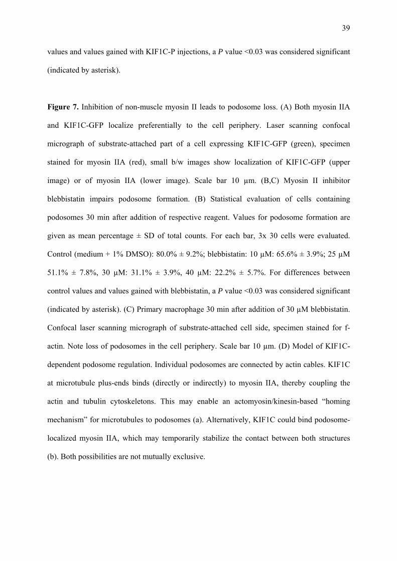
Figure 7. Inhibition of non-muscle myosin II leads to podosome loss. (A) Both myosin IIA
and KIF1C-GFP localize preferentially to the cell periphery. Laser scanning confocal
micrograph of substrate-attached part of a cell expressing KIF1C-GFP (green), specimen
stained for myosin IIA (red), small b/w images show localization of KIF1C-GFP (upper
image) or of myosin IIA (lower image). Scale bar 10 µm. (B,C) Myosin II inhibitor
blebbistatin impairs podosome formation. (B) Statistical evaluation of cells containing
podosomes 30 min after addition of respective reagent. Values for podosome formation are
given as mean percentage ± SD of total counts. For each bar, 3x 30 cells were evaluated.
Control (medium + 1% DMSO): 80.0% ± 9.2%; blebbistatin: 10 µM: 65.6% ± 3.9%; 25 µM
51.1% ± 7.8%, 30 µM: 31.1% ± 3.9%, 40 µM: 22.2% ± 5.7%. For differences between
control values and values gained with blebbistatin, a P value <0.03 was considered significant
(indicated by asterisk). (C) Primary macrophage 30 min after addition of 30 µM blebbistatin.
Confocal laser scanning micrograph of substrate-attached cell side, specimen stained for f-
actin. Note loss of podosomes in the cell periphery. Scale bar 10 µm. (D) Model of KIF1C-
dependent podosome regulation. Individual podosomes are connected by actin cables. KIF1C
at microtubule plus-ends binds (directly or indirectly) to myosin IIA, thereby coupling the
actin and tubulin cytoskeletons. This may enable an actomyosin/kinesin-based “homing
mechanism” for microtubules to podosomes (a). Alternatively, KIF1C could bind podosome-
localized myosin IIA, which may temporarily stabilize the contact between both structures
(b). Both possibilities are not mutually exclusive.

Table 1. Values for podosome formation following various treatments.
cells injected with cells with podosomes, 0 h (%) cells with podosomes, 1 h (%) mock 8.9 ± 7.2 79.2 ± 11.0 AMP-PNP (50 mM) 36.7 ± 9.0 KIF5B antibody (4µg/µl) 72.2 ± 3.0 KIF3B antibody (4µg/µl) 78.9 ± 5.1 dynein antibody (4µg/µl) 80.0 ± 6.8 EHNA (1 mM) 81.1 ± 3.9
kinesin construct (no.) cells with podosomes (%) 4h 6h 8h
1 (control) 93.3% ± 3.3% 94.4% ± 6.9% 87.8% ± 1.9% 2 84.4% ± 5.1% 83.3% ± 12.0% 88.9% ± 1.9% 3 55.6% ± 8.4% 57.8% ± 11.7% 72.2% ± 5.1% 4 34.4% ± 6.9% 50.0% ± 8.8% 56.7% ± 15.3% 5 71.1% ± 5.1% 51.1% ± 7.7% 47.8% ± 8.4% 6 67.8% ± 10.2% 54.4% ± 6.9% 70.0% ± 6.7% 7 63.3% ± 6.7% 62.2% ± 6.9% 76.7% ± 6.7% 8 67.8% ± 10.2% 56.7% ± 3.3% 57.8% ± 10.7% 9 63.3% ± 6.7% 77.8% ± 5.1% 65.6% ± 8.4% 10 93.3% ± 5.8% 93.3% ± 3.3% 92.2% ± 5.1% 11 97.8% ± 1.9% 96.7% ± 3.3% 96.7% ± 3.3% 12 95.6% ± 1.9% 95.6% ± 1.9% 92.2% ± 1.9% 13 92.2% ± 1.9% 95.6% ± 1.9% 91.1% ± 5.1% 14 94.4% ± 1.9% 91.1% ± 5.1% 94.4% ± 1.9% 15 95.6% ± 1.9% 95.6% ± 1.9% 94.4% ± 3.8% 16 94.4% ± 3.8% 96.7% ± 0.0% 96.7% ± 0.0%
siRNA cells with podosomes, 24h (%) cells with podosomes, 48h (%) vector control (pEGFP-N1) 78,0 ± 7.0 80.0 ± 7.0 luciferase 74.0 ± 4.0 68.0 ± 17.0 KIF1C 40.0 ± 18.0 42.0 ± 2.0 KIF3A 63.3 ± 6.7 62.2 ± 6.9 KAP3A 67.8 ± 10.2 56.7 ± 3.3 KIF5B 54.4 ± 8.0 65.6 ± 11.0
shRNA cells with podosomes, 24h (%) cells with podosomes, 48h (%) scrambled 68.9 ± 5.9 65.6 ± 5.3 KIF1C 32.2 ± 3.9 36.7 ± 5.1 dissolution fission no change scrambled 29.6% ± 9.0% 30.6% ± 10.7% 42.5% ± 3.9% KIF1C 23.1% ± 5.0% 13.6% ± 7.4% 63.0% ± 5.9% Statistical evaluation of podosome numbers or behavior in macrophages microinjected with
inhibitors/inhibitory antibodies, transformed with various kinesin constructs, or transformed
with siRNA or shRNA. For each value, each time 30 randomly chosen cells from 3
independent experiments were evaluated. For evaluation of podosome behavior, a total of 473

podosomes for scrambled shRNA, and a total of 501 podosomes for KIF1C shRNA from each
time 3 independent experiments were evaluated. Values are given as mean percentage ± SD of
total counts (s. Fig.2).

Table 2. Effect of KIF1C inhibition after microtubule disruption.
cells incubated with cells transfected with PP2 (25 µM) nocodazole (1 µM)
cells with podosomes (%)
KIF1C-GFP - - 78.8 ± 11.7 KIF1C-GFP + - 77.8 ± 5.1 KIF1C-GFP - + 53.3 ± 8.8 KIF1C-GFP + + 0.0 ± 0.0 KIF1C-K103A-GFP - - 43.3 ± 13.3 KIF1C-K103A-GFP + - 43.3 ± 6.7 KIF1C-K103A-GFP - + 54.4 ± 10.7 KIF1C-K103A-GFP + + 0.0 ± 0.0 Evaluation of podosome numbers in cells expressing KIF1C-GFP or KIF1C-K103A-GFP
following podosome disruption/reformation and/or microtubule disruption. Addition of
podosome-disrupting tyrosine kinase inhibitor PP2 or microtubule-disrupting nocodazole is
indicated by “+”. For each value, 3x 30 cells were evaluated. Values are given as mean
percentage ± SD of total counts (s. Fig. 5D).