Mal3, the Schizosaccharomyces pombe homolog of EB1, changes the microtubule lattice
Upload
wwwunistraCategory
view
0download
0
Andrei V.Popov1,2, Andrei Pozniakovsky1,3,Isabelle Arnal1, Claude Antony4,Anthony J.Ashford1,3, Kazuhisa Kinoshita1,3,Regis Tournebize1,5, Anthony A.Hyman1,3
and Eric Karsenti1
1Cell Biology Program, EMBL, Meyerhofstrasse 1, Heidelberg 69117,3Max Planck Institute for Cell Biology and Genetics, Dresden,Germany and 4Institut Curie, 75248 Paris, Cedex 05, France
5Present address: PathogeÂnie Microbienne MoleÂculaire, InstitutPasteur, 28 rue du Dr Roux, 75724 Paris, Cedex 15, France
2Corresponding authore-mail: [email protected]
XMAP215 belongs to a family of proteins involved inthe regulation of microtubule dynamics. In this studywe analyze the function of different parts ofXMAP215 in vivo and in Xenopus egg extracts.XMAP215 has been divided into three fragments,FrN, FrM and FrC (for N-terminal, middle andC-terminal, respectively). FrN co-localizes with micro-tubules in egg extracts but not in cells, FrC co-localizes with microtubules and centrosomes both inegg extracts and in cells, while FrM does not co-localize with either centrosomes or microtubules. InXenopus egg extracts, FrN stimulates microtubulegrowth at plus-ends by inhibiting catastrophes, whileFrM has no effect, and FrC suppresses microtubulegrowth by promoting catastrophes. Our resultssuggest that XMAP215 is targeted to centrosomes andmicrotubules mainly through its C-terminal domain,while the evolutionarily conserved N-terminal domaincontains its microtubule-stabilizing activity.Keywords: centrosome/MAP/microtubule dynamics/mitotic spindle
Introduction
Microtubules are dynamic polymers that change theirdistribution during cell cycle and development. In vivo,microtubules are found in two main states, either growingor shrinking. Microtubules transit stochastically betweenthese two states, a behavior known as dynamic instability.Such a transition from growth to shrinkage is called a`catastrophe', while the reverse transition from shorteningto extension is called a `rescue' (Walker et al., 1988).Microtubule dynamics has been extensively studied inextracts prepared from Xenopus eggs. Molecules regulat-ing microtubule dynamics have been identi®ed, and theireffects on the assembly of puri®ed tubulin into micro-tubules determined. There are two classes of suchmolecules. Stabilizing factors, such as the microtubule-
associated proteins (MAPs) XMAP215, XMAP230 andXMAP310 (Gard and Kirschner, 1987; Andersen et al.,1994; Andersen and Karsenti, 1997), promote microtubulegrowth by reducing catastrophes and increasing the growthrate. Destabilizing factors, such as OP18/Stathmin(Belmont and Mitchison, 1996), katanin (McNally et al.,1996) and XKCM1, a member of the Kin I kinesin family(Walczak et al., 1996), increase the catastrophe rate.
The primary MAP that stabilizes microtubules inXenopus egg extracts is XMAP215 (Gard and Kirschner,1987; Tournebize et al., 2000). XMAP215 is a 228 kDaprotein that belongs to a conserved family of proteinsrequired for the growth of microtubules and mitoticspindle assembly (Charrasse et al., 1998; Cullen et al.,1999; Tournebize et al., 2000). In extracts, XMAP215exerts its microtubule growth-promoting activity mainlyby antagonizing the activity of the catastrophe factorXKCM1 (Tournebize et al., 2000). Ran-GTP-inducedmicrotubule assembly in egg extracts also depends onXMAP215 (Wilde and Zheng, 1999). Previous work onXMAP215-related proteins from various organisms,including yeast (STU2, p93dis1), Dictyosteliumdiscoideum (DdCP224), Caenorhabditis elegans(Zyg-9), Drosophila melanogaster (Msps) and Homosapiens (ch-TOG), showed that they were all associatedwith the microtubule network and centrosomes or spindlepole bodies in some or all stages of the cell cycle(Nabeshima et al., 1995; Nakaseko et al., 1996; Wang andHuffaker, 1997; Charrasse et al., 1998; Matthews et al.,1998; Cullen et al., 1999; GraÈf et al., 2000). In yeast,C.elegans, Drosophila and Amoeba, mutants show similarphenotypes compatible with defects in microtubuledynamics.
Here we show that, like its orthologs in other organisms,XMAP215 localizes to microtubules and centrosomes. Inorder to examine in more detail how this MAP affectsmicrotubule dynamics and to address its potential functionat the centrosome, we have carried out a functionalanalysis of several fragments. We have correlated theintracellular distribution of various XMAP215 fragmentswith their functional properties in egg extracts. Our resultsindicate that the whole protein is required for properbinding to microtubules in vivo, while its C-terminal parttargets XMAP215 to the centrosome. Although both N-and C-terminal parts bind to microtubules in egg extracts,their effects on microtubule growth in mitotic egg extractsare dramatically different. The conserved N-terminaldomain suppresses catastrophes both in native extractsand in XMAP215-depleted extracts where it fully com-plements the loss of function achieved by depletion. TheC-terminal fragment destabilizes microtubules in eggextracts in a dominant-negative factor manner.
XMAP215 regulates microtubule dynamics throughtwo distinct domains
The EMBO Journal Vol. 20 No. 3 pp. 397±410, 2001
ã European Molecular Biology Organization 397

Results
XMAP215 binds to microtubules and centrosomesthroughout the cell cycleWe have previously shown by immuno¯uorescence thatXMAP215 is localized on microtubules (Tournebizeet al., 2000). Now we have re-examined its localizationin greater detail by both immuno¯uorescence (seeSupplementary ®gure 1, available at The EMBO JournalOnline) and electron microscopy (EM) on ®xed cells andcentrosomes pre-incubated in Xenopus egg extracts. EMstudies revealed the presence of XMAP215 on micro-tubules and associated with the pericentriolar material(Figure 1). We then examined the intracellular distributionof a green ¯uorescent protein (GFP)-tagged version ofXMAP215 in live cells. A cDNA coding for the full-lengthXMAP215 protein was fused at either its N- or C-terminusto GFP and the constructs were electroporated into XL177cells. GFP±XMAP215 localized both to interphase micro-tubules (Figure 2A, C and D) and the centrosome(Figure 2B and D). In mitotic cells, GFP±XMAP215localized predominantly to spindle poles and spindlemicrotubules (Figure 2E). The centrosome staining wasvery strong, independent of the N- or C-terminal local-ization of the GFP tag, or the cell cycle stage, and appearedearly after transfection. To con®rm further the centrosomallocalization of XMAP215, we depolymerized micro-tubules in XL177 cells with nocodazole. Figure 2F showsthat XMAP215 remains associated with centrosomes aftermicrotubule depolymerization (the anti-a-tubulin signalreveals centrioles). We conclude that XMAP215 is amicrotubule-binding protein which localizes along micro-tubules and to the centrosome throughout the cell cycle.
The centrosome and microtubule-binding domainsof XMAP215To determine which domains were required to targetXMAP215 to microtubules and centrosomes, we divided
the molecule into three fragments, termed FrN, FrM andFrC, for N-terminal, middle and C-terminal, respectively(see Figure 3A). They were chosen according to theregions of homology between XMAP215 and othermembers of this family of proteins (see Discussion andFigure 9).
We ®rst examined the localization of these fragmentsin vivo, by expressing GFP-tagged proteins in XL177cells. To exclude the interference of the GFP tag withmicrotubule- or centrosome-binding properties of theseproteins, the N- and C-terminal fragments of XMAP215were tagged with GFP separately, either at the N-(GFP±Fr) or C-terminus (Fr±GFP). These constructsbehaved similarly. Figure 3B shows protein bands corres-ponding to the major GFP-fusion fragments of XMAP215used in this study. When expressed in XL177 cells, bothFrN±GFP (GFP±FrN) and FrM±GFP showed a diffusecytoplasmic ¯uorescence (Figure 3C). In practically alltransfected cells, FrC±GFP (or GFP±FrC) was localized oncentrosomes throughout the cell cycle (see below). Thislocalization was not affected by nocodazole treatment(data not shown). In addition, in some cells overexpressingFrC±GFP, a less prominent punctate staining was observedalong microtubules. Taken together, these results indicatethat the C-terminal part of XMAP215 contains a domainthat targets XMAP215 to centrosomes and participates inmicrotubule binding in vivo.
The sequence of the C-terminal part of XMAP215contains a microtubule-binding domain (consensus:L¼KI/VGS-E/DN-K, amino acids 1865±1879 of theXMAP215 sequence) that targets other MAPs to micro-tubules (Chapin and Bulinski, 1992; Charrasse et al.,1998). In order to de®ne further the centrosome- andmicrotubule-binding domains of the C-terminal fragmentof XMAP215, we created two more deletion mutants:GFP±Fr6 and GFP±XMAP215_DFr6 (Figure 3A). Theboundary between these two fragments was chosenin order to remove the above-mentioned domain.
Fig. 1. Immunoelectron microscopic localization of XMAP215. (A) Electron microscopy on XL177 cells. In cultured epithelial cells, anti-N-terminalXMAP215 antibodies stain microtubules and, to a lesser extent, pericentriolar material. (B) In asters, assembled in vitro using mitotic Xenopus eggextracts and human KE37 cell line centrosomes, XMAP215 is localized on microtubules and pericentriolar material. Arrows indicate gold particlesassociated with pericentriolar material. Scale bar, 200 nm.
A.V.Popov et al.
398

Thus, GFP±XMAP215_DFr6 contains the whole proteinminus the microtubule-targeting domain of the C-terminalregion. The remaining C-terminal part of XMAP215 wastermed Fr6. Similar to the localization of FrC±GFP intransfected cells, GFP±Fr6 intensely stained centrosomesboth in interphase and mitosis (Figure 3C; only GFP±Fr6-containing mitotic cell is shown). XMAP215_DFr6 did notco-localize with centrosomes, but was weakly associatedwith microtubules. However, in contrast to the FrC±GFPstaining, which appeared both continuous and punctate,GFP±XMAP215_DFr6 showed a `smooth' associationwith microtubules (Figure 3C). Therefore, the C-terminalpart of XMAP215 contains the centrosome-targetingdomain of XMAP215. The microtubule-binding propertiesof XMAP215 appear to involve several regions of themolecule, which are necessary for its proper distributionon the microtubule cytoskeleton in vivo.
We next characterized the centrosome-binding domaincontained within the C-terminal part of XMAP215. Aseries of deletion mutants was made within the C-terminalfragments of XMAP215 (Figure 4A). The two smallestfragments, Fr9 and Fr10, were tagged with GFP separ-ately, either on the N- or C-terminus, to exclude interfer-ence of the GFP tag with centrosome binding. For the samereason, the DC6 deletion was made on FrC rather than Fr6.The constructs were electroporated into frog cells and theintensity of the centrosomal staining in live cells wascompared with the intensity of the cytoplasmic GFP¯uorescence (Figure 4B). GFP±Fr6 was found tightly
bound to centrosomes, while deletion of 112, 184 and 201amino acids (constructs DC3±DC5) from the C-terminusgradually decreased the af®nity of the constructs for thecentrosome. Deletion of 282 amino acids from GFP±FrC(construct FrC_DC6) led to the loss of binding tocentrosomes. The results summarized in Figure 4C (andalso presented schematically in Figure 4A) suggest that theregion between amino acids 1784 (C-terminus ofFrC_DC6) and the C-terminus of XMAP215 is necessaryfor the centrosomal localization of XMAP215. This regionincludes the classical microtubule-binding domain ofTau2, MAP4, MAP2b, with a KXGS phosphorylationsite (aa 1871±1874 of the XMAP215 sequence) (Dreweset al., 1997). Phosphorylation of the KXGS motif byMARK kinase reduces the af®nity of some MAPs for themicrotubule lattice (Ebneth et al., 1999). To determinewhether the putative microtubule-binding domain played arole in the centrosomal localization of XMAP215, weperformed a site-speci®c mutagenesis on the FrC±GFP-expressing construct, replacing the KIGS site and two¯anking basic amino acids (KKIGSK) by six irrelevant(NGYLAQ, mutation `M6') amino acids. The centrosome-binding properties of this mutant (FrC_M6±GFP) werecompared with those of the wild-type construct aftertransfecting them into XL177 cells (Figure 4D). Aquanti®cation of ¯uorescence in transfected cells shownin Figure 4E indicates that the intensity of the centrosomalstaining for the M6 mutant was consistently ~50% lowerthan that observed in cells transfected with wild-type FrC.
Fig. 2. GFP±XMAP215 associates with microtubules and centrosomes in interphase and mitosis. (A) A live XL177 cell electroporated withGFP±XMAP215. Note that cultured Xenopus cells frequently have more than one nucleus. (B) Same cell as in (A), showing the plane distal from theglass coverslip. The bright ¯uorescent spot is the centrosome. (C) High-magni®cation image of a live cell, showing GFP±XMAP215 associated withinterphase microtubules at the periphery of the cell. (D) High-magni®cation image showing GFP±XMAP215 on the centrosome and proximal tocentrosome microtubules. (E) In mitosis, GFP±XMAP215 stains the mitotic spindle and spindle poles. (F) Immunolocalization of XMAP215 in ®xedXL177 cells after nocodazole-induced depolymerization of microtubules. Scale bars, 10 mm.
Molecular characterization of XMAP215
399

Therefore, this motif is clearly involved in targeting ofXMAP215 to the centrosome.
Altogether, this analysis shows that XMAP215 istargeted to the centrosome by a domain localized betweenamino acids 1784 and 2065.
Microtubule-binding properties of FrN, FrM andFrCThe results reported in the preceding section indicate howeach of the three fragments of XMAP215 binds tomicrotubules in vivo. Since several parts of the molecule
Fig. 3. Intracellular localization of the XMAP215 fragments expressed as GFP fusions. (A) Schematic representation of fragments and GFP fusionconstructs of XMAP215 and ®ve XMAP215 fragments. Full-length XMAP215, FrN and FrC were separately expressed as either N- or C-terminalfusions with GFP to exclude artifacts created by hindrance of the GFP tag. Sizes of fragments in amino acids (AA) are shown excluding GFP.(B) Immunoblotting analysis showing protein bands corresponding to the full-length GFP±XMAP215 (1), GFP±XMAP215_DFr6 (2), FrN±GFP (3),FrM±GFP (4) and FrC±GFP (5). Lysates from transfected cells were resolved on a polyacrylamide gel, blotted and probed with anti-GFP antibodies.Molecular weight markers are shown on the left-hand side. (C) Expression of the GFP fusions of FrN, FrM, FrC, XMAP215_DFr6 and Fr6 in liveXL177 cells. Scale bars, 10 mm.
Fig. 4. The centrosome-binding site is localized at the C-terminal part of XMAP215. (A) Schematic representation of the GFP fusion constructs ofXMAP215 designed to map the centrosome-binding domain. FrC, Fr9 and Fr10 were separately expressed as either N- or C-terminal fusions with GFPto exclude artifacts created by hindrance of the GFP tag. KKIGS1874K is a six amino acid sequence which was mutated in FrC±GFP to assess its rolein centrosome binding. A vertical line through the C-terminus of XMAP215 shows the position of the KKIGSK sequence in different constructs.Relative intensity of centrosome staining indicated on the right is based on the results shown in (C). (B) Intensity pro®le of GFP ¯uorescence in a cellexpressing FrC±GFP. (C) Ratios of intensities of centrosome/cytoplasm in live XL177 cells expressing different C-terminal fragments (see A). Tocalculate the ratios, the camera background was ®rst subtracted from cytoplasm and centrosome ¯uorescence. The scale of the y-axis begins at 1,representing a cell in which no difference in ¯uorescence intensity between centrosome and cytoplasm exists, for example, in cells transfected withGFP fusions of Fr9, Fr10 and FrC_DC6. For each bar, 20 cells were assessed in a blind assay. Values of intensity ratios are indicated under thecolumns. GFP±FrC and FrC±GFP represent fusion proteins with N- or C-terminal positions of GFP. (D) Intensity pro®le of GFP ¯uorescence in a liveXL177 cell expressing FrC_M6±GFP, where M6 stands for the mutation introduced into FrC with amino acids 1870±1875 (KKIGSK) replaced by sixirrelevant amino acids (NGYLAQ). Note that the level of GFP ¯uorescence in the cytoplasm is higher than in (B). (E) Ratios of intensities ofcentrosome/cytoplasm in live XL177 cells, electroporated with FrC±GFP and FrC_M6±GFP. For each bar, 40 cells were measured. Note that thecentrosome/cytoplasm ¯uorescence ratio of FrC±GFP in this experiment is very close to the data in (C). The scale of the y-axis begins at 1 (as in C).
A.V.Popov et al.
400

seemed to be required to provide the staining pattern ofthe full-length XMAP215, we decided to compare themicrotubule-binding properties of these fragments in vitro.First, we used bacterially expressed fusions of FrN, FrMand FrC with GFP. These proteins were incubated withtaxol-stabilized microtubules and sedimented through aglycerol cushion without ®xation. Results shown in
Figure 5A indicate that all three fragments could bind tomicrotubules while GFP alone did not. To assess thisbinding quantitatively, we used the same three XMAP215fragments, which were tagged with a c-myc peptideinstead of GFP. Various amounts of these fragmentswere incubated with 1 mM taxol-stabilized microtubules.Figure 5B and C shows that microtubule-binding capacity
Molecular characterization of XMAP215
401

for fragments N and M was exceeded in a 1:4 ratio,whereas FrC bound to microtubules in a 1:1 ratio. Morethan half of FrC co-pelleted with microtubules at 0.5 and0.25 mM, whereas <30% of FrN and FrM were found in themicrotubule pellet at these concentrations. These resultsindicate that FrN and FrM have a very low af®nity formicrotubules on their own, and bind to microtubules at alow stoichiometry relative to tubulin dimers. In contrast,
FrC can bind to microtubules proportionally to theconcentration of the polymerized tubulin.
Functional effects of XMAP215 fragments onmicrotubule dynamics in egg extractsWe then examined the effect of the XMAP215 fragmentson microtubule dynamics in egg extracts. We ®rstexamined the localization properties of the fragments in
Fig. 5. The C-terminal fragment of XMAP215 has the highest af®nity for pure tubulin microtubules. (A) GFP fusion proteins of FrN, FrM and FrCwere incubated with pure tubulin microtubules and sedimented without ®xation. CY5-labeled microtubule seeds are shown in a blue pseudocolor. Notethat all three of the fragments studied are associated with microtubules whilst GFP alone does not bind to microtubules. (B) Microtubule spin-downassay. c-myc-tagged FrN, FrM and FrC were incubated with 1 mM pure tubulin microtubules. After sedimentation, pellets and supernatants wereanalyzed by SDS±electrophoresis followed by Coomassie Blue staining. BSA was present in reactions to prevent precipitation and/or absorption ofXMAP215 fragments on the walls of test tubes. Note that the FrN band (62 kDa) is just under the BSA band. (C) Immunoblotting results of the sameexperiment shown in (B).
A.V.Popov et al.
402

extracts. When added at a low concentration of no morethan 0.1 mM, both GFP-tagged N- and C-terminal frag-ments bound to the microtubule asters growing fromcentrosomes (Figure 6A), whereas fragment M did not.These results suggested that FrN and FrC were more likelyto have an effect on microtubule dynamics in extracts.
To investigate this issue, we added His6-tagged FrN,FrM and FrC to mitotic egg extracts and measured astersize and dynamic parameters of microtubule growth. TheN-terminal fragment markedly increased the aster size(Figure 6B), while FrM did not have any effect onmicrotubule length (not shown). At 2.7 mM, FrN increasedaverage microtubule length 3- to 4-fold and reduced thecatastrophe rate 2- to 3-fold. This addition of FrNrepresents an ~10-fold molar excess over the amounts ofendogenous XMAP215 (0.3 mM, calculated from theresults of Gard and Kirschner, 1987). FrN addition had nosigni®cant effect on the growth or shrinkage rates. We nextinvestigated the effect of FrC on microtubule dynamics. At1.8 mM, the C-terminal fragment destabilized micro-tubules, yielding centrosomes practically devoid ofmicrotubules (Figure 6C). To determine which parametersof microtubule growth were affected by FrC, we titrateddown the ®nal concentration of FrC 3-fold to 0.6 mM andmeasured the dynamics of individual microtubules. Underthese conditions, FrC decreased microtubule length 1.6-to 2-fold depending on different extracts. The mainparameter affected was the catastrophe frequency. Italmost doubled relative to the control (50 and 83%increase in two different experiments). To see whether thiseffect of FrC was speci®c to egg extracts, we added FrC topreformed microtubule asters growing from centrosomesin the presence of pure tubulin. Figure 7 shows that notonly did FrC not induce microtubule depolymerizationunder these conditions, but it also brought about micro-tubule bundling. The total mass of polymerized tubulin inthe asters increased 2.8-fold in the presence of 2 mM FrCcompared with the control reaction. Therefore, the catas-trophe-inducing activity of FrC is speci®c to egg extracts.
Since the addition of FrN to extracts diminished thenumber of catastrophes, we wondered whether it couldrescue the phenotype of an XMAP215-depleted extract.Therefore, we added FrN to an XMAP215-depletedextract (see Figure 8A for an example of the XMAP215depletion). If >90% of XMAP215 is depleted, centrosomesnucleate microtubules that are so short they are barelyvisible (Tournebize et al., 2000; see also Figure 6D) andno microtubule dynamics parameters can be measured.FrN reversed this effect. Microtubule length, recordedafter addition of 1 mM FrN, was 61% of the length ofmicrotubules in the mock-depleted extract, with a catas-trophe frequency of 5.1 events/min (compared with that of3.7 events/min in the control; Figure 6D). In contrast, FrMeven at 2.5 mM did not have any effect on microtubulegrowth. When FrN was added at 2.7 mM, the resultingmicrotubules were 2.7 times longer (6.6 mm comparedwith 2.46 mm) than in the control reaction, re¯ecting a>2-fold drop in the catastrophe frequency.
We conclude that the N- and C-terminal parts ofXMAP215 have opposite activities on the microtubuledynamics in egg extracts: the N-terminal fragmentsuppresses catastrophes, while the C-terminal fragmentpromotes them.
The N-terminal fragment rescues spindle assemblyin XMAP215-depleted extracts whereas theC-terminal fragment blocks spindle assemblyWe have recently shown that immunodepletion ofXMAP215 from egg extracts abolished spindle formation(Tournebize et al., 2000). We reproduced this experimentby adding XMAP215 fragments to depleted extracts. Afterremoval of >90% of XMAP215 from the extracts(Figure 8A), no spindles were observed (Figure 8D andE). Adding back FrN (1 mM) partially rescued thedepletion phenotype, resulting in the formation ofminispindles or multipolar spindles around chromosomesin 50±60% of nuclei-associated structures. Addition ofFrC at 1.8 mM resulted in a dramatic inhibition ofmicrotubule growth. In control mock-depleted extracts,addition of FrN (1 mM) and FrM (2.5 mM) had only aminor effect on the percentage of structures formed aroundsperm nuclei, while FrC markedly inhibited microtubulegrowth (Figure 8B and C).
In conclusion, we have found that FrN rescues spindleformation in XMAP215-depleted egg extracts, whereasFrC prevents microtubule polymerization both in depletedand control extracts.
Discussion
Localization of XMAP215 in relation to itsorthologsIn all species examined so far, XMAP215 orthologs havebeen found on microtubules and centrosome/spindle polebodies, although some variability exists among the pub-lished data (Table I). Our results indicate that XMAP215interacts both with microtubules and centrosomes in allstages of the cell cycle (Figures 1±3 and Supplementary®gure 1). The EM data indicate that in the centrosome,XMAP215 is mostly associated with the pericentriolarmaterial. This is also supported by the observation that aD.discoideum analog of XMAP215 (DdCP224; GraÈfet al., 2000) is a resident centrosomal protein. SinceD.discoideum possesses a very compact centrosomelacking centrioles (Moens, 1976), this ®nding suggeststhat DdCP224 and other members of the XMAP215 familybind to the pericentriolar material, rather than directly tocentrioles. The microtubule localization of XMAP215seems different from that reported for the closest orthologstudied, ch-TOG (Charrasse et al., 1998). The reason forthis difference is unclear but could be due to the fact thatthis group used antibodies directed against the C-terminaldomain of ch-TOG. In our hands, using cells in interphase,antibodies raised against the C-terminal domain ofXMAP215 did not stain either microtubules or centro-somes (not shown). However, anti-N-terminal antibodiesused on ®xed human HeLa cells show a pattern of stainingidentical to that of XMAP215 both in interphase andmitosis (see Supplementary ®gure 2).
Our conclusion is that, as with other proteins of thisfamily, XMAP215 interacts both with the centrosomes andmicrotubules.
Functional and structural homologies betweenmembers of the XMAP215 familySequence analyses reveal that, although all relatives ofXMAP215 in species ranging from yeast to human do have
Molecular characterization of XMAP215
403

A.V.Popov et al.
404

regions of considerable homology, their size and domaincomposition vary. This is shown in Figure 9, whichcompares schematically the primary structure of theXMAP215 orthologs. On this scheme, ch-TOG representsproteins like the Msps protein (product of the mini spindlesgene), DdCP224 and a putative protein from Arabidopsisthaliana (DDBJ/EMBL/GenBank No. 4263790), whichshares homology with XMAP215 over the whole proteinlength. Yeast proteins like p93dis1, STU2 and an 809amino acid putative protein from Schizosaccharomycespombe (DDBJ/EMBL/GenBank No. 4164426) are onlyhomologous to the N-terminus of XMAP215. The struc-ture of Zyg-9 is unusual because it contains a duplicateddomain 1 in the N-terminus, which is highly conserved inall the proteins discussed. As illustrated in Figure 9, in allspecies examined, the N-terminal part contains the con-served domain 1 (see also Cullen et al., 1999; GraÈf et al.,2000).
We have shown that FrN is responsible for thecatastrophe-suppressing activity of XMAP215.Moreover, additional experiments (data not shown) indi-cate that a recombinant protein much shorter than FrN,containing only domain 1, is suf®cient to stabilize
microtubule growth in egg extracts. This observation isimportant because it de®nes a new domain that stabilizesmicrotubules most likely by opposing the `catastrophic'kinesins. FrN binds to asters in egg extracts, but does notseem to be associated with microtubules in live cells.However, this can be explained by the low af®nity of FrNfor microtubules; a high amount of GFP±FrN in thecytoplasm of living cells would prevent observation ofindividual microtubules by reducing the contrast.
Deletion/mutation analysis shows that the centrosome-targeting domain of XMAP215 includes a classicalmicrotubule-binding motif, `L-- ---KI/VGS-E/DN-K'.This domain is conserved in XMAP215, ch-TOG,DdCP224 and to a lesser extent in the putative proteinfrom A.thaliana (DDBJ/EMBL/GenBank No. 4263790)and the Msps protein from D.melanogaster. However, acentrosomal (or spindle pole) localization was alsoreported for STU2, p93dis1 and Zyg-9, in which thisdomain is absent, and the whole C-terminal part of themolecule has no homology to the XMAP215/ch-TOG/DdCP224/Msps proteins. Mutational analysis of p93dis1(Nakaseko et al., 1996) is interesting because thisgroup used a deletion/expression of GFP-tagged proteins
Fig. 6. N- and C-terminal parts of XMAP215 bind to centrosomal asters in egg extracts and have opposite effects on microtubule dynamics. (A) OnlyGFP±FrN and GFP±FrC bind to the microtubule asters assembled in a mitotic Xenopus egg extract. Asters were assembled in the presence ofrhodamine±tubulin and 0.1 mM recombinant XMAP215 fragments fused to GFP. Scale bar, 10 mm. (B) Addition of FrN to mitotic egg extract reducesthe number of catastrophes. At the top are the images of microtubule asters as observed in mitotic Xenopus egg extracts during recording by¯uorescence video microscopy. Results in the table summarize two separate experiments performed in two different extracts. Control reactions showmicrotubule dynamics after the addition of the buffer. The ®nal concentration of FrN in the reaction was 2.7 mM. (C) FrC augments the catastropherate in mitotic egg extracts. Note that at 1.8 mM FrC practically no microtubules are visible (corresponding catastrophe rate is shown in the chart as ?).Therefore, effects of FrC on microtubule dynamics were recorded at a ®nal concentration of 0.6 mM. Results from two different extracts are presented.(D) The phenotype of the XMAP215 depletion can be reversed by addition of FrN. Results from two different extracts are presented. Note that at1 mM, FrN partially restores microtubule dynamics to the level of the mock-depleted extract, but at 2.7 mM the number of catastrophes was evenlower than before the depletion of XMAP215. Scale bar, 5 mm. Note that the number of rescues is not indicated in the tables of (B), (C) and (D)because in most cases the number of events observed was too low.
Fig. 7. FrC does not have an intrinsic microtubule-depolymerizing activity. (A) FrC was added to preformed microtubule asters growing fromcentrosomes in the presence of pure tubulin. Note the extensive bundling of microtubules in the presence of FrC. (B) Coomassie-stained gel withsamples from the experiment shown in (A). (C) Quanti®cation of the tubulin protein bands shown in (B). The mass of polymerized tubulin in theasters increased 2.8-fold in the presence of 2 mM FrC compared with the control reaction.
Molecular characterization of XMAP215
405

approach similar to ours, and the deletion of the C-terminalregion of p93dis1 resulted in a lack of spindle pole bodylocalization. This suggests that while p93dis1, XMAP215and related proteins share a catastrophe-suppressingdomain in the N-terminal region (domain 1 in Figure 9),they are targeted to the MTOC through another sequencein the C-terminal domain. Taken together, this suggests the
existence of a functional homology between these twogroups of proteins (XMAP215/ch-TOG/DdCP224/Mspsand STU2/p93dis1/Zyg-9) even in the absence of sequencesimilarity in the C-terminal region.
The function of the microtubule-targeting motif foundin XMAP215 and other proteins is intriguing. InXMAP215, this site seems to be part of the centrosomal-
Fig. 8. The N-terminal part of XMAP215 partially rescues mitotic spindle formation in XMAP215-depleted extracts. The C-terminal part ofXMAP215 inhibits microtubule growth around sperm nuclei. (A) Immunoblot with anti-XMAP215 antibodies showing the ef®ciency of XMAP215depletion from Xenopus egg mitotic extracts. Lanes 1±3 represent 0.5, 0.25 and 0.1 ml of the extract resolved on a 6% SDS±gel. Lanes 4±5 show 0.5and 0.25 ml of an XMAP215-depleted extract (DXMAP215). Lanes 6±7 show 0.5 and 0.25 ml of a mock-depleted extract (Rb IgG, non-immune rabbitserum IgG). XE, eluate from Dynabeads loaded with anti-XMAP215 antibodies after depletion. ME, eluate from Dynabeads loaded with Rb IgG aftermock depletion. (B) Formation of mitotic spindles in the cycled extracts after the addition of recombinant XMAP215 fragments. Most frequentphenotypes are shown. (C) Quanti®cation of the phenotypes shown in (B). More than one hundred nuclei per reaction were observed and themicrotubule structures associated with these nuclei were classi®ed. (D) Formation of spindles and other structures in the cycled XMAP215-depletedextracts in the presence of recombinant XMAP215 fragments. (E) Quanti®cation of the phenotypes shown in (D). More than one hundred nuclei perreaction were estimated. Scale bar, 10 mm.
A.V.Popov et al.
406
targeting domain. Indeed, mutation of the KIGS motif,which was shown to be a target site for the MARK kinase,clearly reduced the amount of FrC±GFP bound to the
centrosomes. However, the actual centrosome localizationdomain goes beyond the mutated KKIGSK sequence, andwe do not know yet whether the phosphorylation of Ser1874
in the KIGS motif affects the af®nity of XMAP215 forcentrosomes or microtubules. Also, although FrC ofXMAP215 clearly binds to microtubules, the C-terminalpart of ch-TOG was not found to bind microtubules in vitro(Spittle et al., 2000). It was instead reported to interactwith tubulin dimers. On the other hand, FrC of XMAP215is not associated with a/b-tubulin dimers in Xenopus eggextracts when analyzed by centrifugation in sucrosegradients (data not shown). These differences could beexplained either by a functional difference between thech-TOG and XMAP215 molecules, or by the fact thatSpittle et al. (2000) used a C-terminal fragment of ch-TOGmuch shorter than FrC of XMAP215 (743 aa ofTOGD14±1129 versus 899 amino acids of FrC). Anotherinteresting possibility is that FrC of XMAP215 may targetin live cells/extracts not only a/b-tubulin dimers but alsoother centrosome-speci®c tubulins such as g-tubulin and/ore/d-tubulins (Chang and Stearns, 2000; Smrzka et al.,2000).
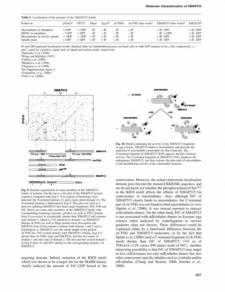
Fig. 10. Model explaining the activity of the XMAP215 fragmentsin egg extracts. XMAP215 binds to microtubules and prevents theinduction of microtubule catastrophes by Kin I kinesins. TheN-terminal fragment of XMAP215 (FrN) opposes the Kin I kinesinactivity. The C-terminal fragment of XMAP215 (FrC) displaces theendogenous XMAP215, and thus exposes the plus-ends of microtubulesto the destabilizing activity of the catastrophic kinesins.
Table I. Localization of the proteins of the XMAP215 family
Found on p93dis1a STU2b Mspsc Zyg-9d ch-TOGe ch-TOG (this work)f XMAP215 (this work)g DdCP224h
Microtubules in interphase + GFP 6 GFP ± IF ± IF ± IF + IF + IF, GFP + IF, GFPMTOC in interphase ±? GFP + GFP ± IF ± IF ± IF ± IF ± IF, + GFP + IF, GFPMicrotubules in mitotic spindle 6 GFP 6 GFP + IF + IF + IF + IF + IF, GFP + IF, GFPSpindle poles + GFP + GFP + IF + IF + IF + IF + IF, GFP + IF, GFP
IF and GFP represent localization results obtained either by immuno¯uorescence on ®xed cells or with GFP mutants in live cells, respectively. +, ±and ? stand for a positive signal, lack of signal and unclear result, respectively.aNakaseko et al. (1996).bWang and Huffaker (1997).cCullen et al. (1999).dMatthews et al. (1998).eCharrasse et al. (1998).fSee Supplementary ®gure 2.gTournebize et al. (2000).hGraÈf et al. (2000).
Fig. 9. Domain organization of some members of the XMAP215family of proteins. On the top is a dot plot of the XMAP215 proteinsequence compared with Zyg-9. Two regions of homology weredetected: the N-terminal domain (1) and a more distal domain (2). TheN-terminal domain is duplicated in Zyg-9. This plot was used as abasis for splitting XMAP215 into three major fragments: FrN, FrM andFrC. Below are some other members of the XMAP215 family withcorresponding homology domains. p93dis1 (as well as STU2 proteinfrom S.cerevisiae) is considerably shorter than XMAP215 and containsonly domain 1, which is 31% identical to domain 1 in XMAP215.Human ch-TOG (as well as Msps protein from Drosophila andDdCP224 from D.discoideum) contains both domain 1 and 2 and ishomologous to XMAP215 over the whole length of the protein.ch-TOG has 76% overall identity with XMAP215. Finally, Zyg-9 isshorter than ch-TOG, msps and DdCP224, and has two copies ofdomain 1 and one copy of domain 2. The ®rst and the second domain 1in Zyg-9 show 41 and 45% identity to the corresponding domain 1 inXMAP215.
Molecular characterization of XMAP215
407

How does XMAP215 regulate microtubuledynamics?Mutational analysis of XMAP215 made it possible for usto draw several conclusions. We have identi®ed themicrotubule-stabilizing and centrosome-binding domainsin the N- and C-terminal parts of the protein, respectively.We have found that the whole protein is required forproper binding to microtubules in vivo and, most interest-ingly, we found that the C-terminal fragment has anactivity in egg extracts opposite to that of full-lengthXMAP215: it strongly destabilizes microtubules andblocks spindle assembly. However, it is also the fragmentthat binds most avidly to microtubules.
The stabilizing effect of fragment N could be explainedin the following ways. It could bind directly to microtubuleends. Indeed, the N-terminal end of ch-TOG bindspreferentially to short microtubules, rather than to longones (Spittle et al., 2000) and we do see a binding of theN-terminal fragment of XMAP215 to microtubulesin vitro. However, we have not seen GFP±XMAP215 orFrN±GFP exclusively co-localized with microtubule endseither in extracts or in live cells (although its activity therecannot be excluded). The second possibility is that FrNdoes not need to bind to microtubules to opposecatastrophes in vivo, but rather interacts with Kin Ikinesins near growing microtubule ends (see model inFigure 10). More work will be required to understand howthe conserved domain contained within FrN stabilizesmicrotubules in egg extracts.
The destabilizing effect of fragment C could beexplained by a dominant-negative effect: it binds tomicrotubules and prevents the endogenous XMAP215from carrying out its function (Figure 10). Anotherpossibility is that the C-terminal fragment directlyincreases the catastrophe frequency by binding to micro-tubules. However, this does not seem to be the casebecause it does not affect the length of microtubulesassembled from pure tubulin and actually increases themass of polymerized tubulin (Figure 7).
In summary, we have identi®ed the microtubule-stabilizing domain of the XMAP215 family of proteins.This N-terminal domain has a powerful microtubulestabilization effect that seems to be achieved by opposingdestabilizing factors such as Kin I kinesins. We haveshown that the whole protein length is required for itsproper distribution on microtubules in live cells. We havealso identi®ed the centrosome-binding domain ofXMAP215 at the C-terminal part of the protein, althoughthe function of XMAP215 and its orthologs at thecentrosome is much less clear. By being concentrated onMTOCs, these molecules could locally increase thegrowth of microtubules and, therefore, stabilize micro-tubules nucleated from the g-tubulin ring complexes(Wiese and Zheng, 2000).
Materials and methods
Antibody productionThe ®rst 560 amino acid N-terminal fragment of XMAP215 (FrN; seeFigure 3A) was expressed as a His6-tagged recombinant protein, puri®ed,and used to obtain af®nity-puri®ed rabbit polyclonal antibodies accordingto standard protocols (Harlow and Lane, 1988; Tournebize et al., 2000).C-terminal antibodies used for XMAP215 depletion from egg extractswere raised against a peptide containing the last 15 amino acids of
XMAP215, and af®nity puri®ed against this peptide by Sigma-Genosys,UK.
Cell culture, electroporation and live cell microscopyXL177 cells (Miller and Daniel, 1977) were grown in 70% Leibowitz-15medium with 10% fetal calf serum. For electroporation, cells from onecon¯uent 90 mm Petri dish were collected, washed in 70% phosphate-buffered saline (PBS), resuspended in 200 ml of 70% PBS with 20 mg ofsupercoiled plasmid DNA and electroporated in 0.2 cm cuvettes at 220 V,200 W and 250 mF. Following electroporation, cells were plated on 15 mmpoly-L-lysine-coated glass coverslips, and the phenotype was scored24±96 h later. For immuno¯uorescence visualization of XMAP215 oncentrosomes after microtubule depolymerization, the cells were incubatedwith 20 mM nocodazole at 4°C for 4 h before ®xing. For live cell imagingthe coverslips were mounted in 70% serum-free Dulbecco's modi®edEagle's medium in slide chambers ®tted for 15 mm round coverslips, andthe images were acquired using a Zeiss Axioscope2 and an F-View CCD12-bit black and white camera (Soft Imaging Systems). After mountingthe coverslips with live cells, another person changed labels on them, sothat a blind assay could be assured.
To quantify the amounts of GFP±XMAP215 fusion constructs bound tocentrosomes, we took pictures of randomly observed green cells aftertransient transfection. For measuring the centrosome/cytoplasm ratios, weused images of cells expressing low amounts of the GFP±XMAP215fragments, de®ned as those showing peak intensity of the staining at thecentrosome <800 U (after subtracting the background of the camera).This threshold was accepted as it was found that cells showing intensitiesof GFP ¯uorescence above this level did not differ signi®cantly in theircentrosome/cytoplasm GFP ratio. Before calculating the ratio ofintensities of centrosome/cytoplasm, the camera background in unitswas subtracted from the brightness of the centrosome and cytoplasm.These ratios were found to be reproducible between experiments. Foreach bar, at least 20 cells were assessed in a blind assay.
Immuno¯uorescence and immunoblottingFor indirect immuno¯uorescence we used the same conditions asdescribed in Tournebize et al. (2000). Pictures were acquired with anLSM 510 confocal microscope (Zeiss) or Zeiss Axioscope2.
For immunoblotting analysis, cells were electroporated with therespective GFP-fusion expression constructs, lysed 24±96 h later in theSDS±electrophoresis loading buffer and resolved on a 6 or 8%polyacrylamide gel. Proteins were blotted onto nitrocellulose membranesand probed with anti-GFP antibodies (Roche, Catalog No. 1 814 460),followed by anti-mouse IgG peroxidase conjugate and the ECL detectionsystem (Amersham).
EM localization of XMAP215 in XL177 cells and oncentrosomal astersFor the EM localization of XMAP215 in XL177 cells and microtubuleasters assembled in egg extracts, we used the same conditions asdescribed in Le Bot et al. (1998) and Wittmann et al. (1998). Cells andasters were stained with anti-N-terminal XMAP215 antibody at 10 mg/ml(see Supplementary data).
Recombinant fragments of XMAP215Cloning of the full-length XMAP215 was described elsewhere(Tournebize et al., 2000). In this study we used the followingXMAP215 fragments: FrN (aa 1±560), FrC (aa 1168±2065), FrM(aa 543±1167), Fr6 (aa 1565±2065), XMAP215_DFr6 (aa 1±1584), Fr8(aa 1847±2065), Fr9 (aa 1882±2065), Fr10 (aa 1922±2065), Fr6_DC3(aa 1565±1982), Fr6_DC4 (aa 1565±1881), Fr6_DC5 (aa 1565±1864) andFrC_DC6 (aa 1168±1784). Information on the cloning, expression andpuri®cation of these fragments is included in the Supplementary data.
Microtubule sedimentation assaysInformation on microtubule sedimentation assays is presented in theSupplementary data.
Xenopus egg extracts, centrosomal asters, spindle assemblyand XMAP215 immunodepletionXenopus egg extracts and spindle assembly were performed as previouslydescribed (Murray, 1991; Sawin and Mitchison, 1991). Depletion ofXMAP215 was performed using 30 mg of anti-C-terminal peptide (the last15 amino acids of XMAP215) antibodies and 10 mg of anti-N-terminalXMAP215 antibodies coupled to 40 ml of protein A Dynabeads (Dynal)per 130 ml of CSF-arrested Xenopus egg extract (CSF-ME). For control`depletion' (`mock-depleted' extracts), we used the same amount (40 mg)
A.V.Popov et al.
408

of rabbit serum IgG (Dianova). After binding the antibodies, beads werewashed twice with PBST and then three times with XB-PI buffer (XB:100 mM KCl, 0.1 mM CaCl2, 1 mM MgCl2, 50 mM sucrose and 10 mMK±HEPES pH 7.7; PI: protease inhibitors, ®nal concentration of 10 mg/mleach of leupeptin, pepstatin and aprotinin). After removing as muchbuffer as possible, 130 ml of egg extract were added to the beads andincubated on ice for 1.5 h with occasional gentle pipetting.
Depleted and mock-depleted extracts were then used to assay spindleassembly. The ef®ciency of XMAP215 depletion was estimated byimmunoblotting the same amounts of XMAP215- and mock-depletedextracts. Both antibodies used for depletion recognized the same~230 kDa band in egg extracts, which was also eluted from theantibody-coated Dynabeads after depletion (see Figure 8A). Spindleswere assembled around sperm nuclei. These 15 ml of XMAP215- ormock-depleted extract were mixed with 1 ml of Xenopus sperm nuclei and0.12 ml of rhodamine±tubulin at 7 mg/ml. The extract was driven into theinterphase by addition of 2 ml of 4 mM CaCl2 for 90 min. At this pointanother 15 ml of the XMAP215- or mock-depleted extract were addedtogether with a protein fragment of XMAP215 in the CSF-XB at one-tenth of the total extract volume. FrN and FrM were added at 1 mM ®nalconcentration, while FrC was added at 2 mM ®nal concentration. In acontrol reaction, the same volume of the CSF-XB buffer with 0.1 mg/mlbovine serum albumin (BSA) was added. The proteins were added to theextract on ice and left for 10 more minutes on ice before transferring thetubes to 20°C for a 30±60 min incubation to allow the extract to cycle intomitosis and form mitotic spindles around sperm DNA. To score theresults, 2 ml of extract were ®xed in the spindle ®x (Heald et al., 1998) andimages were collected using Axiophot 2.
To study the localization of the GFP-labeled FrN, FrM and FrC onmicrotubule asters growing from centrosomes, frog egg extract wasprepared as described above and supplemented with rhodamine±tubulinat 140 mg/ml. Human KE37 cell line centrosomes at a concentration of2 3 108 per ml (prepared according to Moudjou and Bornens, 1998) wereadded in a 2 ml volume to 40 ml of extracts, followed by 1 ml of the GFP-labeled proteins at a ®nal concentration of 0.1 mM. As a control we usedGFP alone, which did not stain centrosome asters (data not shown).Extracts were incubated at 20°C for 10 min and ®xed by the addition of1 ml of ®xing solution (BRB80, 30% glycerol, 0.5% Triton X-100 and0.25% glutaraldehyde). Fixed asters were immediately centrifugedthrough a 40% glycerol cushion in BRB80 in an HB6 rotor in a Sorvallcentrifuge at 12 000 r.p.m., 16°C for 15 min onto coverslips as described(Sawin and Mitchison, 1991). Glutaraldehyde was quenched byincubation in 1 mg/ml NaBH4 in PBS and the coverslips were mountedin Mowiol. Images were obtained using Axiophot 2 and an F-View CCD12-bit black and white camera (Soft Imaging Systems).
Recording of microtubule dynamicsFor microtubule dynamics, single microtubules growing from centro-somes were recorded as described (Tournebize et al., 1997), using a ZeissAxioskop, a 1003 Apochromate lens (NA 1.4) and a rhodamine ®ltercube. Images were recorded every 2 s on an 8-bit black and white camera(Sony SSC-M370CE) with an image processor (model Argus 10;Hamamatsu) and stored on a Macintosh using NIH Image software.Data analysis was performed using a Microsoft Excel macro. For eachcondition (control experiment and in the presence of XMAP215fragments), ~25 microtubules were analyzed for no longer than 10 min.In control reactions, only the buffer, into which individual fragments weredialysed, was added. We found that addition of the PIPES buffer (80 mMPIPES±HCl pH 6.8, 150 mM NaCl, 1 mM EDTA, 1 mM dithiothreitol,0.1 mg/ml BSA) led to a higher number of catastrophes in controlreactions than that of CSF-XB supplemented with 0.1 mg/ml BSA.Differences between experiments, with and without a speci®c XMAP215fragment, were evaluated using a Student's t-test with a con®dence levelof 95%. The average length of microtubules (`steady-state' length) wascalculated from the dynamic parameters as described previously using thefollowing equation (Verde et al., 1992):
<L> = (VgFres ± VsFcat)/(Fcat + Fres)where Vg, Vs, Fcat and Fres are growth rate, shrinkage rate, catastrophefrequency and rescue frequency, respectively. In some experiments, onlya few rescues were observed, therefore the calculated averagemicrotubule length should be considered as an approximation.
Supplementary dataSupplementary data for this paper are available at The EMBO JournalOnline.
Acknowledgements
We are grateful to Fabrice Senger for cell culture, Torsten Wittmann formuch advice, Vladimir Gelfand for advice on Xenopus cell electropora-tion, Alena Bogdanova for help with cloning, and Torsten Wittmann andCelia Antonio for numerous egg extract donations. We would like tothank Torsten Wittmann, Heidi McBride and Wofgang Zachariae forcritical reading and suggestions for the manuscript. A.V.P. and K.K. wereboth supported by HFSPO long-term fellowships.
References
Andersen,S. and Karsenti,E. (1997) XMAP310: a Xenopus rescue-promoting factor localized to the mitotic spindle. J. Cell Biol., 139,975±983.
Andersen,S.S., Buendia,B., Dominguez,J.E., Sawyer,A. and Karsenti,E.(1994) Effect on microtubule dynamics of XMAP230, a microtubule-associated protein present in Xenopus laevis eggs and dividing cells[published erratum appears in J. Cell Biol., 1995, 128, following 988].J. Cell Biol., 127, 1289±1299.
Belmont,L. and Mitchison,T.J. (1996) Identi®cation of a protein thatinteracts with tubulin dimers and increases the catastrophe rate ofmicrotubules. Cell, 84, 623±631.
Chang,P. and Stearns,T. (2000) d-tubulin and e-tubulin: two new humancentrosomal tubulins reveal new aspects of centrosome structure andfunction. Nature Cell Biol., 2, 30±35.
Chapin,S.J. and Bulinski,J.C. (1992) Microtubule stabilization byassembly-promoting microtuble-associated proteins: a repeat per-formance. Cell Motil. Cytoskeleton, 23, 236±243.
Charrasse,S., Schroeder,M., Gauthier-Rouviere,C., Ango,F.,Cassimeris,L., Gard,D.L. and Larroque,C. (1998) The TOGp proteinis a new human microtubule-associated protein homologous to theXenopus XMAP215. J. Cell Sci., 111, 1371±1383.
Cullen,C.F., Deak,P., Glover,D.M. and Ohkura,H. (1999) mini spindles:a gene encoding a conserved microtubule-associated protein requiredfor the integrity of the mitotic spindle in Drosophila. J. Cell Biol., 146,1005±1018.
Drewes,G., Ebneth,A., Preuss,U., Mandelkow,E.M. and Mandelkow,E.(1997) MARK, a novel family of protein kinases that phosphorylatemicrotubule-associated proteins and trigger microtubule disruption.Cell, 89, 297±308.
Ebneth,A., Drewes,G. and Mandelkow,E. (1999) Phosphorylation ofMAP2c and MAP4 by MARK kinases leads to the destabilization ofmicrotubules in cells. Cell Motil. Cytoskeleton, 44, 209±224.
Gard,D. and Kirschner,M. (1987) A microtubule-associated protein fromXenopus eggs that speci®cally promotes assembly at the plus end. J.Cell Biol., 105, 2203±2215.
GraÈf,R., Daunderer,C. and Schliwa,M. (2000) Dictyostelium DdCP224 isa microtubule-associated protein and a permanent centrosomalresident involved in centrosome duplication. J. Cell Sci., 113,1747±1758.
Harlow,E. and Lane,D. (1988) Antibodies. A Laboratory Manual. ColdSpring Harbor Laboratory Press, Cold Spring Harbor, NY.
Heald,R., Tournebize,R., Vernos,I., Murray,A., Hyman,A. andKarsenti,E. (1998) In vitro assays for mitotic spindle assembly andfunction. In Celis,J.E. (ed.), Cell Biology: A Laboratory Handbook,2nd edn. Academic Press, San Diego, CA, pp. 326±335.
Le Bot,N., Antony,C., White,J., Karsenti,E. and Vernos,I. (1998) Role ofxklp3, a subunit of the Xenopus kinesin II heterotrimeric complex, inmembrane transport between the endoplasmic reticulum and the Golgiapparatus. J. Cell Biol., 143, 1559±1573.
Matthews,L.R., Carter,P., Thierry-Mieg,D. and Kemphues,K. (1998)ZYG-9, a Caenorhabditis elegans protein required for microtubuleorganization and function, is a component of meiotic and mitoticspindle poles. J. Cell Biol., 141, 1159±1168.
McNally,F.J., Okawa,K., Iwamatsu,A. and Vale,R.D. (1996) Katanin,the microtubule-severing ATPase, is concentrated at centrosomes. J.Cell Sci., 109, 561±567.
Miller,L. and Daniel,J.C. (1977) Comparison of in vivo and in vitroribosomal RNA synthesis in nucleolar mutants of Xenopus laevis. InVitro, 13, 557±563.
Moens,P.B. (1976) Spindle and kinetochore morphology ofDictyostelium discoideum. J. Cell Biol., 68, 113±122.
Moudjou,M. and Bornens,M. (1998) Method of centrosome isolationfrom cultured animal cells. In Celis,J.E. (ed.), Cell Biology: A
Molecular characterization of XMAP215
409

Laboratory Handbook, 2nd edn. Academic Press, San Diego, CA,pp. 111±119.
Murray,A. (1991) In Kay,B.K. and Peng,H.B. (eds), Cell Cycle Extracts.Vol. 36. Academic Press, San Diego, CA, pp. 581±605.
Nabeshima,K., Kurooka,H., Takeuchi,M., Kinoshita,K., Nakaseko,Y.and Yanagida,M. (1995) p93dis1, which is required for sisterchromatid separation, is a novel microtubule and spindle pole body-associating protein phosphorylated at the Cdc2 target sites. GenesDev., 9, 1572±1585.
Nakaseko,Y., Nabeshima,K., Kinoshita,K. and Yanagida,M. (1996)Dissection of ®ssion yeast microtubule associating protein p93Dis1:regions implicated in regulated localization and microtubuleinteraction. Genes Cells, 1, 633±644.
Sawin,K.E. and Mitchison,T.J. (1991) Mitotic spindle assembly by twodifferent pathways in vitro. J. Cell Biol., 112, 925±940.
Smrzka,O.W., Delgehyr,N. and Bornens,M. (2000) Tissue-speci®cexpression and subcellular localisation of mammalian d-tubulin.Curr. Biol., 10, 413±416.
Spittle,C., Charrasse,S., Larroque,C. and Cassimeris,L. (2000) Theinteraction of TOGp with microtubules and tubulin. J. Biol. Chem.,275, 20748±20753.
Tournebize,R., Andersen,S.S., Verde,F., Doree,M., Karsenti,E. andHyman,A.A. (1997) Distinct roles of PP1 and PP2A-likephosphatases in control of microtubule dynamics during mitosis.EMBO J., 16, 5537±5549.
Tournebize,R. et al. (2000) Control of microtubule dynamics by theantagonistic activities of XMAP215 and XKCM1 in Xenopus eggextracts. Nature Cell Biol., 2, 13±19.
Verde,F., Dogterom,M., Stelzer,E., Karsenti,E. and Leibler,S. (1992)Control of microtubule dynamics and length by cyclin A and cyclin Bdependent kinases in Xenopus egg extracts. J. Biol. Chem., 118,1097±1108.
Walczak,C., Mitchison,T.J. and Desai,A.B. (1996) XKCM1: A Xenopuskinesin-related protein that regulates microtubule dynamics duringmitotic spindle assembly. Cell, 84, 37±47.
Wang,P.J. and Huffaker,T.C. (1997) Stu2p: a microtubule-bindingprotein that is an essential component of the yeast spindle polebody. J. Cell Biol., 139, 1271±1280.
Walker,R.A., O'Brien,E.T., Pryer,N.K., Sobeiro,M.F., Voter,W.A.,Erickson,H.P. and Salmon,E.D. (1988) Dynamic instability ofindividual microtubules analyzed by video light microscopy: rateconstants and transition frequencies. J. Cell Biol., 107, 1437±1448.
Wiese,C. and Zheng,Y. (2000) A new function for the g-tubulin ringcomplex as a microtubule minus-end cap. Nature Cell Biol., 2,358±364.
Wilde,A. and Zheng,Y. (1999) Stimulation of microtubule asterformation and spindle assembly by the small GTPase Ran. Science,284, 1359±1362.
Wittmann,T., Boleti,H., Antony,C., Karsenti,E. and Vernos,I. (1998)Localization of the kinesin-like protein Xklp2 to spindle poles requiresa leucine zipper, a microtubule-associated protein and dynein. J. CellBiol., 143, 673±685.
Received October 24, 2000; revised December 4, 2000;accepted December 6, 2000
A.V.Popov et al.
410
Copyright © 2022 FDOKUMEN