Kinesin family member 12 is a candidate polycystic kidney disease modifier in the cpk mouse
12
Kinesin Family Member 12 Is a Candidate Polycystic Kidney Disease Modifier in the cpk Mouse Michal Mrug,* Renhua Li, § Xiangqin Cui, § Trenton R. Schoeb, † Gary A. Churchill, § and Lisa M. Guay-Woodford* †‡ Departments of *Medicine, † Genetics, and ‡ Pediatrics University of Alabama at Birmingham, Birmingham, Alabama; and § The Jackson Laboratory, Bar Harbor, Maine The cpk mouse is the most extensively characterized model of autosomal recessive polycystic kidney disease (ARPKD). The major ARPKD-related renal and biliary phenotypes are modulated in F2 mutants by genetic background, suggesting that quantitative trait loci (QTL) modulate disease severity. In 461 F2 cpk mice, kidney length, weight, and volume were scored as quantitative traits (QT), and a semiquantitative method to assess biliary duct number, area (BDA), portal vein area, and total area of each portal field, as well as the severity of cholangitis, was developed. QTL mapping was performed with Pseudomar- ker v1.02. Candidate genes were identified within the QTL intervals on the basis of expression profiling, reverse transcriptase– PCR, haplotypes, and sequence analysis. The renal QT were normally distributed in the F2 cohort and strongly correlated (P < 0.001). Among the biliary QT, only BDA correlated with the renal QT (P < 0.01). Genome-wide scan identified a major effect QTL on chromosome (Chr) 4 for the renal traits, adjusted BDA, and cholangitis with logarithm of odds scores of 18, 8, and 5, respectively. Regression modeling refined the Chr 4 main effect into an approximately 50-cM region with three distinct QTL peaks at 16, 34, and 54 cM. Kif12, a gene encoding a novel kinesin, mapped beneath the 34 cM QTL peak and has expression level variants and strain-specific sequences that were associated with renal disease severity in affected mice. Therefore, the positional candidate gene, Kif12, fulfills the major criteria for QTL gene discovery established by the Complex Trait Consortium, and, thus, it is proposed that Kif12 is a cpk modifier gene. J Am Soc Nephrol 16: 905-916, 2005. doi: 10.1681/ASN.2004121083 P olycystic kidney disease (PKD) is one of the most com- mon genetic disorders, affecting 600,000 people in the United States, and is a major cause of ESRD in children and adults (1). Autosomal dominant PKD (ADPKD; MIM 173900; 173910) occurs in 1:400 to 1:1000 individuals and is caused by mutations in one of two genes, PKD1 or PKD2 (2–5). Autosomal recessive PKD (ARPKD; MIM 263200) is less fre- quent (1:20,000 live births) and occurs as a result of defects in a single gene, PKHD1 (6,7). In both disorders, interfamilial and intrafamilial phenotypic variability is observed (8,9), the latter suggesting that variation at nondisease (modifier) loci may modulate disease expression. Given the genetic complexity of human populations, efforts to identify modifier genes or quantitative trait loci (QTL) in single-gene disorders have largely focused on experimental models. Numerous murine (mouse and rat) PKD models have been described in which the mutant phenotypes closely resem- ble human PKD with respect to cyst morphology, cellular and extracellular matrix abnormalities, and disease progression (re- viewed in 10). Mapping strategies have identified several pu- tative QTL intervals in experimental crosses segregating differ- ent PKD gene mutations. Of note, proximal chromosome (Chr) 4 reportedly harbors putative modifying gene(s) for the renal cystic disease expressed in the cpk, bpk, jck, and pcy models (11–14). To date, however, the specific genetic variants under- lying this or any other major effect PKD QTL have yet to be identified. With respect to PKD experimental models, the cpk mouse is one of the most extensively characterized (15,16). The cpk mu- tation (cpk or Cys1 cpk ) arose spontaneously in the C57BL/6J- Pldn pa (B6) strain, and the Cys1 cpk allele involves a tandem deletion that is predicted to cause truncation of the novel, cilia-associated protein, cystin (17,18). In cpk mutants, disease expression and severity are modulated by genetic background (11,19). The renal phenotype is dominated by massive cystic involvement of the collecting ducts, a pattern that strongly resembles human ARPKD (20). In the liver, the ductal plate malformation (DPM) gives rise to a biliary dysgenesis lesion that is similar to ARPKD-related congenital hepatic fibrosis (CHF). Although this biliary phenotype is not observed in B6-cpk/cpk mice (16), it is fully penetrant and variable when Cys1 cpk is expressed in crosses with other strains, e.g., Mus mus castaneus (CAST/Ei), DBA/2J, BALB/c, or CD1 (19,21–23). In the current study, we examined both the cpk-associated renal and biliary phenotypes as sets of quantitative traits (QT). Consistent with recent observations in human ARPKD cohorts (9), we observed that in our cpk cross, the severity of the renal Received December 14, 2004. Accepted January 5, 2005. Published online ahead of print. Publication date available at www.jasn.org. Address correspondence to: Dr. Lisa M. Guay-Woodford, Division of Genetic and Translational Medicine, University of Alabama at Birmingham, 740 Kaul Human Genetics Building, 720 20th Street South, Birmingham, AL 35294. Phone: 205-934-7308; Fax: 205-975-5689; E-mail: [email protected] Copyright © 2005 by the American Society of Nephrology ISSN: 1046-6673/1604-0905
-
Upload
independent -
Category
Documents
-
view
0 -
download
0
Transcript of Kinesin family member 12 is a candidate polycystic kidney disease modifier in the cpk mouse

Kinesin Family Member 12 Is a Candidate Polycystic KidneyDisease Modifier in the cpk Mouse
Michal Mrug,* Renhua Li,§ Xiangqin Cui,§ Trenton R. Schoeb,† Gary A. Churchill,§ andLisa M. Guay-Woodford*†‡
Departments of *Medicine, †Genetics, and ‡Pediatrics University of Alabama at Birmingham, Birmingham, Alabama;and §The Jackson Laboratory, Bar Harbor, Maine
The cpk mouse is the most extensively characterized model of autosomal recessive polycystic kidney disease (ARPKD). Themajor ARPKD-related renal and biliary phenotypes are modulated in F2 mutants by genetic background, suggesting thatquantitative trait loci (QTL) modulate disease severity. In 461 F2 cpk mice, kidney length, weight, and volume were scored asquantitative traits (QT), and a semiquantitative method to assess biliary duct number, area (BDA), portal vein area, and totalarea of each portal field, as well as the severity of cholangitis, was developed. QTL mapping was performed with Pseudomar-ker v1.02. Candidate genes were identified within the QTL intervals on the basis of expression profiling, reverse transcriptase–PCR, haplotypes, and sequence analysis. The renal QT were normally distributed in the F2 cohort and strongly correlated (P <0.001). Among the biliary QT, only BDA correlated with the renal QT (P < 0.01). Genome-wide scan identified a major effectQTL on chromosome (Chr) 4 for the renal traits, adjusted BDA, and cholangitis with logarithm of odds scores of 18, 8, and 5,respectively. Regression modeling refined the Chr 4 main effect into an approximately 50-cM region with three distinct QTLpeaks at 16, 34, and 54 cM. Kif12, a gene encoding a novel kinesin, mapped beneath the 34 cM QTL peak and has expressionlevel variants and strain-specific sequences that were associated with renal disease severity in affected mice. Therefore, thepositional candidate gene, Kif12, fulfills the major criteria for QTL gene discovery established by the Complex TraitConsortium, and, thus, it is proposed that Kif12 is a cpk modifier gene.
J Am Soc Nephrol 16: 905-916, 2005. doi: 10.1681/ASN.2004121083
P olycystic kidney disease (PKD) is one of the most com-mon genetic disorders, affecting �600,000 people in theUnited States, and is a major cause of ESRD in children
and adults (1). Autosomal dominant PKD (ADPKD; MIM173900; 173910) occurs in 1:400 to 1:1000 individuals and iscaused by mutations in one of two genes, PKD1 or PKD2 (2–5).Autosomal recessive PKD (ARPKD; MIM 263200) is less fre-quent (1:20,000 live births) and occurs as a result of defects in asingle gene, PKHD1 (6,7). In both disorders, interfamilial andintrafamilial phenotypic variability is observed (8,9), the lattersuggesting that variation at nondisease (modifier) loci maymodulate disease expression.
Given the genetic complexity of human populations, effortsto identify modifier genes or quantitative trait loci (QTL) insingle-gene disorders have largely focused on experimentalmodels. Numerous murine (mouse and rat) PKD models havebeen described in which the mutant phenotypes closely resem-ble human PKD with respect to cyst morphology, cellular andextracellular matrix abnormalities, and disease progression (re-viewed in 10). Mapping strategies have identified several pu-
tative QTL intervals in experimental crosses segregating differ-ent PKD gene mutations. Of note, proximal chromosome (Chr)4 reportedly harbors putative modifying gene(s) for the renalcystic disease expressed in the cpk, bpk, jck, and pcy models(11–14). To date, however, the specific genetic variants under-lying this or any other major effect PKD QTL have yet to beidentified.
With respect to PKD experimental models, the cpk mouse isone of the most extensively characterized (15,16). The cpk mu-tation (cpk or Cys1cpk) arose spontaneously in the C57BL/6J-Pldnpa (B6) strain, and the Cys1cpk allele involves a tandemdeletion that is predicted to cause truncation of the novel,cilia-associated protein, cystin (17,18). In cpk mutants, diseaseexpression and severity are modulated by genetic background(11,19). The renal phenotype is dominated by massive cysticinvolvement of the collecting ducts, a pattern that stronglyresembles human ARPKD (20). In the liver, the ductal platemalformation (DPM) gives rise to a biliary dysgenesis lesionthat is similar to ARPKD-related congenital hepatic fibrosis(CHF). Although this biliary phenotype is not observed inB6-cpk/cpk mice (16), it is fully penetrant and variable whenCys1cpk is expressed in crosses with other strains, e.g., Mus muscastaneus (CAST/Ei), DBA/2J, BALB/c, or CD1 (19,21–23).
In the current study, we examined both the cpk-associatedrenal and biliary phenotypes as sets of quantitative traits (QT).Consistent with recent observations in human ARPKD cohorts(9), we observed that in our cpk cross, the severity of the renal
Received December 14, 2004. Accepted January 5, 2005.
Published online ahead of print. Publication date available at www.jasn.org.
Address correspondence to: Dr. Lisa M. Guay-Woodford, Division of Geneticand Translational Medicine, University of Alabama at Birmingham, 740 KaulHuman Genetics Building, 720 20th Street South, Birmingham, AL 35294. Phone:205-934-7308; Fax: 205-975-5689; E-mail: [email protected]
Copyright © 2005 by the American Society of Nephrology ISSN: 1046-6673/1604-0905

and biliary disease was only weakly correlated. Genome scan-ning identified a major effect QTL complex on Chr 4 thatmodulates the renal cystic disease, components of the biliarylesion, and cholangitis. Gene expression analyses suggest thatvariations in Kif12, the gene encoding a novel kinesin, underliethe Chr 4 main effect on the renal and biliary phenotypes.
Materials and MethodsMice
Mice that were used in these experiments were bred from stockcolonies of C57BL/6J-�/cpk (B6-�/cpk) mice and CAST/Ei (CAST) micemaintained at the University of Alabama at Birmingham. F1 progenyheterozygous for the Cys1cpk allele were identified using an allele-specific assay (17) and intercrossed to generate F2 mutants. All F2 micewere killed 10 d after birth. One kidney and the right liver lobe werefixed in 10% buffered formalin, embedded in paraffin, and analyzedusing standard histologic methods. The remaining kidney and liverwere snap-frozen and stored in liquid nitrogen. The cpk genotype wasconfirmed by PCR with DNA prepared from spleens using standardprocedures. All protocols were approved by the University of Alabamaat Birmingham Animal Care and Use Committee. The University ofAlabama at Birmingham is fully accredited by the American Associa-tion of the Accreditation of Laboratory Animal Care.
Phenotypic CharacterizationThe following parameters were measured in the F2 kidneys: Kidney
length (KL), kidney weight (KW), kidney volume (Kvol; measured bywater displacement), crown to rump length (CR), and body weight(BW). In addition, we calculated the commonly used ratios KL/CR andKW/BW.
In the liver, the DPM associated with human ARPKD is characterizedby three key features: (1) “chaotic” branching and dilation of the bileductules, (2) biliary epithelial hyperplasia, and (3) immature stromaand fibrosis within the portal triad (24). Therefore, we designed asemiquantitative, histopathologic scoring system and used digital im-aging to assess the peripheral portal triads for the following: (1) thenumber of bile ducts/ductules per portal vein (BDN; an index of ductalbranching), (2) the bile duct area (BDA), (3) the portal vein area (PVA),and (4) the total area of the portal tract (TOTA). We also computed theratio BDA/PVA to “normalize” the trait for area distortions that canoccur when cutting tubular structures in cross-section. A minimum oftwo portal triads were analyzed per section with two or more sectionsevaluated per liver.
In addition, we scored the cholangitis severity using a five-pointsystem that assessed the extent of inflammatory cell infiltrate aroundthe biliary ducts and in the liver parenchyma, as well as the degree ofbiliary ductal changes ranging from epithelia hyperplasia to epithelialnecrosis. According to this scoring system, “0” represented no/mini-mal inflammation and “4” reflected severe and extensive cholangio-hepatitis.
Images were acquired using a Nikon E600 microscope equipped witha SPOT Insight digital camera (Diagnostic Instruments, SterlingHeights, MI). Image analyses were performed with Sigma Scan Pro v5image analysis software (SPSS Inc., Chicago, IL). Statistical evaluationsof the phenotypic data, including trait distributions and correlations,were performed with SPSS 11.5 statistical software package (SPSS Inc.).
PCR-Based GenotypingMicrosatellite markers from Whitehead/MIT Genome Database
(http://www-genome.wi.mit.edu) that were spaced at approximately25-cM intervals throughout the genome and whose B6 and CAST alleles
differed by at least 10 bp were typed by PCR and resolved on agarosegels using standard methods. When the initial genome scan identifieda major effect QTL on Chr 4, additional typing was performed withmarkers spaced at approximately 10-cM intervals along the chromo-some. Primers were purchased from Research Genetics (MapPairs,Huntsville, AL). Primer information is available upon request.
Single-nucleotide polymorphisms (SNP) within the Kif12 gene wereidentified from the Celera Mouse SNP Database (http://myscience.appliedbiosystems.com/servlet/com.celera.web.cds_servlets. [mouse_refsnp-Gene_ID]). Primers NT_039262(Kif12)-1245F (5�-TTAGTGCTC-CGCAAAGAAGC-3�) and NT_039262(Kif12)-1643R (5�-TTCCACATA-GAAGCCTCGTG-3�) amplified a 398-bp product that included SNPmCV24480597. Primers NT_039262(Kif12)-2867F (5�GGGCTAGAGT-TCTGGCATCA-3�) and NT_039262(Kif12)-3266R (5�CTGCTGCAA-GAGCAGGAAG-3�) amplified a 399-bp product that included SNPmCV24480596 and mCV24480595. Primers NT_039262(Kif12)-4222F (5�-ACACCACGGGTATGAGCTTC-3�) and NT_039262(Kif12)-4756R (5�-CTGAAGCCAATGATCAGACG-3�) amplified a 534-bp product thatincluded SNP mCV24480588. Primers NT_039262(Kif12)-5638F (5�-AG-GCCCCAGGTCACATATC-3�) and NT_039262(Kif12)-6078R (5�-TAC-CCTTTCCCCAAGTTTCC-3�) amplified a 440-bp product that includedSNP mCV24480587. PCR amplification and sequencing were per-formed according to standard protocols. Primers NT_039262(Kif12)-4222F and NT_039262(Kif12)-4756R were specific for the B6 allele, andno amplification product was detected for the CAST allele.
QTL MappingQTL analyses were performed with the Pseudomarker software
package (release 1.02; http://www.jax.org/staff/churchill/labsite) us-ing the method for genome-wide scans developed by Sen and Churchill(25). Genome-wide significance thresholds were determined by permu-tation testing for each trait (26). Significant or suggestive QTL meet orexceed the 95 and 37% genome-wide thresholds, respectively. Confi-dence intervals (CI) were determined by computing the region of theposterior density curve that contained 95% of the total area as describedby Sen and Churchill (25).
For detecting epistatic interactions, the initial search for main effectQTL was followed by simultaneous search for pairs among QTL. Pair-wise genome scans can detect QTL that modulate a phenotype byinteracting with another quantitative trait locus, including instances inwhich neither locus alone reaches the significance threshold. Finally, allmain effect and interacting QTL were used to fit multiple regressionmodels. The type III sum of squares associated with each locus wasused to compute the adjusted percentage of variance in the trait.
Identification of Positional QTL Candidate GenesUsing the National Center for Biotechnology Information (NCBI)
online resources (http://ncbi.nlm.nih.gov), we obtained physical andgene content maps corresponding to the 95% CI for the candidate QTL.The gene content of these mouse intervals was verified by comparisonswith syntenic human intervals. Gene expression (tissue origin andtiming) was evaluated in silico using the Unigene Expressed SequenceTag (EST) database (http://ncbi.nlm.nih.gov) and the Serial Analysis ofGene Expression (SAGE)-based Virtual Northern databases (http://cgap.nci.nih.gov). Additional computational analyses included com-parison of parental strain-specific gene sequences (when available).
Validation of Predicted Strain-Specific Sequence VariationsTotal kidney RNA was isolated from three 10-d-old mice from each
parental strain (B6 and CAST) using TRIzol Reagent (Invitrogen, Carls-bad, CA), according to the manufacturer’s recommendations, and con-
906 Journal of the American Society of Nephrology J Am Soc Nephrol 16: 905-916, 2005
verted to cDNA using the Superscript II reverse transcriptase kit (In-vitrogen). PCR amplification and sequence analyses were performedusing standard methods. The B6-associated Kif12 variant was definedby comparison with the GenBank nucleotide (BC02225) and protein(AAAH22225) Kif12 sequences.
Gene Expression ProfilingKidneys at the phenotypic extremes of renal disease severity were
identified by averaging the severity ranks for all three renal QT. Theprocedures described in the GeneChip Expression Analysis Manual(Affymetrix, Inc., Santa Clara, CA) were followed to prepare cRNAtargets. In brief, RNA was extracted with TRIzol, and 20 �g of RNAwas used for cDNA synthesis (Superscript II Kit; Life Technologies,Rockville, MD). Biotin-labeled cRNA was generated from cDNA by invitro transcription (ENZO BioArray High Yield RNA Transcript Label-ing Kit; Affymetrix). Fragmented cRNA (15 �g) was hybridized to theGeneChip Mouse Genome 430 2.0 Array. The arrays were washed andstained according to the manufacturer’s instructions.
The summary signal intensity for each gene was computed withRMA default method (27) that included Rma background correction,quantile normalization, and medium polish. The summary signal in-tensities for individual genes then were used to assess variance of thearray data. These analyses included use of Fs- (28) and MAANOVA-(29) based function and adjustment of tabulated P values with q-valuesoftware (30,31).
Quantitative PCRQuantitative analysis of Kif12 expression was performed in three
kidney cDNA templates from each parental strain using the LightCy-cler and the FastStart DNA Master SYBR Green I (Roche DiagnosticsGmBH, Penzberg, Germany) according to the manufacturer’s recom-mendations. Primers Kif12-F (5�-ACGAGGCTTCTATGTGGAACAG-3�) and Kif12-R (5�-GAGGTACCTGCTGAGAAGTTGG-3�) amplified a184-bp product. We designed primers for three “housekeeping” refer-ence genes: (1) Actb (encoding �-actin) Actb-F (5�-GGAGGGGCCG-GACTCATCGTACTC-3�) and Actb-R (5�-CCGCATCCTCTTCCTC-CCTGGAGAA-3�) amplified a 416-bp product; (2) Ubb (encodingubiquitin) Ubb-F (5�-CGGTCTTTCTGTGAGGGTGT-3�) and Ubb-R (5�-TTCACGTTCTCGATGGTGTC-3�) amplified a 159-bp product; and (3)Ywhaz (encoding tyrosine-3-monooxygenase zeta peptide) Ywhaz-F (5�-AGCAGGCAGAGCGATATGAT-3�) and Ywhaz-R (5�-GAAGCATT-GGGGATCAAGAA-3�) amplified a 288-bp product.
The amplification program consisted of initial denaturation at 95°Cfor 10 s, 20°C/s temperature transition ramp up to 95°C for 10 s, 60°Cfor 20 s. and 72°C for 30 s, repeated for 36 cycles. The threshold cycleswere calculated using the LightCyler software version 3.3. To assess thesignificance of Kif12 differential expression, we calculated a gene ex-pression normalization factor for each kidney cDNA sample on thebasis of the geometric mean of the housekeeping genes Actb, Ubb, andYwhaz using the geNorm program (32) (http://medgen31.ugent.be/jvdesomp/genorm).
ResultsPhenotypic Characterization
Of the 2031 F2 progeny generated from the (B6-cpk/� �
CAST/Ei)F1 intercross, 461 (23%) cpk mutants were identifiedby both phenotype and genotype criteria, a frequency consis-tent with the Mendelian distribution of an autosomal recessivetrait. Renal disease severity was assessed by measurements ofKL and KW, a standard approach used to gauge renal cysticdisease severity in mice (11,13). In addition, we measured Kvol,
a trait that has been recently recognized as a prognostic markerof PKD progression (33). All renal traits (KL, KW, and Kvol)varied significantly in the test cohort (Figure 1A). With KW, forexample, the average weight among the 10 most minimallyaffected kidneys was slightly greater than normal kidneys (ap-proximately 40 to 60 mg), whereas among the 10 most severelyaffected kidneys, the average weight was more than eight timeshigher (Table 1). All renal QT were approximately normallydistributed and strongly intercorrelated (Table 2). In contrast,correlations between renal traits and CR length or BW wereweak (Table 2), suggesting that additional independent QTLmodulate body parameters in this cohort.
From the 461 F2 mice, 458 livers were scored by a singleblinded observer. For validating the initial scoring, repeat anal-yses were performed on a second set of sections. Of the original458 samples, adequate sections and consistent scores were ob-tained for 325 livers. Repeat scoring in the remaining 133 liversamples was confounded by insufficient tissue, suboptimal par-affin embedding, or insufficient portal triads per section. These
Figure 1. Quantitative traits. Histograms show the distributionof the quantitative traits (QT) for renal cystic disease (A), biliarydysgenesis (B), and cholangitis (C). Whereas the renal traits andthe biliary duct number (BDN) were near-normally distributed,the distribution of the biliary duct area (BDA), portal vein area(PVA), and total portal field area (TOTA) traits were skewed tothe right. (C) Grade 4 cholangitis with inflammatory cell infil-trates surrounding and filling the lumen of dilated biliary ducts(*). The arrow indicates the portal vein. The distribution of thecholangitis severity was prominently right skewed. Plotsshown here and Pearson correlation coefficients shown in Table1 demonstrate strong correlations among the renal traits; mod-erate correlations among BDN, BDA, and PVA; and significantcorrelations only between BDA and the renal traits.
J Am Soc Nephrol 16: 905-916, 2005 Kif12 is a Candidate PKD Modifier Gene in the cpk Mouse 907

samples were excluded from phenotypic correlation studiesand QTL analyses of the liver phenotypes.
We scored the components of the DPM as quantitative traits:BDN, BDA, and TOTA as a surrogate index of peribiliaryfibrosis. The PVA was also assessed to provide an internalcross-sectional standard for each portal triad. BDN and BDAwere normalized as the ratios BDN/PVA and BDA/PVA toadjust for potential variance in the angle of sectioning. Whereas
BDN was near-normally distributed in the test cohort, thedistributions of BDA, PVA, and TOTA were skewed to the right(Figure 1B). Biliary traits (BDN and BDA) and PVA were mod-erately intercorrelated (Table 2). Stronger correlations amongBDA, PVA, and TOTA (r � 0.80 to 0.89) most likely reflect thatBDA and PVA are major components of TOTA. The correlationof renal and biliary QT was minimal, with a significant rela-tionship evident only for BDA (r � 0.15 to 0.19; P � 0.01). This
Table 1. Renal phenotypic extremes and Kif12 haplotypesa
Phenotype Gender KL (mm) KW (g) Kvol (ml of Water Displacement) Kif12 Haplotypes
Mild F 7.0 0.09 0.09 BMild F 7.0 0.08 0.09 BMild M 7.0 0.09 0.08 BMild F 7.5 0.08 0.09 BMild F 7.0 0.08 0.09 BMild M 6.0 0.04 0.03 BMild F 7.3 0.11 0.12 BMild M 8.3 0.12 0.12 BMild F 7.5 0.10 0.10 BMild M 8.0 0.09 0.11 BSevere M 15.0 0.91 0.75 CSevere F 14.0 0.76 0.71 CSevere F 14.0 0.78 0.68 HSevere M 13.0 0.57 0.49 CSevere M 14.0 0.64 0.52 HSevere M 14.0 0.78 0.58 HSevere F 14.0 0.73 0.67 BSevere M 14.3 0.72 0.74 HSevere F 13.0 0.67 0.64 HSevere M 15.0 0.85 0.78 HaKL, kidney length; KW, kidney weight; Kvol, kidney volume; Kif12 haplotypes, composed of single-nucleotide
polymorphisms mCV24480597, mCV24480596, mCV24480595, mCV24480588, and mCV24480587; B, B6; H, heterozygote; C,CAST.
Table 2. Pearson correlation of quantitative traitsa
Traits KL KW Kvol KL/CR CR BW BDN BDA PVA TOTA
KW 0.89b
Kvol 0.88b 0.95b
KL/CR 0.82b 0.76b 0.73b
KW/BW 0.76b 0.86b 0.83b 0.84b
CR 0.13c 0.12d 0.10d �0.45b
BW 0.28b 0.29b 0.26b �0.22b 0.84b
BDN — — — — — —BDA 0.15c 0.18c 0.19c 0.19c �0.11d — 0.40b
PVA — — — — — — 0.58b 0.63b
TOTA — — — — — 0.12d 0.65b 0.80b 0.89b
Cholangitis 0.10d — — 0.16c �0.14c �0.11d — — — —aCR, crown-rump length; BW, body weight; BDN, bile duct number; BDA, bile duct area; PVA, portal vein area; TOTA,
total portal field area.bP � 0.001.cP � 0.01.dP � 0.05.
908 Journal of the American Society of Nephrology J Am Soc Nephrol 16: 905-916, 2005

correlation increased to 0.31 (P � 0.001) when the three renalQT were converted to a single composite metric, renal PC1, andBDA was adjusted to log (BDA/PVA; see below).
Among the 458 livers evaluated, 257 (56%) had histopatho-logic evidence of suppurative cholangitis and/or hepatitis.Some sections contained focal collections of Gram-negativebacteria (data not shown). We defined this inflammatory lesionas a quantitative trait and determined that its distribution wasright skewed (Figure 1C). There was virtually no correlationbetween the cholangitis QT and biliary duct dilation, as mea-sured by BDA, or between cholangitis and the degree of peri-portal fibrosis assessed by TOTA (Table 2). Weak positivecorrelation was found between cholangitis and KL (r � 0.10),and a weak negative correlation was found between cholangitisand CR length (r � �0.14) or BW (r � �0.11).
Genotyping and QTL AnalysesWe genotyped the affected F2 mice with 79 microsatellite
markers, five of which were replaced because of segregationdistortion. Using the genotypes for the final 79 markers, wecomputed a genetic map with Pseudomarker software version1.02 and confirmed the designated marker order in our inter-cross.
As the renal QT (KL, KW, and Kvol) were strongly intercor-related, we converted these individual traits to a single com-posite metric (renal PC1) using principal component analysis(34). In addition, logarithmic and square root transformationswere applied to traits with skewed distributions (e.g., BDA,PVA, TOTA; Figure 1B) to normalize their distributions andoptimize subsequent QTL analyses. The statistical power ofthese analyses was further enhanced by the logarithmic trans-formation of the BDA to PVA ratio (log[BDA/PVA]), an indexof biliary ductal expansion.
Genome-wide scans with Pseudomarker 1.02 identified asignificant main effect QTL on Chr 4 that was common for thecomposite renal trait (renal PC1), biliary ductal expansion (ex-pressed as log[BPA/PVA]), and cholangitis (Figure 2). Thebroad QTL peak and high logarithm of odds (LOD) scoresuggest that the Chr 4 interval may harbor multiple loci affect-ing these traits. Additional significant QTL mapped to Chr 1and 2 for renal PC1 and Chr 13 for biliary ductal expansion(log[BDA/PVA]; Figure 2).
The Chr 4 main effect was fitted with models that comprisedone, two, and three QTL; maximum LOD scores were calcu-lated for each; and permutation testing was used to determinesignificance thresholds. These analyses suggest the presence ofat least three QTL intervals on Chr 4 that contribute to renalPC1 (Table 3, Figure 3A). Both double-QTL and triple-QTLmodels had similar significance levels with maximal LODscores 21 to 22. Such high LOD scores allowed “conditioning”on individual QTL peaks to determine their impact on thecomplex main effect. On the basis of these analyses, we ex-cluded the putative QTL at 42 and 26 cM as a false or “ghost”QTL and refined the Chr 4 main effect to a three-QTL complexextending from 10 to 58 cM (95% CI) with QTL peaks at 16, 34,and 54 cM (Table 3).
All main effect renal QTL (Figure 3B) and their interactions
were tested in multiple regression models. Whereas CAST al-leles at the Chr 4 QTL exerted dominant effects, CAST alleles atthe Chr 1 and Chr 2 loci had additive effects on disease severity(Table 3, Figure 3B).
Initial QTL analyses identified a main effect for biliary ductexpansion log(BDA/PVA) on Chr 4 (Figure 4A). In addition tothe Chr 4 QTL, multiple regression modeling confirmed a sig-nificant QTL on Chr 13 (Table 3, Figure 4B), as well as two setsof interactions between Chr 1 at 62 cM and Chr 2 at 96 cM andChr 4 at 14 cM and Chr 18 at 17 cM (Figure 4C). Gender wasused as an additive covariate. This model explained approxi-mately 37% of the phenotypic variability. Although the multi-ple regression model for cholangitis included the Chr 4 QTLand two additional significant QTL on Chr 3 and 19 (Figure 4,D and E), no significant QTL interactions were observed. Gen-der again was used as an additive covariate. This model ex-plained approximately 10% of the observed variability. All ofthe individual, non–Chr 4 QTL were phenotype specific. Thechromosomal positions of these QTL and their dominance char-acteristics are summarized in Table 3.
Identification and Characterization of Positional CandidateGenes for the Renal QT
Among the strategies to identify genes that underlie majorQTL effects, refined interval mapping using congenic strains is
Figure 2. A major-effect quantitative trait loci (QTL) complex onchromosome (Chr) 4. Whole-genome scans identified a major-effect QTL complex on Chr 4 that modulates the three traits:Renal principal component 1 (PC1; see text), biliary ductalexpansion, and cholangitis. Renal PC1 incorporated all threerenal traits (kidney length [KL], kidney weight [KW], and kid-ney volume [Kvol]); biliary ductal expansion represented thelogarithmic transformation of the BDA to PVA ratio log(BDA/PVA). The upper and lower horizontal lines in each scan rep-resent significant (P � 0.05) and suggestive (P � 0.1) logarithmof odds (LOD) thresholds, respectively, as determined by per-mutation testing among 1000 permutations.
J Am Soc Nephrol 16: 905-916, 2005 Kif12 is a Candidate PKD Modifier Gene in the cpk Mouse 909

the most standard but also the most time and labor intensive.Therefore, we developed a set of alternative strategies usinggene-expression profiling, computational analyses, and SNP-based haplotyping to prioritize the positional candidate genesfor the renal QTL. Similar strategies have led to the successfulidentification of QTL candidate genes in other systems (35–37).
Our search for candidate genes focused on the 10- to 58-cMinterval on Chr 4. This interval corresponds to 24 to 124 Mbp onthe physical map and contains approximately 800 genes(http://ncbi.nlm.nih.gov). Virtually all of these genes are rep-resented on the GeneChip Mouse Genome 430 2.0 Array. Weexamined the gene expression profiles in kidneys from 10-d-oldmice representing the phenotypic extremes within the F2 cohort(seven kidneys from each phenotypic tail). We detected approx-imately 2500 genes that were differentially expressed at P �
0.0005. Greater than twofold change in expression was ob-served in 374 genes, among which 23 genes mapped to the Chr4 candidate QTL interval (Table 4). Using computational anal-yses, we determined that seven of these 23 genes are expressedin both kidney and liver, organs that express the major cpkphenotypes (Table 4).
To prioritize the analysis of positional candidate QTL genes,Wang et al. (37) evaluated the SNP-based haplotypes for eachcandidate gene and excluded those with identical parentalstrain haplotypes, because these genes originated in a commonmouse ancestor. We therefore examined the SNP-based haplo-types for our seven positional candidate genes. B6 and CAST-specific haplotypes were identified for two genes, Kif12 (encod-ing kinesin family member 12) and Ptprd (encoding proteintyrosine phosphatase, receptor D), whereas the parental haplo-types were identical in the remaining five genes. We identifiedallele-specific sequence variants for both the Kif12 and Ptprdtranscripts. However, whereas the variation in Ptprd involved arelatively conservative tyrosine to phenylalanine transition(38), the Kif12 variant involves a 15-bp deletion in the B6 allele(c.217_231del15) that corresponds to a five–amino acid deletion(p.61_65del5) in the predicted L2 loop of the catalytic domain ofthe kinesin molecular motor (Figure 5).
Kif12 Is a Candidate cpk Modifier GeneAdditional analyses further supported Kif12 as a strong QTL
candidate gene. We examined the Kif12 haplotype in the 10
Table 3. Main-effect QTLa
Traits Chr Location (cM) 95% CI Max LOD % Variance CAST Effect
Renal PC1 1 26 10 to 39 2.5 2.1 A2 8 0 to 18 3.2 1.8 A4a 16 10 to 58 5.0 2.2 D4b 34 10 to 58 7.0 2.1 D4c 54 10 to 58 6.0 5.2 D
Biliary ductal expansion 4a 14 0 to 58 1.8 2 D4b 34 0 to 58 1.2 1.4 D4c 52 0 to 58 5.8 6.8 D
13 16 2 to 32 6.8 6.5 ODCholangitis 3 6 0 to 30 3.3 3.1 A
4 34 32 to 50 4.8 4.5 OD19 40 8 to 48 2.7 2.5 A
aQTL, quantitative trait loci; Chr, chromosome; LOD, logarithm of odds; CI, confidence interval; A, additive; D, dominant,R, recessive; OD, overdominant.
Figure 3. Main-effect QTL and an interaction model for renal disease severity. (A) Chr 4 major-effect QTL complex. “Conditioning”on the individual QTL peaks revealed that the Chr 4 QTL consists of a triple-QTL complex instead of the five-peak QTL observedin the initial analysis. The true Chr 4 peaks map to 16 (A), 34 (B), and 54 cM (C; see also Table 1); the false or “ghost” peaks at26 and 42 cM are designated by “*.” (B) All main effect loci and mean value for allelic combinations at each locus. BB denoteshomozygosity for B6 alleles, and CC denotes homozygosity for CAST alleles. Approximate 95% confidence intervals (CI; mean �2 SEM) are shown for each allelic combination. The Chr 4 QTL exerted a dominant CAST effect, whereas the CAST allele at theChr 1 and Chr 2 loci had an additive effect on severity.
910 Journal of the American Society of Nephrology J Am Soc Nephrol 16: 905-916, 2005

most severely versus 10 least severely affected F2 cpk mice anddetermined that the CAST haplotype was strongly associatedwith disease severity (�2 16.36, P � 0.001). Consistent with thedominant CAST effect on renal disease severity, 90% of theseverely affected mice had at least one CAST-derived Kif12haplotype, whereas 100% of the mildly affected mice werehomozygous for the B6 haplotype (Table 1).
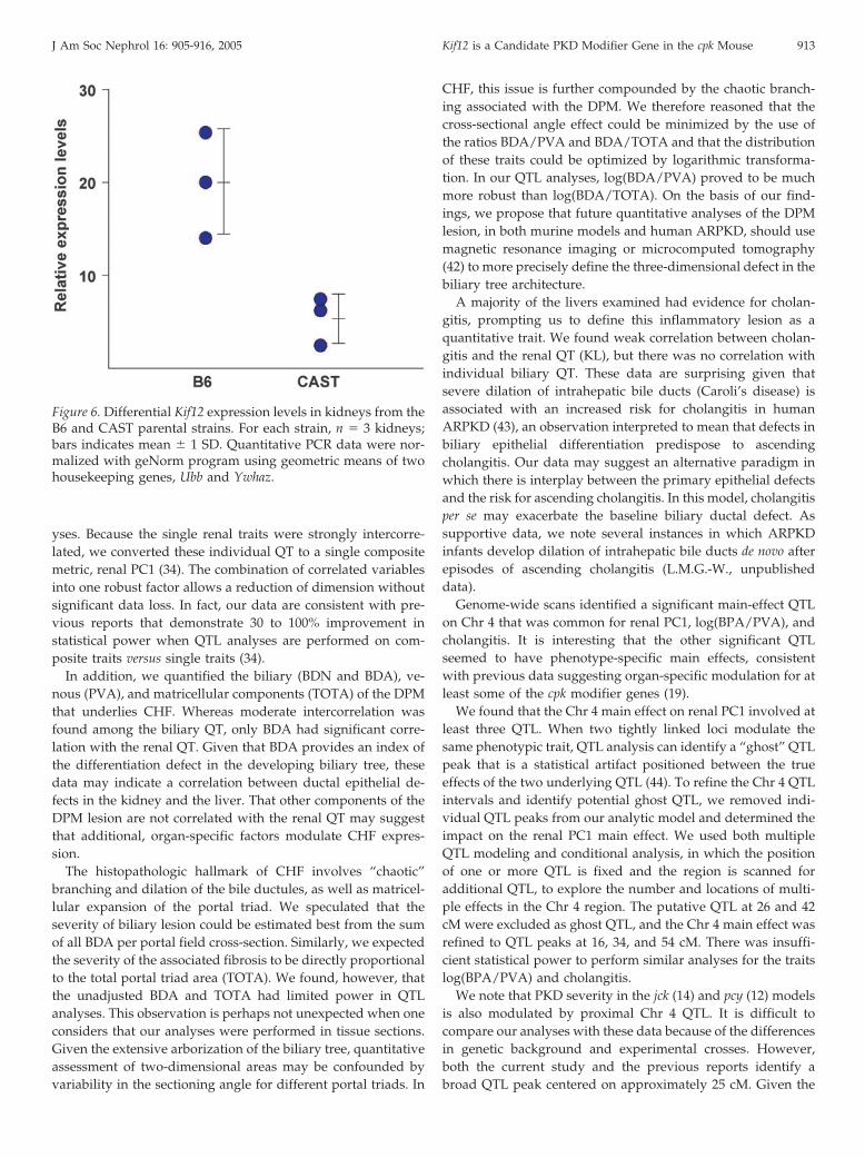
When normalized for the reference “housekeeping genes”Ubb and Ywhaz, Kif12 expression was significantly different(P � 0.016) in kidneys from 10-d-old wild-type B6 mice whencompared with age-matched wild-type CAST mice (Figure 6).These data suggest that the differences observed with respect toKif12 expression in F2 cpk kidneys likely reflects differences inparental strain-specific expression, rather than a secondarychange related to cpk-related disease severity.
Furthermore, analysis of the NCBI EST database reveals that50% of the Kif12-related EST are expressed in neonatal mousekidney, with an additional 29% of the EST derived from kid-
neys of unspecified age. The remaining Kif12 EST derive fromfour other tissues, including liver and pancreas, organs thatexpress the major cpk phenotypes (22). SAGE-based analysis ofthe human nephron segments (GEO ID: GDS503) (39) demon-strates that KIF12 is predominantly expressed in collectingducts, the principal nephron segment that undergoes cysticchange in the cpk mouse.
DiscussionQTL that exert main effects on renal cystic disease have been
reported for several murine PKD models (reviewed in 10). Incontrast to the extensive search for renal QTL, limited effort hasbeen directed toward identifying loci that modulate the pro-gression of CHF, a major ARPKD-associated phenotype andsignificant cause of morbidity and mortality in older children(24).
The current study represents the first quantitative analyses ofboth the renal and biliary phenotypes expressed in the cpk
Figure 4. Main-effect QTL and pairwise interactions for biliary traits and cholangitis. The Ch 4 QTL complex also exerted a majoreffect on biliary ductal expansion (A) and cholangitis (D). Main effects and pairwise interactions are shown for the biliary trait (Band C) and cholangitis (E). The mean value of each trait for allelic combinations of one or two loci is indicated. BB denoteshomozygosity for B6 alleles, and CC denotes homozygosity for CAST alleles. Approximate 95% CI (mean � 2 SEM) are shown.For biliary ductal expansion, a dominant CAST effect was observed for the Chr 4 QTL, whereas the CAST effect was overdominantfor the Chr 13 locus. Two pairwise interactions were identified for biliary trait severity. The Chr 1 and Chr 2 loci show a complexpattern of interaction in that the effect of the Chr 1 locus on log(BDA/PVA) is absent when the Chr 2 locus is heterozygous butdisplays opposing overdominant (heterotic) patterns when the Chr 2 locus is homozygous for BB or CC. The effect of the Chr 4QTL seemed to be enhanced and more distinctly dominant when there was at least one B6 allele at the Chr 18 locus. For cholangitisseverity, the CAST allele at the Chr 3 and Chr 19 QTL had additive effects, whereas the CAST effect was overdominant for theChr 4 locus.
J Am Soc Nephrol 16: 905-916, 2005 Kif12 is a Candidate PKD Modifier Gene in the cpk Mouse 911

model of ARPKD. Several lines of evidence indicate that geneticbackground modulates both cpk phenotypes. In a preliminarystudy, Woo et al. (11) demonstrated that progression of renalcystic disease in cpk mice is a quantitative trait under polygeniccontrol. Whereas B6-cpk/cpk mice do not express a biliary lesion(16), our group and others have shown that the phenotype isfully penetrant in cpk mutants derived from experimentalcrosses and congenic strains (19,22). These observations suggestthat whereas genetic background modulates the expression ofboth the renal cystic and the biliary traits, at least some of thecpk modifier genes exert an organ-specific effect (19).
In the F2 cohort that we generated from a (B6-�/cpk�
CAST)F1 intercross, we defined the renal cystic disease as threequantitative traits based on KL, KW, and Kvol. KL is a common
parameter used in pediatric renal imaging, and Kvol measuredby magnetic resonance imaging has emerged as a prognosticmarker of PKD progression (33). In our test cohort, we used awater displacement technique to measure Kvol ex vivo. Thevalidity of this metric is supported by its strong correlationwith the other renal QT.
Previous studies have used the ratios KL/CR and KW/BW toadjust for runting in severely affected mice. Therefore, weperformed QTL analyses with these ratios as well as KL, KW,and Kvol. Although similar main-effect QTL were identified forall of these traits (data not shown), we note that multiple QTLhave main effects on body size alone, e.g., BW (41). We thereforeexpect that these loci may modulate CR and BW independent ofPKD severity, and we excluded these ratios from further anal-
Table 4. Differential expression in Chr 4 positional QTL candidate genes
# ProbeID
Over-ExpressingPhenotype
N-foldIncreased
ExpressionP Value GenBank# Gene
Symbol DescriptionNCBIESTsTotal
KidneyESTs[%]
LiverESTs[%]
MapChr 4QTLPeak
KidneyESTs
LiverESTs
1 1450771 mild 2.3 0.001123999 NM_010243 Fut9 fucosyltransferase 9 79 27 0 chr4:25818239-25818812 (�) 16 cM 21 01 1457409 mild 2.2 0.001086276 BB484070 Fut9 fucosyltransferase 9 79 27 0 chr4:25822652-25823075 (�) 16 cM 21 02 1435308 mild 2.5 0.001086276 AU067636 — Mus musculus adult female vagina cDNA 37 35 0 chr4:25827189-25827638 (�) 16 cM 13 03 1425150 severe 2.7 0.0015318 BC010829 C730036D15Rik RIKEN cDNA C730036D15 gene 29 52 31 chr4:49023779-49024167 (�) 34 cM 15 94 1453193 mild 2.3 0.001141442 AK018598 Kif12 kinesin family member 12 42 79 2 chr4:61730529-61731682 (�) 34 cM 33 15 1432591 severe 2.8 0.001086276 BB635017 — Mus musculus 0 day neonate head cDNA 9 0 0 chr4:63921632-63922133 (�) 34 cM 0 06 1429052 mild 2.2 0.001073569 AK003303 — Mus musculus 12 days embryo head cDNA 52 4 0 chr4:74394044-74394565 (�) 34 cM 2 07 1424886 mild 2.8 0.001073569 BC025145 Ptprd protein tyrosine phosphatase, receptor D 112 14 3 chr4:74397622-74406923 (�) 34 cM 16 38 1443056 mild 2.2 0.001073569 BB374993 — Mus musculus 16 days embryo head cDNA 5 0 0 chr4:74654255-74654694 (�) 34 cM 0 09 1441498 mild 2.5 0.001086276 AW546318 — Mus musculus transcribed sequences 13 0 0 chr4:74770555-74770941 (�) 34 cM 0 010 1440624 mild 4 0.001073569 BB477150 — Mus musculus transcribed sequences — — — chr4:74771233-74771778 (�) 34 cM — —11 1442837 mild 4.3 0.001066266 BB134628 — Mus musculus adult male bone cDNA 4 0 0 chr4:74840328-74840812 (�) 34 cM 0 012 1444622 mild 2.1 0.001073569 BM118846 — Mus musculus 16 days neonate thymus cDNA 5 40 0 chr4:74851048-74851463 (�) 34 cM 2 013 1443053 mild 3.7 0.001086276 BB523104 — Mus musculus 15 days embryo head cDNA 5 0 0 chr4:74888287-74888702 (�) 34 cM 0 014 1443075 mild 2.7 0.001086276 AW556334 — Mus musculus transcribed sequences — — — chr4:74894293-74894788 (�) 34 cM — —15 1429488 mild 2.1 0.001201747 AK017682 Zdhhc21 zinc finger, DHHC domain containing 21 83 2 0 chr4:81405230-81405727 (�) 54 cM 2 016 1439852 mild 2.2 0.001157847 BB036443 — Mus musculus transcribed sequences — — — chr4:82940006-82940550 (�) 54 cM — —17 1453345 severe 2.1 0.001073569 AK014427 3830408G10Rik RIKEN cDNA 3830408G10 gene 79 0 14 chr4:90819741-90820162 (�) 54 cM 0 1118 1417531 mild 2.2 0.001095959 BF783609 Cyp2j5 cytochrome P450, family 2, subfamily j5 195 68 15 chr4:95677584-95677956 (�) 54 cM 133 2919 1421830 mild 2.4 0.001528605 NM_009647 Ak4 adenylate kinase 4 240 6 1 chr4:100326137-100326387 (�) 54 cM 15 320 1433596 mild 2.4 0.001073569 BQ175337 Dnajc6 DnaJ (Hsp40) homolog, subfamily C6 179 2 0 chr4:100501957-100502430 (�) 54 cM 3 021 1427308 severe 2.4 0.001086276 BB644109 C630028C02Rik RIKEN cDNA C630028C02 gene 58 5 0 chr4:103602026-103602403 (�) 54 cM 3 022 1417991 mild 2.3 0.001115732 NM_007860 Dio1 deiodinase, iodothyronine, type I 45 22 40 chr4:106151238-106151765 (�) 54 cM 10 1823 1417462 severe 2.6 0.001528427 NM_007598 Cap1 CAP, adenylate cyclase-associated protein 1 526 2 2 chr4:121924877-121927503 (�) 54 cM 13 1123 1417461 severe 2.3 0.001743061 NM_007598 Cap1 CAP, adenylate cyclase-associated protein 1 526 2 2 chr4:121927936-121928323 (�) 54 cM 13 11
bold font indicates expression in both kidney and liver.
Figure 5. Kif12 allelic variants. Sequence analysis of B6 and CAST transcripts revealed a 15-bp deletion variant (A) that correspondsto 5–amino acid deletion in the kinesin motor domain (red box; B). In the three-dimensional model of the related kinesin motordomain encoded by yeast kar3, the predicted deletion involves the L2 loop, indicated by a red line (C).
912 Journal of the American Society of Nephrology J Am Soc Nephrol 16: 905-916, 2005
yses. Because the single renal traits were strongly intercorre-lated, we converted these individual QT to a single compositemetric, renal PC1 (34). The combination of correlated variablesinto one robust factor allows a reduction of dimension withoutsignificant data loss. In fact, our data are consistent with pre-vious reports that demonstrate 30 to 100% improvement instatistical power when QTL analyses are performed on com-posite traits versus single traits (34).
In addition, we quantified the biliary (BDN and BDA), ve-nous (PVA), and matricellular components (TOTA) of the DPMthat underlies CHF. Whereas moderate intercorrelation wasfound among the biliary QT, only BDA had significant corre-lation with the renal QT. Given that BDA provides an index ofthe differentiation defect in the developing biliary tree, thesedata may indicate a correlation between ductal epithelial de-fects in the kidney and the liver. That other components of theDPM lesion are not correlated with the renal QT may suggestthat additional, organ-specific factors modulate CHF expres-sion.
The histopathologic hallmark of CHF involves “chaotic”branching and dilation of the bile ductules, as well as matricel-lular expansion of the portal triad. We speculated that theseverity of biliary lesion could be estimated best from the sumof all BDA per portal field cross-section. Similarly, we expectedthe severity of the associated fibrosis to be directly proportionalto the total portal triad area (TOTA). We found, however, thatthe unadjusted BDA and TOTA had limited power in QTLanalyses. This observation is perhaps not unexpected when oneconsiders that our analyses were performed in tissue sections.Given the extensive arborization of the biliary tree, quantitativeassessment of two-dimensional areas may be confounded byvariability in the sectioning angle for different portal triads. In
CHF, this issue is further compounded by the chaotic branch-ing associated with the DPM. We therefore reasoned that thecross-sectional angle effect could be minimized by the use ofthe ratios BDA/PVA and BDA/TOTA and that the distributionof these traits could be optimized by logarithmic transforma-tion. In our QTL analyses, log(BDA/PVA) proved to be muchmore robust than log(BDA/TOTA). On the basis of our find-ings, we propose that future quantitative analyses of the DPMlesion, in both murine models and human ARPKD, should usemagnetic resonance imaging or microcomputed tomography(42) to more precisely define the three-dimensional defect in thebiliary tree architecture.
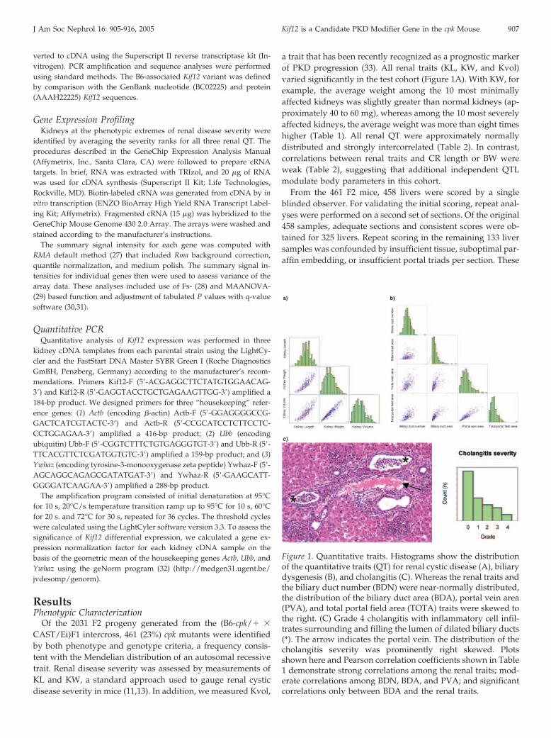
A majority of the livers examined had evidence for cholan-gitis, prompting us to define this inflammatory lesion as aquantitative trait. We found weak correlation between cholan-gitis and the renal QT (KL), but there was no correlation withindividual biliary QT. These data are surprising given thatsevere dilation of intrahepatic bile ducts (Caroli’s disease) isassociated with an increased risk for cholangitis in humanARPKD (43), an observation interpreted to mean that defects inbiliary epithelial differentiation predispose to ascendingcholangitis. Our data may suggest an alternative paradigm inwhich there is interplay between the primary epithelial defectsand the risk for ascending cholangitis. In this model, cholangitisper se may exacerbate the baseline biliary ductal defect. Assupportive data, we note several instances in which ARPKDinfants develop dilation of intrahepatic bile ducts de novo afterepisodes of ascending cholangitis (L.M.G.-W., unpublisheddata).
Genome-wide scans identified a significant main-effect QTLon Chr 4 that was common for renal PC1, log(BPA/PVA), andcholangitis. It is interesting that the other significant QTLseemed to have phenotype-specific main effects, consistentwith previous data suggesting organ-specific modulation for atleast some of the cpk modifier genes (19).
We found that the Chr 4 main effect on renal PC1 involved atleast three QTL. When two tightly linked loci modulate thesame phenotypic trait, QTL analysis can identify a “ghost” QTLpeak that is a statistical artifact positioned between the trueeffects of the two underlying QTL (44). To refine the Chr 4 QTLintervals and identify potential ghost QTL, we removed indi-vidual QTL peaks from our analytic model and determined theimpact on the renal PC1 main effect. We used both multipleQTL modeling and conditional analysis, in which the positionof one or more QTL is fixed and the region is scanned foradditional QTL, to explore the number and locations of multi-ple effects in the Chr 4 region. The putative QTL at 26 and 42cM were excluded as ghost QTL, and the Chr 4 main effect wasrefined to QTL peaks at 16, 34, and 54 cM. There was insuffi-cient statistical power to perform similar analyses for the traitslog(BPA/PVA) and cholangitis.
We note that PKD severity in the jck (14) and pcy (12) modelsis also modulated by proximal Chr 4 QTL. It is difficult tocompare our analyses with these data because of the differencesin genetic background and experimental crosses. However,both the current study and the previous reports identify abroad QTL peak centered on approximately 25 cM. Given the
Figure 6. Differential Kif12 expression levels in kidneys from theB6 and CAST parental strains. For each strain, n � 3 kidneys;bars indicates mean � 1 SD. Quantitative PCR data were nor-malized with geNorm program using geometric means of twohousekeeping genes, Ubb and Ywhaz.
J Am Soc Nephrol 16: 905-916, 2005 Kif12 is a Candidate PKD Modifier Gene in the cpk Mouse 913

statistical power of our analyses, we were able to performappropriate conditioning and exclude this peak as a ghost QTL.It therefore is conceivable that the Chr 4 QTL reported previ-ously are distinct from the QTL found to modulate renal PC1 inthis study. Alternatively, the single reported QTL may repre-sent the effect of two adjacent QTL, perhaps one more proxi-mal, corresponding to the QTL at 16 cM, and one more distal,representing the main effect at 34 cM. Further studies thatcontrol for genetic background and ghost QTL will be requiredto resolve this issue.
QTL intervals are broad, and defining a set of plausiblecandidate genes typically involves construction of recombinantconsomic/congenic strains and bioinformatics approaches torefine the QTL interval. Ultimately, a transgenic or knockout/knock-in strategy is required to validate the candidate gene(45).
Alternative approaches for identifying genes that underlieQTL main effects recently have allowed effective prioritizationamong positional candidates and the subsequent identificationof QTL genes. One approach that prioritized candidate geneson the basis of differential expression in mildly and severelyaffected individuals has successfully identified CD36 as insulinresistance QTL1 (35) and C5 as a susceptibility locus for asthma(36). Therefore, we used gene expression profiling to determinepositional candidate genes that were differentially expressed incpk mice with mild versus severe cystic kidney disease. We thendetermined which of these candidates was expressed in kidneyand liver, organs involved in the traits associated with Chr 4QTL, and excluded QTL candidate genes with identical paren-tal haplotype blocks, reflecting derivation from a common re-cent ancestor (37).
To complement these strategies we then comprehensivelyevaluated the expression and sequence variants of positionalcandidate genes using computational analyses through NCBI,e.g., UniGene, Virtual Northern, and LocusLink. Specifically,we examined all positional QTL candidates and identifiedgenes with expression in expected target tissue(s), includingappropriate temporal expression and protein localization.Then, we further prioritized selected candidates on the basis ofbiologic function and differential expression. Finally, we exam-ined available information regarding variation in coding orregulatory sequences and putative alternative splicing. Usingthis approach, we have identified Kif12, which encodes thekinesin family member 12, as a prime candidate gene for theChr 4 QTL effect.
Kif12 fulfills the major criteria for QTL gene discovery estab-lished by the Complex Trait Consortium (45). Specifically, wehave (1) identified a major QTL on Chr 4; (2) refined this Chr 4main effect to QTL peaks at 16, 34, and 54 cM; (3) determinedthat Kif12 maps beneath the QTL peak at 34 cM; (4) demon-strated that Kif12 strain-specific haplotypes and Kif12 expres-sion levels significantly correlate with renal disease severity inour F2 intercross; (5) confirmed corresponding strain-specificKif12 expression levels in wild-type parental strains; and (6)identified functionally relevant sequence variants among Kif12transcripts derived from the B6 and CAST parental strains. Aswould be predicted for a cpk modifier gene, Kif12 is expressed
primarily in kidney, as well as in liver and pancreas. Within thekidney, Kif12 is predominantly expressed in collecting ducts,the principal segment from which cysts arise in cpk kidneys.
Kif12 encodes a recently described kinesin that has yet to beassigned to a specific kinesin family (http://www.proweb.org/kinesin/). However, in the Kif12-encoded protein, the mo-lecular motor is located in the N-terminus, thus putativelyassigning this protein to the KinN family group that translo-cates cargo toward the plus end of microtubuli (46). Whereasthe functional effect of a 5–amino acid (AA) deletion within theL2 loop of this family of proteins is not known, deletions in theL2 loop of a KinI kinesin diminished its microtubule depoly-merization activity (47). Therefore, we speculate that the 5-AAdeletion in the L2 loop of the kinesin family member 12 maymodulate its interaction with microtubules. Given that micro-tubule instability and degradation is a critical feature underly-ing disease progression in the cpk mouse (48), the B6 allele mayattenuate disease progression through functions similar tothose of taxanes, drugs that markedly retard disease progres-sion in the cpk mouse by promoting microtubule polymeriza-tion and stabilization (49).
Two additional lines of evidence support our assertion thatKif12 is a candidate cpk modifier gene (modifier of cpk, Mocpk).First, yeast two-hybrid data indicate that the protein encodedby Kif1a, one of the closest protein relatives of Kif12 (50), inter-acts with the Cys1cpk gene product cystin (L.M.G.-W., submit-ted). Second, a renal-specific knockout of a second kinesinfamily gene, Kif3a, causes PKD (51). For further validating Kif12as a Mocpk candidate, studies to assess the intracellular distri-bution of kinesin family member 12 and to determine whetherit interacts directly with cystin are in progress. In addition, weare generating transgenic mice that express the CAST allele ofKif12 and will evaluate the transgene effect on disease severityin B6-cpk/cpk mice.
Finally, we note that the Invs gene is a strong candidate forthe most proximal Chr 4 QTL at 16 cM. Mutations in Invs causethe inv phenotype, a PKD-related disorder in mice, andnephronophthisis type 2 (NPHP2) in human infants (52). Alter-native splice variants encode several isoforms of the proteinproduct inversin, and these localize to different subcellularcompartments, including the nucleus and cell–cell adhesionsites (53), as well as the primary apical cilium (54). However,although both cystin and inversin localize to the primary apicalcilia, it is not clear which Invs transcripts encode the cilia-targeted isoforms, and, thus, it is impractical to determinewhether the B6 and CAST parental strains harbor functionallyrelevant sequence variants. Further studies are in progress (CLPhillips, Indiana University, IN, personal communication, Oc-tober 2004).
In summary, this is the first study to define ARPKD-likephenotypes in an experimental model as quantitative traits. Amajor effect QTL complex on Chr 4 modulates the renal cysticdisease and components of the biliary lesion, as well as cholan-gitis. Multiple regression models identify additional pheno-type-specific QTL and several pairwise interactions. These ad-ditional QTL may identify organ-specific genetic pathways thatare important in the pathogenesis of disease phenotypes. Ex-
914 Journal of the American Society of Nephrology J Am Soc Nephrol 16: 905-916, 2005
pression and sequence analyses indicate that variations inKif12, the gene encoding a novel kinesin, underlie the Chr 4main effect on all three major phenotypes. Therefore, we pro-pose that Kif12 is a cpk modifier gene (Mocpk) and as such is thefirst candidate gene identified for a PKD QTL.
AcknowledgmentsThis work was supported by a Polycystic Kidney Disease Foundation
Fellowship Award (M.M.), a Clinical Scientist Award in TranslationalResearch from the Burroughs-Wellcome Foundation (L.M.G.-W.), andthe National Institutes of Health (G.A.C.).
We thank Gbolakan Folayan for technical support and Sylvie Mrugfor assistance with data analysis.
References1. Gabow P: Autosomal dominant polycystic kidney disease.
N Engl J Med 329: 332–342, 19932. The European Polycystic Kidney Disease Consortium: The
polycystic kidney disease 1 gene encodes a 14 kb transcriptand lies within a duplicated region on chromosome 16. Cell77: 881–894, 1994
3. The American PKD1 Consortium: Analysis of the genomicsequence for the autosomal dominant polycystic kidneydisease (PKD1) gene predicts the presence of a leucine-richrepeat. Hum Mol Genet 4: 575–582, 1995
4. The International Polycystic Kidney Disease Consortium:Polycystic kidney disease: The complete structure of thePKD1 gene and its protein. Cell 8: 289–298, 1995
5. Mochizuki T, Wu G, Hayashi T, Xenophontos S,Veldhuisen B, Saris J, Renolds D, Cai Y, Gabow P, PieridesA, Kimberling WJ, Breuning MH, Deltas CC, Peters DJ,Somlo S: PKD2, a gene for polycystic kidney disease thatencodes an integral membrane protein. Science 272: 1339–1342, 1996
6. Onuchic L, Furu L, Nagasawa Y, Hou X, Eggermann T, RenZ, Bergmann C, Senderek J, Esquivel E, Zeltner R, Rudnik-Schoneborn S, Mrug M, Sweeney W, Avner ED, Zerres K,Guay-Woodford LM, Somlo S, Germino GG: PKHD1, thepolycystic kidney and hepatic disease 1 gene, encodes anovel large protein containing multiple IPT domains andPbH1 repeats. Am J Hum Genet 70: 1305–1317, 2002
7. Ward C, Hogan M, Rossetti S, Walker D, Sneddon T, WangX, Kubly V, Cunningham J, Bacallao R, Ishibashi M, Milli-ner DS, Torres VE, Harris PC: The gene mutated in auto-somal recessive polycystic kidney disease encodes a large,receptor-like protein. Nat Genet 30: 259–269, 2002
8. Hateboer N, Lazarou L, Williams A, Holmans P, Ravine D:Familial phenotype differences in PKD1. Kidney Int 56:34–40, 1999
9. Guay-Woodford LM, Desmond RA: Autosomal recessivepolycystic kidney disease: The clinical experience in NorthAmerica. Pediatrics 111: 1072–1080, 2003
10. Guay-Woodford L: Murine models of polycystic kidneydisease: Molecular and therapeutic insights. Am J PhysiolRenal Physiol 285: F1034–F1049, 2003
11. Woo D, Miao S, Tran T: Progression of polycystic kidneydisease in cpk mice is a quantitative trait under polygeniccontrol [Abstract]. J Am Soc Nephrol 6: 731A, 1995
12. Woo D, Nguyen D, Khatibi N, Olsen P: Genetic identifica-
tion of two major modifier loci of polycystic kidney diseaseprogression in pcy mice. J Clin Invest 100: 1934–1940, 1997
13. Guay-Woodford L, Walz G, Wright C, Churchill G: Quan-titative trait loci (QTLs) that influence renal cystic diseaseseverity in the mouse bpk model. J Am Soc Nephrol 11:1253–1260, 2000
14. Kuida S, Beier D: Genetic localization of interacting mod-ifiers affecting severity in a murine model of polycystickidney disease. Genome Res 10: 49–54, 2000
15. Preminger G, Koch W, Fried F, McFarland E, Murphy E,Mandell J: Murine congenital polycystic kidney disease: Amodel for studying development of cystic disease. J Urol127: 556–560, 1982
16. Fry J, Koch W, Jennette J, McFarland E, Fried F, Mandell J:A genetically determined murine model of infantile poly-cystic kidney disease. J Urol 134: 828–833, 1985
17. Hou X, Mrug M, Yoder B, Lefkowitz E, Kremmidiotis G,D’Eustachio P, Beier D, Guay-Woodford L: Cystin, a novelcilia-associated protein, is disrupted in the cpk mousemodel of polycystic kidney disease. J Clin Invest 109: 533–540, 2002
18. Yoder B, Hou X, Guay-Woodford L: The polycystic kidneydisease proteins, polycystin-1, polycystin-2, polaris, andcystin, are co-localized in renal cilia. J Am Soc Nephrol 13:2508–2516, 2002
19. Guay-Woodford L, Green W, Lindsey J, Beier D: Germlineand somatic loss of function of the mouse cpk gene causesbiliary ductal pathology that is genetically modulated.Hum Mol Genet 9: 769–778, 2000
20. Avner E, Studnicki F, Young M, Sweeney W, Piesco W,Ellis D, Fetterman G: Congenital murine polycystic kidneydisease. I. The ontogeny of tubular cyst formation. PediatrNephrol 2: 2l0–2l8, 1987
21. Gattone V, Ricker J, Trambaugh C, Klein R: MultiorganmRNA misexpression in murine autosomal recessive poly-cystic kidney disease. Kidney Int 62: 1560–1569, 2002
22. Ricker J, Gattone V, Calvet J, Rankin C: Development ofautosomal recessive polycystic kidney disease in BALB/c-cpk/cpk mice. J Am Soc Nephrol 11: 1837–1847, 2000
23. Gattone V, MacNaughton K, Kraybill A: Murine autosomalrecessive polycystic kidney disease with multiorgan in-volvement induced by the cpk gene. Anat Rec 245: 488–499, 1996
24. Guay-Woodford L, Jafri Z, Bernstein J: Other cystic kidneydiseases. In: Comprehensive Clinical Nephrology, edited byJohnson R, Feehally J, Mosby International, London, 2003,pp 611–626
25. Sen S, Churchill GA: A statistical framework for quantita-tive trait mapping. Genetics 159: 371–387, 2001
26. Churchill G, Doerge R: Empirical threshold values forquantitative trait mapping. Genetics 138: 963–971, 1994
27. Irizarry R, Bolstad B, Collin F, Cope L, Hobbs B, Speed T:NARF: Summaries of Affymetrix GeneChip probe leveldata. Nucleic Acids Res 31: e15, 2003
28. Cui X, Churchill G: Statistical tests for differential expres-sion in cDNA microarray experiments. Genome Biol 4: 210,2003
29. Wu G, Tian X, Nashimura S, Markowitz G, Agati V, ParkJ, Yao L, Li L, Geng L, Zhao H, Edelmann W, Somlo S:Trans-heterozygous Pkd1 and Pkd2 mutations modify ex-pression of polycystic kidney disease. Hum Mol Gen 11:1845–1854, 2002
J Am Soc Nephrol 16: 905-916, 2005 Kif12 is a Candidate PKD Modifier Gene in the cpk Mouse 915

30. Storey J: A direct approach to false discovery rates. J R StatSoc B 64: 479–498, 2002
31. Storey J, Tibshirani R: Statistical significance for genome-wide studies. Proc Natl Acad Sci U S A 100: 9440–9445, 2003
32. Vandesompele J, Preter KD, Pattyn F, Poppe B, Roy NV,Paepe AD, Speleman F: Accurate normalization of real-time quantitative RT-PCR data by geometric averaging ofmultiple internal control genes. Genome Biol 3: 0034.I–0034.II, 2002
33. Chapman A, Guay-Woodford L, Grantham J, Torres V, BaeK, Baumgarten D, Kenney PJ, King BF Jr, Glockner J,Wetzel L, Brummer ME, O’Neill WC, Robbin ML, BennettWM, Klahr S, Hirschman GH, Kimmel PL, Thompson PA,Miller JP: Renal structure in early autosomal-dominantpolycystic kidney disease (ADPKD): The Consortium forRadiologic Imaging Studies of Polycystic Kidney Disease(CRISP) cohort. Kidney Int 64: 1035–1045, 2003
34. Gilbert H, Le Roy P: Comparison of three multitrait meth-ods for QTL detection. Genet Sel Evol 35: 281–304, 2003
35. Aitman T, Glazier A, Wallace C, Cooper L, Norsworthy P,Wahid F, Al-Majali K, Trembling P, Mann C, Shoulders C,Graf D, St Lezin E, Kurtz TW, Kren V, Pravenec M, Ibra-himi A, Abumrad NA, Stanton LW, Scott J: Identificationof Cd36 (Fat) as an insulin-resistance gene causing defec-tive fatty acid and glucose metabolism in hypertensiverats. Nat Genet 21: 76–83, 1999
36. Karp C, Grupe A, Schadt E, Ewart S, Keane-Moore M,Cuomo P, Kohl J, Wahl L, Kuperman D, Germer S, Aud D,Peltz G, Wills-Karp M: Identification of complement factor5 as a susceptibility locus for experimental allergic asthma.Nat Immunol 1: 221–226, 2000
37. Wang X, Korstanje R, Higgins D, Paigen B: Haplotypeanalysis in multiple crosses to identify a QTL gene. GenomeRes 14: 1767–1772, 2004
38. Miller M, Kumar S: Understanding human disease muta-tions through the use of interspecific genetic variation.Hum Mol Genet 21: 2319–2328, 2001
39. Chabardes-Garonne D, Mejean A, Aude J, Cheval L, Ste-fano AD, Gaillard M, Imbert-Teboul M, Wittner M, BalianC, Anthouard V, Robert C, Segurens B, Wincker P, Weis-senbach J, Doucet A, Elalouf JM: A panoramic view of geneexpression in the human kidney. Proc Natl Acad Sci U S A100: 13710–13715, 2003
41. Cheverud JM, Routman EJ, Duarte FA, van Swinderen B,Cothran K, Perel C: Quantitative trait loci for murinegrowth. Genetics 142: 1305–1319, 1996
42. Masyuk TV, Huang BQ, Ward CJ, Masyuk AI, Yuan D,Splinter PL, Punyashthiti R, Ritman EL, Torres VE, HarrisPC, LaRusso NF: Defects in cholangiocyte fibrocystin ex-pression and ciliary structure in the PCK rat. Gastroenter-ology 125: 1303–1310, 2003
43. Kashtan C, Primack W, Kainer G, Rosenberg A, McDonaldR, Warady B: Recurrent bacteremia with enteric pathogensin recessive polycystic kidney disease. Pediatr Nephrol 13:678–682, 1999
44. Frisch M, Quint M, Lubberstedt T, Melchinger A: Dupli-cate marker loci can result in incorrect locus orders onlinkage maps. Theor Appl Genet 109: 305–316, 2004
45. Abiola O, Angel JM, Avner P, Bachmanov AA, BelknapJK, Bennett B, Blankenhorn EP, Blizard DA, Bolivar V,Brockmann GA, Buck KJ, Bureau JF, Casley WL, CheslerEJ, Cheverud JM, Churchill GA, Cook M, Crabbe JC,Crusio WE, Darvasi A, de Haan G, Dermant P, DoergeRW, Elliot RW, Farber CR, Flaherty L, Flint J, Gershen-feld H, Gibson JP, Gu J, Gu W, Himmelbauer H, Hitze-mann R, Hsu HC, Hunter K, Iraqi FF, Jansen RC, John-son TE, Jones BC, Kempermann G, Lammert F, Lu L,Manly KF, Matthews DB, Medrano JF, Mehrabian M,Mittlemann G, Mock BA, Mogil JS, Montagutelli X, Mo-rahan G, Mountz JD, Nagase H, Nowakowski RS,O’Hara BF, Osadchuk AV, Paigen B, Palmer AA, PeirceJL, Pomp D, Rosemann M, Rosen GD, Schalkwyk LC,Seltzer Z, Settle S, Shimomura K, Shou S, Sikela JM,Siracusa LD, Spearow JL, Teuscher C, Threadgill DW,Toth LA, Toye AA, Vadasz C, Van Zant G, Wakeland E,Williams RW, Zhang HG, Zou F; Complex Trait Consor-tium: The nature and identification of quantitative traitloci: A community’s view. Nat Rev Genet 4: 911–916, 2003
46. Vale RD, Reese TS, Sheetz MP: Identification of a novelforce-generating protein, kinesin, involved in microtubule-based motility. Cell 42: 39–50, 1985
47. Shipley K, Hekmat-Nejad M, Turner J, Moores C, Ander-son R, Milligan R, Sakowicz R, Fletterick R: Structure of akinesin microtubule depolymerization machine. EMBO J23: 1422–1432, 2004
48. Woo D, Miao S, Pelayo J, Woolf A: Taxol inhibits progres-sion of congenital polycystic kidney disease. Nature 368:750–753, 1994
49. Woo D, Tabancay A, Wang C: Microtubule activate taxanesinhibit polycystic kidney disease progression in cpk mice.Kidney Int 51: 1613–1618, 1997
50. Miki H, Setou M, Hirokawa N: Kinesin superfamily pro-teins (KIFs) in the mouse transcriptome. Genome Res 13:1455–1465, 2003
51. Lin F, Hiesberger T, Cordes K, Sinclair A, Goldstein L,Somlo S, Igarashi P: Kidney-specific inactivation of theKIF3A subunit of kinesin-II inhibits renal ciliogenesis andproduces polycystic kidney disease. Proc Natl Acad SciU S A 100: 5286–5291, 2003
52. Otto E, Schermer B, Obara T, O’Toole J, Hiller K, MuellerA, Ruf R, Hoefele J, Beekmann F, Landau D, Foreman JW,Goodship JA, Strachan T, Kispert A, Wolf MT, GagnadouxMF, Nivet H, Antignac C, Walz G, Drummond IA, BenzingT, Hildebrandt F: Mutations in INVS encoding inversincause nephronophthisis type 2, linking renal cystic diseaseto the function of primary cilia and left-right axis determi-nation. Nat Genet 34: 413–420, 2003
53. Nurnberger J, Bacallao R, Phillips C: Inversin forms acomplex with catenins and N-cadherin in polarized epithe-lial cells. Mol Biol Cell 13: 3096–3106, 2002
54. Morgan D, Eley L, Sayer J, Strachan T, Yates L, CraigheadA, Goodship J: Expression analyses and interaction withthe anaphase promoting complex protein Apc2 suggest arole for inversin in primary cilia and involvement in thecell cycle. Hum Mol Genet 11: 3345–3350, 2002
916 Journal of the American Society of Nephrology J Am Soc Nephrol 16: 905-916, 2005